Embed Size (px)
Citation preview
Heliyon 5 (2019) e02648
Contents lists available at ScienceDirect
Heliyon
journal homepage: www.heliyon.com
Research article
Characterization of phospholipid vesicles containing lauric acid:physicochemical basis for process and product development
Laura Farkuh a, Paulo T. Hennies b, Cl�audia Nunes c, Salette Reis c, Luisa Barreiros c,Marcela A. Segundo c, Pedro L. Oseliero Filho d, Cristiano L.P. Oliveira d, Alexandre Cassago e,Rodrigo V. Portugal e, Rodrigo A. Muramoto a, Gustavo P.B. Carretero a, Shirley Schreier a,Hernan Chaimovich a, Iolanda M. Cuccovia a,*
a Department of Biochemistry, Institute of Chemistry, University of S~ao Paulo, S~ao Paulo, Brazilb Líbera Tecnologia e Inovaç~ao Ltda., S~ao Paulo, Brazilc LAQV, REQUIMTE, Faculty of Pharmacy, University of Porto, Porto, Portugald Department of Experimental Physics, Institute of Physics, University of S~ao Paulo, S~ao Paulo, Brazile Brazilian Nanotechnology National Laboratory (LNNano), Brazilian Center for Research in Energy and Materials (CNPEM), Campinas, Brazil
A R T I C L E I N F O
Keywords:BiotechnologyPharmaceutical ScienceCryo-TEMLauric acidDSCLipid VesiclesDLSIonizable soluteSAXS
* Corresponding author.E-mail address: [email protected] (I.M. Cucco
https://doi.org/10.1016/j.heliyon.2019.e02648Received 6 May 2019; Received in revised form 122405-8440/© 2019 The Author(s). Published by Els
A B S T R A C T
Lauric acid (LAH) strongly inhibits the growth of acne-causing bacteria. LAH is essentially water-insoluble and thesolubility of laurate (LA) salts are medium and temperature dependent. Hence, LAH/LA preparations are difficultto formulate. Here we fully characterized phospholipid vesicles containing up to 50 mol% LAH. Vesicles ofdipalmitoylphosphatidylcholine (DPPC) containing LAH, at pHs 7.4 and 5.0, were characterized measuring size,charge, bilayer phase transition temperature (Tm) and permeability of water-soluble probes. Small angle X-rayscattering and cryotransmission electron microscopy showed multilamellar vesicles at low LAH %. Increasing LAH %had a negligible effect on particle size. An internal aqueous compartment in all vesicle's preparations, even atequimolar DPPC: LAH fractions, was demonstrated using water-soluble probes. At pH 5.0, the interaction betweenDPPC and LAH increased the Tm and phase transition cooperativity showing a single lipid phase formed byhydrogen-bonded DPPC: LAH complexes. At pH 7.4, vesicles containing 50 mol% LAH exhibited distinct phases,ascribed to complex formation between LAH and LA or LAH and DPPC. LAH incorporated in the vesicles mini-mally permeated a skin preparation at both pHs, indicating that the primary sites of LAH solubilization were theskin layers. These results provide the foundations for developing processes and products containing DPPC: LAH.
1. Introduction
The inhibition of bacterial growth by free fatty acids (FFA's) is wellestablished [1]. Lauric acid (LAH), the saturated fatty acid with thehighest antimicrobial potential, found in coconut (oil and milk), is alsopresent in small amounts in sebum [2, 3, 4]. LAH inhibits the growth ofPropionibacterium acnes, Staphylococcus aureus, and Staphylococcusepidermis, and, therefore, LAH could treat acne vulgaris [5]. LAH is poorlysoluble in water, and the solubility of laurate (LA) depends critically onthe counterion and the solution temperature [6, 7, 8]. The water solu-bility properties of LAH and LA are significant drawbacks for the use ofthis antimicrobial agent in pharmaceutical formulations.
Phospholipid vesicles are efficient drug carriers [9] and both LAH and
via).
September 2019; Accepted 9 Ocevier Ltd. This is an open access
LA intercalate in their bilayers, changing structure and interfering in themembrane permeability to water-soluble compounds [10,11]. Under-standing the fundamental properties of these aggregates is essential toallow the applicability of these formulations to treat infectious diseases.
The mixture of phospholipids vesicles and fatty acids can lead tochanges in the vesicle's membrane intrinsic structure because the pres-ence of fatty acids can disrupt the well-organized phospholipid bilayer.As fatty acids dissociate, the pH of the environment can also modify themembrane properties due to different ratios of charged/neutral forms ofthe carboxylic group. Addition of fatty acids to pre-formed dimyristoylphosphatidylcholine (DMPC) vesicles changes both turbidity and thephase transition temperature (Tm) [9]. Release of DMPCvesicle-entrapped fluorescent probes accompanies the turbidity changes,
tober 2019article under the CC BY license (http://creativecommons.org/licenses/by/4.0/).
L. Farkuh et al. Heliyon 5 (2019) e02648
demonstrating the disrupting effect of externally added fatty acids on thebilayer structure [11]. Despite the disruptive effect of fatty acids onphospholipid membranes, some fatty acids, such as oleic acid in the pHrange 8–9, form multilamellar vesicles with bilayers stabilized byhydrogen bonding between the protonated and anionic forms [12]. Theseseemingly diverse actions of fatty acids make the study of these phe-nomena necessary.
The present study focuses on the physicochemical properties of largeunilamellar vesicles (LUVs) prepared with dipalmitoylphosphatidylcho-line (DPPC): LAHmixtures. The effect of LAH on the properties of vesiclesprepared with variable proportions of dipalmitoylphosphatidylcholine,DPPC, was studied by mixing the fatty acid with DPPC before vesiclespreparation, at different DPPC: LAH molar ratios and different pHs. Thismethod allowed the formation of vesicles where the phospholipid bi-layers are already in its equilibrium state. We analysed the bilayerstructure of these mixed vesicles, as well as the Tm, size, charge, andpermeability of water-soluble probes. Additionally, the delivery of lauricacid from the vesicles to a skin preparation was studied. Our results mayprovide the foundations for developing processes and products contain-ing DPPC: LAH.
2. Materials and methods
2.1. Materials
1,2-Dipalmitoyl-sn-glycero-3-phosphocholine (DPPC) [Avanti PolarLipids (Alabaster, USA)]; lauric acid (LAH), L-ascorbic acid, tris(hydroxymethyl) aminomethane (Tris), polyoxyethylene-9-lauryl ether(polydocanol), Sephadex G-25 medium, acetic acid, chloroform, andmethanol [Sigma Aldrich (St. Louis, USA)] were used as received. 5(6)-Carboxyfluorescein (CF) [Sigma Aldrich] was purified and stocked asthe sodium salt [13]. The probes 4-trimethylammonium-2,2,6,6-tetrame-thylpiperidine-1-oxyl iodide (CAT1) and tetrasodium 1,3,6,8-pyrenete-trasulfonate (PTS) [Molecular Probes (Eugene, USA)] were used asreceived.
For LC-MS/MS analysis, acetonitrile (LiChrosolv LC-MS grade) wasfrom Merck (Darmstadt, Germany) and water was from purificationsystem (resistivity > 18 MΩ cm, Sartorius, G€ottingen, Germany). Mobilephase components were filtered through 0.45 μm Millipore (Billerica,MA) HVHP and 0.22 μm Millipore GVWP filters, respectively, anddegassed in an ultrasonic bath for 15 min.
2.2. Lauric acid quantification (LC-MS/MS analysis)
LAH concentration was determined by liquid chromatographycoupled to triple quadrupole tandem mass spectrometry (LCMS-8040)equipped with an electrospray ionization source (ESI) (ShimadzuCorporation).
2.3. Liposome preparation
Stock solutions of DPPC 0.05 M and LAH 0.05 M in chloroform:methanol 3:2 (v/v) were prepared. The stock solution of DPPC wasquantified by determining the phosphate concentration of the sample[13] and the concentration of the LAH solution was taken by weight.Lipid mixtures, at the desired molar ratio of DPPC: LAH, were preparedby mixing aliquots of the stock solutions into glass tubes. The followinglipid compositions were used: 100 % DPPC; DPPC: LAH 90:10; DPPC:LAH 80:20; DPPC: LAH 70:30; DPPC: LAH 60:40 and DPPC: LAH 50:50.The solvents were eliminated under an N2 flux forming a film and solventtraces were removed in a vacuum chamber (2 h). Multilamellar vesicles(MLVs) were prepared by adding the desired solution to the lipid film andvortexing the mixture until the film detached from the tube wall. Largeunilamellar vesicles (LUVs) were obtained by extruding MLVs suspen-sions in an Avanti mini-extruder, Alabaster, USA, using two membraneswith 100 nm pore size (eleven passages) at 60 �C.
2
2.4. Dynamic light scattering (DLS)
The LUVs hydrodynamic diameter (DH), the polydispersity index(PdI), zeta potential and the number of particles were determined byDynamic Light Scattering (DLS), in triplicate, at room temperature usinga Zetasizer Nano ZS, Malvern Instruments Ltd.
2.5. Small angle X-ray scattering (SAXS)
Small angle X-ray scattering (SAXS) spectra were obtained with aBruker Nanostar (with optimized optics by Xenocs) at 25 �C. DPPC: LAHvesicles (10 mM total lipid) in 50 mM Tris-HCl buffer, pH 7.4, or 50 mMNa acetate, pH 5.0 were placed in 1.5 mm diameter glass capillaries. Theexperimentally accessible range of the modulus of the transfer momentvector, q ¼ 4π sinðθÞ=λ (where θ is half the scattering angle and λ the X-
ray wavelength), was 0:018�A�1 � 0:35�A
�1. SUPERSAXS program [14]
was used for data treatment, allowing correction of the raw data usingbackground scattering, empty capillary and sample transmission, andthen normalization to absolute scale using water as standard. The finaldata correspond to one-dimensional scattered intensity IðqÞ, withrespective uncertainties, versus q values.
2.6. Cryotransmission electron microscopy (Cryo-TEM)
For the cryogenic transmission electron microscopy (Cryo-TEM)analysis, DPPC: LAH 70:30 and DPPC: LAH 50:50 (mole: mole) vesicleswere used at 2.5 mM total lipid in 10 mM Tris-HCl, pH 7.4, or 10 mM Naacetate, pH 5.0, buffers. A 3 μL droplet was deposited on a 300 meshlacey carbon-coated copper grid (TED Pella) using an easyGlow dischargesystem (Pelco) with 15 mA negative current for 10 s. Specimens wereprepared at 22 �C and humidity 100 % in an automated vitrificationsystem (Vitrobot Mark IV, FEI, The Netherlands) with a blot time of 3 s,blot force of 0, and 20 s waiting time before blotting. Specimens wereanalyzed in low dose condition, with a defocus range of�2 μm to�4 μm,using a Jeol JEM-2100 (Pleasanton, USA) microscope equipped with anF-416 CMOS camera (TVIPS, Germany), operating at 200 kV.
2.7. Differential scanning calorimetry (DSC)
Phase transition temperature (Tm) and cooperativity (ΔT1/2) weredetermined on a MicroCal VP-DSC microcalorimeter, Malvern In-struments Ltd. (Malvern, UK). Demineralized water was used as a refer-ence. The data were analyzed with the Origin 8.5 program.
2.8. Entrapment of hydrophilic probes
2.8.1. Fluorescent probesSolutions (500 μL) containing 2 mM CF in 50 mM Tris-HCl buffer, pH
7.4, or 1 mM PTS in 50 mM Na acetate buffer, pH 5.0, were added toDPPC: LAH films (10 mM total lipid concentration), and the resultingMLVs were extruded.
A Sephadex G25 column (1.5 cm diameter, 45 cm height) was used toseparate CF- or PTS-containing vesicles from the non-encapsulatedfluorescent probes. The sample (300 μL) was applied to the column,eluted with the desired buffer, and 1 mL fractions were collected. Thepercentage of CF (or PTS) encapsulation was calculated according to Eq.(1):
% encapsulation¼ 100*moles Vo
moles Voþ moles Vi(1)
where Vo and Vi are the excluded and internal column volumes respec-tively andmoles Vo andmoles Vi are the number of moles eluted at Vo andVi, respectively. Fluorescence was measured using a Shimadzu RF-5301PC fluorimeter, with λexc 490 nm, λem 520 nm for CF, and λexc355 nm, λem 405 nm for PTS [15]. For the fractions containing vesicles,
L. Farkuh et al. Heliyon 5 (2019) e02648
fluorescence were measured after the addition of 25 μL of an aqueoussolution of 10% polidocanol (v/v) to eliminate vesicle-produced lightscattering.
2.8.2. Electron paramagnetic resonance (EPR) probeCAT1 solutions (3.75 mM) were prepared in 50 mM Tris-HCl, pH 7.4,
or 50 mM Na acetate, pH 5.0. Ascorbic acid (0.04 M), prepared in thesame buffers, was used to suppress the CAT1 signal [16, 17, 18].
DPPC: LAH films were hydrated with CAT1 solutions (10 mM lipid),vortexed and extruded. Vesicles with CAT1 (170 μL) were placed in flatquartz cells (Wilmad, USA) and the EPR spectrum was recorded (totalsignal); then, 10 μL of sodium ascorbate were added, supressing CAT1signal outside the liposomes (final signal). Controls assured that ascorbateconcentration was sufficient to reduce external CAT1.
EPR spectra were acquired at room temperature on a Bruker EMX-200spectrometer with 5 mW microwave power, 1 G modulation amplitude,sweeping a scan range of 16 G with the field centered at 3455 G. The gainwas adjusted according to the sample concentration. Spectra wereanalyzed with the WINEPR software (Bruker), and the peaks were inte-grated twice to obtain the area, which is proportional to the probe con-centration. The percentage of encapsulated CAT1 was calculatedaccording to Eq. (2):
% encapsulated¼ 100 � final signaltotal signal
(2)
2.9. Lauric acid skin permeation
The skin permeation of LAH was studied using a vertical staticFranz diffusion cells with pig ear skin as membrane. Skin was cleaned,the hypodermis removed and stored at -20 �C until its use. Five mlbuffer were placed in the receptor chamber, and 500 μL of sample (0.5mM total lipid) or buffer solution (control) in the donor chamber. A500 μL aliquot of solution from the receptor chamber (37 �C) wastaken every 1 hour (for 8 h) to quantify LAH. To maintain sink con-ditions, 500 μL of buffer solution was replenished in the receptorchamber. The skin permeation (μg/cm2) was calculated dividing theamount of LAH in the acceptor chamber per skin area (0.64 cm2). Thesample was transferred to a tube, 0.2 mL of HCCl3 was added and LAHwas extracted. This process was repeated 3 times. The solvent wasevaporated with an N2 flux at 45 �C and the LAH was solubilized in500 μL of a mixture of acetonitrile:water 80:20 v/v. From each sample,three injections were made into the HPLC and the data in Fig. 6 areaverages.
Chromatographic analysis were performed in a Nexera X2 UHPLCsystem comprising two LC-30AD pumps, a DGU-20A5R degassing unit, aSIL-30AC autosampler, and a CTO-20AC oven (Shimadzu Corporation,Kyoto, Japan).
Chromatographic separation was achieved using a reversed phaseKinetex core-shell C8 column (150 mm � 2.1 mm, 2.6 μm particle size;Phenomenex, Torrance, CA, USA), at 30 �C. Elution was performed in anisocratic mode using acetonitrile:water (80:20, v/v) at a flow rate of 0.2mL min�1. The injection volume was 2 μL and total analysis time was 12min.
The mass spectrometer was operated in negative ionization mode(ESI-), and data were acquired using multiple reaction monitoring(MRM) mode to enhance selectivity. The m/z transition 199.30 >
181.25, corresponding to the neutral loss of one water moleculefrom the molecular ion [M-H]-, was used for quantification andidentification purposes. The nebulizing gas (N2) flow rate was 3 Lmin�1, desolvation line temperature 250 �C, heat block temperature400 �C, detector voltage, 1.76 kV and collision gas (argon) 230 kPa.For calibration, LA standard solutions were between 0.1 and 10 μMin the mobile phase. Peak detection and quantification were per-formed using LabSolutions software version 5.60 SP2 (ShimadzuCorporation).
3
3. Results and discussion
3.1. Small angle X-ray scattering (SAXS)
Fig. 1 shows SAXS data for vesicles with different DPPC: LAH molarratios and at different pHs. The treated data of electron density contrastprofile in Fig. 1A (open symbols), were characteristic of uni- or multi-lamellar vesicles. The main difference between them was related to theMLV exhibiting an oscillation on the bump in the region
0:06 �1
< q < 0:2 �1, indicating that the number of correlated lipid
bilayers (N) was>1. For instance, considering Fig. 1A, pH 5.0, the curvescorresponding to 40 % and 50 % LAH are typical of unilamellar vesicles,
since there was no oscillation on the bump in the 0:06 �1
< q < 0:2 �1
regions, differently from the remaining curves, typical of MLV.DPPC vesicles, at pH 5.0 and pH 7.4, were multilamellar (Fig. 1A).
Since the overall shapes of the SAXS curve were similar, the vesiclesexhibited similar structures in this q range. Increasing the amount of LAHto 10 %, a decrease of the multilamellar vesicle population was observedat pH 7.4. The same transition was observed at pH 5.0 only above 40 %LAH. This result indicates, qualitatively, the importance of pH on thenanostructure of this system. The fact that vesicles have a greater ten-dency to be unilamellar at pH 7.4 is probably due to the negative chargeimparted on the surface by the negatively charged laurate ion (LA),which promotes inter-bilayer repulsion, leading to a decrease in thenumber of lamellae.
The clear difference between the effects of LAH and LA on theresulting structures bring to attention that few investigations on bindingof ionizable solutes to biological and model membranes consider thatpartitioning of the charged and uncharged forms of these compoundsdiffers, the uncharged form usually incorporating to a more significantextent. In Scheme 1, HA and A- are the uncharged and anionic forms ofthe solute, respectively, Kw and Km are the ionization constants in theaqueous and membrane phases, respectively, and P0 and P- are thepartition coefficients of both forms.
P is defined as:
P¼ nm:Vw
nw:Vm(3)
where n and V represent the number of moles and volume, respectively,and the indexes m and w the membrane and aqueous phases, respec-tively. Thus, pKw and pKm are, respectively, the pHs of the bulk aqueousphase where the populations of charged and uncharged forms in theaqueous phase are equal and where the populations of charged and un-charged forms in the membrane are equal. ΔpK (pKm – pKw) is a necessaryconsequence of the fact that P0 6¼ P-.
This analysis was applied in studies of ionizable compounds bindingto micelles [19] and lipid vesicles [20]. An apparent pK, pKapp, wasdefined, corresponding to the aqueous phase pH where the population ofthe charged species equals the population of the uncharged species, bothbeing the sum of their fractions in the membrane and the aqueous phaseand depend on membrane concentration.
The apparent pK of carboxylic acids in membranes significantly in-creases, and the pKw of the free acid, around 5.0, increases to ca. 7.0–7.5[21,22]. The membrane-bound fraction will depend on total membraneconcentration. For LA, at pH 7.4, the total partitioning of LA into themembrane will decrease due to the contribution of both P0 and P� to Pav,and the much lower value of P�. At pH 5.0 essentially all LAH is pro-tonated and fully incorporated in the membrane, as the partition coef-ficient (P0) is sufficiently high.
To obtain quantitative information of overall structural changes in thesystem, the experimental SAXS curves were fitted using the GaussianDeconvolution Method [14], which allows simultaneous determinationof both form and structure factor. Since the form factor is calculated fromthe modeled electron density contrast profile, ΔρðrÞ, it is possible todetect structural bilayer changes either in ΔρðrÞor in bilayer thickness ðδÞ
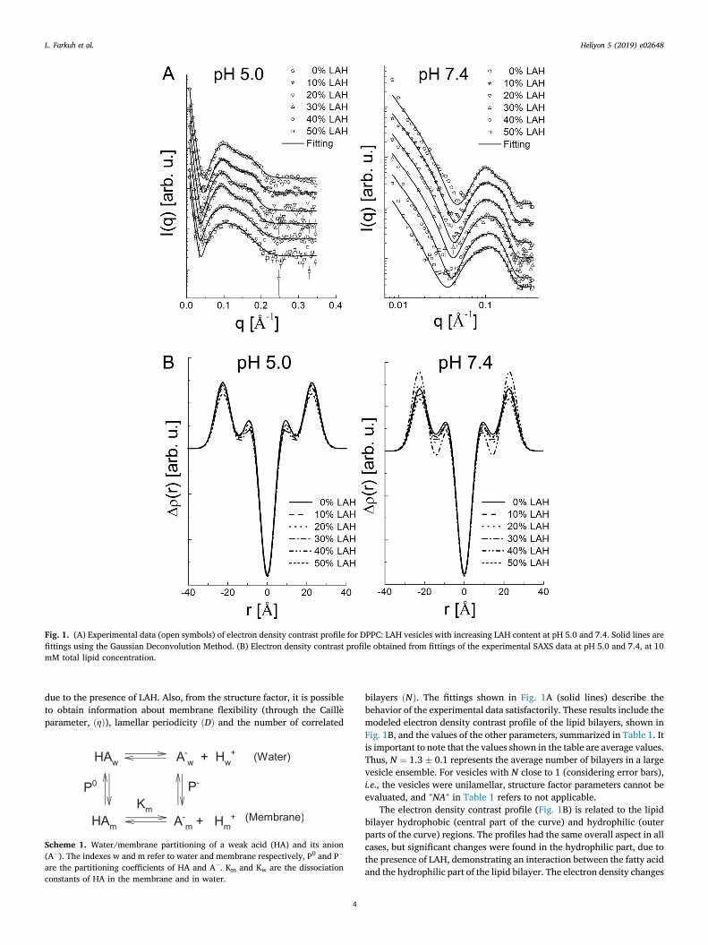
Fig. 1. (A) Experimental data (open symbols) of electron density contrast profile for DPPC: LAH vesicles with increasing LAH content at pH 5.0 and 7.4. Solid lines arefittings using the Gaussian Deconvolution Method. (B) Electron density contrast profile obtained from fittings of the experimental SAXS data at pH 5.0 and 7.4, at 10mM total lipid concentration.
L. Farkuh et al. Heliyon 5 (2019) e02648
due to the presence of LAH. Also, from the structure factor, it is possibleto obtain information about membrane flexibility (through the Caill�eparameter, ðηÞ), lamellar periodicity ðDÞ and the number of correlated
Scheme 1. Water/membrane partitioning of a weak acid (HA) and its anion(A�). The indexes w and m refer to water and membrane respectively, P0 and P�
are the partitioning coefficients of HA and A�. Km and Kw are the dissociationconstants of HA in the membrane and in water.
4
bilayers ðNÞ. The fittings shown in Fig. 1A (solid lines) describe thebehavior of the experimental data satisfactorily. These results include themodeled electron density contrast profile of the lipid bilayers, shown inFig. 1B, and the values of the other parameters, summarized in Table 1. Itis important to note that the values shown in the table are average values.Thus, N ¼ 1.3 � 0.1 represents the average number of bilayers in a largevesicle ensemble. For vesicles with N close to 1 (considering error bars),i.e., the vesicles were unilamellar, structure factor parameters cannot beevaluated, and "NA" in Table 1 refers to not applicable.
The electron density contrast profile (Fig. 1B) is related to the lipidbilayer hydrophobic (central part of the curve) and hydrophilic (outerparts of the curve) regions. The profiles had the same overall aspect in allcases, but significant changes were found in the hydrophilic part, due tothe presence of LAH, demonstrating an interaction between the fatty acidand the hydrophilic part of the lipid bilayer. The electron density changes
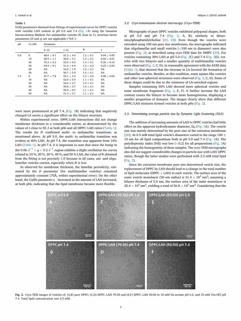
Table 1SAXS parameters obtained from fittings of experimental curves for DPPC vesicleswith variable LAH content at pH 5.0 and 7.4 (Fig. 1A) using the GaussianDeconvolution Method. For unilamellar vesicles (N close to 1), structure factorparameters (D and η) are not applicable (“NA”).
pH % LAH Parameter
D ð�AÞ δ ð�AÞ N η
5.0 0 68.0 � 9.1 51.2 � 4.9 1.3 � 0.1 0.04 � 0.0510 68.9 � 1.1 50.8 � 5.1 1.3 � 0.1 0.02 � 0.0120 70.3 � 0.2 52.5 � 9.2 1.4 � 0.1 0.26 � 0.1030 69.9 � 9.1 51.4 � 6.9 1.3 � 0.2 0.23 � 0.0340 NA 52.0 � 1.9 1.0 � 0.1 NA50 NA 50.7 � 5.9 1.0 � 0.1 NA
7.4 0 67.7 � 7.8 52.1 � 3.2 1.3 � 0.2 0.08 � 0.0210 NA 52.0 � 6.9 1.1 � 0.1 NA20 NA 52.6 � 2.7 1.0 � 0.1 NA30 NA 50.8 � 3.7 1.0 � 0.1 NA40 NA 50.9 � 0.7 1.1 � 0.1 NA50 NA 51.3 � 5.1 1.1 � 0.1 NA
L. Farkuh et al. Heliyon 5 (2019) e02648
were more pronounced at pH 7.4, (Fig. 1B) indicating that negativelycharged LA exerts a significant effect on the bilayer structure.
Within experimental error, DPPC-LAH interactions did not changemembrane thickness to a considerable extent, as demonstrated by thevalues of δ close to 50 Å at both pHs and all DPPC: LAH ratios (Table 1).The results for N confirmed multi- to unilamellar transitions, asmentioned above. At pH 5.0, the multi- to unilamellar transition wasevident at 40% LAH. At pH 7.4, the transition was apparent from 10%LAH (Table 1). At pH 7.4, it is important to note that since the bump in
the 0:06 �1
< q < 0:2 �1 region exhibits a slight oscillation for curves
related to 10%, 20%, 30%, 40%, and 50% LAH, the value ofN obtainedfrom the fitting is not precisely 1.0 because in all cases, uni- and oligo-lamellar vesicles coexist, especially when N is low.
As observed for membrane thickness, the lamellar periodicity, esti-mated by the D parameter (for multilamellar vesicles) remainedapproximately constant (70Å, within experimental error). On the otherhand, the Caill�e parameter η increased as the amount of LAH increased,at both pHs, indicating that the lipid membrane became more flexible.
Fig. 2. Cryo-TEM images of vesicles of: (A,B) pure DPPC; (C,D) DPPC: LAH 70:30 an7.4. Total lipid concentration was 2.5 mM.
5
3.2. Cryo-transmission electron microscopy (Cryo-TEM)
Micrographs of pure DPPC vesicles exhibited polygonal shapes, bothat pH 5.0 and pH 7.4 (Fig. 2, A, B), similarly to dimyr-istoylphosphatidylcholine [23, 24]. Even though the vesicles wereextruded using 100 nm pore size membranes, the micrographs indicatedthat oligolamellar and small vesicles (<100 nm in diameter) were alsopresent (Fig. 2), as described using cryo-TEM data for DMPC [25]. Forvesicles containing 30% LAH at pH 5.0 (Fig. 2C) and 7.4 (Fig. 2D), ves-icles with two bilayers and a smaller quantity of multilamellar vesicleswere observed (Fig. 2, C, D), in reasonable agreement with the SAXS data(Table 1), that showed that the increase in LA favored the formation ofunilamellar vesicles. Besides, at this condition, some square-like vesiclesand other non-spherical structures were observed (Fig. 2, C, D). Some ofthese shapes could be due to the existence of LA-enriched regions.
Samples containing 50% LAH showed more spherical vesicles andsome membrane fragments (Fig. 2, E, F). A further increase the LAHcontent causes the bilayer to become more homogeneous, displaying asmaller proportion of domains. The images clearly show that differentDPPC/LAH mixtures formed vesicles at both pHs (Fig. 2).
3.3. Determining average particle size by Dynamic Light Scattering (DLS)
The addition of increasing amounts of LAH to DPPC vesicles had littleeffect on the apparent hydrodynamic diameter, DH (Fig. 3A). The vesiclesize was mainly determined by the pore size of the extrusion membrane[26]. At 0.5 mM total lipid vesicle's diameters varied in the range 100 �10 nm for all lipid compositions both at pH 5.0 and 7.4 (Fig. 3A). Thepolydispersity index (PdI) was low (<0.2) for all preparations (Fig. 3A)indicating the homogeneity of these samples. The cryo-TEMmicrographsalso did not suggest considerable changes in particle size with LAH: DPPCratios, though the latter studies were performed with 2.5 mM total lipid(Fig. 2).
Since the extrusion membrane pore size determined vesicle size, thereplacement of DPPC by LAH should lead to a change in the total numberof lipid molecules (DPPC þ LAH) in each vesicle. The surface area of theouter vesicle monolayer (50 nm radius) is 31.4 � 103 nm2; assuming abilayer thickness of 5.0 nm, the surface area of the inner monolayer is25.4 � 103 nm2, yielding a total of 56.8 � 103 nm2. Considering that the
d (E,F) DPPC: LAH 50:50 in 10 mM Na acetate pH 5.0, and 10 mM Tris-HCl pH
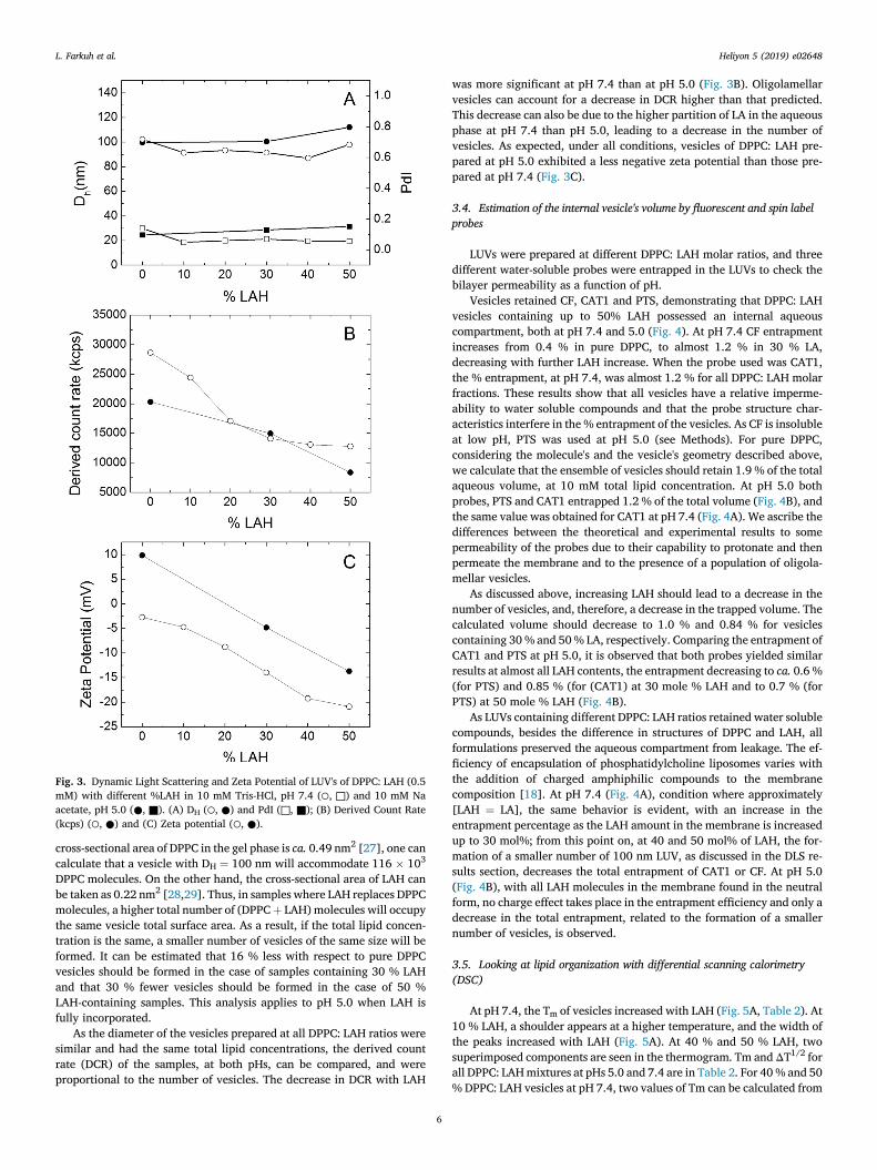
Fig. 3. Dynamic Light Scattering and Zeta Potential of LUV's of DPPC: LAH (0.5mM) with different %LAH in 10 mM Tris-HCl, pH 7.4 (○, □) and 10 mM Naacetate, pH 5.0 (●, ■). (A) DH (○, ●) and PdI (□, ■); (B) Derived Count Rate(kcps) (○, ●) and (C) Zeta potential (○, ●).
L. Farkuh et al. Heliyon 5 (2019) e02648
cross-sectional area of DPPC in the gel phase is ca. 0.49 nm2 [27], one cancalculate that a vesicle with DH ¼ 100 nm will accommodate 116 � 103
DPPC molecules. On the other hand, the cross-sectional area of LAH canbe taken as 0.22 nm2 [28,29]. Thus, in samples where LAH replaces DPPCmolecules, a higher total number of (DPPCþ LAH) molecules will occupythe same vesicle total surface area. As a result, if the total lipid concen-tration is the same, a smaller number of vesicles of the same size will beformed. It can be estimated that 16 % less with respect to pure DPPCvesicles should be formed in the case of samples containing 30 % LAHand that 30 % fewer vesicles should be formed in the case of 50 %LAH-containing samples. This analysis applies to pH 5.0 when LAH isfully incorporated.
As the diameter of the vesicles prepared at all DPPC: LAH ratios weresimilar and had the same total lipid concentrations, the derived countrate (DCR) of the samples, at both pHs, can be compared, and wereproportional to the number of vesicles. The decrease in DCR with LAH
6
was more significant at pH 7.4 than at pH 5.0 (Fig. 3B). Oligolamellarvesicles can account for a decrease in DCR higher than that predicted.This decrease can also be due to the higher partition of LA in the aqueousphase at pH 7.4 than pH 5.0, leading to a decrease in the number ofvesicles. As expected, under all conditions, vesicles of DPPC: LAH pre-pared at pH 5.0 exhibited a less negative zeta potential than those pre-pared at pH 7.4 (Fig. 3C).
3.4. Estimation of the internal vesicle's volume by fluorescent and spin labelprobes
LUVs were prepared at different DPPC: LAH molar ratios, and threedifferent water-soluble probes were entrapped in the LUVs to check thebilayer permeability as a function of pH.
Vesicles retained CF, CAT1 and PTS, demonstrating that DPPC: LAHvesicles containing up to 50% LAH possessed an internal aqueouscompartment, both at pH 7.4 and 5.0 (Fig. 4). At pH 7.4 CF entrapmentincreases from 0.4 % in pure DPPC, to almost 1.2 % in 30 % LA,decreasing with further LAH increase. When the probe used was CAT1,the % entrapment, at pH 7.4, was almost 1.2 % for all DPPC: LAH molarfractions. These results show that all vesicles have a relative imperme-ability to water soluble compounds and that the probe structure char-acteristics interfere in the % entrapment of the vesicles. As CF is insolubleat low pH, PTS was used at pH 5.0 (see Methods). For pure DPPC,considering the molecule's and the vesicle's geometry described above,we calculate that the ensemble of vesicles should retain 1.9 % of the totalaqueous volume, at 10 mM total lipid concentration. At pH 5.0 bothprobes, PTS and CAT1 entrapped 1.2 % of the total volume (Fig. 4B), andthe same value was obtained for CAT1 at pH 7.4 (Fig. 4A). We ascribe thedifferences between the theoretical and experimental results to somepermeability of the probes due to their capability to protonate and thenpermeate the membrane and to the presence of a population of oligola-mellar vesicles.
As discussed above, increasing LAH should lead to a decrease in thenumber of vesicles, and, therefore, a decrease in the trapped volume. Thecalculated volume should decrease to 1.0 % and 0.84 % for vesiclescontaining 30% and 50% LA, respectively. Comparing the entrapment ofCAT1 and PTS at pH 5.0, it is observed that both probes yielded similarresults at almost all LAH contents, the entrapment decreasing to ca. 0.6 %(for PTS) and 0.85 % (for (CAT1) at 30 mole % LAH and to 0.7 % (forPTS) at 50 mole % LAH (Fig. 4B).
As LUVs containing different DPPC: LAH ratios retained water solublecompounds, besides the difference in structures of DPPC and LAH, allformulations preserved the aqueous compartment from leakage. The ef-ficiency of encapsulation of phosphatidylcholine liposomes varies withthe addition of charged amphiphilic compounds to the membranecomposition [18]. At pH 7.4 (Fig. 4A), condition where approximately[LAH ¼ LA], the same behavior is evident, with an increase in theentrapment percentage as the LAH amount in the membrane is increasedup to 30 mol%; from this point on, at 40 and 50 mol% of LAH, the for-mation of a smaller number of 100 nm LUV, as discussed in the DLS re-sults section, decreases the total entrapment of CAT1 or CF. At pH 5.0(Fig. 4B), with all LAH molecules in the membrane found in the neutralform, no charge effect takes place in the entrapment efficiency and only adecrease in the total entrapment, related to the formation of a smallernumber of vesicles, is observed.
3.5. Looking at lipid organization with differential scanning calorimetry(DSC)
At pH 7.4, the Tm of vesicles increased with LAH (Fig. 5A, Table 2). At10 % LAH, a shoulder appears at a higher temperature, and the width ofthe peaks increased with LAH (Fig. 5A). At 40 % and 50 % LAH, twosuperimposed components are seen in the thermogram. Tm andΔT1/2 forall DPPC: LAHmixtures at pHs 5.0 and 7.4 are in Table 2. For 40% and 50% DPPC: LAH vesicles at pH 7.4, two values of Tm can be calculated from
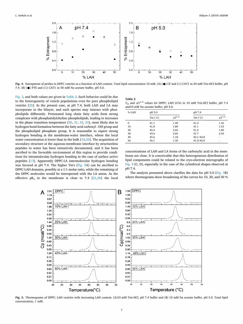
Table 2Tm and ΔT1/2 values for DPPC: LAH LUVs in 10 mM Tris-HCl buffer, pH 7.4and10 mM Na acetate buffer, pH 5.0.
% LAH pH 5.0 pH 7.4
Tm (�C) ΔT1/2 Tm (�C) ΔT1/2
0 41.1 1.49 41.4 1.3510 41.8 1.89 41.1 1.5320 42.4 2.65 41.5 1.8030 43.0 2.65 41.7 2.4340 45.6 1.18 42.1/42.8 -50 45.1 1.35 41.9/42.9 -
Fig. 4. Entrapment of probes in DPPC vesicles as a function of LAH content. Total lipid concentration 10 mM. (A) (●) CF and (○) CAT1 in 50 mM Tris-HCl buffer, pH7.4. (B) (●) PTS and (○) CAT1 in 50 mM Na acetate buffer, pH 5.0.
L. Farkuh et al. Heliyon 5 (2019) e02648
Fig. 3, and both values are given in Table 2. Such behavior could be dueto the heterogeneity of vesicle populations even for pure phospholipidvesicles [25]. In the present case, at pH 7.4, both LAH and LA mayincorporate in the bilayer, and each species may interact with phos-pholipids differently. Protonated long chain fatty acids form strongcomplexes with phosphatidylcholine phospholipids, leading to increasesin the phase transition temperature [30, 31, 32, 33], most likely due tohydrogen bond formation between the fatty acid carboxyl –OH group andthe phospholipid phosphate group. It is reasonable to expect stronghydrogen bonding at the membrane-water interface, where the localwater concentration is lower than in the bulk [34,35]. The acquisition ofsecondary structure at the aqueous-membrane interface by structurelesspeptides in water has been extensively documented, and it has beenascribed to the favorable environment of this region to provide condi-tions for intramolecular hydrogen bonding in the case of surface activepeptides [13]. Apparently DPPC-LA intermolecular hydrogen bondingwas favored at pH 7.4. The higher Tm's (Fig. 5A) can be ascribed toDPPC-LAH domains, possibly at a 1:1 molar ratio, while the remaining ofthe DPPC molecules would be interspersed with the LA anion. As theeffective pKa in the membrane is close to 7.4 [21,36] the local
Fig. 5. Thermograms of DPPC: LAH vesicles with increasing LAH content. (A)10 mMconcentration, 1 mM.
7
concentrations of LAH and LA forms of the carboxylic acid in the mem-brane are close. It is conceivable that this heterogeneous distribution oflipid components could be related to the cryo-electron micrographs ofFig. 4 (C, D), especially in the case of the cylindrical shapes observed atpH 7.4.
The analysis presented above clarifies the data for pH 5.0 (Fig. 5B)where thermograms show broadening of the curves for 10, 20, and 30 %
Tris-HCl, pH 7.4 buffer and (B) 10 mM Na acetate buffer, pH 5.0. Total lipid
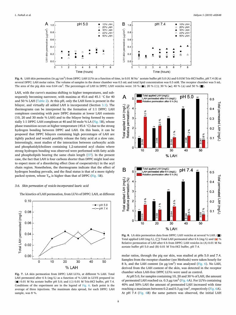
Fig. 6. LAH skin permeation (in μg/cm2) from DPPC: LAH LUVs as a function of time, in 0.01 M Naþ acetate buffer pH 5.0 (A) and 0.01M Tris-HCl buffer, pH 7.4 (B) atseveral DPPC: LAH molar ratios. The volume of samples in the donor chamber was 0.5 mL and total lipid concentration was 0.5 mM. The receptor chamber was 5 mL.The area of the pig skin was 0.64 cm2. The percentages of LAH in DPPC: LAH vesicles were: 10 % (●); 20 % (○); 30 % (▴); 40 % (Δ) and 50 % (■).
L. Farkuh et al. Heliyon 5 (2019) e02648
LAH, with the curve's maxima shifting to higher temperatures, and sub-sequently becoming narrower, with maxima at 45.6 and 45.1 �C for 40and 50 % LAH (Table 2). At this pH, only the LAH form is present in thebilayer, and virtually all added LAH is incorporated (Section 3.1). Thethermograms can be interpreted by the formation of 1:1 DPPC: LAHcomplexes coexisting with pure DPPC domains at lower LAH contents(10, 20 and 30 mole % LAH) and to the bilayer being formed by essen-tially 1:1 DPPC: LAH complexes at 40 and 50 mole % LA (Fig. 5B), whosephase transition occurs at higher temperature (45.6 �C) due to the stronghydrogen bonding between DPPC and LAH. On this basis, it can beproposed that DPPC bilayers containing high percentages of LAH aretightly packed and would possibly release the fatty acid at a slow rate.Interestingly, most studies of the interaction between carboxylic acidsand phosphatidylcholines containing 1,2-saturated acyl chains wherestrong hydrogen bonding was observed were performed with fatty acidsand phospholipids bearing the same chain length [37]. In the presentcase, the fact that LAH is four carbons shorter than DPPC might lead oneto expect more of a disordering effect (loss of cooperativity) in the acylchain region. Nonetheless, the thermograms indicate that the effect ofhydrogen bonding prevails, and the final status is that of a more tightlypacked system, whose Tm is higher than that of DPPC (Fig. 5B).
3.6. Skin permeation of vesicle-incorporated lauric acid
The kinetics of LAH permeation, from LUVs of DPPC: LAH, at different
Fig. 7. LA skin permeation from DPPC: LAH LUVs, at different % LAH. TotalLAH permeated after 6 h (mg/L) as a function of % LAH in LUVS prepared in:(●) 0.01 M Na acetate buffer pH 5.0; and (○) 0.01 M Tris-HCl buffer, pH 7.4.Conditions of the experiment are in the legend of Fig. 6. Each point is theaverage of three injections. The maximum data spread, for each DPPC: LAHsample, was 8 %.
Fig. 8. LA skin permeation data from DPPC: LAH vesicles at several % LAH. (■)Total applied LAH (mg/L), (□) Total LAH permeated after 6 h (mg/L) and ( ) %Relative permeation of LAH after 6 h from DPPC: LAH vesicles in (A) 0.01 M Naacetate buffer pH 5.0 and (B) 0.01 M Tris-HCl buffer, pH 7.4.
8
molar ratios, through the pig ear skin, was studied at pHs 5.0 and 7.4.Samples from the receptor chamber (see Methods) were taken hourly for8 h, and the LAH content (in μg/cm2) was analyzed (Fig. 6). No LAH,derived from the LAH content of the skin, was detected in the receptorchamber when LAH-free DPPC LUVs were used as control.
At pH 5.0, for samples containing 10, 20 and 30% of LAH, the amountof permeated LAH reached ca. 0.3 μg/cm2 (Fig. 6A). For LUVs containing40% and 50% LAH the amount of permeated LAH increased with timereaching a maximum between 0.2 and 0.3 μg/cm2, respectively (Fig. 6A).At pH 7.4 (Fig. 6B) the same pattern was observed, the initial LAH
L. Farkuh et al. Heliyon 5 (2019) e02648
permeation was between 0.05 and 0.3 μg/cm2 after the first hour, at low% LAH, and reached between 0.2 to 0.3 μg/cm2 after 6 h for all % LAH,with exception of the sample containing 50% LAHwhich reached 0.4 μg/cm2.
The total amounts of permeated LAH (mg/L) after 6 h incubation,using LUVs with different % LAH, is shown in Fig. 7. At pH 5.0, the totalLAH that in the receptor chamber increased with the LUV % LAH,reaching a maximum for 40 % LAH. Increasing the % LAH in the LUVsfrom 10 to 50 % only doubled the total permeated LAH (Fig. 7).
At pH 7.4, the maximum permeation was observed with LUVs con-taining 30 % LAH. Further increases in the % LAH decreased the amountof permeated LAH (Fig. 7). At high %LAH, at pH 7.4, the diffusion ofLUVs through the skin could be hampered because of the excess ofnegative charge on the skin surface [38] leading to the maximumobserved in 30 % LAH. The low relative amount of LAH reaching thereceptor fluid in the time selected is convenient. Rather, the retention ofthe active substance, such as LAH, in the stratum corneum, hair follicle,dermis, and epidermis of the skin can be considered to represent cuta-neous permeation [39].
In Fig. 8, we compare our data of LAH permeation through pig earskin. The amount of LA recovered in the receptor liquid after 6 h is alsoplotted vs the % LAH and can be compared the initial amount of LAH inall samples studied, at pH 5.0 and 7.4.
For LUVs containing from 10 to 50 % LAH, the percentage ofpermeated LAH, was small at both pHs. Even increasing the total amountof LAH from 1 mg/mL to 5 mg/mL, less than 0.05 mg/mL permeated theskin for all vesicle's compositions. At pH 5.0, after 6 h, for LUVs with 10%and 50 % LAH, the percentages of permeated LAH were 3.2 % and 0.8 %,respectively (Fig. 8A). At pH 7.4, ca. 1.6 % of LAH permeated the skin forLUVS with 10 % LAH and 0.7 % for those with 50 % LAH (Fig. 8B). Inconclusion, the increase in the total amount of LAH in the donor liquiddid not lead to a proportional increase of LAH in the receptor chamber.
With respect to LAH retention in the skin, Blank and Gould, using 14Clabeled LAH, demonstrated that LAH dissolved in buffered solutions, atdifferent pH's, penetrates in the human skin dermis and that the LAHpenetration is higher as the pH decreases from 8.5 to 7.5 [40]. Theypointed out that they could not perform the experiments below pH 7.5due to the flocculation of the acid form of LAH. Our results show that LAHpermeation through the skin, from LUVs of DPPC: LAH, is observed atpHs 5.0 and 7.4 allowing to bypass the insolubility problem of LAH de-livery to the skin. However, the skin permeation of LAH is low, as desiredfor a drug that has to penetrate the skin and have no effect at the systemiclevel.
LAH-containing LUVs allow the preparation of acid pH stable for-mulations containing high LAH concentrations, high stability, thusavoiding the flocculation of LAH. This is an exciting result, since in thetreatment of acne vulgaris the action of lauric acid on the hair folliclerather than skin permeation has to be considered [5,41].
4. Conclusions
Here we described a detailed physicochemical characterization of thepotential delivery system consisting of DPPC: LAH vesicles. SAXS andcryo-TEM showed that unilamellar vesicles co-existed with differentpopulations of oligo- or multilamellar DPPC: LAH-containing LUVs andthat closed and impermeable vesicles were formed containing an aqueousinner compartment. DSC studies showed that the charged and unchargedforms of lauric acid interact in different manners with the DPPC bilayerand that, at pH 7.4, at least two phases coexist, one possibly beingenriched with LAH. At pH 5.0, a hydrogen bonded-mediated complexbetween DPPC and LAH may be responsible for a system with higherphase transition temperature. The small amounts of LAH detected in thereceptor fluid of Franz cell is suggestive of the ability of LAH-containingDPPC vesicles to deliver the fatty acid to the skin. As we demonstratedthat the LAH-containing lipid vesicles incorporate water soluble probes,the DPPC: LAH vesicle system may serve, at the same time, to deliver the
9
antimicrobial lauric acid and a water-soluble active compound to theskin.
Declarations
Author contribution statement
I. Midea Cuccovia: Conceived and designed the experiments;Analyzed and interpreted the data; Contributed reagents, materials,analysis tools or data; Wrote the paper.
L. Farkuh: Conceived and designed the experiments; Performed theexperiments; Analyzed and interpreted the data; Wrote the paper.
P. T. Hennies: Conceived and designed the experiments.C. Nunes, M. A. Segundo, L. Barreiros, P. L. Oseliero Filho, C. L. P.
Oliveira, A. Cassago, R. V. Portugal, R. A. Muramoto, G. P. B. Carretero:Performed the experiments.
S. Reis: Analyzed and interpreted the data.S. Schreier: Analyzed and interpreted the data; Wrote the paper.H. Chaimovich: Analyzed and interpreted the data; Contributed re-
agents, materials, analysis tools or data; Wrote the paper.
Funding statement
L. Farkuh thanks Ph.D. B�arbara Bianca Gerbelli (IF-USP), CNPq,FAPESP (Proc. 2013/08166-5), Santander Universidades (COPGRAD.01- 044/2014). I.M.Cuccovia thanks the National Council for Scientific andTechnological Development (CNPq – 465259/2014-6), the Coordinationfor the Improvement of Higher Education Personnel (CAPES), the Na-tional Institute of Science and Technology Complex Fluids (INCT-FCx),and the S~ao Paulo Research Foundation (FAPESP – 2014/50983-3). Weacknowledge the Brazilian Nanotechnology National Laboratory(LNNano), CNPEM, for the use of cryo-TEM facilities. This work wassupported by The European Union (FEDER funds) and National Funds(FCT/MEC, Fundaç~ao para a Ciencia e a Tecnologia and Minist�erio daEducaç~ao e Ciencia) under the Partnership Agreement PT2020 UID/QUI/50006/2013 - POCI/01/0145/FEDER/007265. L. Barreiros thanks FCTand POPH (Programa Operacional Potencial Humano) for her Post-Docgrant (SFRH/BPD/89668/2012). C. Nunes thanks FCT for her Investi-gator Grant (IF/00293/2015). G.P.B.Carretero acknowledges the Pro-grama CAPES: INCT -Institutos Nacionais de Ciencia e Tecnologia (Proc.88887.137085/2017-00), R.A. Muramoto acknowledges CNPq. I.M.Cuccovia, H. Chaimovich, and S. Schreier are research fellows of CNPq.
Competing interest statement
The authors declare no conflict of interest.
Additional information
No additional information is available for this paper.
References
[1] N.R.F. Tomarelli, M. Rudolph, C.S. Rose, Gy€orgy Paul, The effect of fatty acids onthe growth of strains of lactobacillus bifidus, J. Biol. Chem. 187 (1950) 197–204.
[2] T. Nakatsuji, M.C. Kao, J.-Y. Fang, C.C. Zouboulis, L. Zhang, R.L. Gallo, C.-M. Huang, Antimicrobial property of lauric acid against Propionibacterium acnes:its therapeutic potential for inflammatory acne vulgaris, J. Investig. Dermatol. 129(2009) 2480–2488.
[3] S. Michael, M. Laurie, R. Evelyn, V.M. Paul, H.N. A., G.S. F., W.C. J., Antimicrobialeffects of virgin coconut oil and its medium-chain fatty acids on Clostridiumdifficile, J. Med. Food 16 (2013) 1079–1085.
[4] B. Ouattara, R.E. Simard, R.A. Holley, G.J. Piette, A. Begin, Antibacterial activity ofselected fatty acids and essential oils against six meat spoilage organisms, Int. J.Food Microbiol. 37 (1997) 155–162.
[5] D. Yang, D. Pornpattananangkul, T. Nakatsuji, M. Chan, D. Carson, C.-M. Huang,L. Zhang, The antimicrobial activity of liposomal lauric acids againstPropionibacterium acnes, Biomaterials 30 (2009) 6035–6040.
[6] B. Lin, A.V. McCormick, H.T. Davis, R. Strey, Solubility of sodium soaps in aqueoussalt solutions, J. Colloid Interface Sci. 291 (2005) 543–549.
L. Farkuh et al. Heliyon 5 (2019) e02648
[7] P.A. Kralchevsky, K.D. Danov, C.I. Pishmanova, S.D. Kralchevska, N.C. Christov,K.P. Ananthapadmanabhan, A. Lips, Effect of the precipitation of neutral-soap, acid-soap, and alkanoic acid crystallites on the bulk pH and surface tension of soapsolutions, Langmuir 23 (2007) 3538–3553.
[8] F.E. Stanley, A.M. Warner, E. Schneiderman, A.M. Stalcup, Rapid determination ofsurfactant critical micelle concentrations using pressure-driven flow with capillaryelectrophoresis instrumentation, J. Chromatogr. A 1216 (2009) 8431–8434.
[9] Z. Vanic, Phospholipid vesicles for enhanced drug delivery in dermatology, J. DrugDiscov. Dev. Deliv. 2 (1) (2015) 1010.
[10] S. Lonchin, P. Luisi, W. Peter, B.H. Robinson, A matrix effect in mixedphospholipid/fatty acid vesicle formation, J. Phys. Chem. B 103 (49) (1999)10910–10916.
[11] M.L. Rogerson, B.H. Robinson, S. Bucak, P. Walde, Kinetic studies of the interactionof fatty acids with phosphatidylcholine vesicles (liposomes), Colloids Surfaces BBiointerfaces 48 (2006) 24–34.
[12] J.M. Gebicki, M. Hicks, Preparation and properties of vesicles enclosed by fatty acidmembranes, Chem. Phys. Lipids 16 (1976) 142–160.
[13] M.C. Manzini, K.R. Perez, K.A. Riske, J.C. Bozelli, T.L. Santos, M.A. da Silva,G.K.V. Saraiva, M.J. Politi, A.P. Valente, F.C.L. Almeida, H. Chaimovich,M.A. Rodrigues, M.P. Bemquerer, S. Schreier, I.M. Cuccovia, Peptide:lipid ratio andmembrane surface charge determine the mechanism of action of the antimicrobialpeptide BP100. Conformational and functional studies, Biochim. Biophys. ActaBiomembr. 1838 (2014) 1985–1999.
[14] C.L.P. Oliveira, B.B. Gerbelli, E.R.T. Silva, F. Nallet, L. Navailles, E.A. Oliveira,J.S. Pedersen, Gaussian deconvolution: a useful method for a form-free modeling ofscattering data from mono- and multilayered planar systems, J. Appl. Crystallogr.45 (2012) 1278–1286.
[15] M. Aschi, A.A. D’Archivio, A. Fontana, A. Formiglio, Physicochemical properties offluorescent probes: experimental and computational determination of theoverlapping pKa values of carboxyfluorescein, J. Org. Chem. 73 (2008) 3411–3417.
[16] C.M. Paleos, P. Dais, Ready reduction of some nitroxide free radicals with ascorbicacid, J. Chem. Soc. Chem. Commun. (1977) 345–346.
[17] S. Schreier-Muccillo, D. Marsh, I.C. Smith, Monitoring the permeability profile oflipid membranes with spin probes, Arch. Biochem. Biophys. 172 (1976) 1–11.
[18] Y. Aracava, S. Schreier, R. Phadke, R. Deslauriers, I.C.P. Smith, Spin label reductionkinetics, a procedure to study the effect of drugs on membrane permeability: theeffects of monosodium urate, dimethyl sulfoxide and amphotericin B, J. Biochem.Biophys. Methods 5 (1981) 83–94.
[19] F.H. Quina, H. Chaimovich, Ion-exchange in micellar solutions .1. Conceptual-framework for ion-exchange in micellar solutions, J. Phys. Chem. 83 (1979)1844–1850.
[20] S. Schreier, W.A. Frezzatti, P.S. Araujo, H. Chaimovich, I.M. Cuccovia, Effect oflipid-membranes on the apparent pk of the local-anesthetic tetracaine - spin labeland titration studies, Biochim. Biophys. Acta 769 (1984) 231–237.
[21] A.A. Pashkovskaya, M. Vazdar, L. Zimmermann, O. Jovanovic, P. Pohl, E.E. Pohl,Mechanism of long-chain free fatty acid protonation at the membrane-waterinterface, Biophys. J. 114 (2018) 2142–2151.
[22] J.R. Kanicky, D.O. Shah, Effect of premicellar aggregation on the pKa of fatty acidsoap solutions, Langmuir 19 (2003) 2034–2038.
[23] A. Nasedkin, J. Davidsson, M. Kumpugdee-Vollrath, Determination of nanostructureof liposomes containing two model drugs by X-ray scattering from a synchrotronsource, J. Synchrotron Radiat. 20 (2013) 721–728.
[24] G. Wu, H.A. Khant, Chiub W and Lee KYC Effects of bilayer phases on phospholipid-poloxamer interactions, Soft Matter 5 (2009) 1496–1503.
10
[25] J. Drazenovic, H. Wang, K. Roth, J. Zhang, S. Ahmed, Y. Chen, G. Bothun,S.L. Wunder, Effect of lamellarity and size on calorimetric phase transitions insingle component phosphatidylcholine vesicles, Biochim. Biophys. Acta 1848(2015) 532–543.
[26] F. Olson, C.A. Hunt, F.C. Szoka, W.J. Vail, D. Papahadjopoulos, Preparation ofliposomes of defined size distribution by extrusion through polycarbonatemembranes, Biochim. Biophys. Acta Biomembr. 557 (1979) 9–23.
[27] R. Hartkamp, Timothy C. Moore, Christopher R. Iacovella, Michael A. Thompson,Pallav A. Bulsara, David J. Moore, C. McCabe, Investigating the structure ofmulticomponent gel-phase lipid bilayers, Biophys. J. 111 (2016) 813–823.
[28] M. H€oltje, T. F€orster, B. Brandt, T. Engels, W. von Rybinski, H.-D. H€oltje, Moleculardynamics simulations of stratum corneum lipid models: fatty acids and cholesterol,Biochim. Biophys. Acta Biomembr. 1511 (2001) 156–167.
[29] L.I. Burke, G.S. Patil, R.V. Panganamala, J.C. Geer, D.G. Cornwell, Surface areas ofnaturally occurring lipid classes and the quantitative microdetermination of lipids,J. Lipid Res. 14 (1973) 9–15.
[30] G. Cevc, J.M. Seddon, R. Hartung, W. Eggert, Phosphatidylcholine-fatty acidmembranes .1. Effects of protonation, salt concentration, temperature and chain-length on the colloidal and phase properties of mixed vesicles, bilayers andnonlamellar structures, Biochim. Biophys. Acta 940 (1988) 219–240.
[31] J.M. Seddon, R.H. Templer, N.A. Warrender, Z. Huang, G. Cevc, D. Marsh,Phosphatidylcholine–fatty acid membranes: effects of headgroup hydration on thephase behaviour and structural parameters of the gel and inverse hexagonal (HII)phases, Biochim. Biophys. Acta Biomembr. 1327 (1997) 131–147.
[32] T. Inoue, S. Yanagihara, Y. Misono, M. Suzuki, Effect of fatty acids on phasebehavior of hydrated dipalmitoylphosphatidylcholine bilayer: saturated versusunsaturated fatty acids, Chem. Phys. Lipids 109 (2001) 117–133.
[33] G. Ma, H.C. Allen, Condensing effect of palmitic acid on DPPC in mixed Langmuirmonolayers, Langmuir 23 (2007) 589–597.
[34] E.A. Disalvo, F. Lairion, F. Martini, E. Tymczyszyn, M. Frías, H. Almaleck,G.J. Gordillo, Structural and functional properties of hydration and confined waterin membrane interfaces, Biochim. Biophys. Acta Biomembr. 1778 (2008)2655–2670.
[35] V. Soldi, J. Keiper, L.S. Romsted, I.M. Cuccovia, H. Chaimovich, Arenediazoniumsalts: new probes of the interfacial compositions of association colloids. 6.Relationships between interfacial counterion and water concentrations andsurfactant headgroup size, sphere-to-rod transitions, and chemical reactivity incationic micelles, Langmuir 16 (2000) 59–71.
[36] M. Langner, T. Isac, S.W. Hui, Interaction of free fatty acids with phospholipidbilayers, Biochim. Biophys. Acta Biomembr. 1236 (1995) 73–80.
[37] A. Arouri, K.E. Lauritsen, H.L. Nielsen, O.G. Mouritsen, Effect of fatty acids on thepermeability barrier of model and biological membranes, Chem. Phys. Lipids 200(2016) 139–146.
[38] K. Egbaria, N. Weiner, Liposomes as a topical drug delivery system, Adv. DrugDeliv. Rev. 5 (1990) 287–300.
[39] C. Guidelines, Cosmetics Europe: Guidelines for the Evaluation of the Efficacy ofCosmetics Products, 2008.
[40] I.H. Blank, E. Gould, Penetration of anionic surfactants into skin III. Penetrationfrom buffered sodium laurate solution, J. Investig. Dermatol. 37 (1961) 3.
[41] C. Sinico, M. Manconi, M. Peppi, F. Lai, D. Valenti, A.M. Fadda, Liposomes ascarriers for dermal delivery of tretinoin: in vitro evaluation of drug permeation andvesicle-skin interaction, J. Control. Release 103 (2005) 123–136.