Embed Size (px)
Citation preview
INFECTION AND IMMUNITY, May 2010, p. 2272–2282 Vol. 78, No. 50019-9567/10/$12.00 doi:10.1128/IAI.01374-09Copyright © 2010, American Society for Microbiology. All Rights Reserved.
Chlamydia muridarum T-Cell Antigens Formulated with the AdjuvantDDA/TDB Induce Immunity against Infection That Correlates with a
High Frequency of Gamma Interferon (IFN-�)/Tumor NecrosisFactor Alpha and IFN-�/Interleukin-17
Double-Positive CD4� T Cells�
Hong Yu,1 Xiaozhou Jiang,1 Caixia Shen,1 Karuna P. Karunakaran,1 Janina Jiang,1Nicole L. Rosin,2 and Robert C. Brunham1*
British Columbia Centre for Disease Control, Vancouver, BC, Canada V5Z 4R4,1 and Department of Biochemistry andMolecular Biology, University of British Columbia, Vancouver, BC, Canada V6T 1Z32
Received 10 December 2009/Returned for modification 13 January 2010/Accepted 3 March 2010
Major impediments to developing a Chlamydia vaccine lie in identifying immunologically relevant T-cellantigens and delivery in a manner to stimulate protective immunity. Using an immunoproteomic approach, wepreviously identified three immunodominant Chlamydia T-cell antigens (PmpG-1, PmpE/F-2, and RplF).Because RplF has high homology to a human ortholog, it may not be suitable for human vaccine development.Therefore, in this study, we evaluated protection against Chlamydia infection in the genital tract in C57BL/6mice immunized with Chlamydia-specific membrane proteins PmpG-1, PmpE/F-2, and major outer membraneprotein (MOMP; as a reference) or a combination of them formulated with one of three adjuvants, CpGoligodeoxynucleotide (CpG-ODN), AbISCO-100 (AbISCO), or DDA/TDB (dimethyldioctadecylammonium bro-mide/D-(�)-trehalose 6,6�-dibehenate). The results show that immunization with the CpG-ODN formulationfailed to provide protection against Chlamydia infection; the AbISCO formulation conferred moderate protec-tion, and the DDA/TDB formulation showed the highest degree of protective efficacy. The combination ofPmpG-1, PmpE/F-2, and MOMP proteins formulated with DDA/TDB exhibited the greatest degree of protec-tion among all vaccine groups studied. Moreover, this vaccine combination also engendered significantprotection in BALB/c mice, which have a different major histocompatibility complex (MHC) background. Wemeasured cell-mediated immune cytokine responses in mice immunized with PmpG-1 mixed with each of thethree adjuvants. The results demonstrate that mice immunized with the DDA/TDB formulation induced thestrongest gamma interferon (IFN-�) and interleukin-17 (IL-17) responses, characterized by the highestfrequency of IFN-�/tumor necrosis factor alpha (TNF-�) and IFN-�/IL-17 double-positive CD4� T cells. Inconclusion, a Chlamydia vaccine based on the recombinant proteins PmpG-1, PmpE/F-2, and MOMP deliveredin a DDA/TDB adjuvant conferred protection against infection that correlated with IFN-�/TNF-� and IFN-�/IL-17 double-positive CD4� T cells.
Chlamydia trachomatis is a major cause of sexually transmit-ted bacterial disease, with over 92 million new cases of C.trachomatis-related disease annually occurring worldwide (39).C. trachomatis infects epithelial cells lining the reproductivetract and causes severe complications such as pelvic inflamma-tory disease, ectopic pregnancy, and infertility in women. Pub-lic health programs to prevent and control Chlamydia based onearly case identification and antibiotic treatment appear to befailing, as case rates continued to rise during the past decade(6). Thus, the development of an effective vaccine is of para-mount importance.
Early vaccination trials involved both human and nonhumanprimates immunized with whole, inactivated C. trachomatiselementary bodies (EBs). The results demonstrated incom-plete and short-lived protection, with even worse inflammation
postvaccination among infected subjects (12, 42), and thus,current C. trachomatis vaccine research has focused on theproduction of subunit vaccines that are based on individualprotective C. trachomatis proteins (7, 38). To date, the Chla-mydia major outer membrane protein (MOMP) has been themost widely investigated subunit vaccine. Although immuneresponses against MOMP can be elicited in animal models,studies indicate that vaccines based on MOMP alone do notafford complete protection (18, 32–34).
Immunity to Chlamydia is currently understood as beingmediated by T cells and mainly relies on tissue traffickingCD4� T cells producing gamma interferon (IFN-�) (28, 40,41). One major impediment to developing a vaccine againstthis intracellular pathogen lies in identifying relevant T-cellantigens (Ags). Recently, we used a combination of affinitychromatography and tandem mass spectrometry to successfullyidentify 8 major histocompatibility complex (MHC) class II(I-Ab)-bound Chlamydia peptides eluted from C. muridarum-infected dendritic cells (DCs) from C57BL/6 mice. Adoptivetransfer of DCs pulsed with a pool of the 8 peptides partiallyprotected mice against challenge infection (19). Further study
* Corresponding author. Mailing address: University of British Co-lumbia Centre for Disease Control, 655 West 12th Avenue, Vancou-ver, BC V5Z 4R4, Canada. Phone: (604) 707-2405. Fax: (604) 707-2401. E-mail: [email protected].
� Published ahead of print on 15 March 2010.
2272
on August 30, 2018 by guest
http://iai.asm.org/
Dow
nloaded from

demonstrated that 3 of the 8 Chlamydia antigens, polymorphicmembrane protein G (PmpG-1), polymorphic membrane pro-tein F (PmpE/F-2), and ribosomal protein L6 (RplF) wereimmunodominant, and vaccination with DCs individuallytransfected with these proteins induced significant protectiveimmunity against lung and genital tract infections (49).
Another challenge in developing an effective Chlamydia vac-cine is to identify suitable adjuvants and the appropriate routesand methods of delivery of the vaccine (15). CpG oligode-oxynucleotide (CpG-ODN) is a potent Th1-polarizing adju-vant which activates antigen-presenting cells through Toll-likereceptor 9 (TLR9), which leads to interleukin-12 (IL-12) pro-duction, driving Th1 immune responses (21). AbISCO-100(AbISCO), an effective immunostimulating complex (ISCOM)adjuvant generates a balanced immune response with both Th1and Th2 characteristics. Compared with other adjuvants suchas alum, Freund’s incomplete adjuvant (FIA), and a syntheticmonophosphoryl lipid A adjuvant (Ribi), AbISCO generatedthe highest antibody titers, the highest proliferation index andgave consistently higher production levels of IFN-�, IL-4, andIL-5. Moreover, animal studies demonstrated that AbISCOadjuvant induced greater protection against challenge infec-tion with human immunodeficiency virus (HIV)/simian immu-nodeficiency virus (SIV), herpesvirus, and influenza virus (24,27). The adjuvant DDA/TDB consists of cationic liposomeDDA (dimethyldioctadecylammonium bromide) and an im-munomodulator, TDB [D-(�)-trehalose 6,6�-dibehenate], aless-toxic, synthetic mycobacterial cord factor analogue (31,36), inserted into liposomes. DDA/TDB was found to be aparticularly promising adjuvant capable of inducing bothstrong Th1 responses and high antibody titers. It was reportedthat DDA/TDB makes an effective tuberculosis subunit vac-cine adjuvant with the ability to raise high levels of protectivememory immunity, comparable to that found in Mycobacteriumbovis BCG-vaccinated mice. The specific Th1-type immuneresponse was characterized by substantial production of IFN-�and high levels of IgG2b isotype antibodies. The lymphocytesubset releasing IFN-� was identified as CD4 T cells (11). Arecent study demonstrated that mice vaccinated with Chla-mydia MOMP protein formulated with DDA/TDB generatedhigh titers of IgG2b, IFN-�, and tumor necrosis factor alpha(TNF-�) and had significantly reduced vaginal Chlamydiashedding relative to control mice (13).
Among the three immunodominant Chlamydia antigensPmpG-1, RplF, and PmpE/F-2, RplF has high homology to thehuman ortholog and therefore may be not suitable for devel-opment as a human vaccine. In the present study, we evaluatedthe protective effect against Chlamydia genital tract infectionin mice immunized with individual PmpG-1, PmpE/F-2, andMOMP proteins or a combination of them formulated with theadjuvant CpG-ODN, AbISCO, or DDA/TDB. We observedthat the protein antigen combination of PmpG-1, PmpE/F-2,and MOMP formulated with DDA/TDB exhibited the greatestdegree of protective immunity among all groups tested. TheAbISCO formulation conferred moderate protection againstchallenge, whereas the CpG-ODN formulation failed to induceprotection. To explore correlates of protection, we measuredcell-mediated immune cytokine responses in mice immunizedwith PmpG-1 mixed with the three adjuvants. The results dem-onstrated that mice immunized with the DDA/TDB formula-
tion had the strongest IFN-� and IL-17 responses, character-ized by the highest frequency of double-positive IFN-�/TNF-�and IFN-�/IL-17 CD4� T cells.
MATERIALS AND METHODS
Chlamydia. Chlamydia muridarum mouse pneumonitis (MoPn)-causing strainNigg was used in this study. This MoPn-causing strain was grown in HeLa 229cells, and elementary bodies (EBs) were purified by discontinuous density gra-dient centrifugation as previously described (8). Purified EBs were aliquoted andstored at �80°C in sucrose-phosphate-glutamic acid buffer and thawed immedi-ately before use. The infectivity of the stock Chlamydia EBs was determined byinfection of HeLa 229 cells and enumeration of inclusions that were stained byanti-EB mouse polyclonal antibody followed by biotinylated anti-mouse IgG(Jackson ImmunoResearch) and DAB (3,3-diaminobenzidine) substrate (VectorLaboratories) (47). Inactivation of EBs was carried out by heating to 56°C for30 min.
Chlamydia proteins. The production and purification of Chlamydia recombi-nant proteins PmpG-1 and PmpE/F-2 have been described previously (49).Briefly, pmpG-1 and pmpE/F-2 gene fragments (representing amino acids 25 to500 and 25 to 575, respectively) from C. muridarum were generated by PCR andcloned into vector pET32a (Novagen) for expression. The omp-1 gene charac-teristically encodes the MOMP protein without first 22 amino acids, was clonedas BamHI/XhoI fragment into the vector pET30a. Plasmids containing pmpG-1,pmpE/F-2, and omp-1 were transformed into Escherichia coli strain BL21(DE3)(Stratagene). The expressed PmpG-1, PmpE/F-2, and MOMP proteins, with anN-terminal His tag, were purified with a nickel column by using the His-Bindpurification system (Qiagen). Lipopolysaccharide (LPS) removal was carried outby adding 0.1% Triton X-114 in the wash buffers during purification.
Adjuvants. Three adjuvants (CpG-ODN 1826, AbISCO-100, and DDA/TDB)were evaluated in the present study. CpG-ODN 1826 (5�-TCCATGACGTTCCTGACGTT-3�, phosphorothioate modified; Integrated DNA Technologies, Inc.)was used in either a free form (free CpG) or a form conjugated with liposomalnanoparticles (LN-CpG). AbISCO-100 adjuvant (ISCONOVA Sweden) is aselection of purified fractions of quillaja saponins formulated with a mixture ofcholesterol (ovine wool) and phosphatidyl choline (egg). DDA (dimethyldiocta-decylammonium bromide [product no. 890810P]) and TDB [D-(�)-trehalose6,6�-dibehenate (product no. 890808P)] were purchased from Avanti Polar Lip-ids (Alabaster, AL). For the DDA/TDB formulation (11, 14), DDA was mixedinto 10 mM Tris buffer at pH 7.4 to a concentration of 1.67 mg/ml, heated to 80°Cwhile being stirred continuously on a magnetic hot plate for 20 min, and thencooled to room temperature. TDB was suspended in distilled water (dH2O)containing 2% dimethyl sulfoxide to a concentration of 5 mg/ml by repeatedpassaging through a fine-tipped pipette followed by 30 s of vortexing. This stepwas repeated three times before the solution was frozen at �20°C until use. A5-ml volume of TDB (1 mg/ml) was added to 15 ml DDA (1.67 mg/ml). Theresulting solution was then vortexed briefly and stored at 4°C until use. The finalconcentration of DDA was 1.25 mg/ml, and the final concentration of TDB was0.25 mg/ml. Each inoculation dose of 200 �l for immunization contained 250 �gDDA and 50 �g TDB.
Mice. Female C57BL/6 mice and BALB/c mice (8 to 10 weeks old) werepurchased from Charles River Canada (Saint Constant, Canada). The mice weremaintained and used in strict accordance with University of British Columbiaguidelines for animal care.
Mouse immunization. Four mouse trials were conducted in this study. All miceexcept the live-EB group were immunized three times subcutaneously (s.c.) inthe base of the tail at 2-week intervals. Mice intranasally infected with 1,500inclusion-forming units (IFU) of live C. muridarum (EBs) were used as positivecontrols.
In the first trial, groups of six C57BL/6 mice were immunized with 20 �gChlamydia protein (PmpG-1 or MOMP) mixed with 700 �g LN-CpG or 700 �gfree CpG. Groups of mice immunized with LN-CpG alone and phosphate-buffered saline (PBS) were used as negative controls. In the second trial, groupsof eight C57BL/6 mice were immunized with 5 �g individual Chlamydia proteinsPmpG-1, PmpE/F-2, and MOMP or a combination (1.67 �g for each protein)formulated with AbISCO-100 (12 �g) or DDA/TDB (250 �g DDA–50 �g TDB)as follows: (i) PmpG-1 plus AbISCO-100 (PmpG�AbISCO), (ii) PmpE/F-2 plusAbISCO-100 (PmpF�AbISCO), (iii) MOMP plus AbISCO-100 (MOMP�AbISCO), (iv) PmpG-1 plus PmpE/F-2 plus MOMP plus AbISCO-100(G�F�M�AbISCO), (v) AbISCO-100 alone (AbISCO alone), (vi) PmpG-1plus DDA/TDB (PmpG�DDA/TDB), (vii) PmpE/F-2 plus DDA/TDB(PmpF�DDA/TDB), (viii) MOMP plus DDA/TDB, (ix) PmpG-1 plus PmpE/
VOL. 78, 2010 IMPORTANCE OF CHLAMYDIA ANTIGEN/ADJUVANT IN PROTECTION 2273
on August 30, 2018 by guest
http://iai.asm.org/
Dow
nloaded from
F-2 plus MOMP plus DDA/TDB (G�F�M�DDA/TDB), (x) DDA/TDB alone,(xi) PBS, or (xii) EBs. In the third trial, three groups of eight BALB/c mice wereimmunized with (i) G�F�M�DDA/TDB, (ii) DDA/TDB alone, or (iii) EBs.Two weeks after the final immunization, the mice in the three animal trialsdescribed above were then challenged with live EBs for protection evaluation.
In the fourth trial, groups of 16 C57BL/6 mice were immunized with 5 �gPmpG-1 formulated with DDA/TDB (250 �g DDA–50 �g TDB), AbISCO-100(12 �g), or CpG (20 �g). Two weeks after the last immunization, half of the micein each group were sacrificed to isolate splenocytes for lymphocyte multicolorflow cytometry, enzyme-linked immunosorbent assay (ELISA), and enzyme-linked immunospot (ELISPOT) assays; the other half of the mice were chal-lenged with live EBs and sacrificed seven days later to isolate splenocytes andiliac lymph nodes for multicolor flow cytometry.
Genital tract infection and determination of Chlamydia titer. One week afterthe last immunization, mice were injected s.c. with 2.5 mg of medroxyprogester-one acetate (Depo-Provera; Pharmacia and Upjohn). One week after medroxy-progesterone acetate treatment, mice were challenged intravaginally with 1,500IFU of C. muridarum. Cervicovaginal washes were taken at selected dates afterinfection and stored at �80°C for titration on HeLa cells as described previ-ously (5).
ELISPOT assay. The cytokine-specific ELISPOT assay was performed asdescribed previously (16). Briefly, 96-well MultiScreen-HA filtration plates (Mil-lipore) were coated overnight at 4°C with 2 �g/ml of murine-IFN-�-specificmonoclonal antibody (clone R4-6A2; BD PharMingen) or murine-IL-17-specificmonoclonal antibody (clone TC11-18H10.1; BioLegend). Splenocytes isolatedfrom mice in complete RPMI 1640 medium (Sigma-Aldrich) were added to thecoated plates at 106 cells per well in the presence of PmpG-1 protein (1 �g/ml)or heat-killed EBs (HK-EBs; 5 � 105 IFU/ml). After 20 h of incubation at 37°Cand 5% CO2, the plates were washed and then incubated with biotin anti-mouseIFN-� (clone XMG1.2; BD PharMingen) or biotin anti-mouse IL-17 (cloneTC11-8H4; BioLegend) at 2 �g/ml. This was followed by incubation with strepta-vidin-alkaline phosphatase (BD PharMingen) at a 1:1,000 dilution. The spotswere visualized with a substrate consisting of 5-bromo-4-chloro-3-indolyl phos-phate and nitroblue tetrazolium (Sigma-Aldrich).
Cytokine measurement. The culture supernatants of the splenocytes stimu-lated with PmpG-1 protein or HK-EBs for 48 h were collected and analyzed withrespect to TNF-� production with a sandwich ELISA using corresponding spe-cific capture and detection antibodies (BD PharMingen). TNF-� levels werecalculated using a standard curve constructed with recombinant murine TNF-�(BD PharMingen).
Multiparameter flow cytometry. Two weeks after the last immunization orseven days after live-EB challenge, the mice from specified groups were sacri-ficed and the cells harvested from spleen and iliac lymph nodes (after challenge)were stimulated with 2 �g/ml antibody to CD28 and PmpG-1 protein (1 �g/ml)or HK-EBs (5 � 105 IFU/ml) in complete RPMI 1640 for 4 h at 37°C. BrefeldinA was added at a final concentration of 1 �g/ml, and cells were incubated for anadditional 12 h before intracellular cytokine staining. Cells were surface stainedfor CD3, CD4, and CD8 as well as with the viability dye, red fluorescent reactivedye (RViD) (L23102; Molecular Probes), followed by staining for IFN-�, TNF-�,and IL-17 by using the BD Cytoperm kit according to the manufacturer’s in-structions. Finally, the cells were resuspended in a 4% formaldehyde solution.All antibodies and all reagents for intracellular cytokine staining were purchasedfrom BD PharMingen except where noted. We acquired 200,000 live lympho-cytes per sample by using an Aria flow cytometer and analyzed the data by usingFlowJo software (Tree Star).
Statistical analysis. All data were analyzed with the aid of the GraphPadPrism software program. The Kruskal-Wallis test was performed to analyze datafor IFU (Chlamydia shedding) from multiple groups, and the Mann-Whitney Utest was used to compare medians between pairs. Two-way repeated-measureanalysis of variance (ANOVA) was used to compare IFU values over the courseof infection in BALB/c mice. Comparison of cytokine productions as determinedby ELISPOT assay, ELISA, and flow cytometry in multiple groups were analyzedusing one-way ANOVA followed by the Tukey posttest. P values of �0.05 wereconsidered significant. Data are presented as means standard errors of themeans (SEM). No statistic analyses were done for those data sets generated fromlymph nodes, as they were pooled from each group.
RESULTS
Multiple Chlamydia antigens formulated with DDA/TDB ex-hibit protection against challenge with live C. muridarum. Inorder to discover a Th1-polarizing adjuvant that efficiently
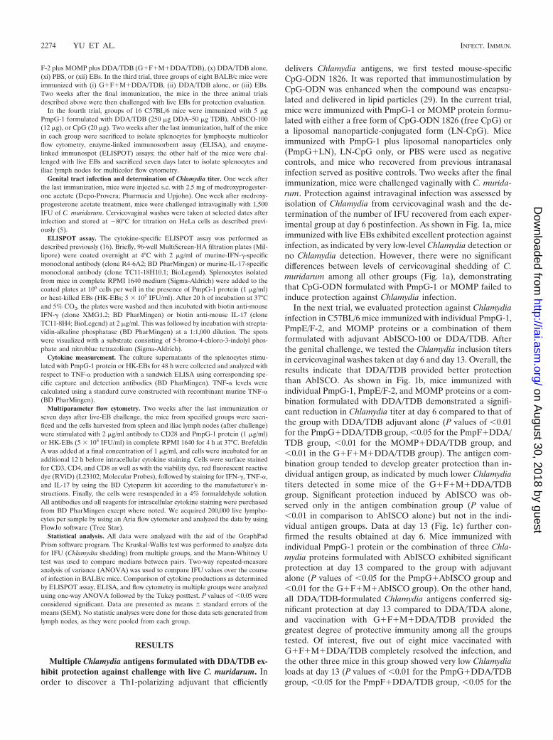
delivers Chlamydia antigens, we first tested mouse-specificCpG-ODN 1826. It was reported that immunostimulation byCpG-ODN was enhanced when the compound was encapsu-lated and delivered in lipid particles (29). In the current trial,mice were immunized with PmpG-1 or MOMP protein formu-lated with either a free form of CpG-ODN 1826 (free CpG) ora liposomal nanoparticle-conjugated form (LN-CpG). Miceimmunized with PmpG-1 plus liposomal nanoparticles only(PmpG�LN), LN-CpG only, or PBS were used as negativecontrols, and mice who recovered from previous intranasalinfection served as positive controls. Two weeks after the finalimmunization, mice were challenged vaginally with C. murida-rum. Protection against intravaginal infection was assessed byisolation of Chlamydia from cervicovaginal wash and the de-termination of the number of IFU recovered from each exper-imental group at day 6 postinfection. As shown in Fig. 1a, miceimmunized with live EBs exhibited excellent protection againstinfection, as indicated by very low-level Chlamydia detection orno Chlamydia detection. However, there were no significantdifferences between levels of cervicovaginal shedding of C.muridarum among all other groups (Fig. 1a), demonstratingthat CpG-ODN formulated with PmpG-1 or MOMP failed toinduce protection against Chlamydia infection.
In the next trial, we evaluated protection against Chlamydiainfection in C57BL/6 mice immunized with individual PmpG-1,PmpE/F-2, and MOMP proteins or a combination of themformulated with adjuvant AbISCO-100 or DDA/TDB. Afterthe genital challenge, we tested the Chlamydia inclusion titersin cervicovaginal washes taken at day 6 and day 13. Overall, theresults indicate that DDA/TDB provided better protectionthan AbISCO. As shown in Fig. 1b, mice immunized withindividual PmpG-1, PmpE/F-2, and MOMP proteins or a com-bination formulated with DDA/TDB demonstrated a signifi-cant reduction in Chlamydia titer at day 6 compared to that ofthe group with DDA/TDB adjuvant alone (P values of �0.01for the PmpG�DDA/TDB group, �0.05 for the PmpF�DDA/TDB group, �0.01 for the MOMP�DDA/TDB group, and�0.01 in the G�F�M�DDA/TDB group). The antigen com-bination group tended to develop greater protection than in-dividual antigen group, as indicated by much lower Chlamydiatiters detected in some mice of the G�F�M�DDA/TDBgroup. Significant protection induced by AbISCO was ob-served only in the antigen combination group (P value of�0.01 in comparison to AbISCO alone) but not in the indi-vidual antigen groups. Data at day 13 (Fig. 1c) further con-firmed the results obtained at day 6. Mice immunized withindividual PmpG-1 protein or the combination of three Chla-mydia proteins formulated with AbISCO exhibited significantprotection at day 13 compared to the group with adjuvantalone (P values of �0.05 for the PmpG�AbISCO group and�0.01 for the G�F�M�AbISCO group). On the other hand,all DDA/TDB-formulated Chlamydia antigens conferred sig-nificant protection at day 13 compared to DDA/TDA alone,and vaccination with G�F�M�DDA/TDB provided thegreatest degree of protective immunity among all the groupstested. Of interest, five out of eight mice vaccinated withG�F�M�DDA/TDB completely resolved the infection, andthe other three mice in this group showed very low Chlamydialoads at day 13 (P values of �0.01 for the PmpG�DDA/TDBgroup, �0.05 for the PmpF�DDA/TDB group, �0.05 for the
2274 YU ET AL. INFECT. IMMUN.
on August 30, 2018 by guest
http://iai.asm.org/
Dow
nloaded from
MOMP�DDA/TDB group, and �0.001 for the G�F�M�DDA/TDB group).
Since all the protection results described above were ob-served in C57BL/6 mice, the strain in which the antigens wereoriginally discovered by use of immunoproteomics, we chal-lenged mice with a different MHC genetic background to de-termine if immunization with multiple Chlamydia protein an-tigens and DDA/TDB conferred protection. BALB/c micewere immunized with G�F�M�DDA/TDB or DDA/TDBalone, and mice infected with live EBs were used as positivecontrols. Chlamydia inclusion titers in the cervicovaginalwashes were detected postchallenge. As shown in Fig. 1d,BALB/c mice immunized with live EBs demonstrated excellentprotection against infection, as indicated by very low bacterial
loads at day 6 and no chlamydiae detected at day 13 and day20. Vaccination with G�F�M�DDA/TDB in BALB/c micesignificantly decreased the Chlamydia load in the cervicovagi-nal washes compared with DDA/TDB alone (P � 0.001). Atday 20 after challenge, all BALB/c mice vaccinated withG�F�M�DDA/TDB completely resolved infection.
Among the three tested adjuvants CpG-ODN 1826,AbISCO-100, and DDA/TDB, the CpG-ODN formulation wasnot able to engender protection against Chlamydia infection atany level in vaccinated mice. The AbISCO formulation con-ferred moderate protection, while the DDA/TDB formulationshowed the greatest efficacy. The combination of PmpG-1,PmpE/F-2, and MOMP formulated with DDA/TDB generatedan additive or synergistic effect that exhibited the greatest
FIG. 1. Vaccine-elicited protection against Chlamydia genital tract infection. Mice were intravaginally challenged with 1,500 IFU of live C.muridarum after immunization with a variety of vaccine formulations. Cervicovaginal washes were taken at selected dates after infection, andbacterial titers were measured on HeLa 229 cells. *, **, and *** indicate P values of �0.05, �0.01, and �0.001, respectively, in comparison tothe corresponding adjuvant-alone group. (a) Failure to induce protection after vaccination with PmpG-1 or MOMP protein formulated withCpG-ODN. (b and c) Resistance to Chlamydia infection in C57BL/6 (C57) mice immunized with PmpG-1, PmpE/F-2, and MOMP proteins or acombination of them formulated with adjuvant AbISCO-100 or DDA/TDB. Cervicovaginal washes were taken at day 6 (b) and day 13 (c) afterinfection. (d) Resistance to Chlamydia infection in BALB/c mice (n 8) immunized with the combination of PmpG-1, PmpE/F-2, and MOMPproteins formulated with the adjuvant DDA/TDB.
VOL. 78, 2010 IMPORTANCE OF CHLAMYDIA ANTIGEN/ADJUVANT IN PROTECTION 2275
on August 30, 2018 by guest
http://iai.asm.org/
Dow
nloaded from
degree of protective immunity among all groups studied.Moreover, G�F�M�DDA/TDB vaccination also gave a one-log reduction of infection burden at day 6 and a 5-log reductionat day 20 in BALB/c mice, with an MHC background differentfrom that of C57BL/6 mice.
PmpG-1 formulated with DDA/TDB induced strong IFN-�,TNF-�, and IL-17 responses, characterized by a high fre-quency of double-positive IFN-�/TNF-� and IFN-�/IL-17CD4� T cells in immunized mice. In order to explore thecellular immune mechanisms for different degrees of protec-tion induced by the three adjuvants, C57BL/6 mice were im-munized with PmpG-1 formulated with DDA/TDB, AbISCO-100, and CpG-ODN 1826 and then challenged with live C.muridarum. The magnitude and quality of T cells producingIFN-�, TNF-�, and IL-17 were assessed before and after chal-lenge using an ELISPOT assay, ELISA, and multiparameterflow cytometry.
In this study, the ELISPOT assay was performed to detectIFN-�- and IL-17-producing cells in immune splenocytes stim-ulated with PmpG-1 protein or HK-EBs. ELISA was per-formed to measure the TNF-� level in the supernatant ofstimulated immune splenocytes.
After immunization with PmpG-1 formulated with DDA/TDB, AbISCO-100, or CpG-ODN 1826, splenocytes exhibitedmarkedly different levels of IFN-� (Fig. 2a), TNF-� (Fig. 2c),and IL-17 (Fig. 2b) responses. The PmpG�DDA/TDB im-mune splenocytes exposed to PmpG-1 protein or HK-EBs de-veloped the highest numbers of IFN-�- and IL-17-secretingcells; the PmpG�AbISCO immune splenocytes demonstratedless-strong IFN-� and IL-17 responses but similar levels ofTNF-� compared with those corresponding to PmpG�DDA/TDB immunization; and the weakest IFN-�/TNF-� response(as well as an absent IL-17 response) was found followingPmpG�CpG immunization. In addition, splenocytes frommice immunized with adjuvant alone, which served as negativecontrols, showed nearly blank background levels, indicatingthat cytokine responses detected in the experimental systemare all Chlamydia Ag specific. The differing levels of IFN-� andIL-17 responses in mice immunized with different adjuvantsare remarkably consistent with the differing degrees of protec-tion against challenge infection (Fig. 1), suggesting that a cor-relate of vaccine-mediated protection against Chlamydia is themagnitude of specific cytokine responses.
To characterize the distinct populations of Th1 and Th17responses, multiparameter flow cytometry was used to simul-taneously analyze multiple cytokines at the single-cell level. Asshown in Fig. 3a, a seven-color flow cytometry panel and gatingstrategy was used to identify IFN-�-, TNF-�-, and IL-17-pro-ducing CD4� T cells in splenocytes from a representativemouse immunized with PmpG�DDA/TDB. Since an individ-ual responding cell could be present in more than one of thetotal cytokine gates, we used Boolean combinations of thecytokine gates to discriminate responding cells based on theirfunctionality or quality of IFN-�/TNF-� (Fig. 3b) and IFN-�/IL-17 (Fig. 3c) production.
Using the Boolean combination of IFN-� gating and TNF-�gating, frequencies of three distinct populations (IFN-� posi-tive/TNF-� negative [IFN-�� TNF-��], IFN-�� TNF-��, andIFN-�� TNF-��) from immune splenocytes stimulated withPmpG-1 and HK-EBs are shown in Fig. 3b-1 and -2, respec-
FIG. 2. Chlamydia antigen-specific cytokine responses after immu-nization with PmpG-1 protein formulated with DDA/TDB, AbISCO,and CpG adjuvants. Two weeks after the final immunization, mousesplenocytes from different vaccine groups were harvested and stimu-lated with 1 �g/ml PmpG-1 protein or 5 � 105 inclusion-forming units(IFU)/ml HK-EBs. Adjuvants consisting of DDA/TDB alone, AbISCOalone, or CpG alone were used as negative controls. The results rep-resent the averages of duplicate wells and are expressed as themeans SEM for groups of six mice. *, **, and *** indicate P valuesof �0.05, �0.01, and �0.001, respectively, in comparison to thePmpG�CpG group. (a) IFN-� responses to PmpG-1 or HK-EBs de-tected by ELISPOT assay. (b) IL-17 responses to PmpG-1 or HK-EBsdetected by ELISPOT assay. (c) TNF-� response to PmpG-1 or HK-EBs detected by ELISA.
2276 YU ET AL. INFECT. IMMUN.
on August 30, 2018 by guest
http://iai.asm.org/
Dow
nloaded from

FIG. 3. Functional characterization of distinct populations of Chlamydia antigen-specific cytokine responses after immunization. Splenocytesfrom different vaccine groups before challenge were analyzed by multiparameter flow cytometry as described in Materials and Methods. Three orfour mice were in each group. Shown are results representative of two experiments. * and ** indicate P values of �0.05 and �0.01, respectively,in comparison to the PmpG�CpG group. A, area; W, width; FSC, forward scatter; SSC, side scatter; PerCP, peridinin chlorophyll protein; APC,allophycocyanin; PE, phycoerythrin. (a) The staining panel and gating strategy used to identify IFN-�-, TNF-�-, and IL-17-producing CD4� T cellsin the splenocytes from a representative mouse immunized with PmpG�DDA/TDB. (b) Comparison of the levels of quality of CD4� IFN-�/TNF-� responses to PmpG-1 protein (b-1) or HK-EBs (b-2) in different vaccine groups. (c) Comparison of the levels of quality of CD4�
IFN-�/IL-17 responses to PmpG-1 protein (c-1) or HK-EBs (c-2) in different vaccine groups.
2277
on August 30, 2018 by guest
http://iai.asm.org/
Dow
nloaded from

tively. The results demonstrate that the response after immu-nization with PmpG�DDA/TDB was dominated by IFN-� andTNF-� double-positive cells, and about half of the response inthe PmpG�AbISCO group was IFN-� and TNF-� doublepositive, whereas the PmpG�CpG vaccine induced the weak-est IFN-� and TNF-� double-positive response, with the sin-gle-positive response dominating. Importantly, this analysisalso showed a correlation between the frequency of multifunc-tional (IFN-� and TNF-� double positive) CD4� T cells andthe degree of protection in mice vaccinated with PmpG�DDA/TDB, PmpG�AbISCO, and PmpG�CpG.
The protective effects of the newly identified lineage of Th17in host defense against bacterial, mycobacterial, and fungalmucosal pathogens have been reported previously (4, 44). Inthis study, the quality of IFN-�/IL-17 cytokine response fromimmune splenocytes stimulated with PmpG (Fig. 3c-1) or HK-EBs (Fig. 3c-2) was also evaluated by multiparameter flowcytometry. Quantifying the fraction of IFN-�/IL-17 response,we found that over half of the response in the best-protectedgroup (PmpG�DDA/TDB) was IFN-� and IL-17 double pos-itive; the PmpG�AbISCO group had a moderate IFN-� andIL-17 double-positive response. The no-protection group(PmpG�CpG) did not develop a detectable IL-17 response.These data also indicate a correlation between the degree ofprotection in vaccinated mice and the frequency of IFN-� andIL-17 double-positive CD4� T cells.
The magnitude and quality of IFN-�, TNF-�, and IL-17responses in spleens and lymph nodes after challenge infec-tion. To define the magnitude of the response on day 7 after C.muridarum challenge, the frequencies of the total PmpG-spe-cific CD4� T-cell cytokine responses comprising IFN-�-, TNF-�-, and IL-17-producing cells in spleens (Fig. 4a) and draininglymph nodes (iliac lymph nodes) (Fig. 4b) from each vaccinegroup are presented. The results demonstrate that amongspleen cells, immunization with PmpG�DDA/TDB inducedstatistically higher frequencies of IFN-�- and IL-17-producingCD4� T cells. The PmpG�AbISCO vaccine induced a fre-quency of TNF-�-producing cells similar to that of the
PmpG�DDA/TDB vaccine but lower frequencies of IFN-�-and IL-17-producing cells. The PmpG�CpG and PBS groupsdeveloped similar frequencies of IFN-�- and TNF-�-producingcells, but these were the lowest frequencies. Notably,PmpG�CpG induced an extremely weak IL-17 response, whilePBS induced a response about one-third of the magnitude ofthe IL-17 response of the PmpG�DDA/TDB group (Fig. 4a).
The data from pooled regional draining lymph node cellsfollowing genital challenge showed that prior immunizationwith PmpG�DDA/TDB resulted in strong IFN-� and TNF-�responses. The PmpG�AbISCO and PBS groups developedsimilar moderate IFN-� and TNF-� responses. The PmpG�CpG group had the weakest IFN-� and TNF-� responses.Surprisingly, and contrary to the spleen cell results, the IL-17responses in lymph nodes were very low in the PmpG�DDA/TDB and PmpG�AbISCO groups, and no IL-17-producingcells were observed in the PmpG�CpG and PBS groups(Fig. 4b).
We further analyzed the quality of cytokine-producing cellsin spleens and iliac lymph nodes from immunized mice at day7 following genital challenge. Immunization with PmpG�DDA/TDB induced the strongest IFN-� and TNF-� double-positive response (Fig. 5a-1) in spleens and an IFN-� andTNF-� double-positive response similar to that induced byPmpG�AbISCO (Fig. 5a-2) in lymph nodes. We found veryfew or no IFN-� and TNF-� double-positive cells in thePmpG�CpG group and in the PBS group (Fig. 5a-1 and -2).Analysis of the IFN-�/IL-17 response in spleen (Fig. 5b-1)after challenge revealed the strongest IFN-� and IL-17 double-positive response in the PmpG�DDA/TDB group, a moderateresponse in the PmpG�AbISCO group, and the weakest re-sponse in the PmpG�CpG group. These findings show a pat-tern similar to what was seen prechallenge (Fig. 3). However,low-level-IL-17-producing cells, and especially few IFN-�/IL-17 double-positive cells, were detected in the lymph nodes(Fig. 5b-2) after challenge. The PBS group developed IFN-�,TNF-�, and IL-17 responses after challenge, although all three
FIG. 4. The magnitude of Chlamydia antigen-specific cytokine responses in spleens and draining lymph nodes after challenge. Splenocytes andpooled draining lymph nodes (iliac lymph node) from different vaccine groups after challenge were stimulated with PmpG-1 and analyzed bymultiparameter flow cytometry as described in Materials and Methods. Four mice were studied in each group. * and ** indicate P values of �0.05and �0.01, respectively, in comparison to the PBS group. Shown are results representative of two experiments. (a) The percentage of IFN-�-,TNF-�-, or IL-17-producing CD4� T cells in spleens. (b) The percentage of IFN-�-, TNF-�-, or IL-17-producing CD4� T cells in pooled iliaclymph nodes.
2278 YU ET AL. INFECT. IMMUN.
on August 30, 2018 by guest
http://iai.asm.org/
Dow
nloaded from
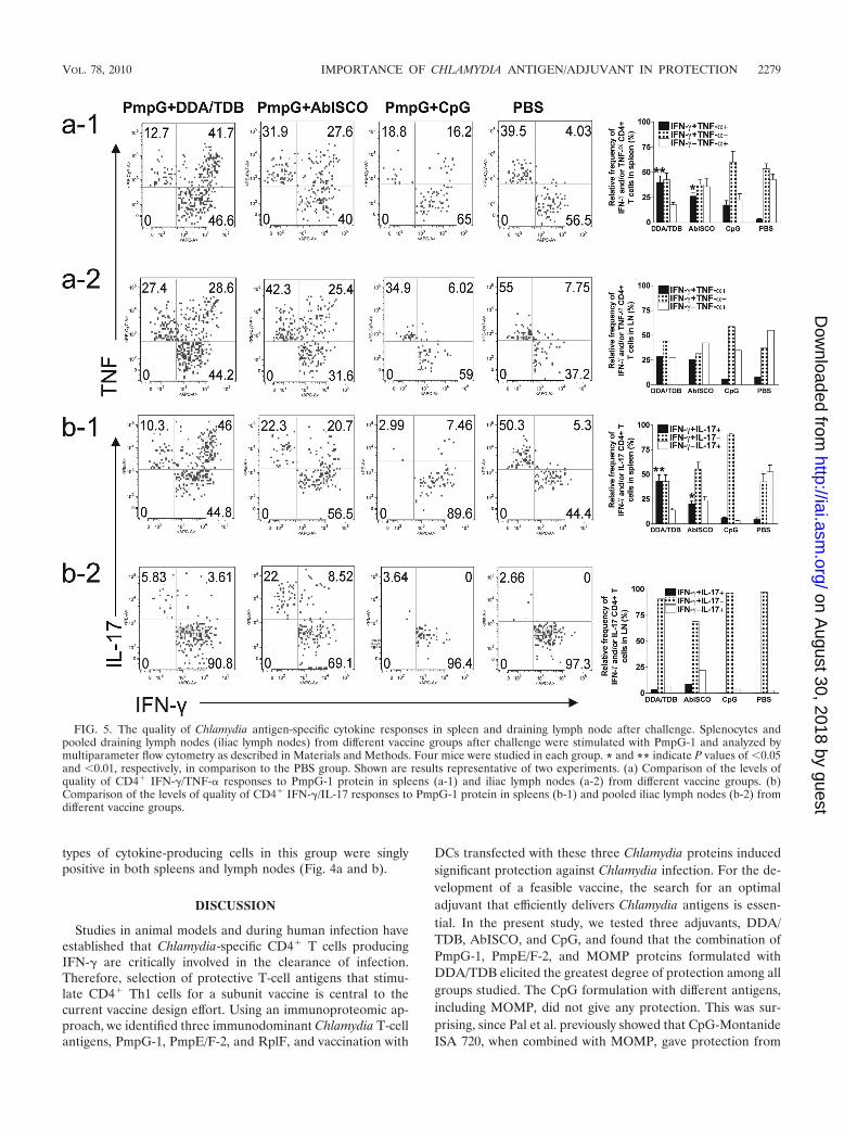
types of cytokine-producing cells in this group were singlypositive in both spleens and lymph nodes (Fig. 4a and b).
DISCUSSION
Studies in animal models and during human infection haveestablished that Chlamydia-specific CD4� T cells producingIFN-� are critically involved in the clearance of infection.Therefore, selection of protective T-cell antigens that stimu-late CD4� Th1 cells for a subunit vaccine is central to thecurrent vaccine design effort. Using an immunoproteomic ap-proach, we identified three immunodominant Chlamydia T-cellantigens, PmpG-1, PmpE/F-2, and RplF, and vaccination with
DCs transfected with these three Chlamydia proteins inducedsignificant protection against Chlamydia infection. For the de-velopment of a feasible vaccine, the search for an optimaladjuvant that efficiently delivers Chlamydia antigens is essen-tial. In the present study, we tested three adjuvants, DDA/TDB, AbISCO, and CpG, and found that the combination ofPmpG-1, PmpE/F-2, and MOMP proteins formulated withDDA/TDB elicited the greatest degree of protection among allgroups studied. The CpG formulation with different antigens,including MOMP, did not give any protection. This was sur-prising, since Pal et al. previously showed that CpG-MontanideISA 720, when combined with MOMP, gave protection from
FIG. 5. The quality of Chlamydia antigen-specific cytokine responses in spleen and draining lymph node after challenge. Splenocytes andpooled draining lymph nodes (iliac lymph nodes) from different vaccine groups after challenge were stimulated with PmpG-1 and analyzed bymultiparameter flow cytometry as described in Materials and Methods. Four mice were studied in each group. * and ** indicate P values of �0.05and �0.01, respectively, in comparison to the PBS group. Shown are results representative of two experiments. (a) Comparison of the levels ofquality of CD4� IFN-�/TNF-� responses to PmpG-1 protein in spleens (a-1) and iliac lymph nodes (a-2) from different vaccine groups. (b)Comparison of the levels of quality of CD4� IFN-�/IL-17 responses to PmpG-1 protein in spleens (b-1) and pooled iliac lymph nodes (b-2) fromdifferent vaccine groups.
VOL. 78, 2010 IMPORTANCE OF CHLAMYDIA ANTIGEN/ADJUVANT IN PROTECTION 2279
on August 30, 2018 by guest
http://iai.asm.org/
Dow
nloaded from
infection and pathology (32). The G�F�M�DDA/TDB for-mulation also provided protection in BALB/c mice, indicatingthat T-cell antigens identified by immunoproteomics inC57BL/6 mice were also recognized by Chlamydia-specific Tcells with a different MHC genetic background. The superiorprotection conferred onto the protein combination group rel-ative to that of the individual protein groups suggests that asuccessful Chlamydia vaccine will need to be composed ofmultiple proteins in order to provide broad coverage in anoutbred population and to cross-protect against multiple vari-ants of C. trachomatis.
It is commonly assumed that Chlamydia vaccine studies re-quire Th1 responses as measured by the frequency and mag-nitude of IFN-�-producing cells as the primary immune cor-relate of protection. Although IFN-� is clearly necessary, usingit as a single immune parameter may not be sufficient to pre-dict protection. Recently, a number of reports dealing withvarious disease targets in different animal models have dem-onstrated a correlation between protection and high-quality Tcells that coexpress multiple cytokines (10, 46). TNF is anothereffector cytokine produced by Th1 cells that can mediate con-trol of intracellular infection, including that due to Chlamydia(23, 30, 35). In the present study, we assessed antigen-specificIFN-�- and TNF-�-producing CD4� T cells in vaccinatedmice. Our data demonstrated that the PmpG�DDA/TDAgroup that conferred the greatest protection showed the high-est frequency of double-positive IFN-� and TNF-� CD4� Tcells as well as the highest magnitude of IFN-� production.These data suggest that there is an association between IFN-�and TNF-� double-positive CD4� T cells and protectionagainst Chlamydia infection. IFN-� and TNF-� double-positiveCD4� T cells may provide a more accurate predictor of Chla-mydia vaccine-primed protective immunity than solely IFN-�-producing CD4� T cells.
Th17 cells are known to play important roles in mucosaldefense against infection with extracellular bacterial and fun-gal pathogens by recruiting neutrophils into local sites of in-fection. Recently, several studies also indicated that Th17augments host defense against intracellular bacteria like My-cobacterium tuberculosis, Listeria monocytogenes, and Salmo-nella enterica (20, 26, 37). In the present study, we investigatedTh17 cells in Chlamydia vaccine-primed protective immunity.We found that the PmpG�DDA/TDA group, which exhibitedthe greatest protection among the three adjuvant formulations,also showed the highest magnitude of IL-17 production beforeand after challenge. Importantly, this group also developed thehighest frequency of IFN-� and IL-17 double-positive CD4� Tcells, whereas the CpG group or the PBS group, with no pro-tection, induced very low levels of IFN-� and IL-17 double-positive CD4� T cells or none. These results indicate thatIL-17 may play cooperative roles with IFN-� in vaccine-primedprotective immunity against Chlamydia. Our data also suggesta potential protective role for Chlamydia-specific IFN-� andIL-17 double-positive CD4� T cells as effector T cells againstChlamydia infection.
Generally, Th1 and Th17 cells represent separate and dis-tinct CD4 T-cell lineages that individually produce IFN-� andIL-17, respectively. To date, most in vitro studies demonstratedthat cytokines that promote CD4 T-cell differentiation intoeach lineage not only strongly reinforce differentiation of other
cells into the same lineage but also potently inhibit differenti-ation into other CD4 lineages. In this study, however, we dem-onstrated that IFN-� and IL-17 were coexpressed in single cellsin a manner similar to what has been found in two other studiesthat observed IFN-� and IL-17 double-positive cells. One studyon human Th17 identified the existence of a remarkable num-ber of double-positive (IFN-�/IL-17) freshly derived cells orT-cell clones. The incubation of Th17 clones with IL-12 al-lowed these cells to produce IFN-� in addition to IL-17, andthis effect was associated with reduced Th17 transcription fac-tor ROR�t and increased Th1 transcription factor T-bet ex-pression, suggesting the existence of flexibility between Th1and Th17 cells and a possible developmental relationship be-tween the two cell types (2). A recent study using a rodentmodel of autoimmune encephalomyelitis observed IFN-� andIL-17 double-positive cells and demonstrated that T-bet ex-pression was critical for cell pathogenicity, regardless of cyto-kine expression by the encephalitogenic T cells. These datasuggest that encephalitogenicity of myelin-specific T cells ap-pears to be mediated by a pathway dependent on T-bet and notnecessarily pathway-specific cytokine end products, such asinterferon and IL-17 (48). In the present study, even though weobserved a correlation between double-positive cells and pro-tection, we are unclear whether there is cause-effect relation-ship between them.
Recent studies on Chlamydia showed the importance ofIL-17 in both innate immunity and adaptive immunity. Themechanism of IL-17-mediated protection against Chlamydiainfection involves neutrophil induction to infected sites andregulation of DC function and Th1 responses through cyto-kines and chemokines induced by Th17 or other IL-17-produc-ing cells (3, 50). Since these studies were conducted using themurine lung model, it is uncertain whether the same protectiverole for IL-17-producing T cells will be seen in the genital tractmodel. In a vaccination model targeting a protective antigenfrom Mycobacterium tuberculosis, it was demonstrated that my-cobacterial Ag-specific Th17 responses enhance protective im-munity through induction of chemokines (CXCR3 ligandsCXCL9, CXCL10, and CXCL11) that recruit mycobacterialAg-specific protective Th1 T cells into the infected lung andinhibit bacterial growth (20). Therefore, vaccination that in-duces both IFN-� and IL-17 could represent a new strategy toprevent Chlamydia infection. However, uncontrolled produc-tion of IL-17 may also lead to immune pathology in infectedorgans (51). We need further information on the regulation ofTh17, innate IL-17-producing cells, and the effects of IL-17itself to establish safe and effective methods to utilize IL-17.
It is of interest that we did not observe IL-17-producing Tcells in lymph nodes. This may be due to proliferative andtrafficking kinetics differing between IL-17- and IFN-�-produc-ing T cells. We observed that in immune and immunized mice,the number of IL-17-secreting cells in spleens as determined byintracellular staining declined dramatically 4 h after antigenstimulation in vitro. In contrast, the number of IFN-�-secretingcells continued to increase even after 24 h following antigenstimulation (unpublished data). Since cells in the draininglymph nodes harvested after challenge had already contactedChlamydia antigens for 7 days in vivo, we may have beenunable to detect IL-17-secreting cells in vitro because of thesekinetic differences. The other possible explanation may be that
2280 YU ET AL. INFECT. IMMUN.
on August 30, 2018 by guest
http://iai.asm.org/
Dow
nloaded from
IL-17-producing cells directly trafficked to the mucosal surfaceof the oviduct.
DDA/TDB is a recently discovered adjuvant that consists ofliposomes and a synthetic mycobacterial cord factor as animmune modulator. The small cationic molecule DDA formsliposomes acting as an antigen “depot” to ensure the long-termrelease of antigen. Incorporation of TDB into the liposomebilayers effectively stabilizes the DDA liposomes (11). Wheninjected subcutaneously, DDA forms depots that last up to 60days in mice without any evidence of overt toxicity. One recentstudy reported that TDB selectively activates the FcR�-Syk-Card9 pathway in antigen-presenting cells to induce a uniqueinnate immune activation program that directs protective Th1and Th17 immunity after subunit Mycobacterium tuberculosisvaccination (45), demonstrating that the TDB adjuvant withthis mode of action is distinct from TLR-triggering adjuvantsengaging Myd88 (CpG and IC31) (1, 22) or Trif (monophos-phoryl lipid A) (25). Ishikawa et al. recently reported that themonocyte-inducible C-type lectin (Mincle) is the essential cellreceptor for TDB (17). According to the current concept ofmurine and human Th17 differentiation, TDB likely operatesthrough the combined induction of IL-1�, -6, and -23 fromantigen-presenting cells, whereas CpG may inhibit Th17 dif-ferentiation through IL-12 and IFN-� (9, 43). This may explainwhy the Th17 Chlamydia antigen-specific response was notfound in mice vaccinated with the CpG formulation.
Since PmpG-1 is an outer membrane protein, we testedPmpG-1-specific antibody responses, and the results showedthat all adjuvant formulations (DDA/TDB, ABISCO, andCpG) induced very high total IgG, IgG1, and IgG2a responsesto recombinant PmpG-1 antigen. Using these sera, we failed todetect neutralizing antibody responses by using C. muridarumin HeLa cell culture (data not shown). These data suggest thatprotection is not correlated with antibody responses.
While the vaccine formulated with the three Chlamydia an-tigens and DDA/TDB adjuvant generated statistically signifi-cant protection against infection, it remains important to eval-uate protection against oviductal pathology. We therefore haveinitiated experiments in mice immunized with G�F�M/DDA-TDB in order to examine for pathology as determined byvisually apparent hydrosalpinx in one or both oviducts. Patho-logical results similar to those reported by Hansen et al., usingthe same adjuvant with MOMP as an antigen (13), were ob-served in the majority of mice 60 days after challenge infection(data not shown). Importantly, mice primed by a prior intra-nasal infection were completely protected against the develop-ment of oviductal pathology following intravaginal challengewith Chlamydia. It remains important to discover the mecha-nism behind the different outcomes for oviductal pathologyfollowing infection-induced immunity and vaccine-induced im-munity. Clearly, an ongoing challenge for Chlamydia vaccineresearch remains the discovery of strategies that maximize theprotective effects of immune T cells while simultaneously pre-venting such cells from causing immune response-mediatedtissue damage.
In conclusion, our study demonstrated that a Chlamydiavaccine based on the multiple purified recombinant proteins(PmpG-1, PmpE/F-2, and MOMP) formulated with a liposomeadjuvant, DDA/TDB, efficiently protects against vaginal infec-tion with C. muridarum. This protection correlates with strong
IFN-�, TNF-�, and IL-17 responses characterized by the highfrequency of IFN-�/TNF-� and IFN-�/IL-17 double-positiveCD4� T cells.
ACKNOWLEDGMENTS
This work was supported by a grant from the National Institutes ofHealth (grant no. R01AI076483).
We are grateful to Lindsey Laycock, David Ko, and Gayle Thorn-bury in the Terry Fox Laboratory at the British Columbia CancerResearch Centre for providing flow cytometry services and technicalassistance and to Norbert Maurer and Kaley D. Wilson at the Centrefor Drug Research and Development, Vancouver, for providing lipo-somal nanoparticle-conjugated CpG.
REFERENCES
1. Agger, E. M., I. Rosenkrands, A. W. Olsen, G. Hatch, A. Williams, C.Kritsch, K. Lingnau, A. von Gabain, C. S. Andersen, K. S. Korsholm, and P.Andersen. 2006. Protective immunity to tuberculosis with Ag85B-ESAT-6 ina synthetic cationic adjuvant system IC31. Vaccine 24:5452–5460.
2. Annunziato, F., L. Cosmi, V. Santarlasci, L. Maggi, F. Liotta, B. Mazzinghi,E. Parente, L. Fili, S. Ferri, F. Frosali, F. Giudici, P. Romagnani, P. Par-ronchi, F. Tonelli, E. Maggi, and S. Romagnani. 2007. Phenotypic andfunctional features of human Th17 cells. J. Exp. Med. 204:1849–1861.
3. Bai, H., J. Cheng, X. Gao, A. G. Joyee, Y. Fan, S. Wang, L. Jiao, Z. Yao, andX. Yang. 2009. IL-17/Th17 promotes type 1 T cell immunity against pulmo-nary intracellular bacterial infection through modulating dendritic cell func-tion. J. Immunol. 183:5886–5895.
4. Bettelli, E., T. Korn, and V. K. Kuchroo. 2007. Th17: the third member of theeffector T cell trilogy. Curr. Opin. Immunol. 19:652–657.
5. Bilenki, L., S. Wang, J. Yang, Y. Fan, A. G. Joyee, and X. Yang. 2005. NK Tcell activation promotes Chlamydia trachomatis infection in vivo. J. Immu-nol. 175:3197–3206.
6. Brunham, R. C., B. Pourbohloul, S. Mak, R. White, and M. L. Rekart. 2005.The unexpected impact of a Chlamydia trachomatis infection control pro-gram on susceptibility to reinfection. J. Infect. Dis. 192:1836–1844.
7. Brunham, R. C., and J. Rey-Ladino. 2005. Immunology of Chlamydia infec-tion: implications for a Chlamydia trachomatis vaccine. Nat. Rev. Immunol.5:149–161.
8. Caldwell, H. D., J. Kromhout, and J. Schachter. 1981. Purification andpartial characterization of the major outer membrane protein of Chlamydiatrachomatis. Infect. Immun. 31:1161–1176.
9. Chu, R. S., O. S. Targoni, A. M. Krieg, P. V. Lehmann, and C. V. Harding.1997. CpG oligodeoxynucleotides act as adjuvants that switch on T helper 1(Th1) immunity. J. Exp. Med. 186:1623–1631.
10. Darrah, P. A., D. T. Patel, P. M. De Luca, R. W. Lindsay, D. F. Davey, B. J.Flynn, S. T. Hoff, P. Andersen, S. G. Reed, S. L. Morris, M. Roederer, andR. A. Seder. 2007. Multifunctional TH1 cells define a correlate of vaccine-mediated protection against Leishmania major. Nat. Med. 13:843–850.
11. Davidsen, J., I. Rosenkrands, D. Christensen, A. Vangala, D. Kirby, Y.Perrie, E. M. Agger, and P. Andersen. 2005. Characterization of cationicliposomes based on dimethyldioctadecylammonium and synthetic cord fac-tor from M. tuberculosis (trehalose 6,6�-dibehenate)-a novel adjuvant induc-ing both strong CMI and antibody responses. Biochim. Biophys. Acta 1718:22–31.
12. Grayston, J. T., and S. P. Wang. 1978. The potential for vaccine againstinfection of the genital tract with Chlamydia trachomatis. Sex. Transm. Dis.5:73–77.
13. Hansen, J., K. T. Jensen, F. Follmann, E. M. Agger, M. Theisen, and P.Andersen. 2008. Liposome delivery of Chlamydia muridarum major outermembrane protein primes a Th1 response that protects against genital chla-mydial infection in a mouse model. J. Infect. Dis. 198:758–767.
14. Holten-Andersen, L., T. M. Doherty, K. S. Korsholm, and P. Andersen. 2004.Combination of the cationic surfactant dimethyl dioctadecyl ammoniumbromide and synthetic mycobacterial cord factor as an efficient adjuvant fortuberculosis subunit vaccines. Infect. Immun. 72:1608–1617.
15. Igietseme, J. U., F. O. Eko, and C. M. Black. 2003. Contemporary ap-proaches to designing and evaluating vaccines against Chlamydia. ExpertRev. Vaccines 2:129–146.
16. Ioannou, X. P., P. Griebel, R. Hecker, L. A. Babiuk, and S. van DrunenLittel-van den Hurk. 2002. The immunogenicity and protective efficacy ofbovine herpesvirus 1 glycoprotein D plus Emulsigen are increased by for-mulation with CpG oligodeoxynucleotides. J. Virol. 76:9002–9010.
17. Ishikawa, E., T. Ishikawa, Y. S. Morita, K. Toyonaga, H. Yamada, O. Takeu-chi, T. Kinoshita, S. Akira, Y. Yoshikai, and S. Yamasaki. 2009. Directrecognition of the mycobacterial glycolipid, trehalose dimycolate, by C-typelectin Mincle. J. Exp. Med. 206:2879–2888.
18. Kari, L., W. M. Whitmire, D. D. Crane, N. Reveneau, J. H. Carlson, M. M.Goheen, E. M. Peterson, S. Pal, L. M. de la Maza, and H. D. Caldwell. 2009.
VOL. 78, 2010 IMPORTANCE OF CHLAMYDIA ANTIGEN/ADJUVANT IN PROTECTION 2281
on August 30, 2018 by guest
http://iai.asm.org/
Dow
nloaded from
Chlamydia trachomatis native major outer membrane protein induces partialprotection in nonhuman primates: implication for a trachoma transmission-blocking vaccine. J. Immunol. 182:8063–8070.
19. Karunakaran, K. P., J. Rey-Ladino, N. Stoynov, K. Berg, C. Shen, X. Jiang,B. R. Gabel, H. Yu, L. J. Foster, and R. C. Brunham. 2008. Immunopro-teomic discovery of novel T cell antigens from the obligate intracellularpathogen Chlamydia. J. Immunol. 180:2459–2465.
20. Khader, S. A., G. K. Bell, J. E. Pearl, J. J. Fountain, J. Rangel-Moreno, G. E.Cilley, F. Shen, S. M. Eaton, S. L. Gaffen, S. L. Swain, R. M. Locksley, L.Haynes, T. D. Randall, and A. M. Cooper. 2007. IL-23 and IL-17 in theestablishment of protective pulmonary CD4� T cell responses after vacci-nation and during Mycobacterium tuberculosis challenge. Nat. Immunol.8:369–377.
21. Klinman, D. M., D. Currie, I. Gursel, and D. Verthelyi. 2004. Use of CpGoligodeoxynucleotides as immune adjuvants. Immunol. Rev. 199:201–216.
22. Kwissa, M., R. R. Amara, H. L. Robinson, B. Moss, S. Alkan, A. Jabbar, F.Villinger, and B. Pulendran. 2007. Adjuvanting a DNA vaccine with a TLR9ligand plus Flt3 ligand results in enhanced cellular immunity against thesimian immunodeficiency virus. J. Exp. Med. 204:2733–2746.
23. Liew, F. Y., Y. Li, and S. Millott. 1990. Tumor necrosis factor-alpha syner-gizes with IFN-gamma in mediating killing of Leishmania major through theinduction of nitric oxide. J. Immunol. 145:4306–4310.
24. Martina, B. E., M. W. van de Bildt, T. Kuiken, G. van Amerongen, and A. D.Osterhaus. 2003. Immunogenicity and efficacy of recombinant subunit vac-cines against phocid herpesvirus type 1. Vaccine 21:2433–2440.
25. Mata-Haro, V., C. Cekic, M. Martin, P. M. Chilton, C. R. Casella, and T. C.Mitchell. 2007. The vaccine adjuvant monophosphoryl lipid A as a TRIF-biased agonist of TLR4. Science 316:1628–1632.
26. Miyamoto, M., M. Emoto, Y. Emoto, V. Brinkmann, I. Yoshizawa, P. Seiler,P. Aichele, E. Kita, and S. H. Kaufmann. 2003. Neutrophilia in LFA-1-deficient mice confers resistance to listeriosis: possible contribution of gran-ulocyte-colony-stimulating factor and IL-17. J. Immunol. 170:5228–5234.
27. Mooij, P., I. G. Nieuwenhuis, C. J. Knoop, R. W. Doms, W. M. Bogers, P. J.Ten Haaft, H. Niphuis, W. Koornstra, K. Bieler, J. Kostler, B. Morein, A.Cafaro, B. Ensoli, R. Wagner, and J. L. Heeney. 2004. Qualitative T-helperresponses to multiple viral antigens correlate with vaccine-induced immunityto simian/human immunodeficiency virus infection. J. Virol. 78:3333–3342.
28. Morrison, S. G., H. Su, H. D. Caldwell, and R. P. Morrison. 2000. Immunityto murine Chlamydia trachomatis genital tract reinfection involves B cellsand CD4(�) T cells but not CD8(�) T cells. Infect. Immun. 68:6979–6987.
29. Mui, B., S. G. Raney, S. C. Semple, and M. J. Hope. 2001. Immune stimu-lation by a CpG-containing oligodeoxynucleotide is enhanced when encap-sulated and delivered in lipid particles. J. Pharmacol. Exp. Ther. 298:1185–1192.
30. Njau, F., U. Wittkop, M. Rohde, H. Haller, A. Klos, and A. D. Wagner. 2009.In vitro neutralization of tumor necrosis factor-alpha during Chlamydiapneumoniae infection impairs dendritic cells maturation/function and in-creases chlamydial progeny. FEMS Immunol. Med. Microbiol. 55:215–225.
31. Olds, G. R., L. Chedid, E. Lederer, and A. A. Mahmoud. 1980. Induction ofresistance to Schistosoma mansoni by natural cord factor and synthetic lowerhomologues. J. Infect. Dis. 141:473–478.
32. Pal, S., E. M. Peterson, and L. M. de la Maza. 2005. Vaccination with theChlamydia trachomatis major outer membrane protein can elicit an immuneresponse as protective as that resulting from inoculation with live bacteria.Infect. Immun. 73:8153–8160.
33. Pal, S., E. M. Peterson, R. Rappuoli, G. Ratti, and L. M. de la Maza. 2006.Immunization with the Chlamydia trachomatis major outer membrane pro-tein, using adjuvants developed for human vaccines, can induce partial pro-tection in a mouse model against a genital challenge. Vaccine 24:766–775.
34. Pal, S., I. Theodor, E. M. Peterson, and L. M. de la Maza. 2001. Immuni-zation with the Chlamydia trachomatis mouse pneumonitis major outer
membrane protein can elicit a protective immune response against a genitalchallenge. Infect. Immun. 69:6240–6247.
35. Perry, L. L., H. Su, K. Feilzer, R. Messer, S. Hughes, W. Whitmire, and H. D.Caldwell. 1999. Differential sensitivity of distinct Chlamydia trachomatisisolates to IFN-gamma-mediated inhibition. J. Immunol. 162:3541–3548.
36. Pimm, M. V., R. W. Baldwin, J. Polonsky, and E. Lederer. 1979. Immuno-therapy of an ascitic rat hepatoma with cord factor (trehalose-6, 6�-dimyco-late) and synthetic analogues. Int. J. Cancer 24:780–785.
37. Schulz, S. M., G. Kohler, C. Holscher, Y. Iwakura, and G. Alber. 2008.IL-17A is produced by Th17, gammadelta T cells and other CD4-lympho-cytes during infection with Salmonella enterica serovar Enteritidis and has amild effect in bacterial clearance. Int. Immunol. 20:1129–1138.
38. Stanberry, L. R., and S. L. Rosenthal. 2005. Progress in vaccines for sexuallytransmitted diseases. Infect. Dis. Clin. North Am. 19:477–490, xi.
39. Starnbach, M. N., and N. R. Roan. 2008. Conquering sexually transmitteddiseases. Nat. Rev. Immunol. 8:313–317.
40. Su, H., and H. D. Caldwell. 1995. CD4� T cells play a significant role inadoptive immunity to Chlamydia trachomatis infection of the mouse genitaltract. Infect. Immun. 63:3302–3308.
41. Wang, S., Y. Fan, R. C. Brunham, and X. Yang. 1999. IFN-gamma knockoutmice show Th2-associated delayed-type hypersensitivity and the inflamma-tory cells fail to localize and control chlamydial infection. Eur. J. Immunol.29:3782–3792.
42. Wang, S. P., J. T. Grayston, and E. R. Alexander. 1967. Trachoma vaccinestudies in monkeys. Am. J. Ophthalmol. 63(Suppl.):1615–1630.
43. Weaver, C. T., L. E. Harrington, P. R. Mangan, M. Gavrieli, and K. M.Murphy. 2006. Th17: an effector CD4 T cell lineage with regulatory T cellties. Immunity 24:677–688.
44. Weaver, C. T., R. D. Hatton, P. R. Mangan, and L. E. Harrington. 2007.IL-17 family cytokines and the expanding diversity of effector T cell lineages.Annu. Rev. Immunol. 25:821–852.
45. Werninghaus, K., A. Babiak, O. Gross, C. Holscher, H. Dietrich, E. M.Agger, J. Mages, A. Mocsai, H. Schoenen, K. Finger, F. Nimmerjahn, G. D.Brown, C. Kirschning, A. Heit, P. Andersen, H. Wagner, J. Ruland, and R.Lang. 2009. Adjuvanticity of a synthetic cord factor analogue for subunitMycobacterium tuberculosis vaccination requires FcRgamma-Syk-Card9-de-pendent innate immune activation. J. Exp. Med. 206:89–97.
46. Wille-Reece, U., B. J. Flynn, K. Lore, R. A. Koup, A. P. Miles, A. Saul, R. M.Kedl, J. J. Mattapallil, W. R. Weiss, M. Roederer, and R. A. Seder. 2006.Toll-like receptor agonists influence the magnitude and quality of memory Tcell responses after prime-boost immunization in nonhuman primates. J.Exp. Med. 203:1249–1258.
47. Yang, X., K. T. HayGlass, and R. C. Brunham. 1996. Genetically determineddifferences in IL-10 and IFN-gamma responses correlate with clearance ofChlamydia trachomatis mouse pneumonitis infection. J. Immunol. 156:4338–4344.
48. Yang, Y., J. Weiner, Y. Liu, A. J. Smith, D. J. Huss, R. Winger, H. Peng, P. D.Cravens, M. K. Racke, and A. E. Lovett-Racke. 2009. T-bet is essential forencephalitogenicity of both Th1 and Th17 cells. J. Exp. Med. 206:1549–1564.
49. Yu, H., X. Jiang, C. Shen, K. P. Karunakaran, and R. C. Brunham. 2009.Novel Chlamydia muridarum T cell antigens induce protective immunityagainst lung and genital tract infection in murine models. J. Immunol. 182:1602–1608.
50. Zhang, X., L. Gao, L. Lei, Y. Zhong, P. Dube, M. T. Berton, B. Arulanandam,J. Zhang, and G. Zhong. 2009. A MyD88-dependent early IL-17 productionprotects mice against airway infection with the obligate intracellular patho-gen Chlamydia muridarum. J. Immunol. 183:1291–1300.
51. Zhou, X., Q. Chen, J. Moore, J. K. Kolls, S. Halperin, and J. Wang. 2009.Critical role of the interleukin-17/interleukin-17 receptor axis in regulatinghost susceptibility to respiratory infection with Chlamydia species. Infect.Immun. 77:5059–5070.
Editor: R. P. Morrison
2282 YU ET AL. INFECT. IMMUN.
on August 30, 2018 by guest
http://iai.asm.org/
Dow
nloaded from


![Chlamydia [Modo De Compatibilidad]](https://img.pdfslide.tips/doc/110x75/556c7061d8b42ad85e8b5027/chlamydia-modo-de-compatibilidad.jpg)