Embed Size (px)
Citation preview

1
Clostridium perfringens Alpha-toxin Recognizes the GM1a/TrkA Complex
Masataka Oda1, Michiko Kabura
1, Teruhisa Takagishi
1, Ayaka Suzue
1, Kaori Tominaga
1, Shiori
Urano1, Masahiro Nagahama
1, Keiko Kobayashi
1, Keiko Furukawa
2, Koichi Furukawa
2, Jun
Sakurai1
1Department of Microbiology, Faculty of Pharmaceutical Sciences, Tokushima Bunri University,
Yamashiro-cho, Tokushima 770-8514, Japan
2Department of Biochemistry 2, Nagoya University School of Medicine, 65 Tsurumai, Showa-ku,
Nagoya 466, Japan
*Running title: GM1a, a novel receptor for Clostridium perfringens alpha-toxin
To whom correspondence should be addressed: Masataka Oda, Faculty of Pharmaceutical Sciences,
Tokushima Bunri University, Yamashiro-cho, Tokushima, 770-8514, Japan, Tel.: +81-88-602-8485;
Fax: +81-88-655-3051; E-Mail: [email protected]
Key words: Alpha-toxin, A549 cell, GM1a, IL-8, TrkA
Background
Gangliosides are receptors for bacterial toxins.
Results
Alpha-toxin from Clostridium perfringens
specifically interacts with GM1a.
Conclusion
Trp-84 and Tyr-85 of alpha-toxin are the
residues that interact with GM1a, leading to
activation of TrkA in A549 cells.
Significance
These results define the role of GM1a/TrkA as a
receptor for alpha-toxin.
SUMMARY
Clostridium perfringens alpha-toxin is the
major virulence factor in the pathogenesis of
gas gangrene. Alpha-toxin is a 43-kDa protein
with 2 structural domains: the N-domain
contains the catalytic site and coordinates the
divalent metal ions, and the C-domain is a
membrane-binding site. The role of the
exposed loop region (72-93 residues) in the
N-domain, however, has been unclear. Here,
we show that this loop contains a
ganglioside-binding motif (H…SxWY…G),
which is the same motif seen in botulinum
neurotoxin, and directly binds to a specific
conformation of the ganglioside GM1a
through a carbohydrate moiety. Confocal
microscopy analysis using fluorescently
labeled BODIPY-GM1a revealed that the
toxin colocalized with GM1a and induced
clustering of GM1a on the cell membranes.
Alpha-toxin was only slightly toxic in
-N-acetylgalactosaminyltransferase
knockout mice, which lack the a-series
gangliosides that contain GM1a, but was
highly toxic in α2,8-sialyltransferase
http://www.jbc.org/cgi/doi/10.1074/jbc.M112.393801The latest version is at JBC Papers in Press. Published on July 30, 2012 as Manuscript M112.393801
Copyright 2012 by The American Society for Biochemistry and Molecular Biology, Inc.
by guest on April 28, 2020
http://ww
w.jbc.org/
Dow
nloaded from
2
knockout mice, which lack both b-series and
c-series gangliosides, similar to the control
mice. Moreover, experiments with
site-directed mutants indicated that Trp-84
and Tyr-85 in the exposed alpha-toxin loop
play an important role in the interaction with
GM1a and subsequent activation of TrkA.
These results suggest that binding of
alpha-toxin to GM1a facilitates the activation
of the TrkA receptor and induces a signal
transduction cascade that promotes the
release of chemokines. Therefore, we
conclude that GM1a is the primary cellular
receptor for alpha-toxin, which can be a
potential target for drug developed against
this pathogen.
Bacterial phospholipase Cs (PLCs) are
secreted proteins that have preferences for
different phospholipids (1,2). Bacterial PLCs
that hydrolyze phosphatidylcholine (PC) and
sphingomyelin (SM) are important virulence
factors in the pathogenesis of diseases caused by
Listeria monocytogenes, Pseudomonas
aeruginosa, and several Clostridium spp. (3-5).
Clostridium perfringens, the pathogenic
bacterium most widely distributed in nature (6),
produces one PLC—alpha-toxin—which is
highly cytotoxic and myotoxic and can lead to
hemolysis and the release of superoxide radicals
and inflammatory cytokines (7-13). The toxin
has been associated with enteritis in domestic
animals, and Crohn’s disease and gas gangrene
in humans (14-17).
We have previously reported that
alpha-toxin-induced activation of endogenous
PLC and sphingomyelinase via pertussis
toxin-sensitive GTP-binding protein plays an
important role in hemolysis of rabbit and sheep
erythrocytes (8,11,18). We also reported that
alpha-toxin simultaneously induced the
formation of diacylglycerol (DG) through the
activation of endogenous PLC and
phosphorylation of ERK1/2, NFκB, and
p38MAPK via activation of tyrosine kinase A
(TrkA) in Human lung adenocarcinoma
epithelial cell line (A549) cells, and that these
events induced the release of interleukin-8
(IL-8) (19). In addition, the toxin-induced
release of TNF-alpha was found to be dependent
on the activation of ERK1/2 signal transduction
via the phosphorylation of TrkA in neutrophils
and macrophages (13). Inhibition of the
toxin-induced phosphorylation of TrkA by
erythromycin led to the arrest of the
toxin-induced events (13), thus supporting the
hypothesis that TrkA is a key signaling molecule
in the action induced by alpha-toxin.
Gangliosides may have either 1 (a-series),
2 (b-series), or 3 (c-series) sialic acid residues
linked to the 3-position of the inner galactose
moiety or may lack sialic acid altogether
(0-series) (20). Gangliosides are present in the
plasma membrane of vertebrate cells with their
oligosaccharide chains exposed to the external
environment, and they have been implicated as
cell surface receptors for several bacterial toxins.
Purified tetanus neurotoxin (TeNT) (21) and
botulinum neurotoxin (BoNT) (22) mainly
interact with the b-series gangliosides GD3,
GD1b, GT1b, and GQ1b. Cholera toxin binds
with high affinity and specificity to the
ganglioside GM1 (23). Mutoh et al. reported
that GM1 can bind to the TrkA protein, where it
regulates the receptor and enhances activation
by the direct interaction of GM1 with TrkA in
by guest on April 28, 2020
http://ww
w.jbc.org/
Dow
nloaded from

3
the lipid rafts on the plasma membrane (24-26).
Ichikawa et al. reported that clustering of GM1
promotes the enrichment of TrkA in the lipid
rafts (27).
In the present study, we have investigated
the role of gangliosides in the pathogenicity of
alpha-toxin, and analyzed the binding region
with which the toxin associates with
gangliosides.
EXPERIMENTAL PROCEDURES
Cell culture and drugs—A549 cells were
cultured at 37°C in growth medium (DMEM
with 10% horse serum). To prepare
ganglioside-depleted A549 cells, the seeded
cells were grown in the presence of medium
containing 1, 5, or 10 μM D
-threo-1-phenyl-2-hexadecanoylamino-
3-morpholino-1-propanol (PPMP) (Matreya,
USA) for 4 days. The cells were used for
immunocytochemistry and cytokine release
measurements 4 days after seeding. GM1a was
obtained commercially (Calbiochem, USA).
Neuraminidase from C. perfringens was
obtained from Sigma-Aldrich.
Alexa488-conjugated cholera toxin B subunit
(CTB) was purchased from Molecular Probes,
USA. All other chemicals were of analytical
grade.
Purification of alpha-toxin—Alpha-toxin was
overexpressed in Bacillus subtilis ISW1214 that
had been transformed with the plasmid vector,
pHY300PLK, carrying the cDNA for
alpha-toxin. The expression and purification of
recombinant alpha-toxin was performed as
described previously (28).
Cy3-coupled toxin—Alpha-toxin (3 mg/mL)
was labeled with the Cy3-labeling kit (GE
healthcare, USA) following the manufacturer's
protocol. The release of IL-8 from A549 cells
treated with Cy3-coupled alpha-toxin
(Cy3-alpha-toxin) was the same as that of cells
treated with non-labeled toxin.
Glycoarray assay—The glycoarray plates
(Sumitomo Bakelite, Japan) were incubated
with 100 g Cy3-alpha-toxin in PBS containing
1 mM CaCl2 for 15 min; this was followed by
washing the plate with PBS. Bound
Cy3-alpha-toxin was measured with an
Affymetrix 428 (Affymetrix, USA).
Binding of alpha-toxin to A549 cells—A549
cells were seeded on a poly-L-lysine glass
bottom dish (MatTek, USA). Cy3-alpha-toxin,
fixed with 2% paraformaldehyde in PBS, was
added at room temperature for 15 min; and the
cells were then washed 3 times with PBS. The
fluorescence level of Cy3-alpha-toxin in each
cell was analyzed by fluorescent microscopy
(BIOREVO BZ-9000; Keyence, Japan) and the
associated analysis software package (BZ-H2A).
IL-8 ELISA—Immunoreactive IL-8 was
quantified in cell culture supernatants by a
double-Ab ELISA kit using rIL-8 as a standard
(R&D Systems, USA) following the
manufacturer’s protocol.
Confocal detection of GM1a—A549 cells were
seeded on a poly-L-lysine glass bottom dish
(MatTek, USA), and 500 nM BODIPY-GM1a
(Molecular Probes, USA) was added to the cells
by guest on April 28, 2020
http://ww
w.jbc.org/
Dow
nloaded from

4
for 5 min at 25° C. The cells were then treated
with alpha-toxin or Cy3-alpha-toxin (1.0 μ
g/mL) at 37°C, fixed with 4% paraformaldehyde
in PBS at room temperature for 15 min, and
washed 3 times with PBS. The nuclei were
stained with Hoechst33342. Stained cells were
visualized using confocal microscopy (Nikon,
A1R, Japan).
Mice—Twenty-five-week-old male wild-type
mice (C57BL/6 strain; Nihon SLC, Japan) were
used.
N-Acetylgalactosaminyltransferase-knock
out (GalNAcT-/-) mice and
α2,8-sialyltransferase-knockout (ST-/-) mice
were constructed previously (29-32).
Experimental protocols were approved by the
Institute Animal Care and Use Committee at
Tokushima Bunri University. The mice were
housed in plastic cages under controlled
environmental conditions (temperature, 22°C;
humidity, 55%). Food and water were freely
available.
Culture of macrophages—Mouse macrophages
were isolated from the cell contents of
peritoneal exudates with 2 mL of phenol
red-free RPMI1640 medium (Wako Pure
Chemical Industries, Japan) supplemented with
5% fetal bovine serum (FBS) (Biowest, USA).
After centrifugation at 170 g for 10 min at 4°C,
the cell pellet was resuspended in phenol
red-free RPMI1640 medium supplemented with
5% FBS. Adherent macrophage monolayers
were obtained by plating the cells in 96- or
48-well plastic trays (Falcon, USA).
Site-directed mutagenesis—The Transformer
Site-Directed Mutagenesis Kit (Takara, Japan)
was used with the following primers to prepare
the modified plasmids: D81A,
5'-AATTTCTCAAAGGCCAATAGTTGGTAT-3
'; N82A,
5'-TTCTCAAAGGACGCGTCTTGGTATTTA-
3'; S83A,
5'-TCAAAGGATAATGCCTGGTATTTAGCT-3
'; W84A,
5'-AAGGATAATAGCGCTTATTTAGCTTAT-3'
; Y85A,
5'-GATAATAGTTGGGCCCTAGCTTATTCT-3'
; Y88A,
5'-TGGTATTTAGCTGCCTCTATACCTGAC-3'.
The genetic sequence of alpha-toxin in each
plasmid was confirmed with an ABI310
PRISMTM
genetic analyzer (Life technologies,
USA).
Preparation of GM1a liposomes—
Phosphatidylcholine from egg yolk (Nacalai,
Japan), sphingomyelin from bovine brain
(Nacalai, Japan), GM1a (Calbiochem, USA),
and cholesterol (Nacalai, Japan) in a 1:1:1:2
molar ratio were dried from
chloroform/methanol (1:2 v/v) under nitrogen
gas, resuspended in HBS-N (0.01 M HEPES
pH7.4, 0.15 M NaCl) (GE healthcare, USA),
and sonicated in a water bath for 5 min to ensure
that the lipid was fully hydrated. The liposome
suspension was passed 21 times (0.5 ml at a
time) through a Liposofast (Avestin, USA)
liposome extruder with 100-nm pore size
polycarbonate membranes.
Alpha-toxin membrane-binding assay—The
membrane-binding assay was performed by
surface plasmon resonance (SPR) analysis using
by guest on April 28, 2020
http://ww
w.jbc.org/
Dow
nloaded from

5
a Biacore3000 system (GE healthcare, USA)
and the associated analysis software package
(BIAevaluation) (GE healthcare, USA).
Alpha-toxin and variants at concentrations of
5.0, 2.5, 1.0, 0.5, and 0 nM were applied to
GM1a liposome-coated L1sensor chips at a flow
rate of 5.0 L/min in a running buffer (0.01 M
HEPES pH7.4, 0.15 M NaCl, 1 mM CaCl2) at
25°C. Dissociation was monitored for at least
150 s in a constant flow of enzyme-free running
buffer.
In silico docking simulation analysis of GM1a
with alpha-toxin—All molecular modeling
studies were performed using the Molecular
Operating Environment software (MOE2011;
Chemical Computing Group, Quebec, Canada).
The X-ray crystallographic structure of
alpha-toxin was obtained from the Protein Data
Bank (PDB ID: 1CA1) (33). This toxin was
prepared for docking studies by adding
hydrogen atoms with standard geometries,
placing the carbohydrate moiety of GM1a in the
vicinity of alpha-toxin residues Trp-84 and
Tyr-85, and coordinating the sialic acid of
GM1a with Trp-84. The structure was
minimized using the MMFF94s force-field.
Statistical analysis—All data presented are
expressed as the mean ± SEM. Mean values
among experimental groups were compared
using the Student's t-test, and P < 0.01 was
considered statistically significant.
RESULTS
Gangliosides mediate the release of IL-8
induced by alpha-toxin—Alpha-toxin induces
the release of IL-8 from A549 cells (19). To
investigate the effect of gangliosides and sialic
acid on the binding of alpha-toxin to A549 cells
and on the release of IL-8, the cells were treated
with various concentrations of PPMP, which is a
ganglioside synthase inhibitor, or neuraminidase
from C. perfringens, which catalyzes the
hydrolysis of terminal sialic acid residues, at
37°C for 4 days or 60 min, respectively. The
inhibition of ganglioside synthesis and depletion
of sialic acid in A549 cells were confirmed with
Alexa488-conjugated CTB that specifically
binds to GM1a and possesses weaker affinity to
GD1b (34). PPMP- or neuraminidase-treated
A549 cells lost their ability to bind to CTB
(Supplemental Fig. S1). After this pre-treatment,
the cells were incubated with 1.0 µg/mL
alpha-toxin. The treatment with both PPMP and
neuraminidase inhibited the release of IL-8 from
A549 cells treated with alpha-toxin in a
dose-dependent manner (Fig. 1A). Furthermore,
the binding of Cy3-alpha-toxin to the cells was
also inhibited by both PPMP and neuraminidase
in a dose-dependent manner (Fig. 1B). In
addition, treatment of HepG2 and HEK293 cells
with these inhibitors resulted in reduction of
binding of alpha-toxin to the same level as in
A549 cells (data not shown). This suggests,
therefore, that gangliosides containing sialic
acid play an important role in the binding of the
toxin.
Alpha-toxin binds directly to GM1a—We
investigated the interaction of alpha-toxin with
various gangliosides using an in vitro glycoarray
system (Fig. 2A). The array plate was incubated
with 100 μg/mL Cy3-alpha-toxin at 37°C for 60
min, and the fluorescence signal of alpha-toxin
by guest on April 28, 2020
http://ww
w.jbc.org/
Dow
nloaded from

6
was strongly localized at the spot corresponding
to the carbohydrate chain of GM1a (Fig. 2A).
The toxin was slightly bound to gangliosides
that contained sialic acid, but not to gangliosides
such as asialo-GM1, which lack sialic acid (Fig.
2A). Binding of alpha-toxin to GM1b, GM2,
and GM3 was significantly weaker than that to
GM1a (Fig. 2A). Because GM1b, GM2, and
GM3 contain sialic acid (Fig. 2B), our results
indicate that sialic acid is not sufficient for the
specific binding of alpha-toxin to gangliosides.
Furthermore, the binding of alpha-toxin to
GD1a, GD1b, GT1a, and GT1c, which have the
same carbohydrate chain as GM1a but contain
different numbers of sialic acid residues (Fig.
2B), was substantially less than the binding to
GM1a (Fig. 2A). Therefore, it appears that the
structural conformation of GM1a is important
for its binding to alpha-toxin.
Colocalization of GM1a and alpha-toxin—We
examined whether alpha-toxin colocalized with
GM1a on the cell membrane by incubating
BODIPY-GM1a-labelled cells with
Cy3-alpha-toxin at 37°C for 15 min. The
BODIPY-GM1a in the cells treated with
alpha-toxin aggregated on the cell membranes,
but aggregation in control cells was
undetectable in our experimental conditions (Fig.
3A). Cy3-alpha-toxin was colocalized at sites of
GM1a aggregation (Fig. 3A), and the percentage
of colocalization was approximately 70% (Fig.
3B). Therefore, it appears that alpha-toxin
specifically binds to GM1a on biological
membranes.
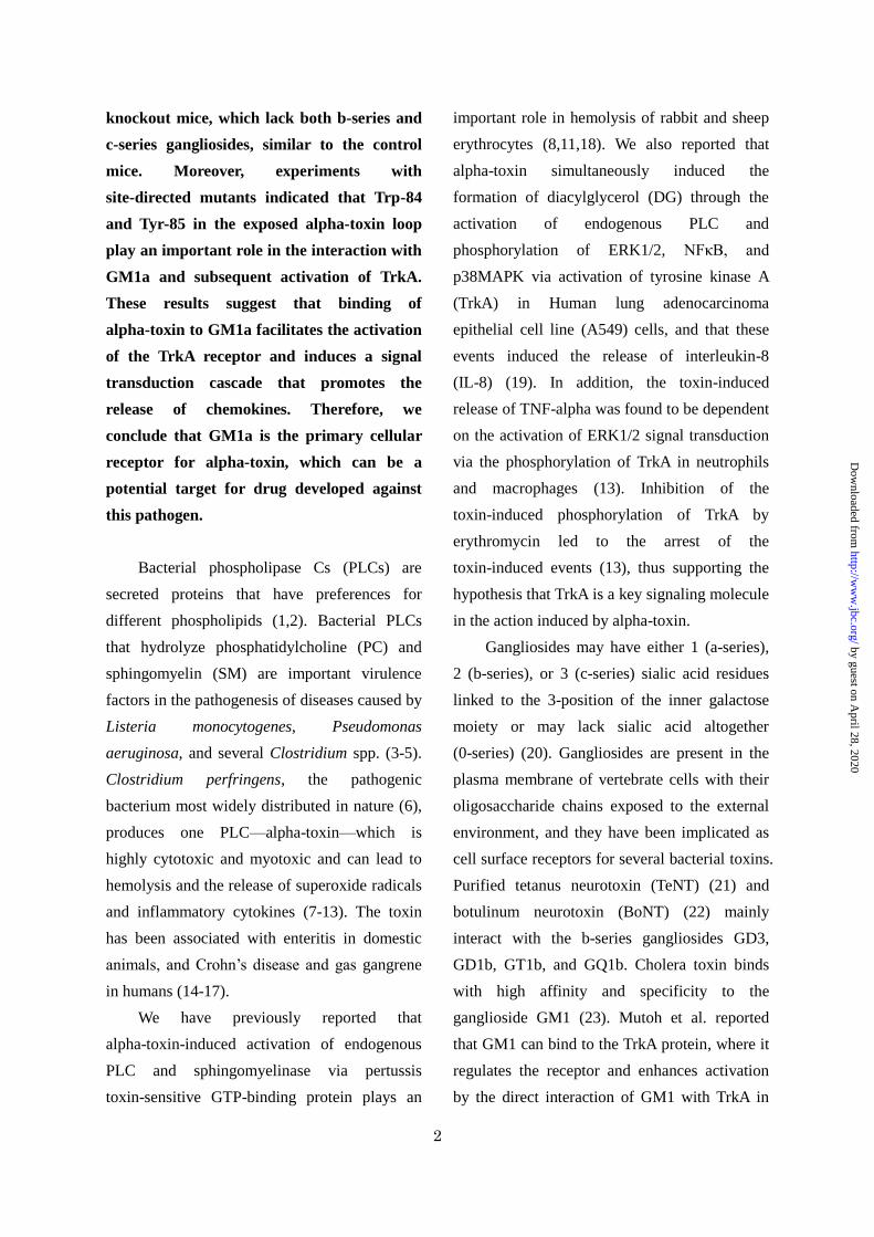
Sensitivity of wild–type, GalNAcT-/-, and ST-/-
mice to alpha-toxin—Next, we examined the
response of GalNAcT-/- mice (lacking GM1a,
GM2, GD2, GD1a, GD1b, GT1a, GT1b, GQ1b,
etc.) and ST-/- mice (lacking b- and c-series
gangliosides) to alpha-toxin (Fig. 4A). We
injected 200 ng of alpha-toxin intraperitoneally
into wild-type, GalNAcT-/-, and ST-/- mice. The
wild-type and ST-/- mice succumbed to the toxic
effects of the toxin within about 10 h while the
average survival time for GalNAcT-/- mice was
36 h (Fig. 4B). Macrophages isolated from wild
type, GalNAcT-/-, and ST-/- mice were
incubated with 1.0 µg/mL alpha-toxin at 37°C
for various periods of time. As shown in Fig. 4C,
the release of GRO/KC (a member of the CXC
chemokine family with homology to human
IL-8) from macrophages isolated from
GalNAcT-/- mice was significantly lower than
that from macrophages isolated from wild-type
or ST-/- mice. Furthermore, the binding of
Cy3-alpha-toxin to the macrophages from
GalNAcT-/- mice was weaker than that to
macrophages from wild-type and ST-/- mice
(Fig. 4D).
Role of amino acids in the loop region of
alpha-toxin—Clark et al. previously suggested
that the 72–93 residue loop in alpha-toxin
participates in membrane binding (35). Previous
experiments with TeNT and BoNT found that
the lactose- and ganglioside-GT1b-binding site
is characterized by the presence of the peptide
motif H…SxWY…G, with the tryptophan and
tyrosine residues being especially important (34,
37). This motif is also found in alpha-toxin (Fig.
5). We focused on Trp-84, Tyr-85, and their
surrounding residues, and constructed the
following alpha-toxin variants with single amino
acid changes in this loop: D81A, N82A, S83A,
by guest on April 28, 2020
http://ww
w.jbc.org/
Dow
nloaded from

7
W84A, Y85A, and Y88A. To determine whether
these variants induce the release of IL-8 from
A549 cells, the cells were incubated with each
of the variants for 3 h at 37°C. As shown in Fig.
6A, the release of IL-8 by D81A, N82A, S83A
and Y88A was the same as that for the wild-type
toxin, but very little release was seen with the
W84A and Y85A variants. In addition, the
binding of Cy3-W84A and Cy3-Y85A to A549
cells was about 20% that of the Cy3-wild-type
toxin (Fig. 6B). Moreover, the W84A and Y85A
variants failed to induce phosphorylation of
TrkA in A549 cells (Fig. 6C).
SPR analysis—We analyzed the affinity of
these variants for GM1a-liposomes by SPR. Fig.
7 shows both the liposome association,
represented by an increase in resonance units,
and dissociation, represented by a decrease in
resonance units. Wild-type toxin rapidly
associated with GM1a-liposomes and slowly
dissociated from them, and the resonance units
of wild-type toxin binding to GM1a-liposomes
increased dose-dependently (red for vehicle,
pink for 0.2 nM, green for 1.0 nM, blue for 2.5
nM, navy for 5.0 nM) (Fig. 7A). The
steady-state binding parameters were
determined with the BIAevaluation software,
and the KD value for the wild-type toxin was
estimated to be 23 nM. The binding curves for
the alpha-toxin variants (Fig. 7B–G) showed
estimated KD values of 34 nM, 54 nM, 68 nM,
520 nM, 448 nM, and 48 nM for D81A, N82A,
S83A, W84A, Y85A and Y88A, respectively.
The KD values of W84A and Y85A were about
20-fold higher than that of the wild-type toxin,
suggesting that the affinity of W84A and Y85A
to GM1a-liposomes is markedly reduced
compared with that of the wild-type toxin.
Model for binding of alpha-toxin to the
carbohydrate moiety of GM1a—Next, we
investigated the mode of binding between
alpha-toxin and GM1a, especially as it relates to
residues Trp-84 and Tyr-85, by in silico docking
simulation analyses of this interaction at the
tertiary structure level using the MOE software.
The analysis was based on the structure of the
complex between BoNT/A and GT1b (PDB ID:
2VU9). Fig. 8A shows the entire structure that
resulted from the docking simulation analysis
between alpha-toxin and GM1a. Trp-84
interacted with the sialic acid of GM1a through
a hydrophobic ring stacking mechanism and a
hydrogen bond, and Tyr-85 interacted with the
galactosamine through a hydrogen bond (Fig.
8B).
DISCUSSION
Alpha-toxin is the major virulence factor in
the pathogenesis of gas gangrene, a devastating
disease associated with traumatic injuries or
surgical wounds (14). Despite the recent
identification of the residues critical for the
toxicity of alpha-toxin (36-38), the binding
mechanism of this toxin is still not completely
understood. In this study, we have shown that
the pretreatment of A549 cells with PPMP
resulted in attenuation of alpha-toxin binding.
Alpha toxin specifically bound to the
ganglioside GM1a and induced the activation of
TrkA, and we found that GM1a plays an
important role in the lethality of alpha-toxin in
mice. Our mutagenesis study has now identified
2 specific amino acid residues, Trp-84 and Tyr-85,
by guest on April 28, 2020
http://ww
w.jbc.org/
Dow
nloaded from

8
that play a critical role in the binding of
alpha-toxin to GM1a.
We measured the binding capacity of
alpha-toxin to various gangliosides and found
that alpha-toxin specifically binds to GM1a.
Sialic acid is required for the binding because
asialo-GM1 (which lacks sialic acid) did not
bind to alpha-toxin. In addition, neuraminidase
dose-dependently inhibited the binding of
alpha-toxin to A549 cells and the subsequent
release of IL-8. These results support those
reported by Flores-Diaz et al. that GM1a
protected artificial and cellular membranes from
the disruption caused by alpha-toxin whereas
asialo-GM1a did not (1). GT1b, GQ1b, and
GD1b, which contain the same sugar chains as
GM1a but have different numbers of sialic acids,
bound to alpha-toxin less tightly than GM1a,
suggesting that the conformation of GM1a for
alpha-toxin binding is important. Alpha-toxin
bound to GM2 through the same carbohydrate
moiety as GM1a, but its binding was less
extensive than to GM1a. This suggests that the
additional Gal1 residue at the 5' end of the
carbohydrate moiety of GM1a is required for
effective binding to alpha-toxin. Flores-Diaz et
al. reported that alpha-toxin has higher affinity
for GT1b and GD1a (b-series) than GM1a and
GD1a (a-series) galactosides (1). This difference
is probably due to the difference in assay
systems. In our binding assays, we used a plate
coated with sugar chains from various
gangliosides, which minimized the
conformational possibilities of the glycolipid
oligosaccharides.
The response of GalNAcT-/- mice, which
lack all complex gangliosides including GM1a,
to alpha-toxin was lower than that of wild-type
mice. On the other hand, the response of ST-/-
mice, which lack b- and c-series gangliosides, to
the toxin was the same as that of wild-type mice.
These results show that the toxic effect of
alpha-toxin in mice is mediated primarily by
a-series gangliosides, and by GM1a in particular.
The b- and c-series gangliosides may not be
essential for alpha-toxin at the initial step during
the intoxication process in mice, and it appears
that GM1a plays an important role in the
pathogenesis of alpha-toxin. In addition, the
GalNAcT-/- mice were not completely inhibited
the death induced by alpha-toxin. The C-domain
of alpha-toxin has ability for binding to
phospholipids of cell membranes (33,39,40). It
therefore appears that the binding of
phospholipids via C-domain of alpha-toxin also
participated in the pathogenesis of the toxin.
Clostridial neurotoxins such as TeNT and
BoNT have a lactose- or ganglioside-binding
site that is characterized by the presence of the
peptide motif H…SxWY…G (41-45), and a
similar motif is conserved in alpha-toxin. We
have generated single-point mutants of 6 amino
acid residues (Asp-81, Asn-82, Ser-83, Trp-84,
Tyr-85, Tyr-88) to clarify the molecular
interaction between alpha-toxin and GM1a.
Substitution of Trp-84 and Tyr-85 residues
located in positions comparable to the
ganglioside-binding pockets of TeNT and
BoNTs dramatically affected the binding to
GM1a-liposome and the release of IL-8. In
addition, we showed that the W84A and Y85A
variants were significantly reduced in their
ability to activate TrkA. GM1a associates with
TrkA on cell membranes and activates TrkA and
ERK1/2 (46-48). Cholera toxin, which is a
high-affinity ligand for GM1, activated TrkA in
by guest on April 28, 2020
http://ww
w.jbc.org/
Dow
nloaded from

9
PC12 cells (47). Ichikawa et al. reported that
GM1 clustering promotes the enrichment of
TrkA in the lipid raft and activation of
downstream signal transduction pathways (27).
We also found the alpha-toxin-induced
clustering of BODIPY-GM1a and the
colocalization of BODIPY-GM1a and
Cy3-alpha-toxin on the cell membranes (Fig. 3).
The high concentration (more than 5 mol%) of
BODIPY-GM1a indicates the red fluorescence.
We could not detect the red fluorescence of
BODIPY-GM1a in A549 cells treated with
non-labeled alpha-toxin in our experimental
condition (data not shown). These results
suggest that the binding of alpha-toxin to GM1a
plays an important role in the clustering and
activation of TrkA.
The results of our current in silico docking
analyses show that Trp-84 interacts with the
sialic acid of GM1a through a hydrophobic ring
stacking mechanism and a hydrogen bond, and
Tyr-85 interacts with the galactosamine of
GM1a through a hydrogen bond. The structures
of BoNT and ganglioside complexes show that
the indole ring of the tryptophan residue in the
loop motif is crucial for the structural integrity
of the sialic acid-binding site (49-51). Thus, our
results support the hypothesis that Trp-84 of
alpha-toxin plays an important role in the
interaction with the sialic acid of GM1a. The
C-domain (C2-like domain) of alpha-toxin
contains a phospholipid-binding site (33,39,40),
and our results suggest that the
ganglioside-binding site in the loop domain is a
second binding site and that binding of
alpha-toxin to gangliosides is the first step in its
cytotoxic mechanism.
In conclusion, Trp-84 and Tyr-85 of alpha-toxin
specifically interact with GM1a, leading to the
clustering of GM1a at the cell membrane and
activation of TrkA in A549 cells. Thus, GM1a
can be a potential target for the development of
drugs against this pathogen.
by guest on April 28, 2020
http://ww
w.jbc.org/
Dow
nloaded from

10
ACKNOWLEGEMENT
The authors thank Riko Hayashi and Kota Igarashi for technical assistance. This research was
supported in part by a Grant-in-aid for Scientific Research from the Ministry of Education, Culture,
Sports, Science and Technology (MEXT) of the Japanese Government (No. 21790431), and
MEXT-supported Program for the Strategic Research Foundation at Private Universities, 2008-2012
(No. S0801078).
by guest on April 28, 2020
http://ww
w.jbc.org/
Dow
nloaded from

11
REFERENCES
1. Flores-Diaz, M., Alape-Giron, A., Clark, G., Catimel, B., Hirabayashi, Y., Nice, E., Gutierrez,
J. M., Titball, R., and Thelestam, M. (2005) A cellular deficiency of gangliosides causes
hypersensitivity to Clostridium perfringens phospholipase C. J. Biol. Chem. 280,
26680-26689
2. Urbina, P., Collado, M. I., Alonso, A., Goni, F. M., Flores-Diaz, M., Alape-Giron, A.,
Ruysschaert, J. M., and Lensink, M. F. (2011) Biochim Biophys Acta 1808, 2618-2627
3. Songer, J. G. (1997) Trends Microbiol 5, 156-161
4. Vazquez-Boland, J. A., Kuhn, M., Berche, P., Chakraborty, T., Dominguez-Bernal, G., Goebel,
W., Gonzalez-Zorn, B., Wehland, J., and Kreft, J. (2001) Clin Microbiol Rev 14, 584-640
5. Stonehouse, M. J., Cota-Gomez, A., Parker, S. K., Martin, W. E., Hankin, J. A., Murphy, R. C.,
Chen, W., Lim, K. B., Hackett, M., Vasil, A. I., and Vasil, M. L. (2002) A novel class of
microbial phosphocholine-specific phospholipases C. Mol. Microbiol. 46, 661-676
6. Shimizu, K., Kawasaki, Y., Hiraga, S., Tawaramoto, M., Nakashima, N., and Sugino, A.
(2002) The fifth essential DNA polymerase phi in Saccharomyces cerevisiae is localized to
the nucleolus and plays an important role in synthesis of rRNA. Proc. Natl. Acad. Sci. U S A
99, 9133-9138
7. Oda, M., Ikari, S., Matsuno, T., Morimune, Y., Nagahama, M., and Sakurai, J. (2006) Signal
transduction mechanism involved in Clostridium perfringens alpha-toxin-induced superoxide
anion generation in rabbit neutrophils. Infect. Immun. 74, 2876-2886
8. Ochi, S., Oda, M., Matsuda, H., Ikari, S., and Sakurai, J. (2004) Clostridium perfringens
alpha-toxin activates the sphingomyelin metabolism system in sheep erythrocytes. J. Biol.
Chem. 279, 12181-12189
9. Oda, M., Saito, Y., Morimune, Y., Nagahama, M., and Sakurai, J. (2011) Induction of
neurite-outgrowth in PC12 cells by alpha-toxin from Clostridium perfringens. Biochem.
Biophys. Res. Commun. 411, 241-246
10. Ochi, S., Miyawaki, T., Matsuda, H., Oda, M., Nagahama, M., and Sakurai, J. (2002)
Clostridium perfringens alpha-toxin induces rabbit neutrophil adhesion. Microbiology 148,
237-245
11. Sakurai, J., Nagahama, M., and Oda, M. (2004) Clostridium perfringens alpha-toxin:
characterization and mode of action. J. Biochem. 136, 569-574
12. Bryant, A. E., and Stevens, D. L. (1996) Phospholipase C and perfringolysin O from
Clostridium perfringens upregulate endothelial cell-leukocyte adherence molecule 1 and
intercellular leukocyte adherence molecule 1 expression and induce interleukin-8 synthesis in
cultured human umbilical vein endothelial cells. Infect. Immun. 64, 358-362
13. Oda, M., Kihara, A., Yoshioka, H., Saito, Y., Watanabe, N., Uoo, K., Higashihara, M.,
by guest on April 28, 2020
http://ww
w.jbc.org/
Dow
nloaded from

12
Nagahama, M., Koide, N., Yokochi, T., and Sakurai, J. (2008) Effect of erythromycin on
biological activities induced by clostridium perfringens alpha-toxin. J. Pharmacol. Exp. Ther.
327, 934-940
14. Titball, R. W., Naylor, C. E., and Basak, A. K. (1999) The Clostridium perfringens
alpha-toxin. Anaerobe 5, 51-64
15. Sakurai, J., Nagahama, M., Oda, M., Tsuge, H., and Kobayashi, K. (2009) Clostridium
perfringens iota-toxin: structure and function. Toxins (Basel) 1, 208-228
16. Keyburn, A. L., Sheedy, S. A., Ford, M. E., Williamson, M. M., Awad, M. M., Rood, J. I., and
Moore, R. J. (2006) Alpha-toxin of Clostridium perfringens is not an essential virulence
factor in necrotic enteritis in chickens. Infect. Immun. 74, 6496-6500
17. Bunting, M., Lorant, D. E., Bryant, A. E., Zimmerman, G. A., McIntyre, T. M., Stevens, D. L.,
and Prescott, S. M. (1997) Alpha toxin from Clostridium perfringens induces
proinflammatory changes in endothelial cells. J. Clin. Invest. 100, 565-574
18. Oda, M., Matsuno, T., Shiihara, R., Ochi, S., Yamauchi, R., Saito, Y., Imagawa, H., Nagahama,
M., Nishizawa, M., and Sakurai, J. (2008) The relationship between the metabolism of
sphingomyelin species and the hemolysis of sheep erythrocytes induced by Clostridium
perfringens alpha-toxin. J. Lipid Res. 49, 1039-1047
19. Oda, M., Shiihara, R., Ohmae, Y., Kabura, M., Takagishi, T., Kobayashi, K., Nagahama, M.,
Inoue, M., Abe, T., Setsu, K., and Sakurai, J. (2012) Biochim Biophys Acta
doi:10.1016/j.bbadis.2012.06.007
20. Tettamanti, G. (2004) Ganglioside/glycosphingolipid turnover: new concepts. Glycoconj. J. 20,
301-317
21. Holmgren, J., Elwing, H., Fredman, P., Strannegard, O., and Svennerholm, L. (1980)
Gangliosides as receptors for bacterial toxins and Sendai virus. Adv. Exp. Med. Biol. 125,
453-470
22. Kitamura, M., Iwamori, M., and Nagai, Y. (1980) Interaction between Clostridium botulinum
neurotoxin and gangliosides. Biochim. Biophys. Acta 628, 328-335
23. Heyningen, S. V. (1974) Cholera toxin: interaction of subunits with ganglioside GM1. Science
183, 656-657
24. Mutoh, T., Tokuda, A., Miyadai, T., Hamaguchi, M., and Fujiki, N. (1995) Ganglioside GM1
binds to the Trk protein and regulates receptor function. Proc. Natl. Acad. Sci. U S A 92,
5087-5091
25. Mutoh, T., Hamano, T., Tokuda, A., and Kuriyama, M. (2000) Unglycosylated Trk protein
does not co-localize nor associate with ganglioside GM1 in stable clone of PC12 cells
overexpressing Trk (PCtrk cells). Glycoconj. J. 17, 233-237
26. Pitto, M., Mutoh, T., Kuriyama, M., Ferraretto, A., Palestini, P., and Masserini, M. (1998)
Influence of endogenous GM1 ganglioside on TrkB activity, in cultured neurons. FEBS Lett.
439, 93-96
by guest on April 28, 2020
http://ww
w.jbc.org/
Dow
nloaded from

13
27. Ichikawa, N., Iwabuchi, K., Kurihara, H., Ishii, K., Kobayashi, T., Sasaki, T., Hattori, N.,
Mizuno, Y., Hozumi, K., Yamada, Y., and Arikawa-Hirasawa, E. (2009) Binding of laminin-1
to monosialoganglioside GM1 in lipid rafts is crucial for neurite outgrowth. J. Cell Sci. 122,
289-299
28. Nagahama, M., Okagawa, Y., Nakayama, T., Nishioka, E., and Sakurai, J. (1995) Site-directed
mutagenesis of histidine residues in Clostridium perfringens alpha-toxin. J. Bacteriol. 177,
1179-1185
29. Kitamura, M., Takamiya, K., Aizawa, S., and Furukawa, K. (1999) Gangliosides are the
binding substances in neural cells for tetanus and botulinum toxins in mice. Biochim. Biophys.
Acta 1441, 1-3
30. Kitamura, M., Igimi, S., and Furukawa, K. (2005) Different response of the knockout mice
lacking b-series gangliosides against botulinum and tetanus toxins. Biochim. Biophys. Acta
1741, 1-3
31. Takamiya, K., Yamamoto, A., Furukawa, K., Yamashiro, S., Shin, M., Okada, M., Fukumoto,
S., Haraguchi, M., Takeda, N., Fujimura, K., Sakae, M., Kishikawa, M., Shiku, H., and
Aizawa, S. (1996) Mice with disrupted GM2/GD2 synthase gene lack complex gangliosides
but exhibit only subtle defects in their nervous system. Proc. Natl. Acad. Sci. U S A. 93,
10662-10667
32. Okada, M., Itoh Mi, M., Haraguchi, M., Okajima, T., Inoue, M., Oishi, H., Matsuda, Y.,
Iwamoto, T., Kawano, T., Fukumoto, S., Miyazaki, H., Furukawa, K., and Aizawa, S. (2002)
b-series Ganglioside deficiency exhibits no definite changes in the neurogenesis and the
sensitivity to Fas-mediated apoptosis but impairs regeneration of the lesioned hypoglossal
nerve. J. Biol. Chem. 277, 1633-1636
33. Naylor, C. E., Eaton, J. T., Howells, A., Justin, N., Moss, D. S., Titball, R. W., and Basak, A.
K. (1998) Structure of the key toxin in gas gangrene. Nat. Struct. Biol. 5, 738-746
34. Arimitsu, H., Tsukamoto, K., Ochi, S., Sasaki, K., Kato, M., Taniguchi, K., Oguma, K., and
Tsuji, T. (2009) Protein Expr. Purif. 67, 96-103
35. Clark, G. C., Briggs, D. C., Karasawa, T., Wang, X., Cole, A. R., Maegawa, T., Jayasekera, P.
N., Naylor, C. E., Miller, J., Moss, D. S., Nakamura, S., Basak, A. K., and Titball, R. W.
(2003) Clostridium absonum alpha-toxin: new insights into clostridial phospholipase C
substrate binding and specificity. J. Mol. Biol. 333, 759-769
36. Alape-Giron, A., Flores-Diaz, M., Guillouard, I., Naylor, C. E., Titball, R. W., Rucavado, A.,
Lomonte, B., Basak, A. K., Gutierrez, J. M., Cole, S. T., and Thelestam, M. (2000)
Identification of residues critical for toxicity in Clostridium perfringens phospholipase C, the
key toxin in gas gangrene. Eur. J. Biochem. 267, 5191-5197
37. Walker, N., Holley, J., Naylor, C. E., Flores-Diaz, M., Alape-Giron, A., Carter, G., Carr, F. J.,
Thelestam, M., Keyte, M., Moss, D. S., Basak, A. K., Miller, J., and Titball, R. W. (2000)
by guest on April 28, 2020
http://ww
w.jbc.org/
Dow
nloaded from

14
Identification of residues in the carboxy-terminal domain of Clostridium perfringens
alpha-toxin (phospholipase C) which are required for its biological activities. Arch. Biochem.
Biophys. 384, 24-30
38. Jepson, M., Bullifent, H. L., Crane, D., Flores-Diaz, M., Alape-Giron, A., Jayasekeera, P.,
Lingard, B., Moss, D., and Titball, R. W. (2001) Tyrosine 331 and phenylalanine 334 in
Clostridium perfringens alpha-toxin are essential for cytotoxic activity. FEBS Lett. 495,
172-177
39. Nagahama, M., Mukai, M., Morimitsu, S., Ochi, S., and Sakurai, J. (2002) Role of the
C-domain in the biological activities of Clostridium perfringens alpha-toxin. Microbiol.
Immunol. 46, 647-655
40. Jepson, M., Howells, A., Bullifent, H. L., Bolgiano, B., Crane, D., Miller, J., Holley, J.,
Jayasekera, P., and Titball, R. W. (1999) Differences in the carboxy-terminal (Putative
phospholipid binding) domains of Clostridium perfringens and Clostridium bifermentans
phospholipases C influence the hemolytic and lethal properties of these enzymes. Infect.
Immun. 67, 3297-3301
41. Tsukamoto, K., Kozai, Y., Ihara, H., Kohda, T., Mukamoto, M., Tsuji, T., and Kozaki, S.
(2008) Identification of the receptor-binding sites in the carboxyl-terminal half of the heavy
chain of botulinum neurotoxin types C and D. Microb. Pathog. 44, 484-493
42. Emsley, P., Fotinou, C., Black, I., Fairweather, N. F., Charles, I. G., Watts, C., Hewitt, E., and
Isaacs, N. W. (2000) The structures of the H(C) fragment of tetanus toxin with carbohydrate
subunit complexes provide insight into ganglioside binding. J. Biol. Chem. 275, 8889-8894
43. Fotinou, C., Emsley, P., Black, I., Ando, H., Ishida, H., Kiso, M., Sinha, K. A., Fairweather, N.
F., and Isaacs, N. W. (2001) The crystal structure of tetanus toxin Hc fragment complexed
with a synthetic GT1b analogue suggests cross-linking between ganglioside receptors and the
toxin. J. Biol. Chem. 276, 32274-32281
44. Rummel, A., Bade, S., Alves, J., Bigalke, H., and Binz, T. (2003) Two carbohydrate binding
sites in the H(CC)-domain of tetanus neurotoxin are required for toxicity. J. Mol. Biol. 326,
835-847
45. Rummel, A., Karnath, T., Henke, T., Bigalke, H., and Binz, T. (2004) Synaptotagmins I and II
act as nerve cell receptors for botulinum neurotoxin G. J. Biol. Chem. 279, 30865-30870
46. Mutoh, T., Tokuda, A., Inokuchi, J., and Kuriyama, M. (1998) Glucosylceramide synthase
inhibitor inhibits the action of nerve growth factor in PC12 cells. J. Biol. Chem. 273,
26001-26007
47. Yamazaki, Y., Horibata, Y., Nagatsuka, Y., Hirabayashi, Y., and Hashikawa, T. (2007)
Fucoganglioside alpha-fucosyl(alpha-galactosyl)-GM1: a novel member of lipid membrane
microdomain components involved in PC12 cell neuritogenesis. Biochem. J. 407, 31-40
48. Duchemin, A. M., Ren, Q., Mo, L., Neff, N. H., and Hadjiconstantinou, M. (2002) GM1
ganglioside induces phosphorylation and activation of Trk and Erk in brain. J. Neurochem. 81,
by guest on April 28, 2020
http://ww
w.jbc.org/
Dow
nloaded from

15
696-707
49. Stenmark, P., Dupuy, J., Imamura, A., Kiso, M., and Stevens, R. C. (2008) Crystal structure of
botulinum neurotoxin type A in complex with the cell surface co-receptor GT1b-insight into
the toxin-neuron interaction. PLoS Pathog. 4, e1000129
50. Strotmeier, J., Lee, K., Volker, A. K., Mahrhold, S., Zong, Y., Zeiser, J., Zhou, J., Pich, A.,
Bigalke, H., Binz, T., Rummel, A., and Jin, R. (2010) Botulinum neurotoxin serotype D
attacks neurons via two carbohydrate-binding sites in a ganglioside-dependent manner.
Biochem. J. 431, 207-216
51. Karalewitz, A. P., Kroken, A. R., Fu, Z., Baldwin, M. R., Kim, J. J., and Barbieri, J. T. (2010)
Identification of a unique ganglioside binding loop within botulinum neurotoxins C and D-SA.
Biochemistry 49, 8117-8126
by guest on April 28, 2020
http://ww
w.jbc.org/
Dow
nloaded from
16
FIGURE LEGENDS
FIGURE 1. Relationship between gangliosides and release of IL-8 induced by alpha-toxin
A549 cells (1.0 × 10⁷ cells/mL) were treated with various concentrations of PPMP or neuraminidase at
37°C for 4 days or 60 min, respectively, and then the cells were incubated with 1.0g/mL alpha-toxin.
A) IL-8 in the culture supernatants 3 h after the addition of the toxin was determined by ELISA.
Values represent the mean ± SEM; n = 3; *P < 0.01, **P < 0.005, compared with the release of IL-8
in the cells treated with alpha-toxin. B) The pretreated cells with these inhibitors were incubated with
Cy3-alpha-toxin for 15 min. The cells were fixed in 4% paraformaldehyde and analyzed using
fluorescence microscopy. The fluorescence intensity in the visual fields was measured as described in
Experimental Procedures. The binding of alpha-toxin to the intact cells was set as the maximal
response (100%) against which all other results were compared. Values represent the mean ± SEM; n
= 3; *P < 0.01, **P < 0.005, compared with the binding of alpha-toxin in the untreated cells.
FIGURE 2. Glycoarray analysis
A) The glycoarray plate was incubated with 100 g/mL Cy3-alpha-toxin for 15 min. Fluorescence
intensity of the toxin on the plate was measured with a fluorescence scanner. Values represent mean ±
S.D. *P < 0.01, (n = 3). B) Schematic figures of representative gangliosides described in the study are
shown.
FIGURE 3. Localization of alpha-toxin and GM1a
A) A549 cells stained with BODIPY-GM1a (green) were incubated with 1.0 µg/mL Cy3-alpha-toxin
(red) at 37°C for 15 min. The cells were fixed in 4% paraformaldehyde and stained with Hoechst
33342. Alpha-toxin, GM1a, and nuclei were visualized using confocal laser microscopy. The data
represent the means for 3 independent experiments. Scale bar: 10 μm. B) The percentage of
colocalization was calculated for each combination of fluorescence markers by analysis of each
confocal plane containing more than 30 cells. Values represent the mean ± SEM; n = 3; *P < 0.003,
compared with the colocalization of alpha-toxin with GM1a in untreated cells.
FIGURE 4. Sensitivity of wild-type and knockout mice to alpha-toxin
A) The red or blue frame indicates the ganglioside biosynthesis pathway that is blocked in the
GalNacT-/- or ST-/- mice, respectively. Cer, ceramide; Glc, glucose; Gal, galactose; GalNAc,
N-acetylgalactosamine; Sia, sialic acid; LacCer, lactosylceramide. B) Twenty-five-week-old male
mice weighing 25 to 28 g were injected intraperitoneally with 200 ng of alpha-toxin. The average
survival time of 4 mice is shown. C) The macrophages from various mice were incubated with
alpha-toxin for the indicated periods. GRO/KC in the serum was assayed with ELISA. Values
represent the mean ± SEM; n = 3; *P < 0.01, compared with the release of GRO/KC from wild-type
mouse macrophages. D) The macrophages from various mice were incubated with Cy3-alpha-toxin
by guest on April 28, 2020
http://ww
w.jbc.org/
Dow
nloaded from

17
(1.0 g/mL) for 15 min. The cells were fixed in 4% paraformaldehyde and analyzed using
fluorescence microscopy. The data represent the mean for 3 independent experiments. Scale bar: 100
μm. E) The fluorescence intensity in the visual fields was measured as described in Experimental
Procedures. The binding of alpha-toxin to the cells from wild type mice was set as the maximal
response (100%), against which all other results were compared. Values represent the mean ± SEM; n
= 3; *P < 0.01, compared with the binding of alpha-toxin to macrophages from wild-type mice.
FIGURE 5. Amino acid sequence alignment
The amino acid residues forming the ganglioside-binding pocket in BoNT/A, BoNT/B, TeNT, and
alpha-toxin are presented as white letters on a black background. Positions of amino acids of
alpha-toxin selected for mutational analyses are highlighted by asterisks above the alpha-toxin
sequence.
FIGURE 6. The role of amino acids coordinated at the loop region of the alpha-toxin
A) A549 cells (1 × 10⁷ cells/mL) were incubated with various toxins (1.0 μg/mL) at 37° C for 3 h.
IL-8 in the culture supernatants was assayed with an ELISA kit. Values represent the mean ± SEM; n
= 3; *P < 0.005, compared with the release of IL-8 in the cells treated with wild-type toxin. B) A549
cells (1 × 10⁷ cells/mL) were incubated with Cy3-variant toxin (1.0 g/mL) for 15 min. The cells were
fixed in 4% paraformaldehyde and analyzed using fluorescence microscopy. The fluorescent intensity
in the visual fields was measured as described in Experimental Procedures. The binding of wild type
toxin to the cells was set as the maximal response (100%), against which all other results were
compared. Values represent the mean ± SEM; n = 3; *P < 0.01, compared with the binding of the
wild-type toxin in A549 cells. C) A549 cells (1 × 10⁷ cells/mL) were incubated with wild-type toxin
or the W84A and Y85A variants at 37°C for 15 and 30 min. The lysates were subjected to SDS-PAGE
followed by immunoblotting with antibodies against phosphorylated TrkA and TrkA. The data
represents the mean for 3 independent experiments.
FIGURE 7. SPR analysis of binding of variants to GM1a-liposomes
Binding of the wild-type toxin (A) and the D81A (B), N82A (C), S83A (D), W84A (E), Y85A (F), and
Y88A (G) variants to GM1-liposomes on L1 sensor chips was measured on a Biacore 3000. Blue,
light blue, green, pink, and red lines indicate the concentrations of the variants with 5.0, 2.5, 1.0, 0.5,
and 0 nM alpha-toxin, respectively. Association and dissociation represent the buffer in the presence
or absence of the input toxins, respectively. A representative result from 1 of 3 experiments is shown.
FIGURE 8. Binding model of alpha-toxin and the carbohydrate moiety of GM1a
A) Simulation analysis of the docking of the carbohydrate moiety of GM1 onto alpha-toxin. B)
Close-up view of the modeled interactions between the carbohydrate moiety of GM1a and Trp-84 and
Tyr-85 of alpha-toxin. Hydrogen bonds are represented by dashed lines.
by guest on April 28, 2020
http://ww
w.jbc.org/
Dow
nloaded from

18
Figures
Figure 1. Relationship between gangliosides and release of IL-8 induced by alpha-toxin
by guest on April 28, 2020
http://ww
w.jbc.org/
Dow
nloaded from

19
Figure 2. Glycoarray analysis
by guest on April 28, 2020
http://ww
w.jbc.org/
Dow
nloaded from

20
Figure 3. Localization of alpha-toxin and GM1a
by guest on April 28, 2020
http://ww
w.jbc.org/
Dow
nloaded from

21
Figure 4. Sensitivity of wild-type and knockout mice to alpha-toxin
by guest on April 28, 2020
http://ww
w.jbc.org/
Dow
nloaded from

22
Figure 5. Amino acid sequence alignment
by guest on April 28, 2020
http://ww
w.jbc.org/
Dow
nloaded from

23
Figure 6. The role of amino acids coordinated at the loop region of the alpha-toxin
by guest on April 28, 2020
http://ww
w.jbc.org/
Dow
nloaded from

24
Figure 7. SPR analysis of binding of variants to GM1a-liposomes
by guest on April 28, 2020
http://ww
w.jbc.org/
Dow
nloaded from

25
Figure 8. Binding model of alpha-toxin and the carbohydrate moiety of GM1a
by guest on April 28, 2020
http://ww
w.jbc.org/
Dow
nloaded from

Furukawa and Jun SakuraiShiori Urano, Masahiro Nagahama, Keiko Kobayashi, Keiko Furukawa, Koichi
Masataka Oda, Michiko Kabura, Teruhisa Takagishi, Ayaka Suzue, Kaori Tominaga, Alpha-toxin Recognizes the GM1a/TrkA ComplexClostridium perfringens
published online July 30, 2012J. Biol. Chem.
10.1074/jbc.M112.393801Access the most updated version of this article at doi:
Alerts:
When a correction for this article is posted•
When this article is cited•
to choose from all of JBC's e-mail alertsClick here
Supplemental material:
http://www.jbc.org/content/suppl/2012/07/30/M112.393801.DC1
by guest on April 28, 2020
http://ww
w.jbc.org/
Dow
nloaded from