Embed Size (px)
Citation preview
Community composition of root-associated fungi in aQuercus-dominated temperate forest: “codominance”of mycorrhizal and root-endophytic fungiHirokazu Toju1,2, Satoshi Yamamoto1, Hirotoshi Sato1, Akifumi S. Tanabe1,3, Gregory S. Gilbert4 &Kohmei Kadowaki1
1Graduate School of Global Environmental Studies, Kyoto University, Sakyo, Kyoto 606-8501, Japan2Graduate School of Human and Environmental Studies, Kyoto University, Sakyo, Kyoto 606-8501, Japan3National Research Institute of Fisheries Science, Fisheries Research Agency, Fukuura, Kanazawa, Yokohama, Kanagawa 236-8648, Japan4Environmental Studies Department, University of California, Santa Cruz, California, 95064
Keywords
454 next-generation sequencing, dark
septate endophytes, fungal communities,
metagenomics, mycorrhizae, network theory.
Correspondence
Hirokazu Toju, Graduate School of Global
Environmental Studies, Kyoto University,
Sakyo, Kyoto, 606-8501, Japan.
Tel: +81-75-753-6766; Fax: +81-75-753-
6722; E-mail: [email protected]
Funding Information
This study was supported by the Funding
Program for Next Generation World-Leading
Researchers of Cabinet Office, the Japanese
government (to H. T.; GS014).
Received: 4 March 2013; Accepted: 9 March
2013
doi: 10.1002/ece3.546
Abstract
In terrestrial ecosystems, plant roots are colonized by various clades of mycor-
rhizal and endophytic fungi. Focused on the root systems of an oak-dominated
temperate forest in Japan, we used 454 pyrosequencing to explore how phyloge-
netically diverse fungi constitute an ecological community of multiple ecotypes.
In total, 345 operational taxonomic units (OTUs) of fungi were found from
159 terminal-root samples from 12 plant species occurring in the forest. Due to
the dominance of an oak species (Quercus serrata), diverse ectomycorrhizal
clades such as Russula, Lactarius, Cortinarius, Tomentella, Amanita, Boletus, and
Cenococcum were observed. Unexpectedly, the root-associated fungal commu-
nity was dominated by root-endophytic ascomycetes in Helotiales, Chaetothyri-
ales, and Rhytismatales. Overall, 55.3% of root samples were colonized by both
the commonly observed ascomycetes and ectomycorrhizal fungi; 75.0% of the
root samples of the dominant Q. serrata were so cocolonized. Overall, this
study revealed that root-associated fungal communities of oak-dominated
temperate forests were dominated not only by ectomycorrhizal fungi but also
by diverse root endophytes and that potential ecological interactions between
the two ecotypes may be important to understand the complex assembly
processes of belowground fungal communities.
Introduction
In terrestrial ecosystems, diverse mycorrhizal fungi are
associated with plant roots, transporting soil nutrients to
their plant hosts (Allen 1991; Smith and Read 2008). In
general, mycorrhizal fungi enhance the growth and sur-
vival of their host plants which in return provide carbo-
hydrates to the fungi (H€ogberg et al. 2001; H€ogberg and
H€ogberg 2002). However, the performance benefits and
energetic costs of mycorrhizal symbiosis for a plant vary
among symbiotic fungal species or strains (Gao et al.
2001; Nara 2006; Hoeksema 2010; Johnson et al. 2012),
and both plants and fungi show strain- or species-specific
compatibility with their symbionts (Bruns et al. 2002;
Sato et al. 2007; Tedersoo et al. 2008; Davison et al.
2011). Such variation in specificity and impacts in plant–fungal symbioses will affect how phylogenetically diverse
fungi can coexist in a community, and hence we need to
understand the community composition of fungi associ-
ated with roots as well as their preference for host plants
at a community-wide scale.
In the Northern Hemisphere, temperate forests are gen-
erally dominated by trees in the Fagaceae and Pinaceae.
Species in these plant families form mycorrhizae with
various phylogenetic clades of ectomycorrhizal fungi
(Jumpponen et al. 2010; Bahram et al. 2012; Sato et al.
2012a,b; Tedersoo et al. 2012). These ectomycorrhizal
fungi extend extraradical mycelia into soil and transport
soil nitrogen and phosphorus to their host plants (Finlay
and Read 1986; Cairney 2005; Wu et al. 2012). In addi-
tion, some ectomycorrhizal fungi protect host roots from
pathogenic fungi or nematodes (Azc�on-Aguilar and Barea
1997; Borowicz 2001). Through such impacts, ectomycor-
rhizal fungi play essential roles in the growth and survival
ª 2013 The Authors. Published by Blackwell Publishing Ltd. This is an open access article under the terms of the Creative
Commons Attribution License, which permits use, distribution and reproduction in any medium, provided
the original work is properly cited.
1
of oaks and pines, presumably affecting the competitive
ability of their hosts in local communities.
Roots of oak and pine trees can be colonized by symbio-
nts in addition to ectomycorrhizal fungi, including
arbuscular mycorrhizal fungi (Dickie et al. 2001) and
various clades of root-endophytic fungi (Girlanda et al.
2002; Wagg et al. 2008; Kernaghan and Patriquin 2011;
Reininger and Sieber 2012). Recent focus on the “hidden
diversity” of root-endophytic fungi has uncovered their
prevalence in various types of terrestrial ecosystems and
their probable benefit to host plants (Jumpponen and
Trappe 1998; Jumpponen 2001; Newsham 2011; Porras-
Alfaro and Bayman 2011). For example, various clades of
“dark septate endophytes” can transform organic nitrogen
to inorganic forms in the rhizosphere, making the nutrient
available to their hosts (Upson et al. 2009; Newsham
2011). Importantly, while ectomycorrhizal fungi generally
associate with a narrow range of host taxa (Sato et al. 2007;
Tedersoo et al. 2008), many of root-endophytic fungi have
broad host ranges (Walker et al. 2011; Knapp et al. 2012;
Mandyam et al. 2012). Therefore, ectomycorrhizal and
root-endophytic fungi may contribute differentially to the
dynamics of forest communities. A comparative assessment
of the community structures of ectomycorrhizal and root-
endophytic fungi in the same ecosystem is needed to help
understand how each contributes to shaping forest tree
communities through plant–fungal interactions.In this study, we describe the community composition
of root-associated fungi in an oak-dominated temperate
forest in Japan based on 454 pyrosequencing of ribosomal
internal transcribed spacer (ITS) sequences. We describe
the community structure of root-associated fungi in terms
of (1) taxonomy, (2) habitat preference (plant root vs.
soil), and (3) host-plant preference. First, molecular identi-
fication from sequence matching and a supplemental phy-
logenetic analysis was used to determine whether each of
the commonly observed fungi were from clades of fungi
known to be mycorrhizal or known to include root endo-
phytes. Second, to infer the role of these root-associated
fungi in providing plants with access to soil nutrients, we
evaluated the prevalence of those fungi in rhizosphere soil.
We predicted that ectomycorrhizal fungi would be com-
mon in soil because they form extraradical mycelia that
extend away from the root (Finlay and Read 1986), whereas
endophytic fungi would be found almost exclusively in
root samples (Rodriguez et al. 2009). Third, we evaluated
preference of the dominant fungal taxa for host plant spe-
cies, expecting ectomycorrhizal fungi to show relatively
high host preference (Sato et al. 2007; Tedersoo et al.
2008) and root-endophytic fungi a broader host range
(Walker et al. 2011; Knapp et al. 2012; Mandyam et al.
2012). Finally, we examined the degree to which ectomy-
corrhizal and root-endophytic fungi co-occur within roots.
Materials and Methods
Sampling
Root samples were collected in a temperate secondary-
growth forest on Mt. Yoshida, Kyoto, Japan (35°02′N,135°47′E; parent material = chert) on 17–18 August 2011.
In the study site, a deciduous oak tree, Quercus serrata, is
dominant, while broad-leaved evergreen trees such as Ilex
pedunculosa (Aquifoliaceae) and Q. glauca co-occur at the
canopy layer. In a 13 m-by-13 m plot, 196 sampling
positions were set at 1-meter intervals. At each sampling
position, two 2-cm segments of terminal root were
collected from the upper part of the A horizon (3 cm below
the soil surface). Terminal roots colonized by ectomycor-
rhizal associates of Quercus species have a characteristic
branching morphology, whereas unbranched root samples
are typical of roots colonized by other types of mycorrhizae
only by endophytes or pathogens. We collected terminal-
root samples indiscriminately in terms of root morphology
or apparent mycorrhizal type so that the samples as a whole
should represent the relative frequency of plant–fungalassociations in the horizon at the study plot (Nielsen and
Bascompte 2007; Montesinos-Navarro et al. 2012).
To examine how much the root-associated fungal
community extends away from roots into rhizosphere
soil, we sampled 1 cm3 soil surrounding root samples,
collected at 2-m intervals across the 169-m2 study site (49
samples; Fig. S1). Both root and soil samples were imme-
diately preserved in absolute ethanol upon collection and
stored at �20°C in the laboratory.
DNA extraction, PCR, and pyrosequencing
One terminal root was randomly chosen from each of 196
sampling positions and subjected to the DNA extraction,
PCR, and sequencing. All soil was carefully removed from
the root samples by placing the roots in 70% ethanol with
1-mm zirconium balls, and then shaking the sample tubes
15 times per second for 2 min using TissueLyser II (Qiagen,
Venlo, Netherlands) (Fig. S2). Samples were frozen at
�20°C and then pulverized by shaking on the TissueLyser
II with 4-mm zirconium balls 20 times per second for
3 min. We extracted plant and fungal DNA from each root
sample using a cetyl trimethyl ammonium bromide (CTAB)
method as detailed elsewhere (Sato and Murakami 2008).
To extract DNA from soil samples, we carefully removed
root and plant debris, and then extracted DNA from
150 mg dried soil per sample, using the CTAB method.
As the concentration of PCR products to be pooled for
massively parallel pyrosequencing must be equalized
among tag-encoded samples, a two-step (nested) PCR
was used to saturate the concentration of the PCR ampli-
2 ª 2013 The Authors. Published by Blackwell Publishing Ltd.
Assembly of Root-Associated Fungi H. Toju et al.
cons of each sample. For each root sample, plant chloro-
plast rbcL sequences were amplified using the primers
rbcL_rvF (5′-CCA MAA ACR GAR ACT AAA GC-3′) andrbcL_R1 (5′-CGR TCY CTC CAR CGC AT-3′) with the
buffer system of Ampdirect Plus (Shimadzu, Kyoto,
Japan) and BIOTAQ HS DNA Polymerase (Bioline, London,
U.K.). PCR was conducted under a temperature profile of
95°C for 10 min, followed by 30 cycles of 94°C for
20 sec, 50°C for 30 sec, and 72°C for 40 sec, and final
extension at 72°C for 7 min. The PCR product of each
root sample was subjected to the second PCR amplifica-
tion of 0.5-kb rbcL gene fragment using the rbcL_rvF
primer fused with the 454 pyrosequencing Adaptor A (5′-CCA TCT CAT CCC TGC GTG TCT CCG ACT CAG-3′)and the 8-mer molecular ID (Hamady et al. 2008) of each
sample, and the reverse primer rbcL_R2 (5′-CCY AAT
TTT GGT TTR ATR GTA C-3′) fused with the 454 Adap-
tor B (5′-CCT ATC CCC TGT GTG CCT TGG CAG
TCT CAG-3′). The second PCR was conducted with the
buffer system of Taq DNA Polymerase with Standard Taq
Buffer (New England BioLabs, Ipswich, MA) under a
temperature profile of 95°C for 1 min, followed by 40
cycles of 94°C for 20 sec, 50°C for 30 sec, and 72°C for
40 sec, and final extension at 72°C for 7 min.
For each root and soil sample, the entire range of fun-
gal ITS sequences were amplified using the fungus-specific
high-coverage primer ITS1F_KYO2 (Toju et al. 2012) and
the universal primer ITS4 (White et al. 1990). The PCR
product of each root or soil sample was subjected to the
second PCR step targeting ITS2 region using the universal
primer ITS3_KYO2 (Toju et al. 2012) fused with the 454
adaptor A and each sample-specific molecular ID, and the
reverse universal primer ITS4 fused with the 454 adaptor
B. The first and second PCR steps of ITS region were
conducted under the same buffer systems and tempera-
ture profiles as those of rbcL. All the rbcL and ITS ampli-
cons of the second PCR steps were pooled and subjected
to a purification process by ExoSAP-IT (GE Healthcare,
Little Chalfont, U.K.) and QIAquick PCR Purification Kit
(Qiagen). As instructed by the manufacturer, 454 pyrose-
quencing was performed on a GS Junior sequencer
(Roche, Basel, Switzerland).
Assembly of sequencing reads
Hereafter, we describe the pyrosequencing procedure as
suggested by Nilsson et al. (2011). For the pyrosequenc-
ing reads output by GS Junior (DDBJ DRA:
DRA000728), trimming of low-quality 3′ tails was con-
ducted with a minimum quality value of 20. Of the
99,101 output reads, 76,818 reads (5112 rbcL and 71,706
ITS reads) passed the filtering process in which rbcL
reads shorter than 400 bp and ITS reads with fewer than
150 bp excluding forward primer and molecular ID posi-
tions were discarded. RbcL and ITS reads were recog-
nized by the primer position sequences and analyzed
separately. For each gene, pyrosequencing reads were
sorted by samples using the sample-specific molecular
IDs. Molecular ID and forward primer sequences were
removed before assembly. Denoising of the pyrosequenc-
ing data was performed based on the assembling of reads
(see below; cf. Li et al. 2012), which did not depend on
computationally intensive methods using flowgram data.
We assembled the sequence data using Assams
v0.1.2012.03.14 (Tanabe 2012a), which is a highly par-
allelized extension of Minimus assembly pipeline (Som-
mer et al. 2007). For host plant rbcL gene, reads in
each sample were assembled with a minimum cutoff
similarity of 97% to remove pyrosequencing errors and
then obtain the consensus rbcL gene sequence of each
root sample. After the elimination of possible chimeras
using the program UCHIME v4.2.40 (Edgar et al.
2011), the consensus sequences for root samples
(within-sample consensus sequences) were further
assembled across samples with a minimum similarity
setting of 99.8%. These consensus sequences (among-
sample consensus sequences) were compared to the ref-
erence rbcL sequences of the plants occurring at the
study sites (AB729077–AB729106) to identify the host
plant species of each root sample.
To process sequence data from the fungal ITS2 region
of root and soil samples, reads were subjected to in silico
detection and removal of chimeras (Edgar et al. 2011). In
each sample, reads were assembled by Assams with a
minimum similarity setting of 97% and then chimera
reads were eliminated using the program UCHIME
v4.2.40 (Edgar et al. 2011) with a minimum score to
report chimera of 0.1. Of the 71,706 ITS reads, 1211 reads
were discarded as chimeras, leaving 70,495 reads.
The within-sample consensus sequences represented by
the 70,495 ITS reads were assembled at a cutoff similarity
of 97%, and the resulting among-sample consensus
sequences assigned as fungal operational taxonomic units
(OTUs; Data S1). Of the 70,495 reads, 556 reads were sin-
gletons and were excluded from further analysis. Since
OTU sequences reconstructed from a small number of
sequencing reads could be susceptible to sequencing
errors, only OTUs representing at least five reads in at
least one sample were included in analyses (Data S1).
Samples with fewer than 100 high-quality reads were
eliminated, leaving 159 root and 38 soil samples. On aver-
age, 357.1 (SD = 80.3; N = 159) or 307.9 (SD = 123.6;
N = 38) reads were obtained for each root or soil sample
(Data S2).
ª 2013 The Authors. Published by Blackwell Publishing Ltd. 3
H. Toju et al. Assembly of Root-Associated Fungi
Molecular identification of fungi
As our samples potentially included not only ectomycor-
rhizal fungi but also diverse and poorly known root-
endophytic and soil fungi, BLAST top-hit sequences in
the NCBI database did not provide enough taxonomically
informative matches, even when we eliminated NCBI-
database sequences registered as “uncultured” fungi (Data
S3). Similarly, comparison of our sequences to the
UNITE database (Abarenkov et al. 2010; http://unite.ut.
ee/), which includes high-quality ITS sequences of fruiting
body specimens identified by experts and deposited in
public herbaria, allowed identification of ectomycorrhizal
OTUs to genus or species (Data S3). Unfortunately, many
other OTUs did not match any UNITE database
sequences (see low query coverage of the UNITE search
in Data S3), making it difficult to identify fungi that were
not ectomycorrhizal.
Therefore, to systematically infer the taxonomy of the
OTUs, we used Claident v0.1.2012.03.14 (Tanabe 2012b),
which integrates BLAST+ (Camacho et al. 2009) and
NCBI taxonomy-based sequence identification engines as
well as utilities to create BLAST databases of sequences
with sufficient taxonomic information. Two BLAST data-
bases were created using Claident and BLAST+, subsets
of the “nt” database downloaded from NCBI ftp server
(http://www.ncbi.nlm.nih.gov/Ftp/) on 8 February 2012.
The first subset database (“genus” database) consisted of
sequences identified at genus or species level (i.e.,
sequences not identifiable to the genus level were elimi-
nated). The “class” database, then, contained sequences
identified at class or lower taxonomic level (i.e.,
sequences unable to be identified to at least the class
level were removed). Because only a small proportion of
fungal sequences in public databases have been deposited
with genus names (Abarenkov et al. 2010; Hibbett et al.
2011), the “genus” database is insufficient for the identi-
fication of many of fungal OTUs in root or soil samples.
Thus, the “class” database was used as well to comple-
ment the identification based on the “genus” database
(see below).
In each “genus” or “class” database, sequences homolo-
gous to each query (OTU) sequence were searched with
the aid of the “clidentseq” command of Claident. Identifi-
cation of OTUs was subsequently performed by the “clas-
signtax” command of Claident based on the lowest
common ancestor (LCA) algorithm (Huson et al. 2007).
The algorithm assigns each query to the lowest taxonomic
level common to the homologous sequences (Huson et al.
2007). However, this algorithm is sometimes too conser-
vative and a high proportion of fungal OTUs remain
unidentified, because even rare sequences with erroneous
taxonomic information in the NCBI database can interrupt
identification. Therefore, each OTU was also identified
using a “relaxed LCA algorithm”. In the relaxed LCA
algorithm, inclusion of 10% of homologous sequences
whose taxonomic information was inconsistent with that
of the remaining 90% homologous sequences was toler-
ated. Thus, each sequence was given a taxonomic identifi-
cation in three ways: the default LCA algorithm with the
“genus” database (LCA/genus), the relaxed LCA algorithm
with the “genus” database (relaxed-LCA/genus), and the
default LCA algorithm with the “class” database (LCA/
class). The final identification results (Data S4) were
obtained by merging LCA/genus, LCA/class, and relaxed-
LCA/genus results with priorities in this order, using the
“clmergeassign” command in Claident.
Molecular phylogeny of commonly observedfungi
For the 10 most common OTUs observed from roots but
whose taxonomy was unidentified at genus level (Data
S3), we conducted a molecular phylogenetic analysis to
further infer the taxonomic identity of the OTUs. Multi-
ple alignments of ITS sequences were performed using
the program MAFFT v6.813b (Katoh et al. 2005), fol-
lowed by elimination of ambiguously aligned nucleotide
sites using GBlocks Server v0.91b (Castresana 2000). Best-
fit substitution models for the aligned sequences were
selected using the program Kakusan v4 (Tanabe 2011).
Maximum likelihood phylogenies were inferred using the
software Treefinder (Jobb et al. 2004) with the tool pack-
age Phylogears v1.5 (Tanabe 2008), whereby parallelized
tree search bootstrapping was conducted.
Data matrix for the analyses of habitat/hostpreference
We created a presence/absence community matrix of fungal
OTUs for all 159 root and 38 soil samples. The number of
sequencing reads varied among samples (106–635 reads),
which could artificially generate variance in estimates of
a-diversity among samples. To reduce this variance in the
following habitat/host preference analyses, we excluded rare
OTUs represented by less than 5% of the sample total
reads. This resulting matrix (Data S5) then was based on 79
to 592 reads per sample after removing any OTUs identi-
fied as representing host plants or Metazoa (Data S4).
Habitat preference
We evaluated whether each fungal OTU occurred prefer-
entially in roots or soil. Habitat preference was evaluated
using the multinomial species classification method
(CLAM; Chazdon et al. 2011) implemented in the “clamtest”
4 ª 2013 The Authors. Published by Blackwell Publishing Ltd.
Assembly of Root-Associated Fungi H. Toju et al.
command of the “vegan” v2.0-2 package (Oksanen et al.
2012) of R (http://cran.r-project.org/). Each OTU was
classified as showing statistically significant habitat prefer-
ence to root samples, to soil samples, or as being
commonly observed in both habitats, based on the
“supermajority” rule (Chazdon et al. 2011).
Host preference
To evaluate the host preference of fungal OTUs, we com-
piled a plant 9 fungal OTU matrix (Data S6) that shows
the number of root samples in which each plant–fungalassociation was observed. Root species presence was based
on the rbcL sequences and fungal OTU presence from the
fungal presence/absence matrix (Data S5). Since the root
samples were washed prior to the PCR and pyrosequenc-
ing, fungi detected from each root sample were consid-
ered physically connected to the plant tissue (“symbiosis”
in the broad sense).
We tested host preference of respective OTUs by calcu-
lating the d’ index of specialization of interspecific inter-
actions (Bl€uthgen et al. 2007) using the “dfun” command
of the “bipartite” v1.17 package (Dormann et al. 2009) of
R. The d’ index measures how strongly a plant species (a
fungus) deviates from a random choice of interacting fun-
gal partners (host plant partners) available. It ranges from
0 (extreme generalization) to 1 (extreme specialization)
(Bl€uthgen et al. 2007). The observed d’ measures were
compared with those of randomized plant 9 fungus
matrices, in which combinations of plants and fungal
OTUs were randomized under “vaznull” model (V�azquez
et al. 2007) using the bipartite package (10,000 permuta-
tions). For simplicity, we show the results of the 10 most
common OTUs.
To visualize the overall architecture of the plant–fun-gal associations represented by the plant 9 fungus
matrix (Data S6), the “gplot” command of the “sna”
v2.2-0 package (Butts 2010) of R was used. The graph of
plant–fungal associations illustrated how host-specific
fungal OTUs and OTUs with broad host range were
distributed within a web of symbiosis. Note that this
does not represent the structure of “common mycelial
network” (Nara 2006; Beiler et al. 2009), which should
be analyzed based on plant–fungal interactions at
individual level.
Co-occurrence of fungal OTUs within roots
Patterns of the co-occurrence of multiple fungal OTUs
within terminal roots were investigated. We calculated the
proportion of root samples that were infected by multiple
ecotypes of fungi (i.e., ectomycorrhizal, arbuscular mycor-
rhizal, or root-endophytic fungi; Data S4).
Results
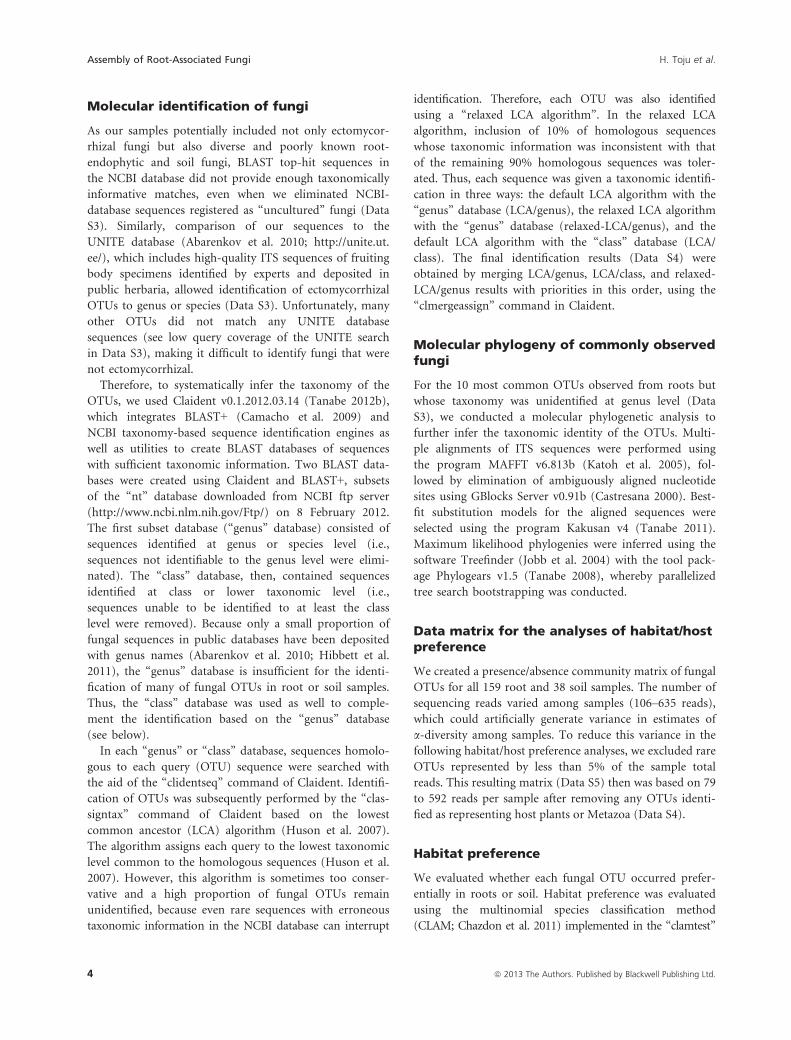
Molecular identification and fungaldiversity within each sample
In total, we found 392 fungal OTUs from the root and soil
samples (Data S2). Among those, 163 and 47 OTUs were
found exclusively from root or soil samples, respectively,
and 182 were common to both sample types. Among the
392 OTUs, 181 were ascomycetes, 108 basidiomycetes, two
chytridiomycetes, and five glomeromycetes, while 96 fungal
OTUs could not be identified to the phylum level. The
mean number of OTUs observed in each sample did not
significantly differ between root samples (12.9 OTUs [SD =4.7]) and soil samples (14.5 OTUs [SD = 5.4]; Fig. 1A and
B) after controlling for the number of sequencing reads per
sample (generalized-linear model with quasi-Poisson error;
t1, 194 = 1.9, P = 0.057). The mean number of arbuscular
and ectomycorrhizal fungal OTUs in a sample was 1.9
(SD = 1.3, N = 159) for roots and 2.7 (SD = 1.8, N = 38)
for soil. For both sample types, the total number of observed
OTUs increased continuously with increasing sample size
(Fig. 1C), reflecting the high diversity of belowground fungi.
Community composition of root-associatedfungi
The analysis of chloroplast rbcL gene sequences revealed
that the 159 terminal-root samples represented 12 plant
species (Fig. 1D). Among the plant species, Q. serrata was
the most dominant, as expected by the dominance of the
plant aboveground in the study site.
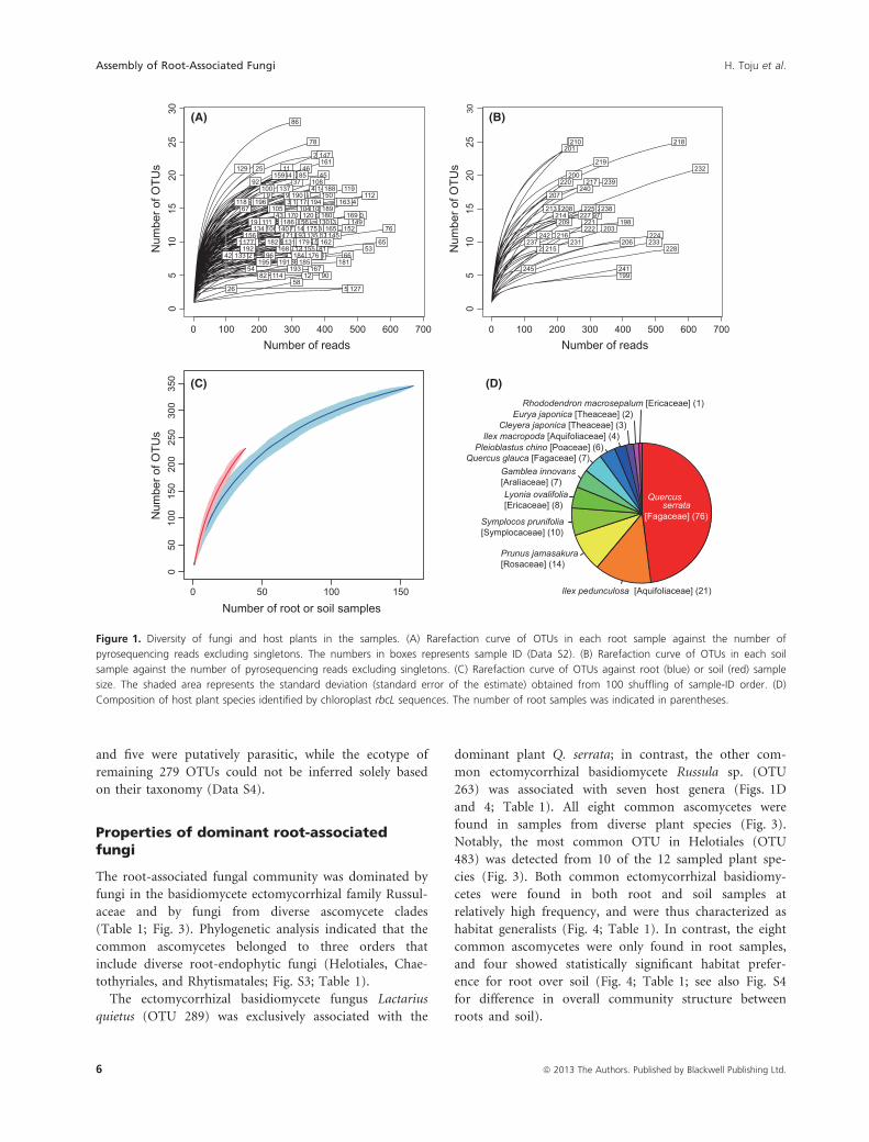
Among the 345 fungal OTUs found from the 159 termi-
nal-root samples, 270 (78.3%) were identified to phylum,
185 (53.6%) were identified to order and 112 (32.5%) were
identified to genus (Fig. 2). At phylum level, 168 fungal
OTUs (62.2%) were ascomycetes, 93 (34.4%) were basidio-
mycetes, five were glomeromycetes (1.9%), and two were
chytridiomycetes (0.7%) (Fig. 2A). At order level,
Helotiales, Russulales, and Agaricales dominated the root-
associated fungal community, while diverse clades such as
Chaetothyriales and Eurotiales were found as well
(Fig. 2B). At the genus level, the ectomycorrhizal taxon
Russula was the most common in the root samples
(Fig. 2C). Besides Russula, fungi in diverse ectomycorrhizal
genera such as Lactarius, Cortinarius, Lactarius, Tomentella,
Amanita, Boletus, and Cenococcum were observed (Fig. 2C).
Meanwhile, we found diverse nonectomycorrhizal fungi
such as Capronia (= Cladophialophola [anamorph]),
Cryptosporiopsis, Oidiodendron, and Hypocrea, genera
known to include root endophytes and plant pathogens.
Of the 345 OTUs found from roots, 56 were putatively
ectomycorrhizal, five were putatively arbuscular mycorrhizal
ª 2013 The Authors. Published by Blackwell Publishing Ltd. 5
H. Toju et al. Assembly of Root-Associated Fungi
and five were putatively parasitic, while the ecotype of
remaining 279 OTUs could not be inferred solely based
on their taxonomy (Data S4).
Properties of dominant root-associatedfungi
The root-associated fungal community was dominated by
fungi in the basidiomycete ectomycorrhizal family Russul-
aceae and by fungi from diverse ascomycete clades
(Table 1; Fig. 3). Phylogenetic analysis indicated that the
common ascomycetes belonged to three orders that
include diverse root-endophytic fungi (Helotiales, Chae-
tothyriales, and Rhytismatales; Fig. S3; Table 1).
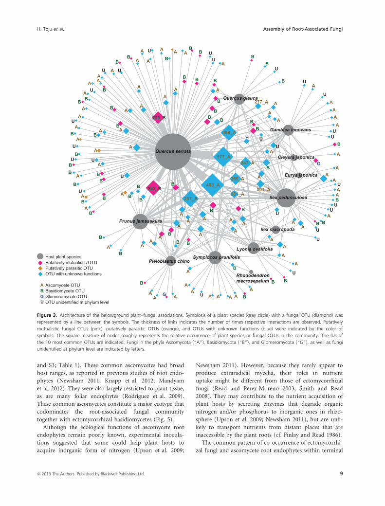
The ectomycorrhizal basidiomycete fungus Lactarius
quietus (OTU 289) was exclusively associated with the
dominant plant Q. serrata; in contrast, the other com-
mon ectomycorrhizal basidiomycete Russula sp. (OTU
263) was associated with seven host genera (Figs. 1D
and 4; Table 1). All eight common ascomycetes were
found in samples from diverse plant species (Fig. 3).
Notably, the most common OTU in Helotiales (OTU
483) was detected from 10 of the 12 sampled plant spe-
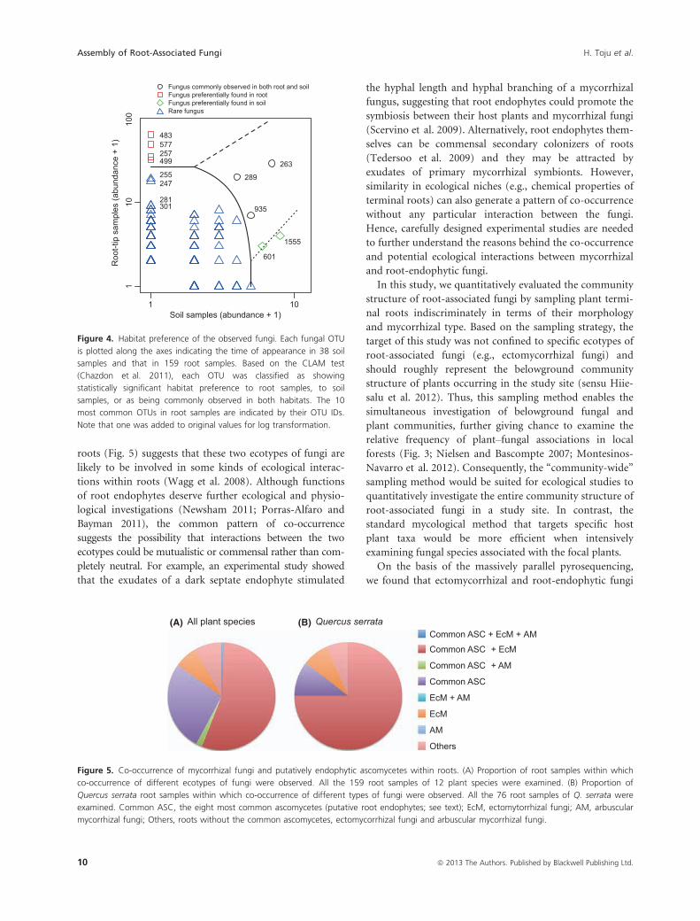
cies (Fig. 3). Both common ectomycorrhizal basidiomy-
cetes were found in both root and soil samples at
relatively high frequency, and were thus characterized as
habitat generalists (Fig. 4; Table 1). In contrast, the eight
common ascomycetes were only found in root samples,
and four showed statistically significant habitat prefer-
ence for root over soil (Fig. 4; Table 1; see also Fig. S4
for difference in overall community structure between
roots and soil).
Quercus serrata
[Fagaceae] (76)
Ilex pedunculosa [Aquifoliaceae] (21)
Prunus jamasakura[Rosaceae] (14)
Symplocos prunifolia[Symplocaceae] (10)
Lyonia ovalifolia[Ericaceae] (8)
Gamblea innovans[Araliaceae] (7)
Quercus glauca [Fagaceae] (7)Pleioblastus chino [Poaceae] (6)
Ilex macropoda [Aquifoliaceae] (4) Cleyera japonica [Theaceae] (3)
Eurya japonica [Theaceae] (2) Rhododendron macrosepalum [Ericaceae] (1)
(D)
(B)
Num
ber o
f OTU
s
0 100 200 300 400 500 600 700
05
1015
2025
30
197198
199
200
201202
203
205206
207
208
209
210
213214
215
216
217
218
219
220
221222
224
225227
228231
232
233
235
237
238
239240
241
242
245
Number of reads
(A)
Num
ber o
f OTU
s
Number of reads0 100 200 300 400 500 600 700
05
1015
2025
301
2
3
4
6
7
89
10
11
12
13
1415
16
1819
22
25
26
27
28
29
32
33
34
35
36
37
3840
4142
43
4546
47
50
5153
54
55
56
5859
6061
6264
65
66
67
68
69
70
71
72
73
7677
78
80
81
82
83
84
85
86
89
90
91
92
93
95
96
97
99
100
101102
103104105
106
107
108
109110
111
112
113
114
115116
117
118
119
120
121
126
127
129
130
131
132133
134135
137
139
140
141
144
145146
147
148149
150
151152153
154155
156157
159
160
161
162
163
165
167
168
169170
172173174
175
176
177
178
179
180
181
182
184185
186
188
189
190
191
192
193
194
195
196
Number of root or soil samples
Num
ber o
f OTU
s
(C)350
0 50 100 150
050
100
150
200
250
300
Figure 1. Diversity of fungi and host plants in the samples. (A) Rarefaction curve of OTUs in each root sample against the number of
pyrosequencing reads excluding singletons. The numbers in boxes represents sample ID (Data S2). (B) Rarefaction curve of OTUs in each soil
sample against the number of pyrosequencing reads excluding singletons. (C) Rarefaction curve of OTUs against root (blue) or soil (red) sample
size. The shaded area represents the standard deviation (standard error of the estimate) obtained from 100 shuffling of sample-ID order. (D)
Composition of host plant species identified by chloroplast rbcL sequences. The number of root samples was indicated in parentheses.
6 ª 2013 The Authors. Published by Blackwell Publishing Ltd.
Assembly of Root-Associated Fungi H. Toju et al.
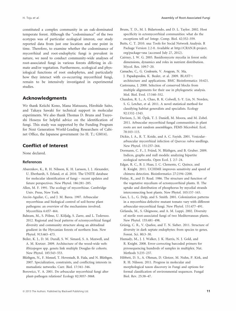
Co-occurrence of fungal OTUs within roots
Of the 159 root samples examined, 84.9% (135/159) were
colonized by at least one of the eight common ascomyce-
tes (Fig. 5). Importantly, most of those roots also colo-
nized by an arbuscular or an ectomycorrhizal fungus. Of
the 159 root samples, 55.3% (88/159) were colonized by
both common ascomycete and ectomycorrhizal fungi,
1.9% (3/159) were colonized by both common ascomy-
cete and arbuscular mycorrhizal fungi, and 0.6% (1/159)
were colonized by all the three ecotypes (Fig. 5). More-
over, of the 76 root samples of the dominant plant Q. ser-
rata, 75.0% (57/76) were colonized by both the common
ascomycetes and ectomycorrhizal fungi; only 7.9% (6/76)
were colonized by ectomycorrhizal fungi but none of the
eight common ascomycetes.
Discussion
We found broad patterns of co-occurrence between
root-endophytic fungi and mycorrhizal fungi in an
oak-dominated temperate forest. Ectomycorrhizal fungi
were found both in root samples and in the soil
surrounding the roots, reflecting the expected nutrient
foraging strategy. In contrast, endophytic ascomycetes
were primarily restricted to root samples. The host ranges
of endophytic ascomycetes were generally broader than
those of ectomycorrhizal basidiomycetes. The structure of
the root fungal communities points to the importance of
studies to understand how co-occurrence of terminal
roots by endophytic and mycorrhizal fungi could
influence host plant performance.
The second-growth forest included many fungal clades
expected in Quercus-dominated North-temperate forests
(Jumpponen et al. 2010; Sato et al. 2012a,b; Tedersoo
et al. 2012). Basidiomycete fungi in Russulaceae were the
most common, while Cortinarius, Tomentella, Amanita,
Boletus, and the ascomycete Cenococcum were also found
in root samples (Fig. 2; Table 1). These fungi were found
in roots as well as in the surrounding soil, as would be
expected from ectomycorrhizal fungi that produce extra-
radical mycelia to forage for nutrients (Finlay and Read
1986).
Somewhat surprisingly, root-endophytic ascomycetes
were more common than ectomycorrhizal basidiomycetes
(Fig. 3; Table 1). The fungal community of roots was
dominated by ascomycetes in diverse taxonomic clades
such as Helotiales, Chaetothyriales, and Rhytismatales (Figs. 3
Ascomycota Basidiomycota Glomeromycota Chytridiomycota Not defined
Helotiales Russulales Agaricales Chaetothyriales Eurotiales Thelephorales Trechisporales Pleosporales Glomerales Hypocreales Boletales Chaetosphaeriales Chytridiales Polyporales Saccharomycetales Sebacinales
Xylariales Atheliales Auriculariales BotryosphaerialesCantharellales Capnodiales Diaporthales Diversisporales Geastrales Leucosporidiales Mortierellales Mucorales Ostropales Tremellales Not defined
Russula Lactarius Capronia Cryptosporiopsis Cortinarius Mycena Penicillium Tomentella Trechispora
Cladophialophora Corynespora Oidiodendron Amanita Candida Cenococcum Hypocrea Meliniomyces Sebacina
Agaricus Boletus Byssocorticium Chaetosphaeria Clavulina Cryptodiscus
Geastrum Glomus Inocybe
Lachnum Lauriomyces Lophiostoma Mollisia Pestalotiopsis Phialocephala Phomopsis Pyrenochaeta Rhodocollybia
Scleropezicula Thelephora Thozetella Thysanophora Tylopilus Umbelopsis
Phylum(A)
(C)
(B) Order
Genus
Identified Identified
Identified
Figure 2. Community composition of root-associated fungi. (A) Phylum level composition of OTUs observed in root samples (270 of 345 OTUs
were identified). (B) Order-level composition of OTUs observed in root samples (185 of 345 OTUs were identified). (C) Genus level composition of
OTUs observed in root samples (112 of 345 OTUs were identified).
ª 2013 The Authors. Published by Blackwell Publishing Ltd. 7
H. Toju et al. Assembly of Root-Associated Fungi
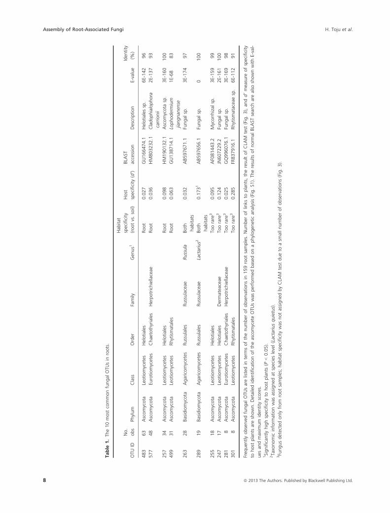
Table
1.Th
e10most
commonfungal
OTU
sin
roots.
OTU
ID
No.
obs
Phylum
Class
Order
Family
Gen
us1
Hab
itat
specificity
(rootvs.soil)
Host
specificity
(d’)
BLA
ST
accession
Description
E-value
Iden
tity
(%)
483
63
Ascomycota
Leotiomycetes
Helotiales
Root
0.027
GU166474.1
Helotiales
sp.
6E-142
96
577
48
Ascomycota
Eurotiomycetes
Chaetothyriales
Herpotrichiellaceae
Root
0.036
HM803232.1
Cladophialophora
carrionii
2E-137
93
257
34
Ascomycota
Leotiomycetes
Helotiales
Root
0.098
HM190132.1
Ascomycota
sp.
3E-160
100
499
31
Ascomycota
Leotiomycetes
Rhytismatales
Root
0.063
GU138714.1
Lophodermium
jiangnan
ense
1E-68
83
263
28
Basidiomycota
Agaricomycetes
Russulales
Russulaceae
Russula
Both
hab
itats
0.032
AB597671.1
Fungal
sp.
3E-174
97
289
19
Basidiomycota
Agaricomycetes
Russulales
Russulaceae
Lactarius2
Both
hab
itats
0.1731
AB597656.1
Fungal
sp.
0100
255
18
Ascomycota
Leotiomycetes
Helotiales
Toorare
30.095
AF081443.2
Mycorrhizal
sp.
3E-159
99
247
17
Ascomycota
Leotiomycetes
Helotiales
Dermateaceae
Toorare
30.124
JN607229.2
Fungal
sp.
2E-161
100
281
8Ascomycota
Eurotiomycetes
Chaetothyriales
Herpotrichiellaceae
Toorare
30.025
GQ996076.1
Fungal
sp.
3E-169
98
301
8Ascomycota
Leotiomycetes
Rhytismatales
Toorare
30.285
FR837916.1
Rhytismataceaesp.
6E-112
91
Freq
uen
tlyobserved
fungal
OTU
sarelistedin
term
softhenumber
ofobservationsin
159rootsamples.
Number
oflinks
toplants,theresultofCLA
Mtest
(Fig.3),an
dd’measure
ofspecificity
tohost
plants
areshown.Detailediden
tificationoftheascomyceteOTU
swas
perform
edbased
onaphylogen
etic
analysis(Fig.S1
).Th
eresultsofnorm
alBLA
STsearch
arealso
shownwithE-val-
ues
andmaxim
um
iden
tity
scores.
1Significantlyhighspecificity
tohost
plants
(P<0.05).
2Taxonomic
inform
ationwas
assigned
atspecieslevel(Lactariusquietus).
3Fungusdetectedonlyfrom
rootsamples;
hab
itat
specificity
was
notassigned
byCLA
Mtest
dueto
asm
allnumber
ofobservations(Fig.3).
8 ª 2013 The Authors. Published by Blackwell Publishing Ltd.
Assembly of Root-Associated Fungi H. Toju et al.
and S3; Table 1). These common ascomycetes had broad
host ranges, as reported in previous studies of root endo-
phytes (Newsham 2011; Knapp et al. 2012; Mandyam
et al. 2012). They were also largely restricted to plant tissue,
as are many foliar endophytes (Rodriguez et al. 2009).
These common ascomycetes constitute a major ecotype that
codominates the root-associated fungal community
together with ectomycorrhizal basidiomycetes (Fig. 5).
Although the ecological functions of ascomycete root
endophytes remain poorly known, experimental inocula-
tions suggested that some could help plant hosts to
acquire inorganic form of nitrogen (Upson et al. 2009;
Newsham 2011). However, because they rarely appear to
produce extraradical mycelia, their roles in nutrient
uptake might be different from those of ectomycorrhizal
fungi (Read and Perez-Moreno 2003; Smith and Read
2008). They may contribute to the nutrient acquisition of
plant hosts by secreting enzymes that degrade organic
nitrogen and/or phosphorus to inorganic ones in rhizo-
sphere (Upson et al. 2009; Newsham 2011), but are unli-
kely to transport nutrients from distant places that are
inaccessible by the plant roots (cf. Finlay and Read 1986).
The common pattern of co-occurrence of ectomycorrhi-
zal fungi and ascomycete root endophytes within terminal
Host plant speciesPutatively mutualistic OTUPutatively parasitic OTUOTU with unknown functions
Ascomycete OTUBasidiomycete OTUGlomeromycete OTUOTU unidentified at phylum levelU
ABG
Cleyera japonica
Eurya japonica
Gamblea innovans
Ilex macropoda
Ilex pedunculosa
Lyonia ovalifolia
Pleioblastus chino
Prunus jamasakura
Quercus glauca
Quercus serrata
Rhododendronmacrosepalum
Symplocos prunifolia
247_A
255_A
257_A
263_B
277_A
281_A
289_B
301_A483_A
499_A
577_A
A
A
AA
AA
A
A
A
AA AA
AA
A
AAA
A
AA
A
AA
A
B
A
B
A
B
A
B
A
B
A
B
A
B
A
B
A
B
A
B
A
B
A
B
A
B
A
BA
B
A
B
A
B
A BA
B
A
B
A
B
A
B
AB
AB
A
B
AB
A
B
A
B
A
B
A
B
A
B
A
B
A
BA
BA
BAB
U
BU
B
U
B
U
B
U
B
GU
B
G
U
U U
U
U
U
UU
U
UU
UU
U
U
UU
U
A
A
A
A
A
AA
AA
A
AAA
A
A
A
B
B
B
B
B
B
B
B
BB
B
B
U
U
U
U
Figure 3. Architecture of the belowground plant–fungal associations. Symbiosis of a plant species (gray circle) with a fungal OTU (diamond) was
represented by a line between the symbols. The thickness of links indicates the number of times respective interactions are observed. Putatively
mutualistic fungal OTUs (pink), putatively parasitic OTUs (orange), and OTUs with unknown functions (blue) were indicated by the color of
symbols. The square measure of nodes roughly represents the relative occurrence of plant species or fungal OTUs in the community. The IDs of
the 10 most common OTUs are indicated. Fungi in the phyla Ascomycota (“A”), Basidiomycota (“B”), and Glomeromycota (“G”), as well as fungi
unidentified at phylum level are indicated by letters.
ª 2013 The Authors. Published by Blackwell Publishing Ltd. 9
H. Toju et al. Assembly of Root-Associated Fungi
roots (Fig. 5) suggests that these two ecotypes of fungi are
likely to be involved in some kinds of ecological interac-
tions within roots (Wagg et al. 2008). Although functions
of root endophytes deserve further ecological and physio-
logical investigations (Newsham 2011; Porras-Alfaro and
Bayman 2011), the common pattern of co-occurrence
suggests the possibility that interactions between the two
ecotypes could be mutualistic or commensal rather than com-
pletely neutral. For example, an experimental study showed
that the exudates of a dark septate endophyte stimulated
the hyphal length and hyphal branching of a mycorrhizal
fungus, suggesting that root endophytes could promote the
symbiosis between their host plants and mycorrhizal fungi
(Scervino et al. 2009). Alternatively, root endophytes them-
selves can be commensal secondary colonizers of roots
(Tedersoo et al. 2009) and they may be attracted by
exudates of primary mycorrhizal symbionts. However,
similarity in ecological niches (e.g., chemical properties of
terminal roots) can also generate a pattern of co-occurrence
without any particular interaction between the fungi.
Hence, carefully designed experimental studies are needed
to further understand the reasons behind the co-occurrence
and potential ecological interactions between mycorrhizal
and root-endophytic fungi.
In this study, we quantitatively evaluated the community
structure of root-associated fungi by sampling plant termi-
nal roots indiscriminately in terms of their morphology
and mycorrhizal type. Based on the sampling strategy, the
target of this study was not confined to specific ecotypes of
root-associated fungi (e.g., ectomycorrhizal fungi) and
should roughly represent the belowground community
structure of plants occurring in the study site (sensu Hiie-
salu et al. 2012). Thus, this sampling method enables the
simultaneous investigation of belowground fungal and
plant communities, further giving chance to examine the
relative frequency of plant–fungal associations in local
forests (Fig. 3; Nielsen and Bascompte 2007; Montesinos-
Navarro et al. 2012). Consequently, the “community-wide”
sampling method would be suited for ecological studies to
quantitatively investigate the entire community structure of
root-associated fungi in a study site. In contrast, the
standard mycological method that targets specific host
plant taxa would be more efficient when intensively
examining fungal species associated with the focal plants.
On the basis of the massively parallel pyrosequencing,
we found that ectomycorrhizal and root-endophytic fungi
Fungus commonly observed in both root and soilFungus preferentially found in rootFungus preferentially found in soilRare fungus
Soil samples (abundance + 1)
Roo
t-tip
sam
ples
(abu
ndan
ce +
1)
1 10
110
100
483577257499 263
289255247
281301
1555
601
935
Figure 4. Habitat preference of the observed fungi. Each fungal OTU
is plotted along the axes indicating the time of appearance in 38 soil
samples and that in 159 root samples. Based on the CLAM test
(Chazdon et al. 2011), each OTU was classified as showing
statistically significant habitat preference to root samples, to soil
samples, or as being commonly observed in both habitats. The 10
most common OTUs in root samples are indicated by their OTU IDs.
Note that one was added to original values for log transformation.
All plant species (B)(A) Quercus serrataCommon ASC + EcM + AM
Common ASC + EcM
Common ASC + AM
Common ASC
EcM + AM
EcM
AM
Others
Figure 5. Co-occurrence of mycorrhizal fungi and putatively endophytic ascomycetes within roots. (A) Proportion of root samples within which
co-occurrence of different ecotypes of fungi were observed. All the 159 root samples of 12 plant species were examined. (B) Proportion of
Quercus serrata root samples within which co-occurrence of different types of fungi were observed. All the 76 root samples of Q. serrata were
examined. Common ASC, the eight most common ascomycetes (putative root endophytes; see text); EcM, ectomytorrhizal fungi; AM, arbuscular
mycorrhizal fungi; Others, roots without the common ascomycetes, ectomycorrhizal fungi and arbuscular mycorrhizal fungi.
10 ª 2013 The Authors. Published by Blackwell Publishing Ltd.
Assembly of Root-Associated Fungi H. Toju et al.
constituted a complex community in an oak-dominated
temperate forest. Although the “codominance” of the two
ecotypes was of particular ecological interest, our study
reported data from just one location and one point in
time. Therefore, to examine whether the codominance of
mycorrhizal and root-endophytic fungi is prevalent in
nature, we need to conduct community-wide analyses of
root-associated fungi in various forests differing in cli-
mate and/or vegetation. Furthermore, ecological and phys-
iological functions of root endophytes, and particularly
how they interact with co-occurring mycorrhizal fungi,
remain to be intensively investigated in experimental
studies.
Acknowledgments
We thank Keiichi Kono, Mana Matsuura, Hirohide Saito,
and Takaya Sawaki for technical support in molecular
experiments. We also thank Thomas D. Bruns and Tsuyo-
shi Hosoya for helpful advice on the identification of
fungi. This study was supported by the Funding Program
for Next Generation World-Leading Researchers of Cabi-
net Office, the Japanese government (to H. T.; GS014).
Conflict of Interest
None declared.
References
Abarenkov, K., R. H. Nilsson, K. H. Larsson, I. J. Alexander,
U. Eberhardt, S. Erland, et al. 2010. The UNITE database
for molecular identification of fungi – recent updates and
future perspectives. New Phytol. 186:281–285.
Allen, M. F. 1991. The ecology of mycorrhizae. Cambridge
Univ. Press, New York.
Azc�on-Aguilar, C., and J. M. Barea. 1997. Arbuscular
mycorrhizas and biological control of soil-borne plant
pathogens: an overview of the mechanisms involved.
Mycorrhiza 6:457–464.
Bahram, M., S. P~olme, U. K~oljalg, S. Zarre, and L. Tedersoo.
2012. Regional and local patterns of ectomycorrhizal fungal
diversity and community structure along an altitudinal
gradient in the Hyrcanian forests of northern Iran. New
Phytol. 913:465–473.
Beiler, K. J., D. M. Durall, S. W. Simard, S. A. Maxwell, and
A. M. Kretzer. 2009. Architecture of the wood-wide web:
Rhizopogon spp. genets link multiple Douglas-fir cohorts.
New Phytol. 185:543–553.
Bl€uthgen, N., F. Menzel, T. Hovestadt, B. Fiala, and N. Bl€uthgen.
2007. Specialization, constraints, and conflicting interests in
mutualistic networks. Curr. Biol. 17:341–346.
Borowicz, V. A. 2001. Do arbuscular mycorrhizal fungi alter
plant-pathogen relations? Ecology 82:3057–3068.
Bruns, T. D., M. I. Bidartondo, and D. L. Taylor. 2002. Host
specificity in ectomycorrhizal communities: what do the
exceptions tell us? Integr. Comp. Biol. 42:352–359.
Butts, C. T. 2010. sna: Tools for Social Network Analysis. R
Package Version 2.2-0. Available at http://CRAN.R-project.
org/package=sna (accessed July 27, 2012).
Cairney, J. W. G. 2005. Basidiomycete mycelia in forest soils:
dimensions, dynamics and roles in nutrient distribution.
Mycol. Res. 109:7–20.
Camacho, C., G. Coulouris, V. Avagyan, N. Ma,
J. Papadopoulos, K. Bealer, et al. 2009. BLAST+:architecture and applications. BMC Bioinformatics 10:421.
Castresana, J. 2000. Selection of conserved blocks from
multiple alignments for their use in phylogenetic analysis.
Mol. Biol. Evol. 17:540–552.
Chazdon, R. L., A. Chao, R. K. Colwell, S.-Y. Lin, N. Norden,
S. G. Letcher, et al. 2011. A novel statistical method for
classifying habitat generalists and specialists. Ecology
92:1332–1343.
Davison, J., M. €Opik, T. J. Daniell, M. Moora, and M. Zobel.
2011. Arbuscular mycorrhizal fungal communities in plant
roots are not random assemblages. FEMS Microbiol. Ecol.
78:103–115.
Dickie, I. A., R. T. Koide, and A. C. Fayish. 2001. Vesicular–
arbuscular mycorrhizal infection of Quercus rubra seedlings.
New Phytol. 151:257–264.
Dormann, C. F., J. Fr€und, N. Bl€uthgen, and B. Gruber. 2009.
Indices, graphs and null models: analyzing bipartite
ecological networks. Open Ecol. J. 2:7–24.
Edgar, R. C., B. J. Haas, J. C. Clemente, C. Quince, and
R. Knight. 2011. UCHIME improves sensitivity and speed of
chimera detection. Bioinformatics 27:2194–2200.
Finlay, R., and D. Read. 1986. The structure and function of
the vegetative mycelium of ectomycorrhizal plants. II. The
uptake and distribution of phosphorus by mycelial strands
interconnecting host plants. New Phytol. 103:157–165.
Gao, L. L., G. Delp, and S. Smith. 2001. Colonization patterns
in a mycorrhiza-defective mutant tomato vary with different
arbuscular-mycorrhizal fungi. New Phytol. 151:477–491.
Girlanda, M., S. Ghignone, and A. M. Luppi. 2002. Diversity
of sterile root-associated fungi of two Mediterranean plants.
New Phytol. 155:481–498.
Gr€unig, C. R., V. Queloz, and T. N. Sieber. 2011. Structure of
diversity in dark septate endophytes: from species to genes.
Forest. Sci. 80:3–30.
Hamady, M., J. J. Walker, J. K. Harris, N. J. Gold, and
R. Knight. 2008. Error-correcting barcoded primers for
pyrosequencing hundreds of samples in multiplex. Nat.
Methods 5:235–237.
Hibbett, D. S., A. Ohman, D. Glotzer, M. Nuhn, P. Kirk, and
R. H. Nilsson. 2011. Progress in molecular and
morphological taxon discovery in Fungi and options for
formal classification of environmental sequences. Fungal
Biol. Rev. 25:38–47.
ª 2013 The Authors. Published by Blackwell Publishing Ltd. 11
H. Toju et al. Assembly of Root-Associated Fungi
Hiiesalu, I., M. €Opik, M. Metsis, L. Lilje, J. Davison, M. Vasar,
et al. 2012. Plant species richness belowground: higher
richness and new patterns revealed by next-generation
sequencing. Mol. Ecol. 21:2004–2016.
Hoeksema, J. D. 2010. Ongoing coevolution in mycorrhizal
interactions. New Phytol. 187:286–300.
H€ogberg, M. N., and P. H€ogberg. 2002. Extramatrical
ectomycorrhizal mycelium contributes one-third of
microbial biomass and produces, together with associated
roots, half the dissolved organic carbon in a forest soil. New
Phytol. 154:791–795.
H€ogberg, P., A. Nordgren, N. Buchmann, A. F. S. Taylor,
A. Ekblad, M. N. H€ogberg, et al. 2001. Large-scale forest
girdling shows that current photosynthesis drives soil
respiration. Nature 411:789–792.
Huson, D. H., A. F. Auch, J. Qi, and S. C. Schuster. 2007.
MEGAN analysis of metagenomic data. Genome Res.
17:377–386.
Jobb, G., A. Von Haeseler, and K. Strimmer. 2004.
TREEFINDER: a powerful graphical analysis environment
for molecular phylogenetics. BMC Evol. Biol. 4:18.
Johnson, D., F. Martin, J. W. G. Cairney, and I. C. Anderson.
2012. The importance of individuals: intraspecific diversity
of mycorrhizal plants and fungi in ecosystems. New Phytol.
194:614–628.
Jumpponen, A. 2001. Dark septate endophytes – are they
mycorrhizal? Mycorrhiza 11:207–211.
Jumpponen, A., and J. M. Trappe. 1998. Dark septate
endophytes: a review of facultative biotrophic root-
colonizing fungi. New Phytol. 140:295–310.
Jumpponen, A., K. L. Jones, J. D. Mattox, and C. Yaege. 2010.
Massively parallel 454-sequencing of fungal communities in
Quercus spp. ectomycorrhizas indicates seasonal dynamics in
urban and rural sites. Mol. Ecol. 19:41–53.
Katoh, K., K. Kuma, H. Toh, and T. Miyata. 2005. MAFFT
version 5: improvement in accuracy of multiple sequence
alignment. Nucleic Acids Res. 33:511–518.
Kernaghan, G., and G. Patriquin. 2011. Host associations
between fungal root endophytes and boreal trees. Microb.
Ecol. 62:460–473.
Knapp, D. G., A. Pintye, and G. M. Kov�acs. 2012. The dark
side is not fastidious – dark septate endophytic fungi of
native and invasive plants of semiarid sandy areas. PLoS
ONE 7:e32570.
Li, W., L. Fu, B. Niu, S. Wu, and J. Wooley. 2012. Ultrafast
clustering algorithms for metagenomic sequence analysis.
Brief. Bioinform. 13:656–668.
Mandyam, K., C. Fox, and A. Jumpponen. 2012. Septate
endophyte colonization and host responses of grasses and
forbs native to a tallgrass prairie. Mycorrhiza 22:109–119.
Montesinos-Navarro, A., J. G. Segarra-Moragues, A. Valiente-
Banuet, and M. Verd�u. 2012. The network structure of
plant-arbuscular mycorrhizal fungi. New Phytol. 194:
536–547.
Nara, K. 2006. Ectomycorrhizal networks and seedling
establishment during early primary succession. New Phytol.
169:169–178.
Newsham, K. K. 2011. A meta-analysis of plant responses to
dark septate root endophytes. New Phytol. 190:783–793.
Nielsen, A., and J. Bascompte. 2007. Ecological networks,
nestedness and sampling effort. J. Ecol. 95:1134–1141.
Nilsson, H. R., L. Tedersoo, B. D. Lindahl, R. Kjøller, T. Carlsen,
C. Quince, et al. 2011. Towards standardization of the
description and publication of next-generation sequencing
datasets of fungal communities. New Phytol. 191:314–318.
Oksanen, J., F. G. Blanachet, R. Kindt, P. Legendre,
P. R. Minchin, R. B. O’Hara, et al. 2012. Vegan: community
ecology package. R package version 2.0-3. Available at http://
CRAN.R-project.org/package=vegan (accessed July 26, 2012).
Porras-Alfaro, A., and P. Bayman. 2011. Hidden fungi,
emergent properties: endophytes and microbiomes. Annu.
Rev. Phytopathol. 49:291–315.
Read, D. J., and J. Perez-Moreno. 2003. Mycorrhizas and
nutrient cycling in ecosystems – a journey towards
relevance? New Phytol. 157:475–492.
Reininger, V., and T. N. Sieber. 2012. Mycorrhiza reduces
adverse effects of dark septate endophytes (DSE) on growth
of conifers. PLoS ONE 7:e42865.
Rodriguez, R. J., J. F. White Jr., A. E. Arnold, and R. S. Redman.
2009. Fungal endophytes: diversity and functional roles.
New Phytol. 182:314–330.
Sato, H., and N. Murakami. 2008. Reproductive isolation
among cryptic species in the ectomycorrhizal genus
Strobilomyces: population-level CAPS marker-based genetic
analysis. Mol. Phylogenet. Evol. 48:326–334.
Sato, H., T. Yumoto, and N. Murakami. 2007. Cryptic species
and host specificity in the ectomycorrhizal genus
Strobilomyces (Strobilomycetaceae). Am. J. Bot. 94:1630.
Sato, H., S. Morimoto, and T. Hattori. 2012a. A thirty-year
survey reveals that ecosystem function of fungi predicts
phenology of mushroom fruiting. PLoS ONE 7:e49777.
Sato, H., R. Tsujino, K. Kurita, K. Yokoyama, and K. Agata.
2012b. Modelling the global distribution of fungal species:
new insights into microbial cosmopolitanism. Mol. Ecol.
21:5599–5612.
Scervino, J. M., A. Gottlieb, V. A. SIlvani, M. P�ergola,
L. Fern�andez, and A. M. Godeas. 2009. Exudates of dark
septate endophyte (DSE) modulate the development of the
arbuscular mycorrhizal fungus (AMF) Gigaspora rosea. Soil
Biol. Biochem. 41:1753–1756.
Smith, S. E., and D. J. Read. 2008. Mycorrhizal symbiosis. 3rd
ed. Elsevier, New York.
Sommer, D. D., A. L. Delcher, S. L. Salzberg, and M. Pop.
2007. Minimus: a fast, lightweight genome assembler. BMC
Bioinformatics 8:64.
Tanabe, A. S. 2008. “Phylogears version 1.5,” software
distributed by the author. Available at http://www.
fifthdimension.jp/ (accessed March 14, 2012).
12 ª 2013 The Authors. Published by Blackwell Publishing Ltd.
Assembly of Root-Associated Fungi H. Toju et al.
Tanabe, A. S. 2011. Kakusan4 and Aminosan: two programs
for comparing nonpartitioned, proportional and separate
models for combined molecular phylogenetic analyses of
multilocus sequence data. Mol. Ecol. Resour. 11:914–921.
Tanabe, A. S. 2012a. Assams v0.1.2012.03.14, a software
distributed by the author. Available at http://www.
fifthdimension.jp/ (accessed January 15, 2012).
Tanabe, A. S. 2012b. Claident v0.1.2012.03.14, a software
distributed by author. Available at http://www.
fifthdimension.jp/ (accessed January 15, 2012).
Tedersoo, L., T. Jairus, B. M. Horton, K. Abarenkov, T. Suvi,
I. Saar, et al. 2008. Strong host preference of
ectomycorrhizal fungi in a Tasmanian wet sclerophyll forest
as revealed by DNA barcoding and taxon-specific primers.
New Phytol. 180:479–490.
Tedersoo, L., K. P€artel, T. Jairus, G. Gates, K. P~oldmaa, and
H. Tamm. 2009. Ascomycetes associated with
ectomycorrhizas: molecular diversity and ecology with
particular reference to the Helotiales. Environ. Microbiol.
11:3166–3178.
Tedersoo, L., M. Bahram, M. Toots, A. G. Di�edhiou,
T. W. Henkel, R. Kjøller, et al. 2012. Towards global
patterns in the diversity and community structure of
ectomycorrhizal fungi. Mol. Ecol. 21:4160–4170.
Toju, H., A. S. Tanabe, S. Yamamoto, and H. Sato. 2012.
High-coverage ITS primers for the DNA-based identification
of ascomycetes and basidiomycetes in environmental
samples. PLoS ONE 7:e40863.
Upson, R., D. J. Read, and K. K. Newsham. 2009. Nitrogen
form influences the response of Deschampsia antarctica to
dark septate root endophytes. Mycorrhiza 20:1–11.
V�azquez, D. P., C. J. Meli�an, N. M. Williams, N. Bl€uthgen,
B. R. Krasnov, and R. Poulin. 2007. Species abundance and
asymmetric interaction strength in ecological networks.
Oikos 116:1120–1127.
Wagg, C., M. Pautler, H. B. Massicotte, and R. L. Peterson.
2008. The co-occurrence of ectomycorrhizal, arbuscular
mycorrhizal, and dark septate fungi in seedlings of four
members of the Pinaceae. Mycorrhiza 18:103–110.
Walker, J. F., L. Aldrich-Wolfe, A. Riffel, H. Barbare,
N. B. Simpson, J. Trowbridge, et al. 2011. Diverse Helotiales
associated with the roots of three species of Arctic Ericaceae
provide no evidence for host specificity. New Phytol.
191:515–527.
White, T. J., T. Bruns, S. Lee, and J. Taylor. 1990.
Amplification and direct sequencing of fungal ribosomal
RNA genes for phylogenetics. Pp. 315–322 in M. A. Innis,
D. H. Gelfand, J. J. Sninsky, and T.J. White eds. PCR
protocols a guide to methods and applications. Academic
Press, MA.
Wu, B., H. Maruyama, M. Teramoto, and T. Hougetsu. 2012.
Structural and functional interactions between extraradical
mycelia of ectomycorrhizal Pisolithus isolates. New Phytol.
194:1070–1078.
Supporting Information
Additional Supporting Information may be found in the
online version of this article:
Data S1. OTU sequences in FASTA format.
Data S2. Summary of reads that passed quality filtering.
Data S3. Comparison of molecular identification results
for the 10 most common OTUs.
Data S4. OTUs observed in root and soil samples (all
OTUs).
Data S5. Matrix representing the presence/absence of each
fungal OTU in each root or soil sample.
Data S6. Matrix representing the symbiosis of plant spe-
cies and fungal OTUs.
Figure S1. Map of the study plot. Schematic illustration
of the sampling design in the study site. Root samples
were collected on crossover points of the 1-m-mesh plot.
Soil samples were collected on crossover points of the
2-m-mesh plot as indicated by the brown circles.
Numbers indicate sample IDs in Data S2.
Figure S2. Example photographs of washed terminal
roots. Example photographs of washed terminal roots.
Each terminal root was washed in 70% ethanol by shak-
ing it with 1-mm zirconium balls 15 times per second for
2 min using TissueLyser II (Qiagen).
Figure S3. Example photographs of washed terminal roots.
Molecular phylogeny of most commonly observed ascomy-
cete OTUs. Maximum-likelihood topology based on ITS
sequences is shown with bootstrap probabilities above the
branches (>50%; 100 replicates). Fungal sequences of “dark
septate endophytes” (Gr€unig et al. 2011; Newsham 2011)
are indicated by asterisks. (A) OTUs in the order Helotiales
(254 bp; J2ef + G model). (B) OTUs in the order Chae-
tothyriales (239 bp; TN93ef + G). (C) OTUs in the order
Rhytismatales (258 bp; TN93ef + G).
Figure S4. Molecular phylogeny of most commonly
observed ascomycetes OTUs. Comparison of fungal
community composition between root and soil samples.
(A) Taxonomic composition of OTUs observed in root
samples (98 of 172 OTUs identified at the order level).
The analysis was conducted after converting the pyrose-
quencing data to the presence/absence matrix (see Materials
and Methods; Data S5). Asterisks indicate OTUs whose
order-level taxonomy is yet to be settled, but genus- or
family-level taxonomic information is available. (B) Taxo-
nomic information of OTUs observed in soil samples (41 of
90 OTUs identified at the order level). (C) Taxonomic
information of OTUs observed in root samples (66 of 172
OTUs identified at the genus level). (D) Taxonomic infor-
mation of OTUs observed in soil samples (30 of 90 OTUs
identified at the genus level).
ª 2013 The Authors. Published by Blackwell Publishing Ltd. 13
H. Toju et al. Assembly of Root-Associated Fungi


![[c1]: The rRole of endophytic fungi on plant disease control · PDF file16 endophytic fungi can enhance plant productivity and they can survive in host plant for long periods. 17 Therefore,](https://img.pdfslide.tips/doc/110x75/5abcfea97f8b9af27d8ea4c6/c1-the-rrole-of-endophytic-fungi-on-plant-disease-control-endophytic-fungi-can.jpg)