Embed Size (px)
Citation preview


____Cranial nerves attach to brain___Spinal nerves attach to spinal cord

Sensation:• The conscious or subconscious
awareness of external or internal stimuli.
Perception:• The conscious awareness and
the interpretation of meaning of sensations.

Peripheral sensory receptors
By location:
• Exteroceptors
– Sensitive to stimuli arising from outside body
• Interoceptors
– Or visceroreceptors, from internal viscera
• Proprioceptors
– Monitor degree of stretch in skeletal muscles, tendons, joints and ligaments

• Pain• Temperature• Light touch• Pressure• Sense of body
and limb position
• Taste• Smell• Vision• Hearing• Balance
General SensesGeneral Senses vs. vs. Special Special SensesSenses

• Mechanoreceptors
• Thermoreceptors
• Photoreceptors
• Chemoreceptors
• Nociceptors
• Osmoreceptors

skin, bones, internal organs, joints
Naked nerve endings surrounded by one or more layers
Pacinian corpuscle
Free nerve endings
Encapsulated Nerve Encapsulated Nerve
EndingsEndingsvs
Unencapsulated Unencapsulated
Nerve EndingsNerve Endings
Deeper tissue, muscles

Free Nerve Endings - Pain & Temperature
Merkel’s Discs - Light Touch & Pressure
Root Hair Plexuses - Light Touch
pain, light touch, and temperature

Pacinian Corpuscles - Deep Pressure
Meissner’s Corpuscles - Discriminative Touch in Hairless Skin Areas
Krause’s End-Bulbs - Discriminative Touch in Mucous Membranes
Ruffini’s Corpuscles - Deep Pressure & Stretch (Proprioception)

Merkel Cells- slow mechanoreceptors (basal layer)
Merkel cell
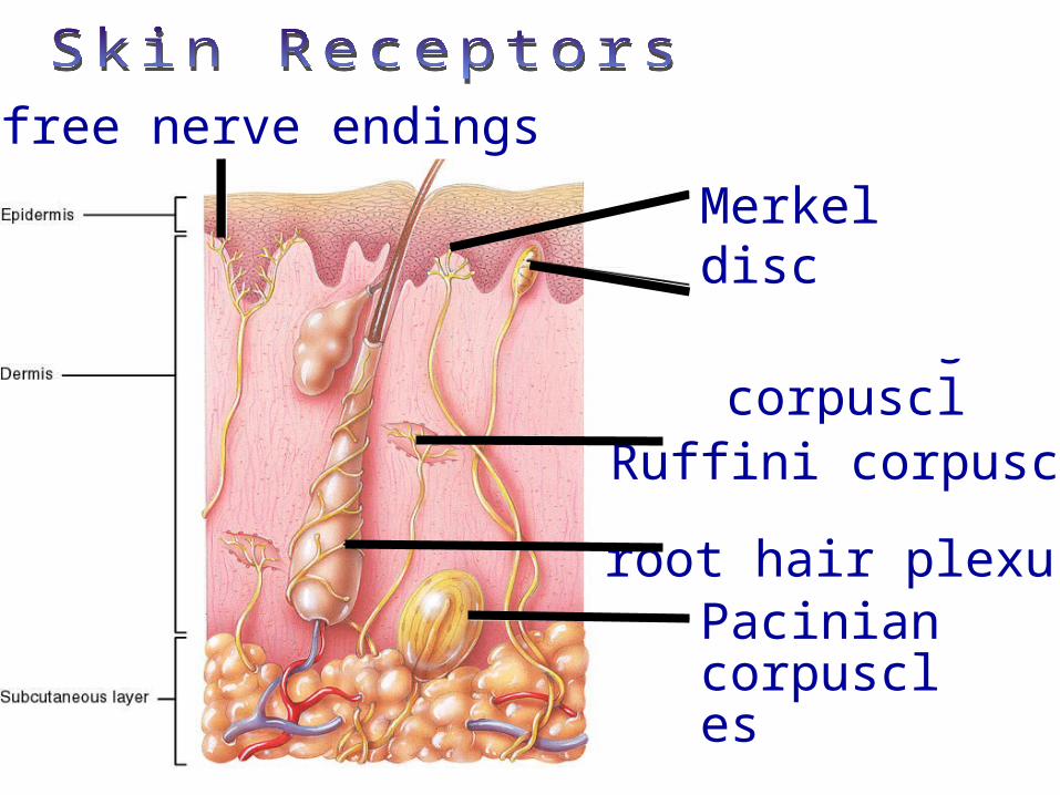
free nerve endings
root hair plexus
Meissner’s corpuscles
Pacinian corpuscles
Ruffini corpuscle
Merkel disc

Muscle Spindles - Skeletal Muscle StretchingGolgi Tendon Organs - Tendon StretchingJoint kinesthetic receptors – monitors stretch
in synovial joints; sends info to cerebellum and spinal reflex arcs
Proprioceptors
– to cerebrum, – cerebellum and – spinal reflex arcs


Peripheral motor endings
• Innervation of skeletal muscle
• Innervation of visceral muscles and glands

• Motor axons innervate skeletal muscle fibers at neuromuscular junctions = motor end plates

Motor unit: motor neuron & all the muscle fibers it innervates
All muscles in motor unit contract together when neuron fires
Stimulation of single motor unit causes weak contraction of entire muscle (spread out)

Innervation of visceral muscles & glands
• Near end organ visceral motor axon swells = presynaptic terminals (vesicles with neurotransmitters): action slow (NT diffuses)

Somatic Pain-results from injuries to skin, muscle, joints, tendon vs.Visceral Pain- pain in body organs

Referred Pain-felt on the body surface

• Mature neurons are amitotic• If the soma of a damaged nerve is intact, axon
will regenerate• Involves coordinated activity among:
– Macrophages—remove debris– Schwann cells—form regeneration tube and
secrete growth factors– Axons—regenerate damaged part
• CNS oligodendrocytes bear growth-inhibiting proteins that prevent CNS fiber regeneration

Figure 13.4 (1 of 4)
Endoneurium
Dropletsof myelin
Fragmentedaxon
Schwann cells
Site of nerve damage
The axonbecomesfragmented atthe injury site.
1

Figure 13.4 (2 of 4)
Schwann cell Macrophage Macrophagesclean out thedead axon distalto the injury.
2

Figure 13.4 (3 of 4)
Fine axon sproutsor filaments
Aligning Schwann cellsform regeneration tube
3 Axon sprouts,or filaments,grow through aregeneration tubeformed bySchwann cells.

Figure 13.4 (4 of 4)
Schwann cell Site of newmyelin sheathformation
4 The axonregenerates anda new myelinsheath forms.
Single enlargingaxon filament

On Old Olympus Towering Tops A Fat Voracious German Viewed A Hop
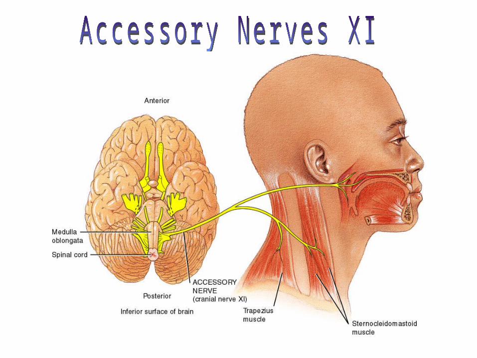
1. Olfactory- smell2. Optic- vision3. Oculomotor- 4 of the 6 extrinsic eye muscles 4. Trochlear- extrinsic eye muscles5. Trigeminal- sensory fibers to the face and motor fibers to
the chewing muscles6. Abducens- controls eye muscles that turn the eye laterally7. Facial- facial expression8. Vestibulocochlear- hearing and balance9. Glosopharyngeal- tongue and pharynx10.Vagus- parasympathetic control of heart, lungs &
abdominal organs11.Accessory- accessory part of vagus nerve, neck & throat
muscles12.Hypoglossal- moves muscles under tongue

Olfactory
Optic
Oculomotor
Trochlear
Trigeminal
Abducens
VestibulocochlearGlossopharyngeal
VagusAccessory Hypoglossal
Facial

Olfactory bulbOlfactory tract
Olfactory receptor cell
Filaments of olfactory nerve


Abducensnerve
Lateral rectus muscle cut


Nerve Pathways into the Spinal Cord
Nerve Pathways into the Spinal Cord sensory
pathway
motor pathway

Spinal nerves

Spinal nerves• Dorsal roots – sensory fibers arising from cell bodies in dorsal root ganglia
• Ventral roots – motor fibers arising from anterior gray column of spinal cord
Ventral root ganglia

• Dorsal and ventral roots join in an intervertebral foramen forming spinal nerve
• Outside foramen, re-branch as rami (sing., ramus):Dorsal and ventral rami (somatic)Rami communicantes (visceral)
Spinal nerve

• Dorsal rami serve the muscles and skin of the posterior trunk– Back, from neck to sacrum, innervated in a
neatly segmented pattern: horizontal strip at same level as emergence from spinal cord
• Ventral rami serve the muscles and skin of the lateral and anterior trunk– In thorax only, a simple segmented pattern
as intercostal nerves– Also serve the limbs

Cross section of thorax showing main roots and branches of a spinal nerve– In the thorax, each ventral ramus
continues as an intercostal nerve
Dorsal ramus
Ventral ramus
Intercostal nerve

Nerve plexuses• Networks of
successive ventral rami that exchange fibers (crisscross & redistribute)
• Mainly innervate the limbs
• Thoracic ventral rami do not form nerve plexuses
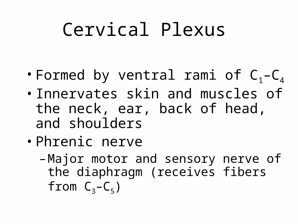
Cervical Plexus
• Formed by ventral rami of C1–C4
• Innervates skin and muscles of the neck, ear, back of head, and shoulders
• Phrenic nerve– Major motor and sensory nerve of the
diaphragm (receives fibers from C3–C5)


Brachial Plexus
• Formed by ventral rami of C5–C8 and T1 (and often C4 and T2)
• It gives rise to the nerves that innervate the upper limb
• Major branches of this plexus: – Roots—five ventral rami (C5–T1)– Trunks—upper, middle, and lower– Divisions—anterior and posterior – Cords—lateral, medial, and posterior

Figure 13.9 (a)
Upper
Middle Trunks
Lower
Roots (ventral rami):
Upper subscapular
Lower subscapular
Thoracodorsal
Medial cutaneousnerves of the armand forearm
Long thoracic
Medial pectoral
Lateral pectoral
Nerve tosubclaviusSuprascapular
Dorsal scapular
Posteriordivisions
Anteriordivisions
Lateral
PosteriorCords
Medial
Axillary
Musculo-cutaneousRadial
Median
Ulnar
Posteriordivisions
Trunks Roots
C4
C5
C6
C7
C8
T1
(a) Roots (rami C5 – T1), trunks, divisions, and cords

Brachial Plexus: Nerves• Axillary—innervates the deltoid, teres minor, and skin
and joint capsule of the shoulder• Musculocutaneous—innervates the biceps brachii and
brachialis and skin of lateral forearm• Median—innervates the skin, most flexors and
pronators in the forearm, and some intrinsic muscles of the hand
• Ulnar—supplies the flexor carpi ulnaris, part of the flexor digitorum profundus, most intrinsic muscles of the hand, and skin of medial aspect of hand
• Radial—innervates essentially all extensor muscles, supinators, and posterior skin of limb

Figure 13.9 (c)
Median nerve
Musculocutaneous nerve
Radial nerveHumerus
Ulna
Ulnar nerveMedian nerve
Radius
Radial nerve (superficial branch)
Superficial branch of ulnar nerveDorsal branch of ulnar nerve
Digital branch of ulnar nerveMuscular branchDigital branch
(c) The major nerves of the upper limb
Axillarynerve
Anteriordivisions
Posteriordivisions
Trunks Roots
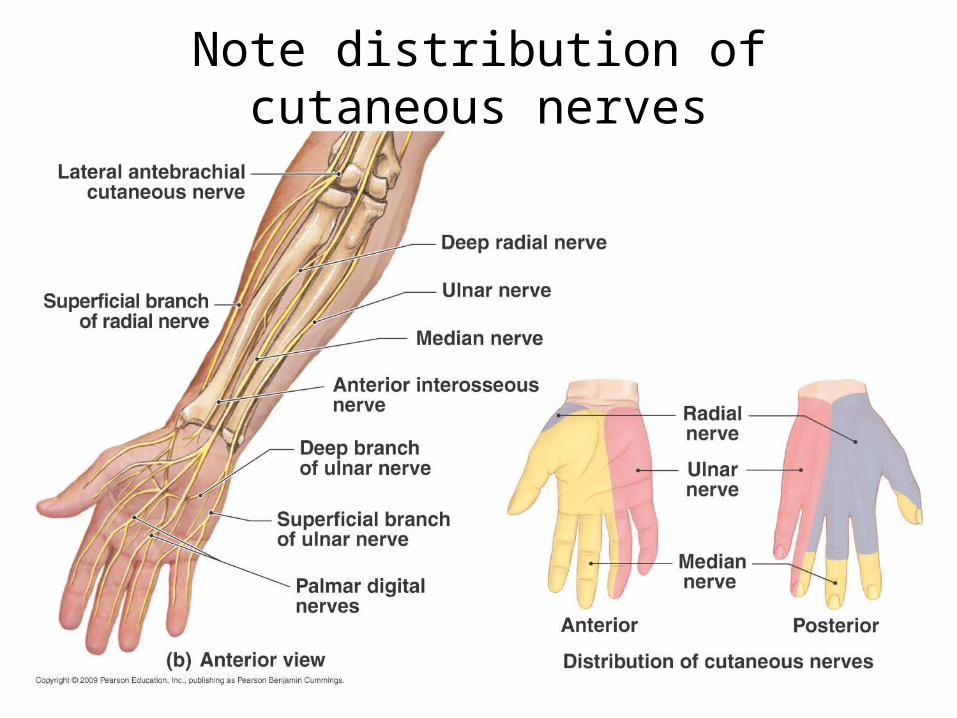
Note distribution of cutaneous nerves

Sensory innervation, palm
1. Ulnar nerve
2. Median nerve
3. Radial nerve

• T3-T12• 11 intercostal nerve• 1 subcostal nerve

Lumbar Plexus
• Arises from L1–L4
• Innervates the thigh, abdominal wall, and psoas muscle
• Femoral nerve—innervates quadriceps and skin of anterior thigh and medial surface of leg
• Obturator nerve—passes through obturator foramen to innervate adductor muscles


Sacral Plexus
• Arises from L4–S4
• Serves the buttock, lower limb, pelvic structures, and perineum
• Sciatic nerve– Longest and thickest nerve of the body– Innervates the hamstring muscles, adductor
magnus, and most muscles in the leg and foot– Composed of two nerves: tibial and common
fibular


Functional Divisions of the Peripheral Nervous System
Afferent Division– Sensory (advances) neuron –
goes toward CNSEfferent Division
– Motor (exits) neuron- leaves CNS– Somatic Nervous System– Autonomic Nervous System

REFLEXES
• Rapid, predictable response to a stimulus.
• Unlearned, involuntary, "hard-wired" into our neuroanatomy at the cellular & tissue level.
The simplest type of nerve circuit regulates a reflex (or autonomic response) and is called a reflex arc.

Methods of Classifying Reflexes

• Innate reflexes–Result from connections that form
between neurons during development
• Acquired reflexes–Learned, and typically more
complex
Reflex classifications
• Cranial reflexes–Reflexes processed in the brain
• Spinal reflexes–Interconnections and processing
events occur in the spinal cord
More reflex classifications

• Somatic reflexes–Control skeletal muscle
• Visceral reflexes (autonomic reflexes)–Control activities of other
systems
still more reflex classifications

• Monosynaptic reflex– Sensory neuron synapses directly on a
motor neuron
• Polysynaptic reflex– At least one interneuron between sensory
afferent and motor efferent
– Longer delay between stimulus and response
and more reflex classifications
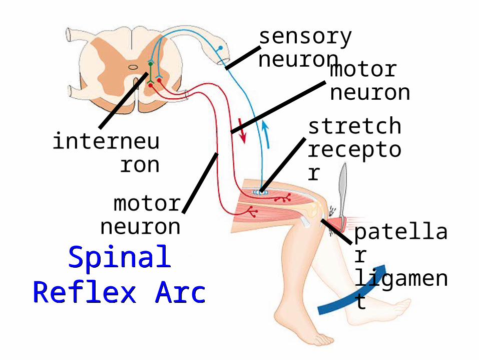
Spinal Reflex Arc
Spinal Reflex Arc
patellar ligament
stretch receptor
motor neuron
sensory neuron
motor neuron
interneuron

+ Excitatory synapse– Inhibitory synapse
Quadriceps strongly contracts. Golgi tendon organs are activated.
Interneurons
Spinal cord
Quadriceps(extensors)
Golgitendon
organHamstrings
(flexors)
1

+ Excitatory synapse– Inhibitory synapse
Quadriceps strongly contracts. Golgi tendon organs are activated.
Afferent fibers synapse with interneurons in the spinal cord.
Interneurons
Spinal cord
Quadriceps(extensors)
Golgitendon
organHamstrings
(flexors)
1 2

+ Excitatory synapse– Inhibitory synapse
Quadriceps strongly contracts. Golgi tendon organs are activated.
Afferent fibers synapse with interneurons in the spinal cord.
Interneurons
Spinal cord
Quadriceps(extensors)
Golgitendon
organHamstrings
(flexors)
1 2
3a The motor neurons (red) send activating impulses to the quadriceps causing it to contract, extending theknee.

+ Excitatory synapse– Inhibitory synapse
Quadriceps strongly contracts. Golgi tendon organs are activated.
Afferent fibers synapse with interneurons in the spinal cord.
Interneurons
Spinal cord
Quadriceps(extensors)
Golgitendon
organHamstrings
(flexors)
1 2
3a 3b
The interneurons (green) make inhibitory synapses with ventral horn neurons (purple) that prevent the antagonist muscles (hamstrings) from resisting the contraction of the quadriceps.
The motor neurons (red) send activating impulses to the quadriceps causing it to contract, extending theknee.
The motor neurons (red) send activating impulses to the quadriceps causing it to contract, extending theknee.

Reflex Arc

Baby Reflexes
• Palmar Grasp reflex• Suckling reflex• Rooting reflex• Babinski/plantar reflex

Pupillary Reflex

Mammalian Dive Reflex
An automated response system for diving in cold water (less than about 21C / 70F).
1. Bradycardia2. Vasoconstriction to extremities3. Apnea

INQUIRY
1. What is a reflex?2. Damage of the spinal cord
above C3 can result in_____?
3. In which portion of the spinal cord do the interneurons lie?
4. What kind of peripheral nerve fiber carries motor impulses outward to smooth muscles and glands of internal organs?