Embed Size (px)
Citation preview
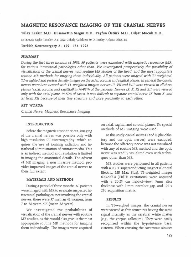
MAGNETIC RESONANCE IMAGING OF THE CRANIAL NERVES
Tiilay Keskin M.D .. Hiisamettin SarglD M.D .. Tayfun Oztiirk M.D .• Dil~at Mucuk M.D ..
BETEMAR Saghk Tesisleri A.~. Ziya Gbkalp Cadddesi 30 fA K.lZIlay Ankara-TURKiYE
Turkish Neurosurgery 2 : 129 - 134. 1992
SUMMARY
During the Brst three months of 1992, 80 patients were examined with magnetic resonance (MR)for various intracranial pathologies other than. We investigated prospectively the possibility ofvisualization of the cranial nerves with routine MR studies of the head. and the most appopriateroutine MR methods for imaging them individually. All patients were imaged with T1 weighted.T2-weighted and proton density images on the axiaL coronal and sagittal planes. In general the cranialnerves were best viewed with T1 -weighted images, nerves III, VIl and VIII were viewed in all threeplanes (axiaL coronal and sagitttal) in 76-98 % of the patients. Nerves IX, X, XI and XIl were viewedonly with the axial plane. in 80% of cases. It was difficult to separate cranial nerve IX from X, andXI from XII because of their tiny structura and dose proximity to each other.
KEY WORDS:
Cranial Nerve, Magnetic Resonance jmaging.
INTRODUCTION
Before the magnetic resonance era, imagingof the cranial nerves was possible only withhigh resolution CT-dsternography, which requires the use of ionizing radiation and intrathecal administration of contrast media. Thisis an indirect method and resolution is limited
in imaging the anatomical details. The adventof MR imaging, a non invasive method. provides improved images of the cranial nerves totheir full extent.
MATERIALS AND METHODS
During a period of three months, 80 patientswere imaged with MR to evaluate suspected intracranial pathologies. not involving the cranialnerves. there were 37 men an 43 women. from
7 to 78 years old (mean 38 years).
We investigated the probabilities ofvisualization of the cranial nerves with routine
MR studies. as this would also give us the mostappropriate routine MR methods for imagingthem individually. The images were acquired
on axial. sagittal and coronal planes. No spedalmethods of MR imaging were used.
In this study cranial nerves I and II (the olfactory and the optic nerves) were excluded,because the olfactory nerve was not visualizedwith any of routine MR method and the opticnerve was readily visualized even with techniques other than MR.
MR studies were performed in all patientswith a 0.5 T superconducting magnet (GeneralElectric. MR Max Plus). Tl-weighted images600/20/2-4 (TRITE exdtations) were acquiredwith a 20-25 an field-oE-view. 5mm slice
thickness with 2 mm interslice gap. and 192 x256 acquisition matrix.
RESULTS
In Tl-weighed images, the cranial nerveswere viewed as thin structures having the samesignal intensity as tha cerebral white matter(e.g., the corpus callosum). They were easilyrecognized within the hypointense basaldsterns. When crossing the cavernous sinuses
129
the cranial nerves ale better visualized because
of the signal void in the carotid arteries and thesignal intensity of the venous blood in the cavernous sinus.
The perineural fat and the venous plexus.make the cranial verves show high signal intensty when passing through their corresponding foraminae.
In proton density images. the highe signalintensity of the CSF almost completely hindersvisualization of cranial nerves.
In T2-weighted images the cranial nerves areseen as having lower signal intensities that thesurrounding hyperintens CSF, but delineationis not of the same quality as with theTl-weighted images.
CRANIAL NERVr; I ( UL MO 0NERVE)
The oculomotor nerve was visualized on the
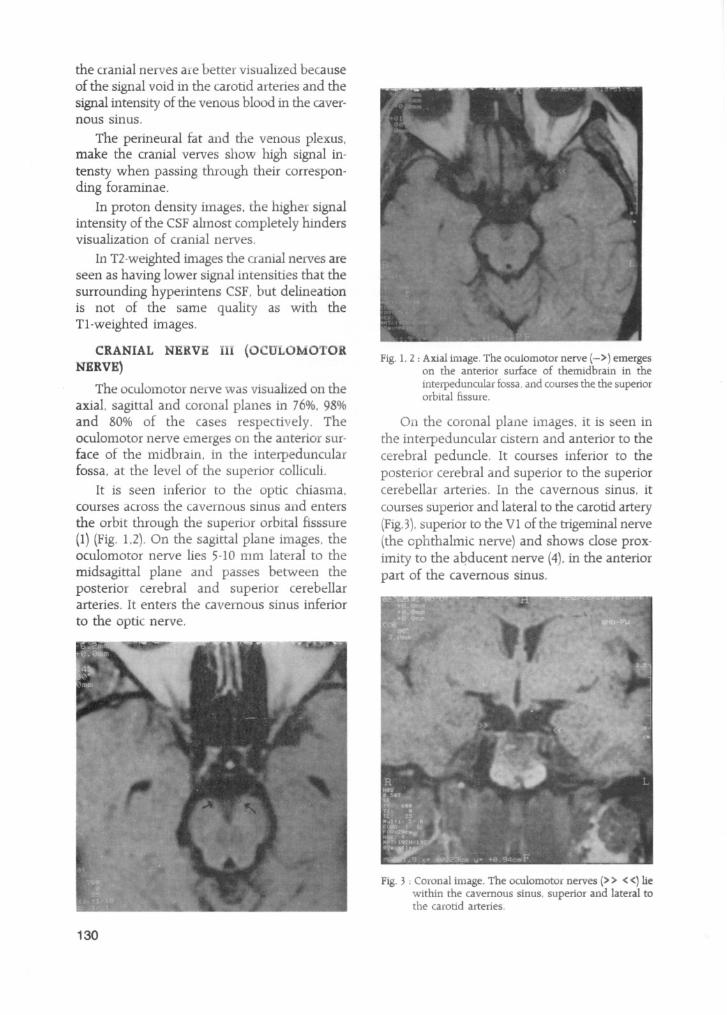
axial. sagittal and coronal planes in 76%, 98%and 80% of the cases respe ti ely. Theoculomotor nerve emerges on the allterio surface of the midbrain. in the interpeduncularfossa. at the level of the superior colliculi.
It is seen inferior to the optic chiasma.courses across the cavernous sinus and enters
the orbit through the superior orbital fisssure(1) (Fig. 1.2). On the sagittal plane images. theoculomotor nerve lies 5-10 rnm lateral to themidsagittal plane and passes between theposterior cerebral and superior cerebellararteries. It enters the cavernous sinus inferior
to the opti nerve.
130
Fig. 1, 2: Axial image. The oculomotor nerve (-» emergeson the anterior surface of themidbrain in theinterpeduncular fossa. and courses the the superiororbital fissure.
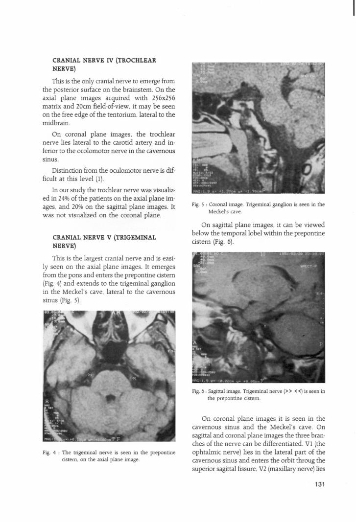
On the coronal plalle images, it is seen inthe interpeduncular cistern and anterior to thecerebral peduncle. It courses inferior to theposterior cerebral and superior to the superiorcerebellar arteries. In the cavernous sinus. it
courses superior and lateral to the carotid artety(FigJ), superior to the VI of the trigeminal nerve(the ophthalmic nerve) and shows close proximity to the aqducent nerve (4). in the anteriorpart of the cavernous sinus.
Fig. 3 : Coronal image. The oculomotor nerves (» «) liewithin the cavernous sinus. superior and lateral tothe carotid arteries.
CRANIAL NERVE IV (TROCHLEAR
NERVE)
This is the only cranial nerve to emerge fromthe posterior surface on the brainstem. On theaxial plane images acquired with 256x256matrix and 20cm field-of-view, it may be seenon the free edge of the tentorium, lateral to themidbrain.
On coronal plane images. the trochlearnerve lies lateral to the carotid artery and inferior to the ocolomotor nerve in the cavernoussinus.
Distinction from the oculomotor nerve is dif
ficult at this level (1).
In our study the trochlear nerve was visualized in 24%of the patients on the axial plane images, and 20% on the sagittal plane images. Itwas not visualized on the coronal plane.
CRANIAL NERVE V (TRIGEMINAL
NERVE)
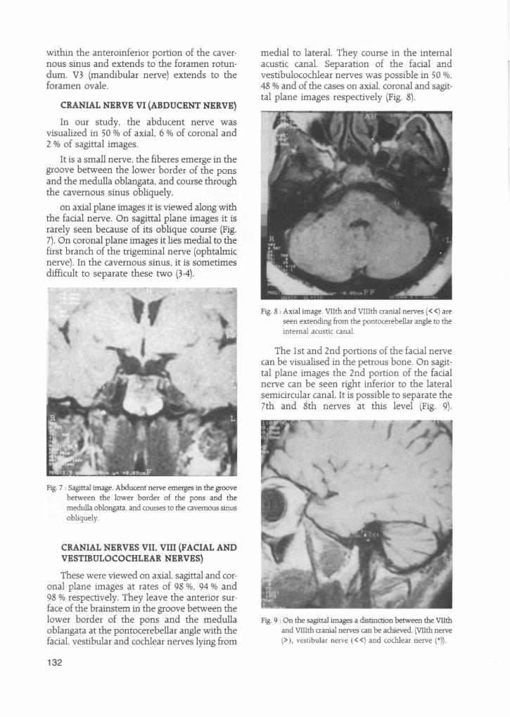
This is the largest cranial nerve and is easily seen on the axial plane images. It emergesfrom the pons and enters the prepontine astern(Fig. 4) and extends to the trigeminal ganglionin the Meckel's cave, lateral to the cavernous
sinus (Fig. 5).
Fig. 4 : The trigeminal nerve is seen in the prepontineastern, on the axial plane image.
Fig. 5 : Coronal image. Trigeminal ganglion is seen in theMeckel's cave.
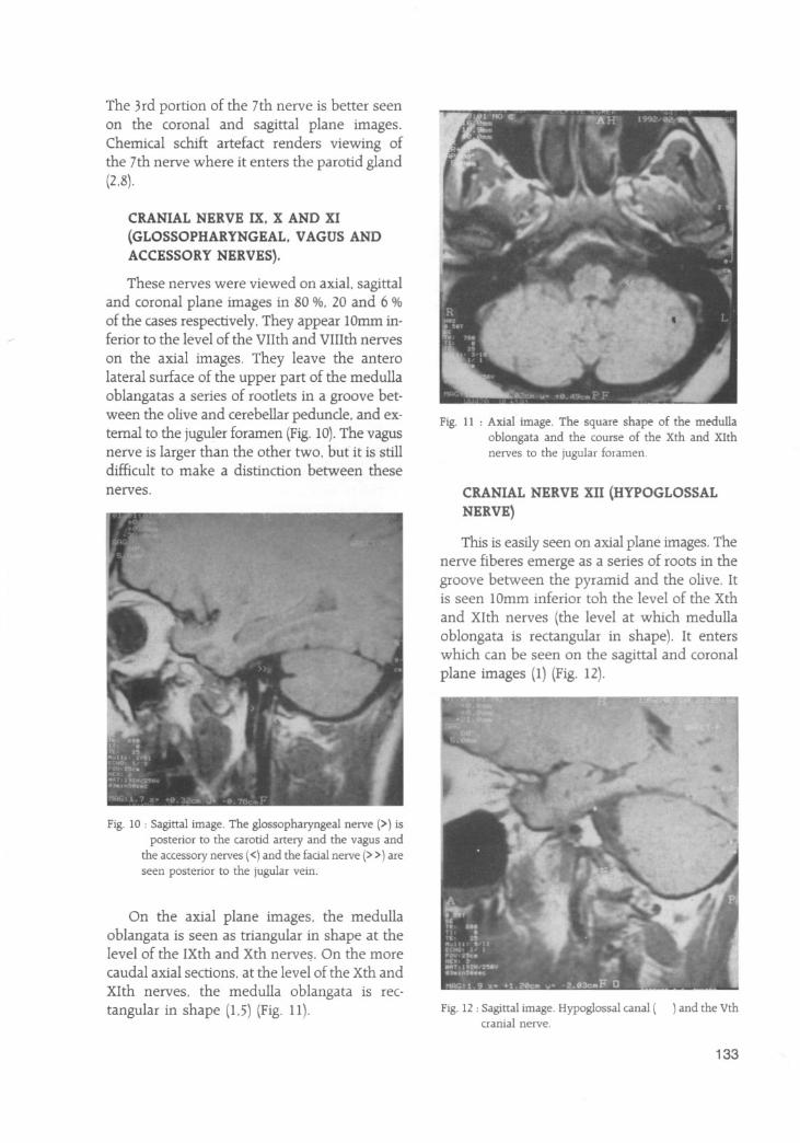
On sagittal plane images, it can be viewedbelow the temporallobel within the prepontineastern (Fig. 6).
Fig. 6 : Sagittal image. Trigeminal nerve (> > < <) is seen inthe prepontine astern.
On coronal plane images it is seen in thecavernous sinus and the Meckel's cave. On
sagittal and coronal plane images the three branches of the nerve can be differentiated. VI (theophtalmic nerve) lies in the lateral part of thecavernous sinus and enters the orbit throug thesuperior sagittal fissure. V2 (maxillary nerve) lies
131
within the anteroinferior portion of the cavernous sinus and extends to the foramen rotun
dum. V3 (mandibular nerve) extends to theforamen ovale.
CRANIAL NERVE VI (ABDUCENT NERVE)
In our study. the abducent nerve wasvisualized in 50 % of axial. 6 % of coronal and2 % of sagittal images.
It is a small nerve. the fiberes emerge in thegroove between the lower border of the ponsand the medulla oblangata. and course throughthe cavernous sinus obliquely.
on axial plane images it is viewed along withthe factal nerve. On sagittal plane images it israrely seen because of its oblique course (Fig.7). On coronal plane images it lies medial to thefirst branch of the trigeminal nerve (ophtalmicnerve). In the cavernous sinus. it is sometimesdifficult to separate these two (3-4).
Fig. 7 : Sagittal image. Abducent nerve emerges in the groovebetween the lower border of the pons and themedulla oblongata. and courses to the cavernous sinusobliquely.
CRANIAL NERVES VII, VIII (FACIAL ANDVESTIBULOCOCHLEAR NERVES)
These were viewed on axial. sagittal and coronal plane images at rates of 98 %. 94 % and98 % respectively. They leave the anterior surface of the brainstem in the groove between thelower border of the pons and the medullaoblangata at the pontocerebellar angle with thefactal. vestibular and cochlear nerves lying from
132
medial to lateral. They course in the internalacustic canal. Separation of the factal andvestibulocochlear nerves was possible in 50 %.
48 % and of the cases on axial. coronal and sagittal plane images respectively (Fig. 8).
Fig. 8 : Axial image. VIlth and VIIIth cranial nerves « <) areseen extending from the pontocerebellar angle to theinternal acustic canal.
The 1st and 2nd portions of the factal nervecan be visualised in the petro us bone. On sagittal plane images the 2nd portion of the factalnerve can be seen right inferior to the lateralsemictrcular canal. It is possible to separate the7th and 8th nerves at this level (Fig. 9).
Fig. 9 : On the sagittal images a distinction between the VIIthand VIIIth cranial nerves can be achieved. (VIIth nerve(». vestibular nerve « <) and cochlear nerve (*)).
The 3rd portion of the 7th nerve is better seenon the coronal and sagittal plane images.Chemical schift artefact renders viewing ofthe 7th nerve where it enters the parotid gland(2,8).
CRANIAL NERVE IX, X AND XI
(GLOSSOPHARYNGEAL, VAGUS AND
ACCESSORY NERVES).
These nerves were viewed on axial. sagittaland coronal plane images in 80 %. 20 and 6 %
of the cases respectively. They appear lOmm inferior to the level of the VIIth and VIIIth nerves
on the axial images. They leave the anterolateral surface of the upper part of the medullaoblangatas a series of rootlets in a groove between the olive and cerebellar peduncle. and external to the juguler foramen (Fig. 10). The vagusnerve is larger than the other two. but it is stilldifficult to make a distinction between thesenerves.
Fig. 10 : Sagittal image. The glossopharyngeal nerve (» isposterior to the carotid artery and the vagus and
the accessory nerves (<) and the fadal nerve (> >) areseen posterior to the jugular vein.
On the axial plane images, the medullaoblangata is seen as triangular in shape at thelevel of the IXth and xth nerve~. On the morecaudal axial sections, at the level of the Xth and
XIth nerves, the medulla oblangata is rectangular in shape (1.5) (Fig. 11).
Fig. 11 : Axial image. The square shape of the medullaoblongata and the course of the Xth and Xlthnerves to the jugular foramen.
CRANIAL NERVE XII (HYPOGLOSSAL
NERVE)
This is easily seen on axial plane images. Thenerve fiberes emerge as a series of roots in thegroove between the pyramid and the olive. Itis seen lOmm inferior toh the level of the Xth
and XIth nerves (the level at which medullaoblongata is rectangular in shape). It enterswhich can be seen on the sagittal and coronalplane images (1) (Fig. 12).
Fig. 12 : Sagittal image. Hypoglossal canal ( ) and the Vthcranial nerve.
133
In our study the XIlth nerve was visualizedon axial. sagittal and coronal plane images ata rate of 80 %. 20 % and 10 % respectively.
DISCUSSION
In the presence of cerebral atrophy the asterns are larger and the cranial nerves are better visualized (1).Tl-weighted axial images areuseful in evaluation of the cranial nerves but co
ronal images are required to visualize cranialnerves III. V and VI. The oblique course of thecranial nerves makes the orbitomeatal or neu
roocular planes more useful. TERESI et al. claim that planes 35 degrees to the orbitomeatalplane give better views (8).20 em field-of-view.256x256 matrix with 2-4 exatations are useful.
3 DFT applications do not give good results because of high signal-to-noise rations.
In the presence of asymmetry. images in atleast two planes and contrast infusion are required to evaluate the cranial nerves. MR is anon invasive technique and gives high resolution images in the evaluation of the cranial nerves and their pathologies.
134
Correspondence: Dr. Tiilay KESKiNBetemar Saghk Tesisleri A.~.ziya Gbkalp Caddesi 30-AK.lZllay/ANKARATel: (4)433 82 30 (6 line) Fax: 431 71 36
REFERENCES
1. Cailiet H. Delvalle A. Doyon D. et al: Les Nerfs CraniensNormaux IRM. Journal de Radiologie 72: 69-78. 1991
2. Daniels DL. Herfkins R. Koeller R. et al: Magnetic Resonance Imaging of the Internal Auditory Canal. Radiology151: 105-108. 1984
3. Daniels DL. Mark L. Pojunas K.et al: Magnetic Resonance Imaging of the Cavernous Sinus. AJR 144: 1009-1014.1985.
4. Daniels DL. Pech P. Pojunas K. et al: Trigeminal Nerve:Anatomic Correlation with MR imaging. Radiology 159:577-583. 1986.
5. Jacob CJ. Harnsberg HR. Lufkin RB. et al: Vagal Neuropathy: Evaluation CT and MR imaging. Radiology 164:97-102. 1987
6. Laine FJ. Braum IF. Jemsen ME. et al: Perineural Ekstension through the Foramen Ovale: Evaluation with MRimaging. Radiology 174: 66·71. 1990
7. Tash RR.Sze G. Leslie DR: Trigeminal Neuralgie: MR Imaging Features Radiology 172: 767-770. 1989
8. Teresi LM. Kolin E. Lufkin RB. et al: MR Imaging Intraparotid Factal Nerve: Normal Anothomy and PathologyAJNR 8: 253-258. 1987