Embed Size (px)
Citation preview
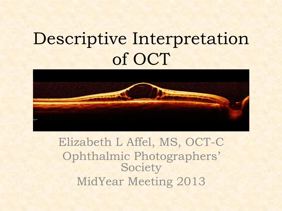
Descriptive Interpretation
of OCT
Elizabeth L Affel, MS, OCT-C
Ophthalmic Photographers’ Society
MidYear Meeting 2013
Describe
• de·scribe/diˈskrīb/
• Verb:Give an account in words of (someone or something), including all the relevant characteristics, qualities, or events.
• Indicate; denote.
• Synonyms:depict - delineate - portray - picture – represent
• wikipedia
Interpret
• in·ter·pre·ta·tion
• noun \in-ˌtər-prə-ˌtā-shən, -pə-\ (Medical Dictionary)
• Medical Definition of INTERPRETATION
• : the act or result of giving an explanation of something <interpretation of the symptoms of disease>; especially : an explanation in understandable terms to a patient in psychotherapy of the deeper meaning according to psychological theory of the material related and the behavior exhibited by the patient during treatment
• —in·ter·pret transitive verb
• —in·ter·pre·tive or in·ter·pre·ta·tive adjective
• Merriam Webster
Interpret
• Verb
• interpret (third-person singular simple present interprets, present participle interpreting, simple past and past participle interpreted)
• To explain or tell the meaning of; to expound; to translate orally into intelligible or familiar language or terms; to decipher; to define; -- applied especially to language, but also to dreams, signs, conduct, mysteries, etc.; as, to interpret the Hebrew language to an Englishman; to interpret an Indian speech.
• wikipedia
Descartian Analysis: *(the physician’s role)
• The science of the intellect is universal, and there can only be one true method, which consists of separating what is already simple and clear in order then to attempt to understand that which is complex and obscure. The method is a collection of reliable, easy rules, observing which there is no risk of mistaking the false for the true. In a logical process, the analyses of each of the possible elements is first performed: Then after this phase, the synthesis of all these elements is performed, and the results of these flow into the conclusions. To replace the apparent chaos of data with an ordered and rationally constructed system.
Role of Ophthalmic Imager • Provide the best quality images
• Facilitate good patient compliance to
ease the process and assure
repeatability
• Understand (interpret) what is being
shown in order to make adjustments to
improve the quality of the information
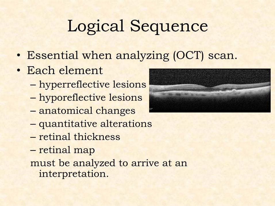
Logical Sequence
• Essential when analyzing (OCT) scan.
• Each element
– hyperreflective lesions
– hyporeflective lesions
– anatomical changes
– quantitative alterations
– retinal thickness
– retinal map
must be analyzed to arrive at an interpretation.
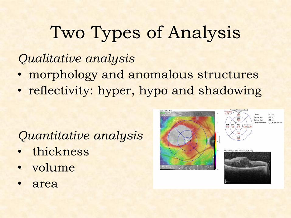
Two Types of Analysis
Qualitative analysis
• morphology and anomalous structures
• reflectivity: hyper, hypo and shadowing
Quantitative analysis
• thickness
• volume
• area
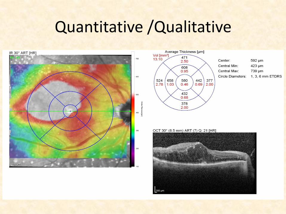
Quantitative /Qualitative
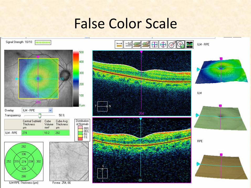
False Color Scale
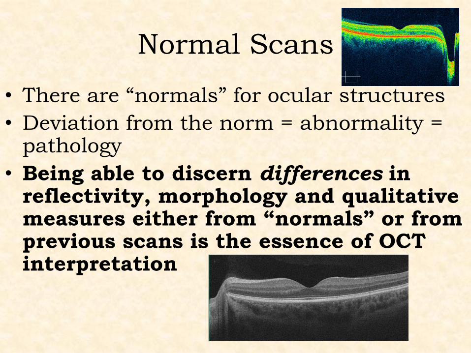
Normal Scans
• There are “normals” for ocular structures
• Deviation from the norm = abnormality = pathology
• Being able to discern differences in reflectivity, morphology and qualitative measures either from “normals” or fromprevious scans is the essence of OCT interpretation
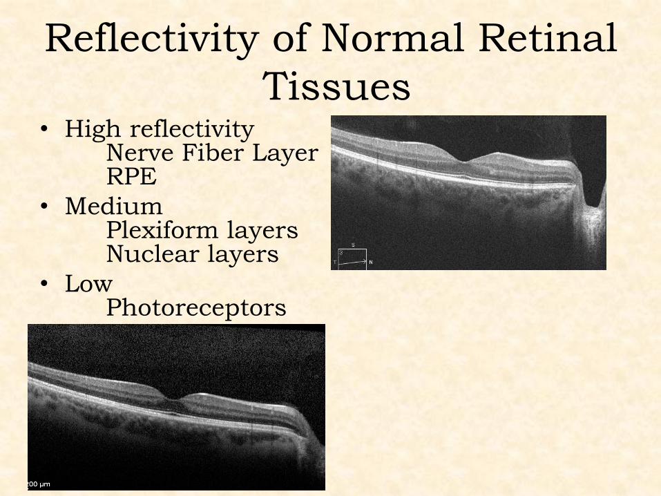
Reflectivity of Normal Retinal
Tissues • High reflectivity
Nerve Fiber Layer RPE
• Medium Plexiform layers Nuclear layers
• Low Photoreceptors
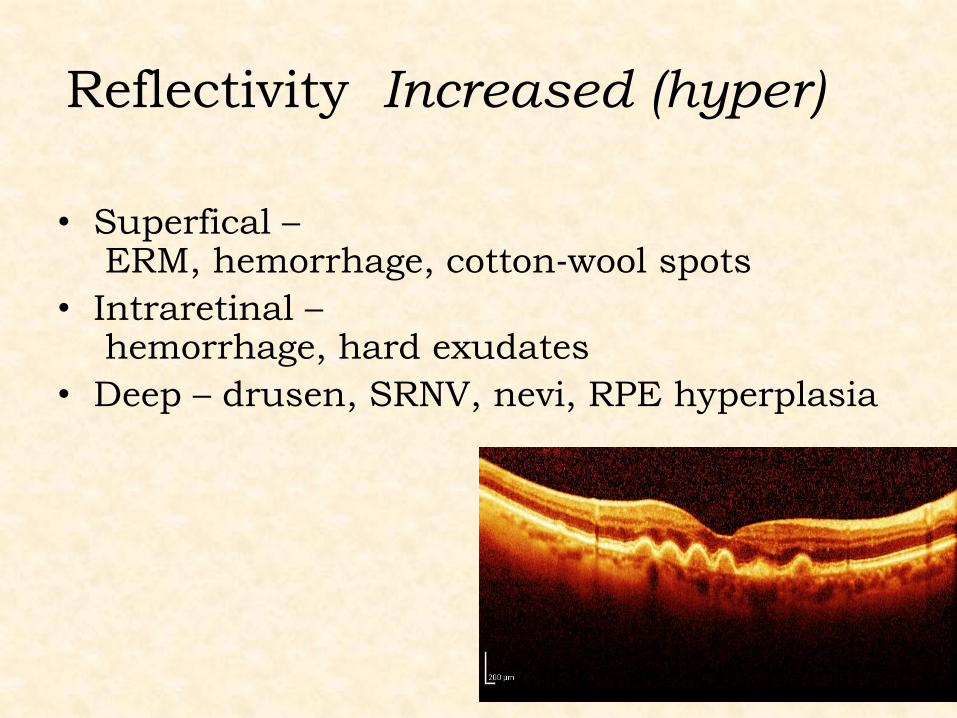
Reflectivity Increased (hyper)
• Superfical – ERM, hemorrhage, cotton‐wool spots
• Intraretinal – hemorrhage, hard exudates
• Deep – drusen, SRNV, nevi, RPE hyperplasia
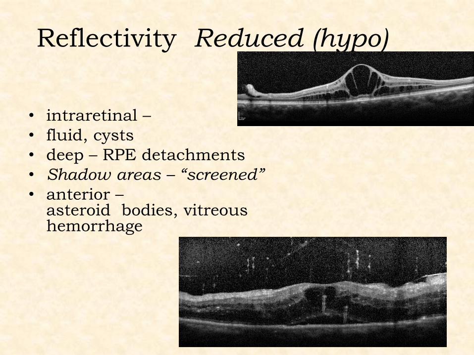
Reflectivity Reduced (hypo)
• intraretinal –
• fluid, cysts
• deep – RPE detachments
• Shadow areas – “screened”
• anterior –asteroid bodies, vitreous hemorrhage
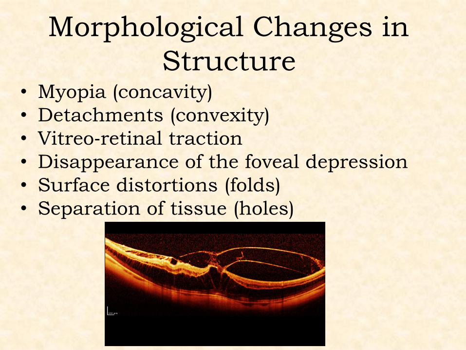
Morphological Changes in
Structure
• Myopia (concavity)
• Detachments (convexity)
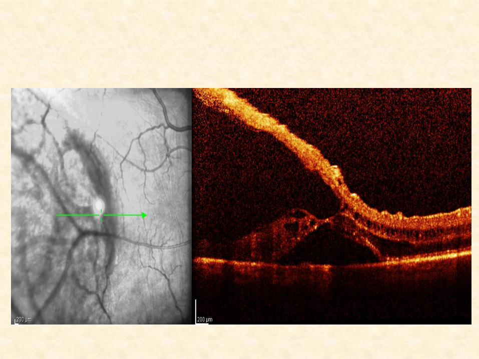
• Vitreo‐retinal traction
• Disappearance of the foveal depression
• Surface distortions (folds)
• Separation of tissue (holes)
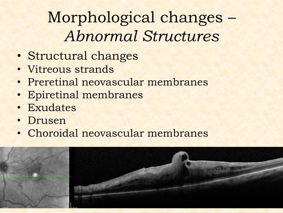
Morphological changes –
Abnormal Structures • Structural changes
• Vitreous strands
• Preretinal neovascular membranes
• Epiretinal membranes
• Exudates
• Drusen
• Choroidal neovascular membranes
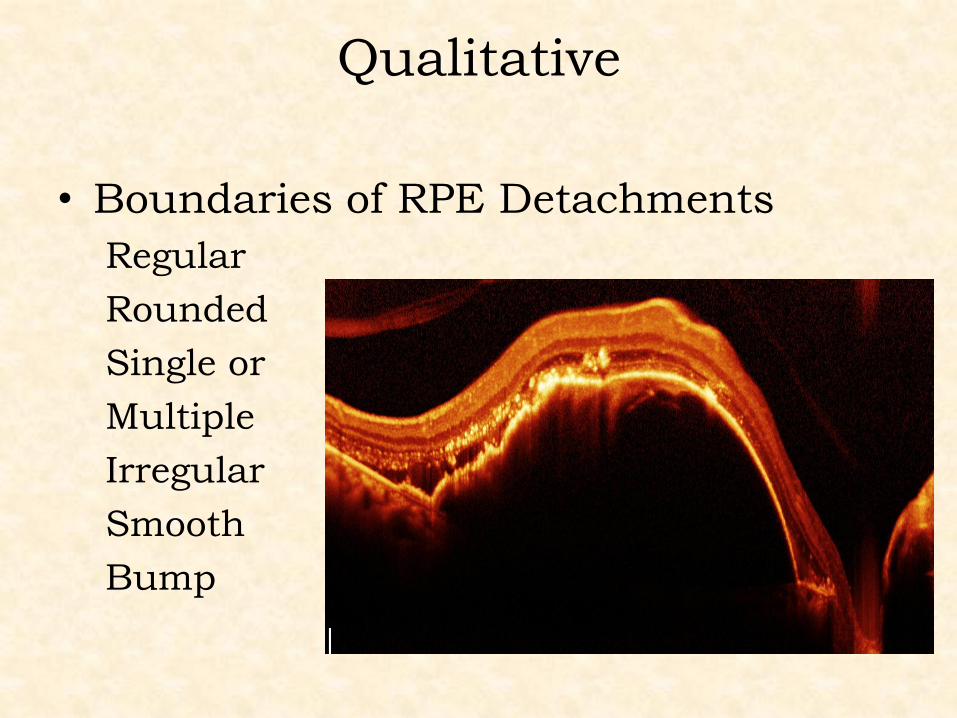
Qualitative
• Boundaries of RPE Detachments
Regular
Rounded
Single or
Multiple
Irregular
Smooth
Bump
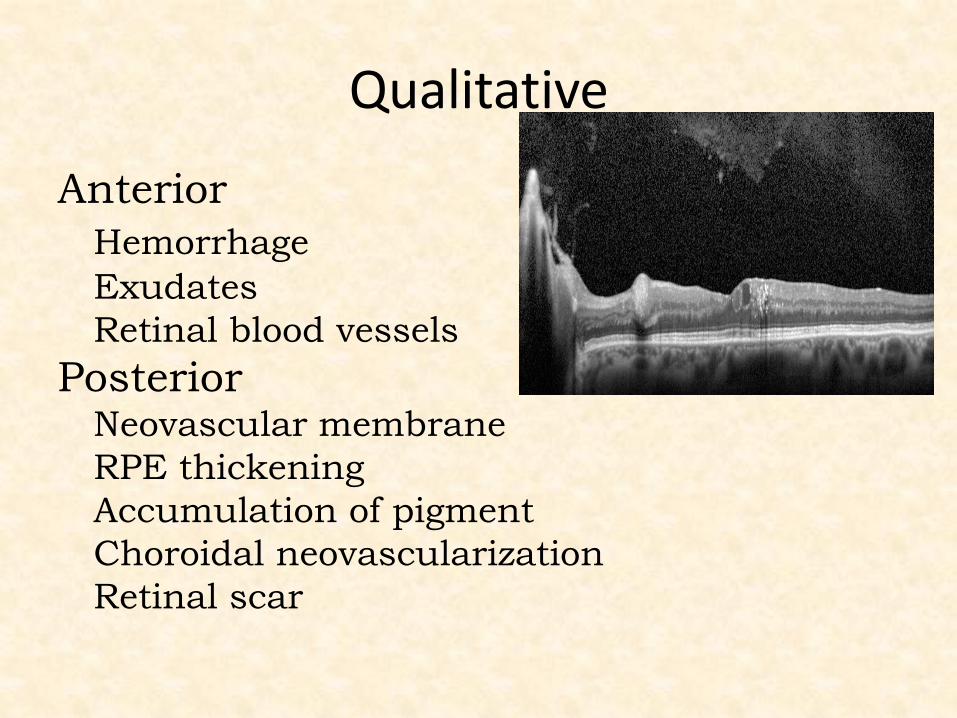
Qualitative
Anterior
Hemorrhage
Exudates
Retinal blood vessels
Posterior Neovascular membrane
RPE thickening
Accumulation of pigment
Choroidal neovascularization
Retinal scar
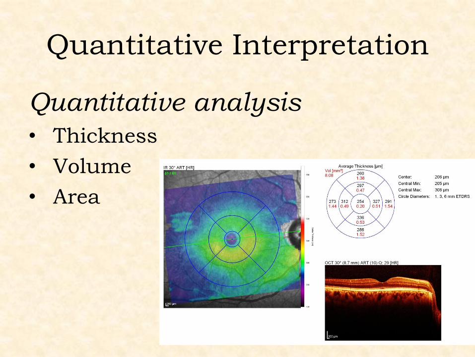
Quantitative Interpretation
Quantitative analysis
• Thickness
• Volume
• Area
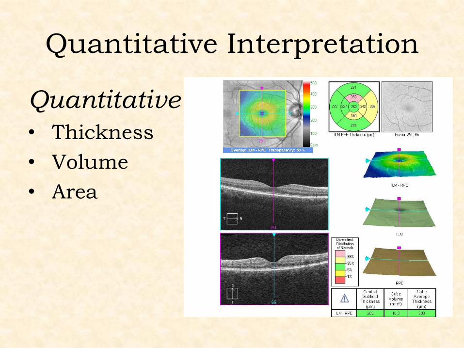
Quantitative Interpretation
Quantitative analysis
• Thickness
• Volume
• Area
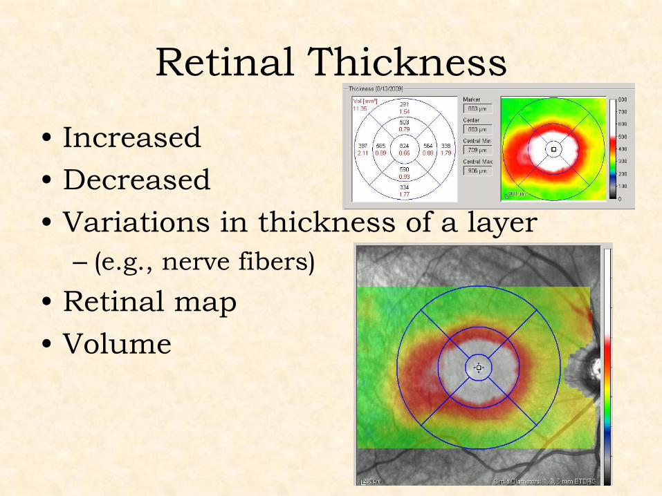
Retinal Thickness
• Increased
• Decreased
• Variations in thickness of a layer
– (e.g., nerve fibers)
• Retinal map
• Volume
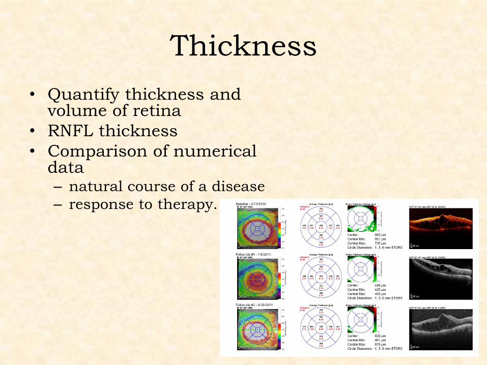
Thickness
• Quantify thickness and volume of retina
• RNFL thickness
• Comparison of numerical data – natural course of a disease
– response to therapy.
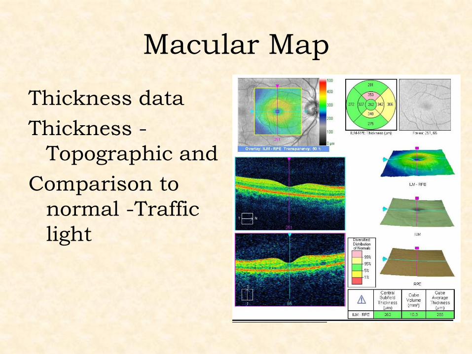
Macular Map
Thickness data
Thickness -
Topographic and
Comparison to
normal -Traffic
light
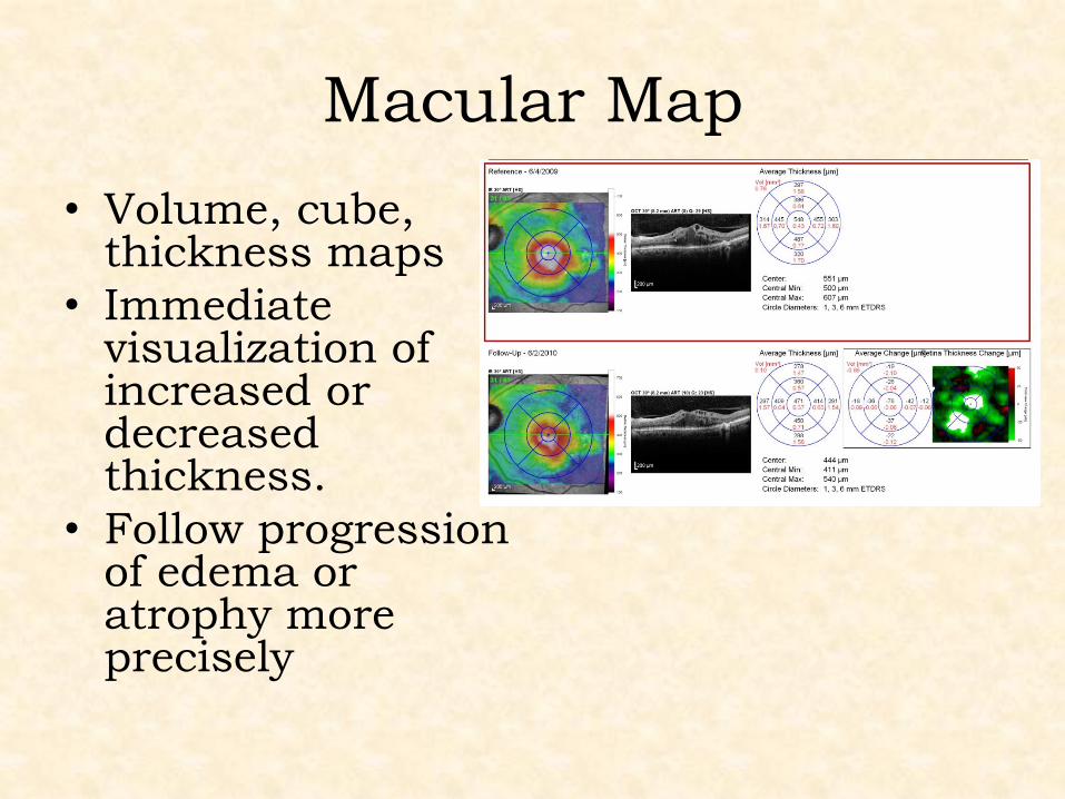
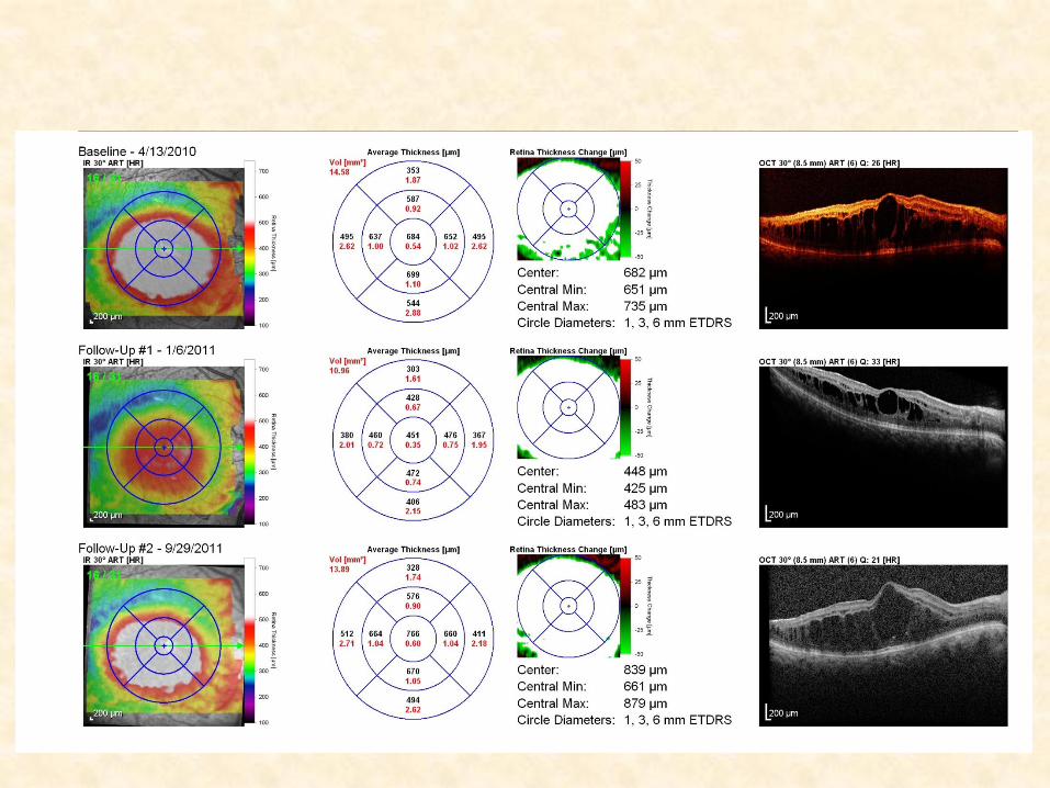
Macular Map
• Volume, cube, thickness maps
• Immediate visualization of increased or decreased thickness.
• Follow progression of edema or atrophy more precisely
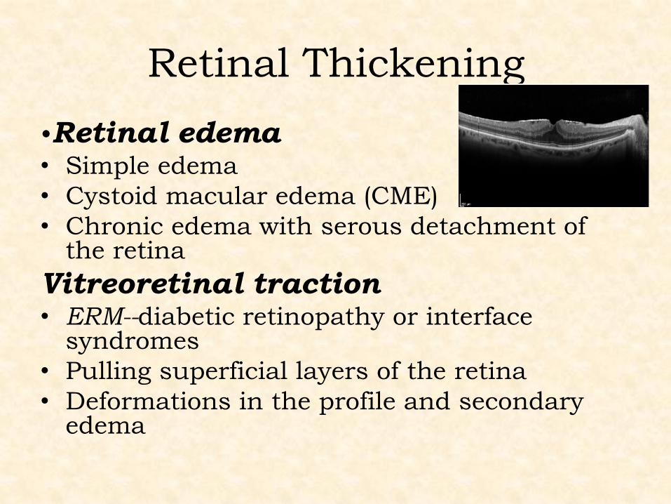
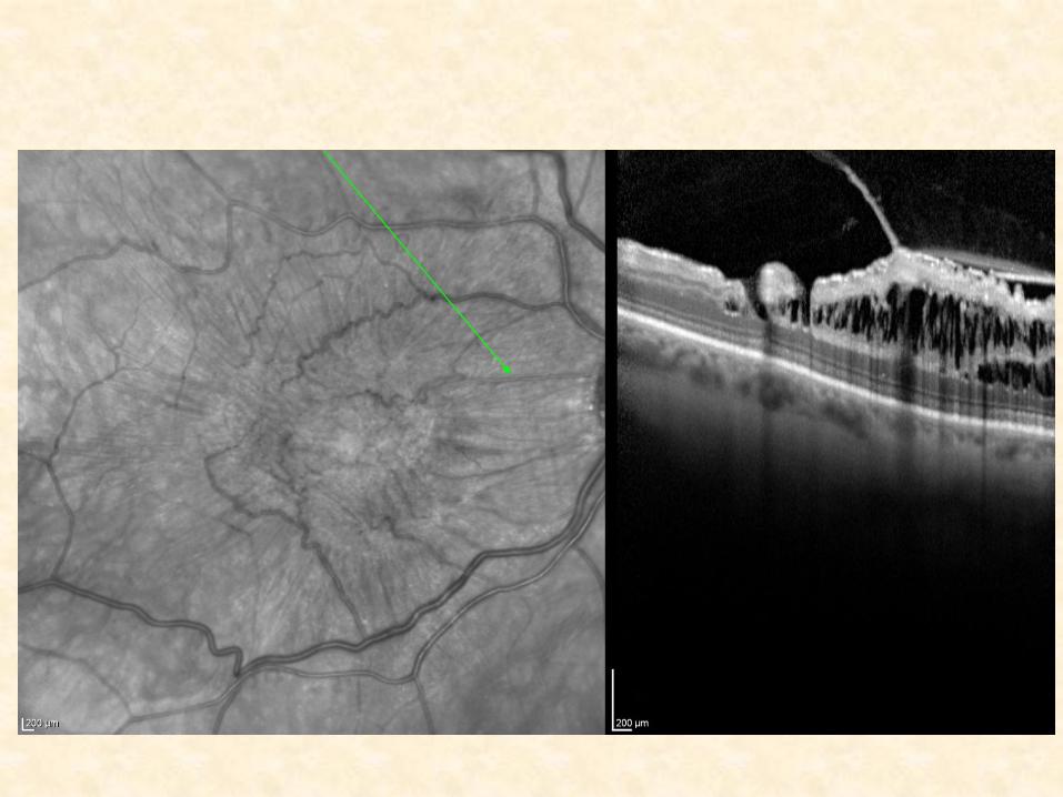
Retinal Thickening
•Retinal edema • Simple edema
• Cystoid macular edema (CME)
• Chronic edema with serous detachment of the retina
Vitreoretinal traction • ERM--diabetic retinopathy or interface
syndromes
• Pulling superficial layers of the retina
• Deformations in the profile and secondary edema
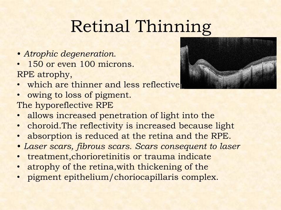
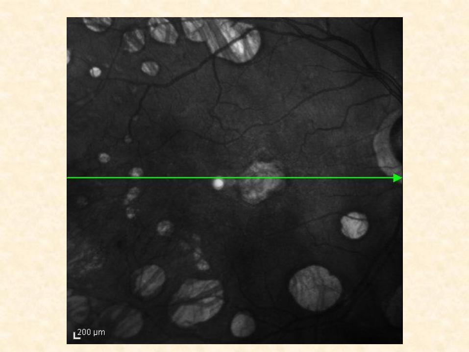
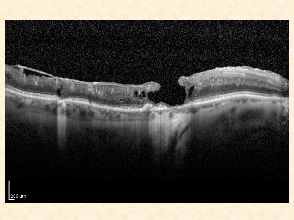
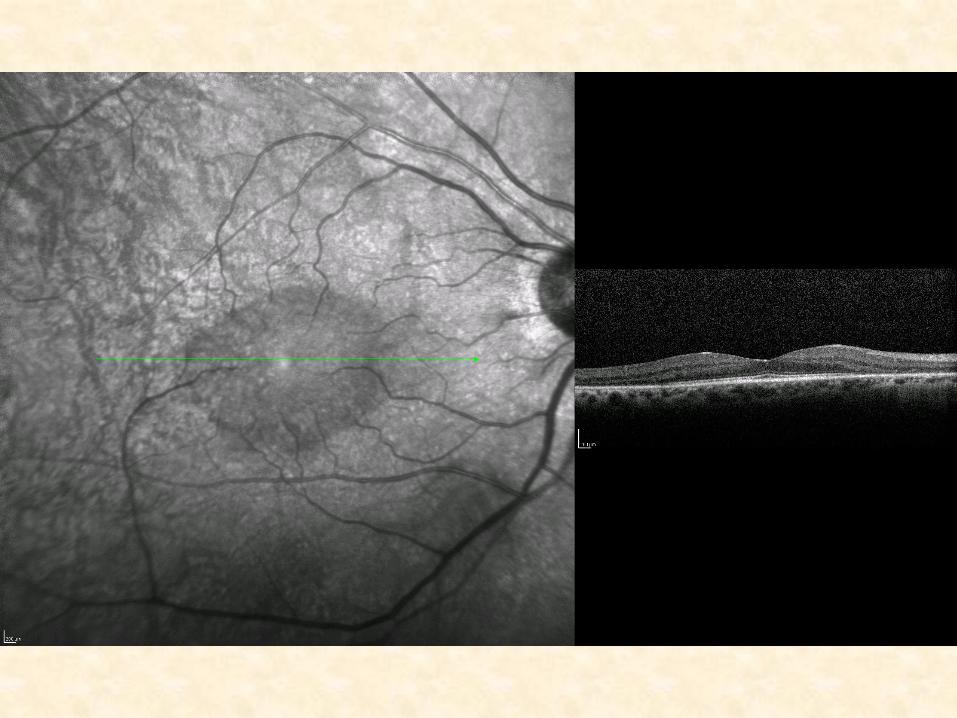
Retinal Thinning
• Atrophic degeneration.
• 150 or even 100 microns.
RPE atrophy,
• which are thinner and less reflective
• owing to loss of pigment.
The hyporeflective RPE
• allows increased penetration of light into the
• choroid.The reflectivity is increased because light
• absorption is reduced at the retina and the RPE.
• Laser scars, fibrous scars. Scars consequent to laser
• treatment,chorioretinitis or trauma indicate
• atrophy of the retina,with thickening of the
• pigment epithelium/choriocapillaris complex.
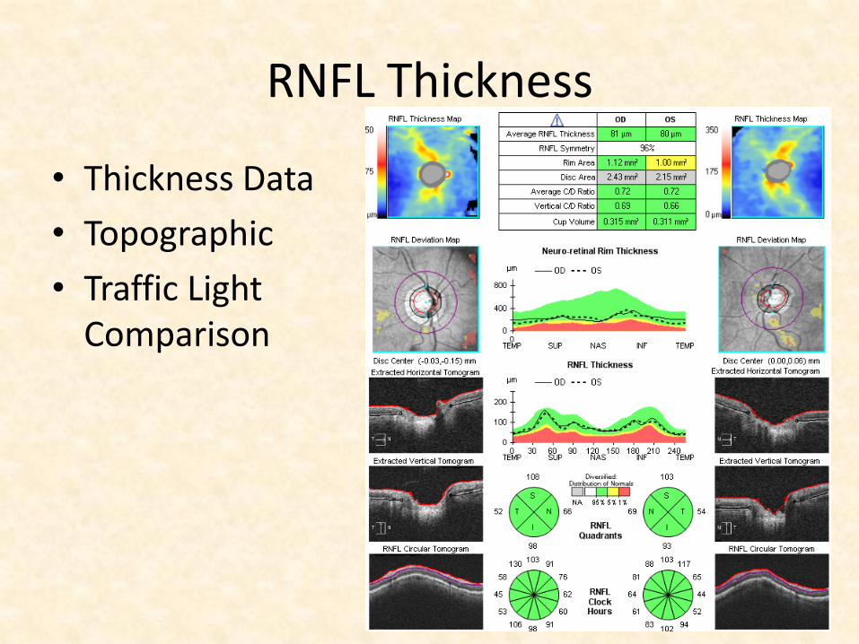
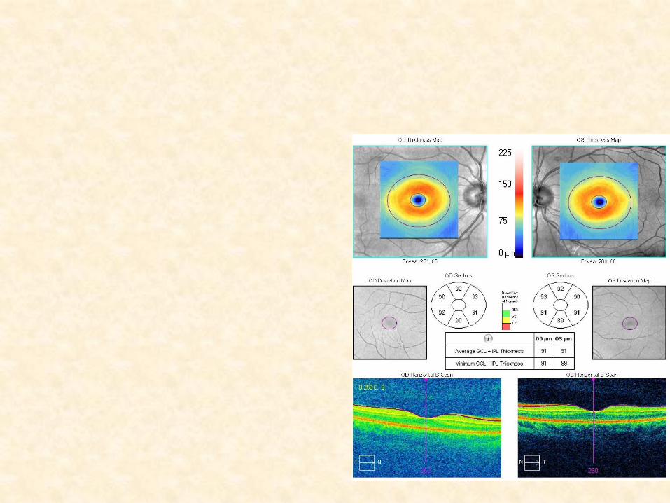
RNFL Thickness
• Thickness Data
• Topographic
• Traffic Light Comparison
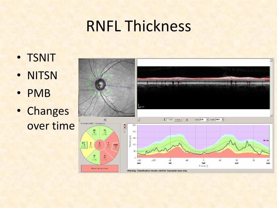
RNFL Thickness
• TSNIT
• NITSN
• PMB
• Changes over time
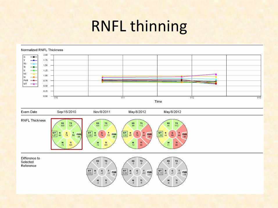
RNFL thinning
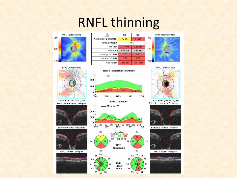
RNFL thinning
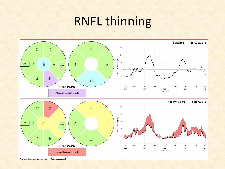
RNFL thinning
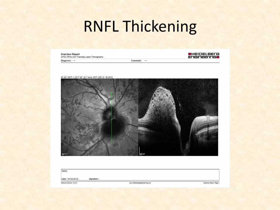
RNFL Thickening
RNFL Thickening
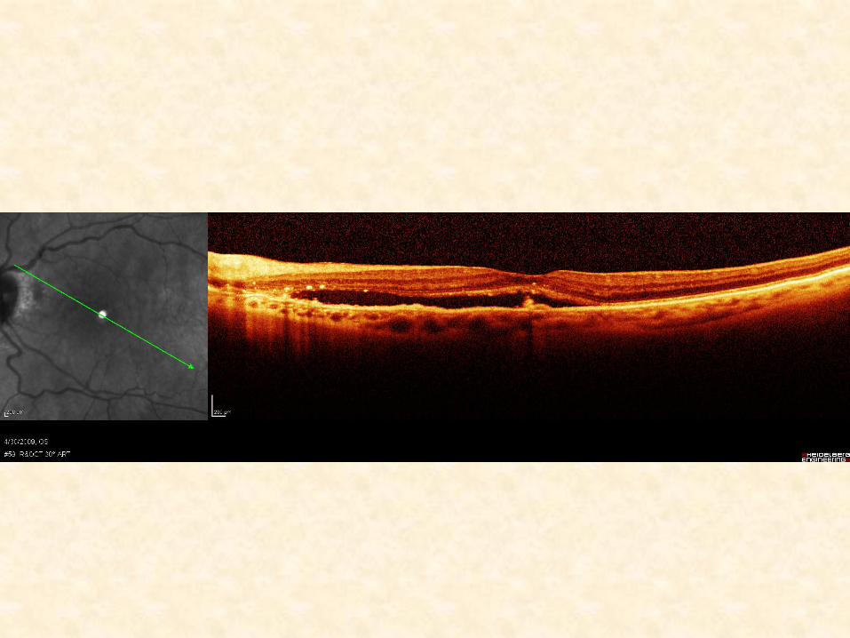
“Descriptive Interpretation”
• Using common terms to describe the
morphology, reflectivity, thickness,
volume and area of tissue being
scanned with OCT
• Excellent reference to help you
• Guide to Optical Coherence Tomogr
aphy Interpretation, Brancato R, Lu
mbroso B et al; I.N.C. Innovation‐ News‐Communication, Roma, IT 2005