Embed Size (px)
DESCRIPTION
This is a guide for chinese vets for neutering dogs and cats
Citation preview
DESEXING OF THE DOG AND CAT FOR CHINESE VETERINARY
PRACTITIONERS
DESEXING OF THE DOG AND CAT FOR CHINESE VETERINARY PRACTITIONERS 2
CONTENTS
Admission and initial examination ……………………………………………...3 Restraint & Handling……………………………………………………………..4 Pre surgery hospitalisation of the patient………………………………………7 Pre surgical preparation of the surgical environment…………………………8 Anaesthesia & Analgesia………………………………………………………..9 Recommended protocol for General Anaesthesia……………………………13 Anaesthetic Monitoring………………………………………………………….14 Surgical preparation……………………………………………………………..22 Preparation of surgical team……………………………………………………24 Surgical procedures……………………………………………………………..26 Ear Marking………………………………………………………………………48 Post surgical monitoring and care……………………………………………..51 APPENDICES Appendix 1 Cleaning procedures for the vet hospital…………………………53 Appendix 2 Infectious diseases…………………………………………………57 Rabies management....……………………………………………..60 Appendix 3 Vaccination Protocols………………………………………………63 Appendix 4 Surgical kit preparation and sterilisation………………………….66 Appendix 5 Anaesthetic and analgesic drugs…………………………………73 Appendix 6 Cardio-pulmonary Cerebral Resuscitation……………………….80 Appendix 7 Antibiotic and Suture Material Selection………………………….86

DESEXING OF THE DOG AND CAT FOR CHINESE VETERINARY PRACTITIONERS 3
ADMISSION AND INITIAL EXAMINATION. All new patients must be given a visual examination and where possible a full physical examination on arrival at the clinic. Contact with other animals should be minimised and care should be taken to avoid scratches and bites, which can spread disease. Feral cats or very aggressive dogs should be examined through the cage as removing them from the cage may stress them and cause them to injure themselves or veterinary staff. On visual examination you should be able to prioritise patients for surgery or quarantine for infectious diseases. Signs of a stressed, scared or aggressive dog may include tail between the legs, ears back, cowering in the corner, growling and/or showing their teeth (see Fig 1). Extra care should be taken when any of these signs are present and muzzling the dog is a good idea (see Fig 4&5) Prioritising patients for surgery should be done by ordering from most important to least important conditions and stress levels. If any wounds or injuries are found on visual examination then these patients should be either prioritised for surgery or treated with medication before having surgery. If any incurable or long term ongoing problems are found then euthanasia may be the kindest option. If any patient is showing signs of being stressed, he/she should also be prioritised. Older patients should always take priority over younger healthy patients. On visual examination any patients showing signs of an infectious disease (see appendix 2) e.g. sneezing, coughing, nasal or ocular discharge, dyspnoea, diarrhoea or vomiting, hypersalivation or neurological signs should be isolated from other patients and treated for these conditions before surgery.
Fig1: A: A normal, relaxed alert dog; B: A Dog with his ears back showing signs of anxiety; C: An anxious dog with ocular discharge
A
B A C
DESEXING OF THE DOG AND CAT FOR CHINESE VETERINARY PRACTITIONERS 4
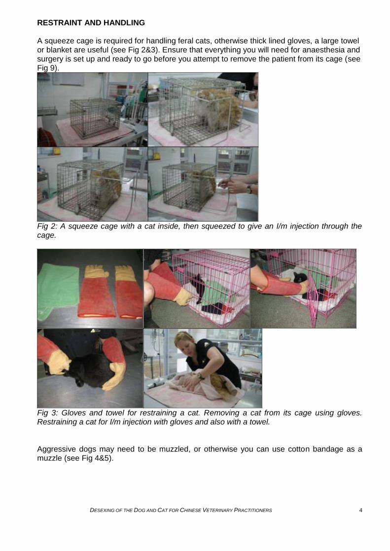
RESTRAINT AND HANDLING A squeeze cage is required for handling feral cats, otherwise thick lined gloves, a large towel or blanket are useful (see Fig 2&3). Ensure that everything you will need for anaesthesia and surgery is set up and ready to go before you attempt to remove the patient from its cage (see Fig 9).
Fig 2: A squeeze cage with a cat inside, then squeezed to give an I/m injection through the cage.
Fig 3: Gloves and towel for restraining a cat. Removing a cat from its cage using gloves. Restraining a cat for I/m injection with gloves and also with a towel. Aggressive dogs may need to be muzzled, or otherwise you can use cotton bandage as a muzzle (see Fig 4&5).

DESEXING OF THE DOG AND CAT FOR CHINESE VETERINARY PRACTITIONERS 5
Fig 4: A dog being restrained using a muzzle, also a towel and if able just with a head hold.
Fig 5: A dog being restrained using a bandage muzzle. Some patients will be more scared than aggressive so give them some time and be very quiet and speak softly when approaching. Each patient will need a different amount of restraint so assess each patient on how they behave in their cage prior to surgery. If they are trying to hide or cowering at the back of the cage looking scared (ears back or down is usually a sign of fear), then they are probably not going to react well to you putting your hands in the cage to remove them so this is when gloves, towel or squeeze cage for cats is probably needed. If the patient is at the front of the cage wanting attention, then slowly try to interact with the patient by gently stroking him/her – do not pat down on the patient‟s head as this can be misinterpreted as violence and the animal may react aggressively. Always have gloves or a towel nearby in case you need them and ideally have an assistant to help you restrain the animal. All animals should be weighed on arrival at the clinic to ensure accurate drug dosing. Aggressive or nervous cats can be weighed in their cage but remember to weigh the cage first so you can subtract this from the total weight (see Fig 6).

DESEXING OF THE DOG AND CAT FOR CHINESE VETERINARY PRACTITIONERS 6
Fig 6: Weighing the squeeze cage first, then with the cat inside and subtract the weight of the cage.

DESEXING OF THE DOG AND CAT FOR CHINESE VETERINARY PRACTITIONERS 7
PRE SURGERY HOSPITALISATION AND HOUSING OF THE PATIENT After the initial physical examination all patients should be placed into a cage with a clean towel or blanket. No animals should be left tied to tables. Ideally dogs and cats should be housed in separate areas as the presence of dogs may cause unnecessary stress to cats. Feral cats scheduled for immediate surgery should be kept in the trap cage until ready for surgery. When scheduling animals for surgery, consider their previous access to food – any animals trapped in the morning should be scheduled for surgery 8+ hours later in case they have recently eaten. Most trap cages will have a slide opening from one end. It is best to put this end into the new holding cage before opening the trap cage. This allows the animal to walk into its new cage and the door can be closed straight away, preventing feral cats from escaping (see Fig 7). All patients should be given a bowl of fresh water and any patients not having surgery that day should be fed. Cats should be given a litter tray (see Fig 8).
Fig 7: A cat arriving in a trap cage and being safely removed from the trap cage into a cage with clean bedding.
Fig 8: A cat and dog waiting for surgery with a blanket, litter tray (cats) and bowl of water. Food should be withheld from animals scheduled for surgery for 8-12 hours for adults and 6-8 hours for puppies and kittens in order to minimize the risk of vomiting and aspiration under anaesthesia. Water should never be withheld from an animal but should be removed from an animal‟s cage after sedation to prevent drowning.

DESEXING OF THE DOG AND CAT FOR CHINESE VETERINARY PRACTITIONERS 8
PRE SURGICAL PREPARATION OF SURGICAL ENVIRONMENT The room in which anaesthesia is administered should be secure, calm and quiet. All equipment and drugs should be prepared in advance to avoid unnecessary handling or stress to the animal (See Fig 9&10). All items should be clean and surgical kits should be sterilely packaged.
Fig 9: Surgery table set up with all necessary equipment.
Fig 10: Surgery trolley with surgical kit, drapes, swabs & sutures.

DESEXING OF THE DOG AND CAT FOR CHINESE VETERINARY PRACTITIONERS 9
ANAESTHESIA AND ANALGESIA: Prior to anaesthesia the patient should be weighed to ensure accurate dosing and a physical examination should be performed to ensure there are no signs of illness or disease, which may increase the anaesthetic risk. If injury or disease is detected, the anaesthetic plan should be reviewed in order to ensure patient safety. Appropriate records pertaining to anaesthesia should be maintained. Where possible the management of anaesthesia should be audited by the veterinary surgeons to ensure that anaesthetic standards are appropriate. Anaesthetic deaths should be minimised through conscientious management of anaesthetic and any deaths under anaesthesia should be recorded (see table 1). Physical condition Dog Cat
Healthy 0.05% 0.11%
Diseased 1.33% 1.40%
Table 1: Table showing levels of anaesthetic-related mortality in dogs and cats in the UK (2005) In order to ensure a minimum standard of quality anaesthesia, every veterinarian who administers anaesthesia should be able to fulfil the following five basic requirements:
1. Ensure the animal‟s airway is patent 2. Administer oxygen 3. Perform manually, intermittent positive pressure ventilation (IPPV) (e.g. using an
Ambu- bag, or an anaesthetic breathing system) 4. Administer IV drugs and fluids, venous access should be secured – ideally with an
IV catheter 5. Perform basic Cardio-Pulmonary Resuscitation (CPR)
This means that all veterinarians should be proficient in the techniques of intubation, intravenous catheterisation, oxygenation and manual ventilation. A safe anaesthetist should be prepared for every procedure – ask yourself the following questions: -Do I have everything required to ensure tracheal intubation? -Do I have enough oxygen and is the equipment ready to deliver it? -Can I immediately perform manual IPPV? -Can I administer intravenous drugs and/or fluids, i.e. is the intravenous catheter in place and functional? If not, is everything ready to gain IV access after induction ? -Is a CPR procedure in place and are the emergency drugs available?
Remember there are no safe anaesthetics – only safe anaesthetists! Anaesthesia is a state of unconsciousness induced in an animal. The three components of anaesthesia are analgesia (pain relief), amnesia (loss of memory) and immobilisation (lack of movement). The most effective anaesthesia is induced through the use of a combination of drugs, which act synergistically. Anaesthesia is not simple – it is a complex interaction with multiple body systems – all drugs have significant effects on the patient‟s ability to manage their own vital functions such as respiration, blood pressure and heart rate. A competent anaesthetist needs to be familiar with each drug, its benefits and side-effects, and what to do in an emergency, in order to ensure the animal‟s safety whilst it is anaesthetised. Monitoring as many vital signs as possible will provide a safer anaesthesia. Multi-modal anaesthesia is desirable. This is the use of several drugs, which act together synergistically to provide surgical anaesthesia, analgesia, muscle relaxation and a reduction in anxiety. No one drug will meet all of these requirements and so by using a safe

DESEXING OF THE DOG AND CAT FOR CHINESE VETERINARY PRACTITIONERS 10
combination of drugs which complement each other, we can achieve these objectives, and because we will need to use lower doses of each individual drug, we will reduce the potential for side effects. Depth of anaesthesia is not only determined by vital signs and reflex activity. The amount of anaesthetic agent administered and surgical stimulation must also be taken into consideration. No one piece of information will provide an adequate assessment of anaesthetic depth. Every patient is different, and will have a different response to an anaesthetic procedure. If at any time during an anaesthetic procedure there is question concerning the depth of anaesthesia, surgical stimulation should cease, and monitoring should continue until anaesthetic depth can be determined and acted upon.
Preparing for anaesthesia Intravenous Access (IV) It is important to maintain patent intravenous access when using injectable anaesthetic agents to ensure anaesthetic depth is easily maintained throughout the procedure and also to have IV access in the case of an anaesthetic emergency. An IV catheter should be placed in the cephalic vessel after clipping and thoroughly cleaning the site at the beginning of a procedure and stabilised using tape. The patient can be safely induced through this access point and anaesthesia maintained during the procedure. Supplies for IV catheter placement and Stabilisation Appropriate size catheter 1 inch tape, 1 -2 pieces long enough to encircle the leg 1 ½ to 2 times Electric clippers (see Fig 11 A& B) Alcohol swabs
Fig 11: Equipment for IV catheterisation: A: 24g, 22g and 20g IV catheter, B: clippers and 1 inch tape Ocular care The eyes of cats and dogs remain open under anaesthesia and so should be protected from injury by applying an ophthalmic lubricant to prevent drying of the cornea (see Fig 12).
A B

DESEXING OF THE DOG AND CAT FOR CHINESE VETERINARY PRACTITIONERS 11
Fig12: Ophthalmic lubricating ointment
Pre-medication and pre-operative analgesia Any procedure considered painful to humans should be considered painful to animals. Post-operative analgesia should be given once pre-operative medications have been metabolised (up to 24 hours for NSAIDs). Prior to giving any anaesthetic drugs, the patient should be given a pre-medication which includes analgesia (a painkiller) and a sedative. The role of pre-medication is to reduce the stress and anxiety caused by handling the patient prior to induction as well as providing appropriate analgesia for the procedure being performed. The degree of activity in the central nervous system (CNS) at the time of anaesthetic induction dictates the amount of anaesthetic used. A pre-medication will decrease the sensitivity of the CNS therefore enhancing the effect of the anaesthetic agent, allowing us to reduce the induction and maintenance doses of our anaesthetic agents of choice and thereby reduce any side-effects. It is important to note that CNS activity is also reduced by wasting disease, age and shock and increased with fear and pain. This has to be taken into consideration on an individual basis when calculating dose rates. Ideally the pre-medication should be an intramuscular (IM) injection of an opioid such as pethidine or buprenorphine in combination with a sedative such as acepromazine or an anxiolytic such as a benzodiazepine. A non-steroidal anti-inflammatory drug such as meloxicam or carprofen can also be given at this time in order to provide analgesia (check with the manufacturer‟s recommendations regarding route of administration). Some of these dugs may not be readily available in China. Pain is difficult to assess in animals because of their inability to communicate directly with people about a painful experience. Instead, indirect signs of pain (e.g. vocalizing, moving, increased heart rate, increased respiratory rate) are often used. Because of the difficulty of determining whether an animal is in pain, animal welfare regulations require that analgesia be provided whenever a procedure is being performed or a condition is present that is likely to cause pain. In the absence of evidence to the contrary, it is assumed that something that is painful in a human will also be painful in an animal. It is best if analgesia can be provided to animals preemptively, or prior to the painful procedure, rather than waiting until after clinical signs of pain are observed. Once an animal is pre-medicated, every effort should be made to minimise stimulation of that animal. Keeping the patient in a quiet environment without handling it will allow the sedative to take effect more rapidly and effectively. This, in turn, can reduce the dosages of induction agents used and provide better anaesthesia.

DESEXING OF THE DOG AND CAT FOR CHINESE VETERINARY PRACTITIONERS 12
Induction Once the pre-medication sedation has had some time to work (typically 30-60minutes), the patient is relaxed and the analgesia has taken affect, the patient should be completely anaesthetised using an intravenous anaesthetic agent. Intravenous formulations work more quickly than intramuscular ones and result in smoother anaesthesia. The plane of anaesthesia can then be maintained by the administration of injectable or inhalational anaesthetics. Regardless of the type of anaesthetic used, all patients should be intubated with an endotracheal (ET) tube once anaesthetised (see Fig 13) – this will protect the airway and prevent respiratory arrest due to airway obstruction. It will also facilitate the administration of oxygen in an emergency. Cats have an extremely sensitive laryngeal reflex and so topical lignocaine 0.2ml should be applied to cats‟ vocal chords 30-60 seconds prior to intubation. Intubation should be performed gently, in an atraumatic manner to prevent soft tissue trauma as well as laryngospasm which can lead to respiratory arrest.
Fig 13: A: Endotracheal tubes, B: cuffed endotracheal tubes If using inhalational anaesthesia, ET tubes should be cuffed sufficiently to prevent the patient from breathing around the tube. However, over-inflation of the cuff may cause tracheal trauma and even tracheal rupture. After intubation the patient‟s respiration should be monitored and breathing around the tube detected by sniffing for gasous anaesthesia and listening for breath sounds around the tube. If detected, the cuff should be inflated until breathing around the tube no longer occurs. Injectable anaesthetics are, in general, metabolised by the liver and excreted by the kidneys. Animals with liver or kidney disease should not be anesthetised with these agents. However, injectable anaesthetics offer the advantage of requiring less expensive equipment. In contrast, while inhalational anaesthetics require more expensive equipment, they are safer for use in sick or debilitated animals. This is because there is minimal metabolism, the amount of anaesthetic administered can be controlled and one can cease administration as the situation dictates.
A B

DESEXING OF THE DOG AND CAT FOR CHINESE VETERINARY PRACTITIONERS 13
RECOMMENDED PROTOCOL FOR GENERAL ANAESTHESIA Dogs: Tramadol 2mg/kg + Acepromazine 0.03 – 0.1mg/kg Plus an NSAID Followed by: Propofol 4mg/kg IV followed by Isofluorane 1-3 % OR Zoletil 4-7mg/kg maintained by 0.5-3mg/kg IV as required Cats: Tramadol 2mg/kg IM/IV + Acepromazine 0.03 – 0.1mg/kg Plus an NSAID Followed by: Propofol 4mg/kg IV followed by Isofluorane 1-3 % OR Zoletil 7 – 10mg/kg IM/IV maintained by zoletil0.5 - 2.5mg/kg IV as required
DESEXING OF THE DOG AND CAT FOR CHINESE VETERINARY PRACTITIONERS 14
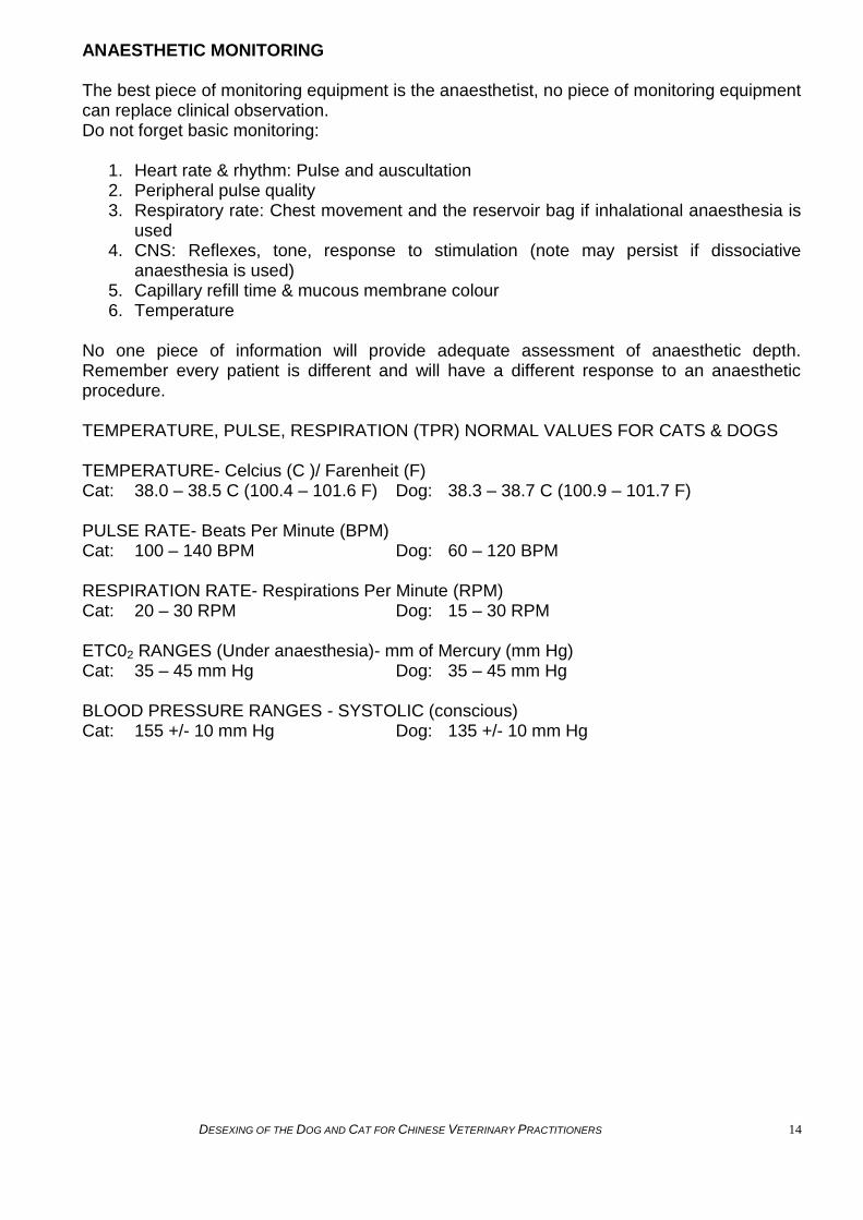
ANAESTHETIC MONITORING The best piece of monitoring equipment is the anaesthetist, no piece of monitoring equipment can replace clinical observation. Do not forget basic monitoring:
1. Heart rate & rhythm: Pulse and auscultation 2. Peripheral pulse quality 3. Respiratory rate: Chest movement and the reservoir bag if inhalational anaesthesia is
used 4. CNS: Reflexes, tone, response to stimulation (note may persist if dissociative
anaesthesia is used) 5. Capillary refill time & mucous membrane colour 6. Temperature
No one piece of information will provide adequate assessment of anaesthetic depth. Remember every patient is different and will have a different response to an anaesthetic procedure. TEMPERATURE, PULSE, RESPIRATION (TPR) NORMAL VALUES FOR CATS & DOGS TEMPERATURE- Celcius (C )/ Farenheit (F) Cat: 38.0 – 38.5 C (100.4 – 101.6 F) Dog: 38.3 – 38.7 C (100.9 – 101.7 F) PULSE RATE- Beats Per Minute (BPM) Cat: 100 – 140 BPM Dog: 60 – 120 BPM RESPIRATION RATE- Respirations Per Minute (RPM) Cat: 20 – 30 RPM Dog: 15 – 30 RPM ETC02 RANGES (Under anaesthesia)- mm of Mercury (mm Hg) Cat: 35 – 45 mm Hg Dog: 35 – 45 mm Hg BLOOD PRESSURE RANGES - SYSTOLIC (conscious) Cat: 155 +/- 10 mm Hg Dog: 135 +/- 10 mm Hg

DESEXING OF THE DOG AND CAT FOR CHINESE VETERINARY PRACTITIONERS 15
Anaesthesia Stages and Planes** ** Please note: below changes may vary depending on the pharmacological combinations being used. Therefore, multiple parameters should be utilized to assess anaesthetic depth and someone well-trained and familiar with the anaesthetic protocol should assess and monitor stages of anaesthesia.
STAGE I disorientation STAGE II excitement STAGE III surgical PLANE 1
STAGE III surgical PLANE 2
STAGE III surgical PLANE 3
STAGE III surgical PLANE 4
STAGE IV death
BEHAVIOR Loss of inhibition, conscious but altered LOC, may see "fight or flight" response
Involuntary struggling, vocalization, and movement; loss of consciousness
Anaesthetized Anaesthetized Deeply anaesthetized Too deeply anaesthetized Moribund
RESPIRATION Normal to increased, may pant or hold breath
May be regular or irregular rhythm and depth; may hold breath or pant
Regular with normal to slightly increased rate
Regular rhythm; normal or shallow depth with normal to slightly decreased rate
Decreased depth and rate; may be irregular
Jerky, diaphragmatic breathing pattern
Apnea; respiratory arrest
CARDIO- VASCULAR FUNCTION
Normal to increased (stress response)
Increased (above pre-anesthetic level); blood pressure may be increased
Heart rate decreasing to pre-anesthetic levels; pulses and blood pressure strong/normal
Heart rate stabilized at pre-anesthetic levels; blood pressure normal to slightly decreased
Heart rate and blood pressure decreased; pulses weaker and CRT prolonged
Heart rate and blood pressure reach critical low; mm color pale and CRT prolonged; pulses weak
Cardiovascular collapse and cardiac arrest imminent
RESPONSE TO SURGERY
Voluntary resistance Exaggerated response to painful stimuli
No voluntary movement; may see movement with painful stimulation
Heart rate, blood pressure, and respiratory rate may increase with painful surgical stimulation
None None None
DEPTH NOT ANESTHETIZED NOT ANESTHETIZED LIGHT MODERATE DEEP OVERDOSE DYING
EYEBALL POSITION
Central Central; may have nystagmus Central or rotated; may have nystagmus
Central or rotated ventrally; third eyelid may partially prolapse
Central; may be ventrally rotated. Corneas dry
Central Central
PUPIL SIZE Normal to constricted Pupils larger than in Stage I and may dilate
Pupils decrease to pre-anaesthetic size or constrict
May be normal to slightly dilated
Moderately dilated Dilated Widely dilated ("blown")
RESPONSE TO LIGHT
Brisk; menace intact Brisk Normal Sluggish Very slow or absent No response No response
MUSCLE TONE High High Poor relaxation Relaxed; jaw tone intact Relaxed; decrease in jaw tone
Flaccid Flaccid
REFLEX RESPONSE
All reflexes present; may be exaggerated
All present; palpebral, corneal, swallow, laryngeal may be exaggerated
Reflexes present but diminished
Palpebral and corneal reflexes may be present; cats retain laryngeal reflex; others absent
All reflexes diminished or absent
No reflexive responses No reflexive responses
NOTES Length of time in Stage I depends on pre-medicants and induction technique. Elongated in gas-only induction.
Length of time in Stage II depends on pre-medicants and induction technique. Elongated in gas-only induction. Dogs may be intubated in late Stage II anesthesia.
Intubation and surgical prep done at this time
Depth appropriate for most routine surgeries
Depth required for painful surgeries such as orthopedics and thoracotomies
Anaesthetic overdose; death is imminent unless reversed immediately
DEATH

DESEXING OF THE DOG AND CAT FOR CHINESE VETERINARY PRACTITIONERS 16
Monitoring Tools Essential monitoring equipment includes a stethoscope, thermometer, an anaesthetic record chart and, of course, a designated person trained to monitor the patient (Fig 14). During anaesthesia the patient‟s vital signs must be closely monitored (every 2-5 minutes) for early detection of any problems. This information should be recorded on an anaesthetic monitoring chart which allows the anaesthetist to see trends and respond to changes quickly and appropriately. If there are any changes such as an increase or decrease in vital signs the Veterinary surgeon should be informed immediately and action taken to prevent the situation from worsening. The anaesthetic chart is also a valuable legal record of the anaesthetic and can provide useful patient information should the animal need future anaesthetics. Advanced anaesthetic monitoring includes capnography and blood pressure monitoring.
Fig 14: Trained Veterinary Nurse monitoring patients heart rate under anaesthesia
Cardiovascular Monitoring The respiratory system works hand in hand with the cardiovascular system to deliver oxygen to the tissues. One cannot work without the other. Many anaesthetic agents have profound effects on the cardiovascular system (see appendix 5) but the anaesthetist must not forget the effects of surgery or even pathological states on the functioning of the cardiovascular system. During anaesthesia the animal‟s heart rate should be carefully monitored and recorded every 5 minutes, any changes in the rate and rhythm of the heart should be immediately reported to the Veterinary surgeon and necessary action taken. Bradycardia (a slow heart rate) may be caused by:
1. Anaesthetic agents e.g. a. Opioids b. Xylazine c. Medetomidine d. Barbituates e. Halothane
2. Anaesthetic depth maintained too deeply 3. Increased vagal tone, which may be caused by:
a. Endotracheal intubation b. Handling of viscera intraoperatively c. Ocular manipulations or ocular surgery d. Periosteal stimulation
4. Hypothermia 5. Increased intracranial pressure 6. Underlying metabolic problems

DESEXING OF THE DOG AND CAT FOR CHINESE VETERINARY PRACTITIONERS 17
7. Shock 8. Hypoxia
Treatment of Bradycardia
1. Reduce anaesthetic depth 2. Ventilate the patient 3. Apply or increase external heat (after checking the temperature) 4. Surgeon can stop stimulation of the vagus nerve if that appears to be the cause 5. Drugs may be used intraoperatively to correct bradycardia (these drugs should not be
used if xylazine or medetomidine have been given) a. Atropine b. Glycopyrrolate c. Dopamine
Tachycardia Tachycardia can decrease cardiac output because there is less time for filling the ventricles. The workload on the heart is increased, as is the myocardial oxygen consumption. Prolonged tachycardia can predispose heart to ventricular arrhythmias that may become detrimental if not addressed Heart rates indicating tachycardia
Large breed dog: > 160bpm Small breed dog: > 180bpm Cat: > 200bpm
Tachycardia may be caused by
1. Administration of some drugs 2. Inadequate depth of anaesthesia 3. Surgical stimulation 4. Pain 5. Hypoxia 6. Hypotension and/or hypovolemia 7. Hypercapnia 8. Anaemia 9. Increased intracranial pressure 10. Hyperthermia
Treatment of Tachycardia
1. Should be directed to the underlying cause 2. Increase anaesthetic depth 3. Increase/administer analgesia 4. Hypoxia – treat with increased ventilation 5. Hypotension or shock - treat with IV fluid administration
Respiratory Monitoring The simplest way to monitor the respiratory system is by observing breathing rate, depth, rhythm, tidal volume of each breath, and mucous membrane colour. The number of breaths the animal takes a minute should be counted and recorded every 5 minutes (dogs and cats need to take at least 3 breaths a minute). Hyperventilation or Tachypnoea Defined as an increase in minute volume due to an increase in tidal volume/respiratory rate Causes include:

DESEXING OF THE DOG AND CAT FOR CHINESE VETERINARY PRACTITIONERS 18
1. Inadequate anaesthetic depth 2. Surgical stimulation 3. Over ventilation 4. Hypoxia 5. Hypotension 6. Pyrexia 7. Hyperthermia
Treatment of hyperventilation
1. Increase anaesthetic depth 2. Administering analgesia 3. Reducing IPPV 4. Treatment of underlying causes
Hypoventilation Defined as reduced minute volume due to a reduction in tidal volume/respiratory rate Leads to hypercarbia, an increased level of CO2 in blood Can simultaneously cause hypoxemia Hypercarbia if left untreated will cause central nervous system depression Causes of hypoventilation include:
1. Over dose of anaesthetic agents, patient “too deep” 2. Pain 3. Accidental endobronchial intubation 4. Hypothermia 5. Severe hypotension
Treatment of hypoventilation
1. Reduction of anaesthetic depth 2. IPPV 3. Treatment of underlying causes
Capnography Capnography is used to measure the adequacy of ventilation, which depends on the respiratory rate and depth (tidal volume). Measurement of the amount of carbon dioxide (CO2) exhaled in the patients end tidal breath (ET CO2) should be around 35-45 mm Hg. Hypoventilation may result in increased ET CO2
Hyperventilation may result in decreased ET CO2 Appropriate concentration of CO2 to activate respiration is what stimulates the brain to tell the body to breathe Low CO2 concentration: stimulus to breathe is not present may result in hypoventilation and/or bradypnea High CO2 concentration: brain is stimulated to remove excess quickly and may result in hyperventilation and/or tachypnea
Mucous Membrane Colour Mucous membrane colour provides information about blood oxygenation and tissue perfusion. However it is very subjective and depends upon many factors such as lighting, drug administration, vasomotor tone, etc.
1. White – anaemia or intense vasoconstriction 2. Yellow – jaundice
DESEXING OF THE DOG AND CAT FOR CHINESE VETERINARY PRACTITIONERS 19
3. Grey/Purple/Cyanotic – poor tissue oxygen delivery, either due to poor cardiac output or insufficient oxygen to meet tissue demands
CNS: Reflexes, Tone & Response to stimulation During anaesthesia it is important not to tie animals to the surgery table. With an appropriate depth of anaesthesia, the patient will not move in response to surgical stimulation, so tying the animal down is not necessary. If the animal moves under anaesthesia, the level of anaesthesia is inadequate and this should be addressed immediately by the anaesthetist. Nerve reflexes such as palpebral reflex and jaw tone are good indicators of depth of anaesthesia – if the animal is not sufficiently anaesthetised, these reflexes will still be present. However if you are using a dissociative anaesthetic such as ketamine or tiletamine, these reflexes may remain even under anaesthesia. If signs such as twitching or moving limbs are seen, the depth of anaesthesia should be immediately increased. It is not acceptable for an animal to move even a little bit during a surgical procedure, as this often means the animal is not anesthetised appropriately and can therefore likely feel pain and this is inhumane. If you are concerned the animal is responding to painful stimuli, stop stimulating the animal, adjust the anaesthetic depth, continue monitoring and wait for the animal to settle before resuming the procedure. Monitoring reflex activity during anaesthesia Palpebral (blink) reflex Observed by gently touching the medial or lateral canthus of the eye, observing if the patient blinks or not Reflex is usually diminished at a surgical plane of anaesthesia unless dissociative agents are used Swallowing reflex Observed by watching ventral aspect of neck for any swallowing activity Reflex is usually diminished at a light or medium plane of anaesthesia, it should be absent when entering a surgical plane of anaesthesia Ear Flick (Pinna) Reflex Observed by gently touching the hair of the inner pinna and watching for a twitch of the ear Reflex may remain intact even into a light plane of anaesthesia, but should be absent in a surgical plane Observation of this reflex is more useful in cats than in dogs Laryngeal Reflex Reflex observed when intubating patient Reflex response is to immediately close epiglottis and vocal cords when stimulated by touch of the endotracheal tube Reflex is most commonly seen in cats and commonly results in laryngospasm – all cats should receive topical lignocaine as previously described Skeletal Muscle Tone Commonly observed in masticating muscles or jaw tone of anesthetised patients Some anaesthetic agents may cause muscle rigidity (ketamine, tiletamine) where muscle tone may not be an adequate indicator of anaesthetic depth

DESEXING OF THE DOG AND CAT FOR CHINESE VETERINARY PRACTITIONERS 20
Eye Position The eyeball is usually central at a light plane of anaesthesia and ventral at a surgical plane. Mydriasis (dilated pupil) is often seen at a surgical plane of anaesthesia Pupillary light response will diminish with deeper levels of anaesthesia Centrally located eye position with mydriasis and absence of pupillary light response may indicate a dangerously deep plane of anaesthesia Atropine administration is known to cause mydriasis, especially in cats
Temperature Anaesthesia generally depresses the patient‟s ability to thermoregulate and temporarily prevents an animal from shivering. Many of the drugs also cause peripheral vasodilation and thereby enhance heat loss. Where large areas of hair are clipped and skin is prepped with alcoholic solutions heat loss is increased through evaporation. Hypothermia is a serious concern especially in smaller animals. Preventing heat loss is easier then re-warming an animal. Body temperature commonly gets lower the longer the anaesthetic continues and should be monitored regularly. Ways to prevent heat loss:
1. Keep animal warm (by using a heat pad or hot water bottles but make sure they are not too hot that they burn the animal)
2. Keep operating theatre warm 3. Minimise surgery time 4. Minimise clipped areas 5. Use warmed IV fluids 6. Use of warm saline intraoperatively
Electric heat pads and hot water bottles should be used with extreme caution: Avoid direct contact with the patient‟s skin as it may result in severe skin burns or sloughing. Patients should be protected with blankets in between the hot water bottle/head pad and their skin. Temperature can be helpful in assessing the cardiovascular status of animals but is also important for a multitude of reasons, especially for the metabolism of anaesthetic drugs. Hypothermia At 36°C (96°F) Shivering will be noted during recovery (note shivering will not occur under anaesthesia) At 32-34°C (90-94°F) Metabolic rates decrease, requiring less anaesthetic agent to maintain anaesthesia Recovery may be prolonged due to decreased metabolic rate At 28-30°C (82-86°F) Little or no anaesthetic agents are required to maintain aneasthesia Recovery will be prolonged Metabolic acidosis may occur due to poor tissue perfusion Hyperthermia Hyperthermia is less common than hypothermia during anaesthesia Hyperthermia can be due to:
1. Increased metabolic rate, commonly seen when the anesthaetic plane is too light 2. Failure to monitor temperature during the use of artificial heating during anaesthesia 3. Obese patients heavily draped during surgery

DESEXING OF THE DOG AND CAT FOR CHINESE VETERINARY PRACTITIONERS 21
Treatment of hyperthermia: 1. Administration of cool or room temperature IV fluids 2. Flushing open body cavities with room temperature sterile saline 3. Soaking skin with wet towels or alcohol, especially pads of feet and ears
DESEXING OF THE DOG AND CAT FOR CHINESE VETERINARY PRACTITIONERS 22
SURGICAL PREPARATION The cat or dog should be positioned appropriately for surgery. Animals should never be tied to the surgery table. Ensure that there is no undue tension of the ET tube on the trachea and that monitoring equipment and positioning do not impede the animal‟s ability to breathe. Thermal support should be provided as described above. Preparation of the surgical site is vital to minimise the risk of surgical infection. Surgical site preparation involves the removal of hair, dirt and debris along with cleaning the site with appropriate scrubbing solutions to remove both contaminant and normal bacteria/microbes found on the skin that can contaminate surgical wounds. Once the patient is anaethetised and you are happy with the depth of anaesthesia then and only then should the surgical area be prepared. It is best practice to wear examination gloves when scrubbing the surgical area to decrease contamination from human hands.
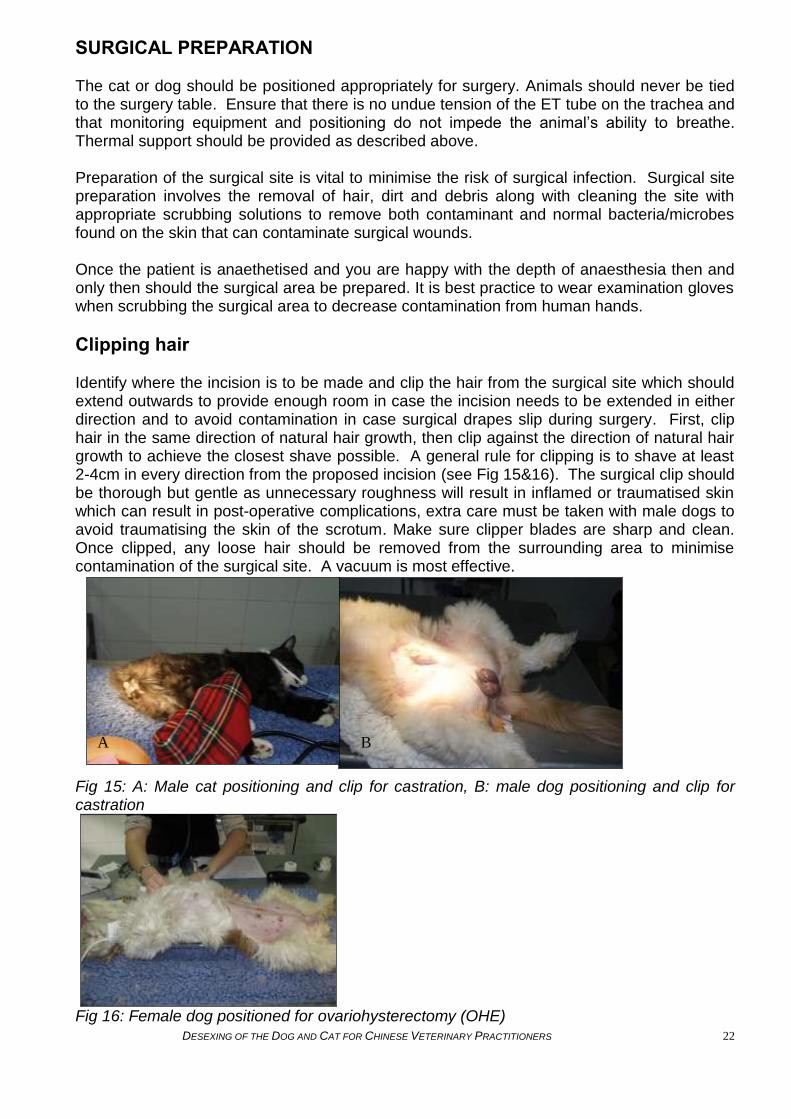
Clipping hair Identify where the incision is to be made and clip the hair from the surgical site which should extend outwards to provide enough room in case the incision needs to be extended in either direction and to avoid contamination in case surgical drapes slip during surgery. First, clip hair in the same direction of natural hair growth, then clip against the direction of natural hair growth to achieve the closest shave possible. A general rule for clipping is to shave at least 2-4cm in every direction from the proposed incision (see Fig 15&16). The surgical clip should be thorough but gentle as unnecessary roughness will result in inflamed or traumatised skin which can result in post-operative complications, extra care must be taken with male dogs to avoid traumatising the skin of the scrotum. Make sure clipper blades are sharp and clean. Once clipped, any loose hair should be removed from the surrounding area to minimise contamination of the surgical site. A vacuum is most effective.
Fig 15: A: Male cat positioning and clip for castration, B: male dog positioning and clip for castration
Fig 16: Female dog positioned for ovariohysterectomy (OHE)
A B

DESEXING OF THE DOG AND CAT FOR CHINESE VETERINARY PRACTITIONERS 23
Pre surgical clean Initial skin preparation is done to remove any loose hair and dirt. Once the area looks clean the surgical scrub can start, using an antiseptic scrub (povidone-iodine or chlorhexidine) (see Fig 17). Scrub the area using circular motions starting from the incision site and working outwards, repeat several times using a new scrub swab. Never go back to the incision area with the swab that has touched the edge of the clipped area. This is to ensure that dirt is not dragged from the edge of the clipped area back to the surgical site. Finish with a spray of alcohol if using chlorhexidine. Once this area has been prepared for surgery only the surgeon with sterile hands should touch this area. If the area is touched by a non sterile person or equipment then the scrub must be repeated.
Fig 17: Types of surgical scrub from left of image; Povidine Iodine, Chlorhexidine & Surgical Spirit 75%

DESEXING OF THE DOG AND CAT FOR CHINESE VETERINARY PRACTITIONERS 24
PREPARATION OF THE SURGICAL TEAM After the patient is anaethetised and the Veterinary surgeon is happy with the depth of anaesthesia, the vet(s) can prepare themselves for the surgical procedure. Ideally hand scrub brushes and towels should be sterilised before use (see Fig 18). The use of a chlorhexidine based or iodine scrub solution should be used to prepare the surgeon‟s hands.
Fig 18: Sterile towel & scrub brush, sterile gloves and face mask. The surgeon should place a clean cap and mask on prior to scrubbing in for surgery as these items are not sterile and should not be touched by the surgeon after he/she has scrubbed in for surgery. There should be a designated area for veterinary surgeons to scrub in for surgery. The surgeon‟s hands and forearms should be thoroughly rinsed in clean water and then an appropriate solution applied to coat the hands and forearms.
A full surgical scrub should take at least 7 minutes (see Fig 19).
Fig 19: Veterinary surgeon scrubbing hands & forearms for a surgical procedure Every surface of the surgeon‟s hands and forearms to the elbows should be thoroughly cleansed including the insides of the fingers and the nails. However, over-scrubbing should be avoided as abrasions to the surgeons skin may lead to post-operative infections in the patient.

DESEXING OF THE DOG AND CAT FOR CHINESE VETERINARY PRACTITIONERS 25
During and after scrubbing, the surgeon should not touch the water tap or the container of scrubbing solution with his/her hands. After the scrub is complete, the hands should be rinsed with clean water. It is vital that at all times the hands are raised above the elbows so that dirty water from the elbows does not run down and contaminate the hands. The hands should be dried with a sterile towel or sterile paper towel (see Fig 20).
Fig 20: Hand drying using sterile towel Sterile gloves should be put on in a sterile manner (see Fig 21). If a sterile gown is worn, the gown should be put on first and then the gloves. Pick up the left glove by its inner cuff using your sterile right hand and slide the glove onto your sterile left hand but do not unfold the cuff Slide your partly gloved left hand into the inner cuff of the right glove and slight your sterile right hand into the right glove, using your partly gloved left hand to place it correctly. Take care not to touch any non-sterile areas such as your arm with your gloved fingers. Once the right glove is properly placed, use your right hand to unfold the cuff of your left glove
Fig 21: Gloving procedure

DESEXING OF THE DOG AND CAT FOR CHINESE VETERINARY PRACTITIONERS 26
Once the surgeon is scrubbed in it is essential that he or she does not touch anything unless it is sterile. If anything non-sterile (e.g. light switches, doors, walls etc) is touched then the the surgeon will need to repeat the scrub and change gloves. Scrubbing, gowning and gloving should be done in a clear area to prevent bumping into objects and contaminating the surgeon. An assistant should be used to open packages and fasten the gown. Everyone in the surgical team should take care not to touch the aseptic surgeon or surgical site.

DESEXING OF THE DOG AND CAT FOR CHINESE VETERINARY PRACTITIONERS 27
SURGICAL PROCEDURES It is helpful to allow cats and dogs to urinate and defecate prior to surgery. Alternatively, bladders may be gently expressed manually once the animal is anesthetised as this will help provide better access to the female reproductive tract during desex surgery.
Ovariohysterectomy Common Indications: To prevent unwanted puppies and kittens To prevent the development of mammary tumours To prevent pyometra (infected uterus) To prevent the development of cancer of the reproductive tract To prevent twisting or prolapse of the uterus Surgical Approach Ovariohysterectomy: the removal of both ovaries and the uterus As with any anesthetic and surgery, complications can occur. Many complications can be avoided by following aseptic technique, performing good hemostasis, using gentle tissue handling, and confirming anatomy. It is important once the uterine horn has been located to confirm its identity by following the uterine horn to the respective ovary and/or to the uterine bifurcation. This helps to avoid accidental ligation of intestines or ureters which can lead to life-threatening complications. Similarly, when placing ligatures, ensure that no excess tissue is being entrapped within the ligatures. Smaller incisions cause less trauma to the patient. However, the surgeon must feel comfortable and confident with the amount of visibility and space to be able to perform the necessary procedure safely and efficiently. If struggling to clearly visualize anatomy or to place ligatures because the incision is too small, then extend the incision to facilitate the procedure. Incisions will take the same amount of time to heal regardless of how long they are. With experience, the incisions tend to become smaller/shorter.
Fig 22: Patient positioned in dorsal recumbency, umbilical scar is circled.

DESEXING OF THE DOG AND CAT FOR CHINESE VETERINARY PRACTITIONERS 28
DOG OVARIOHYSTERECTOMY Start incision at or just caudal to the umbilical scar as this will make it easier to access and exteriorise the ovaries (See Figure 22) Using scalpel, incise through skin and subcutaneous fat, stay in midline with smooth incisions, extending the incision 4-8 cm (see Fig 23)
Fig 23: Incising through skin and subcutaneous tissue
Once linea alba is identified, lift and tent it using rat tooth forceps. Turn the scalpel blade upside down to pierce through abdominal wall to minimize the risk of damaging any underlying, internal structures/organs
Fig 24: Lifting and tenting linea albea, preparing to incise with scalpel blade turned upwards
to pierce through linea alba
With Mayo scissors (blunt tipped) that are closed, sweep from side to side along the underside of the abdominal wall/linea alba to ensure no adhesions, can also use your finger Use atraumatic tissue forceps to handle the abdominal wall, take care not to traumatise tissues as this will impede healing

DESEXING OF THE DOG AND CAT FOR CHINESE VETERINARY PRACTITIONERS 29
Extend incision in both directions using Mayo scissors or scalpel blade, taking care not to incise or damage any other organs Some find it helpful to gently lift the linea alba upwards using open haemostats and incising the linea alba with the scalpel blade to prevent accidental incision of underlying tissues and organs (see Fig 25)
Fig 25: Gently lifting abdominal wall with opened haemostats to facilitate incising linea alba to
avoid lacerating internal organs Do not undercut (do not make the linea alba incision longer than the skin incision as this will make it difficult to close)
Using a spey hook or your finger, sweep along the body wall to feel for uterine horns and gently pull upwards to follow the horn to identify the ovary If unable to locate the uterine horns with this method, gently lift the bladder to locate the uterine body which sits between the bladder and the colon Follow the uterine horn to the ovary and also to the bifurcation of the uterine body to confirm your anatomical location prior to placing the haemostats Occasionally the ovary can be covered or embedded within fat or the bursa, so it may be helpful to locate the other horn and/or the uterine body to ensure you have located the reproductive tract (see Fig 26)
Uterine Horn Ovarian Bursa
Suspensory Ligament
Ovary embedded in fat
DESEXING OF THE DOG AND CAT FOR CHINESE VETERINARY PRACTITIONERS 30
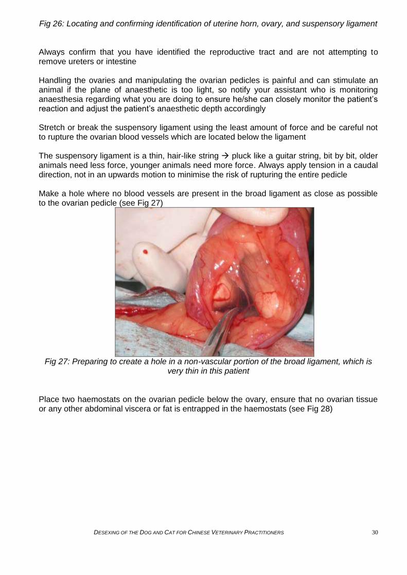
Fig 26: Locating and confirming identification of uterine horn, ovary, and suspensory ligament
Always confirm that you have identified the reproductive tract and are not attempting to remove ureters or intestine
Handling the ovaries and manipulating the ovarian pedicles is painful and can stimulate an animal if the plane of anaesthetic is too light, so notify your assistant who is monitoring anaesthesia regarding what you are doing to ensure he/she can closely monitor the patient‟s reaction and adjust the patient‟s anaesthetic depth accordingly
Stretch or break the suspensory ligament using the least amount of force and be careful not to rupture the ovarian blood vessels which are located below the ligament The suspensory ligament is a thin, hair-like string pluck like a guitar string, bit by bit, older animals need less force, younger animals need more force. Always apply tension in a caudal direction, not in an upwards motion to minimise the risk of rupturing the entire pedicle Make a hole where no blood vessels are present in the broad ligament as close as possible to the ovarian pedicle (see Fig 27)
Fig 27: Preparing to create a hole in a non-vascular portion of the broad ligament, which is
very thin in this patient
Place two haemostats on the ovarian pedicle below the ovary, ensure that no ovarian tissue or any other abdominal viscera or fat is entrapped in the haemostats (see Fig 28)

DESEXING OF THE DOG AND CAT FOR CHINESE VETERINARY PRACTITIONERS 31
Fig 28: placing first clamp on ovarian pedicle through hole in broad ligament, ensuring no
other tissue is entrapped in the clamp
Place first ligature around the ovarian pedicle, remove the first haemostats (most proximal to the cat or dog) and ensuring no fat or viscera is caught in the ligature, tighten it into the crush mark. When tightening down on this ligature, release the ratchet of the second haemostats to release tension on the pedicle and facilitate tightening of the ligature. Re-ratchet the second haemostats once the knot is secure
Use an appropriate sterile, absorbable suture material and ensure that an appropriate number of throws are placed: 6-8throws (3-4 knots) for monofilament suture and 4-6 throws (2-3 knots) for multifilament suture
Place a second ligature, ensuring that it does not overlap the first (see Fig 29)
Fig 29: two catgut ligatures are visible around the ovarian pedicle
Place a third haemostat between the second haemostat and the ovary, or, if there is not enough space here, place it on the uterine horn next to the ovary (see Fig 30)

DESEXING OF THE DOG AND CAT FOR CHINESE VETERINARY PRACTITIONERS 32
Fig 30: Two haemostats placed above the ligatures of the ovarian pedicle and below the ovary; preparing to incise between the two clamps to separate the ovary from the ovarian
pedicle
Incise the ovarian pedicle between the second haemostats and the ovary (see Fig 31)
Fig 31: One haemostat remains on the ovarian pedicle, the other just below the ovary
Hold the ovarian pedicle with rat tooth forceps (placed so that they do not touch the ligature) and remove the second haemostat. Monitor the pedicle for bleeding (see Fig 32)

DESEXING OF THE DOG AND CAT FOR CHINESE VETERINARY PRACTITIONERS 33
Fig 32: Holding the ovarian pedicle and checking for any bleeding or oozing prior to releasing
back into the abdomen
If no oozing or bleeding is noted then release the pedicle and watch the pedicle retract back into the abdomen
Repeat same procedure on alternate side
Once both ovarian pedicles are ligated, retract the excised ovaries and uterine horns caudally, breaking down the broad ligament attachments as you go (see Fig 33)
In bigger dogs, or dogs in heat or if the broad ligament is very fatty and vascular, the broad ligament blood vessels should be ligated to avoid prolonged oozing or hemorrhage
Fig 33: Preparing to gently retract the uterine horns to locate the uterine bifurcation and the
cervix for clamping and ligation
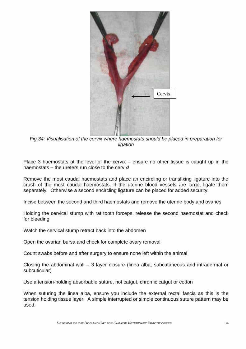
Identify the cervix, not the bilateral uterine blood vessels (see Fig 34)
DESEXING OF THE DOG AND CAT FOR CHINESE VETERINARY PRACTITIONERS 34
Fig 34: Visualisation of the cervix where haemostats should be placed in preparation for
ligation
Place 3 haemostats at the level of the cervix – ensure no other tissue is caught up in the haemostats – the ureters run close to the cervix!
Remove the most caudal haemostats and place an encircling or transfixing ligature into the crush of the most caudal haemostats. If the uterine blood vessels are large, ligate them separately. Otherwise a second encircling ligature can be placed for added security.
Incise between the second and third haemostats and remove the uterine body and ovaries
Holding the cervical stump with rat tooth forceps, release the second haemostat and check for bleeding
Watch the cervical stump retract back into the abdomen
Open the ovarian bursa and check for complete ovary removal
Count swabs before and after surgery to ensure none left within the animal
Closing the abdominal wall – 3 layer closure (linea alba, subcutaneous and intradermal or subcuticular)
Use a tension-holding absorbable suture, not catgut, chromic catgut or cotton
When suturing the linea alba, ensure you include the external rectal fascia as this is the tension holding tissue layer. A simple interrupted or simple continuous suture pattern may be used.
Cervix

DESEXING OF THE DOG AND CAT FOR CHINESE VETERINARY PRACTITIONERS 35
A simple continuous pattern ensures that if one suture dislodges, the other sutures will continue to keep the abdomen closed. However, a simple interrupted pattern means there will be more knots and a potential for more suture reactions. A simple continuous pattern is a bit quicker to place, but if a portion of suture breaks, then the whole incision can open up
Subcutaneous tissue sutures are placed to close dead space. Subcutaneous tissue can be closed with a simple continuous pattern using absorbable suture material
Skin should be closed using a buried, subcuticular or intradermal pattern with absorbable suture material. This pattern reduces scarring and also alleviates the need to remove sutures at a later date and also reduces dogs and cats chewing at their incisions and dislodging sutures (see Fig 35)
Fig 35: Following closure of the abdominal incision using buried, intradermal skin sutures
CAT OVARIOHYSTERECTOMY Prior to surgery, examine the genitalia to ensure the correct sexing of the animal. It is better to take a few minutes to ensure you are about to perform surgery on a female and not on a previously neutered male cat. Therefore, always check the sex of the cat prior to surgery. Flank Spay

DESEXING OF THE DOG AND CAT FOR CHINESE VETERINARY PRACTITIONERS 36
Fig 36: Patient positioned in lateral recumbency for flank approach; Hindlegs must be pulled back to avoid incising muscle of the hindlegs.
The incision line is located by placing the thumb on major trochanter of the femur (red arrow), middle finger placed on tuber coxae (white arrow) and pointer finger (black arrow) forms a triangle where vertical incision should be placed (see Fig 37&38).
Fig 37: Landmarks for flank spay incision
Fig 38: Blue line marks where the incision should be made.
DESEXING OF THE DOG AND CAT FOR CHINESE VETERINARY PRACTITIONERS 37
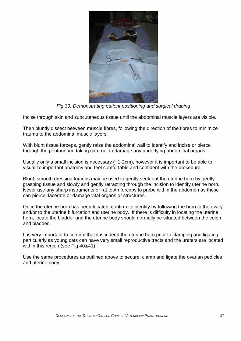
Fig 39: Demonstrating patient positioning and surgical draping
Incise through skin and subcutaneous tissue until the abdominal muscle layers are visible.
Then bluntly dissect between muscle fibres, following the direction of the fibres to minimize trauma to the abdominal muscle layers.
With blunt tissue forceps, gently raise the abdominal wall to identify and incise or pierce through the peritoneum, taking care not to damage any underlying abdominal organs.
Usually only a small incision is necessary (~1-2cm), however it is important to be able to visualize important anatomy and feel comfortable and confident with the procedure.
Blunt, smooth dressing forceps may be used to gently seek out the uterine horn by gently grasping tissue and slowly and gently retracting through the incision to identify uterine horn. Never use any sharp instruments or rat tooth forceps to probe within the abdomen as these can pierce, lacerate or damage vital organs or structures.
Once the uterine horn has been located, confirm its identity by following the horn to the ovary and/or to the uterine bifurcation and uterine body. If there is difficulty in locating the uterine horn, locate the bladder and the uterine body should normally be situated between the colon and bladder.
It is very important to confirm that it is indeed the uterine horn prior to clamping and ligating, particularly as young cats can have very small reproductive tracts and the ureters are located within this region (see Fig 40&41).
Use the same procedures as outlined above to secure, clamp and ligate the ovarian pedicles and uterine body.

DESEXING OF THE DOG AND CAT FOR CHINESE VETERINARY PRACTITIONERS 38
Fig 40: Uterine horn and ovary after ligation of ovarian pedicle
Fig 41: Ovaries and uterine horns to be gently retracted to locate cervix for ligation
DESEXING OF THE DOG AND CAT FOR CHINESE VETERINARY PRACTITIONERS 39
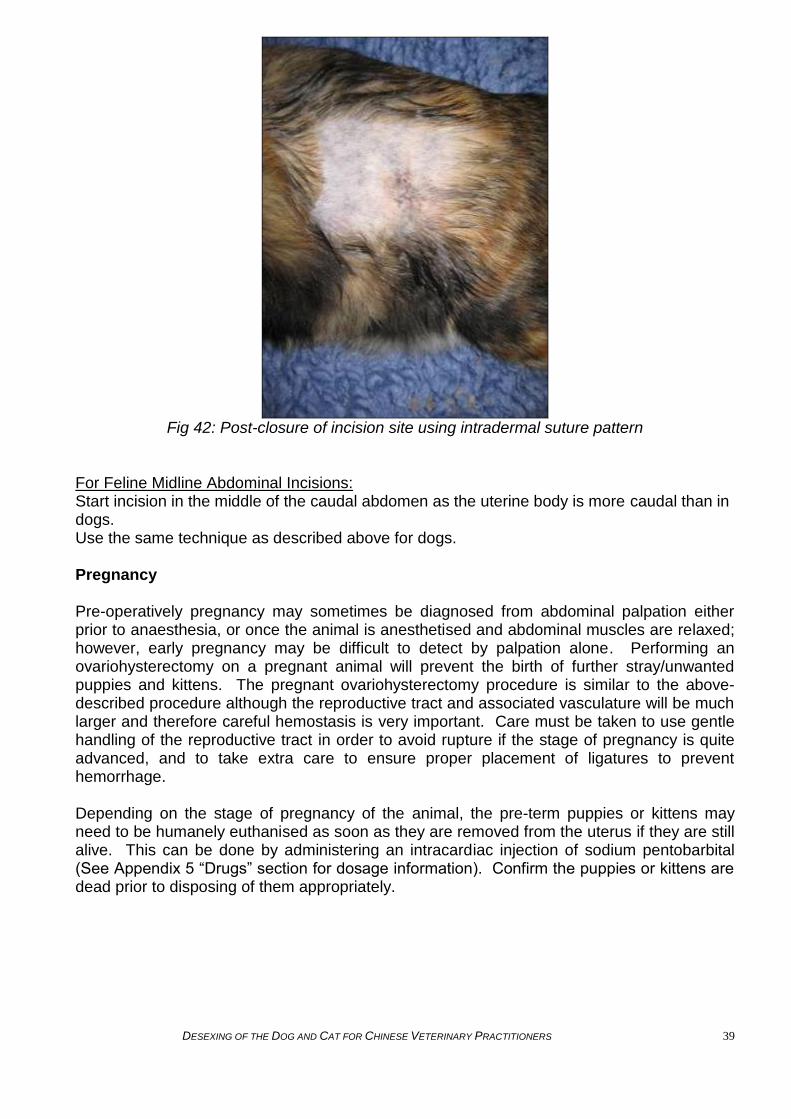
Fig 42: Post-closure of incision site using intradermal suture pattern
For Feline Midline Abdominal Incisions: Start incision in the middle of the caudal abdomen as the uterine body is more caudal than in dogs. Use the same technique as described above for dogs. Pregnancy Pre-operatively pregnancy may sometimes be diagnosed from abdominal palpation either prior to anaesthesia, or once the animal is anesthetised and abdominal muscles are relaxed; however, early pregnancy may be difficult to detect by palpation alone. Performing an ovariohysterectomy on a pregnant animal will prevent the birth of further stray/unwanted puppies and kittens. The pregnant ovariohysterectomy procedure is similar to the above-described procedure although the reproductive tract and associated vasculature will be much larger and therefore careful hemostasis is very important. Care must be taken to use gentle handling of the reproductive tract in order to avoid rupture if the stage of pregnancy is quite advanced, and to take extra care to ensure proper placement of ligatures to prevent hemorrhage. Depending on the stage of pregnancy of the animal, the pre-term puppies or kittens may need to be humanely euthanised as soon as they are removed from the uterus if they are still alive. This can be done by administering an intracardiac injection of sodium pentobarbital (See Appendix 5 “Drugs” section for dosage information). Confirm the puppies or kittens are dead prior to disposing of them appropriately.

DESEXING OF THE DOG AND CAT FOR CHINESE VETERINARY PRACTITIONERS 40
Pyometra Females can develop infections of the uterus, called pyometra. Pyometra is a life-threatening condition during which animals often become very ill and must be treated aggressively and monitored very closely to prevent septicaemia, uterine rupture and death. Patients must be stabilised and surgery performed as soon as possible. The approach is similar to the above described procedure for a standard OHE, the difference being the uterus will be distended to variable degrees due to an accumulation of pus and the tissue may be very friable, gentle handling is therefore very important. The abdomen should be evaluated for signs of free fluid or peritonitis in case the reproductive tract has ruptured. Ensure that the uterus is transected/removed at the level of the cervix. Lavage the vaginal stump to clean away any infected debris. Prior to transecting the uterus, pack sterile laparotomy sponges around the reproductive tract in case any infected material leaks out, so it does not enter the abdominal cavity. Remove laparotomy sponges and change any contaminated surgical instruments, gloves and drapes to minimise contamination of the abdomen. Count all laparotomy sponges pre-operatively and prior to closing the abdomen, recount to ensure all sponges are accounted for and that none are left within the abdomen. Pyometra patients require aggressive fluid therapy, antibiotic treatment, analgesia and monitoring. Surgery is the ONLY therapy for pyometra that will provide longterm success. Antibiotics are a necessary part of therapy but will not cure pyometra alone. Pyometra is 100% preventable by early desexing.
Castration (Orchiectomy) Common Indications: To prevent unwanted puppies and kittens To prevent male aggressiveness To prevent roaming behavior To prevent undesirable urination behavior To prevent diseases such as prostatic diseases, perianal adenomas, perianal hernias To prevent development of cancer of the male reproductive tract DOG ORCHIECTOMY Prescrotal Approach Prior to surgery, palpate the scrotum to ensure two descended testicles are present. Testes should be descended by 6-9 months of age, if not descended by 9-12 months of age, exploratory abdominal surgery and testicular removal should be performed. Retention of intra-abdominal testes increases testicular cancer risks significantly (see below). Open Approach: Position in dorsal recumbency Clip and surgically scrub the caudal abdomen from the prepuce to the scrotum and surrounding areas to the medial thighs (see Fig 43) The skin of the scrotum is sensitive, so gentle handling to avoid irritation and inflammation is important

DESEXING OF THE DOG AND CAT FOR CHINESE VETERINARY PRACTITIONERS 41
Fig 43: Dog in dorsal recumbency for prescrotal castration, note clipped area, arrow points to intended surgical site delineated by blue line (where you aim to push the testicle towards and make the incision overlying the displaced testicle) The opening of the surgical drape should be positioned between the prepuce and the scrotum, thereby covering the prepuce and scrotum to avoid contamination of the incision Using the non-dominant hand, use pressure on the scrotum to push one testicle cranially into the prescrotal area Make an incision through skin, subcutaneous tissue and through spermatic fascia (see Fig 44&45)
Fig 44: Using gentle pressure to hold testicle in place while incising skin and subcutaneous tissue Be careful not to incise and expose testicular parenchyma.

DESEXING OF THE DOG AND CAT FOR CHINESE VETERINARY PRACTITIONERS 42
Fig 45: applying firm but gentle pressure to push testicle upwards while incising skin and subcutaneous tissue facilitates exteriorisation of the testicle through your incision Separate the ligament of the tail of the epididymis from the vaginal tunic, using haemostats (see Fig 46)
Fig 46: Separating the ligament of the tail of the epididymis from the vaginal tunic Gently exteriorise the testicle (see Fig 47)

DESEXING OF THE DOG AND CAT FOR CHINESE VETERINARY PRACTITIONERS 43
Fig 47:Testicle Place two sets of haemostats across the ductus deferens and vascular cord to create a crush mark over which to place a ligature (see Fig 48)
Fig 48: Haemostats across the ductus deferens and vascular cord Using 2/0 or 3/0 absorbable suture material, place an encircling ligature around both the ductus deferens and the vascular cord (see Fig 49). Ensure that your ligature is securely knotted (6-8 throws for monofilament and 4-6 throws for multifilament suture) and tight enough to create complete haemostasis

DESEXING OF THE DOG AND CAT FOR CHINESE VETERINARY PRACTITIONERS 44
Fig 49: Placement of ligature Distal to this encircling ligature (away from the animal) place a second ligature if you feel it is necessary. See Fig 50.
Fig 50: Placing a second distal ligature Using a scalpel blade incise between the two haemostats (see Fig 51)

DESEXING OF THE DOG AND CAT FOR CHINESE VETERINARY PRACTITIONERS 45
Fig 51: Incising ductus deferens and vascular cord Inspect the cord for bleeding by gently grasping the cord using tissue forceps before removing the haemostat to avoid the cord from retracting into the incision Replace the cord into the tunic Close the vaginal tunic using absorbable suture in a continuous pattern or by placing an encircling ligature around the tunic and cremaster muscle Repeat the procedure for the remaining testicle Standard three-layer closure is performed by closing the dense fascial layer with either interrupted or continuous sutures; the subcutaneous tissue layer can be closed with a continuous suture pattern; and the skin layer closed using buried subcuticular or intradermal sutures (see Fig 52&53)
Fig 52: Standard three-layer closure
DESEXING OF THE DOG AND CAT FOR CHINESE VETERINARY PRACTITIONERS 46
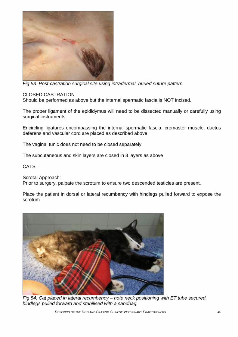
Fig 53: Post-castration surgical site using intradermal, buried suture pattern CLOSED CASTRATION Should be performed as above but the internal spermatic fascia is NOT incised. The proper ligament of the epididymus will need to be dissected manually or carefully using surgical instruments. Encircling ligatures encompassing the internal spermatic fascia, cremaster muscle, ductus deferens and vascular cord are placed as described above. The vaginal tunic does not need to be closed separately The subcutaneous and skin layers are closed in 3 layers as above CATS Scrotal Approach: Prior to surgery, palpate the scrotum to ensure two descended testicles are present. Place the patient in dorsal or lateral recumbency with hindlegs pulled forward to expose the scrotum
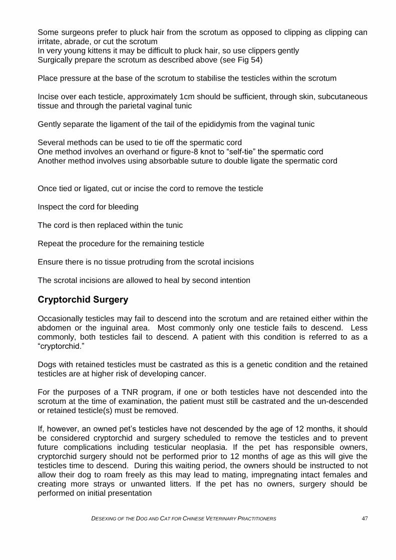
Fig 54: Cat placed in lateral recumbency – note neck positioning with ET tube secured, hindlegs pulled forward and stabilised with a sandbag.
DESEXING OF THE DOG AND CAT FOR CHINESE VETERINARY PRACTITIONERS 47
Some surgeons prefer to pluck hair from the scrotum as opposed to clipping as clipping can irritate, abrade, or cut the scrotum In very young kittens it may be difficult to pluck hair, so use clippers gently Surgically prepare the scrotum as described above (see Fig 54) Place pressure at the base of the scrotum to stabilise the testicles within the scrotum Incise over each testicle, approximately 1cm should be sufficient, through skin, subcutaneous tissue and through the parietal vaginal tunic Gently separate the ligament of the tail of the epididymis from the vaginal tunic Several methods can be used to tie off the spermatic cord One method involves an overhand or figure-8 knot to “self-tie” the spermatic cord Another method involves using absorbable suture to double ligate the spermatic cord Once tied or ligated, cut or incise the cord to remove the testicle Inspect the cord for bleeding The cord is then replaced within the tunic Repeat the procedure for the remaining testicle Ensure there is no tissue protruding from the scrotal incisions The scrotal incisions are allowed to heal by second intention
Cryptorchid Surgery
Occasionally testicles may fail to descend into the scrotum and are retained either within the abdomen or the inguinal area. Most commonly only one testicle fails to descend. Less commonly, both testicles fail to descend. A patient with this condition is referred to as a “cryptorchid.” Dogs with retained testicles must be castrated as this is a genetic condition and the retained testicles are at higher risk of developing cancer. For the purposes of a TNR program, if one or both testicles have not descended into the scrotum at the time of examination, the patient must still be castrated and the un-descended or retained testicle(s) must be removed. If, however, an owned pet‟s testicles have not descended by the age of 12 months, it should be considered cryptorchid and surgery scheduled to remove the testicles and to prevent future complications including testicular neoplasia. If the pet has responsible owners, cryptorchid surgery should not be performed prior to 12 months of age as this will give the testicles time to descend. During this waiting period, the owners should be instructed to not allow their dog to roam freely as this may lead to mating, impregnating intact females and creating more strays or unwanted litters. If the pet has no owners, surgery should be performed on initial presentation

DESEXING OF THE DOG AND CAT FOR CHINESE VETERINARY PRACTITIONERS 48
If the retained testicle is palpable within the inguinal area, it may be possible to manipulate the testicle caudally into the prescrotal area to be removed in the same manner as the descended testicle. Sometimes, however, an inguinal incision overlying the retained testicle is necessary and use the above-mentioned technique for ligation of the spermatic cord for removal. If the retained testicle is not palpable in the inguinal area, abdominal surgery is required. Place the animal in dorsal recumbency Clip and surgically prepare the abdomen as described above for a female dog desex procedure Make a midline abdominal incision from the umbilicus extended paramedian to the prepuce Incise through subcutaneous tissue Using rat tooth forceps, lift/tent the linea alba, turn the scalpel blade upside down and make a small stab incision to enter the abdominal cavity and minimising risk of damaging any underlying structures/organs With Mayo scissors (blunt tipped) that are closed, sweep from side to side to ensure no adhesions, can also use your finger Using tissue forceps to handle the abdominal wall, take care not to traumatise tissues as this will impede healing Extend incision in both directions using Mayo scissors or scalpel blade, taking care not to incise or damage any other organs Do not undercut (do not make the linea alba incision longer than the skin incision as this will make it difficult to close Locate and lift the bladder to identify the ductus deferens by the neck of the bladder Follow this to the testicle If the testicle has only just descended through the inguinal ring, it may be possible to gently manipulate the testicle cranially, back into the abdomen Use the above mentioned techniques to ligate the spermatic cord for removal Close the abdomen using a standard three layer closure as described for an OHE procedure

DESEXING OF THE DOG AND CAT FOR CHINESE VETERINARY PRACTITIONERS 49
Ear clipping/knotching technique It is advised that the ears of feral dogs or cats is tipped or notched to indicate that the animal has been desexed and to prevent future exploratory surgery if the animal is re-trapped. The right ear of females should be clipped, the left ear of males should be clipped. This allows trapping volunteers to see straight away that the animal is already desexed and prevent s time and resources being wasted as the animal can be released and not transported to the vet clinic. Cats should be ear tipped and dogs should be ear knotched as per the pictures below. Cats are ear tipped to prevent knotches in cats ears being mistaken for scratches from fighting. Dogs are ear knotched to prevent excessive bleeding which may occur from tipping.
Cat and dog with left ear knotched The procedure should be performed towards the end of surgery and may be performed by a trained veterinary assistant whilst the vet finishes the desex surgery. Ear knotching may be performed using a custom-designed ear knotcher like the one pictured below, or a scalpel blade may be used. For ear tipping you will need a pair of haemostats and a scalpel blade. Remember ear knotching or ear tipping is painful and should be performed only under general anaesthesia.
Designated ear knotch
1. Preparation:Select the ear (right for female and left for male)
2. If the dog or cat is longhaired you will need to prepare the site by clipping the hair from
the ear. For shorthaired or sparsely haired animals this may not be necessary
3. Check the ears for any signs of infection which may cause complications later.
4. Aseptically prepare the site for the surgical incision using an appropriate disinfectant
(chlorhexidine or povidone iodine). Remember to clean both the inner and outer
surfaces of the pinna, but do NOT allow any disinfectant to trickle into the ear as it

DESEXING OF THE DOG AND CAT FOR CHINESE VETERINARY PRACTITIONERS 50
may cause nerve damage. Plugging the ear canal gently with a ball of cotton wool may
help, but do not push anything down into the canal.
Dog:
1. Using a pair of clean, disinfected ear knotchers or a sharp sterile scalpel, incise a
triangular shape into the margin of the pinnae. Avoid the very centre of the ear as this
is where the auricular artery runs. Discard the skin that is clipped out.
2. The clipped area will bleed profusely and firm pressure should be applied to it using
dry surgical gauze for 5 minutes. Do not poke the clipped area. Do not apply
haemostats to the edges of the clipped area unless the auricular artery is incised (in
which case you may need to ligate it).
3. You may use potassium permanganate or styptic powder to control the bleeding
4. Once the bleeding is controlled, remove any cotton wool from the external canal.
5. The ear may ooze blood for a little while but as long as bleeding is not significant, the
animal may be recovered from anaesthesia.
Cat
1. Place a clean pair of haemostats across the pinna approx. 8-10mm from the tip
2. Using a scalpel blade, remove the ear tip with one clean stroke
3. Apply styptic powder to control the bleeding
4. Remove the haemostats.

DESEXING OF THE DOG AND CAT FOR CHINESE VETERINARY PRACTITIONERS 51
POST SURGICAL MONITORING & CARE Once the procedure is over the surgical wound should be gently cleaned in an aseptic manner. Do not irritate the wound as this may impede healing. The patient must be recovered in a warm quiet area with bedding and no food or water in case he/she falls around after the surgery (risk of aspiration/drowing). Hot water bottles or heat pads should be used but ensure these items are not too hot or hot enough to cause burning or overheating. The patient should be placed in lateral recumbency on a warm bed with the neck extended and the tongue pulled out (see Fig 56). This will help to keep the airway clear and prevent aspiration of vomitus. If vomiting or excess salivation occurs, the patient should remain in lateral recumbency and the head should be held below the body to allow the fluid to drain out. If an ET tube is used, this should be removed only when the patient is able to swallow and has control over the airway. Water should be given once the patient is alert.
Fig 56: A: Dog & B: Cat recovering with bedding and hot water bottles It is vital that animals are closely monitored after anaesthesia until he/she is fully awake (swallowing and sitting up) as most cases of post-operative death occur in the recovery period. Once the patient is standing up and moving around the cage without falling over it can be offered food and water and cats should also be offered a litter tray. Post surgical release of feral patients will vary depending on the animal‟s temperament and the procedure performed. Cat castrations can be passed back to the relevant person when you are happy that the cat is fully awake and there is no bleeding or discharge from the surgery site. This is often later the same day. Patients should always be offered food and water upon recovery and prior to leaving the clinic. Patients should be checked at least twice a day and bedding changed if dirty or wet. Dogs should be taken outside on a lead and given the opportunity to go to the toilet. All patients should be given clean water and fresh food twice a day and monitored for urination and defecation daily, keeping notes of this is a good idea and will help when assessing for release especially if in a large clinic with many staff where different people may take care of the animals. You can help reduce stress of the animal by ensuring that the animal is comfortable, warm, with sufficient bedding, and is receiving adequate nutrition, environmental enrichment (eg a chew toy) and human company. Stress causes immuno-supression and so animals hospitalised in uncomfortable or inappropriate environments will take longer to recover. Spayed animals and dog castrations should be kept for 24 to 48 hours post surgery for observation and the wound examined on a daily basis. If it is possible to examine the wound
A B

DESEXING OF THE DOG AND CAT FOR CHINESE VETERINARY PRACTITIONERS 52
through the cage without stressing the patient this would be ideal but if you are unable to see the wound then the patient needs to removed from the cage with restraint if necessary to examine the wound. The wound should be examined for any redness, swelling or discharge and if none of these symptoms are present and the patient is alert and responsive, eating and drinking well and passing normal urine and faeces then it should be ready to be released. If any of the above symptoms are present then the patient should remain in hospital for further observation and treatment and not released until the problem is resolved. When each patient is released, its cage and all bowls and litter tray used by that patient should be cleaned and disinfected to prevent any potential infectious disease being transmitted to the next patient.

DESEXING OF THE DOG AND CAT FOR CHINESE VETERINARY PRACTITIONERS 53
APPENDIX 1 Hospitalisation & Cleaning
Patients should always be housed in a clean cage, if the bedding gets soiled this must be replaced, cages must be cleaned and bedding replaced in between patients. Patients recovering from surgery must be placed in a clean cage. It is not acceptable for an animal to be left sitting in a soiled cage
Fig 57: Example of appropriate hospitalisation cages for cat (A) & dog (B) Kennel items such as spare leads and collars, food and water bowls, litter trays and bedding should be washed and kept stored in a neat and accessible manner. Cat and dog food should be securely stored in sealed containers or refrigerators to prevent spoiling and discourage insects and rodents which may spread disease.
Fig 58: Storage of equipment & food
A B
DESEXING OF THE DOG AND CAT FOR CHINESE VETERINARY PRACTITIONERS 54
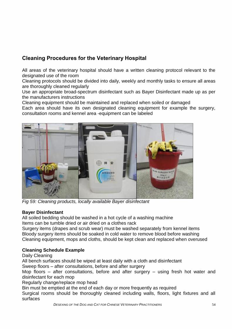
Cleaning Procedures for the Veterinary Hospital All areas of the veterinary hospital should have a written cleaning protocol relevant to the designated use of the room Cleaning protocols should be divided into daily, weekly and monthly tasks to ensure all areas are thoroughly cleaned regularly Use an appropriate broad-spectrum disinfectant such as Bayer Disinfectant made up as per the manufacturers instructions Cleaning equipment should be maintained and replaced when soiled or damaged Each area should have its own designated cleaning equipment for example the surgery, consultation rooms and kennel area -equipment can be labeled
Fig 59: Cleaning products, locally available Bayer disinfectant Bayer Disinfectant All soiled bedding should be washed in a hot cycle of a washing machine Items can be tumble dried or air dried on a clothes rack Surgery items (drapes and scrub wear) must be washed separately from kennel items Bloody surgery items should be soaked in cold water to remove blood before washing Cleaning equipment, mops and cloths, should be kept clean and replaced when overused Cleaning Schedule Example Daily Cleaning All bench surfaces should be wiped at least daily with a cloth and disinfectant Sweep floors – after consultations, before and after surgery Mop floors – after consultations, before and after surgery – using fresh hot water and disinfectant for each mop Regularly change/replace mop head Bin must be emptied at the end of each day or more frequently as required Surgical rooms should be thoroughly cleaned including walls, floors, light fixtures and all surfaces
DESEXING OF THE DOG AND CAT FOR CHINESE VETERINARY PRACTITIONERS 55
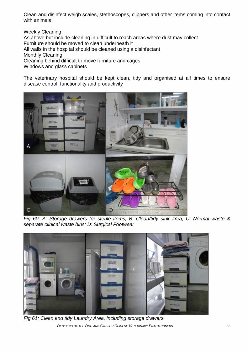
Clean and disinfect weigh scales, stethoscopes, clippers and other items coming into contact with animals Weekly Cleaning As above but include cleaning in difficult to reach areas where dust may collect Furniture should be moved to clean underneath it All walls in the hospital should be cleaned using a disinfectant Monthly Cleaning Cleaning behind difficult to move furniture and cages Windows and glass cabinets The veterinary hospital should be kept clean, tidy and organised at all times to ensure disease control, functionality and productivity
Fig 60: A: Storage drawers for sterile items; B: Clean/tidy sink area; C: Normal waste & separate clinical waste bins; D: Surgical Footwear
Fig 61: Clean and tidy Laundry Area, including storage drawers
A B
C D

DESEXING OF THE DOG AND CAT FOR CHINESE VETERINARY PRACTITIONERS 56
Surgical Suite Should be a designated area where minimal traffic passes through Only the vet and vet assistants should be present during surgical procedures Surgery area walls should be wiped before and after surgery with a disinfectant Floors swept and mopped before and after surgery Surfaces should be wiped with a broad spectrum disinfectant such as Bayer Disinfectant, before and after surgeries The surgical suite should be kept as clean as possible in between patients, for example remove clipped hair, clean clipper blades and clean blood spills A clean towel or blanket should be used for each new patient Wipe the surgery table and instrument table with disinfectant in between procedures The surgical suite should undergo a thorough cleaning and the end of each day which includes cleaning of all surfaces, removing all traces of blood spills and pet hair Bins must be emptied daily
Fig 62: A: Clean surgical suite ready for use; B: Stocked disposables table; C: Surgical kit table
A B C
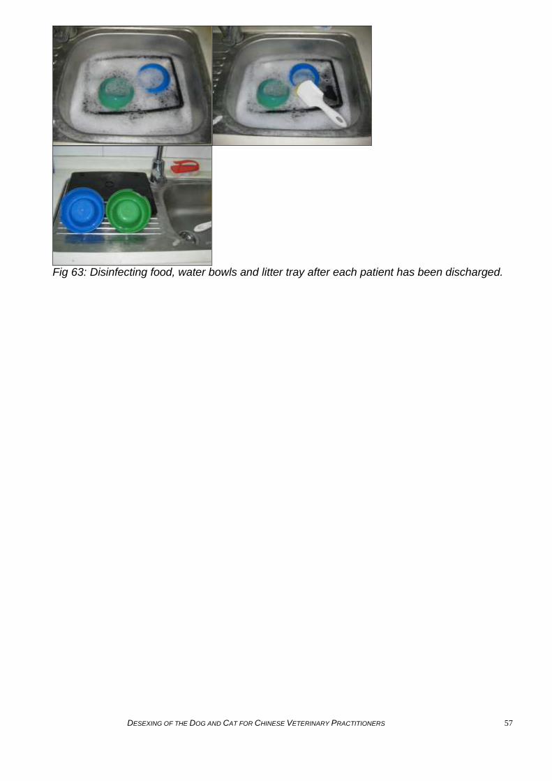
DESEXING OF THE DOG AND CAT FOR CHINESE VETERINARY PRACTITIONERS 57
Fig 63: Disinfecting food, water bowls and litter tray after each patient has been discharged.

DESEXING OF THE DOG AND CAT FOR CHINESE VETERINARY PRACTITIONERS 58
APPENDIX 2 INFECTIOUS DISEASE Identification In-Hospital Management Prevention Rabies IDENTIFYING SUSPECT ANIMALS All animals presented for routine desexing should be examined for infectious disease prior to surgery. Infectious disease may cause serious illness under anaesthesia or during surgery, and routine surgical procedures should not be performed on animals with concurrent disease. Additionally animals that have infectious disease present a risk to other patients and so should be isolated and treated appropriately. All staff members, including reception staff, should be able to recognise when an infectious risk is being presented from the patient‟s history or presenting complaint – ie ocular or nasal discharge, pyrexia, sneezing, coughing, diarrhoea, etc. The receptionist should notify veterinary staff and try to keep a potentially infectious animal separate from other patients waiting in the reception area to avoid spread of disease. Animals identified as possible carriers of infectious disease should be moved directly into an empty examination room or an isolated facility using the shortest route possible and ideally without exposing any other animals. Upon presentation, a history should be taken including the location where the animal lives, estimated age, sex and any noted health problems. A thorough clinical examination should be performed (where possible?) from what about if the animal is aggressive? nose to tail including examination of the eyes, ears, lymph node palpation, mouth examination, capillary refill time and mucous membrane examination, auscultation of the heart and lungs, palpation of the body and limbs and abdomen, and examination of the genitals and mammary tissue. A good recording system must be in place, including the animal‟s identification, clinical condition and symptoms, test results, treatment and outcome. Wherever possible, PCV and TP measurements and microscopic evaluation of blood smears from animals should be done prior to surgery, as this will help in the identification of anaemia and blood-borne disease. IN HOSPITAL MANAGEMENT OF SUSPECT ANIMALS TRANSMISSION OF DISEASE: Infections can be spread through: Faeces Urine Respiratory secretions Blood Reproductive secretions Bites
DESEXING OF THE DOG AND CAT FOR CHINESE VETERINARY PRACTITIONERS 59
Scratches Contact with vectors
Shared food and water dishes Medical or surgical instruments used for multiple animals Human hands, clothing and shoes Shared kennels Cleaning equipment that is inappropriately disinfected Direct contact Aerosolised infectious agents e.g. from animals that are coughing or sneezing
The following simple preventive measures should be used at all times to decrease the chance of spread of disease within the hospital setting:
1. Wash hands before and after each patient contact 2. Place animals with suspected infectious diseases immediately into an examination
room or an isolation area on admission to the hospital 3. Isolate infectious patients in a designated kennel area and ensure strict hygiene
measures such as handwashing and footbaths or shoe-changing facilities are in place. 4. Wear gloves, shoe covers, apron and masks (where appropriate) when handling
patients where infectious or zoonotic diseases are a possibility or suspected 5. Minimize contact with hospital materials (instruments, records, door handles, etc.)
while hands or gloves are contaminated (use a clean, dry paper towel when needed then discard).
6. Change outer garments after each visit/examination of an animal or when soiled by faeces, secretions or exudates
7. Clean and disinfect equipment (stethoscope, thermometers, pens, mops, dishes, litter trays, cleaning buckets, cleaning rags, etc..) after each use with animals likely to have an infectious disease
8. This equipment should remain only in use within the isolation area 9. Do not consume food or drinks in areas where patient care is provided 10. Examination tables, cages, runs should be cleaned and disinfected immediately after
each use 11. Procedures using general hospital facilities, such as surgery and radiology, should be
postponed until the end of the day, after other patients have been returned to their kennels.
12. Biological and hazardous materials should be clearly labelled and disposed of appropriately
13. Disposable materials should be placed in plastic bags within the isolation area and the external surfaces sprayed with disinfectant before removed from the area
14. Contaminated equipment and surfaces should be cleaned and disinfected, remove contaminated gowns/garments and shoe covers
15. Release animals using the shortest route out of the hospital (is there another word not release, sounds like just let cat out of front door) ie should the animals be returned to the area that they were found?
When clinical history or findings suggest the animal may be infectious, it must be isolated in a designated isolation area and not be allowed to contact or share the same environment as non-infectious animals. The isolation area should be a separate area with limited access, or preferably an entirely separate area. No one should enter this area unless required for treatment of isolated animal. The isolation area should have its own handwashing and foot-bath facilities. If only one person is available to treat both healthy and isolated animals, the healthy animals must be attended to first, and ensure that all protective clothing is removed and hands/forearms thoroughly washed before returning from the isolation area. Alcohol hand

DESEXING OF THE DOG AND CAT FOR CHINESE VETERINARY PRACTITIONERS 60
disinfectant is useful but will only work on clean skin and so should be used after hand-washing. A clear protocol for management of animals, cleaning and disinfection should be posted in the isolation area. Cleaning products should be used at the correct concentration and for the correct contact time (for some agents it is up to 10 minutes; consult manufacturer‟s recommendations). NOTE: Phenolic compounds are toxic to cats and should not be used. All equipment and areas within the unit should be cleaned of organic matter (faeces, urine, fur, etc) before being cleaned with a detergent followed by the appropriate disinfectant, such as bleach (sodium hypochlorite). All potentially contaminated surfaces, such as animal pens and cages as well as floors and tables, should be cleaned at least once a day with a 2-5% solution of sodium hypochlorite in clean water. There should be adequate ventilation during use and all surfaces should be thoroughly dried before contact with an animal. An additional thorough clean, which includes walls, doors and all equipment within the unit, as well as the surfaces listed above, should be carried out once every week. Particular care should be paid to the thorough cleaning of an animal‟s pen or cage after the animal has been released. If possible after cleaning and disinfection, the pen or cage should not be used for at least 24 hours before another animal is introduced. All waste from the isolation area should be double-bagged and labelled as coming from the isolation area. It should not be left lying around as it is potentially contaminated. Hands and forearms should be thoroughly washed in 2-4% chlorhexidine pre-prepared solution for a minimum of 1 minute and either air dried or dried thoroughly with clean disposable paper towels. PREVENTION OF DISEASE Local veterinary communities should be consulted regarding their impression of the prevalence and distribution (e.g. age, gender or location) of certain (?infectious or zoonotic?) diseases. Clinical management must be on a case-by-case basis, depending on the aetiology and should be in accordance with recommendations described in standard medical textbooks routinely used in veterinary teaching universities and colleges throughout the world. Owners or guardians of an animal that is sick with an endemic disease should be given advice regarding the treatment options and prognosis for the animal. In some cases, euthanasia of the animal may be the most humane option. Euthanasia should be considered in all cases where the animal‟s welfare is impaired and is unlikely to improve. This may be due to terminal disease, risk to humans and other animals (e.g. rabies), chronic disease (e.g. eye or skin disease) with no guardian to take responsibility for treatment and ensure a good standard of care or a lack of resources at the clinic to treat the animal appropriately and ensure its recovery. See euthanasia protocols for further information.
DESEXING OF THE DOG AND CAT FOR CHINESE VETERINARY PRACTITIONERS 61
RABIES DETECTION AND MANAGEMENT GUIDELINES Rabies is a 100% preventable viral disease of mammals. It is most commonly transmitted by the bite of an infected animal that has rabies virus in its saliva. The rabies virus is shed at high levels in saliva. Contact such as petting or handling an animal, or contact with blood, urine or feces does not necessarily constitute an exposure risk. People usually get rabies from the bite of a rabid animal. It is also possible, but rare, that people may get rabies if infectious material from a rabid animal, such as saliva, gets directly into their eyes, nose, mouth, or a wound. Scratches, abrasions, open wounds, or mucous membranes contaminated with saliva or other potentially infectious material (such as brain tissue) from a rabid animal can be classed as non-bite exposures. If a staff member has an open wound it is advisable they do not come into contact with suspect or confirmed rabies cases. All species of mammals are susceptible to rabies virus infection, but only a few species, such as cats and dogs, are important reservoirs for the disease. The only available method to confirm rabies virus in animals aside from recognizing symptoms is post mortem by submitting the animal‟s brain for microscopic examination. All people working with animals should protect themselves from rabies by taking a course or prophylactic rabies vaccinations. Rabies prevention
1. Remember rabies is 100% preventable – it has been eradicated in many countries. 2. All dogs and cats should be vaccinated against rabies and appropriate booster
vaccinations should be given. 3. Pet owners can reduce the possibility of pets being exposed to rabies by not letting
them roam free. 4. Spaying or neutering your pet may reduce any tendency they might have to roam or
fight and therefore reduce the chance that they will be exposed to rabies. 5. All staff working with animals should seek medical advice with regard to their risk from
rabies. 6. All staff working with animals should be prophylactically vaccinated against rabies –
doing this will significantly reduce your risk of disease should you be bitten by a rabid animal.
7. Wear protective face masks, gloves, clothes, and shoes when handling anything from an animal suspected to have rabies or when cleaning areas where suspected rabid animals are confined.
8. The rabies virus does not survive long outside of animals. It is generally destroyed by heat, sunlight, or air. Detergent, ethyl alcohol, or a one-in-ten dilution of household bleach can also destroy the rabies virus.
9. Routinely disinfect working surfaces, tools and instruments, floors and walls that may have been contaminated with fluids from animals, using procedures established for infection control.
Remember prevention is better than cure! The Rabies virus Rabies is a preventable viral infection. Rabies is zoonotic, which means it can be transmitted from animals to people. Rabies virus causes an acute encephalitis and the outcome is almost always fatal in humans and animals. Rabies is caused by a virus and cannot be treated with antibiotics. Rabies can be transmitted by any warm-blooded mammal, not just by stray mixed-breed dogs.

DESEXING OF THE DOG AND CAT FOR CHINESE VETERINARY PRACTITIONERS 62
Rabies in Cats and Dogs The rabies virus travels through the body when it is introduced into a muscle through a bite from an infected animal. The virus travels along the central nervous system to the brain. The animal does not appear sick during this time. The virus is relatively slow moving and the average time of incubation from exposure to brain involvement is between 3 to 8 weeks in dogs and 2 to 6 weeks in cats. Animals infected with rabies can shed virus in their saliva for up to two weeks prior to the onset of clinical signs. Once the virus has reached the brain it multiplies, causing inflammation, it then progresses to the salivary glands and saliva. After the virus has multiplied in the brain, almost all animals begin to show the first signs of rabies. Usually within 3 to 5 days, the virus has caused enough damage to the brain that the animal begins to show signs of rabies. The first signs of rabies may be nonspecific and include lethargy, fever, vomiting, and anorexia. Signs progress within days to cerebral dysfunction, cranial nerve dysfunction, ataxia, weakness, paralysis, seizures, difficulty breathing, difficulty swallowing, excessive salivation, abnormal behavior, aggression, and/or self-mutilation. After the virus reaches the brain the animal will show one, two, or all three of the following phases: Prodromal phase Includes apprehension, nervousness, anxiety, solitude, and a fever. You may also note a change in the animal‟s normal behaviour: Friendly animals may become shy or irritable and may snap, whereas, aggressive animals may become affectionate and docile. Most animals will constantly lick the site of the bite. In cats, the prodromal phase lasts for only 1-2 days and they usually develop more fever spikes and erratic behaviour than dogs. Furious phase Animals may enter the furious stage; cats are particularly prone to developing this phase. Animals become restless and irritable and are hyperresponsive to auditory and visual stimuli. As they become more restless, they begin to roam and become more irritable and vicious. Animals become disoriented, have seizures and eventually die. Paralytic (dumb) phase Animals may develop the paralytic phase either after the prodromal or furious stage. The paralytic phase usually develops within 2 to 4 days after the first signs are noted. Nerves affecting the head and throat are the first to be involved and animals may begin to salivate as a result of their inability to swallow. Deep laboured breathing and a dropped jaw may result as the diaphragm and facial muscles become increasingly paralyzed. Animals may make a choking sound. The animal will get weaker and eventually go into respiratory failure and die. Management of Suspect Cases Rabies virus might be excreted in the saliva of infected dogs and cats during illness and/or for only a few days before illness or death. A healthy dog or cat that bites a person should be confined and assessed to determine the nature of the aggression. Was this fear response aggression? Is this a normally aggressive animal? Or is this a possible rabies sign? If the animal is suspected to be frightened, take measures to reassure the animal and observe. For animals with a low probability of rabies, observation periods (10 days) may be appropriate to rule out the risk of potential human rabies exposure. If the animal has rabies, it should deteriorate markedly within this time. All staff should be familiar with normal signs of

DESEXING OF THE DOG AND CAT FOR CHINESE VETERINARY PRACTITIONERS 63
canine and feline fear and aggression – remember animals in a strange environment may behave aggressively – this can be normal and should not be confused with a potentially rabid animal. If an animal suspected of having rabies is responsive or manageable, keep it alive and away from other animals or people, until expert assessment can be made. Administration of rabies vaccine to the animal is not recommended during the observation period to avoid confusing signs of rabies with possible side effects of vaccination. Do not touch the animal with bare hands. If an animal is unmanageable and dangerous and cannot be restrained, observe its movements and seek help from qualified experts as soon as possible. If you must handle the animal or carcass, wear protective gloves to prevent infectious material from having contact with cuts or rashes on the skin. Also wear protective masks and goggles to protect against infectious aerosols. Animals in confinement should be evaluated by a veterinarian at the first sign of illness. If signs suggestive of rabies develop, the animal should be humanely euthanized. An animal behaving irrationally and suspected of rabies may need to be humanely euthanized. Do not approach or kill an animal suspected of having rabies except to defend yourself, other people, or other animals. If an animal must be euthanized, follow humane euthanasia guidelines. If/when performing a post-mortem to confirm a potential rabies case, avoid damaging brain tissue as an undamaged brain is important for a quick laboratory diagnosis of rabies. Management of Confirmed Cases All animals showing clinical signs of Rabies or suspected cases of Rabies must be humanely euthanased as per Appendix If you are bitten by a suspect or confirmed rabid animal Act immediately to remove the rabies virus at the site of the infection: Thoroughly wash the wound with soap and water and flush with povidone iodine or alcohol for a minimum of 15 minutes to kill the rabies virus While this is being done, shield the eyes, nose, and mouth from spray from the wound Seek medical attention urgently
DESEXING OF THE DOG AND CAT FOR CHINESE VETERINARY PRACTITIONERS 64
APPENDIX 3 VACCINATION PROTOCOLS
CAT VACCINATION PROTOCOL
Vaccinations are a very important and effective method of preventing disease in animals and minimising spread of disease among cat and dog and human populations. However, vaccines are not always available for all disease pathogens and vaccines are not always 100% effective. Therefore, it is still necessary to follow the above biosecurity measures to prevent or minimize the spread of infection. Healthy cats should be vaccinated with a high quality live attenuated vaccine against feline herpes virus, feline calici virus and feline parvoviral enteritis (panleucopaenia). If cats are less than 12 weeks of age they should be vaccinated twice at an interval of 3 weeks (the second vaccine given after the cat is 12 weeks of age) if retrapping can be achieved. If it is not possible to re-capture the animal, they may be hospitalised or housed during this 3 week period until they can be administered a second vaccine. Healthy cats should also be vaccinated at least once with a high quality inactivated rabies vaccine given subcutaneously. Pregnant animals must never be vaccinated with live attenuated virus. Only killed inactivated vaccines approved by the manufacturer should be used for pregnant animals (pregnant animals may still be desexed and the kittens euthanised humanely rather than adding more kittens to the stray cat population where they can die of disease, trauma or neglect). Animals should receive at least one prophylactic treatment against conditions with zoonotic potential: Echinococcus, Toxocara, Rabies, fleas, mange (Sarcoptes, Cheyletiella) and other endoparasites and ectoparasites depending on local conditions (such as lice, ticks, ear mites). Animals should be weighed accurately to determine a safe and effective dosage of treatment. Care should be taken when using these products in sick animals.
DOG VACCINATION PROTOCOL Vaccinations are a very important and effective method of preventing disease in animals and minimising spread of disease among cat and dog and human populations. However, vaccines are not always available for all disease pathogens and vaccines are not always 100% effective. Therefore, it is still necessary to follow the above biosecurity measures to prevent or minimize the spread of infection. Healthy dogs should be vaccinated with a high quality live attenuated vaccine against Distemper, Parvovirus and Parainfluenza (CAV-2: providing cross protection for Canine Infectious Hepatitis). Dogs over 12 weeks of age are vaccinated once subcutaneously, and dogs less than 12 weeks of age are vaccinated twice at an interval of 2-3 weeks (the second vaccine should be given after the dog is 12 weeks of age) if retrapping can be achieved. Booster vaccinations should be administered according to the vaccine manufacturers schedule. Healthy dogs should also be vaccinated at least once subcutaneously with a high quality inactivated rabies vaccine. Booster vaccinations should be administered according to the vaccine manufacturers schedule.

DESEXING OF THE DOG AND CAT FOR CHINESE VETERINARY PRACTITIONERS 65
Additionally animals should receive at least one prophylactic treatment against: Conditions with zoonotic potential: Echinococcus, Toxocara, Rabies, fleas, mange (Sarcoptes, Cheyletiella). Other endoparasites and ectoparasites depending on local conditions (such as lice, ticks, ear mites). Animals should be weighed accurately before dosage. Care should be taken when using these products in sick animals

DESEXING OF THE DOG AND CAT FOR CHINESE VETERINARY PRACTITIONERS 66
APPENDIX 4 SURGICAL KIT PREPARATION AND STERILISATION
Example of Basic Kit Contents
Large dog Spey Kit Dog Castration/Small Dog/Cat Spey Kit
Cat castration Kit
4 Artery forceps (sized between 10cm and 16cm)
2 13cm Allis tissue forceps
1 14cm Mayo scissor curved
1 14cm needle holders
1 Rat toothed forcep
1 Dressing forcep
1 Size 3 scalpel handle
1 Spey hook (optional)
5 Cotton surgical gauze swabs
Additional information on surgical instruments may be found in surgical textbooks such as “Small animal surgery” by Teresa Welch fossum.
1. If using sterile suture from a cassette, sterile needles must be used. Select an appropriate round-bodied or cutting needle according to the tissue type. If using suture material with a swaged on needle, additional needles are not required.
2. Instruments, including needles, should be cleaned and inspected after each use and removed from the kit and replaced if blunt, damaged or rusted.
3. Instruments must be cleaned and sterilised between each patient 4. Use a new scalpel blade for each surgery, if the blades or needles become blunt
during a surgery, replace it with a new sterile blade or needle. 5. Any sterilised surgical instrument, gauze swab, scalpel blade, suture material etc that
becomes contaminated during surgery must be replaced with a new sterile item
CLEANING INSTRUMENTS POST-SURGERY
1. The veterinary surgeon should discard the scalpel blade and any other sharps into a
sharps bin immediately after surgery. 2. Soak the surgical instruments in fresh, clean, cold water, for 10 minutes immediately
after surgery is complete to remove blood and tissue. Ensure all instrument ratchets are opened during the cleaning process (Fig 65 A).
3. In fresh, clean water add an enzymatic cleaner such as „CSI Liquid Instrument Detergent‟ - Anhui Greenland Disinfection Products Co., Ltd (Fig 64), soak as per manufacturers‟ instructions and using a scrubbing brush thoroughly scrub away all blood and tissue from the instruments (Fig 65 B&C). It is important to wear gloves when using an enzymatic cleaner.
4. Pay special attention to instrument hinges, serrated sections, ends and handles to ensure all blood and tissue is removed. Again ensure all instrument ratchets are opened during the cleaning process.
1 Scalpel blade 2 12cm Artery forceps
1 14cm Mayo scissor curved
1 14cm needle holders
1 Rat toothed forcep
1 Dressing forcep
1 Size 3 scalpel handle
4 Towel clamps
5 Cotton surgical gauze swabs

DESEXING OF THE DOG AND CAT FOR CHINESE VETERINARY PRACTITIONERS 67
Fig 64: CSI Liquid Instrument Detergent’ - Anhui Greenland Disinfection Products Co., Ltd www.ahlvzhou.com.cn
Fig 65: A: Soak instrument immediately after surgery; B & C: scrub instruments clean Manual cleaning alone may not be enough to remove all surgical exudates, enzymatic cleaners are very effective however if available follow with ultrasonic cleaning or alternatively proceed to Step 13. Ultrasonic Cleaning:
5. Ultrasonic cleaning is more effective than manual cleaning 6. It should be performed after debris has been manually removed 7. Place instruments in a solution prepared to manufacturers instructions. 8. Do not place different metals together in solution (corrosion can occur) 9. Instruments should not be piled on top of each other as delicate instruments may be
damaged 10. Ensure that ratchets and jaws on the instruments are open when placed in cleaning
solution. 11. Once the ultrasonic process has finished, proceed to step 12.
Fig 65: Ultrasonic Cleaner- Yongfeng Enterprise Co., Ltd. www.yongfeng-medical.com
A B C

DESEXING OF THE DOG AND CAT FOR CHINESE VETERINARY PRACTITIONERS 68
12. Remove instruments from the cleaner and lay out on a clean towel. 13. Rinse instruments (Fig 66 A) and towel dry soon after cleaning. Water spotting stains
can occur if the instruments are "air" dried. 14. Ensure that all instruments are laid out with ratchets unlocked, in an open position (Fig
66 B).
Fig 66: A: Rinse instruments; B: lay out with ratchets open.
PREPARING SURGICAL INSTRUMENTS FOR STEAM STERILISATION
Sterilising surgical instruments is essential to prevent infection and the spread of disease between patients. All organic material should be removed from instruments prior to sterilising. Instruments should be sterilised with hinges open to ensure proper sterilisation. Double wrap method:
1. Lay out a clean lint free cloth drape on a clean work surface 2. Place a second clean, lint-free cloth drape (the inner drape) onto the first drape (the
outer drape) (Fig: 67 A) 3. Place instruments in a neat pile in the centre of the inner drape (Fig 67 B) 4. Place an indicator strip in the centre of the instruments (Fig 67 C) 5. Place 5 clean gauze swabs on top of the instruments (Fig 67 D) – gauze swabs may
also be wrapped separately
A B
A B
C D
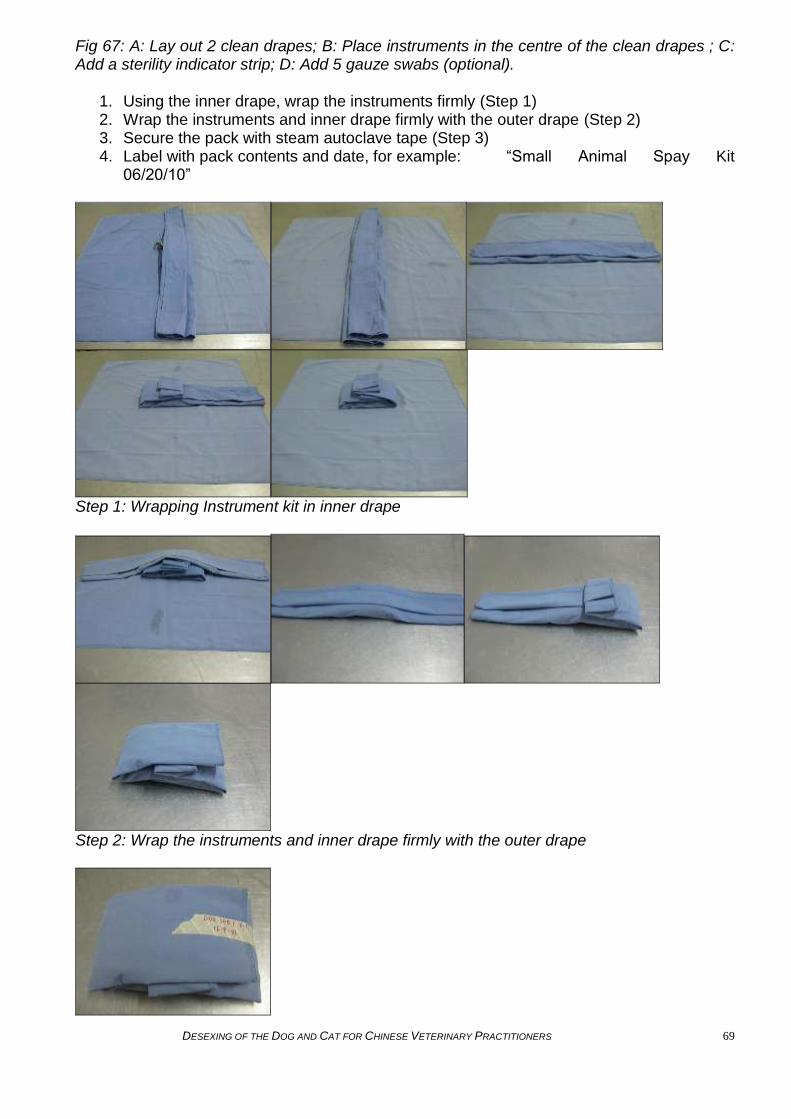
DESEXING OF THE DOG AND CAT FOR CHINESE VETERINARY PRACTITIONERS 69
Fig 67: A: Lay out 2 clean drapes; B: Place instruments in the centre of the clean drapes ; C: Add a sterility indicator strip; D: Add 5 gauze swabs (optional).
1. Using the inner drape, wrap the instruments firmly (Step 1) 2. Wrap the instruments and inner drape firmly with the outer drape (Step 2) 3. Secure the pack with steam autoclave tape (Step 3) 4. Label with pack contents and date, for example: “Small Animal Spay Kit
06/20/10”
Step 1: Wrapping Instrument kit in inner drape
Step 2: Wrap the instruments and inner drape firmly with the outer drape

DESEXING OF THE DOG AND CAT FOR CHINESE VETERINARY PRACTITIONERS 70
Step 3: Secure the pack with steam autoclave tape Alternative methods for kit wrapping: A kit may be single wrapped using the above method and only one drape, however this kit must then be bagged to protect sterility (Fig 68).
Fig 68: Single drape wrapped and bagged kit Gauze swabs (Fig 69 A) and suture needles (Fig 69 B) may also be wrapped separately in an autoclave bag. ALWAYS record the number of swabs being packed so that the surgeon can count them before and after surgery to ensure that none have been left inside the animal.
Fig 69: A: Sterile surgical needles; B: Alternative presentation of 5 sterile swabs Sterilisation Pouches available from Shiny Medical Disposable Products Co., Ltd.
STERILISING INSTRUMENTS
Steam Autoclave (Fig 70): Steam destroys microbes by coagulating and denaturing cellular proteins. There must be a correct relationship between temperature, pressure, and exposure time, in order for all micro-organisms to be destroyed. Minimal Safe Standards for Steam Sterilisation:
Surgical Kit 121˚C/30 minutes
Surgical gowns 132˚C/15 minutes
Surgical swabs 132˚C/15 minutes
Surgical hand towel 132˚C/15
A B

DESEXING OF THE DOG AND CAT FOR CHINESE VETERINARY PRACTITIONERS 71
When packing the autoclave chamber, items should never be stacked on top of one another, but should be placed standing upright for adequate steam flow. Place heavier instruments towards the bottom of the autoclave, and lighter instruments at the top Never overload the autoclave as it impedes steam flow and does not allow for proper drying. Improper wrapping of an instrument kit or overloading the sterilising unit with too many kits may result in improper sterilisation.
Fig 70: Steam Autoclave - Yongfeng Enterprise Co., Ltd. www.yongfeng-medical.com
An indicator of successful sterilisation should always be used to ensure quality of sterility. Examples of indicators are Steam Autoclave Indicator Tape or KDL Sterilisation Indicator Strip - New Era Adhesives Technology Co.,Ltd (Fig 71 A & B) . Indicators should always be packed in the centre of a kit, not at the edge. Or outer wrapping
Fig 71: A: KDL Sterilization Indicator Strip; B: Steam Autoclave Indicator Tape - New Era Adhesives Technology Co. Ltd Storage of Autoclaved/sterile items (Fig 72): Store packs and sterile materials in a non-humid, dust free, well ventilated area Sterile items must be kept dry Store non-sterile and sterile items separately
minutes
Surgical drape 132˚C/15 minutes
A B

DESEXING OF THE DOG AND CAT FOR CHINESE VETERINARY PRACTITIONERS 72
Sterile packets should not be folded or torn If the sterility of an item is questionable, treat it as contaminated and re-sterilise
Standard storage times for autoclaved items:
Double drape wrapped Kit 2 months
Wrapped and bagged kit 6 months
Fig 72: Storage and organisation of sterilised items
Wet/Cold Sterilisation: Used when instruments cannot be autoclaved Use non-corrosive chemicals with this method and avoid using solutions that contain benzyl ammonium chloride, which dissolves tungsten on some instruments Solutions should also be tissue-compatible, but still rinsed with sterile water or saline before use Glutaraldehyde is non-corrosive and can be used at a 2% solution to sterilise instruments, products such as Medical Instrument Glutaraldehyde Disinfectant - Anhui Greenland Disinfection Products Co., Ltd www.ahlvzhou.com.cn Items must be thoroughly cleaned of all blood and organic matter which may prevent successful sterilisation Items must be completely dry to prevent chemical dilution Instruments should be sterilised with hinges open. Chemical sterilisation is harsh on instruments and will shorten their life. Recommended immersion time for instrument sterilisation:
2% Glutaradehyde 10 hours at 68 – 77˚F (20-25˚C) Wet/Cold Disinfection Used when instruments are to be used for non-sterile procedures. Not acceptable for abdominal surgery Use chemicals such as 5% Clorhexidine gluconate concentrate diluted or 2% Glutaraldehyde Recommended instrument immersion time for instrument disinfection:
2% Glutaradehyde 10 minutes at 68 – 77˚F (20-25˚C) 5% Chlorhexidine gluconate Dilute 10 mL 5% Chlorexidine gluconate in 15 mL distilled water, then make up to 100 mL with commercial grade methylated spirits This solution can be used to contain a „wet kit‟ suitable for non-sterile procedures such as minor stitch ups The solution must be replaced weekly or more frequently if contaminated or dirty.

DESEXING OF THE DOG AND CAT FOR CHINESE VETERINARY PRACTITIONERS 73
APPENDIX 5 DRUGS ANALGESICS Tramadol Tramadol is available in both oral and injectable forms. The dose is 2-4mg/kg orally it IV every 12 hours and it may be used in conjunction with a variety of anaesthetics to provide an opioid-like analgesia during and after surgery. Tramadol mimics the effects of opioids on the brain and is an excellent analgesic. Its inclusion in combination with Zoletil and xylazine is documented to provide multimodal anaesthesia. Tramadol has minimal side-effects but may cause sedation at higher doses. Non-steroidal anti-inflammatory drugs (NSAIDs) – Meloxicam (Mobic/Meloxicam, Boeringher-Ingelheim, Tepoxalin (Zubrin, Intervet Schering-Plough), Tolfenamic acid (Tolfedine, Vetoquinol) NSAIDs are anti-inflammatory drugs that inhibit the production of Prostaglandins by interrupting the enzyme COX2 in the inflammatory cascade. However they also inhibit the action of COX1, a necessary enzyme and this may lead to side effects. NSAIDs that select preferentially for COX2 produce fewer side effects. NSAIDs are excellent at reducing surgical pain and inflammation and should be administered prior to surgery unless there is concern the patient may have renal or hepatic disease. Different types of NSAIDs should never be given during the same course of treatment and NSAIDs should never be given with steroids such as prednisone. Combining different NSAIDs or combining NSAIDs with steroids is extremely dangerous and may result in severe side effects. Side effects in dogs and cats: Gastrointestinal ulceration Vomiting or diarrhoea Renal impairment Cats are especially sensitive to the renal effects of NSAIDs and extreme caution should be used when calculating dosages. Always check that the brand of NSAID you administer is suitable in each species Local anaesthetics – Lignocaine Local anaesthetics provide localised loss of sensation by blocking nerves at the site of administration. Local anaesthesia may be useful in providing analgesia to wounds (infiltration) and is helpful in minor procedures as well as dentistry where it may be used to block specific nerves and prevent pain when the patient awakes from anaesthesia. Lignocaine is also applied topically to the vocal chords of cats prior to intubation in order to prevent laryngospasm. Local anaesthesia alone is not sufficient for surgical procedures that enter a body cavity.
SEDATIVES Benzodiazepines – Diazepam and Midazolam Diazepam and Midazolam are anxiolytic drugs that cause good muscle relaxation. They are useful as part of pre-medication sedation and to reduce stress and encourage relaxation in patients. They should not be used alone for surgical procedures but are useful as part of a

DESEXING OF THE DOG AND CAT FOR CHINESE VETERINARY PRACTITIONERS 74
multi-modal anaesthetic approach. Either midazolam or diazepam may be used in combination with ketamine. These drugs are available in China but their use may be restricted. Butyrophenones - Droperidol and haloperidol Butyrophenones are neuroleptic agents and cause tranquilisation and sedation. Haloperidol is available in China in combination with etorphine as Su Mian Xin 846. Droperidol may be used as a pre-medication sedative agent. Side effects in dogs and cats: Doses should be reduced in elderly patients Droperidol may cause cardiac conduction delays in susceptible patients Phenothiazines - Acepromazine maleate and Chlorpromazine hydrochloride Phenothiazines are neuroleptics with antipsychotic, anxiolytic and sedative effects. They also have anti-emetic effects. They may be useful pre-medication drugs. Side effects in dogs and cats: Ataxia and extra-pyramidal signs Priprism Hypotension Opioids – Etorphine, morphine, pethidine, buprenorpine and butorphanol Opioids are generally restricted in China. Su Mian Xin 846 contains etorphine, a potent opioid sedative and an excellent analgesic (see below). The partial opioids buprenorphine and butorphanol are available in Hong Kong. Butorphanol is an extremely poor analgesic and also has the capacity to block the actions of other opioids and so it should not be used as an analgesic or to provide pre-surgical analgesia unless there are no other opioids available. Butorphanol is a useful sedative. Buprenorphine is a good analgesic but has minimal sedative effects, it is useful in combination with other drugs to provide multimodal anaesthesia and analgesia. Opioids may be used together with NSAIDs, and other anaesthetic drugs. Opioids are absorbed across mucous membranes, orally and parenterally and may cause respiratory depression and death in humans at much lower doses than those used in animals. Any clinic using opioid drugs should keep an accessible supply of the reversal agent naloxone in stock which should be administered immediately in case of human exposure. Side effects in dogs and cats: Respiratory depression Bradycardia Excitement (especially in cats) Alpha2 andrenergic agonists - Xylazine (Rompun) and Medetomidine (Domitor) Bayer Animal Health Alpha2-adrenergic agonists stimulate the sympathetic nervous system. Xylazine is available in China in combination with two other unlisted agents. This combination Lu Mian Ning (II) or Su Mian Xin (II) is produced by Chang Chun University and because the two unlisted agents remain secret, it’s use cannot be recommended as potential adverse effects cannot be predicted or managed without knowing the ingredients in the compound. Medetomidine and xylazine are available in Hong Kong. Xylazine causes sedation and CNS depression, provides pain relief and muscle relaxation. The effects of xylazine can be reversed with the use of idazoxan hydrochloride or yohimbine. This can significantly reduce the amount of time needed for recovery. Xylazine is NOT an anaesthetic and should never be used alone for surgical procedures. Xylazine may be used in combination with ketamine or zoletil.

DESEXING OF THE DOG AND CAT FOR CHINESE VETERINARY PRACTITIONERS 75
In dogs and cats the onset of action following an IM or subcutaneous (SQ) dose is approximately 10-15 minutes, and 3-5 minutes if given intravenously (IV). Analgesic effects persist for only 15-30 minutes. Sedative effect may last for 1-2 hours depending on the dose given. Complete recovery can take from 2-4 hours in dogs and cats. Vomiting is often seen in cats and occasionally in dogs receiving xylazine, usually within 3-5 minutes after IM or SQ administration To prevent aspiration it is recommended not to induce further anaesthesia until this time has lapsed. Xylazine should not be used in sick, old or debilitated animals as it has severe effects on the cardiovascular system and on renal perfusion which may cause longterm morbidity. Doses should be minimised whenever possible. Adverse effects in cats and dogs: The adverse effects of Lu Mian Ning (II) or Su Mian Xin (II) cannot be predicted as the Chang Chun University refuses to reveal the additional two ingredients. For this reason we recommend avoiding the use of this product. Despite appearing completely sedated, animals can still move, even kick, bite or scratch, in response to sharp auditory stimulation Muscle tremors Reduced respiratory rate. To treat respiratory depression mechanical support is recommended. Xylazine depresses thermoregulation, and so hypothermia or hyperthermia are possible, patient temperature should be closely monitored. Hypertensive initially followed by a longer period of hypotension Bradycardia - an overall decrease in cardiac output may be seen. Brachycephalic dogs with upper airway disease may develop dyspnoea Xylazine can be contraindicated in diabetic animals Occasionally polyuria is seen in cats, monitor hydration status if polyuria present Increased sensitivity to epinephrine, resulting in cardiac arrhythmias Profound muscle relaxation may exacerbate upper respiratory abnormalities in brachycephalic breeds Overdosage In the event of an overdose, cardiac arrhythmias, hypotension and profound CNS and respiratory depression may occur. Seizures have also been recorded Atipamazole, Yohimbine or tolazoline have been suggested to be used alone to reverse the effects of xylazine and to reduce recovery times.
INJECTABLE ANAESTHETICS Propofol (Diprovan), Propofol is an intravenous anaesthetic commonly used in humans. It is safe and easy to use. Propofol is supplied in glass vials and once opened should be stored in a sterile manner in the refrigerator for no more than 24 hours. It is vital that once opened, propofol is not stored for any longer than this as it deteriorates quickly and may cause severe side-effects if used. Propofol will provide only a few minutes of anaesthesia, so is useful for induction of anaesthesia prior to administration of a volatile anaesthetic. Alfaxalone (Alfaxan) Alfaxalone is an intravenous general anaesthetic agent that is difficult to obtain in China. It may be used for induction Dogs = 2mg/kg given intravenously over 60 seconds Cats = 2-5mg/kg given intravenously over 60 seconds

DESEXING OF THE DOG AND CAT FOR CHINESE VETERINARY PRACTITIONERS 76
Alfaxan may also be used to maintain anaesthesia using a constant rate infusion or injection of boluses (see table 2)
DOG Maintenance Cat Maintenance Constant rate infusi
on
(6-7mg/kg/hour of alfaxalone)
Bolus dose each 10min maintenan
ce
(1.0-1.2mg/kg of alfaxalone)
Constant rate infusion
(7-8mg/kg/hour of alfaxalone)
Bolus dose each 10min
maintenance
(1.1-1.3mg/kg of alfaxalone)
Alfaxan® dose (ml/kg/hour)
Alfaxan® dose (ml/kg/every
10min)
Alfaxan® dose (ml/kg/hour)
Alfaxan® dose
(ml/kg/every 10min)
0.6 - 0.7 0.1 - 0.12 0.7 - 0.8 0.11 - 0.13 Table 2 Tiletamine/Zolezepam (Zoletil), Virbac. Zoletil is a combination of a dissociative anaesthetic agent, tiletamine hydrochloride (chemically related to Ketamine) and a tranquiliser, zolazepam hydrochloride (similar to diazepam). Because Tiletamine hydrochloride is a dissociative anaesthetic, the animal may be anaesthetised and unaware of surgical stimuli but reflexes may still persist. Dissociative agents increase respiratory secretions and salivation. It is vital that the anaesthetist is able to differentiate between persistent reflexes under anaesthesia and an animal who has not received sufficient anaesthesia and is struggling due to pain. Zoletil is presented as a powder that is reconstituted with a sterile diluent. It may be diluted with this diluent or with another drug such as xylazine. Once in solution it remains stable for 4 days at room temperature and 14 days when refrigerated. Zoletil should not be stored in a plastic syringe because it may adsorb to plastic. Discard solutions that contain a precipitate or are discoloured. Administration is commonly intramuscular (IM), but can also be used intravenously (IV), and subcutaneously (SQ). Onset of action may be variable, animals should be closely monitored after injection. If supplementary doses are necessary, ensure the dose is less than the initial dose and do not exceed the recommended total dose. Cats In cats the onset of action is within 1-7 minutes after intramuscular injection (IM). Duration of anaesthesia can range between 30–60 minutes at peak effect. Tiletamine decreases cardiac rate and blood pressure after intramuscular injections and respiratory function must be closely monitored. The duration effect of the zolazepam component is longer than that of tiletamine, so there is a greater degree of tranquillisation than anaesthesia during the recovery period. Recovery times vary between 1-5.5 hours Dogs In dogs the onset of action following IM injection averages 7.5 minutes. The average duration of surgical anaesthesia is approximately 27 minutes, with recovery times averaging 4 hours. The duration of tiletamine effect is longer than that of the zolazepam, so there is a

DESEXING OF THE DOG AND CAT FOR CHINESE VETERINARY PRACTITIONERS 77
shorter duration of tranquillisation than there is anaesthesia. In dogs, tachycardia may be a common effect and may last for 30 minutes. Adverse effects in cats and dogs: Zoletil causes hypothermia, animals should be monitored carefully and supplemental heat supplied. Like Ketamine, Zoletil does not abolish pinnal, palpebral, pedal, laryngeal, and pharyngeal reflexes, reflex activity should not be relied on to determine depth of anaesthesia Involuntary muscular twitching Muscle rigidity Respiratory depression, especially with higher doses Apnoea Pain after IM injection, especially in cats Vomiting Excessive salivation and bronchial/tracheal secretions if atropine not administered before hand Vocalisation Erratic and/or prolonged recovery Hypertonia Cyanosis Cardiac arrest Pulmonary oedema Hypertension or hypotension Athetoid movements (constant succession of slow, writhing, involuntary movements of flexion, extension) may occur, do not give additional Zoletil in the attempt to diminish these actions. Ketamine Ketamine is a restricted drug in China but may be available through approved institutions. Ketamine, like tiletamine hydrochloride, is a dissociative anaesthetic meaning that, although the animal may be anaesthetised and unaware of surgical stimuli, reflexes may persist. It is vital that the anaesthetist is able to differentiate between persistent reflexes under anaesthesia and an animal who has not received sufficient anaesthesia and is struggling due to pain. Ketamine should not be used alone for anaesthesia but should be combined with other analgesics and sedatives to provide appropriate muscle relaxation. Adverse effects in cats and dogs Insufficient muscle relaxation Persistence of reflexes Etorphine/Haloperidol (+/-Xylazine) (Su Mian Xin 846), Veterinary Institute of Military Supplies University. 846 is a combination of etorphine, an opioid and haloperidol, an anxiolytic drug. Some earlier preparations also contain Xylazine, an alpha2 agonist. It provides quite deep anaesthesia. It is administered by intramuscular injection. 1 ml of the current preparation contains 4μg etorphine and 2.5 mg haloperidol Intramuscular injection. For cross breed : 0.08~0.1 ml/kg, Pedigree : 0.04~0.08 ml/kg, cats : 0.2~0.3 ml/kg. User warning: Etorphine is a potent opioid and humans and other primates are extremely sensitive to the respiratory depressant effects of opioids. Etorphine is absorbed across mucous membranes, orally and parenterally and may cause respiratory depression and death in humans. Gloves should always be worn when handling Su Mian Xin 846, and eye protection and facemasks are recommended also. Any clinic using this drug should keep an

DESEXING OF THE DOG AND CAT FOR CHINESE VETERINARY PRACTITIONERS 78
accessible supply of the reversal agent naloxone in stock which should be administered immediately in case of self-injection. Adverse effects in dogs and cats: Etorphine is a potent respiratory depressant Poor muscle relaxation Bradycardia Hypotension Sodium pentobarbital An intravenous anaesthetic. Sodium pentobarbital occurs as white crystalline powder or granules and is freely soluble in sterile water, lactated ringers solution or sodium chloride solution. The aqueous solution is not very stable and should not be used if it forms a precipitate. It is very alkaline (pH 9 – 10.5) and care should be taken not to administer extravascularly. Sedation = 2-4mg/kg IV General anaesthesia = 30-35mg/kg IV Can be used for euthanasia at a dose of 120mg/kg for the first 4kg of bodyweight followed by 60mg/kg for every 4kg of bodyweight thereafter. Adverse effects in dogs and cats: Use cautiously in hypovolaemic patients or those with respiratory or cardiac disease. Sodium pentobarbital is contra-indicated in patients with severe liver disease. Should there be a graph somewhere with what drugs we recommend and what drug combo works well together as a pre-med and anaesthetic drug? Just found this slightly confusing.
INHALATION ANAESTHESIA In order to administer inhalational anaesthesia, essential anaesthetic equipment is required. This equipment comprises three primary components: Compressed gas source
1. Usually oxygen alone, can sometimes be a mixture of oxygen and nitrous oxide 2. Pressure gauge showing gas pressure inside tank 3. Pressure regulator or pressure reducing valve – reduces gas pressure as gas flows
from tank to anaesthetic machine, constant pressure of 50psi Anaesthetic machine
1. Flowmeter – precisely controls the amount of gas administered to the patient 2. Vaporiser – contains a volatile anaesthetic agent such as isofluorane or halothane and
controls the amount of vapourised anaeathetic agent carried by the oxygen passing through
Breathing Circuit
1. Unidirectional inhalation and exhalational valves ensuring that gas passes through the system in one direction only
2. Hoses and reservoir bag - filled with gas that enters the circuit when the patient exhales; empties when the patient inhales: stores gas, indicates proper endotracheal tube placement, allows you to observe patient respirations, and deliver O2 by IPPV
3. Pop-off valve - “pressure relief,” allows excess gas to exit the system to a scavenging system.
DESEXING OF THE DOG AND CAT FOR CHINESE VETERINARY PRACTITIONERS 79
4. Oxygen flush valve - O2 bypasses the flowmeter and flushes through the circuit 5. Pressure manometer - measures the pressure of gases within the breathing system.
Indicates pressure when performing IPPV (not to exceed 15-20 cm H2O in small animals).
6. Negative pressure relief valve - open and admit room air into circuit if there is a negative pressure build up (vacuum) detected (i.e., O2 flow too low, active scavenging system).
Breathing circuits are classified into re-breathing (Circle or To-and-fro circuits) and non-rebreathing (Ayre‟s T-piece, Lack, Bain circuits). Different oxygen flow rates should be calculated depending on the size of the patient and the type of system being used – see anaesthetic textbooks for more information. Before using the anaesthetic machine:
1. Inspect anaesthetic machine: fill vaporisers, tighten filler caps, turn off vaporisers; If using a rebreathing system, make sure CO2 absorbent is not exhausted; change if necessary.
2. Central O2 and N20 supplies should be checked for quantity and pressure. Turn on gas cylinders slowly with the flowmeter “off” to assure a minimum pressure of 500psi. The cylinder should be checked for slow leaks (drop in pressure over a 10 min. period).
3. Check breathing circuit for leaks. 4. Connect pop-off valve to scavenger system. 5. Attach reservoir bag to machine. 6. Visually inspect machine for defects and improper connections
Volatile anaesthetics: Volatile anaesthetics are generally safe and well-tolerated. Administration of volatile anaesthesics allows for better anaesthetic control during long or complicated surgical procedures. Examples of volatile anaesthetic agents include isofluorane and halothane. There are some diffferences between different volatile agents, in humans only 0.2% of isofluorane administered is metabolised by the body, but 20-46% of halothane adminstered is metabolised by the liver, which means that the halothane may result in longer recovery times and may not be well metabolised in patients with liver disease. All volatile anaesthetics cause some cardiorespiratory depression but this is generally mild compared to injectable agents., Nitrous oxide Nitrous oxide is a true gaseous anaesthetic, however it is not very powerful and when used alone will not provide suitable anaesthesia. However it may be administered concurrently with either isofluorane or halothane and is an excellent analgesic. Nitrous oxide is administered from a tank like oxygen and mixed with the other anaesthetic gases administered to the patient. Care should be taken that the patient receives an appropriate concentration of oxygen along with the nitrous oxide.
DESEXING OF THE DOG AND CAT FOR CHINESE VETERINARY PRACTITIONERS 80
APPENDIX 6 CARDIO-PULMONARY CEREBRAL RESUSCITATION (CPCR) Anaesthesia causes respiratory and cardiac depression, which may increase the risk of cardiopulmonary arrest (the sudden cessation of ventilation and effective circulation that requires rapid emergency intervention to prevent death.) See Cardiopulmonary arrest section below. Cardiopulmonary Arrest (CPA): can be defined as a sudden cessation of functional ventilation and systemic perfusion. This results in reduced oxygen delivery to tissues with decreased removal of carbon dioxide and cellular death. Cardiac arrest and respiratory arrest may occur simultaneously but often respiratory arrest occurs first and if not quickly treated, cardiac arrest will soon follow. Cardiopulmonary Cerebral Resuscitation (CPCR): is an emergency procedure utilised in cardiac and respiratory arrest to promote forward blood flow and improve oxygen delivery to brain, heart, and other vital organs. Signs of Impending Cardiopulmonary Arrest (CPA) Weak, irregular pulses, irregular heart sounds, tachycardia Sudden Bradycardia Changes in patient‟s respiratory rate, depth, pattern and effort Cyanotic, grey or pale mucous membranes Prolonged capillary refill time Decreasing end-tidal carbon dioxide Hypotension Sudden unexplained increase in anaesthetic depth Hypothermia despite warming efforts Signs of Cardiopulmonary Arrest No detectable heart sounds on auscultation No palpable pulses Fixed, dilated pupils (occurs within 45 seconds of arrest) Apnea or agonal gasping – may be masked if the patient is being artificially ventilated Pale, grey, cyanotic mucous membranes. NOTE: mucous membranes and capillary refill time can remain normal for several minutes after arrest Absence of bleeding at the surgical site, blood may appear dark if the arrest is due to hypoxemia Abnormal ECG tracing – note ECG may remain normal for several minutes after arrest = not reliable Collapse and loss of consciousness of the awake patient Loss of skeletal muscle tone and cranial nerve reflexes Note: Equipment such as an electrocardiograph and pulse oximeter may continue to show readings (even normal readings) after the heart has stopped beating. As soon as CPA occurs turn off the anaesthetic machine (if applicable) reverse injectable anaesthetic agents if possible.

DESEXING OF THE DOG AND CAT FOR CHINESE VETERINARY PRACTITIONERS 81
Reasons for Arrest Overdose of anaesthetic agent Hypothermia Hypotension Hypoglycemia Severe trauma Sepsis Hypoxemia Hypercarbia Hypovolemia Vagal stimulation Pre-existing cardiac disease
EMERGENCY/CRASH BOX There must always be an emergency box readily available that is stocked with essential drugs and equipment for an emergency situation (Fig:73). All members of the surgical team should be familiar with these drugs and equipment and how to use them. Contents: Adrenaline hydrochloride Lignocaine hydrochloride (Fig 74: A, B & C) Atropine sulphate Antagonist drugs such as naloxone or atipamazole tolazoline Endotracheal tubes (ETT) various sizes, ties to secure the tube, laryngoscope (Fig 75: A) IV catheters (Fig 75: B) Needles and syringes of various sizes Ambu bag if available Emergency drug dose chart Attached to the crash box should be an emergency drug chart that gives dose in mL per kg for all drugs required in CPCR. Emergency drugs can be stored decanted into multi-dose injection vials, labeled with drug name, concentration and expiry date. This allows for quick response when drugs are required for an emergency situation.
Fig 73: Emergency box example

DESEXING OF THE DOG AND CAT FOR CHINESE VETERINARY PRACTITIONERS 82
Fig 74: A: Adrenaline Hydrochloride; B: Lignocaine Hydrochloride; C: Atropine sulphate
Fig 75: A: Laryngoscope, ET Tubes, ET tube tie; B: IV catheters, clippers and tape. Cardiopulmonary Cerebral Resuscitation (CPCR) The minimum number of people to run a resuscitation team is two – one to compress the chest and another to ventilate the patient. A third is preferable to interchange during compressions or to draw up drugs. CPCR is usually performed by the manual administration of artificial respiration and cardiac compressions so that adequate oxygenation of the heart and brain can be maintained. Start CPCR as soon as arrest is recognised. Medical records must reflect all that was done for the patient in the case of arrest. Basic Life Support Venous Access Venous access is critical in the successful treatment of cardiac arrest. An animal being anaesthetised should already have an intravenous catheter placed. If no catheter is present, one should be placed as soon as possible during resuscitation. Large bore catheters are most useful during an emergency procedure as they provide the least resistance to large amounts of fluids. However large bore catheters can be difficult to place in the case of severe circulatory collapse so a smaller catheter may need to be used as this may be easier to place. It is therefore recommended to have a range of sizes available in the crash box. Resuscitation is classified as the ABCs A - Airway
A B C
A B

DESEXING OF THE DOG AND CAT FOR CHINESE VETERINARY PRACTITIONERS 83
B - Breathing C – Circulation A - Airway Management: make sure the patient has a patent airway It is important to quickly establish a patent airway. The most common and effective method is endotracheal intubation with an endotracheal tube (ETT). Intubating all patients routinely will facilitate prompt emergency resuscitation. If the patient is already intubated check the ETT is not blocked or dislodged. B – Breathing: deliver breaths to the patient Intermittent positive pressure ventilation (IPPV) with 100% oxygen should commence. This may be administered using an anaesthetic machine (with the vapouriser turned off) and an anaesthetic circuit. Initially patients should be given two breaths with the inspiratory period lasting 1-2 seconds and then assessed for signs of spontaneous ventilation. If spontaneous ventilation does not occur, ventilation at a rate of 10-12 breaths per minute should continue. For dogs, peak inspiratory pressure should be kept below a maximum of 20cm H2O on the manometer if using an anaesthetic machine. Cats and neonates require 12-15 breaths per minute, at a pressure of 10-15cm H2O. If capnography is available you should monitor ETC02 and a reading of ETCO2>15mmHg suggests improved prognosis. C – Circulation: in the absence of a detectable heartbeat/pulse, circulation is achieved by cardiac compression Circulation should be reassessed following intubation and initial commencement of ventilation. In the absence of a detectable heart beat or pulse, circulation is achieved by cardiac compression. Compressions should be started as soon as possible and not interrupted until the return of spontaneous circulation is achieved or a decision is made to stop CPCR attempts. Coordinating breathing/cardiac compressions is determined by the number of people available. One trained person should administer two breaths then fifteen compressions and repeat. Two trained people should ideally administer one breath to one compression, but one breath to two compressions may be more practical Closed cardiac compression techniques: Cardiac pump mechanism: Method used in patients weighing less than 10kg (cats and small dogs). The chest is compressed directly over the heart. The patient should be positioned in right lateral recumbency. The heel of one or both hands is used to compress the chest at the fifth intercostal space directly over the heart. The chest should be compressed by approximately 1/3 and excessive pressure should be avoided to prevent intrathoracic trauma. In smaller patients the heart is compressed using the thumb and forefingers on either side of the chest. Thoracic pump mechanism: Method suitable for larger patients over 10kg. The patient can be placed in lateral or dorsal recumbency. The chest wall is compressed by approximately 1/3 at its widest point. For both methods the person performing the chest compressions should be positioned so that he or she is above the patient‟s chest. The chest compression rate in dogs and cats should be approximately 100 compressions per minute, depending on the size of the patient, with a 1:1 ratio of compression to relaxation. The effectiveness of chest compressions should be monitored by a person holding a finger on the patients pulse. Effective compressions should produce a palpable pulse. Performing CPCR - Quick reference

DESEXING OF THE DOG AND CAT FOR CHINESE VETERINARY PRACTITIONERS 84
1. Stop anaesthetic administration 2. Establish airway, intubate 3. Reverse anaesthetic drugs 4. Administer 2 long breaths, check for spontaneous breathing/pulse, if none => 5. Begin IPPV, deliver ventilation to patient, visualise chest rising 6. Assess circulation, check for pulse/heart sounds, if none => 7. Begin external cardiac compressions 8. Administer emergency drugs
Advanced Life Support Advanced life support includes further steps with the aim of establishing and maintaining spontaneous ventilation and circulation via the administration of drugs. Ensure basic life support is already underway. Fluid Therapy If cardiopulmonary arrest is due to hypovolemia, aggressive fluid therapy may be required during CPCR. Fluid resuscitation should be approached cautiously in patients whose volume status was normal prior to arrest. Cardiac arrest is a rapidly vasodilating process, it is recommended fluids be administered rapidly as calculated boluses so that overhydration, which may predispose the patient to pulmonary and cerebral oedema, is avoided. Following the administration of any emergency drugs, a fluid bolus should be given to create adequate circulation of those drugs throughout the body (Fig 76). For cats boluses of 20ml/kg and for dogs boluses of 40ml/kg are recommended. Be sure not to exceed shock rates of: Cats – 60ml/kg/hr Dogs – 90ml/kg/hr
Fig 76: Sodium chloride 0.9% and fluid administration set Drug Therapy Adrenaline, atropine and lignocaine may all be administered through the endotracheal tube to be absorbed via the mucous membranes and pulmonary tissue; twice the IV dose must be used. Administration via this route must be followed by ventilation to ensure the drug is absorbed. Note that in small patients the emergency drugs may need to be flushed down the ET tube but ideally an intravenous route is preferred. Remember to administer anaesthetic and sedative reversal drugs prior to emergency resuscitation. Drug Indications Naloxone Reversal agent for opioid drugs

DESEXING OF THE DOG AND CAT FOR CHINESE VETERINARY PRACTITIONERS 85
Yohimbine Reversal agent for xylazine Idazoxan hydrochloride Reversal agent for xylazine Atipamazole Reversal agent for medetomidine Atropine sulphate Bradycardia Cardiac arrest Adrenaline Cardiac arrest Potent vasoconstrictor Lignocaine Ventricular arrhythmias Effectiveness Assessment: Every 2 minutes pause to assess patient Evaluate apex beat and peripheral pulses Use stethoscope to listen for bilateral lung sounds Check pupils Check for response to stimuli MM color and CRT If available; Run Electrocardiogram (ECG) Stop CPCR when; Cardiac function/respiration returns Veterinarian ceases CPCR efforts Owner requests no further effort Post Resuscitation Care Careful monitoring of the patient after cardiac arrest is essential as it is common for the patient to arrest again. The patient should be monitored closely for several hours. Parameters that should be monitored include:
Temperature Lung sounds, respiration rate and pattern Pulse quality Heart rate and rhythm Mucous membrane colour and capillary refill time Urine output

DESEXING OF THE DOG AND CAT FOR CHINESE VETERINARY PRACTITIONERS 86
APPENDIX 7 ANTIBIOTICS AND SUTURE MATERIAL SELECTION See anaesthesia section for notes on anaesthetic and analgesic drugs.
ANTIBIOTICS
Antibiotics are indicated in surgeries where there is pre-existing infection or where the sterile surgical field becomes contaminated. Routine antibiotic administration is unnecessary in elective desex procedures assuming the veterinary team maintains adequate aseptic technique. Surgical wounds may be classified according to the likelihood of infection:
Classification Description Infective
Risk (%)
Clean (Class I) Uninfected operative wound. No acute inflammation Primary
closure achieved. Respiratory, gastrointestinal, biliary, and
urinary tracts not entered. No break in aseptic
technique. Closed drainage used if necessary. Example:
routine ovariohysterectomy or castration
<2
Clean-
contaminated
(Class II)
Elective entry into respiratory, biliary, gastrointestinal, urinary
tracts and with minimal spillage No evidence of infection or
major break in aseptic technique. Example: Urolith removal
<10
Contaminated
(Class III)
Nonpurulent inflammation present. Gross spillage from
gastrointestinal tract. Penetrating traumatic wounds <4 hours.
Major break in aseptic technique
About 20
Dirty-infected
(Class IV)
Purulent inflammation present Preoperative perforation of
viscera Penetrating traumatic wounds >4 hours
About 40
Surgical infection risk factors may include:
1. Decreased host resistance can be due to systemic factors affecting the patient's
healing response, local wound characteristics, or operative characteristics.
2. Systemic factors include age, malnutrition, hypovolemia, poor tissue perfusion,
obesity, diabetes, steroids, and other immunosuppressants.
3. Wound characteristics include nonviable tissue in wound; hematoma; foreign material,
including drains and sutures; dead space; poor skin preparation, including shaving;
and preexistent sepsis (local or distant).

DESEXING OF THE DOG AND CAT FOR CHINESE VETERINARY PRACTITIONERS 87
4. Operative characteristics include poor surgical technique; lengthy operation (>2 h); intraoperative contamination, including infected theater staff and instruments and inadequate theater ventilation; prolonged preoperative stay in the hospital; and hypothermia.
A competent surgeon should take care to minimise these risk factors. Most wound infections are caused by the patients own microbial flora (due to improper surgical site preparation, or post-operative licking or scratching) or by contamination of the surgeon‟s microbial flora due to improper aseptic technique. These infections are entirely avoidable with proper technique and aseptic surgical management. Surgical wounds should not be bandaged unless this is required for support e.g. a cast or support bandage after orthopaedic surgery. For routine desex surgeries, the incision should be closed by primary closure and the incision left unbandaged to allow air to ventilate and keep the area dry. A moist sweaty environment underneath a bandage promotes bacterial growth and may increase infection rates.
In situations where a surgical procedure cannot be categorised as „clean‟, judicious antibiotic administration may be necessary. Antibiotic administration may be categorised as:
1. Prophylaxis = Administration of an antibiotic prior to contamination of previously sterile tissues or fluids.
2. Presumptive therapy = Administration of an antibiotic when there is a strong possibility but unproven established infection
3. Treatment = Administration of an antibiotic when an established infection has been identified.
Characteristics of an optimal antibiotic for surgical prophylaxis
1. Effective against suspected pathogens 2. Does not induce bacterial resistance 3. Effective tissue penetration 4. Minimal toxicity 5. Minimal side effects 6. Long half-life 7. Cost effective
Antibiotic selection
1. Must be effective against organisms most likely to be encountered 2. Endogenous organisms related to type of surgical procedure performed 3. Exogenous organisms introduced secondary to poor surgical technique 4. Must provide adequate tissue penetration for effective concentrations 5. Avoid using broad spectrum agents when unnecessary 6. Widespread use facilitates development of resistance 7. Fluoroquinolones (e.g. marbocyl) and 3rd generation cephalosporins have no role
in prophylaxis Antibiotics should be administered based on the results of culture and sensitivity testing of the infection. Where possible antibiotics that are specific to the infectious agent should be used, broad spectrum and powerful antibiotics should be avoided unless specifically indicated

DESEXING OF THE DOG AND CAT FOR CHINESE VETERINARY PRACTITIONERS 88
SUTURE MATERIAL Suture closure permits primary wound healing. Tissues are held in proximity until enough healing occurs to withstand stress without mechanical support. Suture material is a foreign body implanted into human tissues; it elicits a foreign body tissue reaction. During wound closure, a sterile field and meticulous aseptic technique are critical to minimise the risk of wound infection and wound breakdown. The ideal suture has the following characteristics:
1. Sterile 2. All-purpose (composed of material that can be used in any surgical procedure) 3. Causes minimal tissue injury or tissue reaction (ie, nonelectrolytic, noncapillary,
nonallergenic, noncarcinogenic) 4. Easy to handle 5. Holds securely when knotted (ie, no fraying or cutting) 6. High tensile strength 7. Favorable absorption profile 8. Resistant to infection
Natural sutures Natural sutures can be made of collagen from mammal intestines or from synthetic collagen (polymers). Tissue reaction and suture antigenicity lead to inflammatory reactions, especially with natural materials. Surgical gut, fast-absorbing: Tensile strength is maintained for 7-10 days post-implantation (variable with individual patient characteristics), and absorption is complete within 70 days. This type of suture is used for (1) repairing rapidly healing tissues that require minimal support and (2) ligating superficial blood vessels. This type of suture is indicated for epidermal use (required only for 5-7 d) and is not recommended for internal use. Surgical gut, chromic (treated with chromium salt): Tensile strength is maintained for 10-14 days. The absorption rate is slowed by chromium salt (90 d). Tissue reaction is due to the noncollagenous material present in these sutures. Also, patient factors affect rates of absorption and make tensile strength somewhat unpredictable.
Monofilament versus multifilament sutures Monofilament suture is made of a single strand. This structure is more resistant to harboring microorganisms. The monofilament sutures exhibit less resistance to passage through tissue than multifilament suture. Great care must be taken in handling and tying monofilament suture because crushing or crimping of this suture can nick or weaken the suture and lead to undesirable and premature suture failure. Multifilament suture is composed of several filaments twisted or braided together. These materials are less stiff but create more friction. Multifilament suture generally has greater tensile strength and better pliability and flexibility than monofilament suture. This type of suture handles and ties well. Because multifilament materials have increased capillarity, the increased absorption of fluid may act as a tract for the introduction of pathogens. Absorbable versus nonabsorbable sutures Absorbable sutures provide temporary wound support, until the wound heals well enough to withstand normal stress. Absorption occurs by enzymatic degradation in natural materials and by hydrolysis in synthetic materials. Hydrolysis causes less tissue reaction than enzymatic degradation. The surgeon must recognize that accelerated absorption may occur in patients with fever, infection, or protein deficiency and may lead to an excessively rapid decline in tensile strength. Accelerated

DESEXING OF THE DOG AND CAT FOR CHINESE VETERINARY PRACTITIONERS 89
absorption may also occur in a body cavity that is moist or filled with fluid or if sutures become wet or moist during handling prior to implantation. Nonabsorbable sutures elicit a tissue reaction that results in encapsulation of the suture material by fibroblasts. The following absorbable suture materials are freely available and appropriate for intra-abdominal surgery such as ovariohysterectomy, or for castration:
羊肠线 cat gut
合成的多丝不吸收缝线 ghycolide/lactide polymer (Polysorb)
人工合成的单丝可水解吸收缝线 Polyglyconate ( Macon)
Poliglecaprone 25 (Monocryl) Polyglactin 910 (Vicryl) Polydioxanone (PDS II)