Embed Size (px)
Citation preview
รายงานผู้ป่วยCase Report
ศศิธร ก้อนจันทร์เทศ1, ณัชพล จมูศรี21โรงพยาบาลกำาแพงเพชร
2ภาควิชาชีววิทยาช่องปากและวิทยาการวินิจฉัยโรคช่องปาก คณะทันตแพทยศาสตร์ มหาวิทยาลัยเชียงใหม่Sasithorn Konchanthes1, Nutchapon Chamusri2
1Kamphaeng Phet Hospital2Department of Oral Biology and Diagnostic Sciences, Faculty of Dentistry, Chiang Mai University
ชม. ทันตสาร 2561; 39(1) : 127-136CM Dent J 2018; 39(1) : 127-136
Corresponding Author:
ณัชพล จมูศรี ภาควิชาชีววิทยาช่องปากและวิทยาการวินิจฉัยโรคช่องปาก คณะทันตแพทยศาสตร์ มหาวิทยาลัยเชียงใหม่
Nutchapon ChamusriDepartment of Oral Biology and Diagnostic Sciences, Faculty of Dentistry,Chiang Mai University, Chiang Mai, 50200, ThailandE-mail : [email protected]
บทคัดย่อ อะมีโลบลาสโตมาชนิดเดสโมพลาสติกพบได้น้อย
โดยพบประมาณร้อยละ 4-13 ของอะมีโลบลาสโตมา
ทั้งหมด และยังพบว่าอะมีโลบลาสโตมาชนิดเดสโม
พลาสติกยังมีความแตกต่างจากอะมีโลบลาสโตมาชนิด
อื่น ๆ อย่างมีนัยส�าคัญ ทั้งในเรื่องของต�าแหน่งที่เกิด
ลักษณะทางรังสีวิทยา และลักษณะทางจุลพยาธิวิทยา
บทความนี้น�าเสนอรายงานกรณีศึกษาผู้ป่วยที่ได้รับการ
วินิจฉัยเป็นอะมีโลบลาสโตมาชนิดเดสโมพลาสติกใน
กระดูกขากรรไกรจ�านวนสองรายในขากรรไกรบนและ
ขากรรไกรล่าง โดยทั้งสองรายมีอาการส�าคัญคือ กระดูก
ขากรรไกรบวม ไม่ปวด และพบภาพรังสีแพโนรามา มี
ลักษณะเงาขาวร่วมกับเงาด�า ผู้ป่วยทั้งสองรายได้รับการ
Abstract Among the ameloblastomas, the desmoplastic
variation is a rare variant that accounts for
approximately 4–13 %. The desmoplastic
ameloblastoma displays significant differences in
anatomical sites, radiographic features and histologic
appearances from the other ameloblastoma subtypes.
We here present two patients with desmoplastic
ameloblastomas in the maxilla and the mandible.
Clinically, both patients presented with painless
swelling of the jaw bone. An ill-defined, mixed
radiolucent-radiopaque appearance was evident on
panoramic radiographs. The definitive diagnosis of
Desmoplastic Ameloblastoma: A Report of Two Cases
Introduction Despite having a locally invasive behavior,
ameloblastoma is considered a benign neoplasm
derived from the odontogenic epithelium. The term
“ameloblastoma” includes several clinico-radiographic
appearances and different histological subtypes.
Desmoplastic ameloblastoma is rare, accounting for
approximately 4% to 13% of ameloblastomas.(1,2)
Desmoplastic ameloblastoma showed a nearly equal
male to female ratio with a high prevalence within the
fourth and fifth decades. It also showed the striking
tendency to involve the anterior-premolar area of the
jaws.(6) This subtype of ameloblastoma occurred with
the same frequency in the maxilla and mandible.(3,4,5)
Clinically, a painless swelling with buccolingual
expansion is the most common presentation.
Radiographically, the lesion often presented as mixed
radiolucent-radiopaque area with partly ill-defined
border, intense calcification, or calcified foci.(7) The
lamina dura can also be involved.(3) The radiographic
appearance of this lesion may resemble those of other
odontogenic and non-odontogenic tumors, such as
keratocystic odontogenic tumor, calcifying epithelial
odontogenic tumor, odontogenic myxoma, ossifying
fibroma and other fibro-osseous lesions, and giant cell
lesions.(8) Histologically, scattered odontogenic
epithelial nests and strands surrounded by extensively
stromal collagenization or desmoplasia are the
prominent features of desmoplastic ameloblastoma.(10)
Waldron and El Moft described the histologic
appearance of desmoplastic ameloblastoma as small
ovoid islands and narrow cords of odontogenic
epithelium widely separated by dense, moderately
cellular, fibrous connective tissue. Although the
columnar ameloblast-like cells with hyperchromatic,
reverse polarized nuclei may be present at the periphery
of the epithelial islands, they are not the dominant
feature. Spicules of mature lamellar bone trabeculae
have been reported in intimate contact with the tumor,
and an invasion has been demonstrated. This histologic
finding may indicate the potential for local invasion,
and accounts for the diffuse radiographic appearance.(11)
Whereas a treatment with enucleation provided a
recurrence rate of 21.1%, resection reduces this rate
remarkably to 3.1%. The average period until the
recurrence occurs was 36.9 months.(1,9) Marx et al.
recommended surgical resection of at least 1 cm of
normal appearing bone beyond the radiographic
margin.(12) However, recurrence is still possible.
Therefore, the surgical margin of the resected specimen
must be thoroughly reviewed by the oral pathologist
and warrants a close follow-up in patients for possible
recurrence. (12) Recurrence of solid-type ameloblastoma
may take place in the first two years, but some recur
วินิจฉัยทางจุลพยาธิวิทยาว่าเป็นอะมีโลบลาสโตมาชนิด
เดสโมพลาสตกิ ได้รับการรักษาโดยการตัดขากรรไกรออก
บางส่วน และติดตามผล โดยไม่พบมีการกลับเป็นซ�้า
ค�ำส�ำคัญ: อะมีโลบลาสโตมาชนิดเดสโมพลาสติก อะมีโล
บลาสโตมา เนื้องอกเหตุก�าเนิดฟัน
desmoplastic ameloblastomas were achieved by
incisional biopsies. Subsequently, partial resection
of the maxilla or the mandible were performed. The
patients are on routine follow-up. No sign of
recurrence was observed.
Keywords: desmoplastic ameloblastoma,
ameloblastoma, odontogenic tumor
128ชม. ทันตสาร ปีที่ 39 ฉบับที่ 1 2561 CM Dent J Vol. 39 No. 1 2018

after four to five or more years following the initial
surgery. Therefore, patients need to be followed up
longer.(13)
Case Report 1 A 53-year-old healthy male was referred to the
Faculty of Dentistry, Chiang Mai University with a
chief complaint of swelling at the anterior region of
the maxilla. The patient provided a history of painless
and slow growing of the anterior maxilla for two years.
Extraoral examination revealed a left paranasal
swelling extending from the left alar of the nose towards
the left angle of the mouth causing facial asymmetry
on the left side of the face (Figure 1). Upon intraoral
examination, a well-defined swelling of 3 x 3 cm2 was
seen in the upper left anterior maxillary region. The
swelling extended from the permanent maxillary right
central incisor to the permanent maxillary left canine
and obliterated the vestibule (Figure 2). The overlying
mucosa was unremarkable. Upon palpation, the
swelling was found to be firm, bony hard in consistency,
non-tender, non- fluctuant, irreducible, and non-
pulsatile. The teeth in the vicinity of the swelling were
non-tender to percussion. There was a second degree
mobility of the permanent maxillary left central and
รูปที่ 1 แสดงลกัษณะภายนอกช่องปากของผูป่้วยรายที ่1 พบ
การบวมของใบหน้าข้างซ้าย
Figure 1 Extraoral image of case 1 showing the swelling
on the left side of the face
รูปที่ 2 แสดงลักษณะภายในช่องปากของผู้ป่วยรายที่ 1 พบ
การบวมของขากรรไกรบนข้างซ้าย
รูป 2.1 แสดงภาพถ่ายด้านหน้า
รูป 2.2 แสดงภาพถ่ายด้านบดเคี้ยว
Figure 2 Intraoral images of case 1 showing the swelling
on the left maxilla
Figure 2.1 Frontal view
Figure 2.2 Occlusal view
2.1
2.2
129ชม. ทันตสาร ปีที่ 39 ฉบับที่ 1 2561 CM Dent J Vol. 39 No. 1 2018
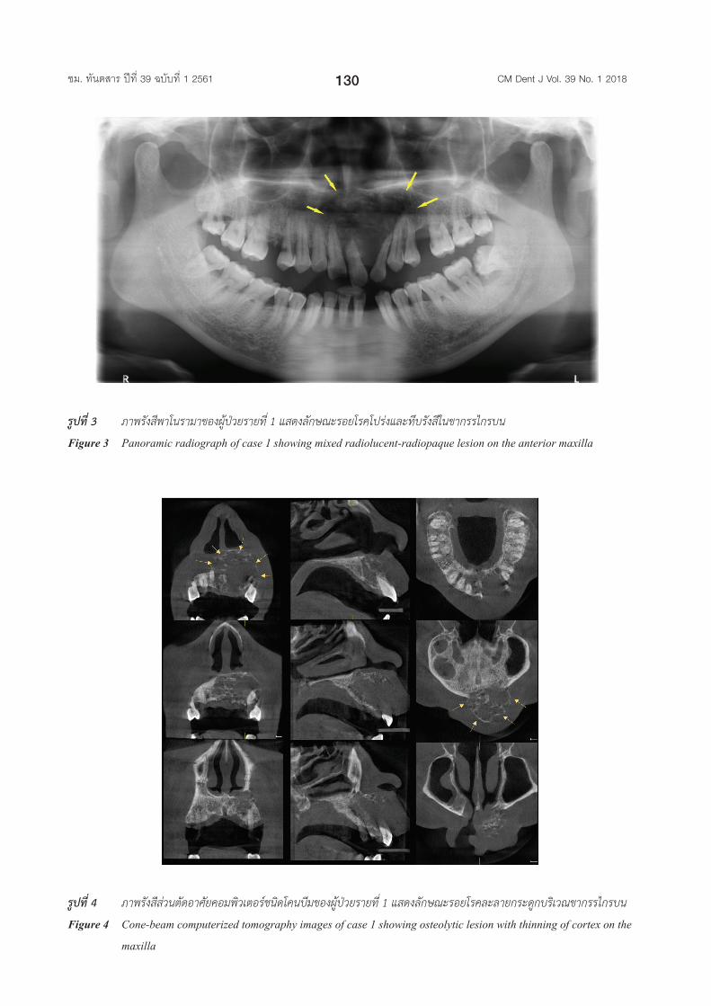
รูปที่ 3 ภาพรังสีพาโนรามาของผู้ป่วยรายที่ 1 แสดงลักษณะรอยโรคโปร่งและทึบรังสีในขากรรไกรบน
Figure 3 Panoramic radiograph of case 1 showing mixed radiolucent-radiopaque lesion on the anterior maxilla
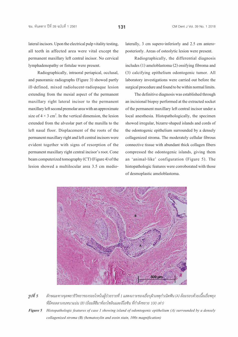
รูปที่ 4 ภาพรังสีส่วนตัดอาศัยคอมพิวเตอร์ชนิดโคนบีมของผู้ป่วยรายที่ 1 แสดงลักษณะรอยโรคละลายกระดูกบริเวณขากรรไกรบน
Figure 4 Cone-beam computerized tomography images of case 1 showing osteolytic lesion with thinning of cortex on the
maxilla
130ชม. ทันตสาร ปีที่ 39 ฉบับที่ 1 2561 CM Dent J Vol. 39 No. 1 2018
lateral incisors. Upon the electrical pulp vitality testing,
all teeth in affected area were vital except the
permanent maxillary left central incisor. No cervical
lymphadenopathy or fistulae were present.
Radiographically, intraoral periapical, occlusal,
and panoramic radiographs (Figure 3) showed partly
ill-defined, mixed radiolucent-radiopaque lesion
extending from the mesial aspect of the permanent
maxillary right lateral incisor to the permanent
maxillary left second premolar area with an approximate
size of 4 × 3 cm2. In the vertical dimension, the lesion
extended from the alveolar part of the maxilla to the
left nasal floor. Displacement of the roots of the
permanent maxillary right and left central incisors were
evident together with signs of resorption of the
permanent maxillary right central incisor’s root. Cone
beam computerized tomography (CT) (Figure 4) of the
lesion showed a multilocular area 3.5 cm medio-
laterally, 3 cm supero-inferiorly and 2.5 cm antero-
posteriorly. Areas of osteolytic lesion were present.
Radiographically, the differential diagnosis
includes (1) ameloblastoma (2) ossifying fibroma and
(3) calcifying epithelium odontogenic tumor. All
laboratory investigations were carried out before the
surgical procedure and found to be within normal limits.
The definitive diagnosis was established through
an incisional biopsy performed at the extracted socket
of the permanent maxillary left central incisor under a
local anesthesia. Histopathologically, the specimen
showed irregular, bizarre-shaped islands and cords of
the odontogenic epithelium surrounded by a densely
collagenized stroma. The moderately cellular fibrous
connective tissue with abundant thick collagen fibers
compressed the odontogenic islands, giving them
an ‘animal-like’ configuration (Figure 5). The
histopathologic features were corroborated with those
of desmoplastic ameloblastoma.
รูปที่ 5 ลักษณะทางจุลพยาธิวิทยาของรอยโรคในผู้ป่วยรายที่ 1 แสดงเกาะของเยื่อบุผิวเหตุกำาเนิดฟัน (A) ล้อมรอบด้วยเนื้อเยื่อพยุง
ที่มีคอลลาเจนหนาแน่น (B) (ย้อมสีฮีมาท็อกไซลินและอีโอซิน ที่กำาลังขยาย 100 เท่า)
Figure 5 Histopathologic features of case 1 showing island of odontogenic epithelium (A) surrounded by a densely
collagenized stroma (B) (hematoxylin and eosin stain, 100x magnification)
131ชม. ทันตสาร ปีที่ 39 ฉบับที่ 1 2561 CM Dent J Vol. 39 No. 1 2018
A partial maxillectomy from the area of the
permanent maxillary right lateral incisor to the
permanent maxillary left second premolar and
immediate reconstruction with the left nasolabial flap
and surgical stent were performed under general
anesthesia. The surgical specimen, consisted of a
segment of the maxilla with the lesion and associated
teeth, was submitted for histopathologic evaluation.
The neoplastic cells were not present at the margins of
the specimen. The post-operative period was uneventful.
The patient was advised for a routine follow-up and
showed no signs of recurrence upon an eight-month
follow-up.
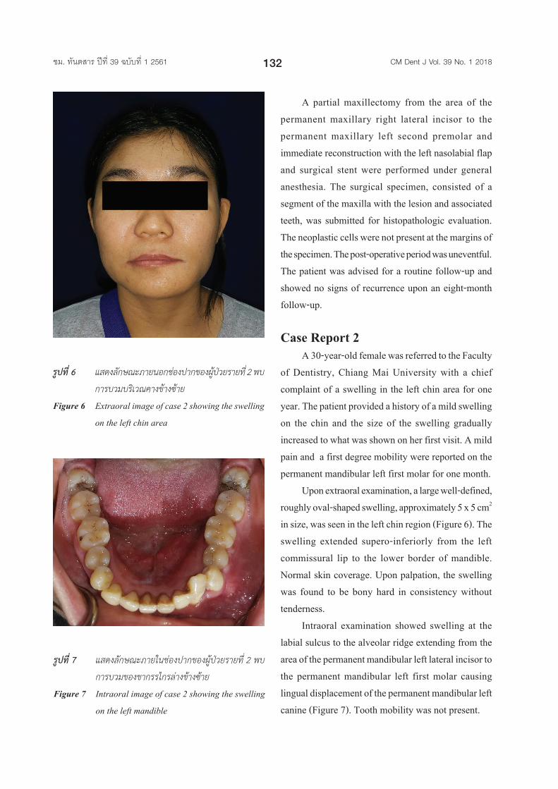
Case Report 2 A 30-year-old female was referred to the Faculty
of Dentistry, Chiang Mai University with a chief
complaint of a swelling in the left chin area for one
year. The patient provided a history of a mild swelling
on the chin and the size of the swelling gradually
increased to what was shown on her first visit. A mild
pain and a first degree mobility were reported on the
permanent mandibular left first molar for one month.
Upon extraoral examination, a large well-defined,
roughly oval-shaped swelling, approximately 5 x 5 cm2
in size, was seen in the left chin region (Figure 6). The
swelling extended supero-inferiorly from the left
commissural lip to the lower border of mandible.
Normal skin coverage. Upon palpation, the swelling
was found to be bony hard in consistency without
tenderness.
Intraoral examination showed swelling at the
labial sulcus to the alveolar ridge extending from the
area of the permanent mandibular left lateral incisor to
the permanent mandibular left first molar causing
lingual displacement of the permanent mandibular left
canine (Figure 7). Tooth mobility was not present.
รูปที่ 7 แสดงลักษณะภายในช่องปากของผู้ป่วยรายที่ 2 พบ
การบวมของขากรรไกรล่างข้างซ้าย
Figure 7 Intraoral image of case 2 showing the swelling
on the left mandible
รูปที่ 6 แสดงลกัษณะภายนอกช่องปากของผูป่้วยรายที ่2 พบ
การบวมบริเวณคางข้างซ้าย
Figure 6 Extraoral image of case 2 showing the swelling
on the left chin area
132ชม. ทันตสาร ปีที่ 39 ฉบับที่ 1 2561 CM Dent J Vol. 39 No. 1 2018
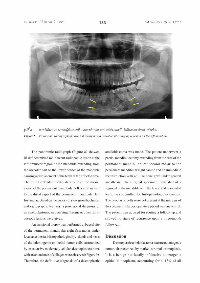
The panoramic radiograph (Figure 8) showed
ill-defined mixed radiolucent-radiopaque lesion at the
left premolar region of the mandible extending from
the alveolar part to the lower border of the mandible
causing a displacement of the teeth at the affected area.
The lesion extended mediolaterally from the mesial
aspect of the permanent mandibular left central incisor
to the distal aspect of the permanent mandibular left
first molar. Based on the history of slow growth, clinical
and radiographic features, a provisional diagnosis of
an ameloblastoma, an ossifying fibroma or other fibro-
osseous lesions were given.
An incisional biopsy was performed at buccal site
of the permanent mandibular right first molar under
local anesthesia. Histopathologically, islands and nests
of the odontogenic epithelial tumor cells surrounded
by an extensive moderately cellular, desmoplastic stroma
with an abundance of collagen were observed (Figure 9).
Therefore, the definitive diagnosis of a desmoplastic
ameloblastoma was made. The patient underwent a
partial mandibulectomy extending from the area of the
permanent mandibular left second molar to the
permanent mandibular right canine and an immediate
reconstruction with an iliac bone graft under general
anesthesia. The surgical specimen, consisted of a
segment of the mandible with the lesion and associated
teeth, was submitted for histopathologic evaluation.
The neoplastic cells were not present at the margins of
the specimen. The postoperative period was uneventful.
The patient was advised for routine a follow- up and
showed no signs of recurrence upon a three-month
follow-up.
Discussion Desmoplastic ameloblastoma is a rare odontogenic
tumor, characterized by marked stromal desmoplasia.
It is a benign but locally infiltrative odontogenic
epithelial neoplasm, accounting for 4–13% of all
รูปที่ 8 ภาพรังสีพาโนรามาของผู้ป่วยรายที่ 2 แสดงลักษณะรอยโรคโปร่งและทึบรังสีในขากรรไกรล่างข้างซ้าย
Figure 8 Panoramic radiograph of case 2 showing mixed radiolucent-radiopaque lesion on the left mandible
133ชม. ทันตสาร ปีที่ 39 ฉบับที่ 1 2561 CM Dent J Vol. 39 No. 1 2018
ameloblastomas.(1,2) Desmoplastic ameloblastoma
mostly occurs in the fourth to fifth decades of life with
no gender predilection. Males and females are equally
affected. More than 70% of the cases are seen in the
anterior region of the maxilla, as against the conventional
ameloblastomas, which are usually found in the
posterior mandibular region.(2) A unique location of
desmoplastic ameloblastoma in the anterior maxilla
was also shown in our first case.(4,10)
Since desmoplastic ameloblastoma shows specific
clinical, radiographic, histologic and biological features,
Philipsen and Reichart(14) suggested desmoplastic
ameloblastoma as a separate variant of ameloblastomas.
In 2005, the world health organization (WHO) classified
desmoplastic ameloblatoma as a separate entity of the
odontogenic tumors.(14) Radiographically, approximately
50 % of desmoplastic ameloblastomas show mottled
and mixed radiolucent-radiopacity with ill-defined
margins, making them difficult to differentiate from
fibro-osseous lesions. It was hypothesized that this
may be due to the infiltrative nature of desmoplastic
ameloblastomas into the trabecular bone. Three
radiological presentations of desmoplastic
ameloblastomas are mentioned in the literature as
follows: type I (osteofibrosis type) which has
radiolucent as well as radiopaque appearances; type II
(radiolucent type) which has a completely radiolucent
appearance; and type III (compound type) which has
radiolucent as well as radiopaque appearances
combined with a large radiolucent change. The type I
is the most common pattern of desmoplastic
ameloblastomas, while the type III is the least common
pattern.(15) The radiographic features of our cases
showed mixed radiolucent-radiopacity, making them
consistent with the osteofibrosis type.
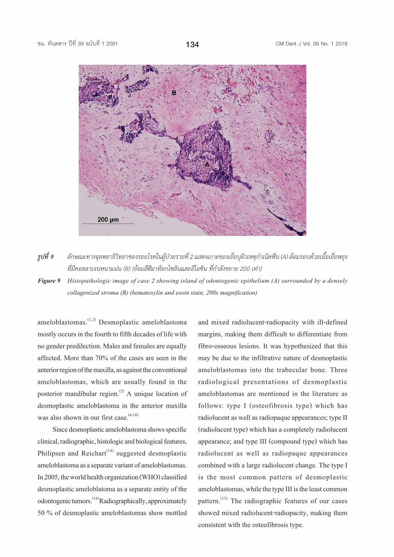
รูปที่ 9 ลักษณะทางจุลพยาธิวิทยาของรอยโรคในผู้ป่วยรายที่ 2 แสดงเกาะของเยื่อบุผิวเหตุกำาเนิดฟัน (A) ล้อมรอบด้วยเนื้อเยื่อพยุง
ที่มีคอลลาเจนหนาแน่น (B) (ย้อมสีฮีมาท็อกไซลินและอีโอซิน ที่กำาลังขยาย 200 เท่า)
Figure 9 Histopathologic image of case 2 showing island of odontogenic epithelium (A) surrounded by a densely
collagenized stroma (B) (hematoxylin and eosin stain, 200x magnification)
134ชม. ทันตสาร ปีที่ 39 ฉบับที่ 1 2561 CM Dent J Vol. 39 No. 1 2018
The definitive diagnosis of desmoplastic
ameloblastomas is based on histopathologic evaluation
of the biopsied specimens. The usual microscopic
features of desmoplastic ameloblastomas are: (1)
extensive stromal desmoplasia with abundance of
collagen and moderate amount of cellular connective
tissue, which is the most consistent and distinguishing
feature; (2) islands of different shapes of the epithelial
component;(3) cells at the periphery of the epithelial
island are usually cuboidal and occasionally
hyperchromatic; and (4) the central area of the islands
are occupied by whorls of spindle-shaped or squamous
epithelial cells.(16) In addition, formation of metaplastic
osteoid trabeculae or osteoplasia may be present. A
pattern of palisaded peripheral cells of the neoplastic
follicles as observed in conventional ameloblastomas
is absent.(14) Myxoid changes of the juxtaepithelial
stroma are often found. A fibrous capsule is not present
corresponding to radiographically poorly defined tumor
margins.(1)
Immunohistochemical studies suggested that the
desmoplasia of desmoplastic ameloblastomas might
be a result of overexpression of transforming growth
factor beta (TGF-β), a potent local factor that modulates
formation of the extracellular matrix. TGF-β can also
influence the synthesis of extracellular matrix proteins
involved in support, adhesion, proliferation, migration
and differentiation of the tumor cells that may lead to
the phenomenon of desmoplasia in desmoplastic
ameloblastomas.(17) Alternatively, desmoplasia in
desmoplastic ameloblastomas may reflect maturational
changes of the stromal connective tissue as this
phenomenon is also observed in long persistent tumors.
Some authors hypothesized that a combination of
desmoplastic ameloblastomas with any other types of
solid ameloblastomas, so-called hybrid ameloblastoma,
may be a transitional phase in the maturation of a solid
multicystic ameloblastoma to the desmoplastic
variation.(16)
Desmoplastic ameloblastomas exhibit a more
aggressive behavior than other types of the
ameloblastoma. This aggressiveness, evidenced by the
diffuse radiographic appearance and the histologic
finding of bone invasion, may be due to their potential
to grow to a large size and the common location of the
tumors in the maxilla leading to an early invasion of
the adjacent structures.(17) Taken together, the treatment
of desmoplastic ameloblastomas should be performed
by surgical resection with adequate margins for
prevention of recurrences.
The biological behavior particularly recurrences
of desmoplastic ameloblastomas remains unclear.
According to the WHO classification, it is stated that
unicystic, peripheral and possibly desmoplastic
ameloblastomas have lower recurrent rates than other
ameloblastomas.(14) However, some authors believed
that it is premature to indicate a low recurrent rate in
desmoplastic ameloblastomas. This is because the
radiologic and histologic findings of desmoplastic
ameloblastomas show a poor encapsulation or total
lack of a capsule. Therefore, further studies on a large
series of cases of desmoplastic ameloblastomas with
a long-term follow-up are suggested.(4)
Conclusion Two rare cases of desmoplastic ameloblastomas
in the maxilla and mandible are reported. Both cases
showed mixed radiopaque-radiolucent radiographic
appearances resembling fibro-osseous lesions.
Resection of the jaw bone was chosen for treatment
based on local aggressiveness of desmoplastic
ameloblastomas. Since recurrences may take place in
desmoplastic ameloblastomas within 3-4 years
postoperatively, a close long-term follow-up for both
patients was planned.
135ชม. ทันตสาร ปีที่ 39 ฉบับที่ 1 2561 CM Dent J Vol. 39 No. 1 2018
Acknowledgement We would like to thank Professor Dr. Anak
lamaroon, Faculty of Dentistry, Chiang Mai University
for his critical reading of this manuscript.
References1. Belgaumi UI, Sundaresh KJ, Varma S, Mallikarjuna
R. Desmoplastic ameloblastoma: a rare odontogenic
neoplasm with unusual radiographic and
histomorphological presentation. BMJ Case Reports
[serial on the internet] 2013 May [cited 2017 Oct 20]
[about 4 p.]. Available from: HYPERLINK https://
www.ncbi.nlm.nih.gov/pmc/articles/PMC3670007/
https://www.ncbi.nlm.nih.gov/pmc/articles/
PMC3670007/
2. Gade L, Patankar S, Khot K, Korde S, Alex S.
Desmoplastic ameloblastoma of maxilla-a case report.
J Clin Exp Dent 2010; 2(4):e204-206.
3. Kaffe I, Buchner A, Taicher S. Radiologic features of
desmoplastic variant of ameloblastoma. Oral Surg
Oral Med Oral Pathol 1993; 76:525–529.
4. Sun ZJ, Wu YR, Cheng N, Zwahlen RA, Zhao YF.
Desmoplastic ameloblastoma—a review. Oral Oncol
2009; 45:752–759.
5. Kawai T, Kishino M, Hiranuma H, Sasai T, Ishida T.
A unique case of desmoplastic ameloblastoma of the
mandible: report of a case and brief review of the
English language literature. Oral Surg Oral Med Oral
Pathol Oral Radiol Endod 1999; 87: 258–263.
6. Takata T, Miyauchi M, Ito H, et al. Clinical and
Histopathological Analyses of Desmoplastic
Ameloblastoma. Pathol Res Pract 1999; 95:669-675.
7. Fujimura M, Sato T, Kawano K, Morita Y, Noikura
T, Yamashita S. Two cases of ameloblastoma with an
unusual radiographic appearance. Dent Radiol 1987;
27:154–160.
8. Masao A, Naoyuki M, Kazuya H, et al. Ameloblastoma,
desmoplastic type: a case report with characteristic
radiological presentation. Oral Radiol 2012; 28:70–73.
9. Kallam SR, Arutla R, Gadwalwari SS, Kubbi JR,
Shylaja SR. Desmoplastic Ameloblastoma – An
Unusual Presentation. J Clin Diagn Res [serial on the
internet] 2015 Oct [cited 2017 Oct 20]; 9(10):[about
2 p]. Available from: HYPERLIN https://www.ncbi.
nlm.nih.gov/pmc/articles/PMC4625356/ https://www.
ncbi.nlm.nih.gov/pmc/articles/PMC4625356/
10. Barnes L, Eveson JW, Reichart P, Sidransky D. World
Health Organization classifications of tumours.
Pathology and Genetics of Head and Neck Tumours.
Lyon: IARC; 2005:300-301.
11. Waldron CA, El Mofty SK. A histopathologic study
of 116 ameloblastomas with special reference to the
desmoplastic variant. Oral Surg Oral Med Oral Pathol
1987; 63:441–451.
12. Marx R, Carlson E. The ameloblastoma: primary,
curative surgical management. J Oral Maxillofac Surg
2006; 64:484–494.
13. Reichart PA, Philipsen HP, Sonner S. Ameloblastoma:
biological profile of 3677 cases. Eur J Cancer B Oral
Oncol 1995; 31:86–99.
14. Philipsen HP, Reichart PA. Revision of the 1992-edition
of the WHO histological typing of odontogenic
tumours. A suggestion. J Oral Pathol Med 2002;
31:253–258.
15. Sheikh S, Pallagatti S, Singla I, Kalucha A. Desmoplastic
Ameloblastoma: A Case Report. J Dent Res Dent
Clinic Dent Prospects 2011; 5(1):27-32.
16. Sharma Lamichhane N, Liu Q, Sun H, Zhang W. A
case report on desmoplastic ameloblastoma of anterior
mandible. BMC research notes 2016; 9:171.
17. Friman T. Extracellular Matrix and Connective Tissue
Cells of the Tumor Microenvironment. Digital
Comprehensive Summaries of Uppsala Dissertation
from the Faculty of Medicine 613. 2010; 37.
136ชม. ทันตสาร ปีที่ 39 ฉบับที่ 1 2561 CM Dent J Vol. 39 No. 1 2018