Embed Size (px)
Citation preview

Determination of Telomerase Enzyme Level in Enzootic Intranasal Tumors of Goats
Şima ŞAHİNDURAN 1,a Özlem ÖZMEN 2,b Reyda KIYICI 1,c
1 Mehmet Akif Ersoy Üniversitesi, Veteriner Fakültesi, İç Hastalıkları Anabilim Dalı, TR-15030 Burdur - TÜRKİYE2 Mehmet Akif Ersoy Üniversitesi, Veteriner Fakültesi, Patoloji Anabilim Dalı, TR-15030 Burdur - TÜRKİYEa ORCID: 0000-0002-7718-2020; b ORCID: 0000-0002-1835-1082; c ORCID: 0000-0002-0667-5477
Article Code: KVFD-2018-19678 Received: 27.02.2018 Accepted: 31.05.2018 Published Online: 31.05.2018
How to Cite This Article
Şahinduran Ş, Özmen Ö, Kıyıcı R: Determination of telomerase enzyme level in enzootic intranasal tumors of goats. Kafkas Univ Vet Fak Derg, 24 (4): 583-588, 2018. DOI: 10.9775/kvfd.2018.19678
AbstractEnzootic intranasal adenocarcinoma (ENA) of goats are contagious tumoral disease that characterized occurrence of tumoral masses in nasal cavity of the animals. The etiological agent of the disease is retrovirus and the disease cause high economical loses in goat industry. Retroviruses, respectively inducing lung or nasal adenocarcinomas. The true economic impact of ENA is not known because affected animals are given insights from the herd before actual diagnosis, and suspected disease incidence is rarely reported. Telomerase regulates the proliferative capacity of cells and this enzyme has critical role in tumor progression. The aim of this study is to evaluate telomerase activity in serum and tumoral tissue in goat with ENA. For this aim 11 goat’s serum were analyses for telomerase activity. Tumoral tissue sections of totally 14 death goats were examined by immunohistochemically for telomerase expression. In addition, 10 normal goat’s serum and nasal tissues that belong slaughtered healthy animals used as control. This study showed that increased serum and tissue telomerase activity in goats with ENA comparing to controls. Also our results showed that telomerase activity becomes a useful prediction marker for tumor development in infected but clinically healthy goats in infected flocks.
Keywords: Enzootic intranasal tumor, ELISA, Immunohistochemistry, Telomerase
Keçilerin Enzootik Intranazal Tümörlerinde Telomeraz Enzim Düzeyinin Belirlenmesi
ÖzKeçilerin enzootik intranazal adenokarsinomu (ENA), hayvanların burun boşluğunda tümoral kitlelerin şekillenmesi ile karakterize bulaşıcı bir hastalıktır. Hastalığın etkeni retrovirüs olup, keçi endüstrisinde ekonomik kayıplara neden olmaktadır. Retroviruslar sırasıyla akciğer veya nazal adenokarsinomlara yol açar. Hastalıktaki ekonomik kayıp tam olarak bilinmemektedir. Çoğu zaman hasta hayvanlar teşhisten önce sürüden çıkarıldıkları için hastalığın gerçek insidansı ile ilgili bildirimler nadirdir. Telomeraz, hücrelerin proliferatif kapasitesini düzenler ve bu enzim tümörün yayılmasında kritik bir role sahiptir. Bu çalışmanın amacı ENA’lı keçilerin kan serumu ve tümöral dokularında telomeraz aktivitesini değerlendirmektir. Bu amaçla, 11 ENA’lı keçi serumunda telomeraz aktivitesi için analizler yapıldı. Ayrıca toplam 14 ölü keçinin tümoral doku kesitleri, telomeraz ekspresyonu için immunohistokimyasal olarak incelendi. Kontrol olarak, kesimi yapılan 10 sağlıklı keçinin serumu ve burun dokusu kullanıldı. Bu çalışmada ENA’lı keçilerde serum ve tümör dokularında telomeraz aktivitesinin kontrol grubundaki keçilere kıyasla arttığı tespit edildi. Enfekte olmuş fakat klinik olarak sağlıklı görünen keçilerde telomeraz aktivitesinin tümör gelişimi için yararlı bir belirteç olabileceği görüldü.
Anahtar sözcükler: Enzootik intranazal tümör, ELISA, İmmunohistokimya, Telomeraz
INTRODUCTIONTelomerase activity is an important determinant of telomere length in mammalian cells, in which lack of telomerase activity could exacerbate cell senescence,
especially in highly proliferative tissues [1,2]. Telomerase activity has been shown to be specifically expressed in immortal cells, cancer and germ cells where it compensates for telomere shortening during DNA replication and thus stabilizes telomere length [3].
İletişim (Correspondence) +90 248 2132202 [email protected]
KafKas Universitesi veteriner faKUltesi Dergisi
JoUrnal Home-Page: http://vetdergi.kafkas.edu.tronline sUbmission: http://submit.vetdergikafkas.org
Research ArticleKafkas Univ Vet Fak Derg24 (4): 583-588, 2018DOI: 10.9775/kvfd.2018.19678

584Determination of Telomerase Enzyme ...
Telomere shortening during the cell division disturbed tumor progression. Because of the continuous cell growth in malign tumors telomerase activity correlated by tumor behavior [4]. Telomerase is a cellular reverse transcriptase and adds new DNA onto the telomeres that are located at the ends of chromosomes [4-6]. Expression of telomerase is associated with the stage of differentiation and inhibition or absence of telomerase may result in cell crisis in cancer cells and cause tumor regression [7]. In addition, telomerase may express in permanently renewing epithelia such as gastrointestinal tract [8]. Shortening of telomeres may contribute to the control of the proliferative capacity in normal cells, and telomerase may be essential for unlimited cell proliferation [9]. An ideal cancer treatment would specifically target cancerous cells and have little or no effect on normal cells. Because of the compelling correlation that most normal cells not expressed telomerase and activity is detected in almost all cancer types, it may be a universal target for cancer therapeutics. Telomerase-based therapies should possess greater specificity, lower toxicity, and reduced side effects compared to conventional chemotherapeutic approaches [10,11].
Many viruses have been shown to be able to increase telomerase activity, for example the LMP1 gene of Epstein Barr virus (EBV) [12], Kaposi’s sarcoma herpesvirus [13], Marek’s disease virus [14], herpes simplex virus type-1 (HSV-1) [15] and Bovine Herpesvirus type1 -all up-regulate telomerase activity. On the other hands some viruses such as human immunodeficiency virus [16], the LMP2A gene of Epstein Barr virus [17] and Hepatitis B virus [18] down-regulate telomerase activity.
Sheep and goats are widely infected by the non-oncogenic Small Ruminant LentiViruses, related to Human Immunodeficiency Virus-1 and responsible for slowly evolving inflammatory and/or degenerative diseases, and by oncogenic retroviruses, and Enzootic Nasal Tumour Virus (ENTV), respectively inducing lung or nasal adeno-carcinomas [19,20]. Enzootic nasal adenocarcinoma (ENA) is an economically important contagious tumor of the nasal mucosa in sheep and goats [21]. The true economic impact of ENA is not known because affected animals are given insights from the herd before actual diagnosis, and suspected disease incidence is rarely reported [22]. Although the exact prevalence of ENA is unknown [23] and often is not diagnosed.
The agent of ENA is a simple retrovirus and it induces unilateral or bilateral neoplastic growth in the mucosal nasal glands of the ethmoidal area [21]. No metastasis has been reported in ENA cases, but disruption of the nasal septum structure as well as erosion of the cribiform plate has been reported [23,24].
Clinical signs of ENA include seromucosal nasal discharge leading to a ‘washed nose’ appearance, accompanied by snoring, coughing, wheezing and dyspnea. The duration
of disease, from the appearance of clinical signs to the time of death, varies from 3 weeks to 1 year or more.
Based on these findings, we investigated the relationship between telomerase value in serum and immuno expression in tissue in enzootic nasal adenocarcinoma in goats.
MATERIAL and METHODS
This study was carried out on a total of eleven (5 males and 6 females) alive and 3 died (all of female) goats (Capra hircus) from a herd numbering 225 goats with a 10-year history of respiratory nasal problems and death with respiratory distress. The goats kept in a small shelter together and they ages were different between 2 to 5 years old.
The farm was visited and blood samples from 11 clinically ill animals (infected group=11) collected for evaluate the serum levels of telomerase. Clinically, signs of dyspnea and mucous nasal discharge were observed in many of the animals. Mild to severe emaciation was noted in all of animals. According to the history that taken from owner, more than 120 goats died from same illness with dyspnea and in some cases nasal bone perforation in recent years. Blood samples were collected in tubes without anticoagulant and were centrifuged at 3000 rpm, at 4°C for 10 min. Furthermore, ten healthy goats (control group=10) were used from another healthy flock. Serum samples were carefully harvested and stored at -20°C until used. These sera were then used to establish the values of telomerase levels using an ELISA commercial kits [Goat Telomerase (TE) ELISA KIT].
The 11 alive goats with clinical symptoms (blood samples collected from) were euthanatized, in addition 3 death goats were underwent to necropsy. At the gross examination of nasal cavity, unilateral intranasal tumors were found in all 14 goats. During necropsy, tissue samples were taken from tumoral masses for histopathological examinations. Samples were fixed in 10% neutral formalin. Using standard methods, tissues were stained with Hematoxylin-Eosin (HE), and examined microscopically.
Selected tumor sections were stained immunohisto-chemically in order to demonstrate telomerase activity in tumor tissue [Anti-Telomerase reverse transcriptase anti- body -C-terminal ab183105, (1/100 dilution)] using a routine streptavidin biotin peroxidase technique. For immunohistochemical examination sections were routinely processed according the manufacturer’s instructions.
Student t test were used for to evaluate the differences of serum telomerase activity between the goats with and without ENA Variables were presented as, “mean±standard deviation” deviations. Calculations were made using the SPSS 15.0 program pack (SPSS Inc., Chicago, IL, USA). P<0.05 was set as the value for significance.
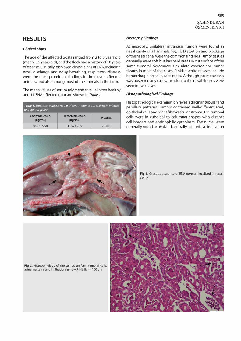
585
ŞAHİNDURANÖZMEN, KIYICI
RESULTS
Clinical Signs
The age of the affected goats ranged from 2 to 5 years old (mean, 3.5 years old), and the flock had a history of 10 years of disease. Clinically, displayed clinical sings of ENA, including nasal discharge and noisy breathing, respiratory distress were the most prominent findings in the eleven affected animals, and also among most of the animals in the farm.
The mean values of serum telomerase value in ten healthy and 11 ENA-affected goat are shown in Table 1.
Necropsy Findings
At necropsy, unilateral intranasal tumors were found in nasal cavity of all animals (Fig. 1). Distortion and blockage of the nasal canal were the common findings. Tumor tissues generally were soft but has hard areas in cut surface of the some tumoral. Seromucous exudate covered the tumor tissues in most of the cases. Pinkish white masses include hemorrhagic areas in rare cases. Although no metastasis was observed any cases, invasion to the nasal sinuses were seen in two cases.
Histopathological Findings
Histopathological examination revealed acinar, tubular and papillary patterns. Tumors contained well-differentiated, epithelial cells and scant fibrovascular stroma. The tumoral cells were in cuboidal to columnar shapes with distinct cell borders and eosinophilic cytoplasm. The nuclei were generally round or oval and centrally located. No indication
Table 1. Statistical analysis results of serum telomerase activity in infected and control groups
Control Group (ng/mL)
Infected Group (ng/mL) P Value
18.97±5.58 49.52±5.39 <0.001
Fig 2. Histopathology of the tumor, uniform tumoral cells, acinar patterns and infiltrations (arrows), HE, Bar = 100 µm
Fig 1. Gross appearance of ENA (arrows) localized in nasal cavity

586Determination of Telomerase Enzyme ...
of pleomorphism and cellular atypia were observed but, slight necrosis, inflammatory infiltrations and mitoses were seen (Fig. 2).
At the immunohistochemistry, increase in telomerase activity in the nucleus and cytoplasm of the tumoral cells was observed in granular appearance (Fig. 3, 4). The severity of the telomerase expression was not uniform and changing slight to severe in the tumoral mass.
DISCUSSION
Enzootic nasal adenocarcinoma is a contagious neoplasm of the secretory epithelial cells of the nasal mucosa of small ruminants and the causative agents of the tumor is retro-virus. ENA is commonly spread horizontally, most likely by the respiratory route. Although clinical symptoms and chronic behavior suspected the diseases, the easiest way for to diagnosis is the examination of nasal cavity of dead animals. Histopathological and/or ultrastructural examinations are necessary for definitive diagnosis [25]. In this study diagnosis of ENA was made base on the clinical, macroscopically, microscopically and ultrastructural findings. Only weld differentiated and characteristic cases included this study.
The telomerase activity plays an important role in maintaining chromosome stability, cellular immortality and oncogenesis. Telomerase is highly active in 90% of human cancers and transformed cells possess longer telomeres at their chromosomes [26]. Beyond its role in telomere maintenance, telomerase provides additional functions in tumorigenesis, DNA repair and cell survival. Telomerase protects cells from apoptosis and necrosis, and stimulates growth in adverse conditions [27].
The telomerase enzyme is ribonucleoprotein reverse transcriptase enzyme that adds telomeric repeats into end of chromosomes [28]. This activity protects the integrity of chromosomes from digestion by exonucleases, fusion to the neighboring chromosomes and occurring the chromosomal defects [29,30]. Possible relation between carcinogenesis and telomere dysfunctions was reported [31,32]. Previous studies cataloging TERT expression and telo-merase activity reported potent suppression in human somatic tissues, while robust expression and activity in germ cells and cancer cells. Progressive telomere shortening from cell division provides a barrier for tumor progression. However, one of the hallmarks of advanced malignancies is continuous cell growth and this almost
Fig 4. A- Higher magnification of increased telomerase immunoreaction (arrows) in nasal epithelial cells, B- Increased telomerase immunoreaction (arrows) in tumoral cells. Streptavidin biotin peroxidase method, Bar = 50 µm
Fig 3. A- Negative telomerase activity of the control nasal tissue, B- Marked increase of the telomerase immuonoreaction (arrows) in tumoral mass, Streptavidin biotin peroxidase method, Bar = 100 µm

587
universally correlates with the reactivation of telomerase [32]. Many authors suggested that determination of telomerase activity may serve as a diagnostic and prognostic tool in oncology. The same results were also observed in tumor cells in animals [33,34]. Telomerase activity was also determined in malignancies of feline tissues and in canine mammary tumors. Results showed that measurement of telomerase activity may be an effective method for detecting malignancy in animals [34,35]. Because it is a viral and tumoral disease, in this study telomerase activity of ENA examined by immunohistochemically method in tissue and by ELISA in serum. Marked increases in serum and tissue telomerase activity observed in both serum and tissue.
Recent studies reported that telomerase activation has an important role in normal somatic cells, and that failure to activate sufficient telomerase also promotes disease. Similarly, some viruses modulate the telomerase activity in animal cells [36,37]. Result of the present study showed that telomerase activity is effected by the disease. This results also indicated that telomerase activity has an important role in ENA. Detection of the telomerase activity in tissue by immunohistochemical methods is recent and reliable method in human tumors. Telomerase expressed in cytoplasm and nucleus of the tumoral cells [38-40]. In this study we used this technique in ENA and our results showed that telomerase expression in tumor tissue was increased compared to normal nasal tissue. The expression was observed both intracytoplasmic and intranuclear localisations.
Effect of virus on telomerase activity in animals previously was reported [36,37] but there is no report about ENA virus and telomerase activity in goats. This study showed that ENA virus up-regulate telomerase activity of infected cells and serum level. Understanding the mechanisms by which ENA virus exerts this further studies are needed.
In recent times, the incidence of ENA has dramatically increased throughout the word. Thus, the prediction of tumor development may require a decrease in the economic damage incurred. Our results showed that telomerase activity becomes a useful prediction marker for tumor development in infected but clinically healthy goats in infected flocks. In this study we have shown that telomerase activity increase in serum and tumoral tissue in ENA cases in goats.
ConfliCts of interest
The authors declare no conflict of interest
REFERENCES
1. Al-Ajmi N, Saretzki G, Miles C, Spyridopoulos I: Dietary restriction ameliorates haematopoietic ageing independent of telomerase, whilst lack of telomerase and short telomeres exacerbate the ageing phenotype. Exp Gerontol, 58, 113-119, 2014. DOI: 10.1016/j.exger.2014.07.010
2. Richardson RB, Allan DS, Le Y: Greater organ involution in highly proliferative tissues associated with the early onset and acceleration of ageing in humans. Exp Gerontol, 55, 80-91, 2014. DOI: 10.1016/j.exger.2014.03.015
3. Chiu CP, Harley CB: Replicative senescence and cell immortality: The role of telomeres and telomerase. Proc Soc Exp Biol Med, 214, 99-106, 1997.
4. Collins K, Mitchell JR: Telomerase in the human organism. Oncogene, 21, 564-579, 2002. DOI: 10.1038/sj.onc.1205083
5. Dikmen G, Doğan P: Telomeraz ve Kanser. T Klin Tıp Bilimleri, 23, 334-341, 2003.
6. Yıldız MG, Aras S, Duman DC: Telomerlerin yaşlanma ve kanser ilişkisindeki rolü. Türk Hij Den Biyol Derg, 66, 187-195, 2009.
7. Kim NW: Clinical implication of telomerase in cancer. Eur J Cancer, 33, 781-786, 1997. DOI: 10.1016/S0959-8049(97)00057-9
8. Bachor C, Bachor OA, Boukamp P: Telomerase is active in normal gastrointestinal mucosa and not up-regulated in precancerous lesions. J Cancer Res Clin Oncol, 125, 453-460, 1999. DOI: 10.1007/s004320050302
9. Chadeneau C, Hay K, Hirte HW, Gallinger S, Bacchetti S: Telomerase activity associated with acquisition of malignancy in human colorectal cancer. Cancer Res, 55, 2533-2536, 1995.
10. Dikmen GZ, Dikmen E, Doğan P: Kanserde telomeraza yönelik tedavi stratejileri. Hacettepe Tıp Der, 37, 49-55, 2006.
11. Shay JW, Wright WE: Role of telomeres and telomerase in cancer. Semin Cancer Biol, 21, 349-353, 2011. DOI: 10.1016/j.semcancer.2011.10.001
12. Yang J, Deng X, Deng L, Gu H, Fan W, Cao Y: Telomerase activation by Epstein-Barr virus latent membrane protein 1 is associated with c-Myc expression in human nasopharyngeal epithelial cells. J Exp Clin Cancer Res, 23, 495-506, 2004.
13. Verma SC, Borah S, Robertson ES: Latency-associated nuclear antigen of Kaposi’s sarcoma-associated herpesvirus upregulates trans-cription of human telomerase reverse transcriptase promoter through interaction with transcription factor Sp1. J Virol, 78, 10348-10359, 2004. DOI: 10.1128/JVI.78.19.10348-10359.2004
14. Djeraba-AitLounis A, Soubieux D, Klapper W, Rasschaert D: Induction of telomerase activity in avian lymphoblastoid cell line transformed by Marek’s disease virus, MDCC-MSB1. Vet Pathol, 41, 405-407, 2004. DOI: 10.1354/vp.41-4-405
15. Yang CT, Song J, Bu X, Cong YS, Bacchetti S, Rennie P, Jia WW: Herpes simplex virus type-1 infection upregulates cellular promoters and telomerase activity in both tumor and nontumor human cells. Gene Ther, 10, 1494-1502, 2003. DOI: 10.1038/sj.gt.3302005
16. Franzese O, Comandini A, Adamo R, Sgadari C, Ensoli B, Bonmassar E: HIV-Tat down-regulates telomerase activity in the nucleus of human CD4+ T cells. Cell Death Differ, 11, 782-784, 2004. DOI: 10.1038/sj.cdd.4401346
17. Chen F, Liu C, Lindvall C, Xu D, Ernberg I: Epstein-Barr virus latent membrane 2A (LMP2A) down-regulates telomerase reverse transcriptase (hTERT) in epithelial cell lines. Int J Cancer, 113, 284-289, 2005. DOI: 10.1002/ijc.20594
18. Fan XG, Huang Y, Tang FQ, Yi H: Telomerase activity of peripheral blood lymphocytes in patients with chronic hepatitis B. Immunol Lett, 73, 7-11, 2000. DOI: 10.1016/S0165-2478(00)00187-5
19. Leroux C, Cruz JC, Mornex JF: SRLVs: A genetic continuum of lentiviral species in sheep and goats with cumulative evidence of cross species transmission. Curr HIV Res, 8, 94-100, 2010. DOI: 10.2174/ 157016210790416415
20. Leroux C, Mornex JF: Retroviral infections in sheep and the associated diseases. Small Ruminant Res, 76, 68-76, 2008. DOI: 10.1016/J.SMALLRUMRES.2007.12.010
21. De las Heras M, Ortin A, Cousens C, Minguijon E, Sharp JM: Enzootic nasal adenocarcinoma of sheep and goats. Curr Top Microbiol Immunol, 275, 201-223, 2003.
22. Caswell J, Williams K: Enzootic nasal tumour of sheep. In, Maxie M (Ed): Jubb, Kennedy & Palmer’s Pathology of Domestic Animals. 5th ed.,
ŞAHİNDURANÖZMEN, KIYICI

588Determination of Telomerase Enzyme ...
640-643, Philadelphia, Elsevier Saunders, 2007.
23. Walsh SR, Linnerth-Petrik NM, Laporte AN, Menzies PI, Foster RA, Wootton SK: Full-length genome sequence analysis of enzootic nasal tumor virus reveals an unusually high degree of genetic stability. Virus Res, 151, 74-87, 2010. DOI: 10.1016/j.virusres.2010.04.002
24. McKinnon AO, Thorsen J, Hayes MA, Misener CR: Enzootic nasal adenocarcinoma of sheep in Canada. Can Vet J, 23, 88-94, 1982.
25. Ozmen O, Sahinduran S, Haligur M, Demir N: Clinical, pathological, immunohistochemical and ultrastructural observations on enzootic nasal adenocarcinoma in five goats. Kafkas Univ Vet Fak Derg, 16, 633-639, 2010. DOI: 10.9775/kvfd.2009.1340
26. Shay JW, Wright WE: Hallmarks of telomeres in ageing research. J Pathol, 211, 114-123, 2007. DOI: 10.1002/path.2090
27. Gorbunova V, Seluanov A: Telomerase as a growth-promoting factor. Cell Cycle, 2, 534-537, 2003. DOI: 10.4161/cc.2.6.515
28. Blackburn EH: Structure and function of telomeres. Nature, 350, 569-573, 1991. DOI: 10.1038/350569a0
29. Nasir L, Devlin P, Mckevitt T, Rutteman G, Argyle DJ: Telomere lengths and telomerase activity in dog tissues: A potential model system to study human telomere and telomerase biology. Neoplasia, 3, 351-359, 2001. DOI: 10.1038/sj.neo.7900173
30. Artandi SE, DePinho RA: Telomeres and telomerase in cancer. Carcinogenesis, 31, 9-18, 2010. DOI: 10.1093/carcin/bgp268
31. Kim NW, Pietyszek MA, Prowse KR, Harley CB, West MD, Ho PLC, Coviello GM, Wright WE, Weinrich SL, Shay JW: Specific association of human telomerase activity with immortal cells and cancer. Science, 266, 2011-2015, 1994. DOI: 10.1126/science.7605428
32. Argyle DJ, Nasir L: Telomerase: A potential diagnosis and therapeutic tool in canine oncology. Vet Pathol, 40, 1-7, 2003. DOI: 10.1354/vp.40-1-1
33. Varon D, Jiang C, Hedican C, Dome JS, Umbricht CB, Carey LA, Thompson HJ, Sukumar S: Telomerase activity in the normal and neoplastic rat mammary gland. Cancer Res, 57, 5605-5609, 1997.
34. Cadile CD, Kitchell BE, Biller BJ, Hetler ER, Balkin RG: Telomerase activity as marker for malignancy in feline tissues. Am J Vet Res, 62, 1578-1581, 2001. DOI: 10.2460/ajvr.2001.62.1578
35. Yazawa M, Okuda M, Setoguchi A, Iwabuchi S, Nishimura R, Sasaki N, Masuda K, Ohno K, Tsujimoto H: Telomere length and telomerase activity in canine mammary gland tumors. Am J Vet Res, 62, 1539-1543, 2001. DOI: 10.2460/ajvr.2001.62.1539
36. Pagnini U, De Martino L, Montagnaro S, Diodato A, Longo M, Pacelli F, Pisanelli G, Iovane G: Bovine herpesvirus type 1 (BHV-1) up-regulates telomerase activity in MDBK cells. Vet Microbiol, 113, 231-236, 2006. DOI: 10.1016/j.vetmic.2005.11.006
37. Suzuki K, Shuto S, Miura Y, Sentsui H: Measurement of telomerase activity in bovine leukaemia virus infected cows. Vet Microbiol, 127, 142-146, 2008. DOI: 10.1016/j.vetmic.2007.08.016
38. Palani J, Lakshminarayanan V, Kannan R: Immunohistochemical detection of human telomerase reverse transcriptase in oral cancer and pre-cancer. Indian J Dent Res, 22, 362-363, 2011. DOI: 10.4103/0970-9290.84281
39. Raghunandan BN, Sanjai K, Kumaraswamy J, Papaiah L, Pandey B, Jyothi BM: Expression of human telomerase reverse transcriptase protein in oral epithelial dysplasia and oral squamous cell carcinoma: An immunohistochemical study. J Oral Maxillofac Pathol, 20, 96-101, 2016. DOI: 10.4103/0973-029X.180953
40. Hiyama E, Hiyama K, Yokoyama T, Shay JW: Immunohistochemical detection of telomerase (hTERT) protein in human cancer tissues and a subset of cells in normal tissues. Neoplasia, 3, 17-26, 2001. DOI: 10.1038/sj.neo.7900134