Embed Size (px)
Citation preview
UNIVERSIDAD AUTÓNOMA DE MADRID
FACULTAD DE CIENCIAS
DEPARTAMENTO DE BIOLOGÍA
EVALUACIÓN DEL METABOLISMO DEL GLUTATIÓN EN LA TOLERANCIA A METALES PESADOS
JUAN SOBRINO PLATA
MADRID, 2014

Memoria para optar al grado de doctor realizada en el Departamento de Biología de la Facultad de Ciencias en la Universidad Autónoma de Madrid y que lleva por título:
EVALUACIÓN DEL METABOLISMO DEL GLUTATIÓN EN LA TOLERANCIA A METALES PESADOS
Presentada por:
Juan Sobrino Plata
Licenciado en Biología y en Bioquímica.
Dirigida por
Madrid, 2014
Luis Eduardo Hernández Rodríguez
Profesor titular
Dpo. Biología
Universidad Autónoma de Madrid
Carolina Escobar Lucas
Profesor titular
Dpo. Ciencias Ambientales
Universidad de Castilla la Mancha


Abreviaturas
APS 5’adenosine phosphosulfate ATPS Adenosine triphosphate sulfurylase Ala Alanine APR APS reductase As Arsenic ASA Ascorbate APX Ascorbate peroxidase Cd Cadmium CAT Catalase Cys Cysteine DHAR Dehydro ascorbate reductase γEC gamma-glutamylcysteine γECS gamma-glutamylcysteine synthetase Glu Glutamate GSH Glutathione GR Glutathione reductase GS Glutathione synthetase Gly Glycine hGSH homo-glutathione MDA Malondialdehyde Hg Mercury MDHAR Monodehydro ascorbate reductase OAS O-acetylserine OAS-TL O-acetylserine (thiol) lyase GSSG Oxidized glutathione POX Peroxidase PC Phytochelatin PCS Phytochelatin synthase PAGE polyacrylamide gel electrophoresis ROS Reactive oxygen species SULTR Sulfate transporter SiR Sulfite reductase SOD Superoxide dismutase

AGRADECIMIENTOS
El desarrollo de esta tesis doctoral fue financiado por las siguientes instituciones:
• Junta de Comunidades de Castilla-La Mancha. Proyectos: POII10-007-6458 (FITOALMA2) y PBI07-0091-3644 (FITOALMA).
• Ministerio de Economía y Competitividad. Proyecto: AGL2010-15151 (PROBIOMET).
• Comunidad de Madrid. Consorcio EIADES, proyecto: S2009/AMB-1478.
• XIV Programa Nacional de Investigación Científica Fundación Ramón Areces

Resumen
Se estudiaron las respuestas de estrés oxidativo inducido por cadmio (Cd) y mercurio (Hg), característico
síntoma de toxicidad por metales, en plantas de alfalfa crecidas en un medio semi-hidropónico con
diferentes dosis de metal (0, 3, 10 y 30 µM) durante una semana. Ambos metales produjeron alteraciones
en enzimas antioxidantes, siendo el Hg más tóxico que el Cd, destacando la inhibición de la actividad
glutatión reductasa (GR), mientras que con Cd hubo mayor síntesis de fitoquelatinas (PCs). Estás
diferencias en los mecanismos de toxicidad nos llevó a plantear la posibilidad de identificar las firmas de
estrés específicas para elementos tóxicos (Cd, Hg y arsénico (As); 0, 6 y 30 µM) en la planta metalofita
Silene vulgaris. Tras una semana de exposición, se confirmó que la inhibición de GR era un indicador
específico de estrés por Hg, siendo de nuevo importante la producción de PCs en plantas tratadas con Cd.
Asimismo, el Cd produjo fotoinhibición y alteración en proteínas relacionadas con el aparato fotosintético,
lo que pudo estar relacionado con la mayor translocación de este metal a hoja. La evidente implicación del
GSH y otros biotioles en la respuesta a metal(loide)s tóxicos nos llevó a plantear el experimentos
funcionales para caracterizar en detalle su contribución en las respuestas Cd y Hg, para lo que utilizaron
mutantes de Arabidopsis thaliana con niveles alterados de GSH y biotioles frente al ecotipo silvestre (Col-
0). Se trabajó con tres alelos mutantes de la enzima γ-glutamilcisteina sintetasa (γECS), pad2-1, cad2-1, y
rax1-1 (acumulan 20, 30 y 45% del GSH detectado en Col-0, respectivamente); y un mutante defectivo en
fitoquelatina sintasa (PCS) cad1-3 (que no acumula PCs). Se diseñaron dos aproximaciones distintas, por
un lado se infiltraron hojas de plantas Col-0, cad2-1, rax1-1 y cad1-3 o se cultivaron en un sistema
hidropónico puro con Cd y Hg (0, 3, 10 o 30 µM) durante 24-72 h. Se observó que pequeñas diferencias
en el contenido de GSH hace que la planta se enfrente al estrés de diferente manera, teniendo
comportamientos similares a Col-0 en rax1-1, mientras que pad2-1 mostró la mayor sensibilidad. También
se estudió el perfil transcripcional de genes de la ruta de asimilación de azufre y metabolismo del GSH en
plantas de Arabidopsis Col-0, mutante de γECS y cad1-3, tratadas con un nivel moderado de Hg (3 µM)
durante 72 h. Se confirmó que los mutantes cad2-1 y pad2-1 fueron particularmente sensibles a Hg, al
tiempo que es importante el nivel de GSH en el patrón de expresión génica. Nuestros datos apoyan la
noción de un umbral crítico de concentración de GSH, observada en los mutantes rax1-1 con niveles de
GSH más parecidos a Col-0, que permite tolerancia a Cd y Hg. Se ha puesto de manifiesto la importancia
del GSH en la tolerancia, tanto a nivel del ajuste metabólico de la respuesta, como en la capacidad de
absorción y transporte de metales y metaloides tóxicos, observándose respuestas específicas para cada
contaminante. Esta información puede ser útil para la optimización de estrategias de descontaminación de
suelos contaminados por elementos tóxicos mediante fitorremediación.
Abstract
We studied the responses of oxidative stress induced by cadmium (Cd) and mercury (Hg), a characteristic
symptom of metal toxicity, in alfalfa plants grown on a semi-hydroponic medium with different doses of
metal (0, 3, 10 and 30 µM) for a week. Both metals produced alterations in antioxidant enzymes, being Hg
more toxic than Cd, in particular the strong inhibition of glutathione reductase (GR) activity, while there
was greater synthesis of phytochelatins (PCs) with Cd. The observed differences in the mechanisms of
toxicity led us to identifying the specific stress signatures for different toxic elements (Cd, Hg and arsenic
(As); 0, 6 and 30 µM) in the metallophyte Silene vulgaris. After a week of exposure, we confirmed that
GR inhibition was a specific indicator of stress by Hg, being Cd a potent inductor of PCs production.
Cadmium also caused photoinhibition and alteration in proteins related to the photosynthetic apparatus,
which could be related to increased translocation of this metal to the shoot. The involvement of GSH and
other biothiols in response to toxic metal(loid)s prompted us to perform functional experiments to
characterize in detail its contribution in the responses to Cd and Hg, using mutants of Arabidopsis thaliana
with altered levels of GSH and biothiols compared with the wild type (Col-0). We studied three mutant
alleles of the enzyme γ-glutamylcysteine synthetase (γECS), pad2-1, cad2-1and rax1-1 (that contain 20,
30 and 45% of Col-0 GSH levels, respectively); and a mutant defective in phytochelatin synthase (PCS)
cad1-3 (does not accumulate PCs). Two different approaches were designed, on the one hand infiltrated
leaves of plants Col-0, cad2-1, rax1-1 and cad1-3, or were cultivated in a pure hydroponic system with Cd
and Hg (0, 3, 10 or 30 µM) for 24-72 h. It was observed that small differences in the content of GSH
makes the plant to face stress differently, showing similar behaviors Col-0 and rax1 -1, while pad2-1 was
more sensitivity. It was also studied the transcriptional profile of genes of the assimilation of sulfur
pathway and GSH metabolism in Arabidopsis Col-0, the γECS mutant plants of and cad1-3, treated with a
moderate level of Hg (3 µM) for 72 h. It was confirmed that the cad2-1 and pad2-1 mutants were
particularly sensitive to Hg, at the time that the level of GSH is important for the gene expression profile.
Our data support the notion of a critical threshold of GSH concentration, observed in the rax1-1 mutant
with GSH levels more similar to Col-0, which allows tolerance to Cd and Hg. It has been highlighted the
importance of GSH in tolerance, both at the level of metabolic adjustment of the response, and at the level
of toxic metal and metalloids absorption and transport, with specific responses for each pollutant. This
information can be useful for the optimization of the decontamination strategies of soils polluted with
toxic elements using phytoremediation technologies.
Índice
Capítulo 1: Biothiols metabolism is crucial for plant cell tolerance to toxic metals and metalloids: the case of mercury
Abstract ................................................................................................................................... 1
1. The great challenge of environmental pollution with toxic elements ..................................... 1
2. Remediation strategies. Phytoremediation. ........................................................................... 7
3. Damages induced by toxic elements in plants. .................................................................... 11
4. Plant tolerance and detoxification mechanisms. .................................................................. 14
5. Glutathione, the key metabolite in redox homeostasis. ........................................................ 19
6. Glutathione metabolism and metal(loid) tolerance: the case of Hg. ..................................... 27
References ............................................................................................................................. 28
Objetivos ........................................................................................................................................ 43
Capítulo 2: Differential alterations of antioxidant defenses as bioindicators of mercury and cadmium toxicity in alfalfa
Abstract ................................................................................................................................. 45
Introduction ........................................................................................................................... 46
Materials and Methods ........................................................................................................... 48
Results ................................................................................................................................... 51
Discussion .............................................................................................................................. 59
Acknowledgments .................................................................................................................. 64
References ............................................................................................................................. 65
Supplementary Material ......................................................................................................... 69
Capítulo 3: Specific stress responses to cadmium, arsenic and mercury appear in the metallophyte Silene vulgaris when grown hydroponically
Abstract ................................................................................................................................. 71
Introduction ........................................................................................................................... 72
Experimental .......................................................................................................................... 74
Results ................................................................................................................................... 77
Discussion .............................................................................................................................. 82
Conclusion ............................................................................................................................. 86
Acknowledgments .................................................................................................................. 86
References ............................................................................................................................. 86
Supplementary Material ......................................................................................................... 90
Capítulo 4: The role of glutathione in mercury tolerance resembles its function under cadmium
stress in Arabidopsis
Abstract ................................................................................................................................. 93
Introduction ........................................................................................................................... 94
Experimental .......................................................................................................................... 96
Results ................................................................................................................................. 100
Discussion ............................................................................................................................ 109
Acknowledgments ................................................................................................................ 112
References ........................................................................................................................... 113
Supplementary Material ....................................................................................................... 116
Capítulo 5: Glutathione is a key antioxidant metabolite to cope with mercury and cadmium stress
Abstract ............................................................................................................................... 121
Introduction ......................................................................................................................... 122
Materials and Methods ......................................................................................................... 124
Results ................................................................................................................................. 126
Discussion ............................................................................................................................ 133
Conclusions ......................................................................................................................... 137
Acknowledgments ................................................................................................................ 137
References ........................................................................................................................... 137
Supplementary Material ....................................................................................................... 144
Capítulo 6: Characterization of sulfur and glutathione metabolism responses to mercury in
glutathione defective Arabidopsis mutants
Introduction ......................................................................................................................... 147
Materials and Methods ......................................................................................................... 150
Results ................................................................................................................................. 154
Discussion ............................................................................................................................ 162
References ........................................................................................................................... 166
Supplementary Material ....................................................................................................... 172
Consideraciones generales y conclusiones .................................................................................... 177
CAPÍTULO 1. INTRODUCCIÓN Biothiols metabolism is crucial for plant cell tolerance to
toxic metals and metalloids: the case of mercury

Tesis doctoral Juan Sobrino Plata
1
Capítulo 1. Introducción:
Biothiols metabolism is crucial for plant cell tolerance to toxic metals and
metalloids: the case of mercury
Juan Sobrino-Plata1,2, Carolina Escobar2, Luis E. Hernández1
1Laboratory of Plant Physiology, Department of Biology, Universidad Autónoma de Madrid, Cantoblanco, ES-28049 Madrid. 2Departamento de Ciencias del Medioambiente, Universidad de Castilla-La Mancha, Campus Fábrica de Armas, ES-45070 Toledo, Spain.
ABSTRACT
In this review we summarize the latest findings about the involvement of biothiol
metabolism in metal tolerance in plants, focusing our attention to mercury (Hg), one of the
most hazardous metals to the environment. The assimilation of sulfur, the synthesis of
glutathione (GSH) and the accumulation of phytochelatins (PCs) are processes of biothiol
metabolism critical for tolerance to toxic elements, as they contribute to maintain the redox
cellular homeostasis and limit the concentration of free metals and metalloids ions.
1. The great challenge of environmental pollution with toxic elements.
1.1 Heavy metals or toxic elements?
The term ‘heavy metal’ has no consensus meaning to identify an element, as there are
different connotations in the literature based on its density, atomic weight, atomic number,
chemical properties or toxicity (Duffus 2002). In general, heavy metals include those
elements with a specific density higher than 5 g/cm3, although frequently some metalloids
such as arsenic (As) are included in this group only by means of their well-known toxicity.
Among the different ways to classify heavy metals, perhaps one of the most accurate from a
physiological point of view is based on their functions in living organisms. Thus, several
heavy metals are classified as ‘essential’ when are required for metabolic processes in the
cell, such as iron (Fe), copper (Cu), cobalt (Co), manganese (Mn), magnesium (Mg) or zinc
(Zn), which are mainly enzymatic cofactors, and are only toxic above a threshold
concentration. On the other hand, non-essential heavy metals are toxic even at low
Glutatión y tolerancia a metales tóxicos
2
concentrations, being cadmium (Cd), chromium (Cr), lead (Pb), mercury (Hg), aluminum
(Al) and As the most relevant to the environment (Tchounwou et al. 2012); elements that
we prefer to name as toxic elements.
1.2 Sources of toxic elements and the public concern.
Important amounts of toxic elements are released by the Earth geological (volcanic or
geothermal) activity and rock weathering and erosion, as these elements are found
frequently in nature associated with metal sulfide ores, such as occurs with Cd together
with Zn (sphalerite, ZnS), the presence of As with pyrite (FeS2), chalcopyrite (CuFeS2) or
galena (PbS), or the release of Hg from cinnabar (HgS) (Ziemacki et al. 1989). However,
the largest proportion of toxic elements accumulating in the environment comes from
several human activities during centuries. Nriagu (1996) affirms that anthropogenic
accumulation of metals stated with the domestication of fire and the development of
metallurgy, which augmented remarkably with the beginning of the Industrial Revolution.
The use of metals and metalloids in modern society and industry, with novel uses in current
technology, has increased enormously. Examples of this are the spread of fertilizers and
pesticides containing Cd or As in agricultural environments, the addition of lead to
gasoline, manufacturing of metallic paintings and batteries, elaboration of metal containing
plastics, use of Hg in lighting systems and electronics or in medicine (dental amalgams,
antiseptics) (Järup, 2003). Besides these uses, mining activities during many years, the
increase of exhaust fumes or the release of industrial wastes have also contaminated vast
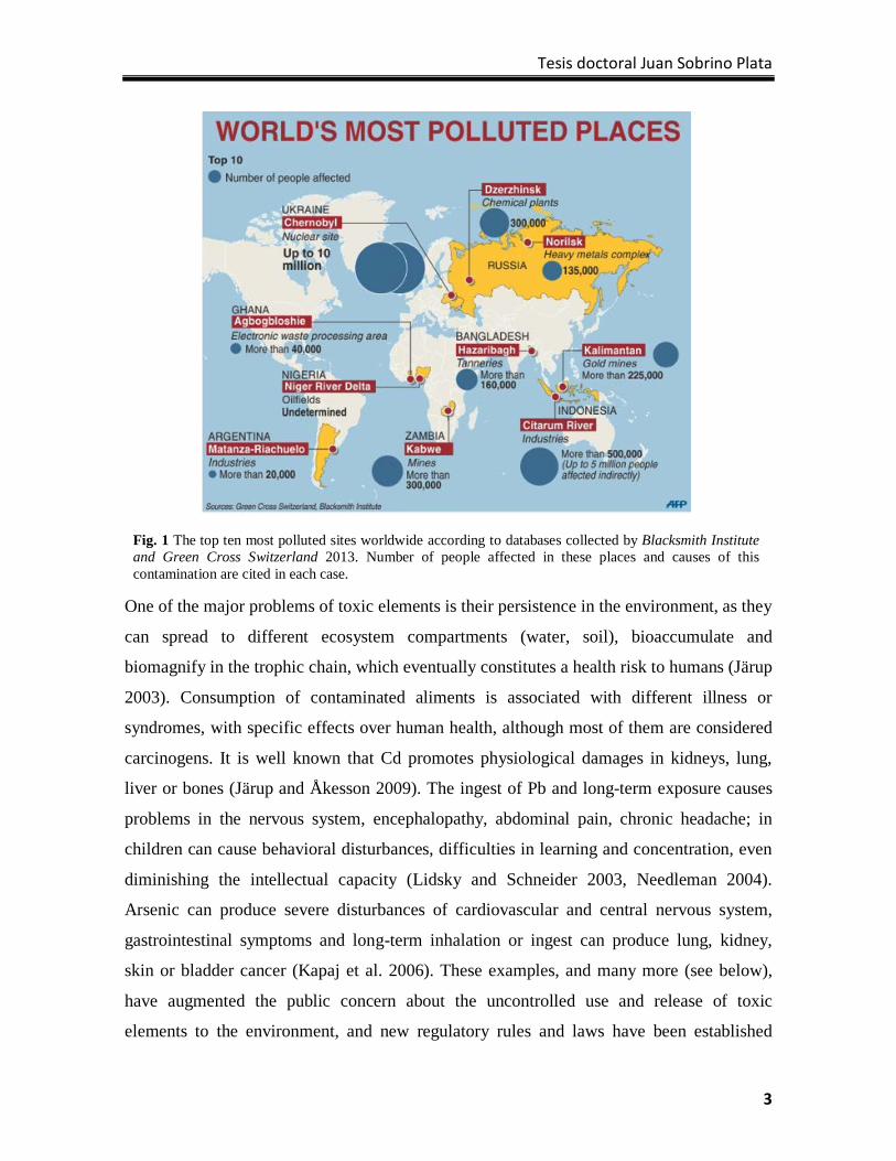
areas (Alloway 2012). It is relevant to mention that seven of the top ten sites most polluted
in the World in 2013 were drastically contaminated by metals and metalloids (Blacksmith
Institute and Green Cross Switzerland 2013; see Fig. 1 for details). Some examples are The
Citarum River in Indonesia, which covers an area of approximately 13.000 Km2 with a
population of 9 million people that consume water with concentrations of Al, Mn and Fe
four-times higher than recommended levels for water consume by the EPA (United Sates
Environmental Protection Agency). Another examples are the Matanza Riachuelo in
Argentina, which contains great amounts of Zn, Pb, Cu, Ni and Cr released from 15000
industries in the area, or alarming Pb contamination in Kabwe (Zambia), caused by intense
and uncontrolled mining activity since 1902, that is causing that children’s blood Pb levels
exceed 5-10 times the recommended level.
Tesis doctoral Juan Sobrino Plata
3
One of the major problems of toxic elements is their persistence in the environment, as they
can spread to different ecosystem compartments (water, soil), bioaccumulate and
biomagnify in the trophic chain, which eventually constitutes a health risk to humans (Järup
2003). Consumption of contaminated aliments is associated with different illness or
syndromes, with specific effects over human health, although most of them are considered
carcinogens. It is well known that Cd promotes physiological damages in kidneys, lung,
liver or bones (Järup and Åkesson 2009). The ingest of Pb and long-term exposure causes
problems in the nervous system, encephalopathy, abdominal pain, chronic headache; in
children can cause behavioral disturbances, difficulties in learning and concentration, even
diminishing the intellectual capacity (Lidsky and Schneider 2003, Needleman 2004).
Arsenic can produce severe disturbances of cardiovascular and central nervous system,
gastrointestinal symptoms and long-term inhalation or ingest can produce lung, kidney,
skin or bladder cancer (Kapaj et al. 2006). These examples, and many more (see below),
have augmented the public concern about the uncontrolled use and release of toxic
elements to the environment, and new regulatory rules and laws have been established
Fig. 1 The top ten most polluted sites worldwide according to databases collected by Blacksmith Institute and Green Cross Switzerland 2013. Number of people affected in these places and causes of this contamination are cited in each case.
Glutatión y tolerancia a metales tóxicos
4
recently by different international and national committees and governing institutions
(EPA, EU, FAO, etc.). Therefore, limited used of some metal(loid)s, improved waste
management, controlled pollutant-emissions or developing of sustainable procedures to
cleanup contaminated sites are now tasks for the benefit of human health (Blacksmith
Institute and Green Cross Switzerland 2013).
1.3 Mercury, the silent poison.
Mercury has chemical and physical properties make it a unique element, being one of the
most hazardous metal(loid)s in nature. In 1997 the EPA elaborated a report about Hg
sources, risk for human health or potential control technologies, recommending the
reduction of the use of Hg (Keating et al. 1997). Mercury is today a global environmental
problem: the Environment Programme of United Nations (UNEP) has a special ad-hoc
work group on Hg where the scientific community can contribute to the negotiations of an
internationally legal instrument for control of Hg (UNEP Chemicals Branch 2008). This
metal can be found in most ecosystems in three different oxidation forms: metallic (Hg0),
monovalent (Hg22+) or divalent (Hg2+), being the last form the most abundant in well-
aerated environments. It is found frequently in minerals associated with Cl—, OH— and
reduced sulfur (i.e. cinnabar). Mercury can also be associated with carbon to form chemical
species of ‘organic Hg’, such as methylHg (CH3Hg) or dimethylHg (CH3HgCH3), the most
toxic and abundant forms. These types of Hg are mainly produced by sulfate-reducing
bacteria, and can bioaccumulate and biomagnify in the food chain, principally in the
consumption of fish, shellfish, seaweed and marine mammals (Clarkson 1997). Fig. 2A
shows the major ecosystem compartments where Hg accumulate, with the inter-conversion
of Hg species among them.
Mercury can be released to the air, water and soil from both natural and anthropogenic
sources (UNEP Chemicals Branch 2008). Natural sources of Hg emissions come from
natural weathering of Hg-containing rocks, geothermal and volcanic activities, these last
can be separated in cataclysmic volcanoes that have the potential to release large amounts
of Hg and alter its concentration in the atmosphere for years, fumes of moderate but more
constant eruptions that can affect to the local environment (Nriagu and Becker 2003). On
the other hand, the anthropogenic sources are divided in primary and secondary sources.

Tesis doctoral Juan Sobrino Plata
5
Combustion of coal accounts for a large amount of Hg released in the atmosphere; for
instance cement production is also an important source of Hg emissions due to the burning
of coal or fuel to heat the materials. An important primary source of Hg is mining, with the
processing of different ores that release this metal to the environment. In this respect, we
can find the largest Hg-contaminated area of the World in the mining area of Almadén
(Spain), as a consequence of cinnabar extraction and processing for centuries (since the
days of the Roman Empire until 2003 when the mines were closed). It is estimated that one
third of Hg used in the World was extracted from these mines (Millán et al. 2006). An
important part of this Hg was exported to America for the extraction of gold and silver
using Hg amalgams, practice that has been maintained now-a-days in several regions of
Latin America, such as in Brazilian Amazonia, which has produced an enormous
accumulation of Hg in sediments and rivers (Salomons 1995). Another example of Hg
pollution in gold-mining is Kalimantan (Indonesia), where more than 1000 tons of Hg per
year are released into the environment through this activity, implying about 30% of the
world’s anthropogenic mercury emissions (Spiegel 2012).
Fig. 2 A. Mercury cycle in the major compartments of an ecosystem cycle: 1. Emissions of Hg from rock weathering, soils, surface waters, volcanoes and human activities. 2. Circulation through the atmosphere. 3. Deposition of Hg. 4. Conversion into other inorganic and organic forms. 5. Precipitation or bioconversion into more volatile or soluble forms. 6. Re-mobilization into the atmosphere or bioaccumulation in food chains (source: http://www.mercury.utah.gov/atmospheric_transport.htm). B. Main mechanisms of Hg phytoremediation. Mercury (red circles) can be stabilized (phytostabilization) in the rhizosphere, sequestered and accumulated in harvestable parts of tissue (phytoextraction), or modified into a volatilize form (phytovolatilization; modified from Pilon-Smits 2005).
Glutatión y tolerancia a metales tóxicos
6
Secondary anthropogenic sources are considered those products, that could be disposed of,
and industries, which emit wastes (fumes, effluents) that contain or use Hg. Multiple
devices, ranging from thermometers, batteries, fluorescent and high-pressure lamps to
dental amalgam fillings, pesticides and fungicides are examples of manufactured products
that contain Hg. Chlor-alkali factories are one of the most contaminating industries that
release Hg to the environment, as occurred in the Minamata bay (Japan) disaster (Ekino et
al. 2007). There is a circulation of Hg in the atmosphere or aquatic ecosystems due to re-
mobilization and re-emission of Hg by rain, chemical reactions and biological activity that
change the chemical speciation of Hg, processes that augmented with human activities
(Selin 2009).
Some of the chemical properties of Hg attracted the attention of ancient civilizations, which
attributed the capacity of awarding power, speed or longevity to people. However since the
Roman Empire it was known the strong toxic effects of Hg-contact. The Romans obliged
the criminals and punished slaves to work in Hg mines, as they found that the prisoners
would become poisoned, showing tremors, suffering poor hearing and vision, or became
insane. However during centuries Hg was also used in medicine and dentistry, as a purging
agent to induce vomiting and diarrhea, Hg-containing lotions were also applied directly to
the skin or as a remedy for syphilis. Thereby famous people like Amadeus Mozart, Ludwig
van Beethoven or the English King George III died probably poisoned with different Hg-
treatments. Although during centuries these controverted uses of Hg contributed to mature
this qualification of “silent poison”, the recent history made us learn drastically the strength
of the Hg toxicity in humans with catastrophes like the poisoning of villagers in Minamata
(Japan) in 1950s by the ingest of Hg-contaminated seafood (Ekino et al. 2007), or the death
of nearly 500 of 6530 hospitalized people in Iraq between 1971-1972 that consumed bread
baked using Hg-treated grain (Myers et al. 2000). The toxicology of Hg is large, it is
reported a long list of damage that could be summarized in neuro-, nephro-, and
immunotoxic, also may cause cardiovascular diseases and reproductive problems. A special
attention has to be put in the development of the child in utero and early in life because Hg
is ubiquitous and persistent and the consumption of, for example, CH3Hg-contaminated fish
by the mother, that could be transferred to the fetus or the child during lactation (Bose-
O'Reilly et al. 2010).
Tesis doctoral Juan Sobrino Plata
7
2. Remediation strategies. Phytoremediation.
Solutions to reduce the environmental impact of these toxic elements had been taken since
two decades ago approximately. First step to achieve this objective is the awareness of the
public about the danger of metals and metalloids accumulation to the environment and the
human health. In recent years several actions have been taken in this regard mainly in
developed countries, but poverty and lack of education in developing countries limit the
progress in this direction, due to the high costs of conventional decontamination techniques
(Sparks, 1995).
The goal of any process of remediation of contaminated sites is the reduction of the
pollutant concentration bellow dangerous levels, and do it with the less cost and lower
environmental impact. Traditional methods are usually aggressive or inefficient with this
regard. Excavation and containment of contaminated soils are the most common strategies
used, but many times the contaminated area makes this mission impracticable because of
the volume to manage or the depth of contaminated soil horizons (Salt et al. 1995).
Furthermore these techniques imply the transport of the contaminated soil and the creation
of restricted zones or landfills to store wastes, which makes this kind of procedures difficult
to implement. Therefore, efforts are underway to improve remediation technologies of soil
decontamination, some achieving a reasonable degree of success like immobilization
strategies and soil washing (Blacksmith Institute and Green Cross Switzerland 2013, EPA
2007). In particular, phytoremediation is envisaged as a more environmental friendly and
less expensive technology, which takes advantage of the natural capacity of plants to take
nutrients from soil.
2.1 Immobilization and soil washing strategies.
The objective in these techniques is to restrict the availability of toxic metal(oid)s in
polluted soils, most of them using amendments to accelerate the reactions.
Solidification/Stabilization (S/S) is the oldest immobilization technology to remediate
metal contaminated sites; hundreds of projects have been performed since the 1980s
(Mulligan et al. 2001). These amendments can be organic or inorganic, and induce
processes to reduce metal leaching and solubility. These processes consist in making the
soil metal(loid)s more geochemically stable by precipitation, adsorbed to surfaces or
Glutatión y tolerancia a metales tóxicos
8
chelated with ligands. Most common immobilization inorganic amendments are Fe-, Al-,
Mn-oxides or hydroxides, clay, cement or ashes (Kumpiene et al. 2008, Wuana and
Okieimen 2011, Fawzy 2008, Fusheng et al. 2012, Napia et al. 2012), whereas organic
matter comprises compost, polyester or polyethylene) (EPA 2007). This technology can be
applied in situ or ex situ, depending of the terrain and the pollutant. For example, Hg
contamination is usually treated ex situ, where the soil is removed and treated afterwards
using mainly Portland cement in the process (Zhang et al. 2009). This technology is
relatively cheap and easy to perform, but some disadvantages are the removal of the soil
polluted with metal(loid)s, and depends on the longevity of the solidified/stabilized
materials; which requires continuous monitoring on site (Wuana and Okieimen 2011).
On the other hand, soil-washing techniques have been employed largely in the United
States, Canada and Europe since 1990. The aim of soil-washing is the separation of
metal(loid) ions from the contaminated soil using physical, chemical approaches or the
combination of both (Dermont et al. 2008). The application of this remediation strategy is
normally ex situ to ensure the complete mobilization of the pollutant. The physical
separation uses procedures typical of mining and mineral processing industry, such as
particle size exclusion, density, magnetism or hydrophobic surface properties (Sierra et al.
2010). Chemical extraction is achieved utilizing different chemical reagents normally in
aqueous solutions to solubilize the metal(loid)s from the treated soils. These chemical
reagents are usually acid/alkali, surfactants, chelating agents or salts. EDTA is the most
common reagent used in the remediation of heavy metal-contaminated soils, but others
such as DTPA, sodium thiosulfate (Na2S2O3), iodide, nitric acid, hydrochloric acid or
sodium hydroxide have been tested were relatively successful (Martin and Ruby 2004,
Mulligan et al. 2001, Wang et al. 2012). This kind of approaches are only feasible when the
objective is to isolate precious metal(loid)s for use in certain industrial applications,
because its high cost (high consume of water, use of specific chelating agents, management
of the wastes and the secondary chemical pollution; Dermont et al. 2008).
2.2 Phytoremediation
Phytoremediation is described as ‘the green solution to the problem of the heavy metal
pollution’ (Ali et al. 2013), which consists basically in the employment of plants to remove
Tesis doctoral Juan Sobrino Plata
9
pollutants from water and/or soils, firstly proposed by Chaney in 1983. Plants have the
ability to absorb pollutants in the same manner as they uptake nutrients from the soil
(Cunningham and Berti 1993). Moreover plants have a potent secondary metabolism and
physiological adaptation mechanisms to respond to stress, which permits the detoxification
and tolerance to contaminants. The use plants to decontaminate soils is generally accepted
by the public, it is cheap, environmental friendly and can be utilized in situ (Peuke and
Rennenberg 2005).
There are several strategies of phytoremediation, which depend on the process to remove
the pollutants from the soil: phytoextraction, phytostabilization, phytovolatilization,
rhizofiltration and phytodegradation. With regard to toxic element contamination,
metal(loid)s are not degraded and only chemical speciation is changed, so most efforts were
directed to implement the first four strategies (Arthur et al. 2005). The efficiency of
phytoremediation strategies of toxic elements depends largely on factors like bioavailability
soil properties and their chemical speciation (Prasad 2003). Phytoremediation may be a
novel component of the Hg cycle in the ecosystems, where plants are used to retrieve this
toxic metal from polluted soils (Fig. 2B).
Phytoextraction is potentially the strategy most useful and economically interesting for
removing metal(loid)s from low or moderate contaminated soils. This strategy relays in the
capacity of plants to absorb the contaminants by the roots, which must be translocated to
aerial organs for harvesting. This only occurs in hyperaccumulating plants, which possess
specific mechanisms of transport (Mendoza-Cózatl et al. 2011). However, many of these
plants are not ideal because have normally low growth rate, poor production of shoot
biomass, and they grow under specific climatic conditions (Ali et al. 2013). Substantial
work is underway to understand the mechanisms of hyperaccumulation and tolerance to
high cytosolic concentration of metal(loid)s, information that could be exploited to
optimize plant species for real field applications. Thus, several model metallophytes (plants
capable to grow in metal(loid)-polluted soils) are studied at the physiological and
biomolecular levels under controlled growing conditions, often in comparison to closely
related non-tolerant species or populations. The major model metallophytes being studied
are various Zn-, Ni- or Cd-hyperaccumulating ecotypes of Thlaspi caerulescens, some Ni-
hyperaccumulating taxa of the genus Alyssum, and Zn-hyperaccumulating populations of
Glutatión y tolerancia a metales tóxicos
10
Arabidopsis halleri (Baker et al. 1994, Weber et al. 2004, Weber et al. 2006). Some
mechanisms of tolerance have been described (see below), traits that could be selected or
introduced by genetic engineering in crop plants, which are easier to cultivate and possess
high biomass production and rapid growth rate (Clemens et al. 2002). Additionally, some
trials were done with crop plants treated with chemical amendments to induce higher
bioavailability of metal(loid)s (Wuana and Okieimen 2011). For instance, Pb-
decontamination with plants is a complicated task due to its poor solubility in most soils,
which can improve with the addition of chelating agents such as EDTA (Meers et al. 2009).
Hernández-Allica et al. (2007) showed that cardoon plants grown in hydroponics system
were able to augment the uptake of Pb, Zn and Cd in the presence of EDTA, while metal
toxicity decreased. However, addition of EDTA has to be also controlled because this
chelating agent can produce phytotoxicity by itself or increase metal-phytotoxicity in some
plant species (Barrutia et al. 2010), as found in metallicolous and non-metallicolous
accessions of Rumex acetosa L. The uptake can also be modulated by the nutritional status
of the plants, as the application of fertilizers increased the uptake of metals in sorrel and
alfalfa plants grown in Pb/Zn- or Hg-contaminated sites respectively (Barrutia et al. 2009,
Carrasco-Gil et al. 2012).
Phytostabilization is the use of plants to reduce the mobility or the leaching of toxic
metals, and thereby preventing the migration to groundwater and the entry into the food
chain (Singh and Prasad 2011). The mechanisms to stabilize metals comprise association
with cell wall components, precipitation, complexation with metabolites or alteration of
redox valence, converting hazardous metals to a relatively less toxic state (Barceló and
Poschenrieder 2003). This strategy is more feasible to use in highly contaminated soils, or
in soils with continuous release of toxic metals from bed rock, as is the case of Hg-polluted
soils in Almadén (Carrasco-Gil et al., 2013). In this respect, recent work showed that
cultivation of red fescue, meadow grass, horseradish or Silene vulgaris prevented Hg
mobilization in polluted soils (Wang et al. 2012).
Phytovolatilization is only possible when the toxic element can be converted to a volatile
chemical species, such is the case of selenium and Hg. Taken up metal(loid)s are
transported from the roots to the shoots through the xylem, and are finally released to the
atmosphere (Ali et al. 2013). Plants lack naturally the capability to reduce Hg2+ to
Tesis doctoral Juan Sobrino Plata
11
elementary Hg (Hg0), the most volatile species, but some transgenic plants have been
engineered to overexpress bacterial mer (Mercuric ion Resistance) genes (Ruiz and Daniell
2009). Arabidopsis and poplar plants overexpressing merA, a mercuric ion reductase, were
more tolerant and were able to limit the Hg accumulation (Rugh et al. 1996, 1998). Other
trangenics were prepared overexpressing merB, which catalyzes the protonolysis of the
carbon–Hg bond to generate Hg2+ from CH3Hg (Bizily et al. 2000), generating less toxic
Hg species. However, this approach has only be tested in controlled environmental
conditions, and could pose serious problems of Hg atmospheric dissemination if used in
highly Hg-polluted soils, as occurs in Almadén.
Finally, rhizofiltration has been designed to remove contaminants from water using plants
cultivated in hydroponics (Salt et al. 1995). Artificial wetlands have been constructed to
ameliorate water contamination, which allows recycling of the metal extracted (Prasad
2003). To implement this technology terrestrial and aquatic plants can be chosen, and
continuous control of pH, well aerated influent solutions, flow rate, periodic harvesting and
plant disposal are required for optimal metal(loid) uptake (Dushenkov et al. 1995). There
are examples of alfalfa (Gardea-Torresdey et al. 1998), water hyacinth (Lytle et al. 1998)
and Pteris vittata (Huang et al. 2004) to eliminate several metals and metalloids like Cr(VI)
or As.
3. Damages induced by toxic elements in plants.
A fundamental factor to implement phytoremediation technologies to clean-up metal(loid)
polluted soils is the selection of tolerant plants, capable of accumulating toxic elements
without suffering physiological damages. The understanding of the mechanisms of
response evoked by the exposure of plants to toxic elements will help in this selection.
Perception, signaling and the subsequent metabolic adjustments is the aim of extensive
research done in recent decades. Behavior depends largely on plant species, which also is
differs between different metal(loid)s, leading to specific stress signatures, as we have
shown in alfalfa and the metallophyte Silene vulgaris when exposed to Cd, Hg or As
(Sobrino-Plata et al., 2009, 2013).
Roots are the part of the plant in charge of nutrients uptake, which can occur by passive
transport or through specific transporters (Clemens et al. 2002). These specific transporters
Glutatión y tolerancia a metales tóxicos
12
of Ni and Cd have been described in soybean plants (Cataldo et al. 1978, 1983), though Cd
can enter by transporters and channels responsible for uptake of essential nutrients such as
Ca2+channels, ZIP transporters, P-type ATPases (HMA) or natural resistance-associated
macrophage proteins (NRAMPs; Clemens 2006). Other toxic elements like the metalloids
As and Se enter in plants via phosphate and sulfate uptake systems respectively (Meharg
and Macnair 1990, Shibagaki et al. 2002).
Main causes of phytotoxicity by heavy metals are (i) generation of reactive oxygen species
(ROS) and induction of oxidative stress; (ii) reactions with thioyl-, histidyl- and carboxyl-
groups of important proteins or structural components in cell; (iii) replacement of essential
cations from specific binding sites; (iv) alteration of water balance; and (v) disruption of
nucleic acid conformation (Hall 2002, Patra et al. 2004). Photosynthesis is one of the
processes in the plant metabolism that could be affected by several toxic metal(loid)s at
different levels. For instance, Cd2+ replaces Ca2+ in the photosystem II reaction centre,
causing the inhibition of PSII photoactivation (Faller et al. 2005), Hg2+ may substitute Zn2+
and Mn2+ of D1 and D2 proteins in the donor site of Photosystem II (PSII) and inhibits
these proteins (Bernier et al. 1993). Leadpromotes reduction of total chlorophyll content
and relative content proportion of Chlorophyll a and b (Van Assche and Clijsters 1990) and
Cu can interact with ferredoxin causing the inhibition of ferredoxin-dependent reactions
(Shioi et al. 1978).
3.1 Oxidative stress and the role of ROS.
The induction of oxidative stress by toxic metal(loid)s accumulation is one of the major
alterations caused in plant cells (Hall 2002). When the cellular redox balance is
compromised, generation of ROS causes the oxidation of different cellular components
such as membrane lipids, proteins and nucleic acids affecting several physiological and
molecular processes. ROS production is innate with aerobic metabolism, and a
sophisticated antioxidant machinery works to maintain ROS levels under control (Foyer
and Noctor 2003). In addition, several physiological processes depend on ROS production
under certain circumstances, and are consider as key components of cell development,
signaling and responses to stress conditions (Jaspers and Kangasjärvi 2010).
Tesis doctoral Juan Sobrino Plata
13
ROS are intermediate products of the reduction of oxygen (O2) to H2O. This occurs in four
steps, and the reaction chain requires initiation at the first step while consecutive steps are
exothermic and can occur spontaneously (Dat et al. 2000):
O2 O2•– H2O2 OH• + H2O 2H2O
The transfer of one, two or three electrons to O2 forms respectively superoxide radical (O2•–
), hydrogen peroxide (H2O2) or hydroxyl radical (OH•). Another ROS can result from the
excitation of O2 to form singlet oxygen (O21) in chlorophylls and carotenoids. This
molecule and OH• are the most reactive and are responsible of large part of the oxidative
damage (Asada 1999).
Some metals interact with ROS and increase their amount by Fenton reactions (Briat and
Lebrun 1999). Redox active metals such as Fe or Cu intervene directly in ROS generation
as catalysts of oxygen reduction, while toxic metals such as Cd or Hg are thought to induce
ROS accumulation indirectly by altering the antioxidant machinery at different levels
(Sharma and Dietz 2009). The formation of ROS takes place in different cellular
compartments where electron transfers occur in aerobic conditions. Chloroplasts,
mitochondria and peroxisomes are the principal ROS producer organelles. Initially it
thought that chloroplasts were the main source of ROS in plants but recent evidences
awarded this prominence to mitochondria (Noctor et al. 2007). Thus, it has been estimated
that 1-5% of the O2 consumption of isolated mitochondria results in ROS production at the
electron transfer chain of the inner membrane (Møller 2001). In peroxisomes, xanthine
oxidase and NADPH oxidase activities generate O2–, and glycolate oxidase, flavin oxidases
and β-oxidation produce H2O2 (Sandalio et al. 2006). Recently, plasma membrane
associated NADPH oxidases are driving mayor attention, as in some biotic and abiotic
stress conditions these enzymes are responsible of oxidative burst at the apoplast (Mittler et
al. 2004). This process is essential for the activity of peroxidases in the cell wall, as H2O2 is
required for the cross-linking of polysaccharides and phenolics during lignification, which
is fundamental for cell architecture (Iiyama et al. 1994). ROS production via NADPH
oxidases is needed for correct cell cycle progress, programed cell death and
growth/expansion of root hairs and pollen tubes, as well as for elongation, proliferation and
Glutatión y tolerancia a metales tóxicos
14
cell differentiation in roots (Foreman et al. 2003, Halliwell 2006, Mittler et al. 2004,
Potocký et al. 2007, Tsukagoshi et al. 2010).
The Plants exposed to toxic metals suffer from the induction of an oxidative burst (Sharma
and Dietz 2009), which can appear after few minutes/hours of the Cd or Hg treatments in
roots of alfalfa plants (Ortega-Villasante et al. 2005). Heyno et al. (2008) proposed that
ROS was generated in plant cells exposed to Cd at the mitochondrial electron chain,
whereas Ortega-Villasante et al. (2007) found that Hg provoked a strong generation of
H2O2 probably at the apoplast though the activation of plasma membrane NADPH-
oxidases. This hypothesis was confirmed in alfalfa and Arabidopsis plants under Hg
exposure, process that may be moderated by the action of the stress related phytohormone
ethylene (Montero-Palmero et al. 2014). It is thus feasible that different toxic metal(loid)s
target distinct cellular components causing particular oxidative stress signatures, and
intensive research work is undergoing to characterize in detail these mechanisms of
responses where different organelles are implied (Romero-Puertas et al. 2004).
4. Plant tolerance and detoxification mechanisms.
Plant cells possess diverse mechanisms of tolerance and detoxification to cope with toxic
metal(oid) accumulation. The cell wall is the first barrier to these pollutants, being a
heterogeneous matrix that contains large number of carboxyl groups (–COOH) in acidic
polysaccharides (pectin, for example homogalacturonan) and phenolic polymers (lignin and
suberin), that play a crucial role in metallic cations binding and retention (Krzesłowska
2011). Cell walls constitute the major pool of toxic metals such as Cd or Hg in root cells, as
was observed in pea, maize, Arabidopsis or alfalfa plants (Lozano-Rodriguez et al. 1996,
Van Belleghem et al. 2007, Carrasco-Gil et al. 2013). Typically, plant cells modify cell
wall composition under metal(oid) stress, accumulating more compounds like certain
pectins and hemicellulose (Zhu et al. 2013). In addition, several proteins with thiol groups
are present in cell wall, such as extensins, which contain characteristic cysteine-rich regions
where Hg could be bound (Carrasco-Gil et al. 2013). Other defense strategy associated with
the apoplast of root cells is the exudation of ligands able to chelate and immobilize metallic
ions in the rhizosphere, as was well described in Al treated plants (Poschenrieder et al.
2008).
Tesis doctoral Juan Sobrino Plata
15
The next barrier of permeability is the plasma membrane, where a number of transport
mechanisms have been characterized. Toxic metal(loid)s can enter the cells using
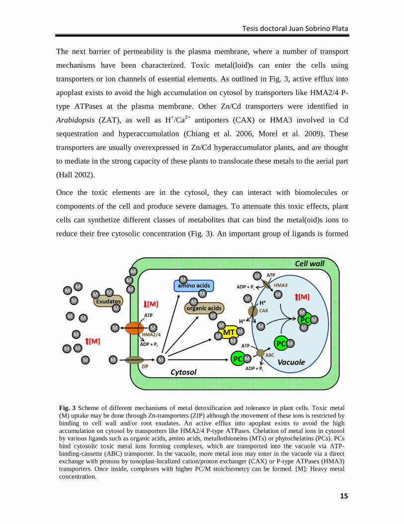
transporters or ion channels of essential elements. As outlined in Fig. 3, active efflux into
apoplast exists to avoid the high accumulation on cytosol by transporters like HMA2/4 P-
type ATPases at the plasma membrane. Other Zn/Cd transporters were identified in
Arabidopsis (ZAT), as well as H+/Ca2+ antiporters (CAX) or HMA3 involved in Cd
sequestration and hyperaccumulation (Chiang et al. 2006, Morel et al. 2009). These
transporters are usually overexpressed in Zn/Cd hyperaccumulator plants, and are thought
to mediate in the strong capacity of these plants to translocate these metals to the aerial part
(Hall 2002).
Once the toxic elements are in the cytosol, they can interact with biomolecules or
components of the cell and produce severe damages. To attenuate this toxic effects, plant
cells can synthetize different classes of metabolites that can bind the metal(oid)s ions to
reduce their free cytosolic concentration (Fig. 3). An important group of ligands is formed
Fig. 3 Scheme of different mechanisms of metal detoxification and tolerance in plant cells. Toxic metal (M) uptake may be done through Zn-transporters (ZIP) although the movement of these ions is restricted by binding to cell wall and/or root exudates. An active efflux into apoplast exists to avoid the high accumulation on cytosol by transporters like HMA2/4 P-type ATPases. Chelation of metal ions in cytosol by various ligands such as organic acids, amino acids, metallothioneins (MTs) or phytochelatins (PCs). PCs bind cytosolic toxic metal ions forming complexes, which are transported into the vacuole via ATP-binding-cassette (ABC) transporter. In the vacuole, more metal ions may enter in the vacuole via a direct exchange with protons by tonoplast-localized cation/proton exchanger (CAX) or P-type ATPases (HMA3) transporters. Once inside, complexes with higher PC/M stoichiometry can be formed. [M]: Heavy metal concentration.
Glutatión y tolerancia a metales tóxicos
16
by cysteine-rich peptides, of which we can distinguish metallothioneins (MTs) and
phytochelatins (PCs) (Cobbett and Goldsbrough 2002). Metallothioneins are low molecular
weight proteins found in most of living organisms, detected also in plants (Zhou and
Goldsbrough, 1995). These proteins were recently classified in four groups (Hassinen et al.
2011): p1 (Class 1), p2 (Class 2), p3 (Class 3) and pec (Class 4). Examples in Arabidopsis
thaliana are MT1a and MT1c (p1 subfamily), MT2a and MT2b (p2 subfamily), MT3 (p3
subfamily), and MT4a and MT4b (pec subfamily). Plant MTs are characterized by two or
three Cys-rich domains separated by long spacer regions that could be organized as Cys–
Cys, Cys–X–Cys or Cys–X–X–Cys repeats, in which X denote any aminoacid (Hossain et
al. 2012). Genes are regulated in a tissue-specific manner and response to diverse stimuli,
included different abiotic stresses as salt or heavy metal stress. Metallothioneins are
directly involved in metal tolerance; different studies of MTs overexpression or loss-of-
function have shown the essentiality of these proteins in metal homeostasis in plant cells
and in the responses to metal toxicity (Hassinen et al. 2011). However, other functions have
been assigned to MTs, such as ROS scavengers, or contributors in development, senescence
or fruit ripening (Ahn et al. 2012).
Phytochelatins are considered the main metal-binding molecule in a wide variety of plant
species. These Cys-rich peptides have the general structure: (γ-Glu-Cys)n-X, where X is
commonly Gly, but may be Ala, Ser, Gln or Glu; and n= 2-11 (Zenk 1996). PCs are
synthesized by the enzyme phytochelatin synthase (PCS) from GSH, tripeptide composed
by Glu, Cys and Gly; and it is involved in many cellular processes that will be discussed
later. PCS activity is induced in plant cells challenged with diverse toxic metals; Cd, Hg,
Cu, As, Ag, Ni, Au or Zn (Rauser 1995). Genes, structure and function of PCS have been
well characterized in last 20 years (Pal and Rai 2010); genes originally sequenced from
Arabidopsis, Saccharomyces pombe and wheat (Clemens et al. 1999, Ha et al. 1999,
Vatamaniuk et al. 1999). This characterization started from the preliminary work done by
Howden et al. (1995b), who isolated several mutants in Arabidopsis with Cd
hypersensitivity and with different degrees of diminished PCs production. Some of these
mutants were defined as cadmium-sensitive (cad1), and were identified as AtPCS1 mutants.
Cadmium is the most potent inductor of PCs, and most studies about metal complexation
and detoxification with PCs have been done with plants treated with this metal. It is known
Tesis doctoral Juan Sobrino Plata
17
that after sequestration of Cd by PCs, the array of complexes formed are transported to the
vacuole (Fig. 3), or can be translocated to the shoots (Ortiz et al. 1995, Mendoza-Cózatl et
al. 2011). If the mutation of PCS produces hypersensitivity to Cd and other toxic
metal(oid)s, paradoxically the overexpression of AtPCS1 in transgenic Arabidopsis seems
to have the same effect (Lee et al. 2003). With novel methodologies based on mass
spectrometry and X-ray spectroscopy our group was able to distinguish the formation of a
wide range of Hg-PC and Hg-biothiol complexes in maize, barley and alfalfa; and,
furthermore, the local distribution of Hg bound to biothiols and organic sulfur in alfalfa
roots (Carrasco‐Gil et al. 2011). These studies together with our recent experiments in this
thesis, in Arabidopsis mutants treated with Cd and Hg (Sobrino-Plata et al. 2014a, 2014b),
support the hypothesis that phytochelatins are important in Cd and Hg tolerance in non-
hyperaccumulator plants.
PC-Cd complexes are introduced in vacuoles but also other transporters are able to
introduce metal ions in vacuole (Mendoza-Cózatl et al. 2011), in a similar manner as
described with the ABC-type transporter (Hmt1) of S. pombe (Ortiz et al. 1995). Mendoza-
Cózatl et al. (2010) identified two classes of ABCC tansporters in vacuoles of Arabidopsis
that are involved in PC-As and PC-Cd vacuolar sequestration.
Other chelators in the cytosol of plant cells are organic acids and aminoacids are able to
bind metal(oid)s with specific stoichiometry (Sharma and Dietz 2006). Citrate, malate, and
oxalate, histidine, nicotianamine or phosphate derivatives (for example phytate) are
documented as ligands for metals in plants, and are possibly involved in metal homeostasis
and tolerance in several hyperaccumulator plants (Callahan et al. 2006).
4.2 Antioxidant System and ROS scavenging.
One of the consequences of toxic metal(loid) accumulation in plant cells is the generation
of ROS, as has been described above. To maintain ROS at controlled levels, plant cells
possess a number of antioxidant enzymes and metabolites involved in cellular redox
homeostasis. This antioxidant system is present in different compartments of the cell,
especially in those with strong oxidative metabolism. Main enzymatic antioxidants are
superoxide dismutases (SODs), ascorbate peroxidases (APXs) and other peroxidases
(POXs), catalases (CATs), glutathione-S-transferases (GSTs), monodehydroascorbate
Glutatión y tolerancia a metales tóxicos
18
reductase (MDHAR), dehydroascorbate reductase (DHAR) or glutathione reductase (GR).
Non-enzymatic antioxidants are ascorbate (ASA), GSH, proline (Pro) or polyamines (PA)
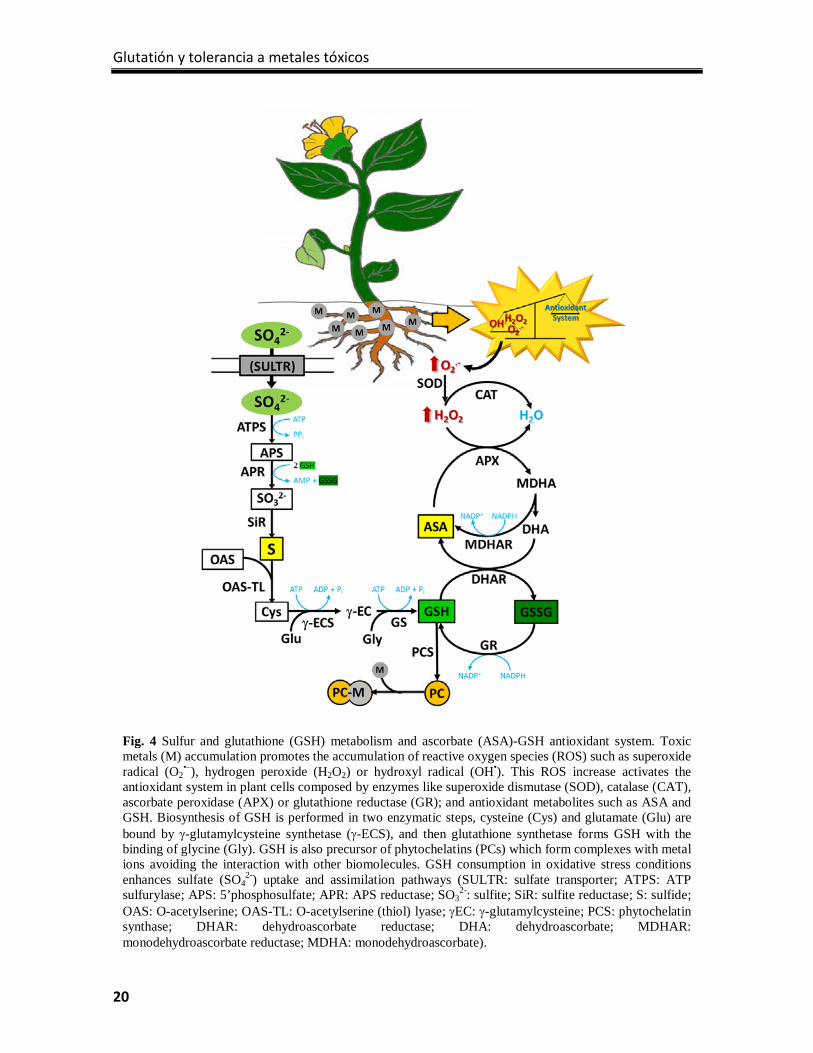
(Gill and Tuteja 2010). See Fig. 4 for details.
The cellular redox homeostasis is maintained basically by the transformation of O2– in
H2O2 by SODs and the posterior scavenging of H2O2 by CATs and POXs (Noctor and
Foyer 1998). In plants there are three groups of SODs based on the metal(s) as co-factor(s):
FeSOD, MnSOD and Cu/ZnSOD. FeSOD is located in chloroplasts, as well as one
Cu/ZnSOD isoform, MnSODs in mitochondria and peroxisomes and the major Cu/ZnSOD
can be found in the cytosol and extracellular space (Alscher et al. 2002). CATs and POXs
have different substrate affinities and catalytic rates although both enzyme families are
involved in the removal of H2O2. Diverse peroxidases isoforms can be found in different
compartments in plant cells; however CATs are mainly present in peroxisomes and absent
in chloroplasts (Sandalio et al. 2006).
Coupled to this antioxidant machinery, the AS-GSH cycle plays a central role (Foyer and
Halliwell 1976). The ROS excess is mainly removed in this cycle where H2O2 is reduced to
H2O by APX using electrons from NADPH (Fig. 4). Ascorbate is the substrate for APX and
it is necessary two molecules to reduce H2O2 to H2O, so the requirement of this metabolite
is totally essential (Nakano and Asada 1987). The consequence of this reaction is the
reduction of ascorbate to MDHA, and after a rapid non-enzymatic reduction is converted to
DHA. To regenerate ASA by the action of DHAR, GSH is oxidized to form oxidized
glutathione (GSSG). GR is then responsible to regenerate GSH using electrons derived
from NADPH. GR is a ubiquitous enzyme located in different cellular organelles
(chloroplasts, mitochondria and cytosol), where the chloroplastic isoform represents 80% of
GR activity of the cell. The importance of this enzyme resides in the maintenance of correct
levels of GSH, because during a redox imbalance having high GSH/GSSG ratio is essential
for an efficient antioxidant defense (Gill et al. 2013).
The induction of this antioxidant machinery has been demonstrated in different plants under
different metal(loid) treatments (Sharma and Dietz, 2009). Several antioxidant enzymes
augmented their activity in Spartina densiflora, an invasive cordgrass, in a dose and time-
dependent manner when treated with different concentrations of Cd (Martínez Domínguez
Tesis doctoral Juan Sobrino Plata
19
et al. 2010). In Jatropha curcas, a plant that usually grows in mining areas where soils are
contaminated by toxic metals, Hg induced oxidative stress and antioxidant responses,
where increments in SOD, CAT and POX activities were observed (Gao et al. 2010).
5. Glutathione, the key metabolite in redox homeostasis.
Glutathione is an essential molecule in all living organisms. Today is known that this
molecule is a multifunctional metabolite. In addition to the antioxidant role described
above, GSH is directly involved with stress signaling, plant development, defense reactions
and redox homeostasis (Noctor et al. 2011). Moreover, GSH is also important in the
detoxification of xenobiotics, through glutathione S-transferases (GST), and detoxification
of toxic metals through the synthesis of phytochelatins (PCs).
Although the most common structure of GSH is γ-L-glutamyl-L-cysteinylglycine, some
plants accumulate homoglutathione (γ-Glu-Cys-Ala; hGSH), where the terminal Gly is
substituted by β-alanine (Ala). This is the case of some in legumes like alfalfa, where
hGSH prevails over GSH and is the major biothiol (Klapheck 1988). Glutathione is found
mostly in its reduced form, serving as one of the main ROS scavengers in plant cells and an
important reservoir of organic sulfur. Glutathione reductases are the responsible for
maintaining in an appropriate balance shifted to GSH from a minor pool of oxidized
glutathione (GSSG). The synthesis of ROS may provoke transient oxidation of GSH via
DHAR, glutaredoxins (GRXs), glutathione peroxidases (GPXs) or adenosine
5’phosphosulfate reductase (APR), levels that should be restored by an activation of GRs
(Davidian and Kopriva 2010, Noctor et al. 2012).
5.1 Glutathione biosynthesis and distribution in cell.
The synthesis of GSH is carried out in two enzymatic steps where amino acids are bound
consecutively. These process is dependent of ATP, and take place in two different cellular
comparments: The first step is restricted to plastids and is catalyzed by γ-glutamylcysteine
synthetase (γECS), encoded by gene GSH1 in Arabidopsis. In this reaction Glu and Cys are
bound to form the intermediate γ-EC, which could be transported to the cytosol or
converted to GSH in plastids. The second step is the addition of Gly to γ-EC, catalyzed by
glutathione synthetase (GS), which is encoded by GSH2 Arabidopsis gene that undergoes
Glutatión y tolerancia a metales tóxicos
20
Fig. 4 Sulfur and glutathione (GSH) metabolism and ascorbate (ASA)-GSH antioxidant system. Toxic metals (M) accumulation promotes the accumulation of reactive oxygen species (ROS) such as superoxide radical (O2
•–), hydrogen peroxide (H2O2) or hydroxyl radical (OH•). This ROS increase activates the antioxidant system in plant cells composed by enzymes like superoxide dismutase (SOD), catalase (CAT), ascorbate peroxidase (APX) or glutathione reductase (GR); and antioxidant metabolites such as ASA and GSH. Biosynthesis of GSH is performed in two enzymatic steps, cysteine (Cys) and glutamate (Glu) are bound by γ-glutamylcysteine synthetase (γ-ECS), and then glutathione synthetase forms GSH with the binding of glycine (Gly). GSH is also precursor of phytochelatins (PCs) which form complexes with metal ions avoiding the interaction with other biomolecules. GSH consumption in oxidative stress conditions enhances sulfate (SO4
2-) uptake and assimilation pathways (SULTR: sulfate transporter; ATPS: ATP sulfurylase; APS: 5’phosphosulfate; APR: APS reductase; SO3
2-: sulfite; SiR: sulfite reductase; S: sulfide; OAS: O-acetylserine; OAS-TL: O-acetylserine (thiol) lyase; γEC: γ-glutamylcysteine; PCS: phytochelatin synthase; DHAR: dehydroascorbate reductase; DHA: dehydroascorbate; MDHAR: monodehydroascorbate reductase; MDHA: monodehydroascorbate).
Tesis doctoral Juan Sobrino Plata
21
alternative splicing depending on the final location of the protein. Shortest form of these
transcripts encoded the cytosolic GS and the longest harbor a signal peptide for chloroplast
(Wachter et al. 2005). When the synthesis of GSH is carried out in cytosol, γ-EC is
exported through chloroquinone-like transporters (CLTs); essential transporters that are
essential in the transfer of γ-EC and GSH from plastids to the cytosol (Maughan et al.
2010).
In the last years modern imaging techniques and advanced mass spectrometry technologies
have provided new information about the compartmentalization and distribution of GSH in
cellular organelles of different plant organs (Ortega-Villasante et al. 2005, Zechmann and
Müller 2010, Carrasco‐Gil et al. 2011, Carrasco-Gil et al. 2013, Koffler et al. 2013).
Glutathione is one of the major forms of organic sulfur translocated in the phloem
(Mendoza‐Cózatl et al. 2008). In Arabidopsis oligopeptide transporter 6 (OPT6) mediates
intercellular transport of GSH, GSSG, GSH-conjugates and GSH-metal complexes (Cagnac
et al. 2004). Intracellularly, GSH may be translocated between organelles and the cytosol.
Apart from CLTs, which transport GSH and γ-EC between cytosol and plastids, there
should be a competent transport system in mitochondria and nuclei, since these organelles
accumulate high amounts of GSH. Although in animals these transporters were
characterized in detail (Voehringer 1999), they have not been found yet in plants. The last
evidences showed higher GSH accumulation in mitochondria and nuclei than in
chloroplasts suggesting a rapid and efficient intracellular transport of GSH from the place
where it is synthetized (plastids and cytosol) to places where is required to deal with the
accumulation of ROS (Zechmann et al. 2008, Queval et al. 2011). Transport of GSSG and
GSH-conjugates into the vacuole is an important mechanism to maintain the cytosolic GSH
redox status. This transport is done through Multidrug Resistance-associated proteins
(MRPs), and their expression is specifically increased in response to oxidative stress
(Queval et al. 2011). Moreover, recent studies show low concentrations of GSH in
chloroplasts in non-stressed conditions, which implies the export to other organelles; but
during oxidative stress there is a rapid and higher GSH synthesis leading to accumulation in
chloroplast, as an efficient defense strategy to limit ROS generation (Koffler et al. 2013).
Glutatión y tolerancia a metales tóxicos
22
5.2 Regulation of glutathione biosynthesis.
This pathway is regulated at different levels, and according to May et al. (1998) there are
five levels to control GSH concentrations: substrate availability, rate limitation by γECS,
feedback inhibition by γEC and GSH, transcriptional control and post-transcriptional
regulations. Mutants in GSH biosynthesis genes and chemicals inhibitors of this pathway
helped to characterize the regulation and functions of GSH in plants. Buthionine
sulfoximine (BSO) is a potent inhibitor of γECS, firstly described by Griffith and Meister
(1979). This molecule is able to bind to active site of γECS and decreases its activity but
not completely. Many functional studies have been performed using BSO to highlight the
fundamental role of GSH in oxidative stress tolerance (May and Leaver 1993, Ortega-
Villasante et al. 2005, Sobrino-Plata et al. 2009).
On the other hand, different Arabidopsis mutants in both enzymes of GSH biosynthesis
have been obtained in the last years. Knockout mutants of GSH1 and GSH2 have embryo-
lethal and seedling-lethal phenotypes respectively (Cairns et al. 2006, Pasternak et al.
2008). These studies made clear the direct implication of GSH in plant development. On
the other hand, γ-EC replaces partially GSH functions and that entails the possibility of
germination but not the normal development of the plant. More useful instruments had been
those γECS mutants with diminished activity respect to wild type (WT), which have been
described as extremely sensitive to several harmful environmental conditions: root-
meristemless 1 (rml1-1) with around 2% of WT GSH levels suffers severe alteration of
plant development (Vernoux et al. 2000); cadmium-sensitive 2-1 (cad2-1) presents
sensitivity to heavy metals and 30% of WT GSH levels (Cobbett et al. 1998, Howden et al.
1995a); the regulator of APX2 1-1 mutant (rax1-1) shows growth inhibition under high
irradiance stress (45% GSH compared to WT; Ball et al. 2004); and the phytoalexin-
deficient 2-1 (pad2-1) which contains 20% of WT GSH levels and is extremely sensitive to
pathogenic interactions (Parisy et al. 2007).
Overexpression of γECS or GS enhances GSH accumulation in plants and, in most cases
transgenic plants show enhanced tolerance to oxidative stress, as occurred under toxic
metal stress (Zhu et al. 1999a, Zhu et al. 1999b) or herbicides (Skipsey et al. 2005).
However, there are also experiments where the overexpression of γECS in some transgenic
Tesis doctoral Juan Sobrino Plata
23
lines caused higher sensitivity and negative effects, such as early leaf senescence or growth
inhibition observed in poplars (Herschbach et al. 2010) or lesion formation and high GSSG
accumulation in tobacco (Creissen et al. 1999). Nonetheless a recent study showed the
expression of the bifunctional γ-glutamylcysteine ligase-glutathione synthetase enzyme
from Streptococcus thermophilus (StGCL-GS) in tobacco plants produced 20-30%
increased GSH levels compare to WT lines, developing resistance to abiotic stresses
produced by accumulation of H2O2 or Cd and no negative effects over growth or
physiological functions of plants (Liedschulte et al. 2010).
One of the first limitations in GSH biosynthesis is the availability of the substrates of the
enzymes (mainly Gly, Cys and ATP); and the activity of γECS (Noctor and Foyer 1998).
However gene transcriptional regulators and post-translational modifications are also
important to maintain a correct level of GSH in plant cells. Accumulation of GSH, GSSG
or H2O2 produce no effects over gene expression, but a depletion of GSH leads to the up
regulation of GSH1 and GSH2 expression (Xiang and Oliver 1998). In general
detoxification strategies that involve GSH such as complexation of PCs with diverse toxic
metal(loid)s, together with the oxidation of GSH under oxidative stress, causes an increase
of GSH1 and GSH2 gene expression by a ‘demand-driven regulation’ (Nocito et al. 2006).
Recent research efforts are clarifying the mechanisms of post-translational regulation of γ-
ECS, the limiting step in GSH biosynthesis. Besides the feedback inhibition of this enzyme
by γ-EC or GSH; or the inhibition by other dithiols in cells (Noctor et al. 2002), there is a
unique regulatory mechanism in plants related with two intramolecular disulfide bridges
that can be formed in γECS. In 2006 the crystallization performed by Hothorn et al. (2006)
of Brassica juncea γECS brought light to this issue. Four Cys residues are involved in the
regulation of this enzyme, Cys341 and Cys356 which form the CC1 disulfide bridge and
established a β-hairpin motif implicated in the entry of molecules to the substrate binding
site; and Cys178 and Cys398 which form a disulfide bridge between two helices located in N-
and C-terminals (CC2). This disulfide bridge is involved in the redox regulation of the
enzyme, where the oxidized form implies a dimerization of the enzyme and the activation,
while the reduction form is monomeric and inactive. The exact regulatory mechanism was
described in Arabidopsis thaliana where the Cys residues occupy the positions 344-364 and
Glutatión y tolerancia a metales tóxicos
24
186-406 (Hicks et al. 2007). The γECS crystallization also helped to explain the low
activity in cad2-1, rax1-1 and rml1 knock-down mutants. In cad2-1 the deletion of six
nucleotides affects positions 237-239 altering the position of important residues in substrate
binding. On the other hand, the substitution of Arg by Lys in position 228 in rax1-1 affects
the recognition of Cys, and the change of Asp258 by Asn in rml1-1 affects the ATP binding.
5.3 Glutathione role in sulfur metabolism.
As the main sink of reduced sulfur in plant cells, the influence of GSH over the sulfur
metabolism is obvious (see Fig. 4 for detailed explanation of the major assimilatory
pathway in plant cells). Sulfur is essential for plants and inorganic sulfate is the major form
that plants acquire from the environment. Sulfate uptake and assimilation pathways have
been well characterized in Arabidopsis in the last years, and the direct implication of GSH
in the regulation of both routes remains clear (Davidian and Kopriva 2010).
The sulfate uptake and distribution in all compartments of the plants is made through
different transporters (AtSULTR; Buchner et al. 2004). First step in sulfate assimilation is
catalyzed by ATP sulfurylase (ATPS), which produce 5’phosphosulfate (APS) by
adenylation of sulfate using ATP (Hatzfeld et al. 2000). APS is reduced to sulfite (SO32-) by
APS reductase (APR) which is located in plastids and using electrons from GSH
(Koprivova et al. 2008). This step together with sulfate uptake is described as key in sulfur
metabolism (Vauclare et al. 2002). In fact, high accumulation of reduced sulfur molecules,
especially GSH, acts as a feedback signal and controls the regulation of sulfur nutrition
repressing SULTRs, APS and APR activities (Kopriva and Rennenberg 2004, Nocito et al.
2006).
Sulfite is reduced to sulfide with the action of the sulfite reductase (SiR) which is
absolutely necessary in this pathway, as this step is no possible in a non-enzymatic reaction
(Nakayama et al. 2000). Cys biosynthesis is the final step of sulfate assimilation and the
link with the GSH metabolism. Cys is synthetized by the cysteine synthase complex (CSC),
which is formed by serine acetyltransferase (SAT) and O-acetylserine (thiol) lyase (OAS-
TL). First enzyme catalyzes the formation of OAS binding acetyl-CoA and serine, and then
OAS-TL synthetizes Cys with the union of sulfide to OAS (Wirtz and Hell 2006).
Tesis doctoral Juan Sobrino Plata
25
Depletion of GSH increases the expression of several genes in sulfate assimilation pathway
in a “demand-driven” regulation (Lappartient and Touraine 1996). In plants exposed to
toxic metal(loid)s GSH is fully implicated in the response to oxidative stress produced by
these elements. The antioxidant function of GSH implies the oxidation of this molecule;
although GSH can be regenerated by GR, in stress situations the proportion of this redox
couple (GSH/GSSG) is displaced to the oxidized form (Semane et al. 2007). This depletion
is further promoted when GSH is used as precursor of PCs, which are needed for the
chelation of the toxic elements (Pal and Rai 2010). Thereby, this GSH requirement in
metal(oid) stress conditions implicates the up-regulation of genes coding the limiting
enzymatic steps of sulfur metabolism such as the sulfate uptake, formation of sulfite or the
synthesis of Cys. In Ni-hyperaccumulator Thalspi goesingense elevated OAS, Cys and
GSH levels and high SAT and GR activities were observed, suggesting a direct implication
of sulfur metabolism in Ni tolerance (Freeman et al. 2004). As well as toxic metals, other
environmental stresses induce the sulfate assimilation pathway, for instance in salt stressed
Arabidopsis plants OAS-TL seems to be involved in the acclimation to high salt, and APR
contributes to the early response, allowing the increase of Cys biosynthesis rate (Koprivova
et al. 2008).
A new model of sulfur metabolism regulation under toxic metal stress was given recently
by Jobe et al. (2012). In this work the authors presented a modification of the “demand-
driven” regulation classic model, adding other factors that are required for the induction of
genes in this pathway. Plants with diminished GSH levels were treated with Cd, Cys and
γEC and the expression of SULTR1;2 was measured: Under GSH depletion the up-
regulation of genes of sulfate uptake and assimilation was possible only if the concentration
of upstream thiols (Cys and γEC) decreased too, while an enhanced oxidative environment
occurs. This new model of regulation gives a significant importance to the GSH redox state
in several metabolic processes in the cell.
5.4 The importance of glutathione redox status.
A reducing environment is required in cells to generate electrochemical gradients that are
essential in many cellular processes. This reducing environment is maintained by different
redox couples, of which GSH/GSSG has a prominent role in the cell due to its abundance
Glutatión y tolerancia a metales tóxicos
26
and contribution in several processes. The redox state of GSH/GSSG is an indicator of the
redox environment in the cell and is defined by redox potential ([GSH]2/GSSG), which
reflects the capacity of GSH redox system to obtain electrons and being reduced (Jozefczak
et al. 2012). As stated before, the GSH redox status is important in plant development, cell
proliferation and regeneration efficiency of somatic embryos (Diaz et al. 2010).
Reduced state of cells is a normal situation, although there are cell types and compartments
that are maintained in an oxidized state, such as endoplasmatic reticulum, root quiescent
center or seeds (Kranner et al. 2002). However the redox environment can change during
the cell cycle. Recent studies showed the flux of GSH from cytosol to nuclei during
mitosis, and at G1 phase GSH is recruited into the nucleus promoting oxidation of the
cytosol and thereby enhanced GSH biosynthesis. This GSH accumulation will be divided
between the daughter cells (Vivancos et al. 2010). The redox environment also marks the
state of cell viability, since the most negative corresponds to cells with high GSH levels at
the beginning of differentiation, whereas the most positive (more oxidative) is found in
apoptotic cells where prooxidant metabolites exceeds the antioxidant capability (Schafer
and Buettner 2004).
Altered the reduced state of GSH pool is a stress indicator and may be produced by
peroxidase activities, disturbance in GSH regeneration processes or direct interaction with
ROS (Noctor et al. 2012). Concretely GPXs and certain GSTs use GSH as reducing
substrate to remove H2O2 or organic peroxides, thereby a high accumulation of these
oxidants will affect the GSH levels promoting a more oxidized environment (Anjum et al.
2012). Although the major detoxification of H2O2 by oxidation of GSH is found in ASA-
GSH cycle as we described above and GRs are the enzymes in charge of maintaining the
GSH/GSSG ratio.
An important consequence of alterations in GSH/GSSG redox couple is the disturbance in
redox signaling. Both GSH/GSSG and NADPH/NADP couples are necessary to be in
equilibrium as part of the cellular oxidative signaling (Queval and Noctor 2007). Defense
responses of plants depend on signals produced by changes in the cellular redox balance.
Changes in GSH redox potential promote signaling cascades that elicit responses front to
different stresses (Schafer and Buettner 2004). In this respect, there are basically two forms
Tesis doctoral Juan Sobrino Plata
27
of signaling mediated by GSH redox status and both are reversible. Modification of redox
state of thiol groups of proteins, also called thiol switches; and glutathionylation of
proteins, conjugation of GSH to Cys residues that regulates its activity (Jozefczak et al.
2012). Thioredoxins (TRXs) and GRXs are able to regulate proteins exchanging the redox
state of thiol residues (Noctor et al. 2012). Examples of GRX implication in this kind of
regulation are seen in assembly of iron-sulfur clusters, regeneration of peroxiredoxins or in
responses to different oxidative stresses (Cheng et al. 2006, Rouhier et al. 2010). The
second kind of GSH redox regulation is the direct and stable union of GSH to Cys residues
or S-glutathionylation. For instance, in a chloroplastic TRX is glutathionylated on a Cys
residue external to the active site reducing its activity against target proteins as NADP-
glyceraldehyde-3-phosphate dehydrogenase (GAPDH; Michelet et al. 2005).
6. Glutathione metabolism and metal(loid) tolerance: the case of Hg.
As was highlighted in this review, proper metabolism of biothiols is important for
metal(loid) tolerance, either as antioxidants or as precursors of PCs (Jozefczak et al. 2012).
The research done in our group also demonstrated the importance of GSH and PCs
metabolism in the tolerance of plants to Hg (Carrasco-Gil et al., 2011). The use of
Arabidopsis mutant plants with defective levels of GSH has permitted us to hypothesize
that a minimum level is required to confer tolerance to Hg and Cd, being affected the
capability to accumulate PCs and Hg-PC complexes (Sobrino-Plata et al. 2014a, 2014b).
Interestingly, Arabidopsis rax1-1 mutants showed the over-expression of γECS possibly as
a counterbalance mechanism to replenish the levels of GSH (Sobrino-Plata et al. 2014b).
This up-regulation depends also on the degree of stress, as plants exposed to high doses of
Hg (30 µM) did not respond as those treated with a lower dose (3 µM); perhaps implying
that readily damaged cells suffer general and chronic stress (Sobrino-Plata et al. 2014a). In
addition, mutant phenotypes with depleted GSH levels accumulated less Hg and Cd than
the wild type, which suggest that processes of uptake and transport to the aerial organs
depend on biothiol metabolism (Sobrino-Plata et al., 2014b). Finally, plants unable to
produce PCs such as Arabidopsis cad1-3 mutants showed a significant increase in GSH
levels when were exposed to moderate doses (3 µM) of Hg and Cd (Carrasco-Gil et al.
Glutatión y tolerancia a metales tóxicos
28
2011, Sobrino-Plata et al. 2014a), possibly related with the activation of the GSH
biosynthetic and sulfate assimilation pathways under stress.
As was discussed earlier, sulfur and GSH metabolism are subjected to strict control, and
multiple factors influence the regulation of limiting steps. Transcriptional, post-
translational modifications, cellular redox perception, antioxidant responses, transport to
vacuoles of metal(loid)-PC complexes, etc. are mediated by GSH status in the cell; perhaps
one of the most versatile metabolite involved in the cellular homeostasis. New available
analytical techniques will be needed to understand in detail these complex mechanisms of
regulation, information that will help to optimize plant tolerance to toxic metal(oid)s and
other stress factors. Future research in this topic will then permit use of plants for
phytoremediation strategies to clean up metal(loid)s-polluled soils, and the subsistence of
plants in hazardous environmental conditions.
REFERENCES
Ahn YO, Kim SH, Lee J, Kim H, Lee H, Kwak S. 2012. Three Brassica rapa metallothionein genes are
differentially regulated under various stress conditions. Molecular biology reports 39, 2059-2067.
Ali H, Khan E, Sajad MA. 2013. Phytoremediation of heavy metals—Concepts and applications.
Chemosphere 91, 869-881.
Alloway BJ. 2012. Trace Metals and Metalloids in Soils and Their Bioavailability. In: Environmental
Pollution: Heavy Metals in Soils, ed.: Springer.
Alscher RG, Erturk N, Heath LS. 2002. Role of superoxide dismutases (SODs) in controlling oxidative
stress in plants. Journal of experimental botany 53, 1331-1341.
Anjum NA, Ahmad I, Mohmood I, Pacheco M, Duarte AC, Pereira E, Umar S, Ahmad A, Khan NA,
Iqbal M. 2012. Modulation of glutathione and its related enzymes in plants’ responses to toxic metals
and metalloids—a review. Environmental and experimental botany 75, 307-324.
Arthur EL, Rice PJ, Rice PJ, Anderson TA, Baladi SM, Henderson KL, Coats JR. 2005.
Phytoremediation—an overview. Critical Reviews in Plant Sciences 24, 109-122.
Asada K. 1999. The water-water cycle in chloroplasts: scavenging of active oxygens and dissipation of
excess photons. Annual review of plant biology 50, 601-639.
Baker A, McGrath S, Sidoli C, Reeves R. 1994. The possibility of in situ heavy metal decontamination of
polluted soils using crops of metal-accumulating plants. Resources, Conservation and Recycling 11, 41-
49.
Tesis doctoral Juan Sobrino Plata
29
Baker AJ. 1981. Accumulators and excluders‐strategies in the response of plants to heavy metals. Journal of
Plant Nutrition 3, 643-654.
Ball L, Accotto G, Bechtold U, Creissen G, Funck D, Jimenez A, Kular B, Leyland N, Mejia-Carranza
J, Reynolds H. 2004. Evidence for a direct link between glutathione biosynthesis and stress defense gene
expression in Arabidopsis. The Plant Cell Online 16, 2448-2462.
Barceló J, Poschenrieder C. 2003. Phytoremediation: principles and perspectives. Contributions to science
2, 333-344.
Barrutia O, Epelde L, García-Plazaola J, Garbisu C, Becerril J. 2009. Phytoextraction potential of two
Rumex acetosa L. accessions collected from metalliferous and non-metalliferous sites: Effect of
fertilization. Chemosphere 74, 259-264.
Barrutia O, Garbisu C, Hernández-Allica J, García-Plazaola JI, Becerril JM. 2010. Differences in
EDTA-assisted metal phytoextraction between metallicolous and non-metallicolous accessions of Rumex
acetosa L. Environmental Pollution 158, 1710-1715.
Bernier M, Popovic R, Carpentier R. 1993. Mercury inhibition at the donor side of photosystem II is
reversed by chloride. FEBS letters 321, 19-23.
Bizily SP, C.L. Rugh CL, Meagher RB. 2000. Phytodetoxification of hazardous organomercurials by
genetically engineered plants. Nature Biotechnology 18, 213–217.
Blacksmith Institute, Green Cross Switzerland. 2013. Top Ten Toxic Threats in 2013: Cleanup, Progress
and Ongoing Challenges. 8.
Bose-O'Reilly S, McCarty KM, Steckling N, Lettmeier B. 2010. Mercury exposure and children's health.
Current problems in pediatric and adolescent health care 40, 186-215.
Bowler C, Montagu Mv, Inze D. 1992. Superoxide dismutase and stress tolerance. Annual review of plant
biology 43, 83-116.
Briat J, Lebrun M. 1999. Plant responses to metal toxicity. Comptes Rendus de l'Académie des Sciences-
Series III-Sciences de la Vie 322, 43-54.
Brookes A, Collins J, Thurman D. 1981. The mechanism of zinc tolerance in grasses. Journal of Plant
Nutrition 3, 695-705.
Buchner P, Takahashi H, Hawkesford MJ. 2004. Plant sulphate transporters: co-ordination of uptake,
intracellular and long-distance transport. Journal of experimental botany 55, 1765-1773.
Cagnac O, Bourbouloux A, Chakrabarty D, Zhang M, Delrot S. 2004. AtOPT6 transports glutathione
derivatives and is induced by primisulfuron. Plant Physiology 135, 1378-1387.
Cairns NG, Pasternak M, Wachter A, Cobbett CS, Meyer AJ. 2006. Maturation of Arabidopsis seeds is
dependent on glutathione biosynthesis within the embryo. Plant Physiology 141, 446-455.
Callahan DL, Baker AJ, Kolev SD, Wedd AG. 2006. Metal ion ligands in hyperaccumulating plants. JBIC
Journal of Biological Inorganic Chemistry 11, 2-12.
Glutatión y tolerancia a metales tóxicos
30
Carrasco‐Gil S, Álvarez‐Fernández A, Sobrino-Plata J, Millán R, Carpena-Ruiz RO, Leduc DL,
Andrews JC, Abadía J, Hernández LE. 2011. Complexation of Hg with phytochelatins is important for
plant Hg tolerance. Plant, Cell and Environment 34, 778-791.
Carrasco-Gil S, Estebaranz-Yubero M, Medel-Cuesta D, Millán R, Hernández LE. 2012. Influence of
nitrate fertilization on Hg uptake and oxidative stress parameters in alfalfa plants cultivated in a Hg-
polluted soil. Environmental and Experimental Botany 75, 16-24.
Carrasco-Gil S, Siebner H, LeDuc DL, Webb SM, Millán R, Andrews JC, Hernández LE. 2013.
Mercury localization and speciation in plants grown hydroponically or in a natural environment.
Environmental Science and Technology 47, 3082-3090.
Cataldo DA, Garland TR, Wildung RE. 1978. Nickel in plants I. Uptake kinetics using intact soybean
seedlings. Plant Physiology 62, 563-565.
Cataldo DA, Garland TR, Wildung RE. 1983. Cadmium uptake kinetics in intact soybean plants. Plant
Physiology 73, 844-848.
Chaney R. 1983. Plant uptake of inorganic waste constituents. In: Land Treatment of Hazardous Wastes,
eds.: JE Parr, PB Marsh, JM Kla, pp. 50-76.
Cheng N, Liu J, Brock A, Nelson RS, Hirschi KD. 2006. AtGRXcp, an Arabidopsis chloroplastic
glutaredoxin, is critical for protection against protein oxidative damage. Journal of Biological Chemistry
281, 26280-26288.
Chiang H, Lo J, Yeh K. 2006. Genes associated with heavy metal tolerance and accumulation in Zn/Cd
hyperaccumulator Arabidopsis halleri: a genomic survey with cDNA microarray. Environmental science
& technology 40, 6792-6798.
Clarkson TW. 1997. The toxicology of mercury. Critical reviews in clinical laboratory sciences 34, 369-
403.
Clemens S, Kim EJ, Neumann D, Schroeder JI. 1999. Tolerance to toxic metals by a gene family of
phytochelatin synthases from plants and yeast. The EMBO journal 18, 3325-3333.
Clemens S, Palmgren MG, Krämer U. 2002. A long way ahead: understanding and engineering plant metal
accumulation. Trends in Plant Science 7, 309-315.
Clemens S. 2006. Toxic metal accumulation, responses to exposure and mechanisms of tolerance in plants.
Biochimie 88, 1707-1719.
Cobbett C, Goldsbrough P. 2002. Phytochelatins and metallothioneins: roles in heavy metal detoxification
and homeostasis. Annual Review of Plant Biology 53, 159-182.
Cobbett CS, May MJ, Howden R, Rolls B. 1998. The glutathione‐deficient, cadmium‐sensitive mutant,
cad2–1, of Arabidopsis thaliana is deficient in γ‐glutamylcysteine synthetase. The Plant Journal 16, 73-
78.
Tesis doctoral Juan Sobrino Plata
31
Creissen G, Firmin J, Fryer M, Kular B, Leyland N, Reynolds H, Pastori G, Wellburn F, Baker N,
Wellburn A. 1999. Elevated glutathione biosynthetic capacity in the chloroplasts of transgenic tobacco
plants paradoxically causes increased oxidative stress. The Plant Cell Online 11, 1277-1291.
Cunningham SD, Berti WR. 1993. Remediation of contaminated soils with green plants: an overview. In
Vitro Cellular & Developmental Biology-Plant 29, 207-212.
Dat J, Vandenabeele S, Vranova E, Van Montagu M, Inzé D, Van Breusegem F. 2000. Dual action of the
active oxygen species during plant stress responses. Cellular and Molecular Life Sciences CMLS 57, 779-
795.
Davidian J, Kopriva S. 2010. Regulation of sulfate uptake and assimilation—the same or not the same?
Molecular plant 3, 314-325.
Dermont G, Bergeron M, Mercier G, Richer-Laflèche M. 2008. Soil washing for metal removal: A review
of physical/chemical technologies and field applications. Journal of hazardous materials 152, 1-31.
Diaz VP, Wolff T, Markovic J, Pallardó F, Foyer C. 2010. A nuclear glutathione cycle within the cell
cycle. Biochemical Journal 431, 169-178.
Duffus JH. 2002. "Heavy metals" a meaningless term? (IUPAC Technical Report). Pure and applied
chemistry 74, 793-807.
Dushenkov V, Kumar PN, Motto H, Raskin I. 1995. Rhizofiltration: the use of plants to remove heavy
metals from aqueous streams. Environmental science & technology 29, 1239-1245.
Ekino S, Susa M, Ninomiya T, Imamura K, Kitamura T. 2007. Minamata disease revisited: an update on
the acute and chronic manifestations of methyl mercury poisoning. Journal of the neurological sciences
262, 131-144.
EPA 2007. Treatment technologies for site cleanup: Annual status report (Twelfth edition).
Faller P, Kienzler K, Krieger-Liszkay A. 2005. Mechanism of Cd2+ toxicity: Cd2+ inhibits photoactivation
of Photosystem II by competitive binding to the essential Ca2+ site. Biochimica et Biophysica Acta (BBA)-
Bioenergetics 1706, 158-164.
Fawzy E. 2008. Soil remediation using in situ immobilisation techniques. Chemistry and Ecology 24, 147-
156.
Foreman J, Demidchik V, Bothwell JH, Mylona P, Miedema H, Torres MA, Linstead P, Costa S,
Brownlee C, Jones JD. 2003. Reactive oxygen species produced by NADPH oxidase regulate plant cell
growth. Nature 422, 442-446.
Foyer CH, Halliwell B. 1976. The presence of glutathione and glutathione reductase in chloroplasts: a
proposed role in ascorbic acid metabolism. Planta 133, 21-25.
Foyer CH, Noctor G. 2003. Redox sensing and signalling associated with reactive oxygen in chloroplasts,
peroxisomes and mitochondria. Physiologia Plantarum 119, 355-364.
Glutatión y tolerancia a metales tóxicos
32
Freeman JL, Persans MW, Nieman K, Albrecht C, Peer W, Pickering IJ, Salt DE. 2004. Increased
glutathione biosynthesis plays a role in nickel tolerance in Thlaspi nickel hyperaccumulators. The Plant
Cell Online 16, 2176-2191.
Fusheng Z, Jingjing L, Kerui C, Long X. 2012. Utilization of cement for solidification/stabilization (s/s) of
heavy metal contaminated soils. DISASTER ADVANCES 5, 1574-1577.
Gao S, Ou-yang C, Tang L, Zhu J, Xu Y, Wang S, Chen F. 2010. Growth and antioxidant responses in
Jatropha curcas seedling exposed to mercury toxicity. Journal of Hazardous Materials 182, 591-597.
Gardea-Torresdey J, Gonzalez J, Tiemann K, Rodriguez O, Gamez G. 1998. Phytofiltration of hazardous
cadmium, chromium, lead and zinc ions by biomass of Medicago sativa (Alfalfa). Journal of Hazardous
Materials 57, 29-39.
Gill SS, Anjum NA, Hasanuzzaman M, Gill R, Trivedi DK, Ahmad I, Pereira E, Tuteja N. 2013.
Glutathione and glutathione reductase: a boon in disguise for plant abiotic stress defense operations. Plant
Physiology and Biochemistry 70, 204-212.
Gill SS, Tuteja N. 2010. Reactive oxygen species and antioxidant machinery in abiotic stress tolerance in
crop plants. Plant Physiology and Biochemistry 48, 909-930.
Griffith OW, Meister A. 1979. Potent and specific inhibition of glutathione synthesis by buthionine
sulfoximine (Sn-butyl homocysteine sulfoximine). Journal of Biological Chemistry 254, 7558-7560.
Groppa M, Benavides M. 2008. Polyamines and abiotic stress: recent advances. Amino Acids 34, 35-45.
Ha S, Smith AP, Howden R, Dietrich WM, Bugg S, O'Connell MJ, Goldsbrough PB, Cobbett CS. 1999.
Phytochelatin synthase genes from Arabidopsis and the yeast Schizosaccharomyces pombe. The Plant
Cell 11, 1153-1163.
Hall J. 2002. Cellular mechanisms for heavy metal detoxification and tolerance. Journal of Experimental
Botany 53, 1-11.
Halliwell B. 2006. Reactive species and antioxidants. Redox biology is a fundamental theme of aerobic life.
Plant Physiology 141, 312-322.
Hassinen V, Tervahauta A, Schat H, Kärenlampi S. 2011. Plant metallothioneins–metal chelators with
ROS scavenging activity? Plant Biology 13, 225-232.
Hatzfeld Y, Lee S, Lee M, Leustek T, Saito K. 2000. Functional characterization of a gene encoding a
fourth ATP sulfurylase isoform from Arabidopsis thaliana. Gene 248, 51-58.
Heckathorn SA, Mueller JK, LaGuidice S, Zhu B, Barrett T, Blair B, Dong Y. 2004. Chloroplast small
heat-shock proteins protect photosynthesis during heavy metal stress. American Journal of Botany 91,
1312-1318.
Hernández-Allica J, Garbisu C, Barrutia O, Becerril JM. 2007. EDTA-induced heavy metal accumulation
and phytotoxicity in cardoon plants. Environmental and Experimental Botany 60, 26-32.
Tesis doctoral Juan Sobrino Plata
33
Herschbach C, Rizzini L, Mult S, Hartmann T, Busch F, Peuke AD, Kopriva S, Ensminger I. 2010.
Over‐expression of bacterial γ‐glutamylcysteine synthetase (GSH1) in plastids affects photosynthesis,
growth and sulphur metabolism in poplar (Populus tremula× Populus alba) dependent on the resulting γ‐
glutamylcysteine and glutathione levels. Plant, Cell and Environment 33, 1138-1151.
Heyno E, Klose C, Krieger-Liszkay A. 2008. Origin of cadmium-induced reactive oxygen species
production: mitochondrial electron transfer versus plasma membrane NADPH-oxidase. New Phytologist
179, 687-699.
Hicks LM, Cahoon RE, Bonner ER, Rivard RS, Sheffield J, Jez JM. 2007. Thiol-based regulation of
redox-active glutamate-cysteine ligase from Arabidopsis thaliana. The Plant Cell 19, 2653-2661.
Hodges DM, DeLong JM, Forney CF, Prange RK. 1999. Improving the thiobarbituric acid-reactive-
substances assay for estimating lipid peroxidation in plant tissues containing anthocyanin and other
interfering compounds. Planta 207, 604-611.
Hossain MA, Hasanuzzaman M, Fujita M. 2010. Up-regulation of antioxidant and glyoxalase systems by
exogenous glycinebetaine and proline in mung bean confer tolerance to cadmium stress. Physiology and
Molecular Biology of Plants 16, 259-272.
Hossain MA, Piyatida P, da Silva, Jaime A Teixeira, Fujita M. 2012. Molecular mechanism of heavy
metal toxicity and tolerance in plants: central role of glutathione in detoxification of reactive oxygen
species and methylglyoxal and in heavy metal chelation. Journal of Botany 2012.
Hothorn M, Wachter A, Gromes R, Stuwe T, Rausch T, Scheffzek K. 2006. Structural basis for the redox
control of plant glutamate cysteine ligase. Journal of Biological Chemistry 281, 27557-27565.
Howden R, Andersen CR, Goldsbrough PB, Cobbett CS. 1995a. A cadmium-sensitive, glutathione-
deficient mutant of Arabidopsis thaliana. Plant Physiology 107, 1067-1073.
Howden R, Goldsbrough PB, Andersen CR, Cobbett CS. 1995b. Cadmium-sensitive, cad1 mutants of
Arabidopsis thaliana are phytochelatin deficient. Plant Physiology 107, 1059-1066.
Huang JW, Poynton CY, Kochian LV, Elless MP. 2004. Phytofiltration of arsenic from drinking water
using arsenic-hyperaccumulating ferns. Environmental Science and Technology 38, 3412-3417.
Iiyama K, Lam TB, Stone BA. 1994. Covalent cross-links in the cell wall. Plant Physiology 104, 315-320.
Järup L, Åkesson A. 2009. Current status of cadmium as an environmental health problem. Toxicology and
Applied Pharmacology 238, 201-208.
Järup L. 2003. Hazards of heavy metal contamination. British Medical Bulletin 68, 167-182.
Jaspers P, Kangasjärvi J. 2010. Reactive oxygen species in abiotic stress signaling. Physiologia Plantarum
138, 405-413.
Jobe TO, Sung DY, Akmakjian G, Pham A, Komives EA, Mendoza‐Cózatl DG, Schroeder JI. 2012.
Feedback inhibition by thiols outranks glutathione depletion: a luciferase‐based screen reveals
Glutatión y tolerancia a metales tóxicos
34
glutathione‐deficient γ‐ECS and glutathione synthetase mutants impaired in cadmium‐induced sulfate
assimilation. The Plant Journal 70, 783-795.
Jozefczak M, Remans T, Vangronsveld J, Cuypers A. 2012. Glutathione Is a Key Player in Metal-Induced
Oxidative Stress Defenses. International Journal of Molecular Sciences 13, 3145-3175.
Kapaj S, Peterson H, Liber K, Bhattacharya P. 2006. Human health effects from chronic arsenic
poisoning–a review. Journal of Environmental Science and Health Part A 41, 2399-2428.
Keating MH, Mahaffey KR, Schoeny R, Rice G, Bullock O. 1997. Mercury study report to Congress.
Volume 1. Executive summary.
Klapheck S. 1988. Homoglutathione: isolation, quantification and occurrence in legumes. Physiologia
Plantarum 74, 727-732.
Koffler BE, Bloem E, Zellnig G, Zechmann B. 2013. High resolution imaging of subcellular glutathione
concentrations by quantitative immunoelectron microscopy in different leaf areas of Arabidopsis. Micron
45, 119-128.
Kopriva S, Rennenberg H. 2004. Control of sulphate assimilation and glutathione synthesis: interaction with
N and C metabolism. Journal of Experimental Botany 55, 1831-1842.
Koprivova A, North KA, Kopriva S. 2008. Complex signaling network in regulation of adenosine 5′-
phosphosulfate reductase by salt stress in Arabidopsis roots. Plant Physiology 146, 1408-1420.
Kranner I, Beckett RP, Wornik S, Zorn M, Pfeifhofer HW. 2002. Revival of a resurrection plant
correlates with its antioxidant status. The Plant Journal 31, 13-24.
Krzesłowska M. 2011. The cell wall in plant cell response to trace metals: polysaccharide remodeling and its
role in defense strategy. Acta Physiologiae Plantarum 33, 35-51.
Kumpiene J, Lagerkvist A, Maurice C. 2008. Stabilization of As, Cr, Cu, Pb and Zn in soil using
amendments–a review. Waste Management 28, 215-225.
Lappartient AG, Touraine B. 1996. Demand-driven control of root ATP sulfurylase activity and SO42-
uptake in intact canola (the role of phloem-translocated glutathione). Plant Physiology 111, 147-157.
Lee S, Moon JS, Ko T, Petros D, Goldsbrough PB, Korban SS. 2003. Overexpression of Arabidopsis
phytochelatin synthase paradoxically leads to hypersensitivity to cadmium stress. Plant Physiology 131,
656-663.
Leshem Y, Kuiper P. 1996. Is there a GAS (general adaptation syndrome) response to various types of
environmental stress? Biologia Plantarum 38, 1-18.
Lidsky TI, Schneider JS. 2003. Lead neurotoxicity in children: basic mechanisms and clinical correlates.
Brain 126, 5-19.
Liedschulte V, Wachter A, Zhigang A, Rausch T. 2010. Exploiting plants for glutathione (GSH)
production: uncoupling GSH synthesis from cellular controls results in unprecedented GSH
accumulation. Plant Biotechnology Journal 8, 807-820.
Tesis doctoral Juan Sobrino Plata
35
Lozano-Rodríguez E, Hernandez LE, Bonay P, Carpena-Ruiz RO. 1997. Distribution of cadmium in
shoot and root tissues of maize and pea plants: physiological disturbances. Journal of Experimental
Botany 48, 123–128.
Lytle CM, Lytle FW, Yang N, Qian J, Hansen D, Zayed A, Terry N. 1998. Reduction of Cr (VI) to Cr
(III) by wetland plants: potential for in situ heavy metal detoxification. Environmental Science and
Technology 32, 3087-3093.
Martin TA, Ruby MV. 2004. Review of in situ remediation technologies for lead, zinc, and cadmium in soil.
Remediation Journal 14, 35-53.
Martínez Domínguez D, Córdoba García F, Canalejo Raya A, Torronteras Santiago R. 2010. Cadmium-
induced oxidative stress and the response of the antioxidative defense system in Spartina densiflora.
Physiologia Plantarum 139, 289-302.
Maughan SC, Pasternak M, Cairns N, Kiddle G, Brach T, Jarvis R, Haas F, Nieuwland J, Lim B,
Müller C. 2010. Plant homologs of the Plasmodium falciparum chloroquine-resistance transporter,
PfCRT, are required for glutathione homeostasis and stress responses. Proceedings of the National
Academy of Sciences USA 107, 2331-2336.
May MJ, Leaver CJ. 1993. Oxidative stimulation of glutathione synthesis in Arabidopsis thaliana
suspension cultures. Plant Physiology 103, 621-627.
May MJ, Vernoux T, Leaver C, Van Montagu M, Inzé D. 1998. Glutathione homeostasis in plants:
implications for environmental sensing and plant development. Journal of Experimental Botany 49, 649-
667.
Meers E, Qadir M, de Caritat P, Tack F, Du Laing G, Zia M. 2009. EDTA-assisted Pb phytoextraction.
Chemosphere 74, 1279-1291.
Meharg AA, Macnair MR. 1990. An altered phosphate uptake system in arsenate‐tolerant Holcus lanatus L.
New Phytologist 116, 29-35.
Mendoza‐Cózatl DG, Butko E, Springer F, Torpey JW, Komives EA, Kehr J, Schroeder JI. 2008.
Identification of high levels of phytochelatins, glutathione and cadmium in the phloem sap of Brassica
napus. A role for thiol‐peptides in the long‐distance transport of cadmium and the effect of cadmium on
iron translocation. The Plant Journal 54, 249-259.
Mendoza-Cózatl DG, Jobe TO, Hauser F, Schroeder JI. 2011. Long-distance transport, vacuolar
sequestration, tolerance, and transcriptional responses induced by cadmium and arsenic. Current Opinion
in Plant Biology 14, 554-562.
Mendoza-Cózatl DG, Zhai Z, Jobe TO, Akmakjian GZ, Song W, Limbo O, Russell MR, Kozlovskyy
VI, Martinoia E, Vatamaniuk OK. 2010. Tonoplast-localized Abc2 transporter mediates phytochelatin
accumulation in vacuoles and confers cadmium tolerance. Journal of Biological Chemistry 285, 40416-
40426.
Glutatión y tolerancia a metales tóxicos
36
Michelet L, Zaffagnini M, Marchand C, Collin V, Decottignies P, Tsan P, Lancelin J, Trost P,
Miginiac-Maslow M, Noctor G. 2005. Glutathionylation of chloroplast thioredoxin f is a redox signaling
mechanism in plants. Proceedings of the National Academy of Sciences of the USA 102, 16478-16483.
Millán R, Gamarra R, Schmid T, Sierra M, Quejido A, Sánchez D, Cardona A, Fernández M, Vera R.
2006. Mercury content in vegetation and soils of the Almadén mining area (Spain). Science of the Total
Environment 368, 79-87.
Mittler R, Vanderauwera S, Gollery M, Van Breusegem F. 2004. Reactive oxygen gene network of plants.
Trends in Plant Science 9, 490-498.
Møller IM. 2001. Plant mitochondria and oxidative stress: electron transport, NADPH turnover, and
metabolism of reactive oxygen species. Annual Review of Plant Biology 52, 561-591.
Montero-Palmero MB, Martín-Barranco A, Escobar C, Hernández LE. 2014. Early transcriptional
responses to mercury: a role for ethylene in mercury-induced stress. New Phytologist 201, 116-130.
Morel M, Crouzet J, Gravot A, Auroy P, Leonhardt N, Vavasseur A, Richaud P. 2009. AtHMA3, a P1B-
ATPase allowing Cd/Zn/Co/Pb vacuolar storage in Arabidopsis. Plant Physiology 149, 894-904.
Mulligan CN, Yong RN, Gibbs BF. 2001. Remediation technologies for metal-contaminated soils and
groundwater: an evaluation. Engineering Geology 60, 193-207.
Myers GJ, Davidson PW, Cox C, Shamlaye C, Cernichiari E, Clarkson TW. 2000. Twenty-Seven Years
Studying the Human Neurotoxicity of Methylmercury Exposure. Environmental Research 83, 275-285.
Nakano Y, Asada K. 1987. Purification of ascorbate peroxidase in spinach chloroplasts; its inactivation in
ascorbate-depleted medium and reactivation by monodehydroascorbate radical. Plant and Cell
Physiology 28, 131-140.
Nakayama M, Akashi T, Hase T. 2000. Plant sulfite reductase: molecular structure, catalytic function and
interaction with ferredoxin. Journal of Inorganic Biochemistry 82, 27-32.
Napia C, Sinsiri T, Jaturapitakkul C, Chindaprasirt P. 2012. Leaching of heavy metals from solidified
waste using Portland cement and zeolite as a binder. Waste Management 32, 1459-1467.
Needleman H. 2004. Lead poisoning. Annual Review Medicine 55, 209-222.
Nocito FF, Lancilli C, Crema B, Fourcroy P, Davidian J, Sacchi GA. 2006. Heavy metal stress and sulfate
uptake in maize roots. Plant Physiology 141, 1138-1148.
Noctor G, De Paepe R, Foyer CH. 2007. Mitochondrial redox biology and homeostasis in plants. Trends in
Plant Science 12, 125-134.
Noctor G, Foyer CH. 1998. Ascorbate and glutathione: keeping active oxygen under control. Annual Review
of Plant Biology 49, 249-279.
Noctor G, Gomez L, Vanacker H, Foyer CH. 2002. Interactions between biosynthesis, compartmentation
and transport in the control of glutathione homeostasis and signalling. Journal of Experimental Botany
53, 1283-1304.
Tesis doctoral Juan Sobrino Plata
37
Noctor G, Mhamdi A, Chaouch S, Han Y, Neukermans J, Márquez‐García B, Queval G, Foyer CH.
2012. Glutathione in plants: an integrated overview. Plant, Cell and Environment 35, 454-484.
Noctor G, Queval G, Mhamdi A, Chaouch S, Foyer CH. 2011. Glutathione. The Arabidopsis
Book/American Society of Plant Biologists 9.
Nriagu J, Becker C. 2003. Volcanic emissions of mercury to the atmosphere: global and regional
inventories. Science of the Total Environment 304, 3-12.
Nriagu JO. 1996. History of global metal pollution. Science 272, 223-224.
Ortega-Villasante C, Rellán-Álvarez R, Del Campo FF, Carpena-Ruiz RO, Hernández LE. 2005.
Cellular damage induced by cadmium and mercury in Medicago sativa. Journal of Experimental Botany
56, 2239-2251.
Ortega-Villasante C, Hernández LE, Rellán-Álvarez R, el Campo FF, Carpena-Ruiz RO. 2007. Rapid
alteration of cellular redox homeostasis upon exposure to cadmium and mercury in alfalfa seedlings. New
Phytologist 176, 96-107.
Ortiz DF, Ruscitti T, McCue KF, Ow DW. 1995. Transport of metal-binding peptides by HMT1, a fission
yeast ABC-type vacuolar membrane protein. Journal of Biological Chemistry 270, 4721-4728.
Pal R, Rai J. 2010. Phytochelatins: peptides involved in heavy metal detoxification. Applied Biochemistry
and Biotechnology 160, 945-963.
Parisy V, Poinssot B, Owsianowski L, Buchala A, Glazebrook J, Mauch F. 2007. Identification of PAD2
as a γ‐glutamylcysteine synthetase highlights the importance of glutathione in disease resistance of
Arabidopsis. The Plant Journal 49, 159-172.
Pasternak M, Lim B, Wirtz M, Hell R, Cobbett CS, Meyer AJ. 2008. Restricting glutathione biosynthesis
to the cytosol is sufficient for normal plant development. The Plant Journal 53, 999-1012.
Patra M, Bhowmik N, Bandopadhyay B, Sharma A. 2004. Comparison of mercury, lead and arsenic with
respect to genotoxic effects on plant systems and the development of genetic tolerance. Environmental
and Experimental Botany 52, 199-223.
Peuke AD, Rennenberg H. 2005. Phytoremediation. EMBO reports 6, 497.
Poschenrieder C, Gunsé B, Corrales I, Barceló J. 2008. A glance into aluminum toxicity and resistance in
plants. Science of the Total Environment 400, 356-368.
Potocký M, Jones MA, Bezvoda R, Smirnoff N, Žárský V. 2007. Reactive oxygen species produced by
NADPH oxidase are involved in pollen tube growth. New Phytologist 174, 742-751.
Prasad M. 2003. Phytoremediation of metal-polluted ecosystems: hype for commercialization. Russian
Journal of Plant Physiology 50, 686-701.
Queval G, Jaillard D, Zechmann B, Noctor G. 2011. Increased intracellular H2O2 availability preferentially
drives glutathione accumulation in vacuoles and chloroplasts. Plant, Cell and Environment 34, 21-32.
Glutatión y tolerancia a metales tóxicos
38
Queval G, Noctor G. 2007. A plate reader method for the measurement of NAD, NADP, glutathione, and
ascorbate in tissue extracts: Application to redox profiling during Arabidopsis rosette development.
Analytical Biochemistry 363, 58-69.
Rauser WE. 1995. Phytochelatins and related peptides. Structure, biosynthesis, and function. Plant
Physiology 109, 1141-1149.
Rea PA. 2006. Phytochelatin synthase, papain's cousin, in stereo. Proceedings of the National Academy of
Sciences USA 103, 507-508.
Romero-Puertas M, Rodríguez-Serrano M, Corpas F, Gomez Md, Del Rio L, Sandalio L. 2004.
Cadmium‐induced subcellular accumulation of O2·− and H2O2 in pea leaves. Plant, Cell and Environment
27, 1122-1134.
Rouhier N, Couturier J, Johnson MK, Jacquot J. 2010. Glutaredoxins: roles in iron homeostasis. Trends in
Biochemical Sciences 35, 43-52.
Rugh CL, Wilde HD, Stack NM, Thompson DM, Summers AO, Meagher RB. 1996. Mercuric ion
reduction and resistance in transgenic Arabidopsis thaliana plants expressing a modified bacterial MerA
gene. Proceedings of the National Academy of Sciences USA 93, 3182–3187.
Rugh CL, Senecoff JF, Meagher RB, Merkle SA. 1998. Development of transgenic yellow poplar for
mercury phytoremediation. Nature Biotechnology 16, 925–928.
Ruiz ON, Daniell H. 2009. Genetic engineering to enhance mercury phytoremediation. Current Opinion in
Chemical Biology 20, 213–219.
Salomons W. 1995. Environmental impact of metals derived from mining activities: processes, predictions,
prevention. Journal of Geochemical Exploration 52, 5-23.
Salt DE, Blaylock M, Kumar NP, Dushenkov V, Ensley BD, Chet I, Raskin I. 1995. Phytoremediation: a
novel strategy for the removal of toxic metals from the environment using plants. Nature biotechnology
13, 468-474.
Sandalio L, Dalurzo H, Gomez M, Romero‐Puertas M, Del Rio L. 2001. Cadmium‐induced changes in the
growth and oxidative metabolism of pea plants. Journal of Experimental Botany 52, 2115-2126.
Sandalio LM, Corpas FJ, Palma JM, Barroso JB. 2006. Reactive oxygen species and reactive nitrogen
species in peroxisomes. Production, scavenging, and role in cell signaling. Plant Physiology 141, 330-
335.
Schafer FQ, Buettner GR. 2004. Signal Transduction by Reactive Oxygen and Nitrogen Species: Pathways
and Chemical Principles. In: Redox state and redox environment in biology, ed.: Springer, pp: 1-14.
Selin NE. 2009. Global biogeochemical cycling of mercury: A review. Annual Review of Environment and
Resources 34, 43-63.
Tesis doctoral Juan Sobrino Plata
39
Semane B, Cuypers A, Smeets K, Van Belleghem F, Horemans N, Schat H, Vangronsveld J. 2007.
Cadmium responses in Arabidopsis thaliana: glutathione metabolism and antioxidative defence system.
Physiologia Plantarum 129, 519-528.
Sharma SS, Dietz K-J. 2006. The significance of amino acids and amino acid-derived molecules in plant
responses and adaptation to heavy metal stress. Journal of Experimental Botany 57, 711–726.
Sharma SS, Dietz K. 2009. The relationship between metal toxicity and cellular redox imbalance. Trends in
Plant Science 14, 43-50.
Shibagaki N, Rose A, McDermott JP, Fujiwara T, Hayashi H, Yoneyama T, Davies JP. 2002. Selenate‐
resistant mutants of Arabidopsis thaliana identify Sultr1;2, a sulfate transporter required for efficient
transport of sulfate into roots. The Plant Journal 29, 475-486.
Shioi Y, Tamai H, Sasa T. 1978. Effects of copper on photosynthetic electron transport systems in spinach
chloroplasts. Plant and Cell Physiology 19, 203-209.
Sierra C, Gallego J, Afif E, Menéndez-Aguado JM, González-Coto F. 2010. Analysis of soil washing
effectiveness to remediate a brownfield polluted with pyrite ashes. Journal of Hazardous Materials 180,
602-608.
Singh A, Prasad SM. 2011. Reduction of heavy metal load in food chain: technology assessment. Reviews in
Environmental Science and Biotechnology 10, 199-214.
Skipsey M, Cummins I, Andrews CJ, Jepson I, Edwards R. 2005. Manipulation of plant tolerance to
herbicides through co‐ordinated metabolic engineering of a detoxifying glutathione transferase and thiol
cosubstrate. Plant Biotechnology Journal 3, 409-420.
Sobrino-Plata J, Carrasco-Gil S, Abadía J, Escobar C, Álvarez-Fernández A, Hernández LE. 2014a.
The contribution of glutathione in Arabidopsis mercury tolerance resembles its role under cadmium
stress. Metallomics DOI: 10.1039/C3MT00329A
Sobrino-Plata J, Meyssen D, Cuypers A, Escobar C, Hernández LE. 2014b. Glutathione is a key
antioxidant metabolite to cope with mercury and cadmium stress. Plant and Soil DOI: 10.1007/s11104-
013-2006-4
Sobrino-Plata J, Herrero J, Carrasco-Gil S, Pérez-Sanz A, Lobo C, Escobar C, Millán R, Hernández
LE. 2013. Specific stress responses to cadmium, arsenic and mercury appear in the metallophyte Silene
vulgaris when grown hydroponically. RSC Advances 3, 4736-4744.
Sobrino-Plata J, Ortega-Villasante C, Laura Flores-Cáceres M, Escobar C, Del Campo FF, Hernández
LE. 2009. Differential alterations of antioxidant defenses as bioindicators of mercury and cadmium
toxicity in alfalfa. Chemosphere 77, 946-954.
Sparks, D. L. 1995. Environmental Soil Chemistry. Academic Press, San Diego,CA.
Spiegel SJ. 2012. Governance Institutions, Resource Rights Regimes, and the Informal Mining Sector:
Regulatory Complexities in Indonesia. World Development 40, 189-205.
Glutatión y tolerancia a metales tóxicos
40
Tchounwou PB, Yedjou CG, Patlolla AK, Sutton DJ. 2012. Heavy metal toxicity and the environment.
Molecular, Clinical and Environmental Toxicology. Experientia Supplementum 101, 133-164.
Tsukagoshi H, Busch W, Benfey PN. 2010. Transcriptional Regulation of ROS Controls Transition from
Proliferation to Differentiation in the Root. Cell 143, 606-616.
Tuteja N, Ahmad P, Panda BB, Tuteja R. 2009. Genotoxic stress in plants: shedding light on DNA
damage, repair and DNA repair helicases. Mutation Research/Reviews in Mutation Research 681, 134-
149.
UNEP Chemicals Branch. 2008. The Global Atmospheric Mercury Assessment: Sources, Emissions and
Transport.
Van Assche F, Clijsters H. 1990. Effects of metals on enzyme activity in plants. Plant, Cell and
Environment 13, 195-206.
Van Belleghem F, Cuypers A, Semane B, Smeets K, Vangronsveld J, d'Haen J, Valcke R. 2007.
Subcellular localization of cadmium in roots and leaves of Arabidopsis thaliana. New Phytologist 173,
495-508.
Vatamaniuk OK, Mari S, Lu Y, Rea PA. 1999. AtPCS1, a phytochelatin synthase from Arabidopsis:
isolation and in vitro reconstitution. Proceedings of the National Academy of Sciences USA 96, 7110-
7115.
Vauclare P, Kopriva S, Fell D, Suter M, Sticher L, Von Ballmoos P, Krähenbühl U, Den Camp RO,
Brunold C. 2002. Flux control of sulphate assimilation in Arabidopsis thaliana: adenosine 5′‐
phosphosulphate reductase is more susceptible than ATP sulphurylase to negative control by thiols. The
Plant Journal 31, 729-740.
Vernoux T, Wilson RC, Seeley KA, Reichheld J, Muroy S, Brown S, Maughan SC, Cobbett CS, Van
Montagu M, Inzé D, May MJ, Sung ZR. 2000. The ROOT MERISTEMLESS1/CADMIUM
SENSITIVE2 Gene Defines a Glutathione-Dependent Pathway Involved in Initiation and Maintenance of
Cell Division during Postembryonic Root Development. The Plant Cell 12, 97-109.
Vivancos PD, Dong Y, Ziegler K, Markovic J, Pallardó FV, Pellny TK, Verrier PJ, Foyer CH. 2010.
Recruitment of glutathione into the nucleus during cell proliferation adjusts whole‐cell redox homeostasis
in Arabidopsis thaliana and lowers the oxidative defence shield. The Plant Journal 64, 825-838.
Voehringer DW. 1999. BCL-2 and glutathione: alterations in cellular redox state that regulate apoptosis
sensitivity. Free Radical Biology and Medicine 27, 945-950.
Wachter A, Wolf S, Steininger H, Bogs J, Rausch T. 2005. Differential targeting of GSH1 and GSH2 is
achieved by multiple transcription initiation: implications for the compartmentation of glutathione
biosynthesis in the Brassicaceae. The Plant Journal 41, 15-30.
Wang J, Feng X, Anderson CW, Xing Y, Shang L. 2012. Remediation of mercury contaminated sites–A
review. Journal of Hazardous Materials 221, 1-18.

Tesis doctoral Juan Sobrino Plata
41
Weber M, Harada E, Vess C, Roepenack‐Lahaye Ev, Clemens S. 2004. Comparative microarray analysis
of Arabidopsis thaliana and Arabidopsis halleri roots identifies nicotianamine synthase, a ZIP transporter
and other genes as potential metal hyperaccumulation factors. The Plant Journal 37, 269-281.
Weber M, Trampczynska A, Clemens S. 2006. Comparative transcriptome analysis of toxic metal
responses in Arabidopsis thaliana and the Cd2+‐hypertolerant facultative metallophyte Arabidopsis
halleri. Plant, Cell and Environment 29, 950-963.
Wirtz M, Hell R. 2006. Functional analysis of the cysteine synthase protein complex from plants: structural,
biochemical and regulatory properties. Journal of Plant Physiology 163, 273-286.
Wollgiehn R, Neumann D. 1999. Metal Stress Response and Tolerance of Cultured Cells from Silene
vulgaris and Lycopersicon peruvianum: Role of Heat Stress Proteins. Journal of Plant Physiology 154,
547-553.
Wuana RA, Okieimen FE. 2011. Heavy metals in contaminated soils: a review of sources, chemistry, risks
and best available strategies for remediation. ISRN Ecology 2011, 1-20.
Xiang C, Oliver DJ. 1998. Glutathione metabolic genes coordinately respond to heavy metals and jasmonic
acid in Arabidopsis. The Plant Cell 10, 1539-1550.
Zechmann B, Mauch F, Sticher L, Müller M. 2008. Subcellular immunocytochemical analysis detects the
highest concentrations of glutathione in mitochondria and not in plastids. Journal of Experimental Botany
59, 4017-4027.
Zechmann B, Müller M. 2010. Subcellular compartmentation of glutathione in dicotyledonous plants.
Protoplasma 246, 15-24.
Zenk MH. 1996. Heavy metal detoxification in higher plants-a review. Gene 179, 21-30.
Zhang X, Wang Q, Zhang S, Sun X, Zhang Z. 2009. Stabilization/solidification (S/S) of mercury-
contaminated hazardous wastes using thiol-functionalized zeolite and Portland cement. Journal of
Hazardous Materials 168, 1575-1580.
Zhou J, Goldsbrough PB. 1995. Structure, organization and expression of the metallothionein gene family in
Arabidopsis. Molecular and General Genetics MGG 248, 318-328.
Zhu YL, Pilon-Smits EA, Jouanin L, Terry N. 1999a. Overexpression of glutathione synthetase in Indian
mustard enhances cadmium accumulation and tolerance. Plant Physiology 119, 73-80.
Zhu YL, Pilon-Smits EA, Tarun AS, Weber SU, Jouanin L, Terry N. 1999b. Cadmium tolerance and
accumulation in Indian mustard is enhanced by overexpressing γ-glutamylcysteine synthetase. Plant
Physiology 121, 1169-1177.
Zhu XF, Wang ZW, Dong F, Lei GJ, Shi YZ, Li GX, Zheng SJ. 2013. Exogenous auxin alleviates
cadmium toxicity in Arabidopsis thaliana by stimulating synthesis of hemicellulose 1 and increasing the
cadmium fixation capacity of root cell walls. Journal Hazardous Materials 263, 398-403.

Glutatión y tolerancia a metales tóxicos
42
Ziemacki G, Viviano G, Merli F. 1989. Heavy metals: sources and environmental presence. Annali
dell'Istituto Superiore di Sanità 25, 531-536.
Tesis doctoral Juan Sobrino Plata
43
OBJETIVOS
La principal meta de este trabajo es esclarecer la importancia del glutatión (GSH) en la
tolerancia y detoxificación de metales tóxicos en las plantas. Dada la especificidad de
respuesta entre distintas especies vegetales se trató de comparar las respuestas de plantas de
cultivo, como la alfalfa (Medicago sativa), y plantas metalofitas, como Silene vulgaris,
crecidas en las mismas condiciones. Asimismo, se utilizaron diversas variantes genéticas de
la planta modelo Arabidopsis thaliana, que ofrece la posibilidad de utilizar técnicas de
Biología y Genética Molecular. Dado el papel del GSH y biotioles en la respuesta a metales
y metaloides tóxicos, se abordaron ensayos funcionales en plantas mutantes de Arabidospis
con niveles alterados de GSH. Centramos nuestro interés en caracterizar el comportamiento
de las plantas frente a estrés por mercurio (Hg), un elemento tóxico sobre el que existe
escasa información bibliográfica, de los procesos de tolerancia y detoxificación en plantas
fundamentalmente, en comparación con otros metales como el cadmio (Cd). Por tanto, nos
planteamos los siguientes objetivos:
1. Evaluar los efectos de Cd y Hg, dos elementos tóxicos con diferentes propiedades
químicas y grado de toxicidad, en plantas de alfalfa crecidas en un sistema semi-
hidropónico. Se examinarán los efectos de estos metales sobre el sistema antioxidante y
el metabolismo del GSH/hGSH.
2. Estudiar la firma de estrés de Cd, Hg y As en Silene vulgaris, para lo que se estudiaran
diversos parámetros fisiológicos de estrés como enzimas y metabolitos antioxidantes, el
metabolismo de biotioles y actividad fotosintética.
3. Dado el papel central del GSH en la tolerancia y detoxificación de metal(loid)es se
realizaron experimentos funcionales con mutantes de Arabidopsis con niveles
disminuidos de GSH, comparando las respuestas a Cd y Hg de tres alelos mutantes de γ-
glutamilcisteina sintetasa (γECS), y un mutante en fitoquelatina sintasa incapaz de
producir fitoquelatinas. Se espera que cambios sutiles en la concentración de GSH
existentes entre los alelos de γECS puedan resultar en distintos grados de respuesta de
las defensas antioxidantes de Arabidopsis frente a estos metales tóxicos.

Tesis doctoral Juan Sobrino Plata
44
CAPÍTULO 2 Differential alterations of antioxidant defenses as bioindicators of mercury and cadmium toxicity in alfalfa

Tesis doctoral Juan Sobrino Plata
45
Capítulo 2:
Differential alterations of antioxidant defenses as bioindicators of
mercury and cadmium toxicity in alfalfa
Juan Sobrino-Plata1,2,, Cristina Ortega-Villasante1,, M. Laura Flores-Cáceres1,
Carolina Escobar2, Francisca F. Del Campo1, Luis E. Hernández1,*
1Laboratorio de Fisiología Vegetal, Departamento de Biología, Universidad Autónoma de Madrid, Campus de Cantoblanco, 28049 Madrid, Spain, and 2Departamento de Ciencias del Medioambiente, Universidad de Castilla la Mancha, Campus Fábrica de Armas, 45070 Toledo, Spain. These authors contributed equally to this work. *Correspondence: Luis E. Hernández. e-mail: [email protected]; fax: (+34) 914978344 Chemosphere (2009) 77: 946–954.
Abstract
Several physiological parameters related to oxidative stress, which is a characteristic of
plants exposed to toxic metals, were studied in three-week-old alfalfa plants treated with
cadmium (Cd) or mercury (Hg) at doses of 0, 3, 10 and 30 µM for 7 days. The
concentrations of biothiols, glutathione (GSH), homoglutathione (hGSH) and
phytochelatins (PCs) increased dramatically in metals-treated plants, in particular in the
presence of Cd. This was accompanied by a remarkable up-regulation of γ-glutamyl
cysteine synthetase gene, probably in response to the higher demand for GSHhGSH
needed for PC synthesis. The presence of metals enhanced lipid peroxidation in shoots,
while chlorophyll content declined in a concentration dependent manner. Ascorbate
peroxidase (APX) activity increased moderately in roots of Cd-exposed plants, and a new
basic root peroxidase isoform was found in both Cd- and Hg-treated plants. Glutathione
reductase (GR) activity was enhanced in shoots of plants exposed to Cd and Hg. However,
this enzymatic activity showed a metal dependent response in roots, and was enhanced in
Cd-treated plants but was severely inhibited in roots of plants treated with Hg. Inhibition
of GR by Hg was confirmed in vitro by incubating a commercially available GR and
control shoot extracts with several doses of Hg and Cd. Ascorbate concentrations were
elevated with treatments of 3 µM Hg, 10 µM Cd and 30 µM Cd, indicating that this
Glutatión y tolerancia a metales tóxicos
46
compound is necessary for redox cellular homeostasis. The different responses observed
with Cd and Hg treatments might be the basis for specific stress bioindicators.
Keywords: alfalfa; ascorbate; cadmium; glutathione; mercury; oxidative stress.
Abbreviations
AA, ascorbic acid; APX, ascorbate peroxidase; BSO, L-buthionine sulfoximine; Cys,
cysteine; GSH, glutathione; hGSH, homoglutathione; GR, glutathione reductase; hGSSGh,
oxidised homoglutathione; PAGE, polyacrylamide gel electrophoresis; PCs,
phytochelatins; POX, peroxidase isoforms; ROS, reactive oxygen species; SOD,
superoxide dismutase.
1. Introduction
The presence of heavy metals in soil can lead to the induction of oxidative stress in plants
and produce relevant changes in metabolism and development (Shützendübel and Polle,
2002). Plants, like other aerobic organisms, generate reactive oxygen species (ROS) as a
side-product of metabolism. Some well-reported examples are the production of singlet
oxygen (1O2), hydrogen peroxide (H2O2) and/or superoxide (O2●―) in photosynthesis and
respiration. However, plant cells have a complex antioxidant system capable of scavenging
ROS in a process known as redox homeostasis. The antioxidant system comprises, among
other components, enzymes such as catalases (CAT), peroxidases (POX), and superoxide
dismutases (SOD), along with redox metabolites, like ascorbate (AA), and glutathione
(GSH; Noctor and Foyer, 1998). Plants subjected to harmful environmental conditions lose
the ability to effectively scavenge ROS, and this loss produces an oxidative burst. In
particular, plants challenged with toxic elements such as Hg and Cd, suffered a remarkable
accumulation of ROS even after short-term treatments (Ortega-Villasante et al., 2005). The
oxidative burst is accompanied by alterations in cellular antioxidant systems, such as
modified antioxidant enzyme activities and/or variations in the cellular pools of antioxidant
metabolites. However, the responses to heavy metal exposure might differ as a function of
doses, plant species, growing conditions and phenology status (extensively reviewed by
Sanita di Toppi and Gabrielli, 1999; Schützendübel and Polle, 2002; and Gratão et al.,
2005).
Tesis doctoral Juan Sobrino Plata
47
Plants cope with cellular damage by sequestration of toxic metals away from the cytosol.
For instance, transporter proteins able to translocate metals to the apoplast or vacuole have
being described in heavy metal tolerant species such as Thlaspi caerulescens and
Arabidopsis halleri (Hall, 2002; Milner and Kochian, 2008). Metal toxicity is also limited
by complexation with phytochelatins (PCs). PCs are synthesized from GSH by the enzyme
phytochelatin synthase, which generates a complex family of peptides rich in cysteine. It is
thought that PCs bound to the metal are transported to the vacuole, as the final step of
detoxification (Cobbett and Goldsbrough, 2002). Given the dual role of GSH as a basic
component of PCs and as an antioxidant metabolite, it might be relevant to study GSH
metabolism in plants challenged with toxic metals as a means to understand several
homeostatic responses (Sharma and Dietz, 2006).
In a previous work, we detected changes in the cellular concentrations of GSH and
homoglutathione (hGSH), the major antioxidant peptide present in alfalfa, in plants
exposed to Cd and Hg for 6 to 24 h, by using the fluorescent probe monochlorobimane
(Ortega-Villasante et al., 2005). The depletion of free GSHhGSH was accompanied by the
induction of an oxidative burst, which was detected with 2´,7´-dichlorofluorescine diacetate
(H2DCFDA). In studies curried up from few minutes up to 24 h, the oxidative burst induced
by Hg was paralleled by higher oxidation of hGSH, increased ascorbate peroxidase activity
(APX) and a remarkable overexpression of GSHhGSH synthesis and glutathione
reductase (GR) genes (Ortega-Villasante et al., 2007). However, some of these responses
were transient in Hg-treated alfalfa seedlings, because metal poisoning caused general
failure of metabolism and eventual cell death. Thus, APX activity and gene expression
declined after 24 h of exposure to Hg (Ortega-Villasante et al., 2005). Based on these
results, it is expected that the metabolism of GSHhGSH might be relevant for the
homeostasis of toxic metals. Our results indicated that the phytotoxic responses of Hg and
Cd were different, and further, that Hg is a potent poison that effected cellular GSHhGSH
balance. The effects of long-term exposure to Hg on redox homeostasis in plants are not
well known, and only limited information is available about antioxidant metabolite
concentrations and redox enzymatic activities in challenged plants (Rellán-Álverez et al.
2006; Zhou et al., 2008).
Glutatión y tolerancia a metales tóxicos
48
The aim of this study was to examine the alteration of GSHhGSH metabolism in alfalfa
plants grown in a semi-hydroponic system and treated for one week with increasing doses
of Cd or Hg. Antioxidant system responses including the alteration of gene expression and
enzyme content in plants grown at more mature phenological states than the seedling stage
previously studied by Ortega-Villasante et al. (2005, 2007) were evaluated. The potential
for developing metal-specific bioindicators based on different physiological responses to
each metal was examined.
2. Materials and Methods
2.1 Plant material, growth conditions and treatments
Alfalfa (Medicago sativa cv. Aragon) seeds were germinated in aqueous 1.5% agarose for
48 h in the dark at 28ºC, and then grown in a semi-hydroponic system (1.0 mM KH2PO4,
0.5 mM MgSO4, 0.4 mM NaCl, 2.0 mM Ca(NO3)2, 1.5 mM KNO3, 1.0 mM Mg(NO3)2,
45.0 µM FeEDDHA, 18.0 µM MnSO4, 6.0 µM CuSO4, 3.0 µM ZnSO4, 23.5 µM H3BO3,
and 2.0 µM (NH4)6Mo7O24, at pH 6.2) for two weeks (see details of culture system in
Supplementary Fig. 1). After this period, either HgCl2 or CdCl2 was added to the nutrient
solution (0, 3, 10 and 30 µM) for 7 d. In parallel, 1 mM buthionine sulfoximine (BSO), an
inhibitor of the γ-glutamyl cysteine synthetase (first step of GSHhGSH synthesis), was
added. At harvest, the plants were dipped in 20 mM Na2EDTA, and then plant length was
measured, and roots and shoots were collected and stored at –80ºC until analysis.
2.2. Chemicals and antibodies
All products were reagent grade. Protease inhibitor cocktail (P2714), Saccharomyces
cerevisiae GR (G3664), and goat anti-rabbit IgG secondary antibody conjugated with
alkaline phosphatase were purchased from Sigma (St. Louis, MO, USA). Rabbit polyclonal
antibodies anti-cytosolic APX (AS06180) and anti-GR (AS06181) were bought from
Agrisera (Vännäs, Sweden). Ampholytes (pH 3-10; BioLyte 163-1113) were purchased
from BioRad (Hercules, CA, USA).
2.3. Stress indexes
Tesis doctoral Juan Sobrino Plata
49
Lipid peroxidation was analyzed according to Ortega-Villasante et al. (2005). Chlorophyll
concentration was determined from an 80% acetone extract by measuring the absorbance at
663 and 645 nm (Arnon 1949).
2.4. Cadmium and mercury tissue concentrations
Plant material was dried at 60ºC for 72 h, and 0.1 g was autoclave-digested as described by
Ortega-Villasante et al. (2007). Cd was determined by atomic absorption
spectrophotometry in a graphite furnace (Perkin Elmer AAnalyst 800), and Hg was
determined by atomic absorption spectrometry using a Perkin Elmer 1100 B coupled with a
cold-vapour generator FIAS system (Perkin Elmer 400).
2.5. Ascorbate peroxidase and glutathione reductase activities
APX and GR activities were determined in gel after separation of protein extracts by non-
denaturing electrophoresis in 10% polyacrylamide gels. Extracts were prepared from 0.5 g
of intact frozen samples in 1 mL extraction solution, freshly prepared by mixing 10 mL
extraction buffer ─ 30 mM MOPS at pH 7.5, 5 mM Na2-EDTA, 10 mM DTT, 10 mM
ascorbic acid, 0.6 % PVP─, 10 µL 100 mM PMSF and 1 mL protease inhibitors cocktail.
After centrifugation (14,000 g) for 15 min at 4ºC, the supernatant was stored as single use
100-200 µL aliquots at ─80ºC. Protein concentration in the extracts was preliminarily
determined with the BioRad Protein Assay reagent, and the final loading for activity
staining was adjusted after denaturing gel electrophoresis and Coomassie-blue staining
(Laemmli, 1970). Protein loading for APX detection was 40 µg and 10 µg of shoot and root
extracts, respectively. Protein loading for GR analysis was 15 µg and 5 µg for shoots and
roots, respectively. APX was detected as described by Jiménez et al. (1998). GR activity
was revealed with the procedure developed by Kaplan (1968), with minor modifications.
Gel slabs were incubated in GR staining solution (250 mM Tris-HCl buffer at pH 7.5,
supplemented with 0.2 mg/mL thizolyl blue tetrazolium bromide, 0.2 mg/mL 2,6-
dichlorophenol indophenol, 0.5 mM NADPH and 3.5 mM oxidised glutathione; GSSG).
Bands corresponding to diaphorase activity, of higher electrophoretic mobility than GRs,
were identified by incubating a second gel in a staining solution without GSSG. The direct
effect of Hg and Cd on GR activity was evaluated by in vitro incubation of purified S.
cerevisiae GR or leaf enzymatic crude extract. Samples were diluted to the appropriate
Glutatión y tolerancia a metales tóxicos
50
activity (4 to 6 mU of purified GR) or extract protein content (15 µg), exposed to Cd or Hg
(up to 1.0 mM), and incubated at room temperature for 1 h. Then, the proteins were
separated by non-denaturing polyacrylamide gel electrophoresis, and GR activity was
assayed in gel as described above.
2.6. Peroxidase and superoxide dismutase activities
The pattern of POX isoforms present in alfalfa extracts was obtained by isoelectric focusing
(IF) in non-denaturing PAGE, prepared with ampholytes that covered the range of pH
between 3.0 and 10.0. The composition of buffers and solutions, and the conditions of
electrophoresis were those described by Ros-Barceló et al. (2002). The amount of protein
from shoot and root extracts (25 and 5 µg, respectively) was adjusted to visualize the major
bands in the linear range of detection. POX activity was revealed by incubating the gels in a
solution containing 100 mM 4-methoxy-α-naphtol and 10 mM H2O2 in a 50 mM sodium
acetate buffer at pH 5.0. SOD activity was detected essentially as described by Rellán-
Álvarez et al. (2006). To improve the resolution of the different isoforms, non-denaturing
gels were elaborated with a polyacrylamide gradient between 12 to 17%.
2.7. Immunodetection of ascorbate peroxidase and glutathione reductase
Immunodetection of APX and GR was performed by Western-blot after denaturing gel
electrophoresis (Laemmli 1970). Once proteins were separated (20 µg total protein), they
were blotted onto a nitrocellulose membrane (BioTraceNT Pall Corporation, East Hills,
NY, USA), by using a semi-dry procedure (Trans Blot SD Semi-Dry Electrophoretic
Transfer Cell; BioRad, Hercules, CA, USA). Gel slabs were briefly incubated in transfer
buffer (48 mM Tris-HCl, 39 mM glycine, 1.3 mM SDS, and 20% methanol at pH 8.3) and
then electroblotted for 1 h. The membrane was blocked with 1% BSA in tris-buffer-saline
(TBS), and incubated overnight at 4ºC with the primary antibodies (anti-cytosolic APX and
anti-GR) diluted 1/2000. After several washes with TBS, the membrane was incubated with
the secondary antibody diluted 1/2000 for 2 h. Alkaline phosphatase was detected by
incubating in staining mixture (18 mL of 100 mM Tris-HCl at pH 9.6; 2 mL of 2 %
nitroblue tetrazolium; 200 µL of 5 mg/mL 5-bromo-4-chloro-3-indolyl phosphate in 100%
dimethylformamide; and 80 µL of 1 M MgCl2).
Tesis doctoral Juan Sobrino Plata
51
2.8. Analysis of biothiols and ascorbate
Biothiols were analyzed by HPLC following the procedure described by Ortega-Villasante
et al. (2005). Extracts (100 µL) were injected in a C18 Spherisorb 5 µm ODS2 column
(250 x 4.6 mm; Waters), using a Waters 2695 HPLC system (Milford, MA, USA). Thiols
were detected after post-column derivatization with Ellman reagent, and quantified against
a GSH standard curve (0, 5, 10, 20 and 40 nmoles). The percentage of oxidised hGSH
(hGSSGh) and ascorbic acid (AA) concentration were determined by HPLC coupled to
electrospray-time of flight mass spectrometry (Rellán-Álvarez et al., 2006).
2.9. Gene Expression
The expression of genes involved in GSHhGSH metabolism was analyzed by RT-PCR
according to Ortega-Villasante et al. (2007).
2.10. Image analysis
Densitometric analysis of the bands in different gels and blots were performed with Kodak
1D Image analysis software (version 3.6, Eastman Kodak, Rochester, NY, USA), according
to manufactures specifications. Only relevant differences are presented, and representative
gels of each experiment are presented in the figures, from three independent assays.
2.11. Statistics
Statistical analysis was performed with the software package SPSS for Windows (ver.
12.0), by using an ANOVA with Duncan tests. Results presented are mean of at least four
replicates ± standard deviation, and were considered statistically different with P < 0.05.
3. Results
3.1. Stress parameters and toxic element concentration
Plant growth was inhibited by addition of Hg and Cd, as reflected by the general trend of
decreased plant size with increasing metal concentrations (Table 1; see also Supplementary
Fig. 1). Chlorophyll content diminished in the treated plants and lipid peroxidation was in
general slightly more pronounced at the highest treatment doses (Table 1). Shoots from
plants treated with Hg showed significantly higher levels of oxidative stress than those
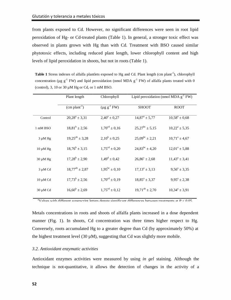
Glutatión y tolerancia a metales tóxicos
52
from plants exposed to Cd. However, no significant differences were seen in root lipid
peroxidation of Hg- or Cd-treated plants (Table 1). In general, a stronger toxic effect was
observed in plants grown with Hg than with Cd. Treatment with BSO caused similar
phytotoxic effects, including reduced plant length, lower chlorophyll content and high
levels of lipid peroxidation in shoots, but not in roots (Table 1).
Metals concentrations in roots and shoots of alfalfa plants increased in a dose dependent
manner (Fig. 1). In shoots, Cd concentration was three times higher respect to Hg.
Conversely, roots accumulated Hg to a greater degree than Cd (by approximately 50%) at
the highest treatment level (30 µM), suggesting that Cd was slightly more mobile.
3.2. Antioxidant enzymatic activities
Antioxidant enzymes activities were measured by using in gel staining. Although the
technique is not-quantitative, it allows the detection of changes in the activity of a
Table 1 Stress indexes of alfalfa plantlets exposed to Hg and Cd. Plant length (cm plant-1), chlorophyll
concentration (µg g-1 FW) and lipid peroxidation (nmol MDA g-1 FW) of alfalfa plants treated with 0
(control), 3, 10 or 30 µM Hg or Cd, or 1 mM BSO.
Plant length Chlorophyll Lipid peroxidation (nmol MDA g-1 FW)
(cm plant-1) (µg g-1 FW) SHOOT ROOT
Control 20,28a ± 3,31 2,40a ± 0,27 14,87a ± 5,77 10,58a ± 0,68
1 mM BSO 18,81b ± 2,56 1,70cd ± 0,16 25,27bc ± 5,15 10,22a ± 5,35
3 µM Hg 19,25ab ± 3,28 2,10b ± 0,25 25,09bc ± 2,21 10,71a ± 4,67
10 µM Hg 18,76b ± 3,15 1,75cd ± 0,20 24,83bc ± 4,20 12,01a ± 5,88
30 µM Hg 17,28b ± 2,90 1,49d ± 0,42 26,86c ± 2,68 11,43a ± 3,41
3 µM Cd 18,77ab ± 2,87 1,95bc ± 0,10 17,13a ± 3,13 9,56a ± 3,35
10 µM Cd 17,73b ± 2,56 1,70cd ± 0,19 18,85a ± 3,37 9,95a ± 2,38
30 µM Cd 16,60b ± 2,69 1,75cd ± 0,12 19,71ab ± 2,70 10,34a ± 3,91
aValues with different superscript letters denote significant differences between treatments at P < 0 05
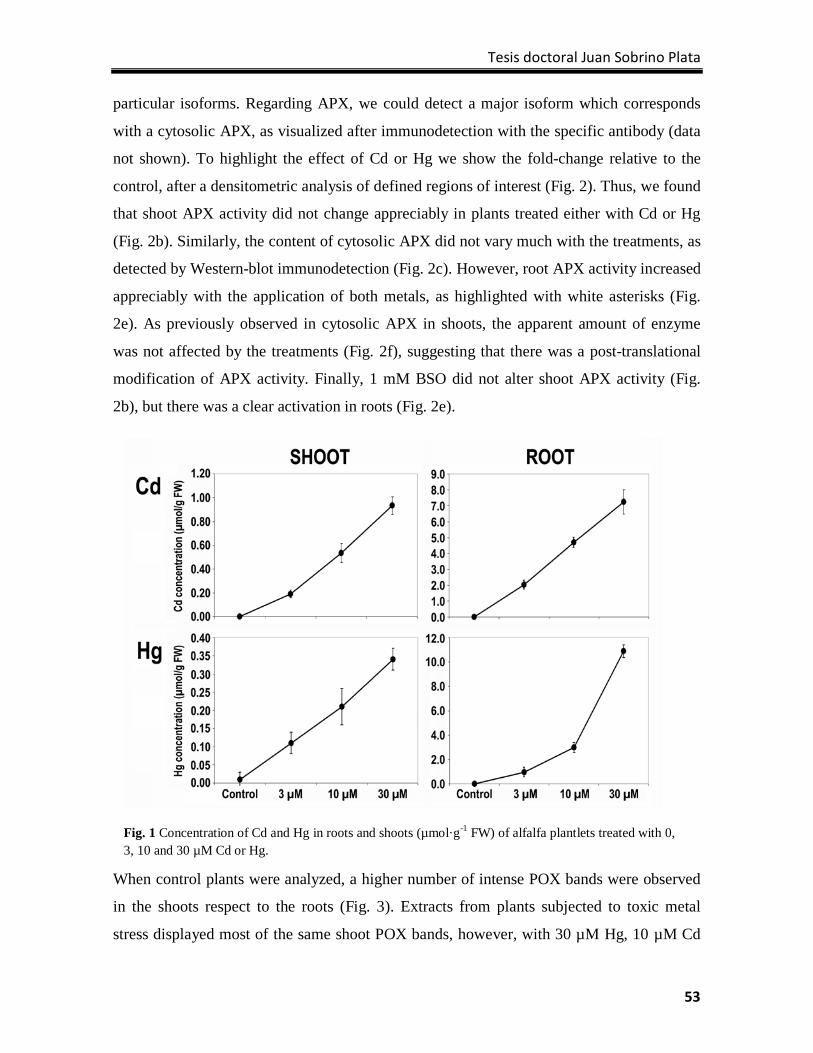
Tesis doctoral Juan Sobrino Plata
53
particular isoforms. Regarding APX, we could detect a major isoform which corresponds
with a cytosolic APX, as visualized after immunodetection with the specific antibody (data
not shown). To highlight the effect of Cd or Hg we show the fold-change relative to the
control, after a densitometric analysis of defined regions of interest (Fig. 2). Thus, we found
that shoot APX activity did not change appreciably in plants treated either with Cd or Hg
(Fig. 2b). Similarly, the content of cytosolic APX did not vary much with the treatments, as
detected by Western-blot immunodetection (Fig. 2c). However, root APX activity increased
appreciably with the application of both metals, as highlighted with white asterisks (Fig.
2e). As previously observed in cytosolic APX in shoots, the apparent amount of enzyme
was not affected by the treatments (Fig. 2f), suggesting that there was a post-translational
modification of APX activity. Finally, 1 mM BSO did not alter shoot APX activity (Fig.
2b), but there was a clear activation in roots (Fig. 2e).
When control plants were analyzed, a higher number of intense POX bands were observed
in the shoots respect to the roots (Fig. 3). Extracts from plants subjected to toxic metal
stress displayed most of the same shoot POX bands, however, with 30 µM Hg, 10 µM Cd
Fig. 1 Concentration of Cd and Hg in roots and shoots (µmol·g-1 FW) of alfalfa plantlets treated with 0, 3, 10 and 30 µM Cd or Hg.
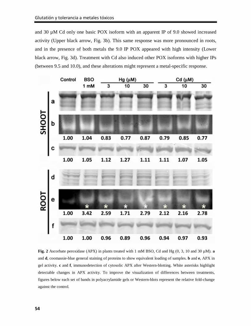
Glutatión y tolerancia a metales tóxicos
54
and 30 µM Cd only one basic POX isoform with an apparent IP of 9.0 showed increased
activity (Upper black arrow, Fig. 3b). This same response was more pronounced in roots,
and in the presence of both metals the 9.0 IP POX appeared with high intensity (Lower
black arrow, Fig. 3d). Treatment with Cd also induced other POX isoforms with higher IPs
(between 9.5 and 10.0), and these alterations might represent a metal-specific response.
Fig. 2 Ascorbate peroxidase (APX) in plants treated with 1 mM BSO, Cd and Hg (0, 3, 10 and 30 µM). a
and d, coomassie-blue general staining of proteins to show equivalent loading of samples. b and e, APX in
gel activity. c and f, immunodetection of cytosolic APX after Western-blotting. White asterisks highlight
detectable changes in APX activity. To improve the visualization of differences between treatments,
figures below each set of bands in polyacrylamide gels or Western-blots represent the relative fold-change
against the control.
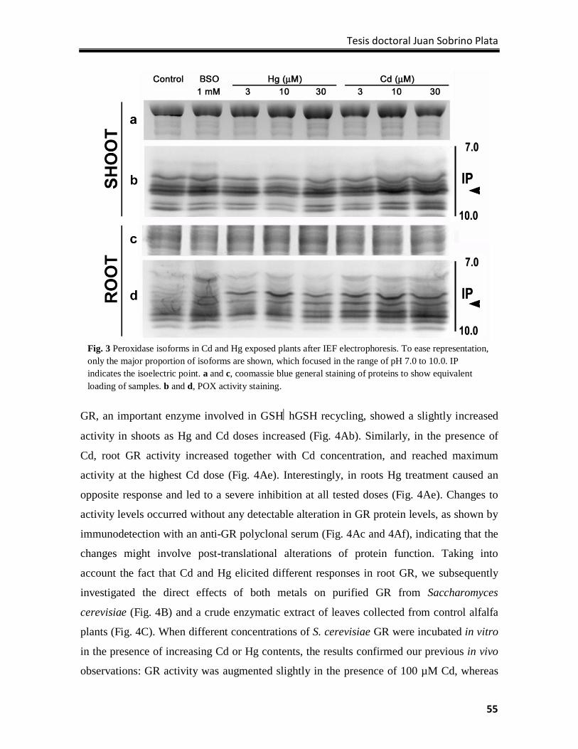
Tesis doctoral Juan Sobrino Plata
55
GR, an important enzyme involved in GSHhGSH recycling, showed a slightly increased
activity in shoots as Hg and Cd doses increased (Fig. 4Ab). Similarly, in the presence of
Cd, root GR activity increased together with Cd concentration, and reached maximum
activity at the highest Cd dose (Fig. 4Ae). Interestingly, in roots Hg treatment caused an
opposite response and led to a severe inhibition at all tested doses (Fig. 4Ae). Changes to
activity levels occurred without any detectable alteration in GR protein levels, as shown by
immunodetection with an anti-GR polyclonal serum (Fig. 4Ac and 4Af), indicating that the
changes might involve post-translational alterations of protein function. Taking into
account the fact that Cd and Hg elicited different responses in root GR, we subsequently
investigated the direct effects of both metals on purified GR from Saccharomyces
cerevisiae (Fig. 4B) and a crude enzymatic extract of leaves collected from control alfalfa
plants (Fig. 4C). When different concentrations of S. cerevisiae GR were incubated in vitro
in the presence of increasing Cd or Hg contents, the results confirmed our previous in vivo
observations: GR activity was augmented slightly in the presence of 100 µM Cd, whereas
Fig. 3 Peroxidase isoforms in Cd and Hg exposed plants after IEF electrophoresis. To ease representation, only the major proportion of isoforms are shown, which focused in the range of pH 7.0 to 10.0. IP indicates the isoelectric point. a and c, coomassie blue general staining of proteins to show equivalent loading of samples. b and d, POX activity staining.
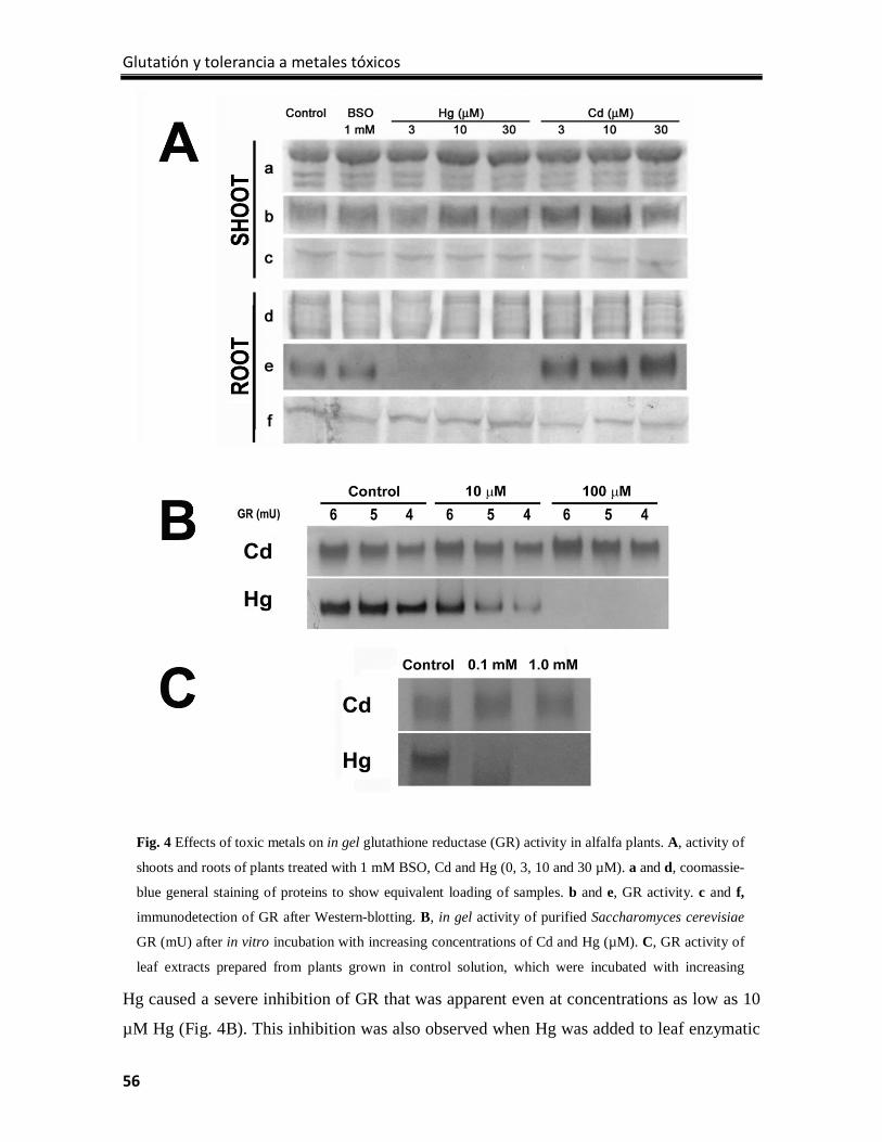
Glutatión y tolerancia a metales tóxicos
56
Hg caused a severe inhibition of GR that was apparent even at concentrations as low as 10
µM Hg (Fig. 4B). This inhibition was also observed when Hg was added to leaf enzymatic
Fig. 4 Effects of toxic metals on in gel glutathione reductase (GR) activity in alfalfa plants. A, activity of
shoots and roots of plants treated with 1 mM BSO, Cd and Hg (0, 3, 10 and 30 µM). a and d, coomassie-
blue general staining of proteins to show equivalent loading of samples. b and e, GR activity. c and f,
immunodetection of GR after Western-blotting. B, in gel activity of purified Saccharomyces cerevisiae
GR (mU) after in vitro incubation with increasing concentrations of Cd and Hg (µM). C, GR activity of
leaf extracts prepared from plants grown in control solution, which were incubated with increasing
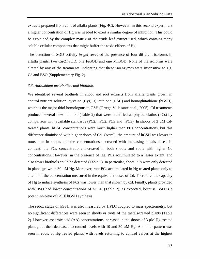
Tesis doctoral Juan Sobrino Plata
57
extracts prepared from control alfalfa plants (Fig. 4C). However, in this second experiment
a higher concentration of Hg was needed to exert a similar degree of inhibition. This could
be explained by the complex matrix of the crude leaf extract used, which contains many
soluble cellular components that might buffer the toxic effects of Hg.
The detection of SOD activity in gel revealed the presence of four different isoforms in
alfalfa plants: two Cu/ZnSOD, one FeSOD and one MnSOD. None of the isoforms were
altered by any of the treatments, indicating that these isoenzymes were insensitive to Hg,
Cd and BSO (Supplementary Fig. 2).
3.3. Antioxidant metabolites and biothiols
We identified several biothiols in shoot and root extracts from alfalfa plants grown in
control nutrient solution: cysteine (Cys), glutathione (GSH) and homoglutathione (hGSH),
which is the major thiol homologous to GSH (Ortega-Villasante et al., 2005). Cd treatments
produced several new biothiols (Table 2) that were identified as phytochelatins (PCs) by
comparison with available standards (PC2, hPC2, PC3 and hPC3). In shoots of 3 µM Cd-
treated plants, hGSH concentrations were much higher than PCs concentrations, but this
difference diminished with higher doses of Cd. Overall, the amount of hGSH was lower in
roots than in shoots and the concentrations decreased with increasing metals doses. In
contrast, the PCs concentrations increased in both shoots and roots with higher Cd
concentrations. However, in the presence of Hg, PCs accumulated to a lesser extent, and
also fewer biothiols could be detected (Table 2). In particular, shoot PCs were only detected
in plants grown in 30 µM Hg. Moreover, root PCs accumulated in Hg-treated plants only to
a tenth of the concentration measured in the equivalent doses of Cd. Therefore, the capacity
of Hg to induce synthesis of PCs was lower than that shown by Cd. Finally, plants provided
with BSO had lower concentrations of hGSH (Table 2), as expected, because BSO is a
potent inhibitor of GSHhGSH synthesis.
The redox status of hGSH was also measured by HPLC coupled to mass spectrometry, but
no significant differences were seen in shoots or roots of the metals-treated plants (Table
2). However, ascorbic acid (AA) concentrations increased in the shoots of 3 µM Hg-treated
plants, but then decreased to control levels with 10 and 30 µM Hg. A similar pattern was
seen in roots of Hg-treated plants, with levels returning to control values at the highest
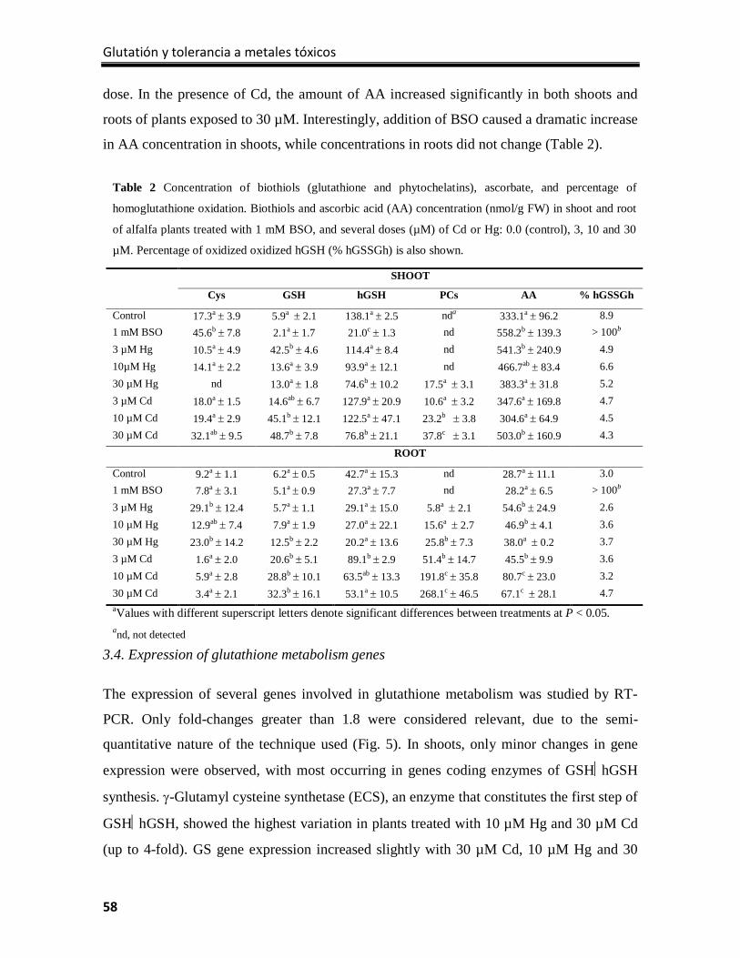
Glutatión y tolerancia a metales tóxicos
58
dose. In the presence of Cd, the amount of AA increased significantly in both shoots and
roots of plants exposed to 30 µM. Interestingly, addition of BSO caused a dramatic increase
in AA concentration in shoots, while concentrations in roots did not change (Table 2).
3.4. Expression of glutathione metabolism genes
The expression of several genes involved in glutathione metabolism was studied by RT-
PCR. Only fold-changes greater than 1.8 were considered relevant, due to the semi-
quantitative nature of the technique used (Fig. 5). In shoots, only minor changes in gene
expression were observed, with most occurring in genes coding enzymes of GSHhGSH
synthesis. γ-Glutamyl cysteine synthetase (ECS), an enzyme that constitutes the first step of
GSHhGSH, showed the highest variation in plants treated with 10 µM Hg and 30 µM Cd
(up to 4-fold). GS gene expression increased slightly with 30 µM Cd, 10 µM Hg and 30
Table 2 Concentration of biothiols (glutathione and phytochelatins), ascorbate, and percentage of
homoglutathione oxidation. Biothiols and ascorbic acid (AA) concentration (nmol/g FW) in shoot and root
of alfalfa plants treated with 1 mM BSO, and several doses (µM) of Cd or Hg: 0.0 (control), 3, 10 and 30
µM. Percentage of oxidized oxidized hGSH (% hGSSGh) is also shown.
SHOOT
Cys GSH hGSH PCs AA % hGSSGh
Control 17.3a ± 3.9 5.9a ± 2.1 138.1a ± 2.5 nda 333.1a ± 96.2 8.9 1 mM BSO 45.6b ± 7.8 2.1a ± 1.7 21.0c ± 1.3 nd 558.2b ± 139.3 > 100b 3 µM Hg 10.5a ± 4.9 42.5b ± 4.6 114.4a ± 8.4 nd 541.3b ± 240.9 4.9 10µM Hg 14.1a ± 2.2 13.6a ± 3.9 93.9a ± 12.1 nd 466.7ab ± 83.4 6.6 30 µM Hg nd 13.0a ± 1.8 74.6b ± 10.2 17.5a ± 3.1 383.3a ± 31.8 5.2 3 µM Cd 18.0a ± 1.5 14.6ab ± 6.7 127.9a ± 20.9 10.6a ± 3.2 347.6a ± 169.8 4.7 10 µM Cd 19.4a ± 2.9 45.1b ± 12.1 122.5a ± 47.1 23.2b ± 3.8 304.6a ± 64.9 4.5 30 µM Cd 32.1ab ± 9.5 48.7b ± 7.8 76.8b ± 21.1 37.8c ± 3.1 503.0b ± 160.9 4.3 ROOT
Control 9.2a ± 1.1 6.2a ± 0.5 42.7a ± 15.3 nd 28.7a ± 11.1 3.0 1 mM BSO 7.8a ± 3.1 5.1a ± 0.9 27.3a ± 7.7 nd 28.2a ± 6.5 > 100b 3 µM Hg 29.1b ± 12.4 5.7a ± 1.1 29.1a ± 15.0 5.8a ± 2.1 54.6b ± 24.9 2.6 10 µM Hg 12.9ab ± 7.4 7.9a ± 1.9 27.0a ± 22.1 15.6a ± 2.7 46.9b ± 4.1 3.6 30 µM Hg 23.0b ± 14.2 12.5b ± 2.2 20.2a ± 13.6 25.8b ± 7.3 38.0a ± 0.2 3.7 3 µM Cd 1.6a ± 2.0 20.6b ± 5.1 89.1b ± 2.9 51.4b ± 14.7 45.5b ± 9.9 3.6 10 µM Cd 5.9a ± 2.8 28.8b ± 10.1 63.5ab ± 13.3 191.8c ± 35.8 80.7c ± 23.0 3.2 30 µM Cd 3.4a ± 2.1 32.3b ± 16.1 53.1a ± 10.5 268.1c ± 46.5 67.1c ± 28.1 4.7 aValues with different superscript letters denote significant differences between treatments at P < 0.05. and, not detected
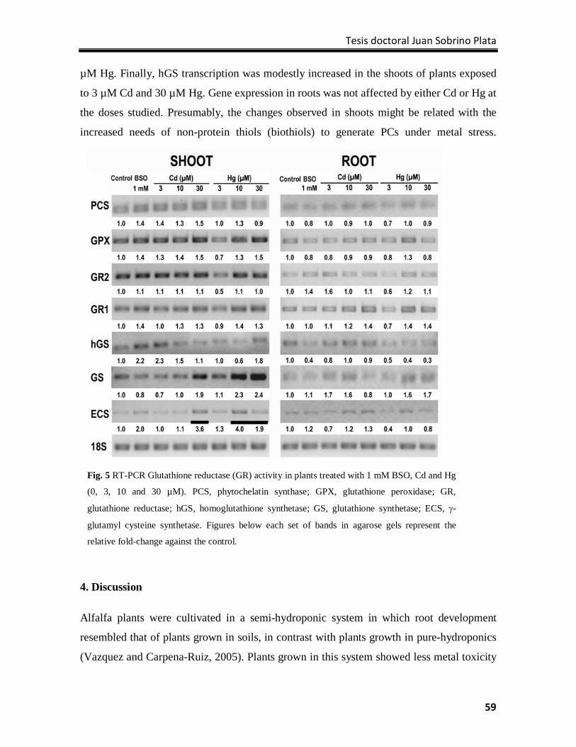
Tesis doctoral Juan Sobrino Plata
59
µM Hg. Finally, hGS transcription was modestly increased in the shoots of plants exposed
to 3 µM Cd and 30 µM Hg. Gene expression in roots was not affected by either Cd or Hg at
the doses studied. Presumably, the changes observed in shoots might be related with the
increased needs of non-protein thiols (biothiols) to generate PCs under metal stress.
4. Discussion
Alfalfa plants were cultivated in a semi-hydroponic system in which root development
resembled that of plants grown in soils, in contrast with plants growth in pure-hydroponics
(Vazquez and Carpena-Ruiz, 2005). Plants grown in this system showed less metal toxicity
Fig. 5 RT-PCR Glutathione reductase (GR) activity in plants treated with 1 mM BSO, Cd and Hg
(0, 3, 10 and 30 µM). PCS, phytochelatin synthase; GPX, glutathione peroxidase; GR,
glutathione reductase; hGS, homoglutathione synthetase; GS, glutathione synthetase; ECS, γ-
glutamyl cysteine synthetase. Figures below each set of bands in agarose gels represent the
relative fold-change against the control.
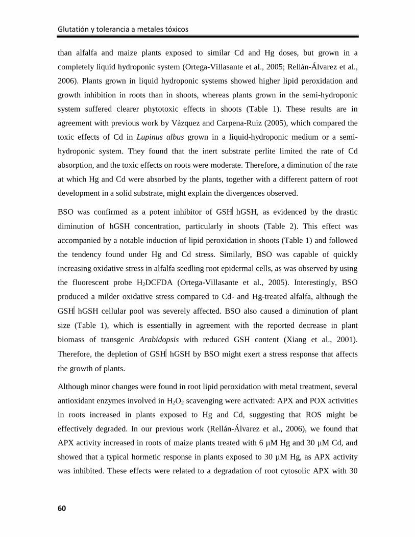
Glutatión y tolerancia a metales tóxicos
60
than alfalfa and maize plants exposed to similar Cd and Hg doses, but grown in a
completely liquid hydroponic system (Ortega-Villasante et al., 2005; Rellán-Álvarez et al.,
2006). Plants grown in liquid hydroponic systems showed higher lipid peroxidation and
growth inhibition in roots than in shoots, whereas plants grown in the semi-hydroponic
system suffered clearer phytotoxic effects in shoots (Table 1). These results are in
agreement with previous work by Vázquez and Carpena-Ruiz (2005), which compared the
toxic effects of Cd in Lupinus albus grown in a liquid-hydroponic medium or a semi-
hydroponic system. They found that the inert substrate perlite limited the rate of Cd
absorption, and the toxic effects on roots were moderate. Therefore, a diminution of the rate
at which Hg and Cd were absorbed by the plants, together with a different pattern of root
development in a solid substrate, might explain the divergences observed.
BSO was confirmed as a potent inhibitor of GSHhGSH, as evidenced by the drastic
diminution of hGSH concentration, particularly in shoots (Table 2). This effect was
accompanied by a notable induction of lipid peroxidation in shoots (Table 1) and followed
the tendency found under Hg and Cd stress. Similarly, BSO was capable of quickly
increasing oxidative stress in alfalfa seedling root epidermal cells, as was observed by using
the fluorescent probe H2DCFDA (Ortega-Villasante et al., 2005). Interestingly, BSO
produced a milder oxidative stress compared to Cd- and Hg-treated alfalfa, although the
GSHhGSH cellular pool was severely affected. BSO also caused a diminution of plant
size (Table 1), which is essentially in agreement with the reported decrease in plant
biomass of transgenic Arabidopsis with reduced GSH content (Xiang et al., 2001).
Therefore, the depletion of GSHhGSH by BSO might exert a stress response that affects
the growth of plants.
Although minor changes were found in root lipid peroxidation with metal treatment, several
antioxidant enzymes involved in H2O2 scavenging were activated: APX and POX activities
in roots increased in plants exposed to Hg and Cd, suggesting that ROS might be
effectively degraded. In our previous work (Rellán-Álvarez et al., 2006), we found that
APX activity increased in roots of maize plants treated with 6 µM Hg and 30 µM Cd, and
showed that a typical hormetic response in plants exposed to 30 µM Hg, as APX activity
was inhibited. These effects were related to a degradation of root cytosolic APX with 30
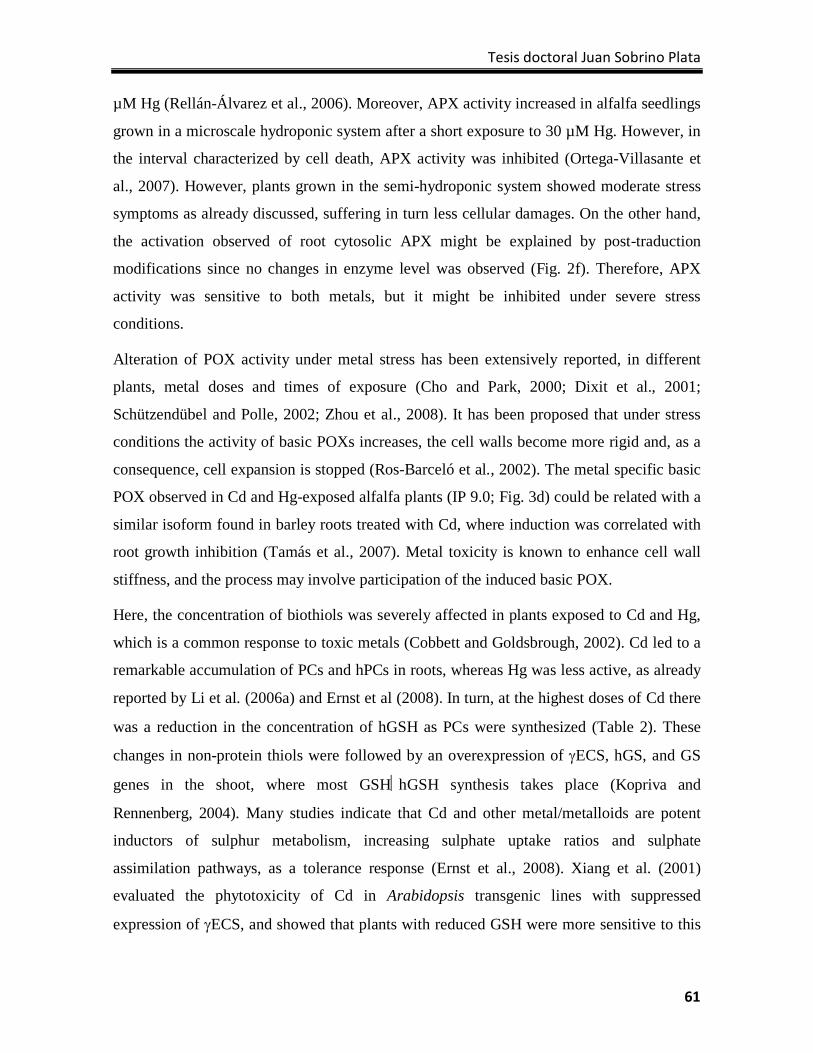
Tesis doctoral Juan Sobrino Plata
61
µM Hg (Rellán-Álvarez et al., 2006). Moreover, APX activity increased in alfalfa seedlings
grown in a microscale hydroponic system after a short exposure to 30 µM Hg. However, in
the interval characterized by cell death, APX activity was inhibited (Ortega-Villasante et
al., 2007). However, plants grown in the semi-hydroponic system showed moderate stress
symptoms as already discussed, suffering in turn less cellular damages. On the other hand,
the activation observed of root cytosolic APX might be explained by post-traduction
modifications since no changes in enzyme level was observed (Fig. 2f). Therefore, APX
activity was sensitive to both metals, but it might be inhibited under severe stress
conditions.
Alteration of POX activity under metal stress has been extensively reported, in different
plants, metal doses and times of exposure (Cho and Park, 2000; Dixit et al., 2001;
Schützendübel and Polle, 2002; Zhou et al., 2008). It has been proposed that under stress
conditions the activity of basic POXs increases, the cell walls become more rigid and, as a
consequence, cell expansion is stopped (Ros-Barceló et al., 2002). The metal specific basic
POX observed in Cd and Hg-exposed alfalfa plants (IP 9.0; Fig. 3d) could be related with a
similar isoform found in barley roots treated with Cd, where induction was correlated with
root growth inhibition (Tamás et al., 2007). Metal toxicity is known to enhance cell wall
stiffness, and the process may involve participation of the induced basic POX.
Here, the concentration of biothiols was severely affected in plants exposed to Cd and Hg,
which is a common response to toxic metals (Cobbett and Goldsbrough, 2002). Cd led to a
remarkable accumulation of PCs and hPCs in roots, whereas Hg was less active, as already
reported by Li et al. (2006a) and Ernst et al (2008). In turn, at the highest doses of Cd there
was a reduction in the concentration of hGSH as PCs were synthesized (Table 2). These
changes in non-protein thiols were followed by an overexpression of γECS, hGS, and GS
genes in the shoot, where most GSHhGSH synthesis takes place (Kopriva and
Rennenberg, 2004). Many studies indicate that Cd and other metal/metalloids are potent
inductors of sulphur metabolism, increasing sulphate uptake ratios and sulphate
assimilation pathways, as a tolerance response (Ernst et al., 2008). Xiang et al. (2001)
evaluated the phytotoxicity of Cd in Arabidopsis transgenic lines with suppressed
expression of γECS, and showed that plants with reduced GSH were more sensitive to this
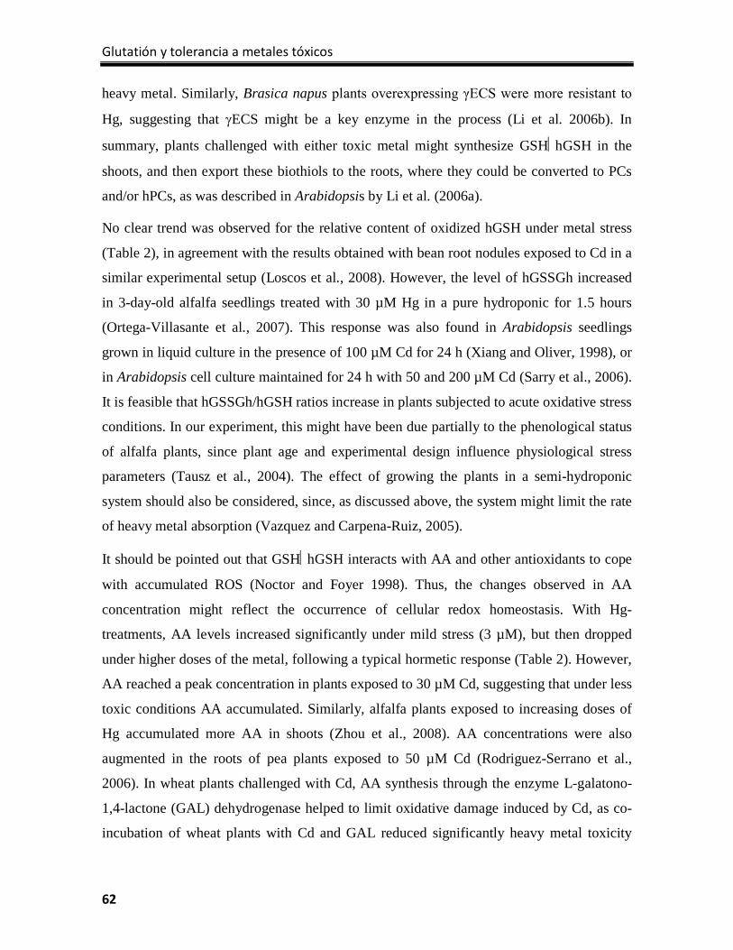
Glutatión y tolerancia a metales tóxicos
62
heavy metal. Similarly, Brasica napus plants overexpressing γECS were more resistant to
Hg, suggesting that γECS might be a key enzyme in the process (Li et al. 2006b). In
summary, plants challenged with either toxic metal might synthesize GSHhGSH in the
shoots, and then export these biothiols to the roots, where they could be converted to PCs
and/or hPCs, as was described in Arabidopsis by Li et al. (2006a).
No clear trend was observed for the relative content of oxidized hGSH under metal stress
(Table 2), in agreement with the results obtained with bean root nodules exposed to Cd in a
similar experimental setup (Loscos et al., 2008). However, the level of hGSSGh increased
in 3-day-old alfalfa seedlings treated with 30 µM Hg in a pure hydroponic for 1.5 hours
(Ortega-Villasante et al., 2007). This response was also found in Arabidopsis seedlings
grown in liquid culture in the presence of 100 µM Cd for 24 h (Xiang and Oliver, 1998), or
in Arabidopsis cell culture maintained for 24 h with 50 and 200 µM Cd (Sarry et al., 2006).
It is feasible that hGSSGh/hGSH ratios increase in plants subjected to acute oxidative stress
conditions. In our experiment, this might have been due partially to the phenological status
of alfalfa plants, since plant age and experimental design influence physiological stress
parameters (Tausz et al., 2004). The effect of growing the plants in a semi-hydroponic
system should also be considered, since, as discussed above, the system might limit the rate
of heavy metal absorption (Vazquez and Carpena-Ruiz, 2005).
It should be pointed out that GSHhGSH interacts with AA and other antioxidants to cope
with accumulated ROS (Noctor and Foyer 1998). Thus, the changes observed in AA
concentration might reflect the occurrence of cellular redox homeostasis. With Hg-
treatments, AA levels increased significantly under mild stress (3 µM), but then dropped
under higher doses of the metal, following a typical hormetic response (Table 2). However,
AA reached a peak concentration in plants exposed to 30 µM Cd, suggesting that under less
toxic conditions AA accumulated. Similarly, alfalfa plants exposed to increasing doses of
Hg accumulated more AA in shoots (Zhou et al., 2008). AA concentrations were also
augmented in the roots of pea plants exposed to 50 µM Cd (Rodriguez-Serrano et al.,
2006). In wheat plants challenged with Cd, AA synthesis through the enzyme L-galatono-
1,4-lactone (GAL) dehydrogenase helped to limit oxidative damage induced by Cd, as co-
incubation of wheat plants with Cd and GAL reduced significantly heavy metal toxicity
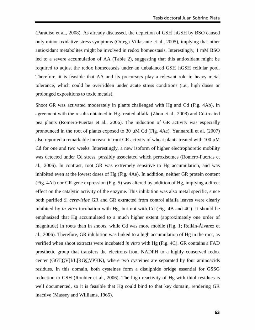
Tesis doctoral Juan Sobrino Plata
63
(Paradiso et al., 2008). As already discussed, the depletion of GSHhGSH by BSO caused
only minor oxidative stress symptoms (Ortega-Villasante et al., 2005), implying that other
antioxidant metabolites might be involved in redox homeostasis. Interestingly, 1 mM BSO
led to a severe accumulation of AA (Table 2), suggesting that this antioxidant might be
required to adjust the redox homeostasis under an unbalanced GSHhGSH cellular pool.
Therefore, it is feasible that AA and its precursors play a relevant role in heavy metal
tolerance, which could be overridden under acute stress conditions (i.e., high doses or
prolonged expositions to toxic metals).
Shoot GR was activated moderately in plants challenged with Hg and Cd (Fig. 4Ab), in
agreement with the results obtained in Hg-treated alfalfa (Zhou et al., 2008) and Cd-treated
pea plants (Romero-Puertas et al., 2006). The induction of GR activity was especially
pronounced in the root of plants exposed to 30 µM Cd (Fig. 4Ae). Yannarelli et al. (2007)
also reported a remarkable increase in root GR activity of wheat plants treated with 100 µM
Cd for one and two weeks. Interestingly, a new isoform of higher electrophoretic mobility
was detected under Cd stress, possibly associated which peroxisomes (Romero-Puertas et
al., 2006). In contrast, root GR was extremely sensitive to Hg accumulation, and was
inhibited even at the lowest doses of Hg (Fig. 4Ae). In addition, neither GR protein content
(Fig. 4Af) nor GR gene expression (Fig. 5) was altered by addition of Hg, implying a direct
effect on the catalytic activity of the enzyme. This inhibition was also metal specific, since
both purified S. cerevisiae GR and GR extracted from control alfalfa leaves were clearly
inhibited by in vitro incubation with Hg, but not with Cd (Fig. 4B and 4C). It should be
emphasized that Hg accumulated to a much higher extent (approximately one order of
magnitude) in roots than in shoots, while Cd was more mobile (Fig. 1; Rellán-Álvarez et
al., 2006). Therefore, GR inhibition was linked to a high accumulation of Hg in the root, as
verified when shoot extracts were incubated in vitro with Hg (Fig. 4C). GR contains a FAD
prosthetic group that transfers the electrons from NADPH to a highly conserved redox
center (GGTCV[I/L]RGCVPKK), where two cysteines are separated by four aminoacids
residues. In this domain, both cysteines form a disulphide bridge essential for GSSG
reduction to GSH (Rouhier et al., 2006). The high reactivity of Hg with thiol residues is
well documented, so it is feasible that Hg could bind to that key domain, rendering GR
inactive (Massey and Williams, 1965).
Glutatión y tolerancia a metales tóxicos
64
Although root GR was severely inhibited in Hg-treated alfalfa, the hGSH/hGSSGh ratio
was not affected (Table 2). It is feasible that the variations observed in the cellular pools of
several biothiols imposed by the synthesis of PCs and hPCs might hide effects on the
hGSSGh/hGSH ratio. Moreover, the accumulation of other antioxidants (i.e., AA) could be
one of the first homeostatic redox responses, with the hGSSGh/hGSH ratio only affected
under severe oxidative damage, as discussed above.
In summary, the results of biothiol concentration, antioxidant enzymatic activity and
oxidative stress indexes provide support for the idea that the toxic mechanisms of Cd and
Hg differ. Although the metal doses used in this study were one or two magnitudes higher
than the concentration bioavailable in polluted soils, the information presented might help
to develop new specific biomarkers for toxicity evaluation. Future experiments are
programmed to assess the usefulness of these putative biomarkers in plants grown in
polluted soils under environmental controlled conditions. On the other hand, further work is
needed to understand the toxic mechanisms triggered by both metals, and describe the
antioxidant systems that might be essential components of homeostatic responses to these
pollutants.
Acknowledgments
We gratefully appreciate Fundación Ramón Areces for supporting LEH’s laboratory
research. This work was also funded by the Spanish Ministry of Science and Innovation
(CTM2005-04809/TECNO; REUSA), Comunidad de Madrid (S-0505/AMB/000296,
Consorcio EIADES) and Junta de Comunidades de Castilla-La Mancha (PBI07-0091-3644;
FITOALMA). COV was supported by a PhD Student Exchange Program Grant provided by
Universidad Autónoma de Madrid. We are utterly indebted to Drs. Javier Abadía and Ana
Álvarez for supporting us with the analyses of GSH/GSSG, hGSH/hGSSGh and AA by
HPLC mass spectrometry at Estación Experimental de Aula Dei-CSIC (Saragossa, Spain).
We also thank Dr. Liz Caldwell (Write Science Right, Las Vegas, NV, USA) for her superb
editing of our manuscript.
Tesis doctoral Juan Sobrino Plata
65
References
Arnon, D.I., 1949) Copper enzymes in isolated chloroplasts: polyphenol oxidase in Beta vulgaris.
Plant Physiol. 24, 1─15.
Buege, J.A., Aust, S.D., 1978. Microsomal lipid peroxidation. Methods Enzymol. 52, 302-310.
Cho, U.H., Park, J.O., 2000. Mercury-induced oxidative stress in tomato seedlings. Plant Sci. 156,
1─9.
Cobbett, C., Goldsbrough, P., 2002. Phytochelatins and metallothioneins: roles in heavy metal
detoxification and homeostasis. Ann. Rev. Plant Biol. 53, 159─182.
Dixit, V., Pandey, V., Shyam, R., 2001. Differential antioxidative responses to cadmium in roots
and leaves of pea (Pisum sativum L. cv. Azad). J. Exp. Bot. 52, 1101─1109.
Ernst, W.H.O., Krauss, G.J., Verkleij, J.A.C., Wesenberg, D., 2008. Interaction of heavy metals
with the sulphur metabolism in angiosperms from an ecological point of view. Plant Cell
Environ. 31, 123─143.
Gratão, P.L., Polle, A., Lea, P.J., Azevedo, R.A., 2005. Making the life of heavy metal-stressed
plants a little easier. Funct. Plant Biol. 32, 481─494.
Hall, J.L., 2002. Cellular mechanisms for heavy metal detoxification and tolerance. J. Exp. Bot. 53,
1–11.
Jiménez, A., Hernández, J.A., Ros-Barceló, A., Sandalio, L.M., del Río, L.A., Sevilla, F., 1998.
Mitochondrial and peroxisomal ascorbate peroxidase of pea leaves. Physiol. Planta. 104, 687–
692.
Kaplan, J.C., 1968. Electrophoretic study of glutathione reductase in human erythrocytes and
leucocytes. Nature 217, 256─258.
Kopriva, S., Rennenberg, H., 2004. Control of sulphate assimilation and glutathione synthesis:
interaction with N and C metabolism. J. Exp. Bot. 55, 1831–1842.
Laemmli, U.K., 1970. Cleavage of structural proteins during the assembly of the head of
bacteriophage T4. Nature 227, 680–685.
Li, Y., Dankher, O.P., Carreira, L., Smith, A.P., Meagher, R.B., 2006a. The shoot-specific expression
of γ-glutamylcysteine synthetase directs the long-distance transport of thiol-peptides to roots
conferring tolerance to mercury and arsenic. Plant Physiol. 141, 288-298.
Glutatión y tolerancia a metales tóxicos
66
Li, Y., Heaton, A.C.P., Carreira, L., Meagher, R.B. 2006b. Enhanced tolerance to and accumulation of
mercury, but not arsenic, in plants overstressing two enzymes required for thiol peptide synthesis.
Physiol. Planta. 128, 48─57.
Loscos, J., Matamoros, M.A., Becana, M., 2008. Ascorbate and homoglutathione metabolism in
common bean nodules under stress conditions and during natural senescence. Plant Physiol.
146, 1282─1292.
Massey, V., Williams, C.H., 1965. On the reaction mechanism of yeast glutathione reductase. J.
Biol. Chem. 240, 4470─4480.
Milner, M.J., Kochian, L.V., 2008. Investigating heavy-metal hyperaccumulation using Thlaspi
caerulescens as a model system. Ann. Bot. 102, 3–13.
Noctor, G., Foyer, C.H., 1998. Ascorbate and glutathione: keeping active oxygen under control.
Ann. Rev. Plant Physiol. Plant Mol. Biol. 49, 249─279.
Ortega-Villasante, C., Rellán-Álvarez, R., del Campo, F.F., Carpena-Ruiz, R.O., Hernández, L.E.,
2005. Cellular damage induced by cadmium and mercury in Medicago sativa. J. Exp. Bot. 56,
2239─2251.
Ortega-Villasante, C., Hernández, L.E., Rellán-Álvarez, R., del Campo, F.F., Carpena-Ruiz, R.O.,
2007. Rapid alteration of cellular redox homeostasis upon exposure to cadmium and mercury
in alfalfa seedlings. New Phytol. 176, 96─107.
Paradiso, A., Berardino, R., de Pinto, M.C., Sanita di Toppi, L., Storelli, M.M., Tomáis, F., De
Gara, L., 2008. Increase in ascorbate-glutathione metabolism as local and precocious systemic
responses induced by cadmium in Durum wheat plants. Plant Cell Physiol. 43, 362─374.
Rellán-Álvarez, R., Ortega-Villasante, C., Álvarez-Fernández, A., del Campo, F.F., Hernández,
L.E., 2006. Stress responses of Zea mays to cadmium and mercury. Plant Soil 279, 41─50.
Rodríguez-Serrano, M., Romero-Puertas, M.C., Zabalza, A., Corpas, F.J., Cómez, M., del Río,
L.A., Sandalio, L.M., 2006. Cadmium effect on oxidative metabolism of pea (Pisum sativum
L.) roots. Imaging of reactive oxygen species and nitric oxide accumulation in vivo. Plant Cell
Environ. 29, 1532─1544.
Romero-Puertas, M.C., Corpas, F.J., Sandalio, L.M., Leterrier, M., Rodríguez-Serrano, M., del Río,
L.A., Palma, J.M., 2006. Glutathione reductase from pea leaves: response to abiotic stress and
characterization of the peroxisomal isoenzyme. New Phytol. 170, 43─52.
Tesis doctoral Juan Sobrino Plata
67
Ros-Barceló, A., Pomar, F., Ferrer, M.A., Martínez, P., Ballesta, M.C., Pedreño, M.A., 2002. In situ
characterization of a NO-sensitive peroxidase in the lignifying xylem of Zinnia elegans.
Physiol. Planta. 114, 33–40.
Rouhier, N., Couturier, J., Jacquot, J.P., 2006. Genome-wide analysis of plant glutaredoxin systems.
J. Exp. Bot. 57, 1685─1696.
Salt, D.E., Smith. R.D., Raskin, I., 1998. Phytoremediation. Ann. Rev. Plant Physiol. Plant Mol.
Biol. 49, 643─668.
Sarry, J.E., Kuhn, L., Ducruix, C., Lafaye, A., Junot, C., Hugouvieux, V., Jourdain, A., Bastien, O.,
Fievet, J.B., Vailhen, D., Amekraz, B., Moulin, C., Ezan, E., Garin, J., Bourguignon, J., 2006.
The early responses of Arabidopsis thaliana cells to cadmium exposure explored by protein
and metabolite profiling analyses. Proteomics 6, 2180─2198.
Semane, B., Cuypers, A., Smeets, K., van Belleghem, F., Horemans, N., Schat, H., Vangronsveld,
J., 2007. Cadmium responses in Arabidopsis thaliana: glutathione metabolism and
antioxidative defence system. Physiol. Planta. 129, 519─528.
Schützendübel, A., Polle, A., 2002. Plant responses to abiotic stresses: heavy metal-induced
oxidative stress and protection by mycorrhization. J. Exp. Bot. 53, 1351─1365.
Sharma, S.S., Dietz, K.J., 2006. The significance of amino acids and amino acid-derived molecules
in plant responses and adaptation to heavy metal stress. J. Exp. Bot. 57, 711─726.
Tamás, L., Ďurčeková, K., Halaušková, L., Huttová, J., Mistrík, I., Ollé, M., 2007. Rhizosphere
localized cationic peroxidase from barley roots is strongly activated by cadmium and correlated
with root growth inhibition. Chemosphere 66, 1292─1300.
Vázquez, S., Carpena-Ruiz, R., 2005. Use of perlite in cadmium plant studies: an approach to
polluted soil conditions. J. Environ. Monitor. 7, 1355─1358.
Yannarelli, G.G., Fernández-Álvarez, A.J., Santa-Cruz, D.M., Tomaro, M.L. 2007. Glutathione
reductase activity and isoforms in leaves and roots of wheat plants subjected to cadmium
stress. Phytochem. 68, 505─512.
Xiang, C., Oliver, D.J., 1998. Glutathione metabolic genes co-ordinately respond to heavy metals
and jasmonic acid in Arabidopsis. Plant Cell 10, 1539─1550.
Xiang, C., Werner, B.L., Christensen, E.M., Oliver, D.J., 2001. The biological functions of
glutathione revisited in Arabidopsis transgenic plants with altered glutathione levels. Plant
Physiology 126, 564-574.

Glutatión y tolerancia a metales tóxicos
68
Zhou, Z.S., Wang, S.J., Yang, Z.M., 2008. Biological detection and analysis of mercury toxicity to
alfalfa (Medicago sativa) plants. Chemosphere 70, 1500─1509.
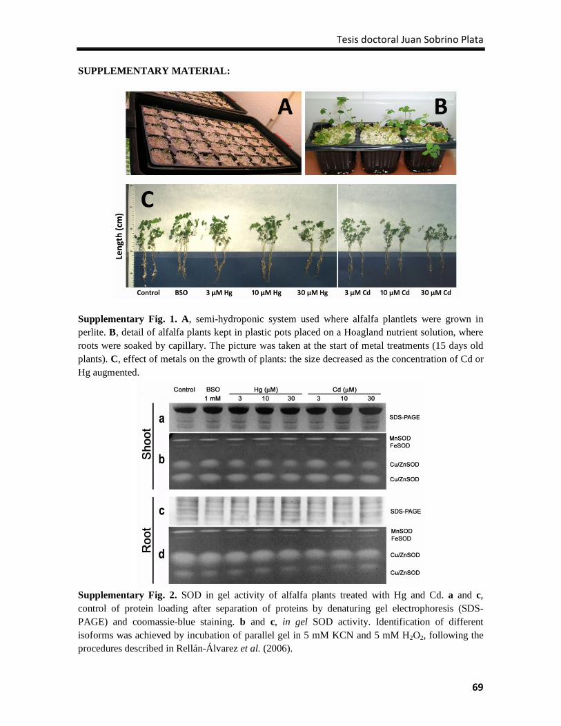
Tesis doctoral Juan Sobrino Plata
69
SUPPLEMENTARY MATERIAL:
Supplementary Fig. 1. A, semi-hydroponic system used where alfalfa plantlets were grown in perlite. B, detail of alfalfa plants kept in plastic pots placed on a Hoagland nutrient solution, where roots were soaked by capillary. The picture was taken at the start of metal treatments (15 days old plants). C, effect of metals on the growth of plants: the size decreased as the concentration of Cd or Hg augmented.
Supplementary Fig. 2. SOD in gel activity of alfalfa plants treated with Hg and Cd. a and c, control of protein loading after separation of proteins by denaturing gel electrophoresis (SDS-PAGE) and coomassie-blue staining. b and c, in gel SOD activity. Identification of different isoforms was achieved by incubation of parallel gel in 5 mM KCN and 5 mM H2O2, following the procedures described in Rellán-Álvarez et al. (2006).

Glutatión y tolerancia a metales tóxicos
70
CAPÍTULO 3 Specific stress responses to cadmium, arsenic and mercury appear in the metallophyte Silene vulgaris when grown hydroponically

Tesis doctoral Juan Sobrino Plata
71
Capítulo 3:
Specific stress responses to cadmium, arsenic and mercury appear in the
metallophyte Silene vulgaris when grown hydroponically
Juan Sobrino-Plataa, d, Joaquín Herreroa, Sandra Carrasco-Gila, b, Araceli Pérez-Sanzc,
Carmen Loboc, Carolina Escobard, Rocío Millánb, Luis E. Hernándeza*
aLaboratorio de Fisiología Vegetal, Dpto. Biología, Universidad Autónoma de Madrid. Cantoblanco, 28049 Madrid. bCentro de Investigaciones Energéticas, Medioambientales y Tecnológicas, Avd. Complutense, 22, 28040 Madrid, Spain. cInstituto Madrileño de Investigación y Desarrollo Rural, Agrario y Alimentario, "El Encín" Autovía A-2 Km 38, 28800 Alcalá de Henares, Madrid dDepartamento de Ciencias del Medioambiente, Universidad de Castilla la Mancha, Campus Fábrica de Armas, 45070 Toledo, Spain. *Corresponding author E-mail: [email protected] RSC Advances (2013) 3: 4736-4744.
Abstract
The tolerance of the metallophyte Silene vulgaris, a plant suitable for the phytostabilisation of
metal(loid)-contaminated soils, to arsenic (As), mercury (Hg) and cadmium (Cd) was
evaluated in a semi-hydroponic culture system under controlled environmental conditions.
The appearance of oxidative stress, alteration of photochemical processes and modification of
biothiol content were studied as physiological parameters of metal(loid) stress in plants
treated with 0, 6 and 30 μM (As, Hg or Cd) for 7 days. In spite of the metal(loid) excluder
behaviour of S. vulgaris, Cd was translocated to the aerial part of the plant at a higher rate
than Hg or As. The major toxic effects were observed in roots, where lipid peroxidation was
increased in a dose-dependent manner. Redox enzymes such as glutathione reductase (GR)
were severely inhibited by Hg, whereas GR was overexpressed. The accumulation of Cd
produced a remarkable production of phytochelatins (PCs) in roots, whereas Hg and As led to
modest PCs synthesis. There was a severe loss of chlorophyll content in Cd-treated plants,
accompanied with a significant decrease in photosystem II efficiency (ΦPSII) and
photochemical quenching (qP). Similar negative effects were observed in Hg- and As-
exposed plants, but to a lesser degree. The exposure to the highest dose of each toxic element
(30 μM) caused depletion of the Light Harvesting Complex b1 protein. In conclusion, specific
stress signatures to each metal(loid) were observed, with As being the least toxic element,
suggesting that different mechanisms of tolerance were exerted. These results could be
Glutatión y tolerancia a metales tóxicos
72
applied in future experiments to select tolerant ecotypes to optimize the phytostabilisation of
metal(loid) multipolluted soils.
Introduction
Numerous soils are heavily polluted by naturally occurring toxic metal(loid)s released from
weathered bedrock minerals, such as the Hg found in the Almadén mining district (Spain).1 In
such mining areas, the phytostabilisation of pollutants using the innate properties of plants to
accumulate them in below-ground organs may be the most feasible phytoremediation
strategy.2 The optimisation of phytostabilisation technologies depends largely on the selection
of plant species with enhanced tolerance to large concentrations of heavy metals, which has
led many researchers to study heavy metal detoxification mechanisms in plants.3 A relatively
small number of higher plant species are capable of tolerating heavily contaminated soils, and
some ecotypes of plant species growing on metalliferous soils have evolved resistance to the
excessive accumulation of certain heavy metals.4 Silene vulgaris (Moench), an Eurasian plant
species of the Pink Family (Caryophyllaceae), is considered a metallophyte, but not a
hyperaccumulator. Its fast, vigorous growth and high colonization capacity make this plant a
promising species for accomplishing phytostabilisation of soils polluted with toxic elements.5-
7 However, the metal-specific resistance mechanisms of this plant species remain to be
elucidated.8
Heavy metals such as mercury (Hg) or cadmium (Cd) and metalloids such as arsenic (As),
which accumulate as a consequence of diverse human activities, are toxic elements that are
extremely persistent in the ecosystem, carrying a risk of bio-accumulation through the food
chain.9 These toxic metal(loid)s can cause alterations in plant metabolism and development
such as reduction of growth,10 disruption of photosynthetic activity,11 or inhibition of nutrient
uptake.12,13
In plants, as in other aerobic organisms, the generation of reactive oxygen species (ROS) such
as superoxide radical (O2―) or hydrogen peroxide (H2O2) is a by-product of respiration and
the oxygenic metabolism.14,15 These ROS are involved in many processes in the plant cell,
controlling growth and development, and have more recently been considered as potential
signalling molecules intervening in the responses to stress. Photosynthesis is a well-
established source of ROS in plants. ROS are generated by the activity of the photosynthetic
electron transport chain (PET) in an aerobic environment. A complex antioxidant system has
evolved to minimize ROS production and to control the cellular redox status.16 An important
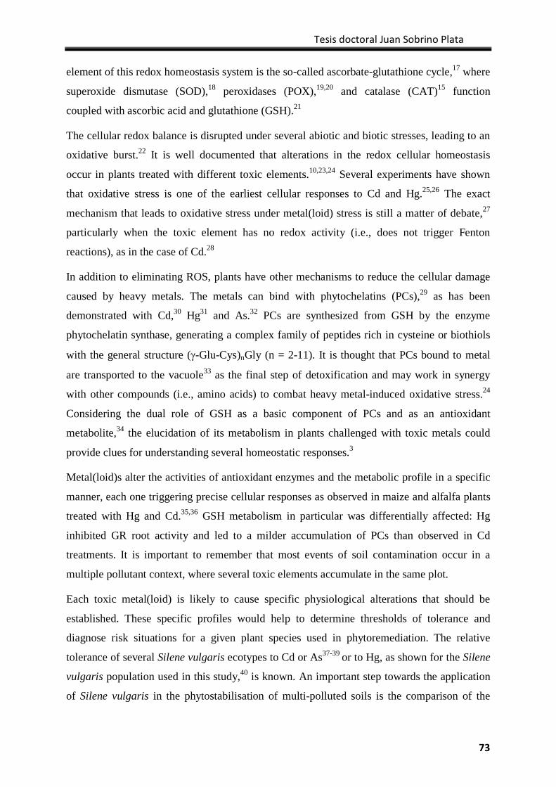
Tesis doctoral Juan Sobrino Plata
73
element of this redox homeostasis system is the so-called ascorbate-glutathione cycle,17 where
superoxide dismutase (SOD),18 peroxidases (POX),19,20 and catalase (CAT)15 function
coupled with ascorbic acid and glutathione (GSH).21
The cellular redox balance is disrupted under several abiotic and biotic stresses, leading to an
oxidative burst.22 It is well documented that alterations in the redox cellular homeostasis
occur in plants treated with different toxic elements.10,23,24 Several experiments have shown
that oxidative stress is one of the earliest cellular responses to Cd and Hg.25,26 The exact
mechanism that leads to oxidative stress under metal(loid) stress is still a matter of debate,27
particularly when the toxic element has no redox activity (i.e., does not trigger Fenton
reactions), as in the case of Cd.28
In addition to eliminating ROS, plants have other mechanisms to reduce the cellular damage
caused by heavy metals. The metals can bind with phytochelatins (PCs),29 as has been
demonstrated with Cd,30 Hg31 and As.32 PCs are synthesized from GSH by the enzyme
phytochelatin synthase, generating a complex family of peptides rich in cysteine or biothiols
with the general structure (γ-Glu-Cys)nGly (n = 2-11). It is thought that PCs bound to metal
are transported to the vacuole33 as the final step of detoxification and may work in synergy
with other compounds (i.e., amino acids) to combat heavy metal-induced oxidative stress.24
Considering the dual role of GSH as a basic component of PCs and as an antioxidant
metabolite,34 the elucidation of its metabolism in plants challenged with toxic metals could
provide clues for understanding several homeostatic responses.3
Metal(loid)s alter the activities of antioxidant enzymes and the metabolic profile in a specific
manner, each one triggering precise cellular responses as observed in maize and alfalfa plants
treated with Hg and Cd.35,36 GSH metabolism in particular was differentially affected: Hg
inhibited GR root activity and led to a milder accumulation of PCs than observed in Cd
treatments. It is important to remember that most events of soil contamination occur in a
multiple pollutant context, where several toxic elements accumulate in the same plot.
Each toxic metal(loid) is likely to cause specific physiological alterations that should be
established. These specific profiles would help to determine thresholds of tolerance and
diagnose risk situations for a given plant species used in phytoremediation. The relative
tolerance of several Silene vulgaris ecotypes to Cd or As37-39 or to Hg, as shown for the Silene
vulgaris population used in this study,40 is known. An important step towards the application
of Silene vulgaris in the phytostabilisation of multi-polluted soils is the comparison of the
Glutatión y tolerancia a metales tóxicos
74
tolerance mechanisms triggered under different metal(loid) stress. The plants were cultivated
in a semi-hydroponic system where root development resembles that of plants grown in soils
(in contrast with plants grown in pure-hydroponics25), avoiding the inconvenience of soil
interactions with metal(loid) speciation and dynamics or the interference of soil components
with several plant physiological parameters analyses (i.e., humic acids reacting with biothiols
and proteins). In this context, we studied the behaviour of several parameters of oxidative
stress, biothiol metabolism and photosynthetic activity in S. vulgaris plants treated with Cd,
Hg and As, toxic elements that trigger differential stress signatures.
Experimental
Plant material, growth conditions and treatments
A natural population of Silene vulgaris (Moench) Garcke (Caryophyllaceae) collected in
central Spain (03º18'05'' W y 40º31'40'' N, at 610 m altitude) was used in this study.
Specimens were collected following the method of Hawkes41 to preserve the original genetic
structure of the natural population. A total of 35 traits related to cotyledon, leaf, flower and
fruit morphology, plant height and flowering time were used to calculate the genetic diversity
index,42 which has a high level of intrapopulation polymorphism according to the Shannon-
Weaver genetic diversity index value (H = 11 1.28).
Silene vulgaris seeds were germinated in aqueous 1.5% agarose for 48 h in the dark at 28ºC,
and then grown in a semi-hydroponic system using perlite as inert substrate (1.0 mM
NH4H2PO4, 0.5 mM MgSO4, 2.0 mM Ca(NO3)2, 3.0 mM KNO3, 20 μM FeEDDHA, 2.0 μM
MnSO4, 0.1 μM CuSO4, 2.0 μM ZnSO4, 25 μM H3BO3, and 0.5 μM (NH4)6Mo7O24, at pH
5.25 adjusted with KOH) for four weeks and maintained in a growth chamber under
controlled environmental conditions (16/8 h at 25/18 ºC; light/night).43 A complete
description of the plant growth cycle used in the experiments is shown in Supplementary Fig.
1. After this period, As, Hg or Cd was added to the nutrient solution (0, 6 and 30 μM) for 7
days. Arsenic was provided as As(V) because arsenate is the predominant As species in
aerated soils and environments.44 At harvest, the plants were dipped in 20 mM Na2EDTA and
thoroughly washed in deionized H2O. Plant length was then measured. Roots and shoots were
collected, snap frozen in liquid N2 and stored at –80ºC until analysis.
Chemicals and antibodies
All products were reagent grade. Protease inhibitor cocktail (P2714) and Saccharomyces
cerevisiae GR (G3664) were purchased from Sigma (St. Louis, MO, USA). LumiSensor
Tesis doctoral Juan Sobrino Plata
75
Chemiluminescent HRP Substrate Kit (L00221 V60) and Goat Anti-Rabbit IgG Antibody
(H&L) conjugated with horseradish peroxidase (α-IgG; A00160) were purchased from
GenScript (Piscataway, NJ, USA). Rabbit polyclonal antibodies anti-cytosolic APX (α-APX;
AS06180), anti-GR (α-GR; AS06181), anti-LHCII type I chlorophyll a/b-binding protein (α-
LHCb1; AS01004) and anti-D1 protein of PSII (α-D1; AS10704) were bought from Agrisera
(Vännäs, Sweden).
GR and APX in gel activity
APX and GR activities were determined in gel after separation of protein extracts by non-
denaturing polyacrylamide electrophoresis following the procedures described by Sobrino-
Plata et al.36 using 10% polyacrylamide gels. The concentration of protein in the extracts was
determined using the BioRad Protein Assay reagent with bovine serum albumin as a standard,
and adequate loading was corrected by denaturing gel electrophoresis according to Laemmli45
prior to electrophoresis and activity staining. Protein loading for APX detection was 60 µg
and 20 µg of shoot and root extracts, respectively. Protein loading for GR analysis was 15 µg
and 10 µg of shoot and root, respectively.
Western-blot and immunodetection
Immunodetection was performed by Western-blot after denaturing gel electrophoresis.45 Once
proteins were separated (10 µg total protein), they were blotted onto a nitrocellulose
membrane (BioTraceNT Pall Corporation, East Hills, NY, USA) by using a semi-dry
procedure (Trans Blot SD Semi-Dry Electrophoretic Transfer Cell; BioRad, Hercules, CA,
USA). Gel slabs were briefly incubated in transfer buffer (48 mM Tris-HCl, 39 mM glycine,
1.3 mM SDS, and 20% methanol at pH 8.3) and then electroblotted for 1 h. The membrane
was blocked with 1% BSA in tris-buffer-saline (TBS), and incubated overnight at 4ºC with
the primary antibodies (α-APX, diluted 1/10000; α-GR, dil. 1/5000; α-LHCb1, dil. 1/2000;
α-D1, dil. 1/2500). After several washes with TBS, the membrane was incubated with the
secondary antibody diluted 1/10000 for 1 h. Proteins were detected by incubating 2 minutes
with LumiSensor Chemiluminescent HRP Substrate Kit. The images were taken with
ChemiDoc™ XRS+ System (BioRad, Hercules, CA, USA).
Analysis of biothiols
HPLC (high performance liquid chromatography) was used to quantify biothiols in extracts.
Plant tissue was ground in liquid N2, and 0.1 g of frozen powder was mixed thoroughly with
Glutatión y tolerancia a metales tóxicos
76
250 μL of 0.5% (w/w) NaBH4 in 1 M NaOH using a mortar and pestle. Prior to injection, the
homogenates were centrifuged for 15 min at 20,000 g and 4 °C, and the supernatants were
acidified to pH 1.0 with 6N HCl and centrifuged again for 10 min at 10,000 g and 4 °C.46
Biothiols were analysed by HPLC following the procedure described by Sobrino-Plata et al.36
A sample size of 100 μL was analysed using a Mediterranea SEA18 column (5 μm, 250 x 4.6
mm; Teknokroma, Spain) and an Agilent 1200 HPLC system (Santa Clara, CA, USA).
Detection was achieved by absorption at 412 nm after post-column derivatization with
Ellman’s reagent.25 Identification of PCs was achieved using commercially available PC2,
PC3 and PC4 standards (Peptide2.0 Inc., Chantilly, VA, USA). The concentration of biothiols
was calculated in samples spiked with N-acetyl cysteine (N-AcCys; 15 μL of a 5 mM
solution) added as internal standard prior to homogenization at a final concentration of 250
μM.
Arsenic, cadmium and mercury tissue concentrations
Plant material was dried at 60ºC for 72 h, and 50 mg was autoclave-digested as described by
Ortega-Villasante et al.26 Concentration of metal(loid)s was determined by ICP-MS Elan 6000
(Perkin-Elmer, San Jose, CA, USA).
Stress indexes
Lipid peroxidation was analysed according to Ortega-Villasante et al.25 Chlorophyll
concentration was determined from an 80% acetone extract by measuring the absorbance at
663 and 645 nm and then using the formula described by Arnon.47
Photochemical parameters
Chlorophyll (Chl) fluorescence was measured in dark-adapted plants (15 min) under different
light regimens to determine different photochemical parameters: quantum efficiency or
photochemical efficiency (ΦPSII), photochemical quenching or proportion of open reaction
centres (qP), and non-photochemical quenching (NPQ) according to Maxwell and Johnson.48
Throughout the fluorescence measurements, an actinic light of 400 μmol m-2 s1 and saturating
pulses light of 8000 μmol m-2 s1 were used sequentially to determine Fo, Fmo, Fm
’, Ft and Fo’
fluorescence data (see Suppl. Fig. 2) in a FMS-2 Pulse Modulated Fluorimeter (Hansatech
Instruments, Norfolk, UK). Due to the non-invasive nature of this technique, photosynthesis
parameters could be determined in plants treated with metal(loid)s for 2, 5 and 7 days.
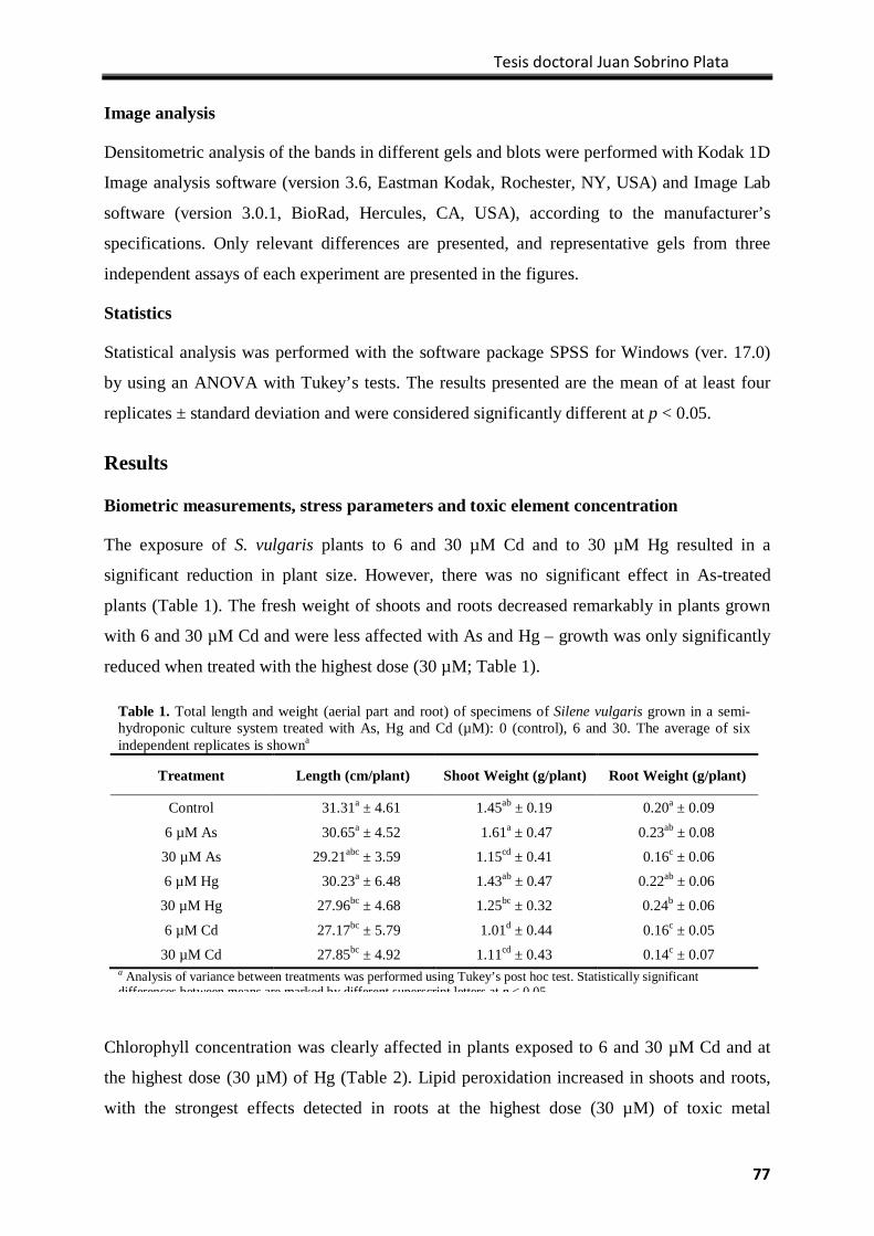
Tesis doctoral Juan Sobrino Plata
77
Image analysis
Densitometric analysis of the bands in different gels and blots were performed with Kodak 1D
Image analysis software (version 3.6, Eastman Kodak, Rochester, NY, USA) and Image Lab
software (version 3.0.1, BioRad, Hercules, CA, USA), according to the manufacturer’s
specifications. Only relevant differences are presented, and representative gels from three
independent assays of each experiment are presented in the figures.
Statistics
Statistical analysis was performed with the software package SPSS for Windows (ver. 17.0)
by using an ANOVA with Tukey’s tests. The results presented are the mean of at least four
replicates ± standard deviation and were considered significantly different at p < 0.05.
Results
Biometric measurements, stress parameters and toxic element concentration
The exposure of S. vulgaris plants to 6 and 30 µM Cd and to 30 µM Hg resulted in a
significant reduction in plant size. However, there was no significant effect in As-treated
plants (Table 1). The fresh weight of shoots and roots decreased remarkably in plants grown
with 6 and 30 µM Cd and were less affected with As and Hg – growth was only significantly
reduced when treated with the highest dose (30 µM; Table 1).
Chlorophyll concentration was clearly affected in plants exposed to 6 and 30 µM Cd and at
the highest dose (30 µM) of Hg (Table 2). Lipid peroxidation increased in shoots and roots,
with the strongest effects detected in roots at the highest dose (30 µM) of toxic metal
Table 1. Total length and weight (aerial part and root) of specimens of Silene vulgaris grown in a semi-hydroponic culture system treated with As, Hg and Cd (µM): 0 (control), 6 and 30. The average of six independent replicates is showna
Treatment Length (cm/plant) Shoot Weight (g/plant) Root Weight (g/plant)
Control 31.31a ± 4.61 1.45ab ± 0.19 0.20a ± 0.09
6 µM As 30.65a ± 4.52 1.61a ± 0.47 0.23ab ± 0.08
30 µM As 29.21abc ± 3.59 1.15cd ± 0.41 0.16c ± 0.06
6 µM Hg 30.23a ± 6.48 1.43ab ± 0.47 0.22ab ± 0.06
30 µM Hg 27.96bc ± 4.68 1.25bc ± 0.32 0.24b ± 0.06
6 µM Cd 27.17bc ± 5.79 1.01d ± 0.44 0.16c ± 0.05
30 µM Cd 27.85bc ± 4.92 1.11cd ± 0.43 0.14c ± 0.07 a Analysis of variance between treatments was performed using Tukey’s post hoc test. Statistically significant differences between means are marked by different superscript letters at p < 0 05
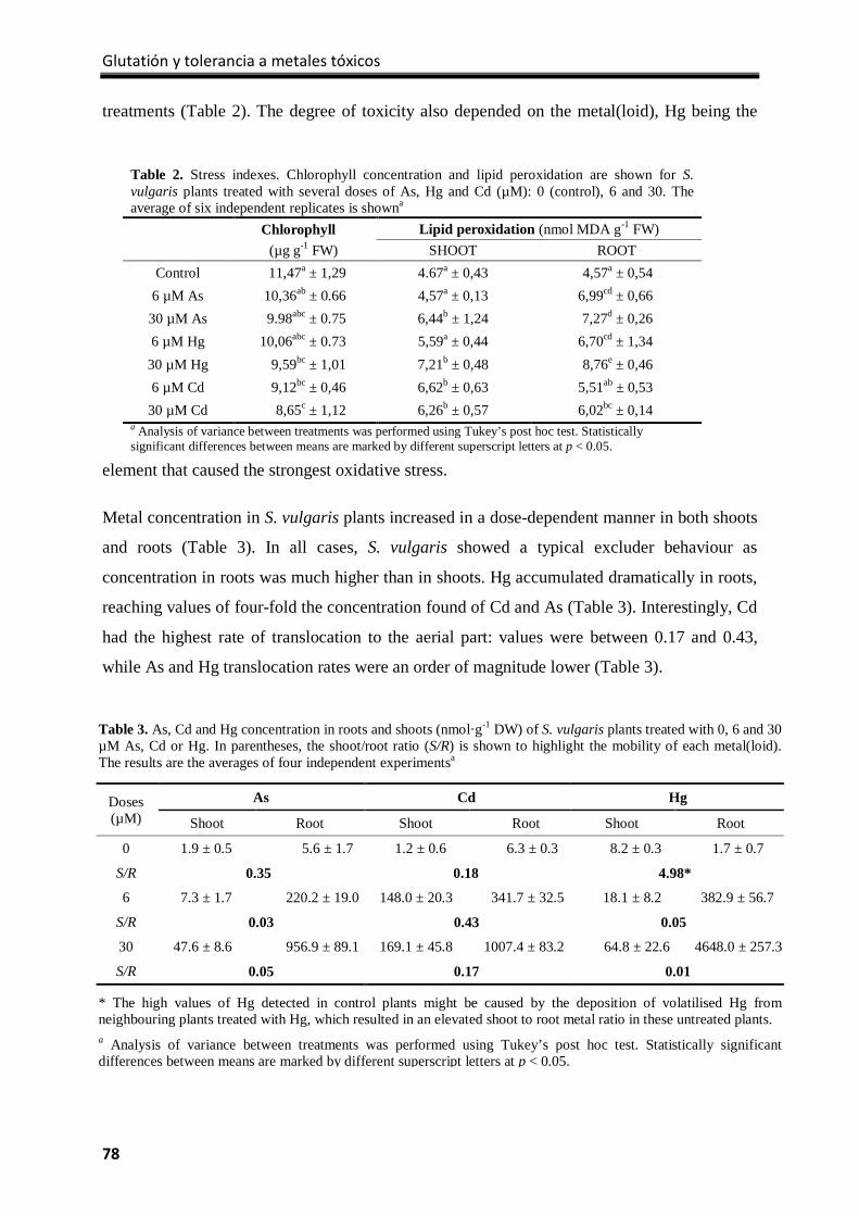
Glutatión y tolerancia a metales tóxicos
78
treatments (Table 2). The degree of toxicity also depended on the metal(loid), Hg being the
element that caused the strongest oxidative stress.
Metal concentration in S. vulgaris plants increased in a dose-dependent manner in both shoots
and roots (Table 3). In all cases, S. vulgaris showed a typical excluder behaviour as
concentration in roots was much higher than in shoots. Hg accumulated dramatically in roots,
reaching values of four-fold the concentration found of Cd and As (Table 3). Interestingly, Cd
had the highest rate of translocation to the aerial part: values were between 0.17 and 0.43,
while As and Hg translocation rates were an order of magnitude lower (Table 3).
Table 3. As, Cd and Hg concentration in roots and shoots (nmol·g-1 DW) of S. vulgaris plants treated with 0, 6 and 30 µM As, Cd or Hg. In parentheses, the shoot/root ratio (S/R) is shown to highlight the mobility of each metal(loid). The results are the averages of four independent experimentsa
Doses (µM)
As Cd Hg
Shoot Root Shoot Root Shoot Root
0 1.9 ± 0.5 5.6 ± 1.7 1.2 ± 0.6 6.3 ± 0.3 8.2 ± 0.3 1.7 ± 0.7
S/R 0.35 0.18 4.98*
6 7.3 ± 1.7 220.2 ± 19.0 148.0 ± 20.3 341.7 ± 32.5 18.1 ± 8.2 382.9 ± 56.7
S/R 0.03 0.43 0.05
30 47.6 ± 8.6 956.9 ± 89.1 169.1 ± 45.8 1007.4 ± 83.2 64.8 ± 22.6 4648.0 ± 257.3
S/R 0.05 0.17 0.01
* The high values of Hg detected in control plants might be caused by the deposition of volatilised Hg from neighbouring plants treated with Hg, which resulted in an elevated shoot to root metal ratio in these untreated plants. a Analysis of variance between treatments was performed using Tukey’s post hoc test. Statistically significant differences between means are marked by different superscript letters at p < 0.05.
Table 2. Stress indexes. Chlorophyll concentration and lipid peroxidation are shown for S. vulgaris plants treated with several doses of As, Hg and Cd (µM): 0 (control), 6 and 30. The average of six independent replicates is showna
Chlorophyll Lipid peroxidation (nmol MDA g-1 FW) (µg g-1 FW) SHOOT ROOT
Control 11,47a ± 1,29 4.67a ± 0,43 4,57a ± 0,54 6 µM As 10,36ab ± 0.66 4,57a ± 0,13 6,99cd ± 0,66 30 µM As 9.98abc ± 0.75 6,44b ± 1,24 7,27d ± 0,26 6 µM Hg 10,06abc ± 0.73 5,59a ± 0,44 6,70cd ± 1,34
30 µM Hg 9,59bc ± 1,01 7,21b ± 0,48 8,76e ± 0,46 6 µM Cd 9,12bc ± 0,46 6,62b ± 0,63 5,51ab ± 0,53
30 µM Cd 8,65c ± 1,12 6,26b ± 0,57 6,02bc ± 0,14 a Analysis of variance between treatments was performed using Tukey’s post hoc test. Statistically significant differences between means are marked by different superscript letters at p < 0.05.
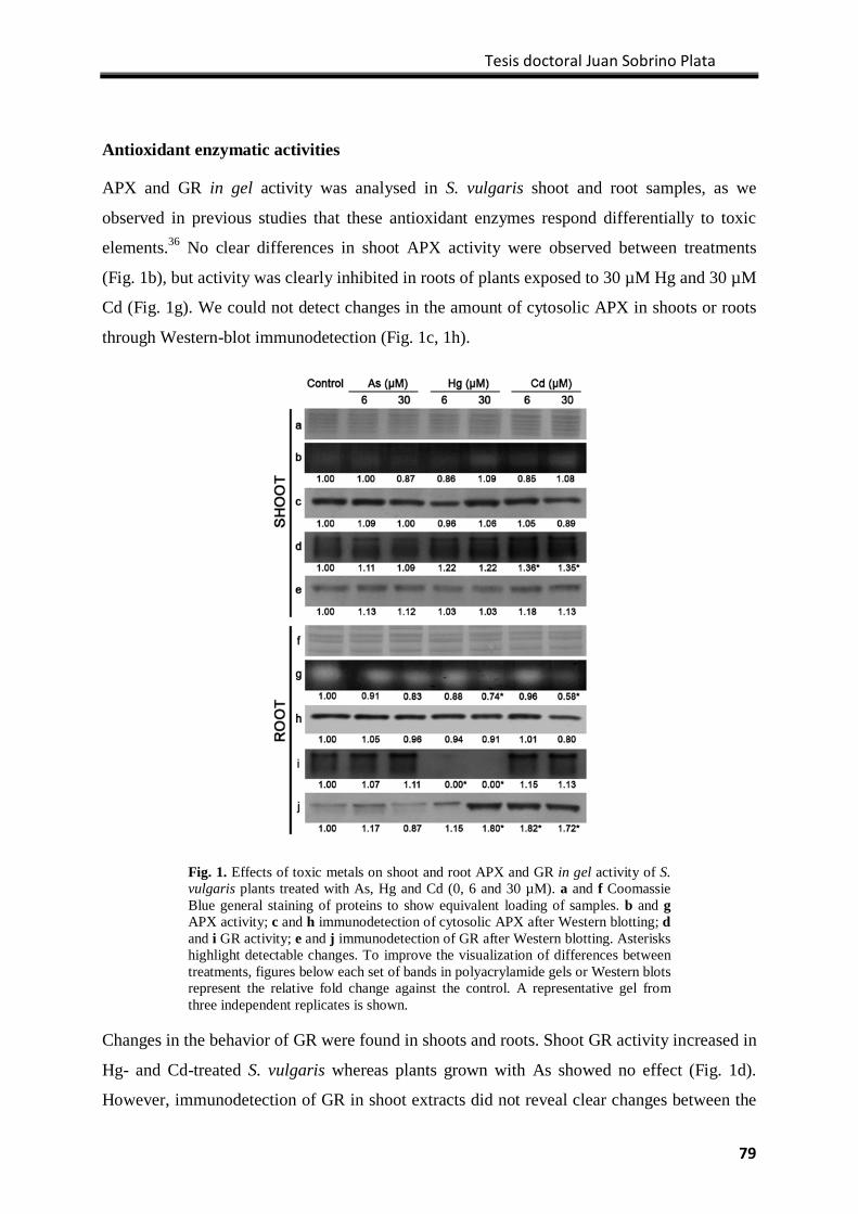
Tesis doctoral Juan Sobrino Plata
79
Antioxidant enzymatic activities
APX and GR in gel activity was analysed in S. vulgaris shoot and root samples, as we
observed in previous studies that these antioxidant enzymes respond differentially to toxic
elements.36 No clear differences in shoot APX activity were observed between treatments
(Fig. 1b), but activity was clearly inhibited in roots of plants exposed to 30 µM Hg and 30 µM
Cd (Fig. 1g). We could not detect changes in the amount of cytosolic APX in shoots or roots
through Western-blot immunodetection (Fig. 1c, 1h).
Changes in the behavior of GR were found in shoots and roots. Shoot GR activity increased in
Hg- and Cd-treated S. vulgaris whereas plants grown with As showed no effect (Fig. 1d).
However, immunodetection of GR in shoot extracts did not reveal clear changes between the
Fig. 1. Effects of toxic metals on shoot and root APX and GR in gel activity of S. vulgaris plants treated with As, Hg and Cd (0, 6 and 30 µM). a and f Coomassie Blue general staining of proteins to show equivalent loading of samples. b and g APX activity; c and h immunodetection of cytosolic APX after Western blotting; d and i GR activity; e and j immunodetection of GR after Western blotting. Asterisks highlight detectable changes. To improve the visualization of differences between treatments, figures below each set of bands in polyacrylamide gels or Western blots represent the relative fold change against the control. A representative gel from three independent replicates is shown.
Glutatión y tolerancia a metales tóxicos
80
treatments (Fig. 1e). Root GR activity also increased in Cd- and As-treated plants, particularly
in samples exposed to the highest dose (30 µM). Interestingly, Hg treatments led to a severe
inhibition of root GR activity at the two doses tested (Fig. 1i). Western blot with α-GR
showed a remarkable increase in the amount of GR protein in plants exposed to Cd (both
doses) and in those grown with 30 µM Hg (Fig. 1j).
Analysis of chlorophyll fluorescence and stability of PSII proteins
ΦPSII, qP and NPQ parameters were calculated from the data of chlorophyll fluorescence
under different light regimens in dark-adapted S. vulgaris plants (Fo, Fmo, Fm
’, Ft and Fo’;
Suppl. Fig. 1); the results were in the range described in the literature for most higher plants.48
Chlorophyll fluorescence yield varied greatly with the treatments; there was a pronounced
increase in Fmo, Fm´ and Ft values in 30 µM Cd-treated plants (Suppl. Fig. 1). This resulted in
significant differences in ΦPSII and qP under metal(loid)s stress. There was a clear decrease
in quantum yield or photosynthetic efficiency (ΦPSII) in plants exposed to Hg and Cd (Fig.
2). A similar effect was observed in qP, a parameter that decreased in all treatments after 7
days, this toxic effect being clearest in Cd-exposed plants (Fig. 2). NPQ showed a rather
erratic behaviour, as this parameter increased slightly under Hg stress mainly in the first 2 to 5
days of exposure, but decreased with the lowest dose of As and Cd (6 µM) after 7 days of
treatment (Fig. 2). All experimental conditions produced only minor changes.
The toxic effects of Cd, As and Hg on the photochemical apparatus was further studied
through Western-blot immunodetection of the state of D1 protein, a phosphoprotein of the
PSII reaction centre, and that of LHCb1, a polypeptide of the light-harvesting chlorophyll a/b
complex II located in the PSII antenna. The α-D1 used recognized two proteins of
approximately 28 KDa (Fig. 3c), upper slower-migrating band potentially representing
phosphorylated D1 protein.49 There was no clear effect on D1 protein, as the intensity neither
band changed appreciably. However, there was a clear dose-dependent decrease in the amount
of LHCb1 in Hg- and Cd-treated plants (Fig. 3b), implying that at the highest doses of
metal(loid)s, there was a loss of this PSII antenna-associated protein.
Analysis of biothiols
Cysteine (Cys) and glutathione (GSH) were the only biothiols that accumulated in shoots and
roots of S. vulgaris plants grown in control nutrient solution (Table 4), with the same order of
concentration observed that was previously described for this plant species.7,50 In general,
exposure to As, Cd and Hg did not alter the levels of Cys and GSH in shoots, with the
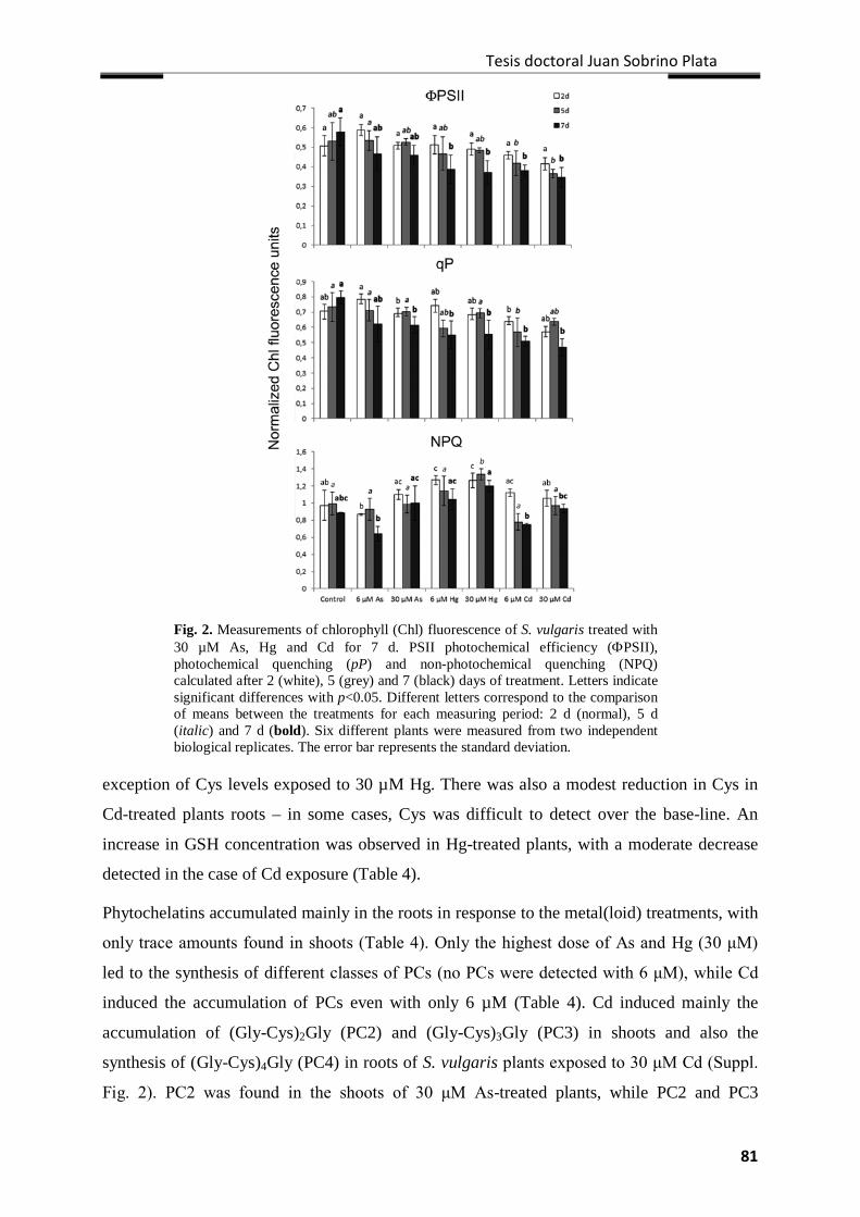
Tesis doctoral Juan Sobrino Plata
81
Fig. 2. Measurements of chlorophyll (Chl) fluorescence of S. vulgaris treated with 30 µM As, Hg and Cd for 7 d. PSII photochemical efficiency (ΦPSII), photochemical quenching (pP) and non-photochemical quenching (NPQ) calculated after 2 (white), 5 (grey) and 7 (black) days of treatment. Letters indicate significant differences with p<0.05. Different letters correspond to the comparison of means between the treatments for each measuring period: 2 d (normal), 5 d (italic) and 7 d (bold). Six different plants were measured from two independent biological replicates. The error bar represents the standard deviation.
exception of Cys levels exposed to 30 µM Hg. There was also a modest reduction in Cys in
Cd-treated plants roots – in some cases, Cys was difficult to detect over the base-line. An
increase in GSH concentration was observed in Hg-treated plants, with a moderate decrease
detected in the case of Cd exposure (Table 4).
Phytochelatins accumulated mainly in the roots in response to the metal(loid) treatments, with
only trace amounts found in shoots (Table 4). Only the highest dose of As and Hg (30 μM)
led to the synthesis of different classes of PCs (no PCs were detected with 6 μM), while Cd
induced the accumulation of PCs even with only 6 µM (Table 4). Cd induced mainly the
accumulation of (Gly-Cys)2Gly (PC2) and (Gly-Cys)3Gly (PC3) in shoots and also the
synthesis of (Gly-Cys)4Gly (PC4) in roots of S. vulgaris plants exposed to 30 μM Cd (Suppl.
Fig. 2). PC2 was found in the shoots of 30 μM As-treated plants, while PC2 and PC3
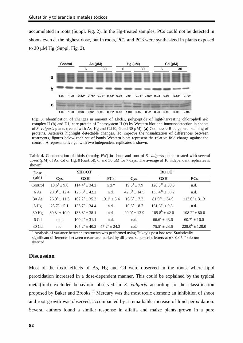
Glutatión y tolerancia a metales tóxicos
82
accumulated in roots (Suppl. Fig. 2). In the Hg-treated samples, PCs could not be detected in
shoots even at the highest dose, but in roots, PC2 and PC3 were synthesized in plants exposed
to 30 μM Hg (Suppl. Fig. 2).
Discussion
Most of the toxic effects of As, Hg and Cd were observed in the roots, where lipid
peroxidation increased in a dose-dependent manner. This could be explained by the typical
metal(loid) excluder behaviour observed in S. vulgaris according to the classification
proposed by Baker and Brooks.51 Mercury was the most toxic element: an inhibition of shoot
and root growth was observed, accompanied by a remarkable increase of lipid peroxidation.
Several authors found a similar response in alfalfa and maize plants grown in a pure
Table 4. Concentration of thiols (nmol/g FW) in shoot and root of S. vulgaris plants treated with several doses (µM) of As, Cd or Hg: 0 (control), 6, and 30 µM for 7 days. The average of 10 independent replicates is showna
Dose (µM)
SHOOT ROOT Cys GSH PCs Cys GSH PCs
Control 18.6a ± 9.0 114.4a ± 34.2 n.d.* 19.5a ± 7.9 128.5ab ± 30.3 n.d.
6 As 23.0a ± 12.4 123.5a ± 42.2 n.d. 42.3b ± 14.5 133.4ab ± 58.2 n.d.
30 As 26.9a ± 11.3 162.2a ± 35.2 13.1a ± 5.4 16.6a ± 7.2 81.9ab ± 34.9 112.6a ± 31.3
6 Hg 25.7a ± 5.1 136.7a ± 34.4 n.d. 10.6a ± 8.7 131.3ab ± 9.8 n.d.
30 Hg 30.3b ± 10.9 133.3a ± 38.1 n.d. 29.0a ± 13.9 189.8b ± 42.0 108.2a ± 80.0
6 Cd n.d. 100.4a ± 31.1 n.d. n.d. 66.6a ± 43.6 60.7a ± 16.0
30 Cd n.d. 105.2a ± 40.3 47.2a ± 24.3 n.d. 75.5a ± 23.6 228.0b ± 128.0 a Analysis of variance between treatments was performed using Tukey’s post hoc test. Statistically significant differences between means are marked by different superscript letters at p < 0.05. b n.d.: not detected
Fig. 3. Identification of changes in amount of Lhcb1, polypeptide of light-harvesting chlorophyll a/b complex II (b) and D1, core protein of Photosystem II (c) by Western blot and immunodetection in shoots of S. vulgaris plants treated with As, Hg and Cd (0, 6 and 30 µM). (a) Coomassie Blue general staining of proteins. Asterisks highlight detectable changes. To improve the visualization of differences between treatments, figures below each set of bands Western blots represent the relative fold change against the control. A representative gel with two independent replicates is shown.
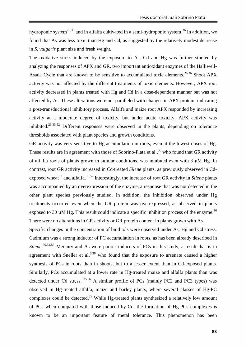
Tesis doctoral Juan Sobrino Plata
83
hydroponic system25,35 and in alfalfa cultivated in a semi-hydroponic system.36 In addition, we
found that As was less toxic than Hg and Cd, as suggested by the relatively modest decrease
in S. vulgaris plant size and fresh weight.
The oxidative stress induced by the exposure to As, Cd and Hg was further studied by
analyzing the responses of APX and GR, two important antioxidant enzymes of the Halliwell-
Asada Cycle that are known to be sensitive to accumulated toxic elements.26,36 Shoot APX
activity was not affected by the different treatments of toxic elements. However, APX root
activity decreased in plants treated with Hg and Cd in a dose-dependent manner but was not
affected by As. These alterations were not paralleled with changes in APX protein, indicating
a post-transductional inhibitory process. Alfalfa and maize root APX responded by increasing
activity at a moderate degree of toxicity, but under acute toxicity, APX activity was
inhibited.26,35,52 Different responses were observed in the plants, depending on tolerance
thresholds associated with plant species and growth conditions.
GR activity was very sensitive to Hg accumulation in roots, even at the lowest doses of Hg.
These results are in agreement with those of Sobrino-Plata et al.,36 who found that GR activity
of alfalfa roots of plants grown in similar conditions, was inhibited even with 3 μM Hg. In
contrast, root GR activity increased in Cd-treated Silene plants, as previously observed in Cd-
exposed wheat53 and alfalfa.36,52 Interestingly, the increase of root GR activity in Silene plants
was accompanied by an overexpression of the enzyme, a response that was not detected in the
other plant species previously studied. In addition, the inhibition observed under Hg
treatments occurred even when the GR protein was overexpressed, as observed in plants
exposed to 30 µM Hg. This result could indicate a specific inhibition process of the enzyme.36
There were no alterations in GR activity or GR protein content in plants grown with As.
Specific changes in the concentration of biothiols were observed under As, Hg and Cd stress.
Cadmium was a strong inductor of PC accumulation in roots, as has been already described in
Silene.50,54,55 Mercury and As were poorer inducers of PCs in this study, a result that is in
agreement with Sneller et al.6,38 who found that the exposure to arsenate caused a higher
synthesis of PCs in roots than in shoots, but to a lesser extent than in Cd-exposed plants.
Similarly, PCs accumulated at a lower rate in Hg-treated maize and alfalfa plants than was
detected under Cd stress. 35,36 A similar profile of PCs (mainly PC2 and PC3 types) was
observed in Hg-treated alfalfa, maize and barley plants, where several classes of Hg-PC
complexes could be detected.29 While Hg-treated plants synthesized a relatively low amount
of PCs when compared with those induced by Cd, the formation of Hg-PCs complexes is
known to be an important feature of metal tolerance. This phenomenon has been
Glutatión y tolerancia a metales tóxicos
84
demonstrated in both alfalfa and Arabidopsis.31 These findings were further supported by the
work of Park et al.,33 who found that Hg and Cd tolerance was compromised in Arabidopsis
mutants with unpaired capacity to translocate metal-PC complexes to the vacuoles. On the
other hand, Arnetoli et al.56 observed that PCs were not completely correlated with As
tolerance. PCs were synthesized in two ecotypes of Silene paradoxa – one As-tolerant and
another non-tolerant. These authors suggested that tolerance depended on alternative defence
mechanisms controlling the degree of As uptake and accumulation.57 Therefore, PCs might be
part of an array of metal(loid) defence mechanisms that reduces the amount of free cytosolic
metal(loid).29
Interestingly, there was a significant increase in GSH root levels in Silene plants exposed to
Hg, indicating that GSH metabolism might be imbalanced. This result is in agreement with
our previous results in alfalfa.36 GSH plays a dual role under metal stress: it is an important
antioxidant, and it is the precursor of PCs, which help to detoxify metal(loid)s.3 Therefore, it
is expected that the activation of the metabolic pathways responsible for keeping the cellular
pool of GSH under tight control either increases its synthesis rate58 or controls its turnover via
oxidation. The observed overexpression of GR under Hg (30 µM) and Cd (6 and 30 µM)
stresses is interesting, as GR is the enzyme that shifts the balance towards GSH from oxidised
GSH.22
The concentration of chlorophyll decreased in general under metal(loid) stress, particularly in
Silene plants treated with 30 µM Cd. Several authors observed similar changes in a number of
different plant species tested with several toxic metals, and chlorophyll concentration has
been used frequently as an index of toxicity.10,59-61 This phenomenon indicated that
photosynthetic reactions might be altered in Silene plants under metal stress, and prompted us
to study the photochemical processes by measuring chlorophyll fluorescence. Quantum
efficiency (ΦPSII) and photochemical quenching (qP) decreased in Cd-treated Silene plants,
confirming that photosynthesis was affected under Cd stress. Similar results were observed in
Oryza sativa,62 Thlaspi caerulescens,11 maize,63 and Populus tremula x P. alba,64 indicating
that the number of open reaction centres decreased. This may result in a decrease in ATP and
NADPH, which could hamper photosynthesis.48,65 With regard to the effects of As and Hg,
the information available in the literature about damage caused to the photosynthesis
processes of higher plants is rather scarce.66 Some experiments have been carried out by direct
application of the metals to chloroplasts or components of thylakoidal photosystems.67
Mercury caused a clear decrease in the photosynthesis efficiency of S. vulgaris. On the other
Tesis doctoral Juan Sobrino Plata
85
hand, As had a modest effect on photosynthesis performance, a result which is in agreement
with the results obtained by Milivojević et al.68 in soybean plants grown in similar conditions.
The test of PSII stability performed by α-D1 and α-Lhcb1 immunodetection showed a
remarkable decrease in the amount of Lhcb1 in S. vulgaris plants treated with 30 µM As, Hg
or Cd, whereas there were no significant differences in the amount of D1 under metal(loid)
exposure. Studies on modifications in the PSII complexes by heavy metal treatments showed
that Cd inhibits the formation of LHCII aggregates, perturbing the photoprotective capability
of the PSII complexes and leading to photoinhibition.60 Baryla et al.59 suggested that Cd
might directly affect enzymes in the chlorophyll synthesis pathway or interfere with the
correct assembly of the pigment-protein complexes of photosystems. Recently, a proteomic
study in spinach plants showed that Cd affected mainly the stability of PSI complexes. The
study also documented a specific reduction in the Lhcb1.1 isomer of the PSII antenna
complex69. In addition, Durand et al. 64 saw a great decrease in the photosynthetic activity in
Cd-exposed Populus tremula x P. alba. This decrease was related to failures in PSII D1
protein turnover and negative effects on the level of chlorophyll a/b binding proteins.
Therefore, it is feasible that the damages observed in S. vulgaris Lhcb1 under metal(loid)
stress could be involved in the observed decrease in photosynthesis efficiency.
All our results exhibited that Cd was more toxic than Hg and As in shoots. Incidentally, the
shoot-to-root accumulation ratio of Cd was an order of magnitude higher than the one
calculated for Hg and As. These data suggest that Cd was more mobile than Hg and As in S.
vulgaris plants. Verkleij et al.54 observed that tolerant and resistant ecotypes of Silene
accumulated Cd preferentially in roots when exposed to low doses. At higher concentrations,
however, the plant suffered phytotoxicity as the rate of Cd translocation to shoots increased.
In addition, Cd was readily translocated to the aerial part of Cd-sensitive S. vulgaris ecotype
plants. The metal possibly follows the transpiration stream to the shoot, where it accumulates
in the leaf epidermis.36 Several authors have proposed that Cd could be translocated to the
shoots either as free ions or in complex with PCs or GSH, as observed in poplar trees.70
Similarly, Mendoza-Cozatl et al.71 found high levels of biothiols in the phloem sap of
Brassica napus, supporting the idea of long-distance transport of Cd assisted at least partially
by PCs. As already observed in S. vulgaris plants, Cd was a potent inductor of PCs. This
result could fit with the above hypothesis.
Glutatión y tolerancia a metales tóxicos
86
Conclusion
Silene vulgaris plants showed specific stress signatures when exposed to different toxic
metal(loid)s. According to our results, As is the least toxic element. Cd accumulation led to an
effect in the photosynthetic apparatus, whereas Hg mainly affected the redox status of roots.
In agreement with Mitler et al.,22 the information obtained could help to perform early and
accurate diagnosis of toxicity. This would help in the selection of tolerant plants to construct
an adequate plant cover for contaminated areas, which is important for optimizing the
phytostabilisation of metal(loid) multipolluted soils. Future research should be aimed at
analysis of sensitive and tolerant ecotypes, to evaluate the different responses between them
and, consequently, to elucidate the mechanisms that confer tolerance to metal(loid) stress.
Acknowledgements
This work was supported by Comunidad de Madrid (EIADES S2009/AMB-1478), Fundación
Ramón Areces (www.fundacionareces.es), the Spanish Ministry of Science and Innovation
(AGL2010-15151-PROBIOMET and CTM2005-04809/TECNO-REUSA), and Junta
Comunidades Castilla-La Mancha (FITOALMA2, POII10-0087-6458). We thank Dr. Pilar
García (IMIDRA) for her technical assistance.
References
1. S. Carrasco-Gil, M. Estebaranz-Yubero, D. Medel-Cuesta, R. Millán, L.E. Hernández.
Environ. Exp. Bot., 2012, 75: 16–24.
2. W.H.O. Ernst. Chemie Erde Geochem., 2005, 65: 29–42.
3. C.S. Seth, T. Remans, E. Keunen, M. Jozefczak, H. Gielen, K. Opdenakker, N. Weyens,
J. Vangronsveld, A. Cuypers. Plant. Cell. Environ., 2012, 35: 334−346.
4. W.H.O. Ernst. Proceedings: International Conference on Heavy Metals in the
Environment, Toronto, 1975.
5. K. Bringezu, O. Lichtenberger, I. Leopold, D. Neumann. J. Plant. Physiol., 1999, 154:
536–546.
6. F.E. Sneller, L.M. Van Heerwaarden, P.L.M. Koevoets, R. Vooijs, H. Schat, J.A.C.
Verkleij. J. Agr. Food. Chem., 2000, 48: 4014−4019.
7. H. Schat, M. Llugany, R. Vooijs, J. Hartley-Whitaker, P.M. Bleeker. J. Exp. Bot., 2002,
53: 2381−2392.
Tesis doctoral Juan Sobrino Plata
87
8. J.A.C. Verkleij, N.A.L.M. VanHoof, A.N. Chardonnens, P.L.M. Koevoets, H. Hakvoort,
V.M. TenBookum, H. Schat, W.H.O.Ernst. Plant. Nutrition., 2001, 446−447.
9. P. Babula, V. Adam, R. Opatrilova, J. Zehnalek, L. Havel, R. Kizek. Environ. Chem. Let.,
2008, 6: 189–213.
10. J.L. Hall. J. Exp. Bot., 2002, 53: 1−11.
11. H. Küpper, A. Parameswaran, M. Leitenmaier, M. Trtílek, I. Setlik. New. Phytol., 2007,
175: 655−674.
12. S. Siripornadulsil, S. Traina, D.P.S. Verm, S.T. Richard. Plant. Cell., 2002, 14:
2837−2847.
13. H. Wenhua, X. Chen, G. Song, Q. Wang, C.C. Chan. Plant. Physiol. Biochem., 2007, 45:
62−69.
14. A. Esteban-Carrasco, M. López-Serrano, J.M. Zapata, B. Sabater, M. Martín. Plant.
Physiol. Biochem., 2001, 39: 521−527.
15. R. Mitler. Trends. Plant. Sci., 2002, 7: 405−410.
16. C.H. Foyer, S. Shigeoka. Plant. Physiol., 2011, 155: 93–100.
17. K. Asada. Plant. Physiol., 2006, 141: 391–396.
18. L.M. Casano, L.D. Gómez, H.R. Lascano, C.A. González, V.S. Trippi. Plant. Cell.
Physiol., 1997, 38: 433–440.
19. J.M. Zapata, B. Sabater, M. Martín. Phytochem., 1998, 48: 1119-1123.
20. A. Ros-Barceló, L.V. Gómez-Ros, A. Esteban-Carrasco. Trends. Plant. Sci., 2007, 12:
486-491.
21. G. Noctor, C.H. Foyer. Ann. Rev. Plant. Physiol. Plant. Mol. Biol., 1998; 49: 249−279.
22. R. Mitler. Trends. Plant. Sci., 2006, 11: 15-19.
23. A. Schützendübel, A. Polle. J. Exp. Bot., 2002, 53, 1351─1365.
24. S.S. Sharma, K.J. Dietz. J. Exp. Bot., 2006, 57: 711−726.
25. C. Ortega-Villasante, R. Rellán-álvarez, F.F. Del Campo, R.O. Carpena-Ruiz, L.E.
Hernández. J. Exp. Bot., 2005, 56: 2239−2251.
26. C. Ortega-Villasante, L.E. Hernández, R. Rellán-Álvarez, F.F. Del Campo, R.O.
Carpena-Ruiz. New. Phytol., 2007, 176: 96−107.
27. P.L. Gratão, A. Polle, P.J. Lea, R.A. Azevedo. Funct. Plant. Biol., 2005, 32: 481−494.
28. A. Cuypers, M. Plusquin, T. Remans, M. Jozefczak, E. Keunen, H. Gielen, K.
Opdenakker, A.R. Nair, E. Munters, T.J. Artois, T. Nawrot, J. Vangronsveld, K. Smeets.
Biometals., 2010, 23: 927-940.
29. R. Pal, J.P.N. Rai. Appl. Biochem. Biotech., 2010, 160: 945−963.
Glutatión y tolerancia a metales tóxicos
88
30. C. Cobbet, P.B. Goldsbrough. Ann. Rev. Plant. Biol., 2002, 53: 159–182.
31. S. Carrasco-Gil, A. Álvarez-Fernández, J. Sobrino-Plata, R. Millán, R.O. Carpena-Ruiz,
D.L. Leduc, J.C. Andrews, J. Abadía, L.E. Hernández. Plant. Cell. Environ., 2011, 34:
778–791.
32. A. Raab, K. Ferreira, A.A. Meharg, J. Feldmann. J. Exp. Bot., 2007, 58: 1333-1338.
33. J. Park, W.Y. Song, D. Ko, Y. Eom, T.H. Hansen, M. Schiller, T.G. Lee, E. Martinoia, Y.
Lee. Plant. J., 2012, 69: 278-288.
34. G. Noctor, L. Gomez, H. Vanacker, C.M. Foyer. J. Exp. Bot., 2002, 53: 1283-1304.
35. R. Rellán-Álvarez, C. Ortega-Villasante, A. Álvarez-Fernández, F.F. Del Campo FF, L.E.
Hernández. Plant. Soil., 2006, 279: 41−50.
36. J. Sobrino-Plata, C. Ortega-Villasante, M.L. Flores-Cáceres, C. Escobar, F.F. Del Campo,
L.E. Hernández. Chemosphere., 2009, 77: 946−954.
37. A.N. Chardonnens, W.M. ten Bookum, L.D.J. Kuijper, J.A.C. Verkleij, W.H.O. Ernst.
Physiol. Plant., 1998, 104: 75-80.
38. H. Schat, R. Voojijs. New. Phytol., 1997, 136: 489-496.
39. F.E. Sneller, L.M. Van Heerwaarden, P.L.M. Koevoets, R. Vooijs, H. Schat, J.A.C.
Verkleij. New. Phytol., 1999, 144: 223−232.
40. A. Pérez-Sanz, R. Millán, M.J. Sierra, R. Alarcón, P. García, M. Gil-Díaz, S. Vázquez,
M.C. Lobo. J. Environ. Manage., 2012, 95: S233-S237.
41. J.G. Hawkes, in: Field collection manual for seed crops, root and tuber crops, tree fruit
crops and related wild species, ed. Department of Plant Biology, University of
Birmingham, England IBPGR and European Association for research of plant breeding
(Eucarpia), Rome,1980, pp. 1-37.
42. R. Alarcón, J. Tardío, P. García. Delpinoa, 2003, 45: 29-35.
43. H. Schat, S.S. Sharma, R. Vooijs. Physiol. Plant., 1997, 101: 477−482.
44. F.J. Zhao, J.F. Ma, A.A. Meharg, S.P. McGrath. New Phytol., 2009, 181: 777–794.
45. U.K. Laemmli. Nature, 1970, 227: 680−685.
46. L. Chen, L. Yang, Q. Wang. Metallomics., 2009, 1: 101–106.
47. R.J. Porra. Photosyn. Res., 2002, 73: 149-156.
48. K. Maxwell, G.N. Johnson. J. Exp. Bot., 2000, 51: 659−668.
49. E. Rintamäki, R. Kettunen, E.M. Aro. J. Biol. Chem., 1996, 25: 14870-14875.
50. J.A. De Knecht, N. VanBaren, W.M. TenBookum, W.F. Sang, P.L.M. Koevoetsa, H.
Schat, J.A.C. Verkleij. Plant. Sci., 1995, 106: 9–18.
51. A.J.M. Baker, R.R. Brooks. Biorecovery., 1989, 1: 81–126.
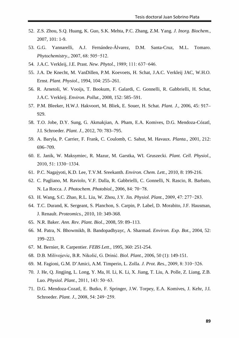
Tesis doctoral Juan Sobrino Plata
89
52. Z.S. Zhou, S.Q. Huang, K. Guo, S.K. Mehta, P.C. Zhang, Z.M. Yang. J. Inorg. Biochem.,
2007, 101: 1-9.
53. G.G. Yannarelli, A.J. Fernández-Álvarez, D.M. Santa-Cruz, M.L. Tomaro.
Phytochemistry., 2007, 68: 505−512.
54. J.A.C. Verkleij, J.E. Prast. New. Phytol., 1989; 111: 637−646.
55. J.A. De Knecht, M. VanDillen, P.M. Koevoets, H. Schat, J.A.C. Verkleij JAC, W.H.O.
Ernst. Plant. Physiol., 1994, 104: 255–261.
56. R. Arnetoli, W. Vooijs, T. Bookum, F. Galardi, C. Gonnelli, R. Gabbrielli, H. Schat,
J.A.C. Verkleij. Environ. Pollut., 2008, 152: 585–591.
57. P.M. Bleeker, H.W.J. Hakvoort, M. Bliek, E. Souer, H. Schat. Plant. J., 2006, 45: 917–
929.
58. T.O. Jobe, D.Y. Sung, G. Akmakjian, A. Pham, E.A. Komives, D.G. Mendoza-Cózatl,
J.I. Schroeder. Plant. J., 2012, 70: 783–795.
59. A. Baryla, P. Carrier, F. Frank, C. Coulomb, C. Sahut, M. Havaux. Planta., 2001, 212:
696–709.
60. E. Janik, W. Maksymiec, R. Mazur, M. Garstka, WI. Gruszecki. Plant. Cell. Physiol.,
2010, 51: 1330−1334.
61. P.C. Nagajyoti, K.D. Lee, T.V.M. Sreekanth. Environ. Chem. Lett., 2010, 8: 199-216.
62. C. Pagliano, M. Raviolo, V.F. Dalla, R. Gabbrielli, C. Gonnelli, N. Rascio, R. Barbato,
N. La Rocca. J. Photochem. Photobiol., 2006, 84: 70−78.
63. H. Wang, S.C. Zhao, R.L. Liu, W. Zhou, J.Y. Jin. Physiol. Plant., 2009, 47: 277−283.
64. T.C. Durand, K. Sergeant, S. Planchon, S. Carpin, P. Label, D. Morabito, J.F. Hausman,
J. Renault. Proteomics., 2010, 10: 349-368.
65. N.R. Baker. Ann. Rev. Plant. Biol., 2008, 59: 89–113.
66. M. Patra, N. Bhowmikb, B. Bandopadhyayc, A. Sharmad. Environ. Exp. Bot., 2004, 52:
199–223.
67. M. Bernier, R. Carpentier. FEBS Lett., 1995, 360: 251-254.
68. D.B. Milivojevic, B.R. Nikolić, G. Drinić. Biol. Plant., 2006, 50 (1): 149-151.
69. M. Fagioni, G.M. D’Amici, A.M. Timperio, L. Zolla. J. Prot. Res., 2009, 8: 310−326.
70. J. He, Q. Jingjing, L. Long, Y. Ma, H. Li, K. Li, X. Jiang, T. Liu, A. Polle, Z. Liang, Z.B.
Luo. Physiol. Plant., 2011, 143: 50−63.
71. D.G. Mendoza-Cozatl, E. Butko, F. Springer, J.W. Torpey, E.A. Komives, J. Kehr, J.I.
Schroeder. Plant. J., 2008, 54: 249−259.
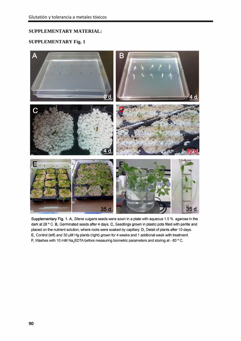
Glutatión y tolerancia a metales tóxicos
90
SUPPLEMENTARY MATERIAL:
SUPPLEMENTARY Fig. 1
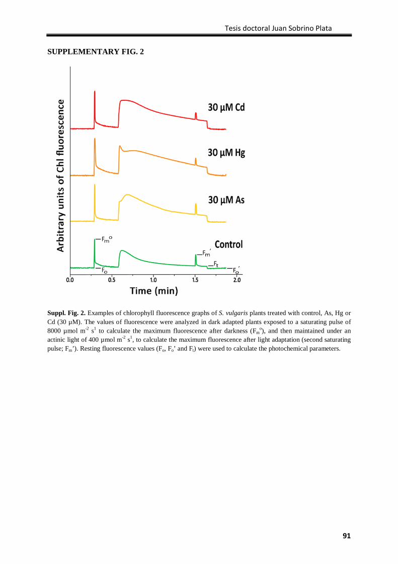
Tesis doctoral Juan Sobrino Plata
91
SUPPLEMENTARY FIG. 2
Suppl. Fig. 2. Examples of chlorophyll fluorescence graphs of S. vulgaris plants treated with control, As, Hg or Cd (30 µM). The values of fluorescence were analyzed in dark adapted plants exposed to a saturating pulse of 8000 µmol m-2 s1 to calculate the maximum fluorescence after darkness (Fm
o), and then maintained under an actinic light of 400 µmol m-2 s1, to calculate the maximum fluorescence after light adaptation (second saturating pulse; Fm’). Resting fluorescence values (Fo, Fo’ and Ft) were used to calculate the photochemical parameters.
Glutatión y tolerancia a metales tóxicos
92
SUPPLEMENTARY FIG. 3
Suppl. Fig. 3. Typical HPLC chromatograms of biothiols obtained from shoot and root extracts of S. vulgaris plants treated with control, As, Cd and Hg (30 µM). Cysteine (Cys) and glutathione (GSH) appeared in all samples, whereas phytochelatins (PCs) were detected mainly in Cd-treated plants: (Gly-Cys)2Gly (PC2), (Gly-Cys)3Gly (PC3), and (Gly-Cys)4Gly (PC4). N-acetyl Cysteine is used as internal standard (I.S.).
CAPÍTULO 4 The role of glutathione in mercury tolerance resembles its function under cadmium stress in Arabidopsis

Tesis doctoral Juan Sobrino Plata
93
Capítulo 4:
The role of glutathione in mercury tolerance resembles its function under cadmium stress in Arabidopsis
Juan Sobrino-Plataa,c, Sandra Carrasco-Gila,b, Javier Abadíab, Carolina Escobarc, Ana Álvarez-Fernándezb, Luis E. Hernándeza
aLaboratory of Plant Physiology, Department of Biology, Universidad Autónoma de Madrid, Cantoblanco, 28049 Madrid, Spain. bDepartment of Plant Nutrition, Estación Experimental de Aula Dei (CSIC), Av. Montañana 1005, E-50080, Zaragoza, Spain. cDepartment of Environmental Sciences, Universidad de Castilla la Mancha, Campus Fábrica de Armas, 45070 Toledo, Spain. *Corresponding author E-mail: [email protected]
Metallomics (2014) DOI: 10.1039/c3mt00329a
Abstract
Recent research efforts have highlighted the importance of glutathione (GSH) as a key
antioxidant metabolite for metal tolerance in plants. Little is known about the mechanisms
involved in stress due to mercury (Hg), one of the most hazardous metals to the
environment and human health. To understand the implication of GSH metabolism in Hg
tolerance, we used two γ-glutamylcysteine synthetase (γECS) Arabidopsis thaliana allele
mutants (rax1-1 and cad2-1) and a phytochelatin synthase (PCS) mutant (cad1-3). The
leaves of these mutants and of wild type (Col-0) were infiltrated with a solution containing
Cd or Hg (0, 3 and 30 µM) and incubated for 24 and 48 h. The formation of phytochelatins
(PCs) in the leaf extracts was followed by two different HPLC-based methods and occurred
in Col-0, cad2-1 and rax1-1 plants exposed to Cd, whereas in the Hg treatments, PCs
accumulated mainly in Col-0 and rax1-1, where Hg-PC complexes were also detected.
ASA and GSH/GSSG levels increased under moderate metal stress conditions,
accompanied by increased GSH reductase (GR) activity and expression. However, higher
metal doses led to a decrease in the analysed parameters, and stronger toxic effects
appeared with 30 µM Hg. The GSH concentration was significantly higher in rax1-1 (70%
of Col-0) than in cad2-1 (40% of Col-0). The leaves of rax1-1were less sensitive than cad2-
1, in accordance with the greater expression of γECS in rax1-1. Our results underline the
Glutatión y tolerancia a metales tóxicos
94
existence of a minimal GSH concentration threshold needed to minimise the toxic effects
exerted by Hg.
Introduction
Important advances have been made in recent years in understanding the mechanisms of
plant tolerance to hazardous metals. Among these toxic elements, mercury (Hg) requires
major attention due to its particular physicochemical properties, which render it an
extremely toxic metal for the environment and for humans. In fact, severe restrictions in its
industrial application and trading have been imposed in recent years to prevent
environmental and health risks.1 This metal can be found in most ecosystems in three
different oxidation forms: metallic (Hg0), monovalent (Hg22+) or divalent (Hg2+), the last
form being the most abundant in well-aerated environments. Mercury is found frequently in
minerals associated with Cl—, OH— and reduced sulphur (i.e., cinnabar) but can also bind
directly to carbon, forming organomercurials, such as methylmercury (CH3Hg; MeHg) or
dimethylmercury (CH3HgCH3), which are the most toxic Hg species in nature. MeHg is
mainly the result of microbial activity in the environment and can bioconcentrate and
biomagnify in the food chain, as occurred in the Minamata (Japan) poisoning incident.2
Plants are able to absorb and accumulate Hg from polluted soils mainly as Hg2+, which may
constitute the main entrance of Hg into terrestrial ecosystems. The accumulation of Hg
causes several toxic symptoms, such as the inhibition of plant growth and the induction of
oxidative stress.3 The strong affinity of Hg2+ for sulfhydryl groups is one of the
mechanisms that provokes severe alterations of protein structure and function (structural
proteins in membranes and the inhibition of several enzymatic processes) in a similar
manner as cadmium (Cd) and copper (Cu) exert when accumulated at phytotoxic
concentrations.4 However, Hg provokes stronger harmful effects than does Cd when plants
are treated with similar doses, causing, in particular, a more rapid induction of oxidative
stress and cell death.5
Many aerobic processes in plant metabolism generate reactive oxygen species (ROS),
which are mostly partially reduced intermediates of oxygen, such as hydrogen peroxide
(H2O2) or superoxide anion radical (O2−). Some of these molecules play important roles in
stress signalling and perception, cell cycle regulation, photosynthesis and cell wall
Tesis doctoral Juan Sobrino Plata
95
lignification.6 However, under biotic or abiotic stresses, the production of ROS is enhanced,
and oxidation reactions occur with different macromolecules,7 leading to several degrees of
cellular damage. In this respect, the relevant role of glutathione (GSH) in counteracting the
phytotoxic effects of diverse toxic metals is highlighted.8,9 Glutathione is a tripeptide that is
synthesised from glycine (Gly), glutamate (Glu) and cysteine (Cys), aminoacid that
harbours a thiol group that provides the chemical properties of this antioxidant metabolite.10
In addition to its cellular redox homeostasis and organic sulphur storage roles, many
important functions of GSH have recently been discovered, from the detoxification of
xenobiotics to cellular signalling and development.11 The biosynthesis of GSH has been
widely studied.12,13 This process is carried out in two ATP-dependent enzymatic steps.
First, gamma-glutamylcysteine synthetase (γECS) binds Cys and Glu to form the
intermediate γ-glutamylcysteine (γEC); second, GSH is formed by glutathione synthetase
(GS) by the binding of glycine (Gly) to γEC. This process is highly compartmentalised, as
γECS is located entirely in the plastids, whereas GS occurs mainly, but not exclusively, in
the cytosol.14
To control the levels of ROS, plant cells possess an antioxidant system composed of
enzymes and metabolites in which the ascorbate (ASA)-glutathione (GSH) cycle plays a
central role.15 In this cycle, excess ROS are removed by the reduction of H2O2 to H2O,
consuming ascorbate via ascorbate peroxidase (APX) and forming monodehydroascorbate
(MDHA), which ultimately is converted to dehydroascorbate (DHA) in a rapid non-
enzymatic reaction.7 The enzyme dehydroascorbate reductase consumes GSH to regenerate
ascorbate, accumulating oxidised glutathione (GSSG).15 The cellular pool of reduced GSH
is maintained, in turn, by glutathione reductase (GR), an enzyme that consumes NADPH.
GR is a homodimer protein located in different organelles, including chloroplasts and
mitochondria, or in the cytosol; however, in photosynthetic cells, up to 80 % of GR activity
is associated with chloroplasts. GR is considered a relevant component of the antioxidant
machinery, maintaining the correct GSH/GSSG ratio, which is fundamental for the redox
homeostasis of the cell.16
An important mechanism of Hg detoxification is based on its high affinity for sulfhydryl
groups through the formation of Hg-phytochelatin (Hg-PCs) complexes.17 Phytochelatins
Glutatión y tolerancia a metales tóxicos
96
are synthesised by the enzyme phytochelatin synthase (PCS), which condenses γEC
molecules into GSH (((γGluCys)nGly), where n is between 2 and 11), which is thought to
be transported into the vacuole while bound to the metal during the final step of metal
detoxification.18
Glutathione may play a dual protective role in plant toxic metal tolerance, both as an
antioxidant and as a precursor of PCs; this hypothesis is based on the drastic changes
observed in its metabolism, particularly under Cd stress.9 However, little is known about
the functionof GSH in Hg homeostasis, despite the recent findings regarding the protection
acquired by the chelation of Hg with PCs.17 Therefore, we studied the behaviour of two
γECS mutant alleles of Arabidopsis thaliana that accumulate lower GSH levels than does
the wild type genotype (Columbia 0; Col-0): cadmium-sensitive 2-1 (cad2-1) and regulator
of APX2 1-1 (rax1-1), which contain 30% and 45% of the GSH found in Col-0 plants,
respectively.19,20 We also evaluated the responses of a PC synthesis-defective mutant,
cadmium-sensitive 1-3 (cad1-3), which expresses a non-functional PCS.21We infiltrated the
leaves of these Arabidopsis genotypes with different concentrations of Hg and Cd (a potent
inducer of PC synthesis) to characterise in detail the short-term adjustments in the GSH
metabolism induced by these toxic metals.
Experimental
Plant material, growth conditions and treatments
Arabidopsis thaliana seeds of wild type (Col-0), cad2-1, rax1-1 and cad1-3 genotypes were
surface-sterilised by agitation in a 15% (v/v) NaOCl solution for 10 min, followed by
several rinses in distilled sterile water. The seeds were germinated in square Petri dishes in
solid Murashige-Skoog nutrient medium (0.6% Phyto-agar, Duchefa Biochemie B.V.,
Haarlem, The Netherlands) supplemented with 2% sucrose for 5 days under a long-day light
regime at 25 ºC. After this period, the seedlings were grown in a perlite:peat (1:3) mixture
for 1 month under a short-day light regime. The leaves were cut in 1 cm diameter disks,
placed into 15 mL glass tubes, sealed with silicone septum stoppers, and infiltrated under
vacuum with deionised water (control), CdCl2 or HgCl2 solutions(3 or 30 µM). The disks
were incubated at 20 ºC with continuous illumination for 24 and 48 hours (Fig. S1). Before
Tesis doctoral Juan Sobrino Plata
97
storing, the disks were washed with 10 mM Na2EDTA and deionised water to remove any
adsorbed superficial heavy metals, snap-frozen in liquid nitrogen and stored at -80 ºC until
analysis.
Chemicals and antibodies
All of the products were analytical grade (≥ 99.0 %). All of the eluents, extraction buffers,
and standard solutions were prepared with analytical-grade type I water (Milli-Q Synthesis,
Millipore, Bedford, MA, USA). Acetonitrile HPLC–gradient grade was purchased from
Scharlab S.L. (Barcelona, Spain); formic acid was purchased from Fluka (Sigma–Aldrich,
St. Louis, MO, USA); GSH, GSSG, ASA, N-acetylcysteine, Protease Inhibitor Cocktail
P2714and Saccharomyces cerevisiae GR were purchased from Sigma–Aldrich (St. Louis,
MO, USA); and labelled GSH ([glycine 1,2-13C,15N]GSH) and ASA ([1-13C]ASA) were
obtained from Cambridge Isotope Laboratories (Andover, MA, USA). PC2, PC3 and PC4
were purchased from Peptide 2.0 (Chantilly, VA, USA). A LumiSensor Chemiluminescent
HRP Substrate Kit (L00221V60) and Goat Anti-Rabbit IgG Antibody (H&L) conjugated
with horseradish peroxidase (α−IgG; A00160) were acquired from GenScript (Piscataway,
NJ, USA). Rabbit polyclonal antibodies anti-GR (α-GR; AS06181) and anti-γECS
(α−γECS; AS06186) were purchased from Agrisera (Vännäs, Sweden).
Cadmium and mercury analyses by ICP-MS
Leaf disks were dried at 50 ºC for 72 h, homogenised with a mortar and pestle, and acid-
digested under pressure in chromatographic vials (4 mL capacity) sealed with
polytetrafluoroethylene stoppers. A 100 mg aliquot of the dried powder was mixed with 1 mL
digestion reagent (HNO3:H2O2:H2O, 0.6:0.4:1 v:v) and autoclaved (Presoclave-75 Selecta,
Barcelona, Spain) at 120ºC and 1.5 atm for 30 min.5 The digests were filtered through a
PVDF filter and diluted in milli-RO water to 5 mL. The Cd and Hg concentrations were
determined by ICP-MS NexION 300 (Perkin-Elmer Sciex, San Jose, CA, USA).
Analysis of biothiols by HPLC/UV-VISandpost-columnderivatisation
Biothiols were analysed by high performance liquid chromatography (HPLC) using an
Agilent 1200 HPLC system (Santa Clara, CA, USA) following the procedure described by
Ortega-Villasante et al.22 A 100 mg aliquot of frozen intact leaf tissue was homogenised in
Glutatión y tolerancia a metales tóxicos
98
a solution of 0.125 N HCl, which contained 0.25 mM N-acetylcysteine as an internal
standard for quantification.21 The homogenate obtained was centrifuged at 14,000 x g for
15 min at 4 ºC. The extracts (100 µL) were injected in a Mediterranea SEA18 column (5
μm, 250 x 4.6 mm; Teknokroma, SantCugat del Vallès, Spain) and eluted with a gradient
mobile phase built with 98:2 H2O:acetonitrile (v/v) in 0.01% TFA (solvent A) and 2:98
dH2O:acetonitrile (v/v) in 0.01% TFA (solvent B). The gradient program, regarding the %
solvent B, was as follows: 2 min, 0%; 25 min, 25%; 26 min, 50%; 30 min, 50%; 35 min,
0%, 45 min, 0%.Detection was achieved after post-column derivatisation with Ellman’s
reagent [1.8 mM 5,5-dithio-bis(2-nitrobenzoic) acid in 300 mM K-phosphate, 15 mM
EDTA at pH 7.0] in a thermostatic 1.8 mL reactor at 38 °C as described by Rauser.23
Biothiols were identified by the derivative compound 5-mercapto-2-nitrobenzoate
(maximum absorption at 412 nm) using commercially available standards (Cys, GSH, PC2,
PC3, and PC4).
Analysis of GSH, GSSG, ASA and complexes of biothiols with mercury by HPLC/ESI-
MS(TOF)
Isotopically labelled GSH (GSH*) and ASA (ASA*) were used as internal standards and
added to the leaf disks immediately before the extraction as described Rellán-Álvarez et al.24
The final concentration of GSH* and ASA* in the injected solutions (standards and plant
extracts) were 75 and 100 µM, respectively. An HPLC/ESI-MS(TOF) analysis was carried out
using an Alliance 2795 HPLC system (Waters, Milford, MA, USA) coupled to a time-of-flight
mass spectrometer (micrOTOF, Bruker Daltonics, Bremen, Germany) equipped with an
electro-spray (ESI) source following the method described by Rellán-Álvarez et al.24 with
minor modifications(Method. S1). The mass axis was calibrated externally using Li-formate
adducts (10 mM LiOH, 0.2% (v/v) formic acid and 50% (v/v) 2-propanol). The HPLC/ESI-
TOF MS system was controlled using the software packages micrOTOF Control v.2.2 and
HyStar v.3.2 (Bruker Daltonics). The data were processed using Data Analysis v.4.0 (Bruker
Daltonics).Ion chromatograms were always extracted with a precision of ± 0.05 m/z units.
Mercury complexes and PC ligands were identified as described by Carrasco-Gil etal.17
Tesis doctoral Juan Sobrino Plata
99
Glutathione reductase activity in gel
The glutathione reductase activity was determined in gel after separating the protein
extracts by non-denaturing electrophoresis in 10% polyacrylamide gels. Five hundred
milligrams of intact frozen material were homogenised in 1 mL extraction solution (30 mM
MOPS at pH 7.5, 5 mM Na2-EDTA, 10 mM DTT, 10 mM ascorbic acid, and 0.6% PVPP
supplemented with 100 µM PMSF and Protease Inhibitor Cocktail) and centrifuged at
14,000 x g and 4ºC for 15 min. The protein concentration was determined using the Protein
Assay Reagent (Bio-Rad, Hercules, CA, USA), and the final protein loading was adjusted
by denaturing gel electrophoresis and Coomassie-blue staining.25 The protein loading for
GR analysis was 15 µg, and the GR activity was revealed as described by Sobrino-Plata et
al.26
Westernblot and immunodetection
Immunodetection was performed by western blot after the denaturing gel electrophoresis.25
Once the proteins were separated (10 µg total protein), they were blotted onto a
nitrocellulose membrane (BioTrace NT Pall Corporation, East Hills, NY, USA) using a
semi-dry procedure (Trans Blot SD Semi-Dry Electrophoretic Transfer Cell; Bio-Rad).
The gel slabs were briefly incubated in transfer buffer (48 mMTris-HCl, 39 mM glycine,
1.3 mM SDS, and 20% methanol at pH 8.3) and then electroblotted for 1 h. The membrane
was blocked with 1% BSA in Tris-buffer-saline (TBS) and incubated overnight at 4ºC with
the primary antibodies (α-GR, dil. 1/5000; α-γECS, dil. 1/2500). After several washes with
TBS, the membrane was incubated with the secondary antibody diluted 1/10000 for 1 h.
The proteins were detected by incubating for 2 minutes with the LumiSensor
Chemiluminescent HRP Substrate Kit, and images were taken with the ChemiDoc™ XRS+
System (Bio-Rad).
Statistics
Statistical analysis was performed using the software package SPSS for Windows (v. 19.0)
using an ANOVA with Tukey’s test. The results presented are means of at least four
replicates ± standard deviation and are considered significantly different at p< 0.05.
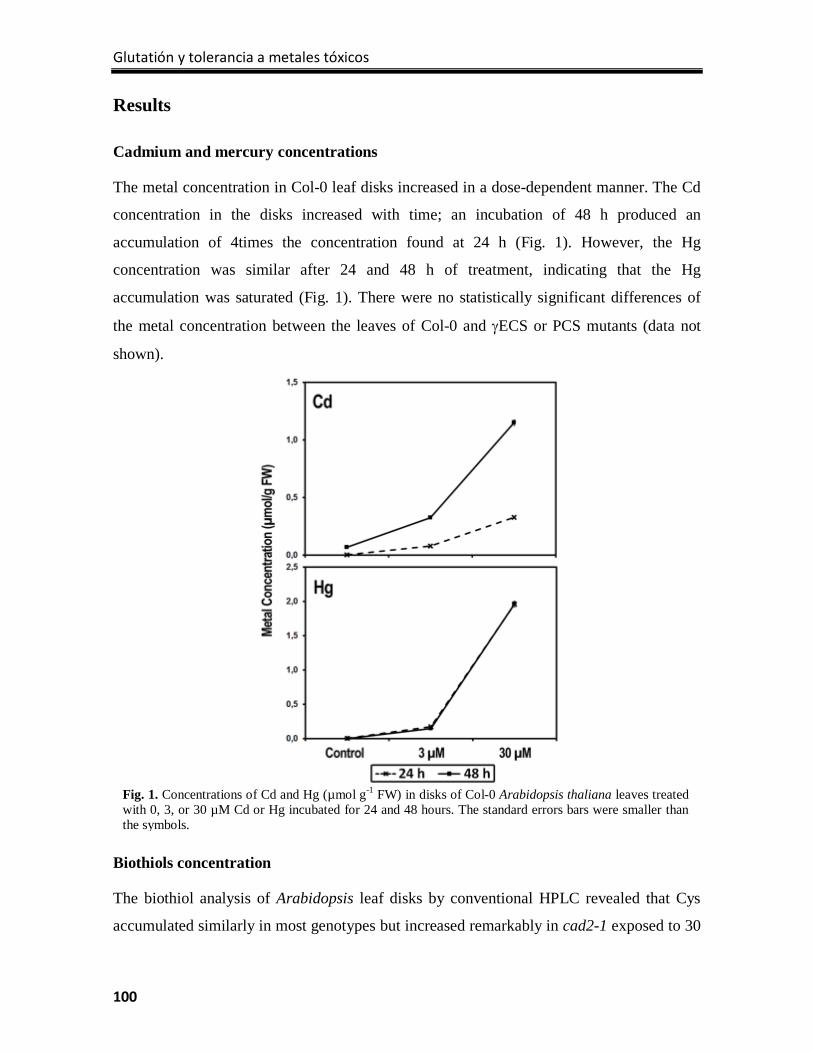
Glutatión y tolerancia a metales tóxicos
100
Results
Cadmium and mercury concentrations
The metal concentration in Col-0 leaf disks increased in a dose-dependent manner. The Cd
concentration in the disks increased with time; an incubation of 48 h produced an
accumulation of 4times the concentration found at 24 h (Fig. 1). However, the Hg
concentration was similar after 24 and 48 h of treatment, indicating that the Hg
accumulation was saturated (Fig. 1). There were no statistically significant differences of
the metal concentration between the leaves of Col-0 and γECS or PCS mutants (data not
shown).
Biothiols concentration
The biothiol analysis of Arabidopsis leaf disks by conventional HPLC revealed that Cys
accumulated similarly in most genotypes but increased remarkably in cad2-1 exposed to 30
Fig. 1. Concentrations of Cd and Hg (µmol g-1 FW) in disks of Col-0 Arabidopsis thaliana leaves treated with 0, 3, or 30 µM Cd or Hg incubated for 24 and 48 hours. The standard errors bars were smaller than the symbols.
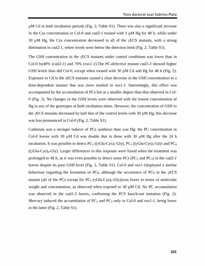
Tesis doctoral Juan Sobrino Plata
101
µM Cd in both incubation periods (Fig. 2; Table S1). There was also a significant increase
in the Cys concentration in Col-0 and cad2-1 treated with 3 µM Hg for 48 h, while under
30 µM Hg, the Cys concentration decreased in all of the γECS mutants, with a strong
diminution in cad2-1, where levels were below the detection limit (Fig. 2; Table S1).
The GSH concentration in the γECS mutants under control conditions was lower than in
Col-0 by40% (cad2-1) and 70% (rax1-1).The PC-defective mutant cad1-3 showed higher
GSH levels than did Col-0, except when treated with 30 µM Cd and Hg for 48 h (Fig. 2).
Exposure to Cd in the γECS mutants caused a clear decrease in the GSH concentration in a
dose-dependent manner that was more marked in rax1-1. Interestingly, this effect was
accompanied by the accumulation of PCs but at a smaller degree than that observed in Col-
0 (Fig. 2). No changes in the GSH levels were observed with the lowest concentration of
Hg in any of the genotypes at both incubation times. However, the concentration of GSH in
the γECS mutants decreased by half that of the control levels with 30 µM Hg; this decrease
was less pronounced in Col-0 (Fig. 2, Table S1).
Cadmium was a stronger inducer of PCs synthesis than was Hg: the PC concentration in
Col-0 leaves with 30 µM Cd was double that in those with 30 µM Hg after the 24 h
incubation. It was possible to detect PC2 ((γGlu-Cys)2-Gly), PC3 ((γGlu-Cys)3-Gly) and PC4
((γGlu-Cys)4-Gly). Larger differences in this response were found when the treatment was
prolonged to 48 h, as it was even possible to detect some PCs (PC2 and PC3) in the cad2-1
leaves despite its poor GSH level (Fig. 2, Table S1). Col-0 and rax1-1displayed a similar
behaviour regarding the formation of PCs, although the occurrence of PCs in the γECS
mutant (all of the PCs except for PC5 (γGlu-Cys)5-Gly))was lower in terms of molecular
weight and concentration, as observed when exposed to 30 µM Cd. No PC accumulation
was observed in the cad1-3 leaves, confirming the PCS knock-out mutation (Fig. 2).
Mercury induced the accumulation of PC2 and PC3 only in Col-0 and rax1-1, being lower
in the latter (Fig. 2, Table S1).
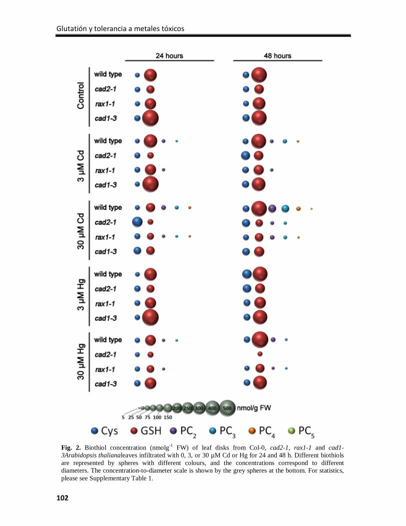
Glutatión y tolerancia a metales tóxicos
102
Fig. 2. Biothiol concentration (nmolg-1 FW) of leaf disks from Col-0, cad2-1, rax1-1 and cad1-3Arabidopsis thalianaleaves infiltrated with 0, 3, or 30 µM Cd or Hg for 24 and 48 h. Different biothiols are represented by spheres with different colours, and the concentrations correspond to different diameters. The concentration-to-diameter scale is shown by the grey spheres at the bottom. For statistics, please see Supplementary Table 1.
Tesis doctoral Juan Sobrino Plata
103
GSH, GSSG and ASA concentrations
The ASA, GSH and GSSG concentrations were determined in disks of A. thaliana leaves
treated with Cd and Hg by HPLC/ESI-MS(TOF) using isotopically labelled ASA and GSH
as internal standards. In agreement with the analysis performed with conventional HPLC,
the GSH levels in the γECS mutants were significantly lower than those in Col-0 and cad1-
3. The GSH concentration increased in Col-0 and rax1-1, but particularly in cad1-3 when
exposed to 3 µM Cd or Hg for 24 h (Fig. 3A). In parallel, the ASA and GSSG
concentrations increased even more in leaves treated with 3 µM Cd, which led to a higher
GSSG-to-total GSH ratio. However, the GSH concentration returned to control values
when the leaf disks were treated with 30 µM Cd or Hg. In this case, the concentrations of
ASA and GSSG decreased in general compared with those of leaves treated with the lowest
metal dose, following a typical hormetic response. Therefore, under 30 µM Cd or Hg, the
GSSG-to-total GSH ratio was lower than in the control samples (Fig. 3A). A different
pattern was observed in leaves treated for 48 h: the GSH concentration decreased in a dose-
dependent manner. The highest levels of GSSG were found in leaves exposed to 3 µM Cd
or Hg, values that were particularly high in cad1-3 (Fig. 3B). Conversely, the GSSG
concentration of leaves incubated with 30 µM Cd or Hg decreased sharply to values below
those of the controls. The ASA concentration remained at a level similar to that of the
controls, but dropped when exposed to 30 µM Cd or Hg (Fig. 3B).
Detection of free PCs and Hg-PC complexes
Ion chromatograms of free ligands and Hg-thiol complexes were extracted at the exact
mass-to-charge ratio (m/z) corresponding to [M-H]–ions from the HPLC/ESI-MS(TOF)
analyses. The Hg-PC complexes were identified based on the characteristic natural Hg
isotopic signature: (196Hg, 198Hg, 199Hg, 200Hg, 202Hg, 204Hg)according to Carrasco-Gil et
al.,17 and the chromatograms were extracted always with the m/z corresponding to the most
abundant stable isotope of Hg (202Hg). Several examples of chromatograms and zoomed-
MS spectra showing signals matching with free PCs and Hg-linked ligands occurring in the
leaf disks of Col-0 (Fig. 4A) and rax1-1 (Fig. 4B) incubated with 30 µM Hg for 48 h can be
found in Fig. 4. The ligands (PC2, Fig. 4C) and Hg-PC complexes (Hg-PC2 and Hg-PC3,
Figs. 4D,H) were identified by the comparison of the analytical and theoretical spectrum,
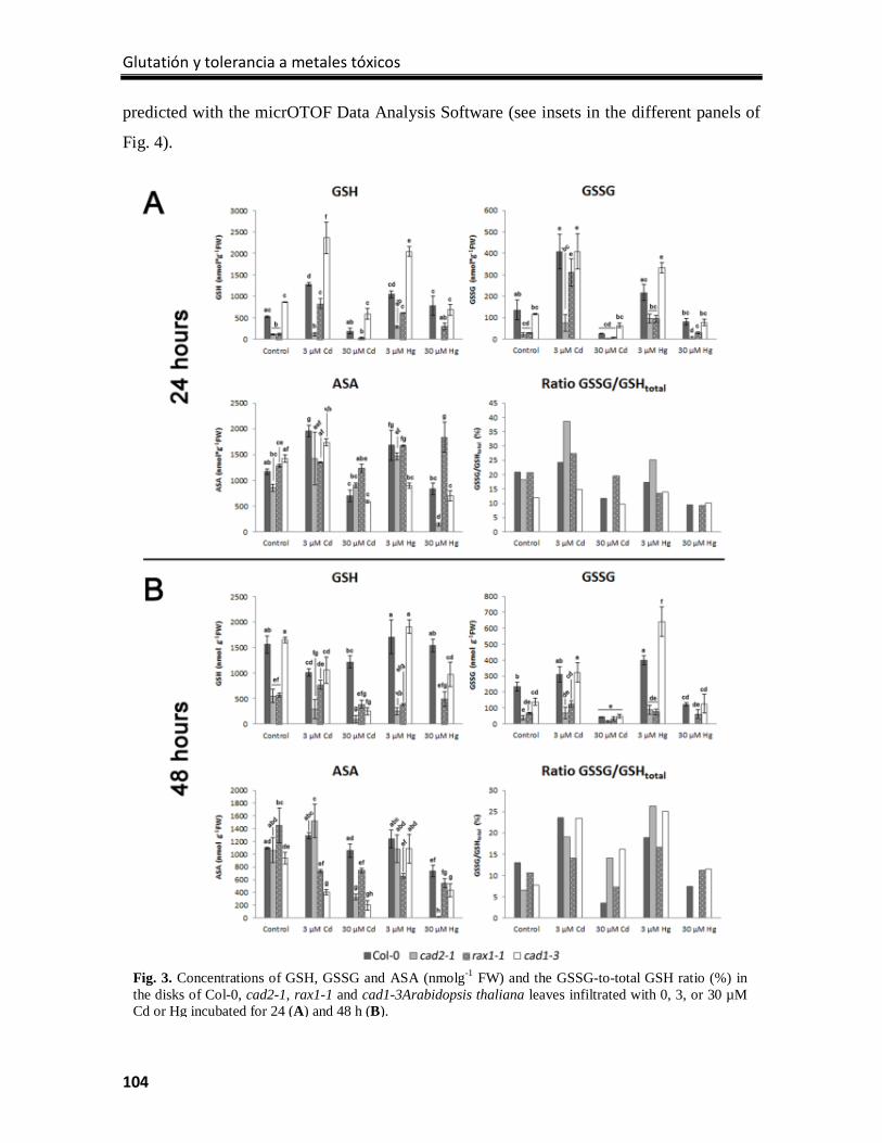
Glutatión y tolerancia a metales tóxicos
104
predicted with the micrOTOF Data Analysis Software (see insets in the different panels of
Fig. 4).
Fig. 3. Concentrations of GSH, GSSG and ASA (nmolg-1 FW) and the GSSG-to-total GSH ratio (%) in the disks of Col-0, cad2-1, rax1-1 and cad1-3Arabidopsis thaliana leaves infiltrated with 0, 3, or 30 µM Cd or Hg incubated for 24 (A) and 48 h (B).
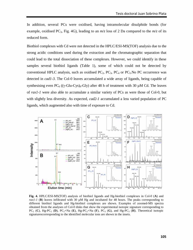
Tesis doctoral Juan Sobrino Plata
105
In addition, several PCs were oxidised, having intramolecular disulphide bonds (for
example, oxidised PC3, Fig. 4G), leading to an m/z loss of 2 Da compared to the m/z of its
reduced form.
Biothiol complexes with Cd were not detected in the HPLC/ESI-MS(TOF) analysis due to the
strong acidic conditions used during the extraction and the chromatographic separation that
could lead to the total dissociation of these complexes. However, we could identify in these
samples several biothiol ligands (Table 1), some of which could not be detected by
conventional HPLC analysis, such as oxidised PC2, PC3, PC4 or PC5.No PC occurrence was
detected in cad1-3. The Col-0 leaves accumulated a wide array of ligands, being capable of
synthesising even PC6 ((γ-Glu-Cys)6-Gly) after 48 h of treatment with 30 µM Cd. The leaves
of rax1-1 were also able to accumulate a similar variety of PCs as were those of Col-0, but
with slightly less diversity. As expected, cad2-1 accumulated a less varied population of PC
ligands, which augmented also with time of exposure to Cd.
Fig. 4. HPLC/ESI-MS(TOF) analysis of biothiol ligands and Hg-biothiol complexes in Col-0 (A) and rax1-1 (B) leaves infiltrated with 30 µM Hg and incubated for 48 hours. The peaks corresponding to different biothiol ligands and Hg-biothiol complexes are shown. Examples of zoomed-MS spectra obtained from the analyses of Col-0 disks that show the experimental isotopic signature corresponding to PC2 (C), Hg-PC2 (D), PC2+Na (E), Hg-PC2+Na (F), PC3 (G), and Hg-PC3 (H). Theoretical isotopic signaturescorresponding to the identified molecular ions are shown in the insets.
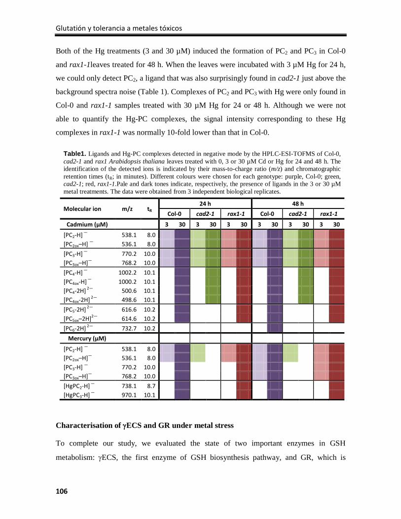
Glutatión y tolerancia a metales tóxicos
106
Both of the Hg treatments (3 and 30 µM) induced the formation of PC2 and PC3 in Col-0
and rax1-1leaves treated for 48 h. When the leaves were incubated with 3 µM Hg for 24 h,
we could only detect PC2, a ligand that was also surprisingly found in cad2-1 just above the
background spectra noise (Table 1). Complexes of PC2 and PC3 with Hg were only found in
Col-0 and rax1-1 samples treated with 30 µM Hg for 24 or 48 h. Although we were not
able to quantify the Hg-PC complexes, the signal intensity corresponding to these Hg
complexes in rax1-1 was normally 10-fold lower than that in Col-0.
Characterisation of γECS and GR under metal stress
To complete our study, we evaluated the state of two important enzymes in GSH
metabolism: γECS, the first enzyme of GSH biosynthesis pathway, and GR, which is
Table1. Ligands and Hg-PC complexes detected in negative mode by the HPLC-ESI-TOFMS of Col-0, cad2-1 and rax1 Arabidopsis thaliana leaves treated with 0, 3 or 30 µM Cd or Hg for 24 and 48 h. The identification of the detected ions is indicated by their mass-to-charge ratio (m/z) and chromatographic retention times (tR; in minutes). Different colours were chosen for each genotype: purple, Col-0; green, cad2-1; red, rax1-1.Pale and dark tones indicate, respectively, the presence of ligands in the 3 or 30 µM metal treatments. The data were obtained from 3 independent biological replicates.
Molecular ion m/z tR 24 h 48 h
Col-0 cad2-1 rax1-1 Col-0 cad2-1 rax1-1
Cadmium (µM) 3 30 3 30 3 30 3 30 3 30 3 30
[PC2-H] — 538.1 8.0 [PC2ox–H] — 536.1 8.0 [PC3-H] — 770.2 10.0 [PC3ox–H]— 768.2 10.0 [PC4-H] — 1002.2 10.1 [PC4ox-H] — 1000.2 10.1 [PC4-2H] 2— 500.6 10.1 [PC4ox-2H] 2— 498.6 10.1 [PC5-2H] 2— 616.6 10.2 [PC5ox–2H]2— 614.6 10.2 [PC6-2H] 2— 732.7 10.2
Mercury (µM)
[PC2-H] — 538.1 8.0 [PC2ox–H]— 536.1 8.0 [PC3-H] — 770.2 10.0 [PC3ox–H]— 768.2 10.0 [HgPC2-H] — 738.1 8.7 [HgPC3-H] — 970.1 10.1
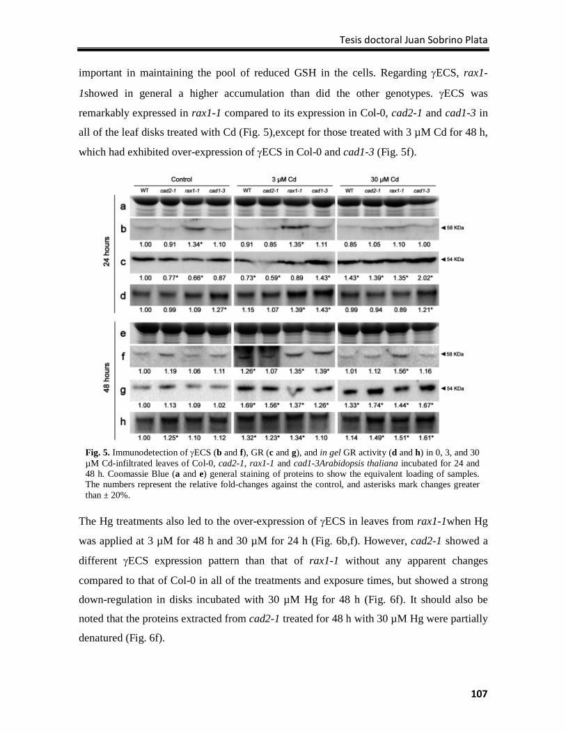
Tesis doctoral Juan Sobrino Plata
107
important in maintaining the pool of reduced GSH in the cells. Regarding γECS, rax1-
1showed in general a higher accumulation than did the other genotypes. γECS was
remarkably expressed in rax1-1 compared to its expression in Col-0, cad2-1 and cad1-3 in
all of the leaf disks treated with Cd (Fig. 5),except for those treated with 3 µM Cd for 48 h,
which had exhibited over-expression of γECS in Col-0 and cad1-3 (Fig. 5f).
The Hg treatments also led to the over-expression of γECS in leaves from rax1-1when Hg
was applied at 3 µM for 48 h and 30 µM for 24 h (Fig. 6b,f). However, cad2-1 showed a
different γECS expression pattern than that of rax1-1 without any apparent changes
compared to that of Col-0 in all of the treatments and exposure times, but showed a strong
down-regulation in disks incubated with 30 µM Hg for 48 h (Fig. 6f). It should also be
noted that the proteins extracted from cad2-1 treated for 48 h with 30 µM Hg were partially
denatured (Fig. 6f).
Fig. 5. Immunodetection of γECS (b and f), GR (c and g), and in gel GR activity (d and h) in 0, 3, and 30 µM Cd-infiltrated leaves of Col-0, cad2-1, rax1-1 and cad1-3Arabidopsis thaliana incubated for 24 and 48 h. Coomassie Blue (a and e) general staining of proteins to show the equivalent loading of samples. The numbers represent the relative fold-changes against the control, and asterisks mark changes greater than ± 20%.
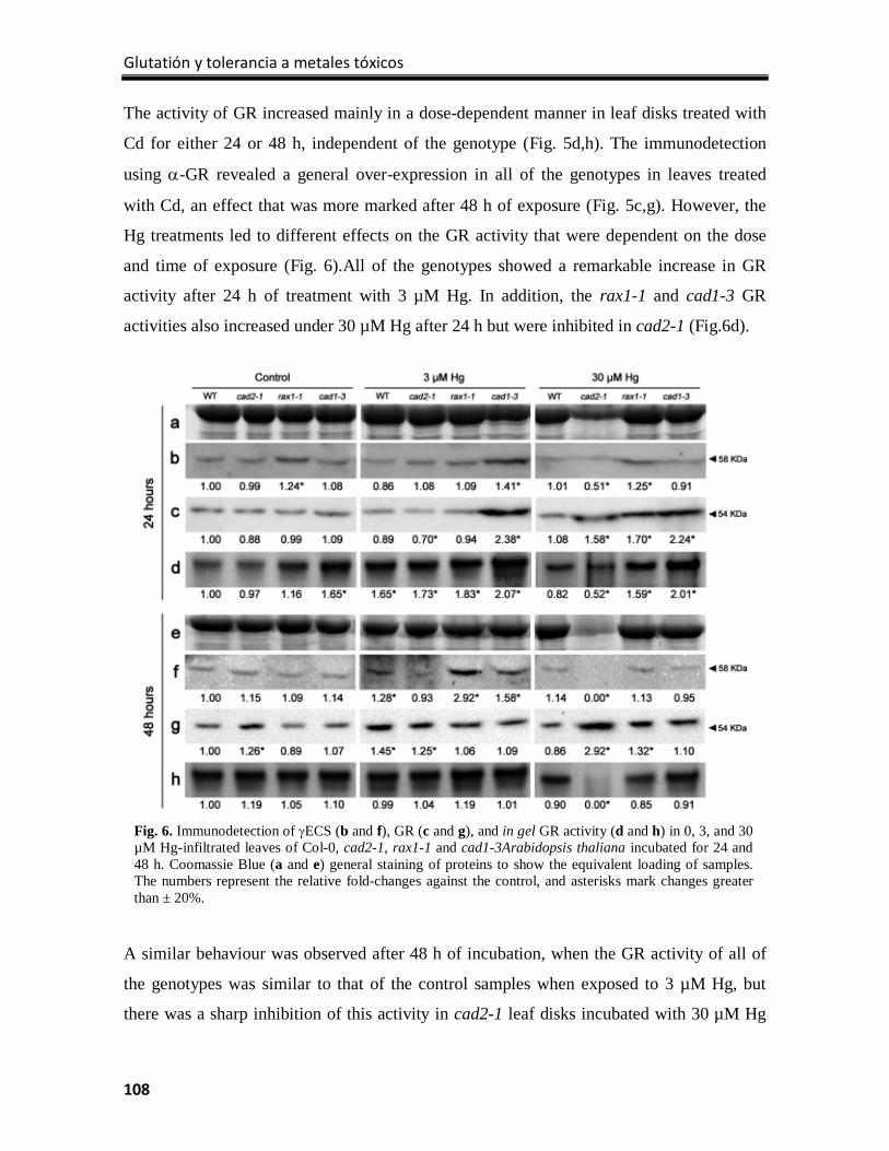
Glutatión y tolerancia a metales tóxicos
108
The activity of GR increased mainly in a dose-dependent manner in leaf disks treated with
Cd for either 24 or 48 h, independent of the genotype (Fig. 5d,h). The immunodetection
using α-GR revealed a general over-expression in all of the genotypes in leaves treated
with Cd, an effect that was more marked after 48 h of exposure (Fig. 5c,g). However, the
Hg treatments led to different effects on the GR activity that were dependent on the dose
and time of exposure (Fig. 6).All of the genotypes showed a remarkable increase in GR
activity after 24 h of treatment with 3 µM Hg. In addition, the rax1-1 and cad1-3 GR
activities also increased under 30 µM Hg after 24 h but were inhibited in cad2-1 (Fig.6d).
A similar behaviour was observed after 48 h of incubation, when the GR activity of all of
the genotypes was similar to that of the control samples when exposed to 3 µM Hg, but
there was a sharp inhibition of this activity in cad2-1 leaf disks incubated with 30 µM Hg
Fig. 6. Immunodetection of γECS (b and f), GR (c and g), and in gel GR activity (d and h) in 0, 3, and 30 µM Hg-infiltrated leaves of Col-0, cad2-1, rax1-1 and cad1-3Arabidopsis thaliana incubated for 24 and 48 h. Coomassie Blue (a and e) general staining of proteins to show the equivalent loading of samples. The numbers represent the relative fold-changes against the control, and asterisks mark changes greater than ± 20%.
Tesis doctoral Juan Sobrino Plata
109
for 48 h (Fig. 6h). Interestingly, GR was also over-expressed when the leaf disks were
challenged with Hg in a dose- and time-dependent manner, with the strongest response
occurring in cad2-1 treated with 30 µM Hg for 48 h (Fig. 6g).
Discussion
Mercury accumulated more than did Cd in the analysed leaf disks, reaching the maximum
concentration after 24 h and showing a characteristic saturation pattern, while the Cd
concentration increased up to 48 h. Similar results were found in plants treated with
identical doses of Hg and Cd, where Hg accumulated to a higher degree in the roots than
did Cd.3,26 The mechanism of Hg uptake in plant cells is not completely understood, but
recent research has shown that Hg accumulates preferentially in the cell walls17,28 and
presumably moves to the aerial part of plants following the apoplastic water flow.27
Cadmium seems to bind to a lesser degree to cell walls, as this metal has a more balanced
distribution between the roots and shoots.26,29 These differences in mobility and binding
capacity to the cell walls might explain at least partially the quick and saturated
accumulation of Hg relative to that of Cd in the leaf disks after metal infiltration.
The concentration of GSH in the leaf disks of the γECS mutant alleles cad2-1 and rax1-1was
significantly lower than that of Col-0, in agreement with previous results.19,20 However, subtle
differences were found when the GSH concentration was analysed by conventional HPLC
(Fig. 2, Table S1) or by HPLC/ESI-MS(TOF) (Fig. 3). This divergence can be explained by
the different levels of sensitivity of these detection methods as discussed by Rellán-
Álvarezet al.24 when alternative analytical procedures are followed. Both of the γECS
mutants displayed a relevant increase in the accumulation of Cys. A similar phenotype was
described in Arabidopsis zin1, another mutant of γECS that contains less than 20% of the GSH
level of Col-0.30 The exposure of cad2-1 and rax1-1 to Cd and Hg resulted in the over-
accumulation of Cys, particularly in the cad2-1 leaf disks (Table S1). These responses are in
agreement with those observed in nrc1 mutants, another Arabidopsis γECS-defective allele,
when treated with 20 µM Cd.31 It is possible that the accumulation of Cys in the γECS mutants
reflects an alteration in the sulphur assimilation pathway, in which several enzymatic steps
could be up-regulated to increase the concentration of Cys required to augment the GSH
concentration under stress.32 Interestingly, the amplitude of the response may depend on the

Glutatión y tolerancia a metales tóxicos
110
GSH concentration, as the increase in the Cys concentration was milder in rax1-1 than in
cad2-1, mutants whose GSH concentrations were, respectively, 70% and 40% that of Col-0
(Fig. 2, Table S1). Jobe et al.31 suggested a complex interplay between GSH, Cys or γEC
concentrations and the cellular redox homeostasis that affects the overall expression of genes
involved in sulphur and GSH metabolisms, such as occurs with the high-affinity AtSULTR1;2
sulphate transporter.
The alteration in the PC production also led to relevant changes in GSH metabolism, as there
was a transient and significant increase in the GSH concentration in cad1-3 leaf disks after 24
h of incubation, particularly greater when treated with 3 µM Cd or Hg (Figs. 2 and 3, Table
S1). These results are in agreement with previous experiments of Cd- and Hg-infiltrated
Arabidopsis leaves.17 The function of PC, an enzyme that is constitutively expressed, in non-
metal-stressed plants is still a matter of debate.33 PC is a soluble protein located in the cytosol,
and it is thought to intervene in the GSH and GSH-conjugate turnover to form γEC or γEC-
conjugates, possibly for Cys recycling.34 It is possible that in the absence of a functional PC in
cad1-3 mutants, lower GSH catabolism occurs, leading to an increase in the GSH
concentration, the catabolic activity of which is enhanced in Cd-treated seedlings.35
Recent work with high light stress in Arabidopsis showed the importance of the accumulation
of ASA and GSH to protect against the overproduction of ROS.36 The exposure of leaf disks
to moderate doses of Cd or Hg (3 µM) for 24 h led to an increase in the GSH and GSSG
concentrations that was significantly greater in non-γECS mutants. This increase was
accompanied by a remarkable increase in the GSSG-to-total GSH ratio and, interestingly, an
accumulation of ASA (Fig. 3A). However, at the highest doses of metals, these values
dropped to those of the control samples, following a typical hormetic response. This pattern
also occurred in leaf disks incubated for 48 h, where changes were less pronounced (Fig. 3B).
A similar pattern was found in Arabidopsis hypocotyl cells treated with 50 μM Cd, where the
GSH and PC concentrations increased after 24 h of incubation, but the GSH concentration
decreased at longer exposure times.37 Transient increases in the GSH and ASA concentrations
were also reported in the root tips of Poplar hybrids under Cd stress (5 and 50 μM), which
was accompanied by an increased GSSG-to-total GSH ratio and increased GR activity.38A
transient accumulation of ASA and GSH occurred also under short-term Cd treatments in the
Tesis doctoral Juan Sobrino Plata
111
roots of wheat seedlings and was accompanied by a significant increase in GR at the highest
dose (40 µM).39 In our case, the increase in ASA, GSH and GSSG concentrations under Cd
and Hg was accompanied by higher GR activity and GR over-expression (Figs. 5 and 6).
Therefore, it is feasible that under moderate metal stress conditions, the ASA-GSH antioxidant
cycle is activated. However, this induction is repressed under acute stress conditions, attained
at metal higher doses or prolonged exposures. In this sense, metal-sensitive genotypes such as
cad2-1 suffered stronger phytotoxic effects, as visualised by the severe inhibition in the
activity of GR, an enzyme that is readily sensitive to Hg, under 30 µM Hg for 48 h (Fig.
6h.26,40 Interestingly, GR was highly over-expressed in the cad2-1 leaf disks, possibly
following a compensatory mechanism to recover the GSH levels under metal stress.
The transient depletion of GSH with 30 µM Cd or Hg could be associated with the elevated
production of PCs.41 The importance of PC accumulation in Cd tolerance has been
described in several plants;42-45 biothiols also play a relevant role in the detoxification of
Hg.17,46 Metal-PC complexes are then sequestered in vacuoles by the action of ABCC
tonoplast transporters.47 In our experiments, both of the metals were clearly able to induce
the synthesis of different PCs, Cd being a stronger inducer than Hg, as found by
conventional HPLC and HPLC/ESI-MS(TOF), in accordance with previous studies
performed in different plant species and with different growing conditions 3,17,26,40.
As expected, the PC synthesis under Cd and Hg exposure depends completely on GSH
metabolism, with remarkable differences between the two γECS allele mutants studied:
cad2-1 and rax1-1 (Fig. 2, Table 1, Table S1). According to the literature, cad2-1has a
limited capability to produce PCs due to its limited synthesis of GSH.19 When exposed to 30
µM Cd for 48 h, cad2-1 accumulated PC2 and PC3, while rax1-1 also synthesised PC4
according to the data of conventional HPLC (Fig. 2, Table S1). This result confirms previous
observations where cad2-1 accumulated a significantly lower concentration of PCs than did
Col-0 when exposed to 30 µM Cd, while Hg failed to induce PCs or Hg-PC complexes.17
However, less is known about the behaviour of rax1-1 under metal stress, a mutant allele that
is able to synthesise more GSH than can cad2-1 and that is less sensitive to biotic and abiotic
stresses.20,48 These characteristics were confirmed by conventional HPLC and HPLC/ESI-
MS(TOF), the data of which showed a lower number of PC ligands and lower values of their
corresponding signal intensities in cad2-1 than in rax1-1, an allele in which we could even
Glutatión y tolerancia a metales tóxicos
112
detect PC5 (Table 1). Therefore, rax1-1 showed a response similar to that of Col-0 when
treated with 30 µM Hg for 48 h, and we could even detect PC6 in its leaf disks. These
differences in PC accumulation between the γECS mutant alleles were also reflected in the
Hg-PC complexes detected in the leaf disks that were exposed to Hg: rax1-1 accumulated Hg-
PC2 and Hg-PC3, although the intensity of these molecular ions was lower than that in Col-0
Hg-treated leaf disks, while Hg-PC complexes were not detected in cad2-1 (compare Figs. 4A
and 4B). Finally, the absence of Hg-PCs (for example in cad2-1 and cad1-3) did not result in
the accumulation of Hg-GSH complexes in vivo, in agreement with previous studies.17,46
In summary, we found that rax1-1exhibited a similar behaviour as that of Col-0 under Hg and
Cd stress, despite the minor differences in the GSH concentration compared with cad2-1. Our
results suggest that a decrease in the GSH concentration over a narrow threshold limits metal
tolerance. The regulation of γECS is complex at the transcriptional and post-translational
levels, as has been recently reported.49,50 This complexity might explain the consistent over-
expression of γECS in rax1-1 leaf disks, reflecting possible modifications in the transcriptional
pattern of S-metabolism genes. In this respect, Jobe et al.31 found that altered levels of Cys or
γECS in conjunction with the GSSG-to-total GSH ratio, as occurs in cad2-1 and rax1-1
mutants under metal stress, mediate the expression of Sultr1;2, a high-affinity sulphate
transporter that is considered the first step in the sulphate assimilation process. Thus, the
complete characterisation of the regulatory components of sulphur metabolism under metal
stress, in particular in GSH-deficient mutants, should be a major task for future experiments to
shed light on the mechanisms of metal tolerance in plants mediated by GSH.
Acknowledgments
This work was funded by the Ministry of Economy and Competitivity (Projects
PROBIOMET AGL2010-15151 and AGL2010-16515), Junta Comunidades Castilla-La
Mancha (FITOALMA2, POII10-007-6458), Comunidad de Madrid (EIADES consortium
S2009/AMB-1478) and the Aragón Government (Group A03).
Tesis doctoral Juan Sobrino Plata
113
References
1. European Commission, Brussels, 28, 2005.
2. N. E. Selin, Annu. Rev. Env. Resour.,2009, 34, 43-63.
3. R. Rellán-Álvarez, C. Ortega-Villasante, A. Álvarez-Fernández, F. F. Campo and L.
E. Hernández, PlantSoil, 2006, 279, 41-50.
4. F. Van Assche and H. Clijsters, Plant Cell Environ.,1990, 13, 195-206.
5. C. Ortega-Villasante, L. E. Hernández, R. Rellán-Álvarez, F. F. Del Campo and R. O.
Carpena-Ruiz, New Phytol.,2007, 176, 96-107.
6. R. Mittler, S. Vanderauwera, M. Gollery and F. Van Breusegem, Trends Plant Sci.,
2004, 9, 490-498.
7. Y. Nakano and K. Asada, Plant Cell physiol., 1987, 28, 131-140.
8. M. A. Hossain, P. Piyatida, , J. A. Teixeira da Silva and M. Fujita, J. Bot., 2012.
doi:10.1155/2012/872875.
9. M. Jozefczak, T. Remans, J. Vangronsveld and A. Cuypers, Int. J. Mol. Sci., 2012,
13, 3145-3175.
10. G. Noctor and C. H. Foyer, Annu. Rev. Plant Biol., 1998, 49, 249-279.
11. G. Noctor, A. Mhamdi, S. Chaouch, Y. Han, J. Neukermans, B. Márquez-García, G.
Queval and C. H. Foyer, Plant Cell Environ.,2012, 35, 454-484.
12. H. Rennenberg, Phytochemistry, 1980, 21, 2771-2781.
13. P. M. Mullineaux and T. Rausch, Photosynthesis Res., 2005, 86, 459-474.
14. A. Wachter, S. Wolf, H. Steininger, J. Bogs and T. Rausch, Plant J., 2005, 41, 15-30.
15. C. H. Foyer and B. Halliwell, Planta, 1976, 133, 21-25.
16. S. S. Gill, N. A. Anjum, M. Hasanuzzaman, R. Gill, D. K. Trivedi, I. Ahmad, E.
Pereira and N. Tuteja, Plant Physiol.Bioch.,2013, 70, 204-212.
17. S. Carrasco-Gil, A. Álvarez-Fernández, J. Sobrino-Plata, R. Millán, R. O. Carpena-
Ruiz, D. L. Leduc, J. C. Andrews, J. Abadía and L. E. Hernández, PlantCell
Environ.,2011, 34, 778–791.
18. C. Cobbett and P. Goldsbrough, Annu. Rev. Plant Biol., 2002, 53, 159-182.
Glutatión y tolerancia a metales tóxicos
114
19. C. S. Cobbett, M. J. May, R. Howden and B. Rolls, Plant J., 1998, 16, 73-78.
20. L. Ball, G. Accotto, U. Bechtold, G. Creissen, D. Funck, A. Jimenez, B. Kular, N.
Leyland, J. Mejia-Carranza and H. Reynolds, Plant Cell, 2004, 16, 2448-2462.
21. R. Howden, P. B. Goldsbrough, C. R. Andersen and C. S. Cobbett, Plant Physiol.,
1995, 107, 1059-1066.
22. C. Ortega-Villasante, R. Rellán-Álvarez, F. F. Del Campo, R. O. Carpena-Ruiz and
L. E. Hernández, J. Exp. Bot., 2005, 56, 2239-2251.
23. W. E. Rauser, Meth. Enzymol.,1991, 205, 319-333.
24. R. Rellán-Álvarez, L. E. Hernández, J. Abadía and A. Álvarez-Fernández, Anal.
Biochem.,2006, 356, 254-264.
25. U. K. Laemmli, Nature, 1970, 227, 680-685.
26. J. Sobrino-Plata, C. Ortega-Villasante, M. Laura Flores-Cáceres, C. Escobar, F. F.
Del Campo and L. E. Hernández, Chemosphere, 2009, 77, 946-954.
27. S. Carrasco-Gil, H. Siebner, D. L. LeDuc, S. M. Webb, R. Millán, J. C. Andrews and
L. E. Hernández, Environ. Sci. Technol., 2013, 47, 3082-3090.
28. M. Válega, A. Lima, E. Figueira, E. Pereira, M. Pardal and A. Duarte, Chemosphere,
2009, 74, 530-536.
29. E. Lozano-Rodriguez, L. E. Hernández, P. Bonay and R. Carpena-Ruiz, J. Exp. Bot.,
1997, 48, 123-128.
30. V. Shanmugam, M. Tsednee and K. Yeh, Plant J., 2012, 69, 1006-1017.
31. T. O. Jobe, D. Y. Sung, G. Akmakjian, A. Pham, E. A. Komives, D. G.
Mendoza-Cózatl and J. I. Schroeder, Plant J., 2012, 70, 783–795.
32. J. Davidian and S. Kopriva, Mol. Plant, 2010, 3, 314-325.
33. S. Lee and S. Korban, Planta, 2002, 215, 689-693.
34. R. Blum, K. C. Meyer, J. Wünschmann, K. J. Lendzian and E. Grill, Plant Physiol.,
2010, 153, 159-169.
35. R. Blum, A. Beck, A. Korte, A. Stengel, T. Letzel, K. Lendzian and E. Grill, Plant J.,
2007, 49, 740-749.
Tesis doctoral Juan Sobrino Plata
115
36. E. Heyneke, N. Luschin-Ebengreuth, I. Krajcer, V. Wolkinger, M. Müller and B.
Zechmann, BMC Plant Biol., 2013, 13, 104.
37. R. De Michele, E. Vurro, C. Rigo, A. Costa, L. Elviri, M. Di Valentin, M. Careri, M.
Zottini, L. S. di Toppi and F. L. Schiavo, Plant Physiol., 2009, 150, 217-228.
38. A. Schützendübel, P. Nikolova, C. Rudolf and A. Polle, Plant
Physiol.Biochem.,2002, 40, 577-584.
39. A. Paradiso, R. Berardino, M. C. de Pinto, L. S. di Toppi, M. M. Storelli, F. Tommasi
and L. De Gara, Plant Cell Physiol., 2008, 49, 362-374.
40. J. Sobrino-Plata, J. Herrero, S. Carrasco-Gil, A. Pérez-Sanz, C. Lobo, C. Escobar, R.
Millán and L. E. Hernández, RSC Advances, 2013, 3, 4736–4744.
41. W. E. Rauser, Plant Physiol., 1995, 109, 1141-1149.
42. J. A. de Knecht, M. van Dillen, P. L. Koevoets, H. Schat, J. A. Verkleij and W. H.
Ernst, Plant Physiol., 1994, 104, 255-261.
43. H. Schat, M. Llugany, R. VOOiJS, J. Hartley-Whitaker and P. M. Bleeker, J. Exp.
Bot., 2002, 53, 2381-2392.
44. J. He, J. Qin, L. Long, Y. Ma, H. Li, K. Li, X. Jiang, T. Liu, A. Polle and Z. Liang,
Physiol. Plantarum, 2011, 143, 50-63.
45. E. C. Bianucci, J. Sobrino-Plata, R. O. C. Ruíz, M. del Carmen Tordable, A. Fabra, L.
Hernandez and S. Castro, Metallomics, 2012, 4, 1119-1124.
46. L. Chen, L. Yang and Q. Wang, Metallomics, 2009, 1, 101-106.
47. J. Park, W. Song, D. Ko, Y. Eom, T. H. Hansen, M. Schiller, T. G. Lee, E. Martinoia
and Y. Lee, Plant J., 2012, 69, 278-288.
48. V. Parisy, B. Poinssot, L. Owsianowski, A. Buchala, J. Glazebrook and F. Mauch,
Plant J., 2007, 49, 159-172.
49. M. Hothorn, A. Wachter, R. Gromes, T. Stuwe, T. Rausch and K. Scheffzek, J. Biol.
Chem., 2006, 281, 27557-27565.
50. L. M. Hicks, R. E. Cahoon, E. R. Bonner, R. S. Rivard, J. Sheffield and J. M. Jez,
Plant Cell, 2007, 19, 2653-2661.
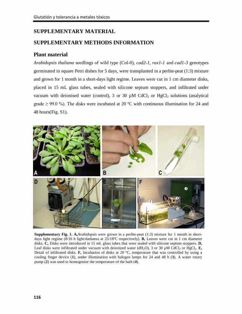
Glutatión y tolerancia a metales tóxicos
116
Supplementary Fig. 1. A,Arabidopsis were grown in a perlite-peat (1:3) mixture for 1 month in short-days light regime (8/16 h light/darkness at 25/18ºC respectively). B, Leaves were cut in 1 cm diameter disks. C, Disks were introduced in 15 mL glass tubes that were sealed with silicone septum stoppers. D, Leaf disks were infiltrated under vacuum with deionized water (dH2O), 3 or 30 µM CdCl2 or HgCl2. E, Detail of infiltrated disks. F, Incubation of disks at 20 ºC, temperature that was controlled by using a cooling finger device (1), under illumination with halogen lamps for 24 and 48 h (3). A water rotary pump (2) was used to homogenize the temperature of the bath (4).
SUPPLEMENTARY MATERIAL
SUPPLEMENTARY METHODS INFORMATION
Plant material Arabidopsis thaliana seedlings of wild type (Col-0), cad2-1, rax1-1 and cad1-3 genotypes
germinated in square Petri dishes for 5 days, were transplanted in a perlite-peat (1:3) mixture
and grown for 1 month in a short-days light regime. Leaves were cut in 1 cm diameter disks,
placed in 15 mL glass tubes, sealed with silicone septum stoppers, and infiltrated under
vacuum with deionised water (control), 3 or 30 µM CdCl2 or HgCl2 solutions (analytical
grade ≥ 99.0 %). The disks were incubated at 20 ºC with continuous illumination for 24 and
48 hours(Fig. S1).
Tesis doctoral Juan Sobrino Plata
117
Chromatography and mass-spectrometry conditions
Chromatographic separation was carried out by injecting 20 µL aliquots of standard solutions
and samples extracts spiked with the internal standards in a Mediterranea SEA18 column (3
μm, 150 x 2.1 mm; Teknokroma, SantCugatdelVallès, Spain). Samples were eluted with a
gradient mobile phase built with 0.1% formic acid in Milli-Q water (solvent A) and 0.1% (v/v)
formic acid in acetonitrile (solvent B) and a flow rate of 250 µL/min. The gradient program
started increasing linearly the concentration of solvent B from 2% to 10% B for 5 min. Then,
to wash the column, this proportion was increased linearly to 50% B for 1 min, this solvent
composition was maintained for 3 min, and returned linearly to initial conditions in 2 min. The
column was then allowed to stabilise for 9 min at the initial conditions before the next
injection. Autosampler and column temperatures were 6 and 30°C, respectively. The total exit
flow from the column was loaded into the ESI interface of the MS(TOF) apparatus from the
HPLC using a 125 µm i.d. PEEK tube (Upchurch Scientific, Oak Harbor, WA). The
MS(TOF) operated in negative ion mode at -500 and 3000 V of endplate and spray tip
voltages, respectively. The orifice voltage was set at -90 V to acquire spectra in the mass-to-
charge ratio (m/z) ranges of 50-1000. The nebulizer gas (N2) pressure, drying gas (N2) flow
rate and drying gas temperature were 2.7 bar, 8.5 L min-1 and 200 °C.
To check the modifications in the method described by Rellán-Álvarezet al.24, basically
regarding to the type of column and chromatographic conditions, we run mixtures of ASA,
GSH and GSSG with internal standards, obtaining a sufficient separation of the analytes:
ASA/ASA* co-eluted at 3.2 min, GSH/GSH* co-eluted at 3.5 min, and GSSG had a longer
retention time of 5.1 min (Fig. S2). Concentrations of GSH, GSSG and ASA were always
quantified by external calibration with internal standardization.
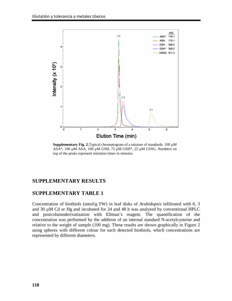
Glutatión y tolerancia a metales tóxicos
118
Supplementary Fig. 2.Typical chromatogram of a mixture of standards: 100 µM ASA*, 100 µM ASA, 100 µM GSH, 75 µM GSH*, 25 µM GSSG. Numbers on top of the peaks represent retention times in minutes.
SUPPLEMENTARY RESULTS SUPPLEMENTARY TABLE 1 Concentration of biothiols (nmol/g FW) in leaf disks of Arabidopsis infiltrated with 0, 3 and 30 µM Cd or Hg and incubated for 24 and 48 h was analyzed by conventional HPLC and postcolumnderivatization with Ellman’s reagent. The quantification of the concentration was performed by the addition of an internal standard N-acetylcysteine and relative to the weight of sample (100 mg). These results are shown graphically in Figure 2 using spheres with different colour for each detected biothiols, which concentrations are represented by different diameters.
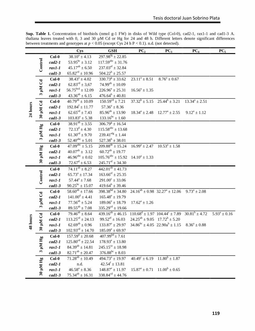
Tesis doctoral Juan Sobrino Plata
119
Sup. Table 1. Concentration of biothiols (nmol g-1 FW) in disks of Wild type (Col-0), cad2-1, rax1-1 and cad1-3 A. thaliana leaves treated with 0, 3 and 30 µM Cd or Hg for 24 and 48 h. Different letters denote significant differences between treatments and genotypes at p < 0.05 (except Cys 24 h P < 0.1). n.d. (not detected).
Cys GSH PC2 PC3 PC4 PC5
24 h
ours
Con
trol
Col-0 38.10a ± 4.13 297.98fg ± 22.85 cad2-1 53.95bc ± 3.12 117.59abc ± 31.76 rax1-1 45.17ab ± 6.50 237.03ef ± 32.84 cad1-3 65.82cd ± 10.96 504.22h ± 25.57
3 µM
Cd Col-0 38.43a ± 4.02 330.73g ± 33.62 23.11a ± 8.51 8.76a ± 0.67
cad2-1 62.83cd ± 3,67 74.99ab ± 10.09 rax1-1 56.75bcd ± 12.09 226.96e ± 25.31 16.56a ± 1.35 cad1-3 43.36ab ± 6.15 476.64h ± 40.81
30 µ
M C
d Col-0 40.79ab ± 10.09 150.59cd ± 7.21 37.32b ± 5.15 25.44b ± 3.21 13.34a ± 2.51 cad2-1 192.84f ± 11.77 57.36a ± 8.36 rax1-1 62.65cd ± 7.43 85.96ab ± 13.90 18.34a ± 2.48 12.77a ± 2.55 9.12a ± 1.12 cad1-3 103.83e ± 5.38 133.16bc ± 1.60
3 µM
Hg Col-0 38.91ab ± 3.55 306.79g ± 16.54
cad2-1 72.13d ± 4.30 115.58abc ± 13.68 rax1-1 61.30cd ± 9.70 239.41efg ± 1.44 cad1-3 52.40abc ± 5.01 527.38h ± 38.01
30 µ
M H
g Col-0 47.09abc ± 5.15 209.88de ± 15.24 16.99a ± 2.47 10.53a ± 1.58 cad2-1 40.07ab ± 3.12 60.72ab ± 19.77 rax1-1 46.96abc ± 0.02 105.76abc ± 15.92 14.10a ± 1.33 cad1-3 72.67d ± 6.53 245.71ef ± 34.30
48 h
ours
Con
trol
Col-0 74.11ab ± 8.27 442.01ab ± 41.73 cad2-1 65.73a ± 17.34 163.66ef ± 25.35 rax1-1 57.44a ± 7.68 291.00c ± 33.06 cad1-3 90.25bc ± 15.07 419.64b ± 39.46
3 µM
Cd Col-0 58.60ab ± 17.66 398.38bd ± 34.80 24.16ab ± 0.98 32.27a ± 12.06 9.73a ± 2.08
cad2-1 141.00d ± 4.41 165.48e ± 19.79 rax1-1 77.56ab ± 5.24 189.06e ± 18.79 17.62a ± 1.26 cad1-3 89.55ab ± 7.08 335.29cd ± 19.66
30 µ
M C
d Col-0 79.46ab ± 8.64 439.16ab ± 46.15 110.68d ± 1.97 104.44c ± 7.89 30.81b ± 4.72 5.93a ± 0.16 cad2-1 113.25cd ± 24.13 99.52ef ± 16.03 24.25ab ± 9.05 17.72b ± 5.20 rax1-1 62.69ab ± 0.96 133.87e ± 29.97 34.86bc ± 4.05 22.90ab ± 1.15 8.36a ± 0.88 cad1-3 102.93cd ± 14.70 185.09e ± 69.97
3 µM
Hg Col-0 157.59d ± 20.68 407.99bd ± 7.61
cad2-1 125.80cd ± 22.54 178.93e ± 13.80 rax1-1 84.39ab ± 14.81 245.15ce ± 18.98 cad1-3 82.71ab ± 20.47 376.88bc ± 8.03
30 µ
M H
g Col-0 71.28ab ± 10.49 494.73a ± 19.97 40.49c ± 6.19 11.80b ± 1.87 cad2-1 n.d. 42.54f ± 13.81 rax1-1 46.58a ± 8.36 148.87e ± 11.97 15.87a ± 0.71 11.00b ± 0.65 cad1-3 75.34ab ± 16.31 338.84cd ± 44.76

Glutatión y tolerancia a metales tóxicos
120
CAPÍTULO 5 Glutathione is a key antioxidant metabolite to cope with mercury and cadmium stress

Tesis doctoral Juan Sobrino Plata
121
Capítulo 5:
Glutathione is a key antioxidant metabolite to cope with mercury and cadmium stress
Juan Sobrino-Plata1,2, Dirk Meyssen3, Ann Cuypers3, Carolina Escobar2, Luis E. Hernández1*
1Laboratory of Plant Physiology, Department of Biology, Universidad Autónoma de Madrid, Cantoblanco, ES-28049 Madrid. 2Departamento de Ciencias del Medioambiente, Universidad de Castilla la Mancha, Campus Fábrica de Armas, ES-45070 Toledo, Spain. 3Centre for Environmental Sciences, Universiteit Hasselt, BE-3590 Diepenbeek, Belgium.
*Correspondence: Luis E. Hernández, e-mail: [email protected]
Plant and Soil (2014) DOI: 10.1007/s11104-013-2006-4
Abstract
Background Glutathione (GSH) plays a dual role under heavy metal stress, as antioxidant
metabolite and as precursor of phytochelatins (PCs). Studying the responses of the GSH
metabolism to heavy metals is important to improve tolerance.
Methods We studied the oxidative stress signature of three γ-glutamylcysteine synthetase
(γECS) Arabidopsis thaliana allele mutants (rax1-1, cad2-1, and pad2-1), first enzymatic
step in the GSH synthetic pathway, when treated with 10 µM Cd or Hg for 72 h.
Results GSH concentration was lower in the mutants (45% rax1-1; 30% cad2-1; and 20%
pad2-1), which was also associated with inferior translocation of Cd or Hg to shoots, than
in wild type Col-0. Glutathione reductase (GR) and NADPH-oxidase activities were
inhibited in roots, phytotoxic effects consistently more pronounced in the mutants,
particularly in pad2-1. Non-photochemical quenching augmented with exposure time to Cd
or Hg in Col-0, but not so in the γECS mutants. Mercury caused severe damage in cad2-1
and pad2-1 root proteins profile; toxic effects confirmed by GR and H+-ATPase
immunodetection. PCs appeared in Col-0 roots under metal stress, and surprisingly
accumulated in rax1-1. γECS immunodetection revealed its overexpression in rax1-1.
Conclusion A minimum amount of GSH may be required for adequate metal tolerance,
where γECS expression could compensate GSH deficiency under stress.
Glutatión y tolerancia a metales tóxicos
122
Key words: Arabidopsis thaliana, biothiols, cadmium, γ-glutamylcysteine synthetase, glutathione, mercury;
phytochelatins
Introduction
Most elements classified as “heavy metals” are essential for plants, like Fe, Cu, Zn or Mn,
but few like Cd or Hg are toxic even at very low concentrations (Weast 1984; Gallego et al.
2012). Mercury and Cd are considered as global environmental threats due to their
bioaccumulation and biomagnification in diverse ecosystems (Dietz et al. 2000). In particular,
there is a concern about the fate of Hg in the mine facilities of Almadén (Spain) area that
has the largest levels of environmental Hg in the world (Berzas et al. 2003).
Phytoremediation is envisaged as an environmentally friendly and cost efficient strategy to
cope with this kind of environmental pollution, which requires selection of tolerant plants
(Wang et al. 2012). In this sense, inorganic Hg and Cd can cause cellular damage due to their
strong affinity for sulfhydryl residues, which finally results in metabolism disruption (Van
Assche and Clijsters 1990; Hall 2002). Mercury has also been found to be a potent inducer of
oxidative stress (Cho and Park 2000; Rellán-Álvarez et al. 2006; Sobrino-Plata et al. 2009),
while Cd also causes oxidative stress damage but at prolonged exposures and higher
concentrations (Ortega-Villasante et al. 2005, 2007).
Oxygenic photosynthesis causes a hyperoxic environment in photosynthetic tissues of the
plants, which entails adaptations in metabolism to avoid dangerous oxidative processes in
the plant cells. These oxidative damages are usually produced by an imbalance between
accumulation of reactive oxygen species (ROS), and the protective action of the cellular
antioxidant system (Foyer and Noctor 2011). ROS cause oxidation of proteins and
polyunsaturated fatty acids in membranes, depolymerization of nucleic acids, and breakage
of peptide bonds (Halliwell 2006). In addition, ROS participate in signaling cascades in
defense responses, cell cycle, apoptosis or lignification of cell walls (Mittler et al. 2004).
Therefore it is necessary to maintain a certain level of ROS for adequate cell metabolism.
This is attained with a complex antioxidant homeostatic system, composed by metabolites
like ascorbate (ASC) and glutathione (GSH), between others, and antioxidant enzymes that
scavenge ROS like superoxide dismutase (SOD), ascorbate peroxidases (APX), catalases
(CAT) or glutathione reductases (GR; Foyer and Noctor 2011). However, pathogenic
Tesis doctoral Juan Sobrino Plata
123
interactions and harmful environmental conditions might imbalance the redox status,
causing the appearance of oxidative stress and ROS accumulation, as occurs under metal
stress (Seth et al. 2012).
Besides the antioxidant role of GSH, this tripeptide is also important in the detoxification of
heavy metals through the synthesis of phytochelatins (PCs; Jozefczak et al. 2012). PCs form a
family of peptides with varying repetitions of the γ-Glu-Cys motif followed by a terminal
Gly (γ-Glu-Cys)n-Gly), as identified in a wide variety of plant species (Cobbett and
Goldsbrough 2002). PCs are synthesized from GSH by the enzyme phytochelatin synthase,
and it is thought that PCs-metal complexes are transported to the vacuole in the final step of
metal detoxification (Park et al. 2012).
The first step in GSH synthesis comprises the formation of a peptide bond between the
cysteine amino and the glutamate γ-carboxyl residues, reaction catalyzed by the enzyme γ-
glutamylcysteine synthetase (γECS). This reaction is followed by the condensation of Gly
to γEC by the enzyme glutathione synthetase (GS) to produce GSH. Several experiments
using buthionine sulfoximine, a potent inhibitor of γECS, have shown that depletion of
GSH in plant cells enhanced sensitivity to metal stress (Howden et al. 1995). On the other
hand, allelic mutations of γECS of Arabidopsis thaliana have been described as extremely
sensitive to several harmful environmental conditions: cadmium-sensitive 2-1 (cad2-1) to
heavy metals (Cobbett et al. 1998); root-meristemless 1 (rml1-1) suffers severe alteration of
plant development (Vernoux et al. 2000); the regulator of APX2 1-1 mutant (rax1-1) under
high irradiance stress (Ball et al. 2004); and the phytoalexin-deficient 2-1 mutant (pad2-1)
to pathogenic interactions (Parisy et al. 2006).
The aim of this work was to study the role of GSH metabolism in Hg and Cd tolerance. Thus,
we used plants of Arabidopsis thaliana Columbia 0 (Col-0) and several γECS mutant alleles
(cad2-1, pad2-1 and rax1-1) that contained different levels of GSH. It is expected that subtle
changes in GSH concentration may result in altered responses of the antioxidant defenses of
Arabidopsis to these toxic metals.
Glutatión y tolerancia a metales tóxicos
124
Materials and methods
Plant material, growth conditions and treatments
Seeds of Arabidopsis thaliana wild type (Col-0), cad2-1, pad2-1 and rax1-1 mutant
genotypes were surface-sterilized by agitation in a 15% (v/v) NaClO solution for 10 min,
followed by several rinses in distilled water. Then seeds were germinated and grown in
Araponics® hydroponic systems (Araponics SA, Liège, Belgium) in seedholders filled with
0.6 % phytoagar. The seeds were grown for 4 weeks in short-days light regime (8 h light and
16 h darkness) at 25/18ºC (day/night), with a modified Hoagland nutrient solution (Tocquin
et al. 2006), after this period either HgCl2 or CdCl2 was added to the nutrient solution (0
and 10 μM) for 72 h. At harvest, shoots and roots were collected separately and stored at -
80 ºC until analysis (Supp. Fig. 1).
Chlorophyll fluorescence measurements
Chlorophyll (Chl) fluorescence was measured at different light regimes to determine
ΦPSII, qP and NPQ, according to Maxwell and Johnson (2000). Throughout the
fluorescence measurements, an actinic light of 400 μmol m-2 s1 and saturating pulses light
of 8000 μmol m-2 s1 were used sequentially to determine Fo, Fm, Fm’, Ft and Fo
’ fluorescence
data in a FMS-2 Pulse Modulated Fluorimeter (Hansatech Instruments, Norfolk, UK).
Photosynthesis parameters were determined in plants treated with metals for 24, 48 and 72
h.
Cadmium and mercury analyses by ICP-MS
Plant material was dried at 50 ºC for 72 h and ground with a mortar and pestle. 100 mg of
dried material were acid digested with 1 mL of a digestion mixture (HNO3:H2O2:H2O,
0.6:0.4:1 v:v) in chromatographic vials (4 mL capacity) sealed with polytetrafluoroethylene
stoppers and autoclaved (Presoclave-75 Selecta, Barcelona, Spain) at 120ºC and 1.5 atm for
30 min (Ortega-Villasante et al. 2007). The digests were filtered through a polyvinylidene
difluoride filter and diluted in miliRO water to 6 mL. Cd and Hg concentration was
determined by ICP-MS NexION 300 (Perkin-Elmer Sciex, San Jose, CA, USA).
GR and NADPH-oxidase in gel activity
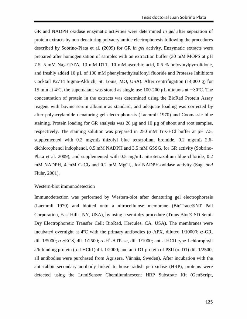
Tesis doctoral Juan Sobrino Plata
125
GR and NADPH oxidase enzymatic activities were determined in gel after separation of
protein extracts by non-denaturing polyacrylamide electrophoresis following the procedures
described by Sobrino-Plata et al. (2009) for GR in gel activity. Enzymatic extracts were
prepared after homogenisation of samples with an extraction buffer (30 mM MOPS at pH
7.5, 5 mM Na2-EDTA, 10 mM DTT, 10 mM ascorbic acid, 0.6 % polyvinylpyrrolidone,
and freshly added 10 µL of 100 mM phenylmethylsulfonyl fluoride and Protease Inhibitors
Cocktail P2714 Sigma-Aldrich; St. Louis, MO, USA). After centrifugation (14,000 g) for
15 min at 4ºC, the supernatant was stored as single use 100-200 µL aliquots at ─80ºC. The
concentration of protein in the extracts was determined using the BioRad Protein Assay
reagent with bovine serum albumin as standard, and adequate loading was corrected by
after polyacrylamide denaturing gel electrophoresis (Laemmli 1970) and Coomassie blue
staining. Protein loading for GR analysis was 20 µg and 10 µg of shoot and root samples,
respectively. The staining solution was prepared in 250 mM Tris-HCl buffer at pH 7.5,
supplemented with 0.2 mg/mL thizolyl blue tetrazolium bromide, 0.2 mg/mL 2,6-
dichlorophenol indophenol, 0.5 mM NADPH and 3.5 mM GSSG, for GR activity (Sobrino-
Plata et al. 2009); and supplemented with 0.5 mg/mL nitrotetrazolium blue chloride, 0.2
mM NADPH, 4 mM CaCl2 and 0.2 mM MgCl2, for NADPH-oxidase activity (Sagi and
Fluhr, 2001).
Western-blot immunodetection
Immunodetection was performed by Western-blot after denaturing gel electrophoresis
(Laemmli 1970) and blotted onto a nitrocellulose membrane (BioTraceNT Pall
Corporation, East Hills, NY, USA), by using a semi-dry procedure (Trans Blot SD Semi-
Dry Electrophoretic Transfer Cell; BioRad, Hercules, CA, USA). The membranes were
incubated overnight at 4ºC with the primary antibodies (α-APX, diluted 1/10000; α-GR,
dil. 1/5000; α-γECS, dil. 1/2500; α-H+-ATPase, dil. 1/1000; anti-LHCII type I chlorophyll
a/b-binding protein (α-LHCb1) dil. 1/2000; and anti-D1 protein of PSII (α-D1) dil. 1/2500;
all antibodies were purchased from Agrisera, Vännäs, Sweden). After incubation with the
anti-rabbit secondary antibody linked to horse radish peroxidase (HRP), proteins were
detected using the LumiSensor Chemiluminescent HRP Substrate Kit (GenScript,
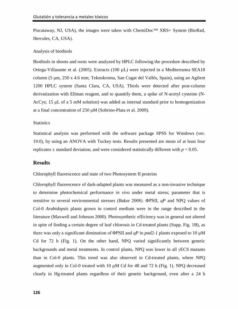
Glutatión y tolerancia a metales tóxicos
126
Piscataway, NJ, USA), the images were taken with ChemiDoc™ XRS+ System (BioRad,
Hercules, CA, USA).
Analysis of biothiols
Biothiols in shoots and roots were analyzed by HPLC following the procedure described by
Ortega-Villasante et al. (2005). Extracts (100 µL) were injected in a Mediterranea SEA18
column (5 μm, 250 x 4.6 mm; Teknokroma, San Cugat del Vallés, Spain), using an Agilent
1200 HPLC system (Santa Clara, CA, USA). Thiols were detected after post-column
derivatization with Ellman reagent, and to quantify them, a spike of N-acetyl cysteine (N-
AcCys; 15 μL of a 5 mM solution) was added as internal standard prior to homogenization
at a final concentration of 250 μM (Sobrino-Plata et al. 2009).
Statistics
Statistical analysis was performed with the software package SPSS for Windows (ver.
19.0), by using an ANOVA with Tuckey tests. Results presented are mean of at least four
replicates ± standard deviation, and were considered statistically different with p < 0.05.
Results
Chlorophyll fluorescence and state of two Photosystem II proteins
Chlorophyll fluorescence of dark-adapted plants was measured as a non-invasive technique
to determine photochemical performance in vivo under metal stress; parameter that is
sensitive to several environmental stresses (Baker 2008). ΦPSII, qP and NPQ values of
Col-0 Arabidopsis plants grown in control medium were in the range described in the
literature (Maxwell and Johnson 2000). Photosynthetic efficiency was in general not altered
in spite of finding a certain degree of leaf chlorosis in Cd-treated plants (Supp. Fig. 1B), as
there was only a significant diminution of ΦPSII and qP in pad2-1 plants exposed to 10 µM
Cd for 72 h (Fig. 1). On the other hand, NPQ varied significantly between genetic
backgrounds and metal treatments. In control plants, NPQ was lower in all γECS mutants
than in Col-0 plants. This trend was also observed in Cd-treated plants, where NPQ
augmented only in Col-0 treated with 10 µM Cd for 48 and 72 h (Fig. 1). NPQ decreased
clearly in Hg-treated plants regardless of their genetic background, even after a 24 h
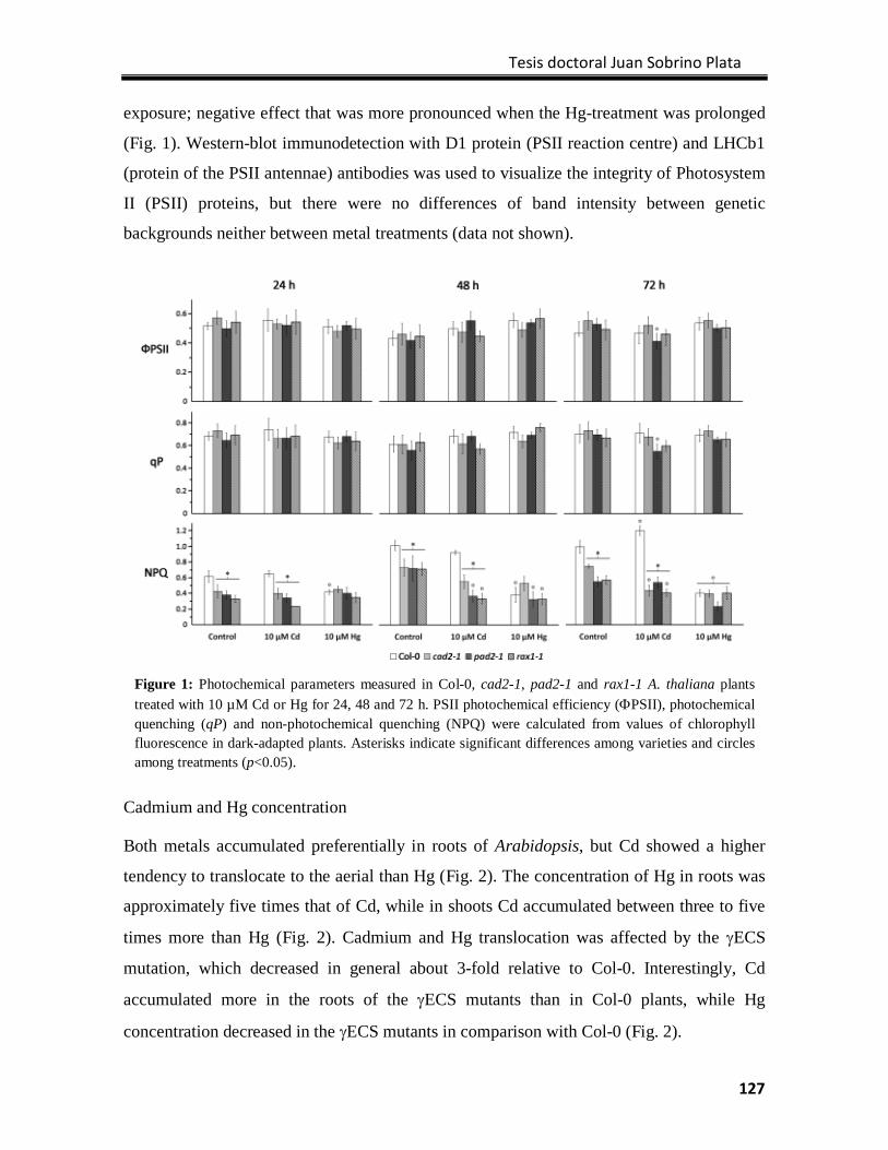
Tesis doctoral Juan Sobrino Plata
127
exposure; negative effect that was more pronounced when the Hg-treatment was prolonged
(Fig. 1). Western-blot immunodetection with D1 protein (PSII reaction centre) and LHCb1
(protein of the PSII antennae) antibodies was used to visualize the integrity of Photosystem
II (PSII) proteins, but there were no differences of band intensity between genetic
backgrounds neither between metal treatments (data not shown).
Cadmium and Hg concentration
Both metals accumulated preferentially in roots of Arabidopsis, but Cd showed a higher
tendency to translocate to the aerial than Hg (Fig. 2). The concentration of Hg in roots was
approximately five times that of Cd, while in shoots Cd accumulated between three to five
times more than Hg (Fig. 2). Cadmium and Hg translocation was affected by the γECS
mutation, which decreased in general about 3-fold relative to Col-0. Interestingly, Cd
accumulated more in the roots of the γECS mutants than in Col-0 plants, while Hg
concentration decreased in the γECS mutants in comparison with Col-0 (Fig. 2).
Figure 1: Photochemical parameters measured in Col-0, cad2-1, pad2-1 and rax1-1 A. thaliana plants treated with 10 µM Cd or Hg for 24, 48 and 72 h. PSII photochemical efficiency (ΦPSII), photochemical quenching (qP) and non-photochemical quenching (NPQ) were calculated from values of chlorophyll fluorescence in dark-adapted plants. Asterisks indicate significant differences among varieties and circles among treatments (p<0.05).
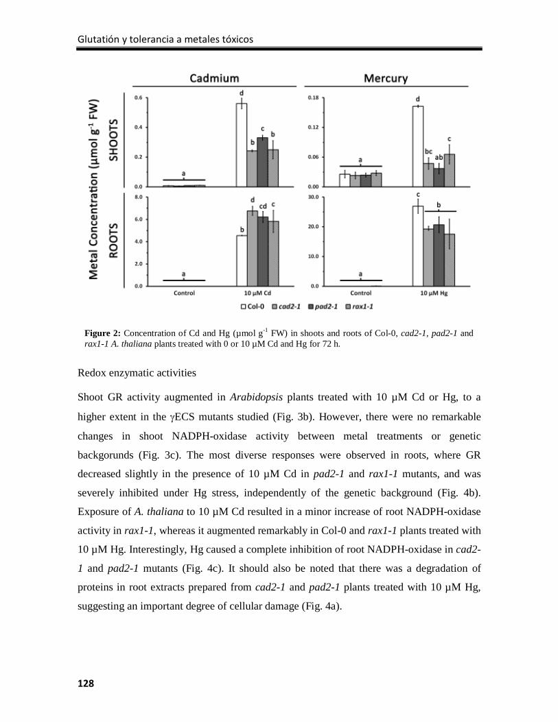
Glutatión y tolerancia a metales tóxicos
128
Redox enzymatic activities
Shoot GR activity augmented in Arabidopsis plants treated with 10 µM Cd or Hg, to a
higher extent in the γECS mutants studied (Fig. 3b). However, there were no remarkable
changes in shoot NADPH-oxidase activity between metal treatments or genetic
backgorunds (Fig. 3c). The most diverse responses were observed in roots, where GR
decreased slightly in the presence of 10 µM Cd in pad2-1 and rax1-1 mutants, and was
severely inhibited under Hg stress, independently of the genetic background (Fig. 4b).
Exposure of A. thaliana to 10 µM Cd resulted in a minor increase of root NADPH-oxidase
activity in rax1-1, whereas it augmented remarkably in Col-0 and rax1-1 plants treated with
10 µM Hg. Interestingly, Hg caused a complete inhibition of root NADPH-oxidase in cad2-
1 and pad2-1 mutants (Fig. 4c). It should also be noted that there was a degradation of
proteins in root extracts prepared from cad2-1 and pad2-1 plants treated with 10 µM Hg,
suggesting an important degree of cellular damage (Fig. 4a).
Figure 2: Concentration of Cd and Hg (µmol g-1 FW) in shoots and roots of Col-0, cad2-1, pad2-1 and rax1-1 A. thaliana plants treated with 0 or 10 µM Cd and Hg for 72 h.
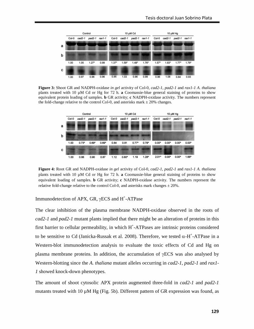
Tesis doctoral Juan Sobrino Plata
129
Immunodetection of APX, GR, γECS and H+-ATPase
The clear inhibition of the plasma membrane NADPH-oxidase observed in the roots of
cad2-1 and pad2-1 mutant plants implied that there might be an alteration of proteins in this
first barrier to cellular permeability, in which H+-ATPases are intrinsic proteins considered
to be sensitive to Cd (Janicka-Russak et al. 2008). Therefore, we tested α-H+-ATPase in a
Western-blot immunodetection analysis to evaluate the toxic effects of Cd and Hg on
plasma membrane proteins. In addition, the accumulation of γECS was also analysed by
Western-blotting since the A. thaliana mutant alleles occurring in cad2-1, pad2-1 and rax1-
1 showed knock-down phenotypes.
The amount of shoot cytosolic APX protein augmented three-fold in cad2-1 and pad2-1
mutants treated with 10 µM Hg (Fig. 5b). Different pattern of GR expression was found, as
Figure 3: Shoot GR and NADPH-oxidase in gel activity of Col-0, cad2-1, pad2-1 and rax1-1 A. thaliana plants treated with 10 µM Cd or Hg for 72 h. a Coomassie-blue general staining of proteins to show equivalent protein loading of samples. b GR activity; c NADPH-oxidase activity. The numbers represent the fold-change relative to the control Col-0, and asterisks mark ± 20% changes.
Figure 4: Root GR and NADPH-oxidase in gel activity of Col-0, cad2-1, pad2-1 and rax1-1 A. thaliana plants treated with 10 µM Cd or Hg for 72 h. a Coomassie-blue general staining of proteins to show equivalent loading of samples. b GR activity; c NADPH-oxidase activity. The numbers represent the relative fold-change relative to the control Col-0, and asterisks mark changes ± 20%.
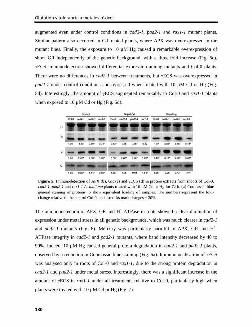
Glutatión y tolerancia a metales tóxicos
130
augmented even under control conditions in cad2-1, pad2-1 and rax1-1 mutant plants.
Similar pattern also occurred in Cd-treated plants, where APX was overexpressed in the
mutant lines. Finally, the exposure to 10 µM Hg caused a remarkable overexpression of
shoot GR independently of the genetic background, with a three-fold increase (Fig. 5c).
γECS immunodetection showed differential expression among mutants and Col-0 plants.
There were no differences in cad2-1 between treatments, but γECS was overexpressed in
pad2-1 under control conditions and repressed when treated with 10 µM Cd or Hg (Fig.
5d). Interestingly, the amount of γECS augmented remarkably in Col-0 and rax1-1 plants
when exposed to 10 µM Cd or Hg (Fig. 5d).
The immunodetection of APX, GR and H+-ATPase in roots showed a clear diminution of
expression under metal stress in all genetic backgrounds, which was much clearer in cad2-1
and pad2-1 mutants (Fig. 6). Mercury was particularly harmful to APX, GR and H+-
ATPase integrity in cad2-1 and pad2-1 mutants, where band intensity decreased by 40 to
90%. Indeed, 10 µM Hg caused general protein degradation in cad2-1 and pad2-1 plants,
observed by a reduction in Coomassie blue staining (Fig. 6a). Immunolocalisation of γECS
was analysed only in roots of Col-0 and rax1-1, due to the strong protein degradation in
cad2-1 and pad2-1 under metal stress. Interestingly, there was a significant increase in the
amount of γECS in rax1-1 under all treatments relative to Col-0, particularly high when
plants were treated with 10 µM Cd or Hg (Fig. 7).
Figure 5: Immunodetection of APX (b), GR (c) and γECS (d) in protein extracts from shoots of Col-0, cad2-1, pad2-1 and rax1-1 A. thaliana plants treated with 10 µM Cd or Hg for 72 h. (a) Coomassie-blue general staining of proteins to show equivalent loading of samples. The numbers represent the fold-change relative to the control Col-0, and asterisks mark changes ± 20%.
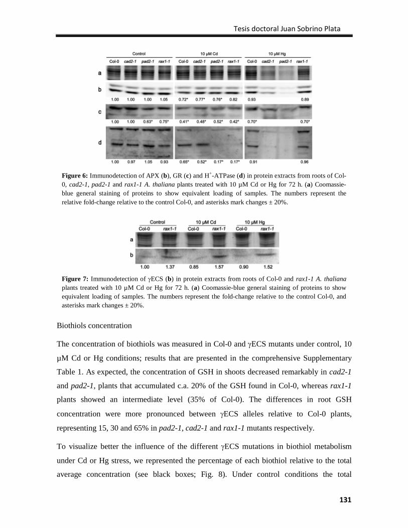
Tesis doctoral Juan Sobrino Plata
131
Biothiols concentration
The concentration of biothiols was measured in Col-0 and γECS mutants under control, 10
µM Cd or Hg conditions; results that are presented in the comprehensive Supplementary
Table 1. As expected, the concentration of GSH in shoots decreased remarkably in cad2-1
and pad2-1, plants that accumulated c.a. 20% of the GSH found in Col-0, whereas rax1-1
plants showed an intermediate level (35% of Col-0). The differences in root GSH
concentration were more pronounced between γECS alleles relative to Col-0 plants,
representing 15, 30 and 65% in pad2-1, cad2-1 and rax1-1 mutants respectively.
To visualize better the influence of the different γECS mutations in biothiol metabolism
under Cd or Hg stress, we represented the percentage of each biothiol relative to the total
average concentration (see black boxes; Fig. 8). Under control conditions the total
Figure 6: Immunodetection of APX (b), GR (c) and H+-ATPase (d) in protein extracts from roots of Col-0, cad2-1, pad2-1 and rax1-1 A. thaliana plants treated with 10 µM Cd or Hg for 72 h. (a) Coomassie-blue general staining of proteins to show equivalent loading of samples. The numbers represent the relative fold-change relative to the control Col-0, and asterisks mark changes ± 20%.
Figure 7: Immunodetection of γECS (b) in protein extracts from roots of Col-0 and rax1-1 A. thaliana plants treated with 10 µM Cd or Hg for 72 h. (a) Coomassie-blue general staining of proteins to show equivalent loading of samples. The numbers represent the fold-change relative to the control Col-0, and asterisks mark changes ± 20%.
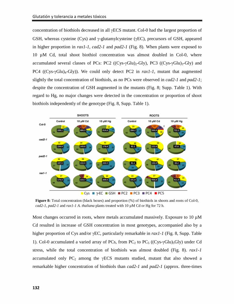
Glutatión y tolerancia a metales tóxicos
132
concentration of biothiols decreased in all γECS mutant. Col-0 had the largest proportion of
GSH, whereas cysteine (Cys) and γ-glutamylcysteine (γEC), precursors of GSH, appeared
in higher proportion in rax1-1, cad2-1 and pad2-1 (Fig. 8). When plants were exposed to
10 µM Cd, total shoot biothiol concentration was almost doubled in Col-0, where
accumulated several classes of PCs: PC2 ((Cys-γGlu)2-Gly), PC3 ((Cys-γGlu)3-Gly) and
PC4 ((Cys-γGlu)4-Gly)). We could only detect PC2 in rax1-1, mutant that augmented
slightly the total concentration of biothiols, as no PCs were observed in cad2-1 and pad2-1;
despite the concentration of GSH augmented in the mutants (Fig. 8; Supp. Table 1). With
regard to Hg, no major changes were detected in the concentration or proportion of shoot
biothiols independently of the genotype (Fig. 8, Supp. Table 1).
Most changes occurred in roots, where metals accumulated massively. Exposure to 10 µM
Cd resulted in increase of GSH concentration in most genotypes, accompanied also by a
higher proportion of Cys and/or γEC, particularly remarkable in rax1-1 (Fig. 8, Supp. Table
1). Col-0 accumulated a varied array of PCs, from PC2 to PC5 ((Cys-γGlu)5Gly) under Cd
stress, while the total concentration of biothiols was almost doubled (Fig. 8). rax1-1
accumulated only PC2 among the γECS mutants studied, mutant that also showed a
remarkable higher concentration of biothiols than cad2-1 and pad2-1 (approx. three-times
Figure 8: Total concentration (black boxes) and proportion (%) of biothiols in shoots and roots of Col-0, cad2-1, pad2-1 and rax1-1 A. thaliana plants treated with 10 µM Cd or Hg for 72 h.
Tesis doctoral Juan Sobrino Plata
133
control concentration, Fig. 8). Treatment with 10 µM Hg caused similar responses than Cd
in the biothiols concentration and profile (i.e. increase in biothiol concentration and
synthesis of PCs), being only rax1-1 the genotype with a behavior closer to that of Col-0
roots (Fig. 8). Interestingly, the concentration of biothiols in cad2-1 and pad2-1 decreased
abruptly under Hg stress, possibly as a symptom of extensive cellular damage (Fig. 8).
Discussion
Photosynthetic efficiency (ΦPSII) and photochemical quenching (qP) only decreased in
pad2-1 plants treated with Cd during 72 h, whereas Hg had little effect in these parameters.
ΦPSII and qP decreased in plants subjected normally to long treatments (more than two
weeks) where substantial Cd accumulation occurred in shoots (doses up to 100 µM), toxic
effects that were accompanied frequently by leaf chlorosis in rice, spinach, wheat or cork
oak plants (Pagliano et al. 2006; Fagioni et al. 2009; Janik et al. 2010; Gogorcena et al.
2011). Similar responses were found in Cd-treated Thlaspi caerulescens that suffered PSII
reaction center inhibition, possibly caused by the substitution of important metal ions by
Cd2+, such as Fe2+, in prosthetic groups of proteins (Küpper et al. 2007). Indeed, Solti et al.
(2008) found that extra supply of Fe prevented the toxic effects on photosynthesis caused
by Cd in Poplar. Recently, mutants lacking metal transport to vacuoles were particularly
sensitive to Cd, implying that toxic elements can interfere with photochemical processes in
chloroplasts (Molins et al. 2013).
Non-photochemical quenching (NPQ) augmented significantly in Col-0 Arabidopsis after
72 h of exposure to 10 µM Cd. Similar short-term NPQ behavior was observed in
Phragmites australis plants, treated with 50 µM Cd during 72 h (Pietrini et al. 2003). It is
feasible that under moderate stress quenching of excess excitons in photosynthetic antenna
via NPQ heat dissipation prevents damages to photosystems (Niyogi et al. 1998). The
violaxanthin to zeaxanthin de-epoxidation cycle is an important feature of NPQ under
environmental stress, acting ascorbate as redox metabolite (Müller et al. 2001). In turn, the
chloroplastic pool of ascorbate is maintained by GSH through the dehydroascorbate
reductase enzyme (Foyer and Noctor 2011), implying that an adequate GSH amount is
required to maintain optimal photosynthetic processes and protection against oxidative
imbalances produced by metals (Bertrand and Poirier 2005; Foyer and Shigeoka 2011).
Glutatión y tolerancia a metales tóxicos
134
Interestingly, NPQ values were significantly lower in all γECS mutant alleles studied under
control conditions, in agreement with the hypothesis that GSH mediates the regulation of
photosynthetic processes (Schürmann and Jacquot 2000). The differences in NPQ between
Col-0 and γECS mutants were even bigger in plants treated with 10 µM Cd for 72 h (Fig.
1), highlighting the importance of the pool of GSH under Cd stress to prevent excess of
stored energy in the thylakoids. On the other hand, Hg caused minor changes in NPQ
independently of the genetic background. This poor toxic effect could be explained by the
extreme accumulation of Hg in roots (Fig.2), toxic metal less prone to mobilize to the
leaves and cause negative effects on photochemical reactions during the short-term
experiment performed in our study.
The strongest toxic effects of metals in roots were detected in Hg-treated plants, which
exhibited reduced biomass, dehydration and brownish roots, typically associated with
oxidative stress (Supp. Fig. 1C). The proteins extracted from the roots of Hg-treated Col-0
and rax1-1 Arabidopsis plants were degraded partially, and were almost completely
degraded in cad2-1 and pad2-1. NADPH-oxidase activity was higher in roots of Hg-treated
Col-0 and rax1-1 plants; plasma membrane intrinsic protein that releases H2O2 and
responds to different biotic and abiotic stresses (Mithöfer et al. 2004). Ortega-Villasante et
al. (2007) observed a quick accumulation of H2O2 under Hg stress in alfalfa roots, partially
caused by NADPH-oxidases, which could account for the oxidative stress found in roots of
different plants treated with Hg (Cho and Park 2000; Cargnelutti et al. 2006; Zhou et al.
2008; Lomonte et al. 2010). On the other hand, the strong inhibition of NADPH-oxidase
activity and disappearance of H+-ATPase in cad2-1 and pad2-1 mutants could reflect
severe cellular damage, with massive alteration of membranes and protein denaturation;
characteristic symptoms of poisoning and cell death under high doses of Hg (Ortega-
Villasante et al. 2007).
The induction of oxidative stress occurs in parallel to changes in antioxidant enzymes, in
particular alterations in the activity of GR regardless of the Arabidopsis γECS genetic
background. Similarly, Cd and Hg caused the induction of GR activity in shoots in pea
(Romero-Puertas et al. 2006), wheat (Yannarelli et al. 2007) and alfalfa plants (Sobrino-
Plata et al. 2009). In roots there were little changes in activity under Cd-stress, but it was
Tesis doctoral Juan Sobrino Plata
135
completely inhibited in Hg-treated Arabidopsis plants. It has been shown recently that GR
is very sensitive to Hg even at low doses (3 µM), as observed in alfalfa (Sobrino-Plata et al.
2009) and Silene vulgaris plants (Sobrino-Plata et al. 2013); enzymatic activity that can be
used as a specific index of Hg accumulation.
APX and GR immunodetection in shoot extracts revealed that Cd and Hg caused the
accumulation of these proteins, in particular in the mutants with less GSH level, cad2-1 and
pad2-1. On the contrary, γECS was overexpressed in Col-0 and rax1-1 plants, both in
shoots and in roots. Interestingly, the concentration of GSH augmented remarkably in the
roots of rax1-1 plants exposed to Cd and Hg, while in cad2-1 and pad2-1 there was a
severe diminution. It is well known that GSH synthesis is located in plastids, mainly in
shoots (Noctor et al. 2011). Under metal stress there is an efficient long-transport of
biothiols (γEC, GSH, or even PCs) from shoots to roots, important feature for metal
tolerance (Li et al. 2006). It is feasible that the unexpected high level of GSH detected in
roots of Cd and Hg-treated Col-0 and rax1-1 plants, and the subsequent production of PCs,
could be a consequence of the augmented expression of γECS in shoots and/or enhanced
translocation to roots.
With regard to biothiols concentration, there were significant differences between γECS
alleles under metal stress. There was a higher concentration GSH in shoots and roots of
Col-0 and rax1-1 than in cad2-1 and pad2-1 when exposed to Cd and Hg, following the
expected pattern of GSH synthesis described in the literature under environmental stress
(Cobbett et al. 1998; Ball et al. 2004; Parisy et al. 2006). γECS mutants, in particular cad2-
1 and pad2-1, had higher proportion of Cys and γEC due to the poorer affinity to Cys of the
mutated γECS enzyme, which resulted in lower levels of GSH (Xiang et al. 2001).
Cadmium caused the synthesis of PCs in Col-0 and rax1-1 plants, but not in cad2-1 and
pad2-1, in agreement with results previously reported with γECS deficient mutants
(Cobbett et al. 1998). However, unexpected results were observed in roots of Hg-treated
rax1-1: there was a strong accumulation of GSH and PCs. γECS plays a central role in GSH
synthesis, an enzyme that is under transcriptional regulation responding to environmental
and developmental cues; in particular the oxidative stress induced by metals is considered
as one of the key stimuli (Rausch et al. 2007). There was indeed an overexpression of γECS
Glutatión y tolerancia a metales tóxicos
136
in Col-0 and rax1-1 clearly observed in Hg treated plants (Fig. 5d), which could permit an
accumulation of GSH required for PCs synthesis. There is an efficient transport of GSH
from cytosol or plastids and vice versa, step in GSH metabolism that is not considered a
barrier for effective dissemination of GSH to different cell compartments (Lim et al. 2013).
It should be noted that rax1-1 plants were much less affected by Hg and Cd than cad2-1
and pad2-1, and showed similar tolerance than Col-0 according to the different
physiological parameters measured; possibly due to sufficient level of GSH and PCs
needed to ameliorate metal toxic effects (Carrasco-Gil et al. 2011). Our results are in
agreement with the work of Parisy et al. (2006) and Dubreuil-Maurizi et al. (2011), who
found that pad2-1 was more sensitive to biotic stress than Col-0, rax1-1 and cad2-1.
Therefore, the maintenance of a certain threshold of GSH cellular pool is required for metal
tolerance.
We found interesting differences in the concentration of Cd and Hg in roots and shoots
between γECS genotypes: Depletion of GSH, and perhaps lower capacity to synthetase
PCs, caused a general diminution of metal translocation to the aerial part of the plants,
particularly more pronounced in the case of Hg (Fig. 2). In this sense, transgenic Indian
mustard plants over-expressing GS (Zhu et al., 1999a) and γECS (Zhu et al., 1999b)
accumulated more Cd and were able to tolerate higher Cd concentrations than un-
transformed plants. Similarly, the concentration of Cd in the leaves of transgenic poplar
over-expressing γECS also augmented when leaf GSH levels were higher than wild type
plants (Arisi et al., 2000). On the other hand, we observed that all γECS Arabidopsis
mutants had higher Cd accumulation in roots than Col-0, particularly cad2-1 and pad2-1,
which is in contradiction with the decrease in root Hg concentration (Fig. 2). This could be
related with the accumulation of Cys detected in these mutants (Fig. 8), since the over-
expression of the enzymes involved in Cys biosynthesis in tobacco (Harada et al. 2001) and
Arabidopsis (Domínguez-Solis et al., 2004) augmented Cd concentration and tolerance.
However, Hg caused severe alterations in root membrane proteins that may regulate uptake
processes, such as NADPH-oxidases or H+-ATPase (Figs. 4 and 6), which could lead to
lesser accumulation of Hg in the roots of Hg-hypersensitive γECS genotypes.
Tesis doctoral Juan Sobrino Plata
137
Conclusions
GSH is a key metabolite in heavy metal tolerance, as found by comparing the responses of
γECS mutants with different GSH concentrations. Mutant plants with lower GSH level,
pad2-1, exhibited the most severe phenotype, in particular when exposed to Hg.
Surprisingly, rax1-1 could respond by accumulating substantial amounts of GSH and PCs
under metal stress, in spite of having between 65 (roots) to 35% (shoots) of the GSH
concentration measured in Col-0. Regulation of γECS appears as a key factor in metal
tolerance, accumulation or translocation, objective of future work that should shed some
light on the mechanisms mediated by biothiols.
Acknowledgments
This work was funded by the Ministry of Economy and Competitivity (PROBIOMET
AGL2010-15151), Fundación Ramón Areces, and Junta Comunidades Castilla-La Mancha
(FITOALMA2, POII10-0087-6458). We thank the comments of two anonymous reviewers
which allowed substantial improvement of the manuscript.
References
Arisi A-C M, Mocquot B, Lagriffoul A, Mench M, Foyer CH, Jouanin L. (2000) Responses
to cadmium in leaves of transformed poplars overexpressing γ-glutamylcysteine
synthetase. Phyiol Plant 109: 143-149.
Baker NR (2008) Chlorophyll fluorescence: A probe of photosynthesis in vivo. Ann Rev
Plant Biol 59: 89-113.
Ball L, Accotto GP, Bechtold U, Creissen G, Funck D, Jiménez A, Kular B, Leyland N,
Mejía-Carranza J, Reynolds H, Karpinski S, Mullineaux PM (2004) Evidence for a
direct link between glutathione biosynthesis and stress defense gene expression in
Arabidopsis. Plant Cell 16: 2448-2462.
Bertrand M, Poirier I (2005) Photosynthetic organisms and excess of metals.
Photosynthetica 43: 345-353.
Berzas JL, Garcia LF, Rodriguez R (2003) Distribution of mercury in the aquatic
environment at Almadén, Spain. Environ Pollut 122: 261-271.
Glutatión y tolerancia a metales tóxicos
138
Cargnelutti D, Tabaldi LA, Spanevello RM, de Oliveira Jucoski G, Battisti V, Redin M,
Linares CE, Dressler VL, de Moraes Flores EM, Nicoloso FT, Morsch VM, Schetinger
MR (2006) Mercury toxicity induces oxidative stress in growing cucumber seedlings.
Chemosphere 65: 999-1006.
Carrasco-Gil S, Álvarez-Fernández A, Sobrino-Plata J, Millán R, Carpena-Ruiz RO, Leduc
D, Andrews JC, Abadía J, Hernández LE (2011) Complexation of Hg with
phytochelatins is important for plant Hg tolerance. Plant Cell Environ 34: 778-791.
Cho UH, Park JO (2000) Mercury-induced oxidative stress in tomato seedlings. Plant Sci
156: 1-9.
Cobbett CS, May MJ, Howden R, Rolls B (1998) The glutathione-deficient, cadmium-
sensitive mutant, cad2-1, of Arabidopsis thaliana is deficient in γ-glutamylcysteine
synthetase. Plant J 16: 73-78.
Cobbet C, Goldsbrough PB (2002) Phytochelatins and metallothioneins: roles in heavy
metal detoxification and homeostasis. Annu Rev Plant Biol 53: 159-182.
Dietz R, Riget F, Cleemann M, Aarkrog A, Johansen P, Hansen JC (2000) Comparison of
contaminants from different trophic levels and ecosystems. Sci Total Environ 245:
221-231.
Domínguez-Solís JR, López-Martín MC, Ager FJ, Ynsa MD, Romero LC, Gotor C (2004)
Increased cysteine availability is essential for cadmium tolerance and accumulation in
Arabidopsis thaliana. Plant Biotechnol J 2: 469-476.
Dubreuil-Maurizi C, Vitecek J, Marty L, Branciard L, Frettinger P, Wendehenne D, Meyer
AJ, Mauch F, Poinssot B (2011) Glutathione deficiency of the Arabidopsis mutant
pad2-1 affects oxidative stress-related events, defense gene expression, and the
hypersensitive response. Plant Physiol 157: 2000-2012.
Fagioni M, D’Amici GM, Timperio AM, Zolla L (2009) Proteomic analysis of multiprotein
complexes in the thylakoid membrane upon cadmium treatment. J Prot Res 8: 310-326.
Foyer CH, Noctor G (2011) Ascorbate and glutathione: The heart of the redox hub. Plant
Physiol 155: 2–18.
Foyer CH, Shigeoka S (2011) Understanding oxidative stress and antioxidant functions to
enhance photosynthesis. Plant Physiol 155: 93-100.
Tesis doctoral Juan Sobrino Plata
139
Gallego SM, Pena LB, Barcia RA, Azpilicueta CE, Iannone MF, Rosales EP, Zawoznik
MS, Groppa MD, Benavides MP (2012) Unravelling cadmium toxicity and tolerance in
plants: Insight into regulatory mechanisms. Environ Exp Bot 83: 33-46.
Gogorcena Y, Larbi A, Andaluz S, Carpena RO, Abadía A, Abadía J (2011) Effects of
cadmium on cork oak (Quercus suber L.) plants grown in hydroponics. Tree
Physiology 31: 1401-1412.
Hall JL (2002) Cellular mechanisms for heavy metal detoxification and tolerance. J Exp
Bot 52: 631-640.
Halliwell B (2006) Reactive species and antioxidants. Redox biology is a fundamental
theme of aerobic life. Plant Physiol 141: 312-322.
Harada E, Choi YE, Tsuchisaka A, Obata H, Sano H (2001) Transgenic tobacco plants
expressing a rice cysteine synthase gene are tolerant to toxic levels of cadmium. J Plant
Physiol 158: 655-661.
Howden R, Goldsbrough PB, Andersen CR, Cobbett CS (1995) Cadmium-sensitive, cad1
mutants of Arabidopsis thaliana are phytochelatin deficient. Plant Physiol 107: 1059-
1066.
Janicka-Russak M, Kabała K, Burzyński M, Kłobus G (2008) Response of plasma
membrane H+-ATPase to heavy metal stress in Cucumis sativus roots. J Exp Bot 59:
3721-3728.
Janik E, Maksymiec W, Mazur R, Garstka M, Gruszecki WI (2010) Structural and
functional modifications of the major light-harvesting complex II in cadmium- or
copper-treated Secale cereale. Plant Cell Physiol 51: 1330-1340.
Jozefczak M, Remans T, Vangronsveld J, Cuypers A (2012) Glutathione is a key player in
metal-induced oxidative stress defences. Int J Mol Sci 13: 3145-3175.
Küpper H, Parameswaran A, Leitenmaier B, Trtílek M, Šetlík I (2007) Cadmium-induced
inhibition of photosynthesis and long-term acclimation to cadmium stress in the
hyperaccumulator Thlaspi caerulescens. New Phytol 175: 655-674.
Laemmli UK (1970) Cleavage of structural proteins during the assembly of the head of
bacteriophage T4. Nature 227: 680-685.
Li Y, Dankher OP, Carreira L, Smith AP, Meagher RB (2006) The shoot-specific
expression of gamma-glutamylcysteine synthetase directs the long-distance transport of
Glutatión y tolerancia a metales tóxicos
140
thiol-peptides to roots conferring tolerance to mercury and arsenic. Plant Physiol 141:
288-298.
Lim B, Pasternak M, Meyer AJ, Cobbett CS (2013) Restricting glutamylcysteine synthetase
activity to the cytosol or glutathione biosynthesis to the plastid is sufficient for normal
plant development and stress tolerance. Plant Biol: May 20. doi: 10.1111/plb.12033.
Lomonte C, Sgherri C, Baker AJM, Kolev SD, Navari-Izzo F (2010) Antioxidative
response of Atriplex codonocarpa to mercury. Environ Exp Bot 69: 9-16.
Maxwell K, Johnson GN (2000) Chlorophyll fluorescence - a practical guide. J Exp Bot
345: 659-668.
Mithöfer A, Schulze B, Boland W (2004) Biotic and heavy metal stress response in plants:
evidence for common signals. FEBS Lett 21; 566(1-3): 1-5.
Mittler R, Vanderauwera S, Gollery M, Van Breusegem F (2004) Reactive oxygen gene
network of plants. Trends Plant Sci 9: 490-498.
Molins H, Michelet L, Lanquar V, Agorio A, Giraudat J, Roach T, Krieger-Liszkay A,
Thomine S (2013) Mutants impaired in vacuolar metal mobilization identify
chloroplasts as a target for cadmium hypersensitivity in Arabidopsis thaliana. Plant
Cell Environ 36: 804-817.
Müller P, Li XP, Niyogi, KK (2001) Non-photochemical quenching. A response to excess
light energy. Plant Physiol 125: 1558–1566.
Niyogi KK, Grossman AR, Björkman O (1998) Arabidopsis mutants define a central role
for the xanthophyll cycle in the regulation of photosynthetic energy conversion. Plant
Cell 10: 1121-1134.
Noctor G, Queval G, Mhamdi A, Chaouch S, Foyer CH (2011) Glutathione. The
Arabidopsis Book, 9. American Society of Plant Biologists, Rockville, M. D., pp. 1–
32.
Ortega-Villasante C, Rellán-Álvarez R, Del Campo FF, Carpena-Ruiz RO, Hernández LE
(2005) Cellular damage induced by cadmium and mercury in Medicago sativa. J Exp
Bot 56: 2239-2251.
Ortega-Villasante C, Hernández L E, Rellán-Álvarez R, del Campo FF, Carpena-Ruiz RO
(2007) Rapid alteration of cellular redox homeostasis upon exposure to cadmium and
mercury in alfalfa seedlings. New Phytol 176: 96−107.
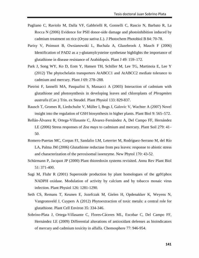
Tesis doctoral Juan Sobrino Plata
141
Pagliano C, Raviolo M, Dalla VF, Gabbrielli R, Gonnelli C, Rascio N, Barbato R, La
Rocca N (2006) Evidence for PSII donor-side damage and photoinhibition induced by
cadmium treatment on rice (Oryza sativa L.). J Photochem Photobiol B 84: 70-78.
Parisy V, Poinssot B, Owsianowski L, Buchala A, Glazebrook J, Mauch F (2006)
Identification of PAD2 as a γ-glutamylcysteine synthetase highlights the importance of
glutathione in disease resistance of Arabidopsis. Plant J 49: 159–172.
Park J, Song WY, Ko D, Eom Y, Hansen TH, Schiller M, Lee TG, Martinoia E, Lee Y
(2012) The phytochelatin transporters AtABCC1 and AtABCC2 mediate tolerance to
cadmium and mercury. Plant J 69: 278–288.
Pietrini F, Iannelli MA, Pasqualini S, Massacci A (2003) Interaction of cadmium with
glutathione and photosynthesis in developing leaves and chloroplasts of Phragmites
australis (Cav.) Trin. ex Steudel. Plant Physiol 133: 829-837.
Rausch T, Gromes R, Liedschulte V, Müller I, Bogs J, Galovic V, Wachter A (2007) Novel
insight into the regulation of GSH biosynthesis in higher plants. Plant Biol 9: 565–572.
Rellán-Álvarez R, Ortega-Villasante C, Álvarez-Fernández A, Del Campo FF, Hernández
LE (2006) Stress responses of Zea mays to cadmium and mercury. Plant Soil 279: 41–
50.
Romero-Puertas MC, Corpas FJ, Sandalio LM, Leterrier M, Rodríguez-Serrano M, del Río
LA, Palma JM (2006) Glutathione reductase from pea leaves: response to abiotic stress
and characterization of the peroxisomal isoenzyme. New Phytol 170: 43-52.
Schürmann P, Jacquot JP (2000) Plant thioredoxin systems revisited. Annu Rev Plant Biol
51: 371-400.
Sagi M, Fluhr R (2001) Superoxide production by plant homologues of the gp91phox
NADPH oxidase. Modulation of activity by calcium and by tobacco mosaic virus
infection. Plant Physiol 126: 1281-1290.
Seth CS, Remans T, Keunen E, Jozefczak M, Gielen H, Opdenakker K, Weyens N,
Vangronsveld J, Cuypers A (2012) Phytoextraction of toxic metals: a central role for
glutathione. Plant Cell Environ 35: 334-346.
Sobrino-Plata J, Ortega-Villasante C, Flores-Cáceres ML, Escobar C, Del Campo FF,
Hernández LE (2009) Differential alterations of antioxidant defenses as bioindicators
of mercury and cadmium toxicity in alfalfa. Chemosphere 77: 946-954.
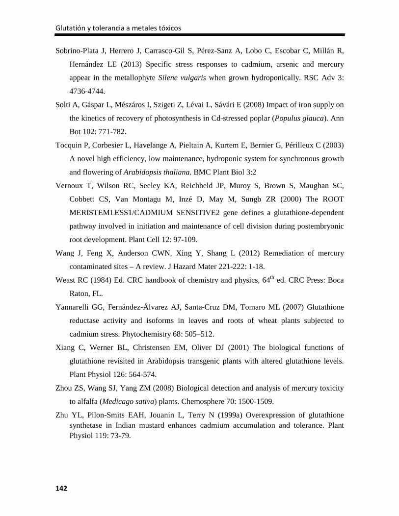
Glutatión y tolerancia a metales tóxicos
142
Sobrino-Plata J, Herrero J, Carrasco-Gil S, Pérez-Sanz A, Lobo C, Escobar C, Millán R,
Hernández LE (2013) Specific stress responses to cadmium, arsenic and mercury
appear in the metallophyte Silene vulgaris when grown hydroponically. RSC Adv 3:
4736-4744.
Solti A, Gáspar L, Mészáros I, Szigeti Z, Lévai L, Sávári E (2008) Impact of iron supply on
the kinetics of recovery of photosynthesis in Cd-stressed poplar (Populus glauca). Ann
Bot 102: 771-782.
Tocquin P, Corbesier L, Havelange A, Pieltain A, Kurtem E, Bernier G, Périlleux C (2003)
A novel high efficiency, low maintenance, hydroponic system for synchronous growth
and flowering of Arabidopsis thaliana. BMC Plant Biol 3:2
Vernoux T, Wilson RC, Seeley KA, Reichheld JP, Muroy S, Brown S, Maughan SC,
Cobbett CS, Van Montagu M, Inzé D, May M, Sungb ZR (2000) The ROOT
MERISTEMLESS1/CADMIUM SENSITIVE2 gene defines a glutathione-dependent
pathway involved in initiation and maintenance of cell division during postembryonic
root development. Plant Cell 12: 97-109.
Wang J, Feng X, Anderson CWN, Xing Y, Shang L (2012) Remediation of mercury
contaminated sites – A review. J Hazard Mater 221-222: 1-18.
Weast RC (1984) Ed. CRC handbook of chemistry and physics, 64th ed. CRC Press: Boca
Raton, FL.
Yannarelli GG, Fernández-Álvarez AJ, Santa-Cruz DM, Tomaro ML (2007) Glutathione
reductase activity and isoforms in leaves and roots of wheat plants subjected to
cadmium stress. Phytochemistry 68: 505–512.
Xiang C, Werner BL, Christensen EM, Oliver DJ (2001) The biological functions of
glutathione revisited in Arabidopsis transgenic plants with altered glutathione levels.
Plant Physiol 126: 564-574.
Zhou ZS, Wang SJ, Yang ZM (2008) Biological detection and analysis of mercury toxicity
to alfalfa (Medicago sativa) plants. Chemosphere 70: 1500-1509.
Zhu YL, Pilon-Smits EAH, Jouanin L, Terry N (1999a) Overexpression of glutathione synthetase in Indian mustard enhances cadmium accumulation and tolerance. Plant Physiol 119: 73-79.
Tesis doctoral Juan Sobrino Plata
143
Zhu YL, Pilon-Smits EAH, Tarun AS, Weber SU, Jouanin L, Terry N (1999b) Cadmium tolerance and accumulation in Indian mustard is enhanced by overexpressing γ-glutamylcysteine synthetase. Plant Physiol 121: 1169-1177.
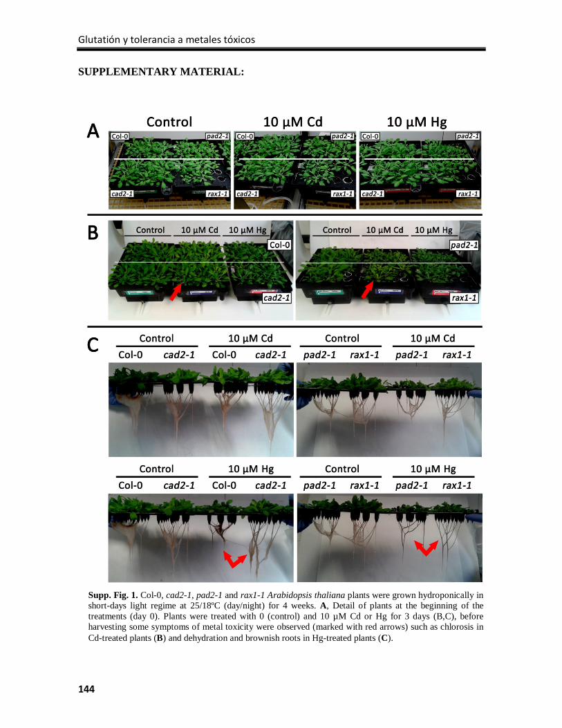
Glutatión y tolerancia a metales tóxicos
144
SUPPLEMENTARY MATERIAL:
Supp. Fig. 1. Col-0, cad2-1, pad2-1 and rax1-1 Arabidopsis thaliana plants were grown hydroponically in short-days light regime at 25/18ºC (day/night) for 4 weeks. A, Detail of plants at the beginning of the treatments (day 0). Plants were treated with 0 (control) and 10 µM Cd or Hg for 3 days (B,C), before harvesting some symptoms of metal toxicity were observed (marked with red arrows) such as chlorosis in Cd-treated plants (B) and dehydration and brownish roots in Hg-treated plants (C).
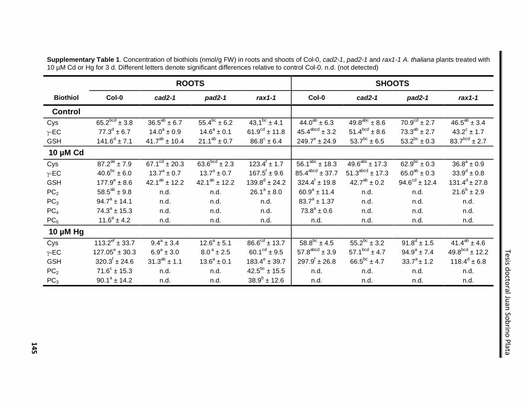
145
Tesis doctoral Juan Sobrino Plata
Supplementary Table 1. Concentration of biothiols (nmol/g FW) in roots and shoots of Col-0, cad2-1, pad2-1 and rax1-1 A. thaliana plants treated with 10 µM Cd or Hg for 3 d. Different letters denote significant differences relative to control Col-0. n.d. (not detected)
ROOTS SHOOTS Biothiol Col-0 cad2-1 pad2-1 rax1-1 Col-0 cad2-1 pad2-1 rax1-1
Control Cys 65.2bcd ± 3.8 36.5ab ± 6.7 55.4bc ± 6.2 43,1bc ± 4.1 44.0ab ± 6.3 49.8abc ± 8.6 70.9cd ± 2.7 46.5ab ± 3.4 γ-EC 77.3d ± 6.7 14.0a ± 0.9 14.6a ± 0.1 61.9cd ± 11.8 45.4abcd ± 3.2 51.4bcd ± 8.6 73.3ab ± 2.7 43.2c ± 1.7 GSH 141.6d ± 7.1 41.7ab ± 10.4 21.1ab ± 0.7 86.8c ± 6.4 249.7e ± 24.9 53.7bc ± 6.5 53.2bc ± 0.3 83.7bcd ± 2.7
10 µM Cd Cys 87.2de ± 7.9 67.1cd ± 20.3 63.6bcd ± 2.3 123.4f ± 1.7 56.1abc ± 18.3 49.6abc ± 17.3 62.9bc ± 0.3 36.8a ± 0.9 γ-EC 40.6bc ± 6.0 13.7a ± 0.7 13.7a ± 0.7 167.5f ± 9.6 85.4abcd ± 37.7 51.3abcd ± 17.3 65.0ab ± 0.3 33.9d ± 0.8 GSH 177.9e ± 8.6 42.1ab ± 12.2 42.1ab ± 12.2 139.8d ± 24.2 324.4f ± 19.8 42.7ab ± 0.2 94.6cd ± 12.4 131.4d ± 27.8 PC2 58.5ab ± 9.8 n.d. n.d. 26.1a ± 8.0 60.9a ± 11.4 n.d. n.d. 21.6b ± 2.9 PC3 94.7a ± 14.1 n.d. n.d. n.d. 83.7a ± 1.37 n.d. n.d. n.d. PC4 74.3a ± 15.3 n.d. n.d. n.d. 73.8a ± 0.6 n.d. n.d. n.d. PC5 11.6a ± 4.2 n.d. n.d. n.d. n.d. n.d. n.d. n.d. 10 µM Hg Cys 113.2ef ± 33.7 9.4a ± 3.4 12.6a ± 5.1 86.6cd ± 13.7 58.8bc ± 4.5 55.2bc ± 3.2 91.8d ± 1.5 41.4ab ± 4.6 γ-EC 127.05e ± 30.3 6.9a ± 3.0 8.0 a ± 2.5 60.1cd ± 9.5 57.8abcd ± 3.9 57.1bcd ± 4.7 94.9a ± 7.4 49.8bcd ± 12.2 GSH 320.3f ± 24.6 31.3ab ± 1.1 13.6a ± 0.1 183.4e ± 39.7 297.9f ± 26.8 66.5bc ± 4.7 33.7a ± 1.2 118.4d ± 6.8 PC2 71.6c ± 15.3 n.d. n.d. 42.5bc ± 15.5 n.d. n.d. n.d. n.d. PC3 90.1a ± 14.2 n.d. n.d. 38.9b ± 12.6 n.d. n.d. n.d. n.d.

Glutatión y tolerancia a metales tóxicos
146
CAPÍTULO 6 Characterization of sulfur and glutathione metabolism responses to mercury in glutathione defective Arabidopsis mutants

Tesis doctoral Juan Sobrino Plata
147
Capítulo 6:
Characterization of sulfur and glutathione metabolism responses to mercury in glutathione defective Arabidopsis mutants
(Work in progress)
INTRODUCTION
Mercury (Hg) is one of the most harmful metals in nature; in 1997 the U.S. Environmental
Protection Agency (EPA) prepared a report about Hg sources, risk for human health or
potential control technologies and described entirely the serious problems of Hg
contamination and its capability of bioaccumulation in trophic chain (Keating et al., 1997).
Mercury is released to the environment from different sources, natural events like volcanic
or geothermal activities; and anthropogenic emissions from mining, ore processing, fossil
fuel burning or waste combustion (UNEP Chemicals Branch., 2008). The largest Hg-
contaminated area of the World is in Almadén (Ciudad Real, Spain), derived from the
extraction of cinnabar for centuries (Millán et al., 2006). Diverse remediation techniques
are being developed during last years to reduce or control Hg contamination in soil, Wang
et al. (2012) reviewed a large number of these technologies including the use of plants to
extract Hg from soil or stabilize in soil. However, to develop effective phytoremediation
strategies it is necessary to know how this metal affects the physiology of plants, but still
nowadays little is known about the mechanisms to cope with Hg induced stress.
The accumulation of Hg in plants produces diverse toxic effects since alteration of normal
plant development and growth, metabolism disruption or induction of severe oxidative
damages (Chen and Yang., 2012). Most of these effects are caused by the interaction with
cellular components such as proteins, due to the strong affinity of Hg for sulphydryl
residues (Van Assche and Clijsters., 1990). Nonetheless the induction of oxidative stress is
a well-known symptom of the Hg accumulation in plant cells (Rellán-Álvarez et al., 2006,
Ortega‐Villasante et al., 2007, Sobrino-Plata et al., 2009). Recent results in our laboratory
have shown the relevant role of the glutathione (GSH) metabolism in the responses of
plants to Hg stress (Sobrino-Plata et al., 2014a, 2014b). This metabolite is essential for
many processes in plant cell; but GSH has principally a dual role in the tolerance to toxic
Glutatión y tolerancia a metales tóxicos
148
metals, firstly as part of the antioxidant system of plant cells, responsible for removing the
excess of reactive oxygen species (ROS) formed by the cellular redox imbalance (Noctor
and Foyer., 1998); and secondly, as the precursor of phytochelatins (PCs), which form
complexes with metals to avoid the interaction with sensitive biomolecules, and is
considered as one of the main mechanisms to heavy metal detoxification (Cobbett and
Goldsbrough., 2002).
As the major reservoir of non-protein reduced sulfur, GSH behaves as the main protagonist
in the regulation of sulfur metabolism in a metal toxicity scenario (Gill and Tuteja., 2011).
Sulfur is essential for plants and inorganic sulfate is the major combined form that plants
acquire from nature. The sulfate uptake and distribution in all compartments of the plants is
controlled through different transporters, concretely in Arabidopsis exist 14 sulfate
transporters, distributed in 5 groups (AtSULTR1-5; Buchner et al., 2004). Group 1 or high-
affinity sulfate transporters are mainly localized in roots and are responsible for sulfate
uptake from soil. Sulfate transporters involved in long-distance transport belong to group 2
or low-affinity sulfate transporters. Group 3, 4 and 5 have no direct relation with S-nutrition
status of the plant; however group 3 transporters seem to be related with the increase of
root-to-shoot sulfate transport and essential in development of embryos and legume root
nodules (Kopriva., 2006). First step in sulfate assimilation is catalyzed by ATP sulfurylase
(ATPS), which produce 5’phosphosulfate (APS) by adenylation of sulfate using ATP. 4
ATPS isoforms are described in Arabidopsis (Hatzfeld et al., 2000). APS is reduced to
sulfite (SO32-) by APS reductase (APR) using electrons from GSH. There are three APR
isoforms presented in Arabidopsis plastids with different regulation and functions
(Koprivova et al., 2008). This step together with sulfate uptake is described as limiting step
in sulfur metabolism (Vauclare et al., 2002). Sulfite is reduced to sulfide with the action of
the sulfite reductase (SiR) which is absolutely necessary in this pathway, as this step is no
possible in a non-enzymatic reaction (Nakayama et al., 2000).
Cysteine (Cys) biosynthesis is the final step of sulfate assimilation and the link for the GSH
metabolism. Cys is synthetized by the cysteine synthase complex (CSC), which is formed
by serine acetyltransferase (SAT) and O-acetylserine (thiol) lyase (OAS-TL). First enzyme
catalyzes the formation of OAS binding acetyl-CoA and serine, and then OAS-TL
synthetizes Cys with the union of sulfide to OAS (Wirtz and Hell., 2006). Cys can be part
Tesis doctoral Juan Sobrino Plata
149
of proteins or can be precursor of GSH. In that case, the enzyme γ-glutamylcysteine
synthetase (γECS) produces γ-glutamylcysteine (γEC) from Cys and Glutamate (Glu), and
glutathione synthetase (GS) forms GSH from Gly and γEC. Both enzymatic steps depend
on ATP and are compartmentalized, being the first reaction localized in plastids, and the
second mainly in the cytosol (Noctor et al., 2012).
Sulfur metabolism is strongly regulated, since several intermediates in this pathway are
toxic and diverse molecules containing sulfur are important for proper cellular function. At
the transcriptional level, we can find both cis- and trans-regulatory elements, some genes of
sulfur metabolism have specific cis boxes of 16 base-pairs in their promoter region known
as ‘Sulfur-Responsive Elements’ (SURE) that are involved in regulation of these genes
(Maruyama‐Nakashita et al., 2005). On the other hand there are transcription factors that
regulate the transcription of genes coding proteins of this metabolic pathway. For instance,
the regulator Sulfur LIMITATION 1 (SLIM1) regulates AtSULTR2;1, AtSULTR4;2 and
ATPS4 expression in Arabidopsis. Other regulators are involved in other metabolic routes
influenced by the sulfur uptake and assimilation process, like members of the MYB
transcription factors family that act as regulators of the glucosinolates metabolism, such as
MYB28 and MYB51, which also regulate the expression of ATPS, APR and SiR genes
(Davidian and Kopriva., 2010).
In most cases, the regulation of this metabolic pathway is exerted in a “demand-driven”
manner (Lappartient and Touraine, 1996), where the need of GSH entails transcriptional
regulation of sulfate uptake and assimilation genes, and apart from GSH biosynthesis
genes. On the other hand, the increase of GSH has a negative effect over the expression of
APR, ATPS and sulfate transporters in a normal S nutrition situation (Vauclare et al., 2002,
Kopriva and Rennenberg., 2004). When plants are exposed to toxic metals, GSH is fully
implicated in the response to oxidative stress produced by these elements. Glutathione
contributes to metal tolerance by removing ROS, with the consequent formation of
oxidized glutathione disulfide (GSSG). Although GSH can be regenerated by glutathione
reductase (GR), in stress situations the proportion of this redox couple (GSH/GSSG) is
displaced to the oxidized form (Semane et al., 2007). Besides that, the chelation of several
metals through the formation of complexes with PCs also consumes part of the cellular
Glutatión y tolerancia a metales tóxicos
150
pool of GSH (Pal and Rai, 2010). In this scenario, the depletion of GSH is thought to
provoke a ‘demand-driven’ up-regulation of the sulfate assimilation pathway (Nocito et al.,
2006, Na and Salt, 2011).
The effects of Hg caused in plant cells differ to that of other toxic metals and metalloids,
showing specific stress signatures (Sobrino-Plata et al., 2009, 2013), and little information
is available about its influence on sulfur metabolism. For this reason, we planned our
experiments to study the alterations in the transcriptional regulation of sulfate uptake and
assimilation processes under Hg stress in Arabidopsis plants. Knowing the direct role of
GSH, we used several Arabidopsis mutants with altered GSH levels: Three γECS mutant
alleles cad2-1, pad2-1 and rax1-1 containing respectively 30, 20 and 45% of wild type
(WT) GSH level (Parisy et al., 2007); and cad1-3, a PCS mutant that is unable to produce
PCs and presents elevated GSH level related to WT (Sobrino-Plata et al., 2014a). We
completed this study by analyzing several Hg-sensitive physiological parameters like
photosynthesis, protein stability and biothiols content. Although most of the work is
finished, we are conscious that in the present study we show some preliminary results that
require the further analysis that will strengthen in the near future the final report.
MATERIALS AND METHODS
Plant material, growth conditions and treatments
Seeds of A. thaliana of the wild type Columbia 0 (Col-0), and the cad2-1, pad2-1, rax1-
1and cad1-3 mutants were surface-sterilized by agitation in a 15% (v/v) NaClO solution for
10 min, followed by several rinses in distilled water. Then seeds were germinated and
grown in Araponics® hydroponic systems (Araponics SA, Liège, Belgium) in seedholders
filled with 0.6 % phytoagar. The seeds were grown for 3 weeks in short-days light regime (8
h light and 16 h darkness) at 25 ºC, with a modified Hoagland nutrient solution (Tocquin et
al., 2006), after this period 0 and 3 μM HgCl2 was added to the nutrient solution for 72
hours. At harvest, shoots and roots were collected separately and stored at -80 ºC until
analysis.
Tesis doctoral Juan Sobrino Plata
151
Chlorophyll fluorescence measurements
Chlorophyll (Chl) fluorescence was measured at different light regimes to determine
ΦPSII, qP and NPQ, according to Maxwell and Johnson (2000). Throughout the
fluorescence measurements, an actinic light of 400 μmol m-2 s1 and saturating pulses light
of 8000 μmol m-2 s1 were used sequentially to determine Fo, Fm, Fm’, Ft and Fo
’ fluorescence
data in a FMS-2 Pulse Modulated Fluorimeter (Hansatech Instruments, Norfolk, UK).
Photosynthesis parameters were determined in plants treated with metals for 24 and 72
hours.
Mercury tissue concentrations
Around 200 mg of leaves and 100 mg of roots were dried at 60 ºC for 72 h, and then were
acid digested in 1 mL of the digestion mixture (HNO3:H2O2:H2O, 0.6:0.4:1 v:v) in an
autoclave (Presoclave-75 Selecta, Barcelona, Spain) at 120ºC and 1.5 atm for 30 min
(Ortega‐Villasante et al., 2007). The digests were filtered through a PVDF filter and diluted
in miliRO water to 6 mL. Hg concentration was determined by ICP-MS NexION 300
Perkin-Elmer Sciex (San Jose, CA, USA).
Sequencing and alignment of γECS mutants
Genomic DNA from shoots of Col-0, cad2-1, pad2-1 and rax1-1 plants were isolated using
illustra DNA Extraction Kit PHYTOPURE (GE Healthcare Life Sciences). Final
concentration of extracted DNA was measured in a NanoDrop ND-1000
spectrophotometer (Thecnologies, Inc., USA). PCR was performed to amplify a 637 bp
fragment of GSH1 gene using the primers g-ECS01F (CGTTCGGATTATTTCTTGGTGT)
and g-ECS02R (GCGGTCCTTGTCAGTGTCTGT). Purification of PCR product was
made with High Pure PCR Product Purification kit (Roche Applied Science). PCR products
were sequenced in a ABI Prism® 3730/3730xl DNA Sequencer (Certified Scientific
Instruments, Inc., USA). The obtained sequences were revised and aligned using the
software Geneious Pro 5.5.3 comparing with GSH1 gene sequence (AT4G23100) taken
from “The Arabidopsis Information Resource” web page (http://www.arabidopsis.org). The
identity of each mutant was verified finding the mutations described in literature (Parisy et
al., 2007, Fig. 1).
Glutatión y tolerancia a metales tóxicos
152
RNA extraction and quantification
Total RNA from shoots and roots of Arabidopsis was isolated with TRI Reagent (Ambion)
as described by (Portillo et al., 2006). The RNA was cleaned using in-column DNAse
treatment with the RNeasy Mini Kit (Qiagen). Final concentration of extracted RNA was
measured in a NanoDrop ND-1000 spectrophotometer (Thecnologies, Inc., USA). RNA
integrity was determined with an Agilent 2100 Bioanalyzer equipped with a RNA 6000
Nano LabChip Kit (Agilent Technologies, Santa Clara, CA, USA). The algorithm RNA
integrity number (RIN) was calculated as the RNA quality of three independent biological
replicates (Fig. 2, Schroeder et al., 2006).
Quantitative reverse transcription polymerase chain reaction (qRT-PCR)
Quantitative RT-PCR was performed with RNA from Arabidopsis shoots and roots of two
completely independent biological experiments (three RNA biological replicates each) to
synthesize the complementary DNA strand (cDNA). Oligonucleotide primers of several
sulfur metabolism genes were designed using the ProbeFinder ver. 2.45 (Roche
Diagnostics, Rotkreuz, Switzerland; Supplementary Table 1). The reverse transcription
(RT) reaction was performed with random hexamers, using the RETROscript® First Strand
Synthesis Kit for RT-PCR (Applied Biosystems-Life technologies, Carlsbad, CA, USA).
Quantitative polymerase chain reaction (qPCR) wascarried out with 50 ng single-stranded
cDNA in a final volume of 20 μL, containing 10 μL of SYBR-Green Master Mix (Applied
Biosystems-Life Technologies) and 250 nM forward and reverse specific primers (Life
Technologies). Amplification reactions were performed in a Real-Time 7000SDS
Termocycler (Applied Biosystems-Life Technologies), at the Genomics Service of Rey
Juan Carlos University (Madrid, Spain). The program used was: denaturation at 95 °C 10
min, followed by 40 cycles of 15 seconds at 95 °C and 1 min annealing and extension at 60
°C. Gene expression quantification was performed using the relative 2−∆∆Ct method (Livak
and Schmittgen., 2001). The glyceraldehyde 3-phosphate dehydrogenase gene (GAPDH)
was used as reference according to Montero‐Palmero et al. (2014).
Glutathione reductase in gel activity
GR enzymatic activities were determined in gel after separation of protein extracts by non-
denaturing polyacrylamide electrophoresis following the procedures described by Sobrino-
Tesis doctoral Juan Sobrino Plata
153
Plata et al. (2009) for GR in gel activity. The concentration of protein in the extracts was
determined using the BioRad Protein Assay reagent with bovine serum albumin as
standard, and adequate loading was corrected by after polyacrylamide denaturing gel
electrophoresis (Laemmli., 1970) and Coomassie blue staining. Protein loading was 20 µg
and 10 µg of shoot and root samples, respectively. The staining solution was prepared in
250 mM Tris-HCl buffer at pH 7.5, supplemented with 0.2 mg/mL thizolyl blue tetrazolium
bromide, 0.2 mg/mL 2,6-dichlorophenol indophenol, 0.5 mM NADPH and 3.5 mM GSSG.
Western-blot immunodetection
Immunodetection was performed by Western-blot after denaturing gel electrophoresis
(Laemmli., 1970) and blotted onto a nitrocellulose membrane (BioTraceNT Pall
Corporation, East Hills, NY, USA), by using a semi-dry procedure (Trans Blot SD Semi-
Dry Electrophoretic Transfer Cell; BioRad, Hercules, CA, USA). The membranes were
incubated overnight at 4ºC with the primary antibodies (α-GR, dil. 1/5000; α−γECS, dil.
1/2500; all antibodies were purchased from Agrisera, Vännäs, Sweden). After incubation
with the anti-rabbit secondary antibody linked to horse radish peroxidase (HRP), proteins
were detected using the LumiSensor Chemiluminescent HRP Substrate Kit (GenScript,
Piscataway, NJ, USA), the images were taken with ChemiDoc™ XRS+ System (BioRad,
Hercules, CA, USA).
Analysis of biothiols
Biothiols in shoots and roots were analyzed by HPLC following the procedure described by
Ortega-Villasante et al. (2005). Extracts (100 µL) were injected in a Mediterranea SEA18
column (5 μm, 250 x 4.6 mm; Teknokroma, San Cugat del Vallés, Spain), using an Agilent
1200 HPLC system (Santa Clara, CA, USA). Thiols were detected after post-column
derivatization with Ellman reagent, and to quantify them, a spike of N-acetyl cysteine (N-
AcCys; 15 μL of a 5 mM solution) was added as internal standard prior to homogenization
at a final concentration of 250 μM (Sobrino-Plata et al., 2009).
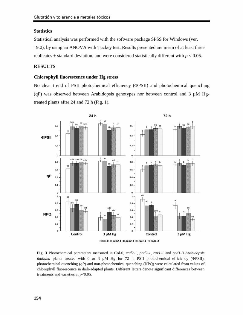
Glutatión y tolerancia a metales tóxicos
154
Statistics
Statistical analysis was performed with the software package SPSS for Windows (ver.
19.0), by using an ANOVA with Tuckey test. Results presented are mean of at least three
replicates ± standard deviation, and were considered statistically different with p < 0.05.
RESULTS
Chlorophyll fluorescence under Hg stress
No clear trend of PSII photochemical efficiency (ΦPSII) and photochemical quenching
(qP) was observed between Arabidopsis genotypes nor between control and 3 µM Hg-
treated plants after 24 and 72 h (Fig. 1).
Fig. 3 Photochemical parameters measured in Col-0, cad2-1, pad2-1, rax1-1 and cad1-3 Arabidopsis thaliana plants treated with 0 or 3 µM Hg for 72 h. PSII photochemical efficiency (ΦPSII), photochemical quenching (qP) and non-photochemical quenching (NPQ) were calculated from values of chlorophyll fluorescence in dark-adapted plants. Different letters denote significant differences between treatments and varieties at p<0.05.
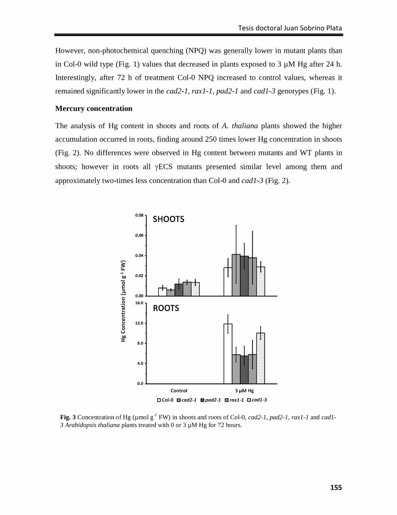
Tesis doctoral Juan Sobrino Plata
155
However, non-photochemical quenching (NPQ) was generally lower in mutant plants than
in Col-0 wild type (Fig. 1) values that decreased in plants exposed to 3 µM Hg after 24 h.
Interestingly, after 72 h of treatment Col-0 NPQ increased to control values, whereas it
remained significantly lower in the cad2-1, rax1-1, pad2-1 and cad1-3 genotypes (Fig. 1).
Mercury concentration
The analysis of Hg content in shoots and roots of A. thaliana plants showed the higher
accumulation occurred in roots, finding around 250 times lower Hg concentration in shoots
(Fig. 2). No differences were observed in Hg content between mutants and WT plants in
shoots; however in roots all γECS mutants presented similar level among them and
approximately two-times less concentration than Col-0 and cad1-3 (Fig. 2).
Fig. 3 Concentration of Hg (µmol g-1 FW) in shoots and roots of Col-0, cad2-1, pad2-1, rax1-1 and cad1-3 Arabidopsis thaliana plants treated with 0 or 3 µM Hg for 72 hours.
Glutatión y tolerancia a metales tóxicos
156
Analysis of sulfur metabolism genes expression
We selected 20 different genes along the sulfur metabolic route to analyze the alterations in
the expression under Hg-stress. The gene expression was normalized to endogenous
glyceraldehyde 3-phosphate dehydrogenase gene (GAPDH) expression and related to Col-0
control in shoots and roots separately.
The expression of three different sulfate transporters was analyzed. The high-affinity
sulfate transporter SULTR1;2 had an induced gene expression in Hg treatment in roots,
although this was 5-10 times higher in mutants than in Col-0 (Fig. 3). SULTR2;1 gene
which codifies a low-affinity sulfate transporter was repressed in all the analyzed samples,
except in roots of non-treated and Hg-treated cad1-3. The third gene analyzed in this group
was SULTR3;5, Hg induced its expression in all plants except cad1-3 (Fig. 3). In shoots
Hg produced an increase in SULTR1;2, SULTR2;1 and SULTR3;5 in Col-0 but the opposite
effect was observed in the mutants (Fig. 4).
In the first step of sulfate assimilation pathway we analyzed the expression of three
isoforms. We found ATPS1 gene repressed in roots of all mutants in control conditions but
Hg produced the induction in the mutants and repression in Col-0. ATPS3 was repressed in
control roots of cad2-1 and rax1-1 and the expression was increased with Hg as in cad1-3.
Hg promoted an increment in the expression of ATPS4 in pad2-1 and clearer in cad2-1
(Fig. 3). ATPS1 was over-expressed in shoots of rax1-1, but with Hg treatment we observed
an induction in shoots of Col-0 and cad1-3 plants. An increased expression of ATPS3 was
observed in shoots of all plants in both treatments. Mercury affected negatively the
expression of ATPS4 in pad2-1 shoots and produced the opposite effect in rax1-1 (Fig. 4).
APS reductase genes also showed different behavior under Hg-stress. In shoots of rax1-1
and cad1-3 we found an increased expression of APR1 related to Col-0 in control treatment.
While Hg treatment increased the expression of Col-0, in these two mutants occurred the
contrary result. In roots occurred a general repression of this gene except in non-treated
pad2-1 and cad1-3. APR2 was induced in shoots of all mutants in control conditions and
Hg treatment reduced this expression. By contrast, Hg affected positively the expression of
Col-0 in roots but nothing occurred in shoots. Curiously, APR3 expression was similar to
APR1 except the increment in shoots of control rax1-1 (Figs. 3 and 4). On the other hand,
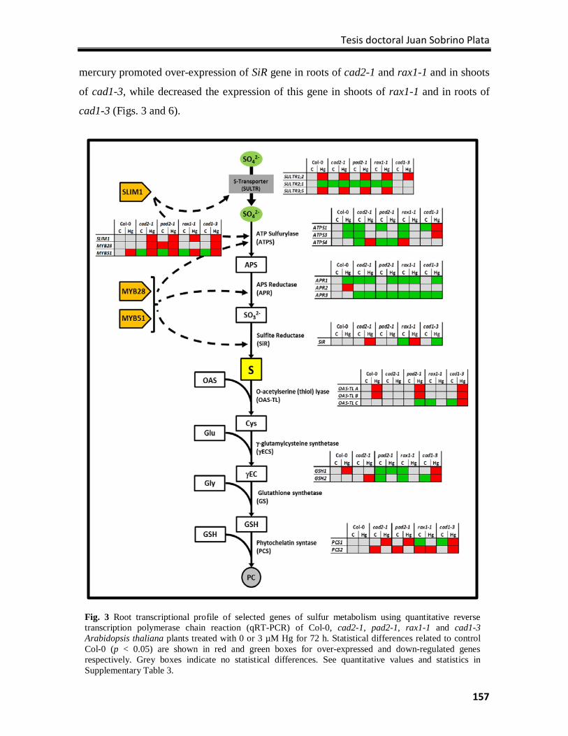
Tesis doctoral Juan Sobrino Plata
157
mercury promoted over-expression of SiR gene in roots of cad2-1 and rax1-1 and in shoots
of cad1-3, while decreased the expression of this gene in shoots of rax1-1 and in roots of
cad1-3 (Figs. 3 and 6).
Fig. 3 Root transcriptional profile of selected genes of sulfur metabolism using quantitative reverse transcription polymerase chain reaction (qRT-PCR) of Col-0, cad2-1, pad2-1, rax1-1 and cad1-3 Arabidopsis thaliana plants treated with 0 or 3 µM Hg for 72 h. Statistical differences related to control Col-0 (p < 0.05) are shown in red and green boxes for over-expressed and down-regulated genes respectively. Grey boxes indicate no statistical differences. See quantitative values and statistics in Supplementary Table 3.
Glutatión y tolerancia a metales tóxicos
158
Sulfur metabolism is strongly regulated, part of this regulation are the transcription factors.
We wanted to observe the alterations in SLIM1, MYB28 and MYB51 which regulate genes
in this first part of the pathway. Hg promoted an induction of SLIM1 in roots of all mutants
but not in Col-0. MYB genes had similar response front to Hg stress in cad2-1 and cad1-3
roots.
Hg-treated Col-0 also showed an increase of MYB51 expression in roots (Fig. 3). Compared
with non-treated Col-0 shoots, cad2-1 and pad2-1 had an increased expression of SLIM1
and this was reduced under Hg-stress, while in Hg-treated Col-0 and cad1-3 we observed
an induction of this gene. Moreover we observed a general decrease of MYB28 expression
in shoots of Hg-treated plants (Fig. 4).
The last part of sulfate assimilation is the synthesis of Cys. We analyzed in this step the
genes of the three isoforms of OAS-TL. Two of them, OAS-TL A and OAS-TL B showed
similar alterations in roots, Hg induced the expression of both genes in Col-0, pad2-1 and
cad1-3 (Fig. 3). Mercury also induced OAS-TL A expression in shoots of cad2-1, rax1-1
and cad1-3. Likewise OAS-TL B and OAS-TL C showed similar alterations but in shoots;
Hg produced the induction of both genes in Col-0, and cad1-3 presented in control
conditions an increased expression and no alteration with Hg. Furthermore Hg promoted
increment of OAS-TL B expression in shoots of pad2-1 and reduction in rax1-1 (Fig. 4).
In roots Hg produced an induction in Col-0, rax1-1 and cad1-3 (Fig. 3), while in shoots
repressed the γECS gene in pad2-1 and rax1-1 (Fig. 4). Glutathione synthetase gene was
induced under Hg treatment in shoots of Col-0 and cad1-3. This gene appeared repressed in
control conditions in shoots of all γECS mutants and no changes were observed with Hg
(Fig. 4). However in roots Hg induced the expression of GS in all mutants (Fig. 3). We
observed a repressed expression of PCS1 in shoots of cad2-1, rax1-1 and cad1-3. Mercury
affected negatively the expression of this gene in pad2-1 shoots while in roots was induced
as in the rest of mutants. Surprisingly PCS2 was over-expressed in roots of all γECS
mutants in control conditions and was repressed with Hg treatment. Nonetheless mercury
promoted induction of this gene in shoots of pad2-1 and both in shoots and in roots of
cad1-3 (Figs. 3 and 4).
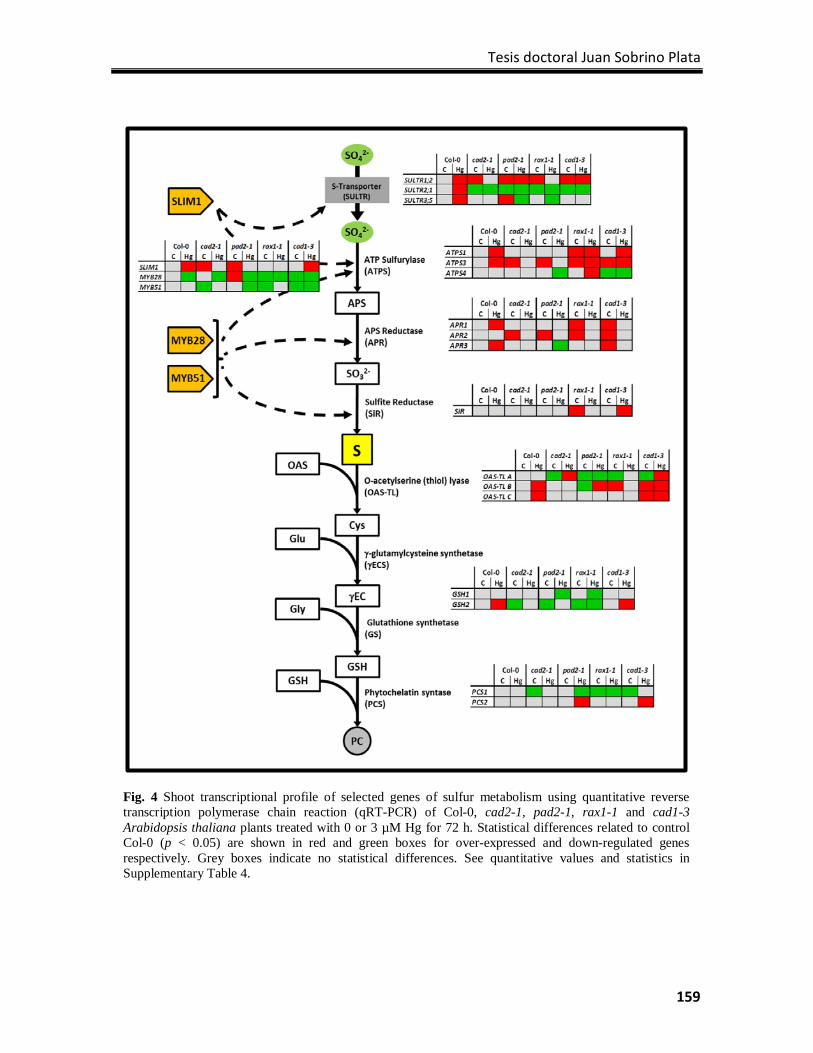
Tesis doctoral Juan Sobrino Plata
159
Fig. 4 Shoot transcriptional profile of selected genes of sulfur metabolism using quantitative reverse transcription polymerase chain reaction (qRT-PCR) of Col-0, cad2-1, pad2-1, rax1-1 and cad1-3 Arabidopsis thaliana plants treated with 0 or 3 µM Hg for 72 h. Statistical differences related to control Col-0 (p < 0.05) are shown in red and green boxes for over-expressed and down-regulated genes respectively. Grey boxes indicate no statistical differences. See quantitative values and statistics in Supplementary Table 4.
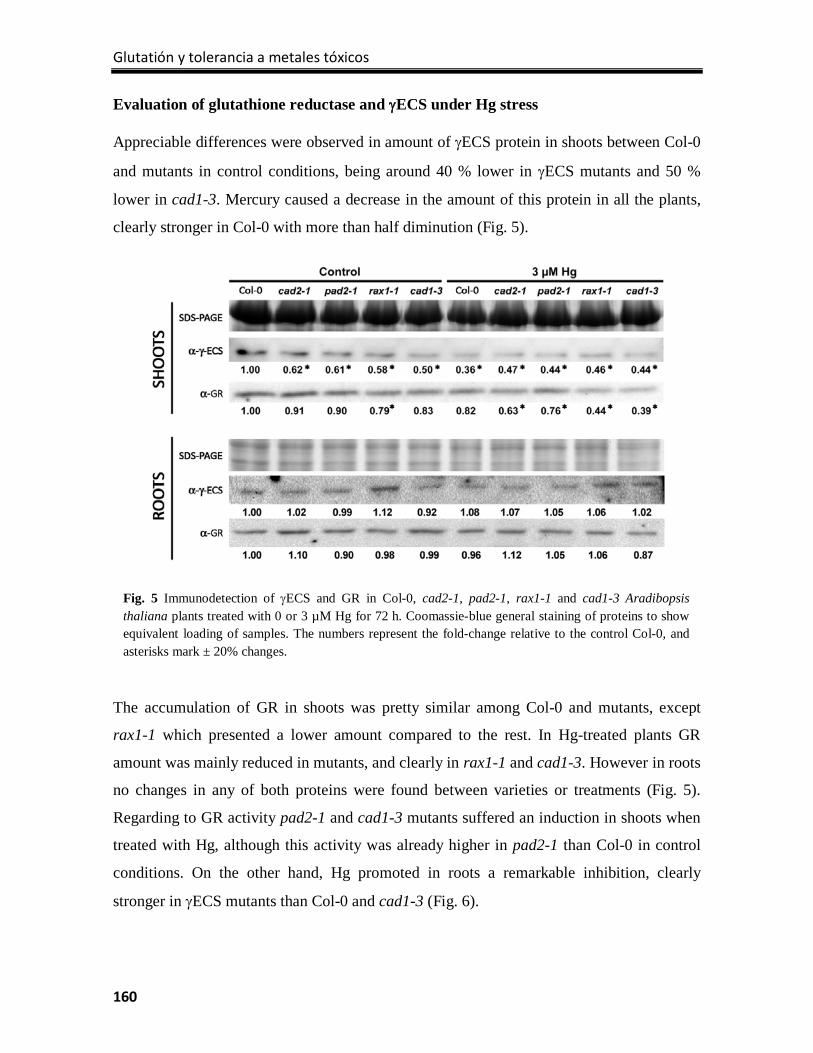
Glutatión y tolerancia a metales tóxicos
160
Evaluation of glutathione reductase and γECS under Hg stress
Appreciable differences were observed in amount of γECS protein in shoots between Col-0
and mutants in control conditions, being around 40 % lower in γECS mutants and 50 %
lower in cad1-3. Mercury caused a decrease in the amount of this protein in all the plants,
clearly stronger in Col-0 with more than half diminution (Fig. 5).
The accumulation of GR in shoots was pretty similar among Col-0 and mutants, except
rax1-1 which presented a lower amount compared to the rest. In Hg-treated plants GR
amount was mainly reduced in mutants, and clearly in rax1-1 and cad1-3. However in roots
no changes in any of both proteins were found between varieties or treatments (Fig. 5).
Regarding to GR activity pad2-1 and cad1-3 mutants suffered an induction in shoots when
treated with Hg, although this activity was already higher in pad2-1 than Col-0 in control
conditions. On the other hand, Hg promoted in roots a remarkable inhibition, clearly
stronger in γECS mutants than Col-0 and cad1-3 (Fig. 6).
Fig. 5 Immunodetection of γECS and GR in Col-0, cad2-1, pad2-1, rax1-1 and cad1-3 Aradibopsis thaliana plants treated with 0 or 3 µM Hg for 72 h. Coomassie-blue general staining of proteins to show equivalent loading of samples. The numbers represent the fold-change relative to the control Col-0, and asterisks mark ± 20% changes.
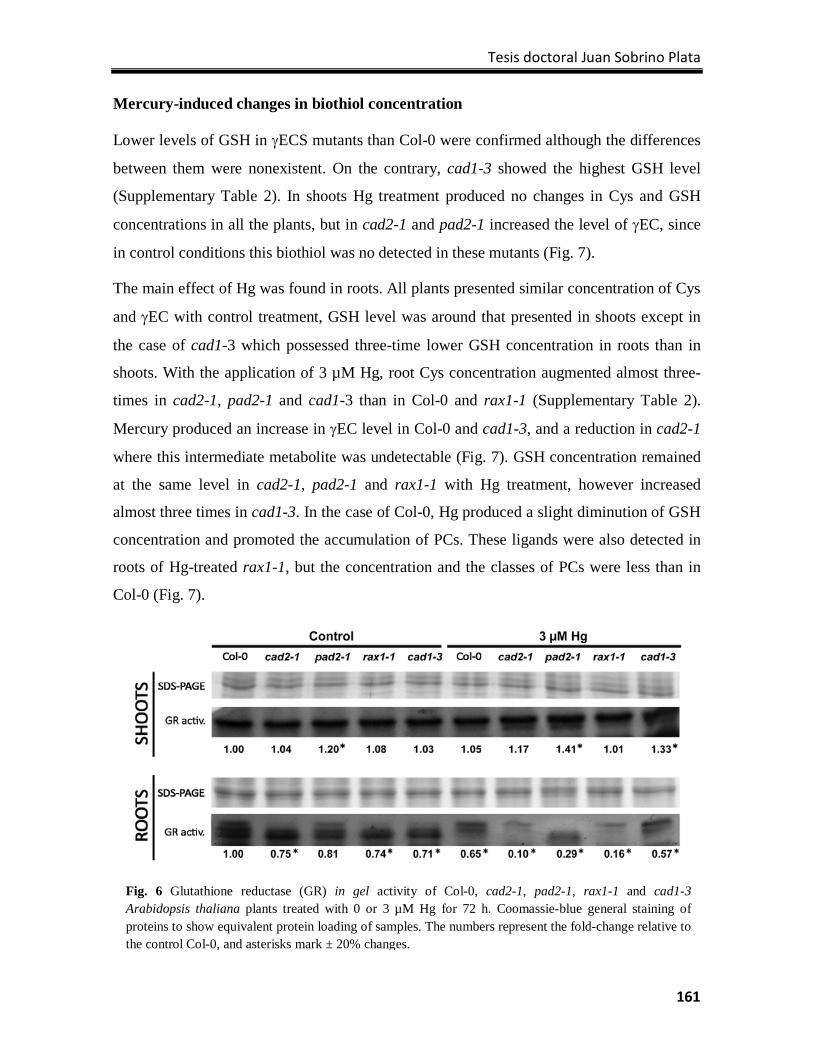
Tesis doctoral Juan Sobrino Plata
161
Mercury-induced changes in biothiol concentration
Lower levels of GSH in γECS mutants than Col-0 were confirmed although the differences
between them were nonexistent. On the contrary, cad1-3 showed the highest GSH level
(Supplementary Table 2). In shoots Hg treatment produced no changes in Cys and GSH
concentrations in all the plants, but in cad2-1 and pad2-1 increased the level of γEC, since
in control conditions this biothiol was no detected in these mutants (Fig. 7).
The main effect of Hg was found in roots. All plants presented similar concentration of Cys
and γEC with control treatment, GSH level was around that presented in shoots except in
the case of cad1-3 which possessed three-time lower GSH concentration in roots than in
shoots. With the application of 3 µM Hg, root Cys concentration augmented almost three-
times in cad2-1, pad2-1 and cad1-3 than in Col-0 and rax1-1 (Supplementary Table 2).
Mercury produced an increase in γEC level in Col-0 and cad1-3, and a reduction in cad2-1
where this intermediate metabolite was undetectable (Fig. 7). GSH concentration remained
at the same level in cad2-1, pad2-1 and rax1-1 with Hg treatment, however increased
almost three times in cad1-3. In the case of Col-0, Hg produced a slight diminution of GSH
concentration and promoted the accumulation of PCs. These ligands were also detected in
roots of Hg-treated rax1-1, but the concentration and the classes of PCs were less than in
Col-0 (Fig. 7).
Fig. 6 Glutathione reductase (GR) in gel activity of Col-0, cad2-1, pad2-1, rax1-1 and cad1-3 Arabidopsis thaliana plants treated with 0 or 3 µM Hg for 72 h. Coomassie-blue general staining of proteins to show equivalent protein loading of samples. The numbers represent the fold-change relative to the control Col-0, and asterisks mark ± 20% changes.
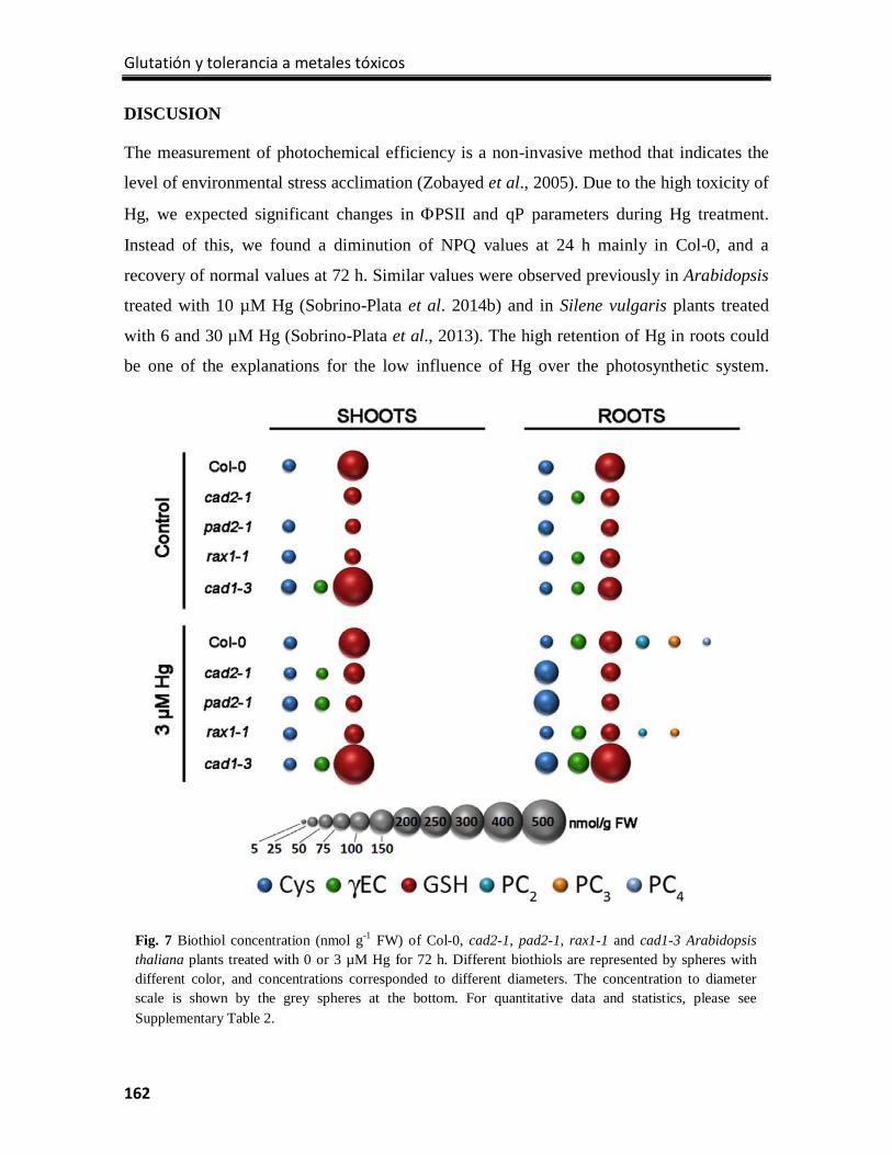
Glutatión y tolerancia a metales tóxicos
162
DISCUSION
The measurement of photochemical efficiency is a non-invasive method that indicates the
level of environmental stress acclimation (Zobayed et al., 2005). Due to the high toxicity of
Hg, we expected significant changes in ΦPSII and qP parameters during Hg treatment.
Instead of this, we found a diminution of NPQ values at 24 h mainly in Col-0, and a
recovery of normal values at 72 h. Similar values were observed previously in Arabidopsis
treated with 10 µM Hg (Sobrino-Plata et al. 2014b) and in Silene vulgaris plants treated
with 6 and 30 µM Hg (Sobrino-Plata et al., 2013). The high retention of Hg in roots could
be one of the explanations for the low influence of Hg over the photosynthetic system.
Fig. 7 Biothiol concentration (nmol g-1 FW) of Col-0, cad2-1, pad2-1, rax1-1 and cad1-3 Arabidopsis thaliana plants treated with 0 or 3 µM Hg for 72 h. Different biothiols are represented by spheres with different color, and concentrations corresponded to different diameters. The concentration to diameter scale is shown by the grey spheres at the bottom. For quantitative data and statistics, please see Supplementary Table 2.
Tesis doctoral Juan Sobrino Plata
163
Recently Carrasco-Gil et al. (2013) showed this poor mobilization of Hg, which is retained
strongly in cell walls of epidermal and xylem root cells. Moreover changes in NPQ could
be explained as an increment in photorespiration or photoinhibition processes caused
indirectly by Hg inhibition of aquaporines, which could result in altered hydric balance in
mesophyll cells (Zhang and Tyerman., 1999, Postaire et al., 2010).
Different groups have described the increment in sulfate uptake and assimilation pathways
when plants are treated with heavy metals (Ernst et al., 2008, Na and Salt, 2011). For
instance the study of Cd, Zn or Cu effects over the sulfur metabolism is highly represented
in literature and all of them show a direct up-regulation of several genes of this route when
these metals are applied (Harmens et al., 1993, Heiss et al., 1999, Domı́nguez-Solı́s et al.,
2001, Nocito et al., 2006, Nocito et al., 2002). Indeed, a recent transcriptomic analysis
revealed the up-regulation of several sulfur metabolism related genes after just 6 h of
exposure to 3 µM Hg (Montero-Palmero et al., 2014). We could observe that 3 µM Hg
treatment in Col-0 plants promoted similar effects. A general induction of sulfate uptake
and assimilation pathways occurred in shoots of Hg-treated Col-0 plants. However in roots
we found repressed several important genes such as SULTR2;1, ATPS1, ATPS3 and APR1.
The high affinity sulfate transporter SULTR1;2 had an induced gene expression in roots
under Hg stress, and that indicates mercury is increased the sulfate uptake in roots but the
sulfate assimilation is more active in shoots caused by the requirement of GSH. Since first
step of GSH biosynthesis is produced in plastids (Wachter et al., 2005), the depletion of
GSH in roots, caused by the formation of PCs, could have an indirect consequence in the
demand of GSH from shoots. In agreement with Lappartient and Touraine (1996), the
concept of “demand-driven regulation” seems to be suitable to explain this situation.
According to Kataoka et al. (2004), an efficient root-to-shoot transport is achieved thanks
to the formation of SULTR2;1 and SULTR3;5 transporter complex, we found both genes
induced in shoots but in the roots SULTR2;1 is repressed, this could be related with a toxic
effect of Hg-accumulation in roots affecting directly to the expression of this and other
genes. On the other hand, there are other genes like OAS-TL that are induced in the
presence of Hg both in roots and shoots. The up-regulation of this gene expression together
with SAT has been described as an important response in Nickel (Ni) and Cd
hyperaccumulator plants (Freeman et al., 2004, Guo et al., 2009), and it has also been
Glutatión y tolerancia a metales tóxicos
164
demonstrated that the increase of OAS-TL transcripts conferences certain higher tolerance
to Cd (Domı́nguez-Solı́s et al., 2001). We found OAS-TL genes induced not only in WT
plants but also in mutants, for instance pad2-1 and cad1-3 presented both in shoots and
roots a general induction of this step, although not all genes are up-regulated.
The comparison with mutants showed some interesting results that could support the
evidence of the fine regulation of this route. For instance Hg promoted in roots of all
mutants an increased expression of SLIM1 and MYB genes, while in Col-0 this effect was
significant only in MYB51 gene expression. By contrast, the effect of Hg was clearly
negative in plants with lower GSH content in comparison with Col-0 and cad1-3 which
presented no effect or augmented expression of these transcription factors. In this sense it is
surprising that in mutants these transcription factors have the opposite effect to what we
expected over the genes that are regulated by them. MYB28 and MYB51 activate the
expression of ATPS, APR and SiR genes (Yatusevich et al., 2010), while in roots of these
mutants the up-regulation of these genes by Hg promoted a general, but not always
significant, induction of ATPS, APR and SiR expression; in shoots this regulation is less
evident. These changes in regulation suggests an implication of other factors like the
increment of Cys or γEC in cad2-1 and pad2-1 when are treated with Hg. It is well reported
by Jobe et al. (2012) that the accumulation of reduced sulfur forms can produce the down-
regulation of sulfate uptake and assimilation genes by an increase of reducing redox
environment, it is probably that for this reason we found some genes in shoots repressed or
non-altered under Hg stress in the γECS mutants depending on the Cys, γEC or GSH level
of each one. On the other hand, the high level of GSH in shoots of cad1-3 produced no
effects or even repression of several genes in this pathway, while the opposite situation
occurs in roots where this mutant possesses a lower GSH level related to Col-0.
Nonetheless, the different behavior to Hg-stress between Col-0 and mutants could show a
direct relation with the lower tolerance in these, since Col-0 plants presented in shoots and
roots an overexpression of ATPS, APR and OAS-TL genes. Several studies with other
metals like Cu or Cd evidenced an increase in the basal tolerance to these metals in plants
when transcripts of these genes were increased (Heiss et al., 1999, Domı́nguez-Solı́s et al.,
2001, Wangeline et al., 2004).
Tesis doctoral Juan Sobrino Plata
165
Interestingly, the accumulation of γECS protein had no relation with the expression of the
gene. While in roots we could observe an induction of GSH1 produced by Hg in Col-0,
rax1-1 and cad1-3, we detected no changes in γECS protein content in this plants. Besides
that the accumulation of γECS is much lower in shoots of Hg-treated plants while the
expression of GSH1 appeared significant repressed only in pad2-1 and rax1-1. This
difference could be related with the complex regulation of this enzyme that includes
translational regulation, feedback inhibition by γEC and GSH or posttranscriptional
modifications (Hothorn et al., 2006, Hicks et al., 2007). Moreover a direct negative effect
of mercury is also taken into consideration since the same behavior is shown in the
accumulation of GR in shoots except in Col-0 plants. However the level of GR activity in
those samples was similar to the WT level in control conditions with the exception of rax1-
1 and cad1-3 which increased the activity with the treatment of Hg. Looking at the amount
of protein and the activity of GR presented in the samples, we could argue a direct Hg-
induction of GR activity in shoots. Possibly this effect is a consequence of an augmented
activity of the antioxidant system in response to the oxidative stress promoted by Hg in
shoots (Chen and Yang, 2012). The opposite is observed in roots where the high
accumulation of Hg in this part of the plant caused the strong inhibition of the GR activity
being more accused in γECS mutants in spite of present lower Hg content than Col-0 and
cad1-3, while the accumulation of protein showed no alterations. All these results in GR
activity are in agreement with previous works realized in our laboratory in different plants
such as Medicago sativa or Silene vulgaris (Sobrino-Plata et al., 2009, Sobrino-Plata et al.,
2013). The poor mobility of Hg to the aerial part causes that the main effects occur in roots.
Besides all results already described we found a high accumulation of PCs in wild type and
rax1-1 plants. This mechanism of tolerance is responsible for a reduced Hg toxicity in these
plants respect to the other mutants of study. The inability of forming PCs in cad1-3
(Howden et al., 1995) is compensated by an increased level of GSH and this could reduce
the damage produced by oxidative stress. However in the case of cad2-1 and pad2-1 the
low GSH level and the inexistent formation of PCs make these mutants vulnerable to the
Hg toxicity, as we could confirm in the quality of the RNA and proteins extracted from
roots of Hg-treated pad2-1 plants.
Glutatión y tolerancia a metales tóxicos
166
This work shows the relation of GSH and sulfur metabolism in the response to stress
produced by low concentration of Hg. The regulation of this pathway seems to be based in
the demand of reduced sulfur which in Hg-treated Col-0 plants is decreased due to the
requirement of GSH to form PCs. The requirement for GSH activates the enzymes involved
in GSH biosynthesis and implies a reduction of Cys level. The reduction of Cys has the
consequent induction of sulfate uptake and assimilation pathways. The use of γECS and
PCS mutants adds variations to this classic model where the low tolerance and the
increased levels of other biothiols (Cys or γEC) may become important factors to take into
account in the regulation of sulfur metabolism under Hg stress. These factors also could
explain the differences between rax1-1 and the other γECS mutants, since the GSH level
was very similar in the three mutants but rax1-1 seemed to be more tolerant than the other
two alleles and its behavior to Hg-stress was similar to Col-0 in spite of the low GSH
concentration in control conditions.
REFERENCES
Buchner P, Takahashi H, Hawkesford MJ. 2004. Plant sulphate transporters: co-ordination of uptake, intracellular and long-distance transport. Journal of Experimental Botany 55, 1765-1773.
Carrasco-Gil S, Siebner H, LeDuc DL, Webb SM, Millán R, Andrews JC, Hernández LE. 2013. Mercury localization and speciation in plants grown hydroponically or in a natural environment. Environmental Science and Technology 47, 3082-3090.
Chen J, Yang ZM. 2012. Mercury toxicity, molecular response and tolerance in higher plants. BioMetals 25, 847-857.
Cobbett C, Goldsbrough P. 2002. Phytochelatins and metallothioneins: roles in heavy metal detoxification and homeostasis. Annual Review of Plant Biology 53, 159-182.
Davidian J, Kopriva S. 2010. Regulation of sulfate uptake and assimilation—the same or not the same? Molecular Plant 3, 314-325.
Domı́nguez-Solı́s JR, Gutiérrez-Alcalá G, Romero LC, Gotor C. 2001. The cytosolic O-acetylserine (thiol) lyase gene is regulated by heavy metals and can function in cadmium tolerance. Journal of Biological Chemistry 276, 9297-9302.
Tesis doctoral Juan Sobrino Plata
167
Ernst WH, Krauss G, Verkleij JA, Wesenberg D. 2008. Interaction of heavy metals with the sulphur metabolism in angiosperms from an ecological point of view. Plant, Cell and Environment 31, 123-143.
Freeman JL, Persans MW, Nieman K, Albrecht C, Peer W, Pickering IJ, Salt DE. 2004. Increased glutathione biosynthesis plays a role in nickel tolerance in Thlaspi nickel hyperaccumulators. The Plant Cell 16, 2176-2191.
Gill SS, Tuteja N. 2011. Cadmium stress tolerance in crop plants: probing the role of sulfur. Plant Signaling and Behavior 6, 215-222.
Guo W, Liang J, Yang X, Chao Y, Feng Y. 2009. Response of ATP sulfurylase and serine acetyltransferase towards cadmium in hyperaccumulator Sedum alfredii Hance. Journal of Zhejiang University Science B 10, 251-257.
Harmens H, Den Hartog PR, Bookum WMT, Verkleij JA. 1993. Increased zinc tolerance in Silene vulgaris (Moench) Garcke is not due to increased production of phytochelatins. Plant Physiology 103, 1305-1309.
Hatzfeld Y, Lee S, Lee M, Leustek T, Saito K. 2000. Functional characterization of a gene encoding a fourth ATP sulfurylase isoform from Arabidopsis thaliana. Gene 248, 51-58.
Heiss S, Schäfer H, Haag-Kerwer A, Rausch T. 1999. Cloning sulfur assimilation genes of Brassica juncea L.: cadmium differentially affects the expression of a putative low-affinity sulfate transporter and isoforms of ATP sulfurylase and APS reductase. Plant Molecular Biology 39, 847-857.
Hicks LM, Cahoon RE, Bonner ER, Rivard RS, Sheffield J, Jez JM. 2007. Thiol-based regulation of redox-active glutamate-cysteine ligase from Arabidopsis thaliana. The Plant Cell Online 19, 2653-2661.
Hothorn M, Wachter A, Gromes R, Stuwe T, Rausch T, Scheffzek K. 2006. Structural basis for the redox control of plant glutamate cysteine ligase. Journal of Biological Chemistry 281, 27557-27565.
Howden R, Goldsbrough PB, Andersen CR, Cobbett CS. 1995. Cadmium-sensitive, cad1 mutants of Arabidopsis thaliana are phytochelatin deficient. Plant Physiology 107, 1059-1066.
Jobe TO, Sung DY, Akmakjian G, Pham A, Komives EA, Mendoza‐Cózatl DG, Schroeder JI. 2012. Feedback inhibition by thiols outranks glutathione depletion: a luciferase‐based screen reveals glutathione‐deficient γ‐ECS and glutathione synthetase mutants impaired in cadmium‐induced sulfate assimilation. The Plant Journal.
Glutatión y tolerancia a metales tóxicos
168
Kataoka T, Hayashi N, Yamaya T, Takahashi H. 2004. Root-to-shoot transport of sulfate in Arabidopsis. Evidence for the role of SULTR3; 5 as a component of low-affinity sulfate transport system in the root vasculature. Plant Physiology 136, 4198-4204.
Keating MH, Mahaffey KR, Schoeny R, Rice G, Bullock O. 1997. Mercury study report to Congress.Volume 1.Executive summary.
Kopriva S. 2006. Regulation of sulfate assimilation in Arabidopsis and beyond. Annals of Botany 97, 479-495.
Kopriva S, Rennenberg H. 2004. Control of sulphate assimilation and glutathione synthesis: interaction with N and C metabolism. Journal of experimental botany 55, 1831-1842.
Koprivova A, North KA, Kopriva S. 2008. Complex signaling network in regulation of adenosine 5′-phosphosulfate reductase by salt stress in Arabidopsis roots. Plant Physiology 146, 1408-1420.
Laemmli UK. 1970. Cleavage of structural proteins during the assembly of the head of bacteriophage T4. Nature 227, 680-685.
Lappartient AG, Touraine B. 1996. Demand-driven control of root ATP sulfurylase activity and SO42-uptake in intact canola (the role of phloem-translocated glutathione). Plant Physiology 111, 147-157.
Livak KJ, Schmittgen TD. 2001. Analysis of Relative Gene Expression Data Using Real-Time Quantitative PCR and the 2− ΔΔCT Method. Methods 25, 402-408.
Maruyama‐Nakashita A, Nakamura Y, Watanabe‐Takahashi A, Inoue E, Yamaya T, Takahashi H. 2005. Identification of a novel cis‐acting element conferring sulfur deficiency response in Arabidopsis roots. The Plant Journal 42, 305-314.
Maxwell K, Johnson GN. 2000. Chlorophyll fluorescence—a practical guide. Journal of Experimental Botany 51, 659-668.
Millán R, Gamarra R, Schmid T, Sierra M, Quejido A, Sánchez D, Cardona A, Fernández M, Vera R. 2006. Mercury content in vegetation and soils of the Almadén mining area (Spain). Science of the Total Environment 368, 79-87.
Montero‐Palmero MB, Martín‐Barranco A, Escobar C, Hernández LE. 2014. Early transcriptional responses to mercury: a role for ethylene in mercury‐induced stress. New Phytologist. 201, 116-130.
Na G, Salt DE. 2011. The role of sulfur assimilation and sulfur-containing compounds in trace element homeostasis in plants. Environmental and Experimental Botany 72, 18-25.
Tesis doctoral Juan Sobrino Plata
169
Nakayama M, Akashi T, Hase T. 2000. Plant sulfite reductase: molecular structure, catalytic function and interaction with ferredoxin. Journal of Inorganic Biochemistry 82, 27-32.
Nocito FF, Lancilli C, Crema B, Fourcroy P, Davidian J, Sacchi GA. 2006. Heavy metal stress and sulfate uptake in maize roots. Plant Physiology 141, 1138-1148.
Nocito FF, Pirovano L, Cocucci M, Sacchi GA. 2002. Cadmium-induced sulfate uptake in maize roots. Plant Physiology 129, 1872-1879.
Noctor G, Foyer CH. 1998. Ascorbate and glutathione: keeping active oxygen under control. Annual Review of Plant Biology 49, 249-279.
Noctor G, Mhamdi A, Chaouch S, Han Y, Neukermans J, MARQUEZ‐GARCIA B, Queval G, Foyer CH. 2012. Glutathione in plants: an integrated overview. Plant, Cell and Environment 35, 454-484.
Ortega-Villasante C, Rellán-Álvarez R, Del Campo FF, Carpena-Ruiz RO, Hernández LE. 2005. Cellular damage induced by cadmium and mercury in Medicago sativa. Journal of Experimental Botany 56, 2239-2251.
Ortega‐Villasante C, Hernández LE, Rellán‐Álvarez R, Del Campo FF, Carpena‐Ruiz RO. 2007. Rapid alteration of cellular redox homeostasis upon exposure to cadmium and mercury in alfalfa seedlings. New Phytologist 176, 96-107.
Pal R, Rai J. 2010. Phytochelatins: peptides involved in heavy metal detoxification. Applied Biochemistry and Biotechnology 160, 945-963.
Parisy V, Poinssot B, Owsianowski L, Buchala A, Glazebrook J, Mauch F. 2007. Identification of PAD2 as a γ‐glutamylcysteine synthetase highlights the importance of glutathione in disease resistance of Arabidopsis. The Plant Journal 49, 159-172.
Portillo M, Fenoll C, Escobar C. 2006. Evaluation of different RNA extraction methods for small quantities of plant tissue: Combined effects of reagent type and homogenization procedure on RNA quality‐integrity and yield. Physiologia Plantarum 128, 1-7.
Postaire O, Tournaire-Roux C, Grondin A, Boursiac Y, Morillon R, Schäffner AR, Maurel C. 2010. A PIP1 aquaporin contributes to hydrostatic pressure-induced water transport in both the root and rosette of Arabidopsis. Plant Physiology 152, 1418-1430.
Rellán-Álvarez R, Ortega-Villasante C, Álvarez-Fernández A, Campo FF, Hernández LE. 2006. Stress responses of Zea mays to cadmium and mercury. Plant and Soil 279, 41-50.
Glutatión y tolerancia a metales tóxicos
170
Schroeder A, Mueller O, Stocker S, Salowsky R, Leiber M, Gassmann M, Lightfoot S, Menzel W, Granzow M, Ragg T. 2006. The RIN: an RNA integrity number for assigning integrity values to RNA measurements. BMC Molecular Biology 7, 3.
Semane B, Cuypers A, Smeets K, Van Belleghem F, Horemans N, Schat H, Vangronsveld J. 2007. Cadmium responses in Arabidopsis thaliana: glutathione metabolism and antioxidative defence system. Physiologia Plantarum 129, 519-528.
Sobrino-Plata J, Ortega-Villasante C, Laura Flores-Cáceres M, Escobar C, Del Campo FF, Hernández LE. 2009. Differential alterations of antioxidant defenses as bioindicators of mercury and cadmium toxicity in alfalfa. Chemosphere 77, 946-954.
Sobrino-Plata J, Herrero J, Carrasco-Gil S, Pérez-Sanz A, Lobo C, Escobar C, Millán R, Hernández LE. 2013. Specific stress responses to cadmium, arsenic and mercury appear in the metallophyte Silene vulgaris when grown hydroponically. RSC Advances. 3, 4736-4744.
Sobrino-Plata J, Carrasco-Gil S, Abadía J, Escobar C, Álvarez-Fernández A, Hernández LE. 2014a. The contribution of glutathione in Arabidopsis mercury tolerance resembles its role under cadmium stress. Metallomics DOI: 10.1039/C3MT00329A
Sobrino-Plata J, Meyssen D, Cuypers A, Escobar C, Hernández LE. 2014b. Glutathione is a key antioxidant metabolite to cope with mercury and cadmium stress. Plant and Soil DOI: 10.1007/s11104-013-2006-4
Tocquin P, Corbesier L, Havelange A, Pieltain A, Kurtem E, Bernier G, Périlleux C. 2003. A novel high efficiency, low maintenance, hydroponic system for synchronous growth and flowering of Arabidopsis thaliana. BMC Plant Biology 3, 2.
UNEP Chemicals Branch. 2008. The Global Atmospheric Mercury Assessment: Sources, Emissions and Transport.
Van Assche F, Clijsters H. 1990. Effects of metals on enzyme activity in plants. Plant, Cell and Environment 13, 195-206.
Vauclare P, Kopriva S, Fell D, Suter M, Sticher L, Von Ballmoos P, Krähenbühl U, Den Camp RO, Brunold C. 2002. Flux control of sulphate assimilation in Arabidopsis thaliana: adenosine 5′‐phosphosulphate reductase is more susceptible than ATP sulphurylase to negative control by thiols. The Plant Journal 31, 729-740.
Wachter A, Wolf S, Steininger H, Bogs J, Rausch T. 2005. Differential targeting of GSH1 and GSH2 is achieved by multiple transcription initiation: implications for the compartmentation of glutathione biosynthesis in the Brassicaceae. The Plant Journal 41, 15-30.
Tesis doctoral Juan Sobrino Plata
171
Wang J, Feng X, Anderson CW, Xing Y, Shang L. 2012. Remediation of mercury contaminated sites–A review. Journal of Hazardous Materials 221, 1-18.
Wangeline AL, Burkhead JL, Hale KL, Lindblom SD, Terry N, Pilon M, Pilon-Smits EA. 2004. Overexpression of ATP Sulfurylase in Indian Mustard. Journal of Environmental Quality 33, 54-60.
Wirtz M, Hell R. 2006. Functional analysis of the cysteine synthase protein complex from plants: structural, biochemical and regulatory properties. Journal of Plant Physiology 163, 273-286.
Yatusevich R, Mugford SG, Matthewman C, Gigolashvili T, Frerigmann H, Delaney S, Koprivova A, Flügge U, Kopriva S. 2010. Genes of primary sulfate assimilation are part of the glucosinolate biosynthetic network in Arabidopsis thaliana. The Plant Journal 62, 1-11.
Zhang W, Tyerman SD. 1999. Inhibition of water channels by HgCl2 in intact wheat root cells. Plant Physiology 120, 849-858.
Zobayed S, Afreen F, Kozai T. 2005. Temperature stress can alter the photosynthetic efficiency and secondary metabolite concentrations in St. John's wort. Plant Physiology and Biochemistry 43, 977-984.
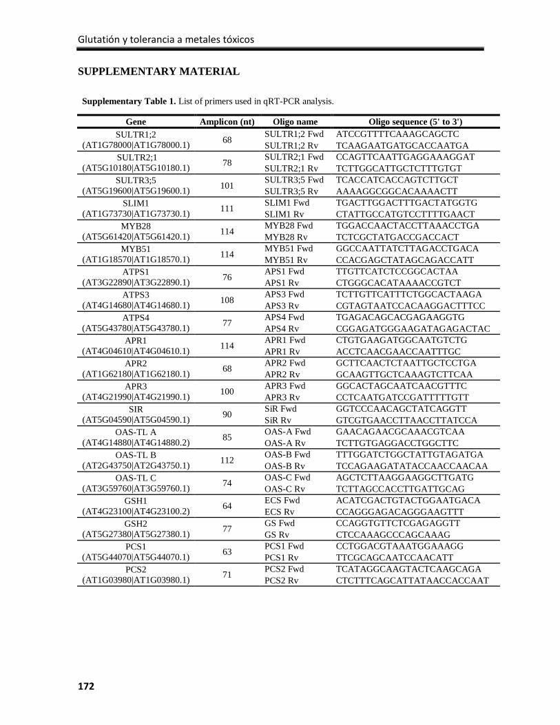
Glutatión y tolerancia a metales tóxicos
172
SUPPLEMENTARY MATERIAL
Supplementary Table 1. List of primers used in qRT-PCR analysis.
Gene Amplicon (nt) Oligo name Oligo sequence (5' to 3') SULTR1;2
(AT1G78000|AT1G78000.1) 68 SULTR1;2 Fwd ATCCGTTTTCAAAGCAGCTC SULTR1;2 Rv TCAAGAATGATGCACCAATGA
SULTR2;1 (AT5G10180|AT5G10180.1) 78 SULTR2;1 Fwd CCAGTTCAATTGAGGAAAGGAT
SULTR2;1 Rv TCTTGGCATTGCTCTTTGTGT SULTR3;5
(AT5G19600|AT5G19600.1) 101 SULTR3;5 Fwd TCACCATCACCAGTCTTGCT SULTR3;5 Rv AAAAGGCGGCACAAAACTT
SLIM1 (AT1G73730|AT1G73730.1) 111 SLIM1 Fwd TGACTTGGACTTTGACTATGGTG
SLIM1 Rv CTATTGCCATGTCCTTTTGAACT MYB28
(AT5G61420|AT5G61420.1) 114 MYB28 Fwd TGGACCAACTACCTTAAACCTGA MYB28 Rv TCTCGCTATGACCGACCACT
MYB51 (AT1G18570|AT1G18570.1) 114 MYB51 Fwd GGCCAATTATCTTAGACCTGACA
MYB51 Rv CCACGAGCTATAGCAGACCATT ATPS1
(AT3G22890|AT3G22890.1) 76 APS1 Fwd TTGTTCATCTCCGGCACTAA APS1 Rv CTGGGCACATAAAACCGTCT
ATPS3 (AT4G14680|AT4G14680.1) 108 APS3 Fwd TCTTGTTCATTTCTGGCACTAAGA
APS3 Rv CGTAGTAATCCACAAGGACTTTCC ATPS4
(AT5G43780|AT5G43780.1) 77 APS4 Fwd TGAGACAGCACGAGAAGGTG APS4 Rv CGGAGATGGGAAGATAGAGACTAC
APR1 (AT4G04610|AT4G04610.1) 114 APR1 Fwd CTGTGAAGATGGCAATGTCTG
APR1 Rv ACCTCAACGAACCAATTTGC APR2
(AT1G62180|AT1G62180.1) 68 APR2 Fwd GCTTCAACTCTAATTGCTCCTGA APR2 Rv GCAAGTTGCTCAAAGTCTTCAA
APR3 (AT4G21990|AT4G21990.1) 100 APR3 Fwd GGCACTAGCAATCAACGTTTC
APR3 Rv CCTCAATGATCCGATTTTTGTT SIR
(AT5G04590|AT5G04590.1) 90 SiR Fwd GGTCCCAACAGCTATCAGGTT SiR Rv GTCGTGAACCTTAACCTTATCCA
OAS-TL A (AT4G14880|AT4G14880.2) 85 OAS-A Fwd GAACAGAACGCAAACGTCAA
OAS-A Rv TCTTGTGAGGACCTGGCTTC OAS-TL B
(AT2G43750|AT2G43750.1) 112 OAS-B Fwd TTTGGATCTGGCTATTGTAGATGA OAS-B Rv TCCAGAAGATATACCAACCAACAA
OAS-TL C (AT3G59760|AT3G59760.1) 74 OAS-C Fwd AGCTCTTAAGGAAGGCTTGATG
OAS-C Rv TCTTAGCCACCTTGATTGCAG GSH1
(AT4G23100|AT4G23100.2) 64 ECS Fwd ACATCGACTGTACTGGAATGACA ECS Rv CCAGGGAGACAGGGAAGTTT
GSH2 (AT5G27380|AT5G27380.1) 77 GS Fwd CCAGGTGTTCTCGAGAGGTT
GS Rv CTCCAAAGCCCAGCAAAG PCS1
(AT5G44070|AT5G44070.1) 63 PCS1 Fwd CCTGGACGTAAATGGAAAGG PCS1 Rv TTCGCAGCAATCCAACATT
PCS2 (AT1G03980|AT1G03980.1) 71 PCS2 Fwd TCATAGGCAAGTACTCAAGCAGA
PCS2 Rv CTCTTTCAGCATTATAACCACCAAT
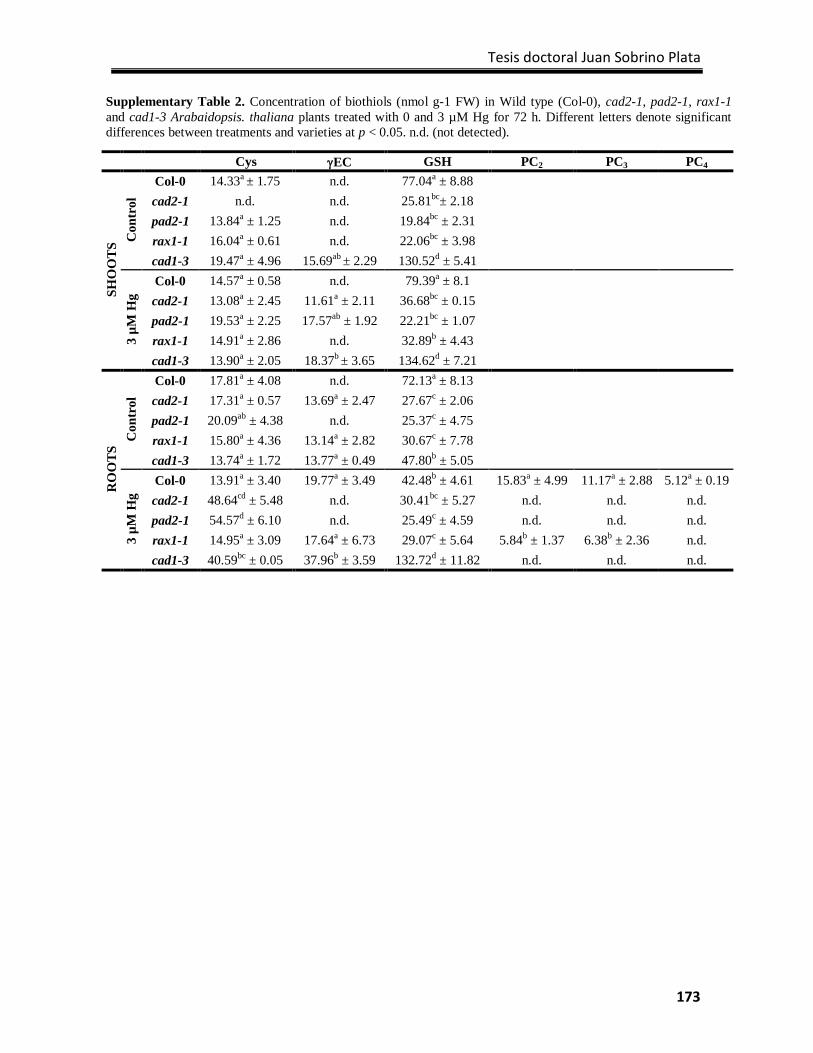
Tesis doctoral Juan Sobrino Plata
173
Supplementary Table 2. Concentration of biothiols (nmol g-1 FW) in Wild type (Col-0), cad2-1, pad2-1, rax1-1 and cad1-3 Arabaidopsis. thaliana plants treated with 0 and 3 µM Hg for 72 h. Different letters denote significant differences between treatments and varieties at p < 0.05. n.d. (not detected).
Cys γEC GSH PC2 PC3 PC4
SHO
OTS
Con
trol
Col-0 14.33a ± 1.75 n.d. 77.04a ± 8.88
cad2-1 n.d. n.d. 25.81bc± 2.18
pad2-1 13.84a ± 1.25 n.d. 19.84bc ± 2.31 rax1-1 16.04a ± 0.61 n.d. 22.06bc ± 3.98
cad1-3 19.47a ± 4.96 15.69ab ± 2.29 130.52d ± 5.41
3 µM
Hg
Col-0 14.57a ± 0.58 n.d. 79.39a ± 8.1
cad2-1 13.08a ± 2.45 11.61a ± 2.11 36.68bc ± 0.15
pad2-1 19.53a ± 2.25 17.57ab ± 1.92 22.21bc ± 1.07 rax1-1 14.91a ± 2.86 n.d. 32.89b ± 4.43
cad1-3 13.90a ± 2.05 18.37b ± 3.65 134.62d ± 7.21
RO
OTS
Con
trol
Col-0 17.81a ± 4.08 n.d. 72.13a ± 8.13
cad2-1 17.31a ± 0.57 13.69a ± 2.47 27.67c ± 2.06
pad2-1 20.09ab ± 4.38 n.d. 25.37c ± 4.75 rax1-1 15.80a ± 4.36 13.14a ± 2.82 30.67c ± 7.78
cad1-3 13.74a ± 1.72 13.77a ± 0.49 47.80b ± 5.05
3 µM
Hg
Col-0 13.91a ± 3.40 19.77a ± 3.49 42.48b ± 4.61 15.83a ± 4.99 11.17a ± 2.88 5.12a ± 0.19 cad2-1 48.64cd ± 5.48 n.d. 30.41bc ± 5.27 n.d. n.d. n.d. pad2-1 54.57d ± 6.10 n.d. 25.49c ± 4.59 n.d. n.d. n.d. rax1-1 14.95a ± 3.09 17.64a ± 6.73 29.07c ± 5.64 5.84b ± 1.37 6.38b ± 2.36 n.d. cad1-3 40.59bc ± 0.05 37.96b ± 3.59 132.72d ± 11.82 n.d. n.d. n.d.
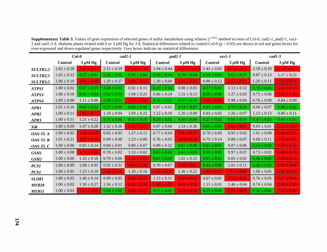
174
Supplementary Table 3. Values of gene expression of selected genes of sulfur metabolism using relative 2−∆∆Ct method in roots of Col-0, cad2-1, pad2-1, rax1-1 and cad1-3 A. thaliana plants treated with 0 or 3 µM Hg for 3 d. Statistical differences related to control Col-0 (p < 0.05) are shown in red and green boxes for over-expressed and down-regulated genes respectively. Grey boxes indicate no statistical differences.
SULTR1;2
Col-0 cad2-1 pad2-1 rax1-1 cad1-3 Control 3 µM Hg Control 3 µM Hg Control 3 µM Hg Control 3 µM Hg Control 3 µM Hg
1.02 ± 0.28 2.88 ± 0.14 2.15 ± 0.59 19.06 ± 2.98 1.94 ± 0.44 11.57 ± 4.06 2.42 ± 0.05 11.58 ± 0.11 2.19 ± 0.10 11.16 ± 0.71
SULTR2;1 1.03 ± 0.32 0.27 ± 0.01 0.28 ± 0.05 0.30 ± 0.04 0.29 ± 0.04 0.18 ± 0.04 0.18 ± 0.01 0.65 ± 0.10 0.87 ± 0.13 1.17 ± 0.23
SULTR3;5 1.00 ± 0.10 1.96 ± 0.08 1.29 ± 0.27 4.00 ± 0.27 1.30 ± 0.04 5.10 ± 0.42 0.88 ± 0.12 2.59 ± 0.15 1.20 ± 0.11 5.13 ± 0.71
ATPS1 1.00 ± 0.02 0.67 ± 0.15 0.48 ± 0.02 0.92 ± 0.11 0.35 ± 0.06 0.98 ± 0.03 0.27 ± 0.01 1.13 ± 0.12 0.70 ± 0.06 2.04 ± 0.15
ATPS3 1.00 ± 0.10 0.40 ± 0.04 0.52 ± 0.08 1.04 ± 0.21 0.66 ± 0.24 1.31 ± 0.13 0.26 ± 0.06 1.27 ± 0.02 0.73 ± 0.06 8.42 ± 1.36
ATPS4 1.00 ± 0.08 1.11 ± 0.06 0.49 ± 0.01 1.49 ± 0.06 0.19 ± 0.01 0.67 ± 0.18 1.34 ± 0.19 0.98 ± 0.04 0.76 ± 0.09 0.84 ± 0.09
APR1 1.01 ± 0.16 0.60 ± 0.22 0.71 ± 0.00 0.64 ± 0.16 0.87 ± 0.02 0.54 ± 0.07 0.43 ± 0.03 0.70 ± 0.10 0.94 ± 0.07 0.48 ± 0.02
APR2 1.00 ± 0.11 1.82 ± 0.71 1.18 ± 0.04 1.69 ± 0.22 1.12 ± 0.10 1.26 ± 0.08 0.83 ± 0.02 1.26 ± 0.07 1.12 ± 0.13 0.80 ± 0.11
APR3 1.00 ± 0.11 1.21 ± 0.22 0.37 ± 0.06 0.55 ± 0.10 0.29 ± 0.02 0.49 ± 0.04 0.21 ± 0.02 0.66 ± 0.10 0.47 ± 0.05 0.41 ± 0.05
SiR 1.00 ± 0.09 1.47 ± 0.26 1.32 ± 0.26 1.51 ± 0.11 0.87 ± 0.04 1.54 ± 0.36 0.60 ± 0.05 1.66 ± 0.07 0.83 ± 0.01 2.54 ± 0.23
OAS-TL A 1.00 ± 0.05 1.62 ± 0.26 0.82 ± 0.05 1.27 ± 0.11 0.77 ± 0.04 1.45 ± 0.17 0.78 ± 0.09 0.95 ± 0.05 1.01 ± 0.08 1.36 ± 0.15
OAS-TL B 1.01 ± 0.21 2.50 ± 0.13 0.99 ± 0.08 1.23 ± 0.08 0.76 ± 0.03 1.63 ± 0.19 0.75 ± 0.14 0.88 ± 0.07 0.69 ± 0.11 1.76 ± 0.01
OAS-TL C 1.00 ± 0.06 0.85 ± 0.14 0.84 ± 0.01 0.80 ± 0.07 0.89 ± 0.12 0.67 ± 0.06 0.61 ± 0.01 0.87 ± 0.06 0.64 ± 0.02 1.93 ± 0.20
GSH1 1.00 ± 0.08 1.79 ± 0.28 0.78 ± 0.02 1.33 ± 0.02 0.65 ± 0.04 0.63 ± 0.03 0.50 ± 0.02 0.97 ± 0.07 0.73 ± 0.02 1.88 ± 0.28
GSH2 1.00 ± 0.06 1.16 ± 0.16 0.79 ± 0.06 1.27 ± 0.01 0.61 ± 0.04 1.02 ± 0.12 0.41 ± 0.12 0.85 ± 0.02 0.46 ± 0.03 1.84 ± 0.12
PCS1 1.00 ± 0.09 1.08 ± 0.01 0.92 ± 0.31 1.60 ± 0.20 0.70 ± 0.07 1.56 ± 0.81 0.42 ± 0.06 1.01 ± 0.11 0.40 ± 0.02 2.67 ± 0.04
PCS2 1.00 ± 0.05 1.23 ± 0.19 1.65 ± 0.12 1.20 ± 0.16 1.59 ± 0.25 1.36 ± 0.22 1.65 ± 0.11 1.61 ± 0.03 1.06 ± 0.05 2.81 ± 0.17
SLIM1 1.00 ± 0.05 1.40 ± 0.14 0.99 ± 0.05 2.66 ± 0.23 1.13 ± 0.11 2.63 ± 0.41 0.67 ± 0.01 2.15 ± 0.29 0.76 ± 0.05 2.57 ± 0.08
MYB28 1.00 ± 0.02 1.30 ± 0.27 1.56 ± 0.12 2.16 ± 0.28 1.98 ± 0.05 2.02 ± 0.33 1.11 ± 0.01 1.46 ± 0.04 0.74 ± 0.04 2.86 ± 0.35
MYB51 1.00 ± 0.03 1.23 ± 0.05 0.69 ± 0.02 3.02 ± 0.12 0.52 ± 0.01 3.24 ± 0.10 0.29 ± 0.00 2.00 ± 0.09 0.32 ± 0.02 7.65 ± 0.40
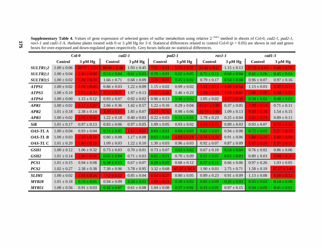
175
Supplementary Table 4. Values of gene expression of selected genes of sulfur metabolism using relative 2−∆∆Ct method in shoots of Col-0, cad2-1, pad2-1, rax1-1 and cad1-3 A. thaliana plants treated with 0 or 3 µM Hg for 3 d. Statistical differences related to control Col-0 (p < 0.05) are shown in red and green boxes for over-expressed and down-regulated genes respectively. Grey boxes indicate no statistical differences.
Col-0 cad2-1 pad2-1 rax1-1 cad1-3 Control 3 µM Hg Control 3 µM Hg Control 3 µM Hg Control 3 µM Hg Control 3 µM Hg
SULTR1;2 1.00 ± 0.06 10.72 ± 2.14 10.59 ± 1.40 1.93 ± 0.45 7.51 ± 0.11 5.74 ± 0.55 16.42 ± 9.21 1.15 ± 0.13 21.81 ± 6.63 5.40 ± 0.05 SULTR2;1 1.00 ± 0.04 1.42 ± 0.08 0.54 ± 0.04 0.61 ± 0.03 0.29 ± 0.01 0.62 ± 0.05 0.72 ± 0.11 0.68 ± 0.04 0.65 ± 0.06 0.45 ± 0.04 SULTR3;5 1.00 ± 0.02 1.84 ± 0.20 1.66 ± 0.71 0.68 ± 0.09 2.29 ± 0.03 0.45 ± 0.02 0.79 ± 0.17 0.54 ± 0.08 0.96 ± 0.07 0.97 ± 0.16
ATPS1 1.00 ± 0.02 1.71 ± 0.45 0.86 ± 0.03 1.22 ± 0.09 1.15 ± 0.02 0.99 ± 0.02 1.41 ± 0.14 1.88 ± 0.14 1.13 ± 0.03 2.47 ± 0.17 ATPS3 1.00 ± 0.10 2.13 ± 0.32 2.73 ± 0.21 1.97 ± 0.13 2.42 ± 0.28 1.46 ± 0.23 2.09 ± 0.69 2.18 ± 0.05 2.80 ± 0.00 5.34 ± 0.83 ATPS4 1.00 ± 0.00 1.15 ± 0.12 0.93 ± 0.07 0.92 ± 0.02 0.96 ± 0.13 0.48 ± 0.02 1.05 ± 0.02 3.17 ± 0.39 0.34 ± 0.02 0.48 ± 0.02
APR1 1.00 ± 0.02 5.17 ± 1.47 2.66 ± 0.36 1.42 ± 0.57 1.22 ± 0.16 0.29 ± 0.04 17.87 ± 2.88 0.37 ± 0.05 4.99 ± 0.53 0.71 ± 0.11 APR2 1.01 ± 0.18 2.38 ± 0.14 4.49 ± 0.15 1.85 ± 0.07 3.14 ± 0.36 0.98 ± 0.06 10.99 ± 2.09 1.09 ± 0.13 9.18 ± 0.58 1.54 ± 0.15 APR3 1.00 ± 0.02 3.34 ± 0.40 1.22 ± 0.18 0.40 ± 0.03 0.22 ± 0.03 0.12 ± 0.02 1.79 ± 0.23 0.25 ± 0.04 5.55 ± 0.71 0.89 ± 0.11
SiR 1.01 ± 0.17 0.97 ± 0.13 0.83 ± 0.06 0.97 ± 0.05 1.09 ± 0.05 0.93 ± 0.02 1.45 ± 0.12 0.89 ± 0.03 0.93 ± 0.07 1.78 ± 0.15
OAS-TL A 1.00 ± 0.04 0.93 ± 0.04 0.54 ± 0.02 1.15 ± 0.01 0.69 ± 0.01 0.84 ± 0.03 0.62 ± 0.03 0.94 ± 0.08 0.72 ± 0.03 1.21 ± 0.13 OAS-TL B 1.00 ± 0.03 1.55 ± 0.10 0.80 ± 0.08 1.17 ± 0.08 0.63 ± 0.04 1.69 ± 0.18 1.31 ± 0.10 0.91 ± 0.06 1.60 ± 0.21 1.69 ± 0.09 OAS-TL C 1.01 ± 0.20 1.43 ± 0.15 1.09 ± 0.03 1.22 ± 0.10 1.30 ± 0.03 0.96 ± 0.03 0.92 ± 0.07 0.87 ± 0.09 1.47 ± 0.05 1.37 ± 0.21
GSH1 1.00 ± 0.12 1.06 ± 0.32 0.73 ± 0.03 0.70 ± 0.01 0.73 ± 0.07 0.63 ± 0.02 0.67 ± 0.10 0.54 ± 0.04 0.76 ± 0.02 0.86 ± 0.06 GSH2 1.01 ± 0.14 1.44 ± 0.20 0.65 ± 0.04 0.71 ± 0.03 0.62 ± 0.01 0.70 ± 0.09 0.51 ± 0.01 0.63 ± 0.03 0.89 ± 0.03 1.94 ± 0.25
PCS1 1.01 ± 0.15 0.94 ± 0.08 0.58 ± 0.13 0.67 ± 0.07 0.28 ± 0.05 0.68 ± 0.12 0.37 ± 0.11 0.66 ± 0.06 0.97 ± 0.26 1.03 ± 0.05 PCS2 1.02 ± 0.27 2.38 ± 0.38 7.30 ± 0.96 3.78 ± 0.95 3.32 ± 0.68 67.57 ± 16.4 1.90 ± 0.03 2.75 ± 0.71 1.58 ± 0.19 27.27 ± 2.40
SLIM1 1.00 ± 0.02 1.61 ± 0.26 1.52 ± 0.12 0.85 ± 0.04 1.54 ± 0.27 0.80 ± 0.05 0.89 ± 0.23 0.91 ± 0.09 1.13 ± 0.08 1.49 ± 0.12 MYB28 1.01 ± 0.18 0.70 ± 0.05 0.94 ± 0.09 0.24 ± 0.01 1.68 ± 0.13 0.30 ± 0.02 0.65 ± 0.09 0.20 ± 0.03 0.55 ± 0.03 0.14 ± 0.00 MYB51 1.08 ± 0.56 0.91 ± 0.03 0.42 ± 0.07 0.61 ± 0.08 1.04 ± 0.08 0.37 ± 0.02 0.15 ± 0.01 0.97 ± 0.15 0.34 ± 0.05 0.41 ± 0.01
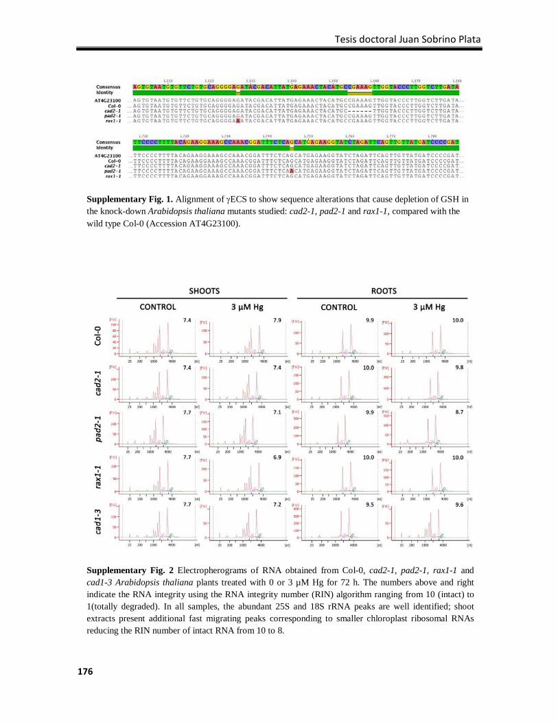
Tesis doctoral Juan Sobrino Plata
176
Supplementary Fig. 1. Alignment of γECS to show sequence alterations that cause depletion of GSH in the knock-down Arabidopsis thaliana mutants studied: cad2-1, pad2-1 and rax1-1, compared with the wild type Col-0 (Accession AT4G23100).
Supplementary Fig. 2 Electropherograms of RNA obtained from Col-0, cad2-1, pad2-1, rax1-1 and cad1-3 Arabidopsis thaliana plants treated with 0 or 3 µM Hg for 72 h. The numbers above and right indicate the RNA integrity using the RNA integrity number (RIN) algorithm ranging from 10 (intact) to 1(totally degraded). In all samples, the abundant 25S and 18S rRNA peaks are well identified; shoot extracts present additional fast migrating peaks corresponding to smaller chloroplast ribosomal RNAs reducing the RIN number of intact RNA from 10 to 8.
CONSIDERACIONES GENERALES
CONCLUSIONES

Tesis doctoral Juan Sobrino Plata
177
CONSIDERACIONES GENERALES
La continua acumulación de elementos tóxicos derivados de actividades antropogénicas y
de distintos procesos naturales a lo largo del tiempo ha provocado que en la actualidad nos
encontremos ante un gran problema ambiental en muchas áreas del planeta. Metales como
el cadmio (Cd) y el mercurio (Hg), o metaloides como el arsénico (As), se encuentran entre
los contaminantes de diversos ecosistemas más abundantes y peligrosos. Desde hace unas
décadas se ha acentuado la preocupación por desarrollar estrategias de descontaminación
que sean baratas y no produzcan efectos secundarios perjudiciales para el medio ambiente.
En este sentido crece el convencimiento de utilizar las plantas para limpiar suelos
contaminados o para limitar la movilización de los contaminantes; estrategias conocidas
como técnicas de fitorremediación. Distintos avances han ido encaminados a mejorar estas
estrategias, en la que es fundamental la elección del tipo de planta, que pueda tolerar la
acumulación del contaminante y resista las condiciones ambientales del ecosistema donde
está el suelo contaminado. Para esto es esencial conocer la dinámica y especiación química
del elemento tóxico en la planta, así como caracterizar en detalle los mecanismos de
tolerancia y detoxificación.
A lo largo de este trabajo se han mostrado distintas aproximaciones con el objetivo común
de evaluar la implicación del metabolismo del glutatión (GSH) en la tolerancia a metales
tóxicos. Inicialmente se analizaron las distintas respuestas que podían inducir dos metales
con diferentes propiedades químicas como son el Cd y el Hg en plantas de alfalfa. A partir
de los resultados obtenidos se pudo concluir que estos metales tienen diferentes
mecanismos de toxicidad produciendo sus efectos negativos en distintos niveles
fisiológicos o bioquímicos. Dado que el Cd tiene una movilidad mayor que el Hg, su
translocación a la parte aérea fue mayor, lo que pudo provocar mayores alteraciones en
clorofilas o induciendo la actividad de enzimas antioxidantes en hoja. Además se pudo
observar una importante acumulación de fitoquelatinas (PCs) en la parte aérea de la planta
con Cd, incluso desde la menor de las concentraciones aplicadas (3 µM), indicando además
de la alta capacidad inductora de la síntesis de biotioles de este metal. El Hg mostró una
movilidad reducida y por tanto un mayor efecto tóxico en las raíces, donde destacó la
capacidad de inhibición de la actividad glutatión reductasa (GR) incluso a la menor dosis de
Glutatión y tolerancia a metales tóxicos
178
metal. También encontramos el mayor nivel de peroxidación lipídica y la inducción de
genes de la síntesis del GSH en presencia de este metal.
El siguiente paso se realizó basándonos en estas diferencias en la toxicidad de los metales
preguntándonos si sería posible determinar una firma de estrés para cada metal lo que
podría ayudar en ocasiones a detectar el grado y el tipo de contaminación en los suelos. La
elección de una planta metalofita como Silene vulgaris nos permitió también evaluar las
diferencias entre ésta y una planta de cultivo en las respuestas frente al estrés por metal. En
este estudio se introdujo un tercer elemento nocivo, el arsénico (As), dando mayor amplitud
en la comparativa que queríamos hacer. Pudimos observar que a pesar de su la relativa
tolerancia a metales de esta planta, tanto Cd como Hg produjeron alteraciones muy
similares a las encontradas en alfalfa, confirmando además los distintos mecanismos de
toxicidad, mientras que As fue el elemento que menor daño causó en las plantas. El análisis
in vivo de la actividad fotoquímica medida por la fluorescencia de clorofila, así como la
evaluación de proteínas relacionadas con el aparato fotosintético, confirmó la mayor
toxicidad de Cd en parte aérea debido mayoritariamente a su mayor translocación en Silene,
lo que podría estar afectando más directamente que otros metal(oid)es tóxicos el
funcionamiento de los sistemas fotosintéticos.
La evidente participación del GSH y biotioles en las distintas respuestas de la planta frente
a estrés por metales y metaloides nos llevó a plantear el uso de mutantes que tuvieran
niveles alterados de GSH. A través de las hojas infiltradas con Cd y Hg pudimos observar
que con tratamientos moderados de ambos metales se producía un incremento en los
niveles de GSH y ácido ascórbico (ASA), mientras que le prolongación del tratamiento o la
aplicación de concentraciones elevadas hacía disminuir los niveles de ambos metabolitos.
Además se pudo ver una relación directa entre la posesión de niveles menores de GSH
deficientes en la actividad de γ-glutamilcisteína sintetasa (γECS), como en el caso de cad2-
1, y una mayor sensibilidad a ambos metales, siendo el Hg mucho más tóxico que el Cd.
Las medidas en rax1-1 mostraron que, a pesar de sus menores niveles iniciales de GSH con
respecto al ecotipo silvestre (Col-0), era capaz de responder a la acumulación de Cd y Hg
promoviendo mayor expresión de γECS para compensar su menor actividad, lo que le
permitió poder sintetizar suficiente concentración de GSH y PCs para no mostrar síntomas
de estrés tan severos como cad2-1 o pad2-1. En el caso del mutante en fitoquelatina sintasa
Tesis doctoral Juan Sobrino Plata
179
(PCS), cad1-3, la incapacidad de formar PCs hacía presumir en un incremento en la
sensibilidad a ambos metales, pero pudimos observar que el presentar niveles iniciales de
GSH incluso mayores a los de Col-0 pudo responder a la acumulación de Hg y Cd de
manera similar a éste.
El empleo de un sistema hidropónico para Arabidopsis facilitó la herramienta para generar
suficiente material de raíz para poder abordar el estudio de los efectos de los metales en
ambas partes de la planta, y así distinguir entre las respuestas diferenciales que aparecieron
frente a metales en cada una de ellas. En estos ensayos la introducción de un mutante con
una concentración de GSH menor incluso a la de cad2-1, como fue pad2-1 permitió obtener
mayor información sobre la necesidad de poseer unos niveles adecuados de GSH dentro de
un umbral, y así poder enfrentarse al daño oxidativo producido por metales tóxicos. Este
mutante, pad2-1, presentó alrededor de un 20% de GSH respecto a Col-0 y mostró los
daños más severos en el tratamiento con Hg 10 µM a nivel de proteína, además de ser el
único afectado en la eficiencia fotosintética en el tratamiento con Cd 10 µM. Por el
contrario rax1-1, con un contenido de GSH de la mitad aproximadamente al del ecotipo
silvestre, mostró un comportamiento similar a éste en todas las medidas realizadas. Al
analizar la acumulación por Western-blot de γECS pudimos observar una cantidad
significativamente mayor que la de Col-0, y que además aumentaba en los tratamientos de
Cd y Hg. Esto podría deberse a un mecanismo de compensación, que podría reponer niveles
de GSH in el mutante rax1-1, llegando a ser similar al de Col-0.
La alta degradación proteica y los daños físicos observados a nivel de raíz con la
concentración tan elevada de Hg en los primeros ensayos en hidroponía pura con
Arabidopsis, nos hizo pensar en utilizar un nivel moderado para abordar la caracterización
de la respuesta transcripcional a Hg. Esta última parte del trabajo no se ha podido
completar, pero los resultados obtenidos nos permiten formular interesantes hipótesis en el
futuro inmediato. El tratamiento de Hg 3 µM fue suficiente como para inducir respuestas en
las plantas, sin llegar a observarse degradación proteica o disminuciones drásticas en los
niveles de los biotioles. Como describimos en los trabajos previos, los mayores efectos de
este metal se produjeron en las raíces, y aquellos mutantes con menor contenido de GSH,
cad2-1 y pad2-1, fueron los más afectados. Por otro lado rax1-1 al igual que en los trabajos
anteriores presentó una inducción en la acumulación de γECS con Hg en raíz y fue capaz de
Glutatión y tolerancia a metales tóxicos
180
producir PCs en presencia del metal. Además al analizar la expresión de distintos genes de
la ruta del azufre, este mutante mostró con Hg un comportamiento similar al de Col-0 y
distinto al de los otros dos mutantes de γECS. El análisis de la expresión de genes del
azufre mostró en las hojas del ecotipo silvestre una clara activación en la mayoría de ellos
con el tratamiento de Hg, respondiendo presumiblemente a la demanda de GSH que se
produce en situaciones de estrés. Sin embargo en cad2-1 y pad2-1 la acumulación de otros
tioles como Cys o γEC produjo inhibición de la expresión de ciertos genes importantes para
la asimilación de azufre. Puede que este efecto junto con el detrimento de la cantidad de
GSH en situaciones de estrés produzca los efectos más severos en estos mutantes en
comparación con rax1-1. Por último, cad1-3 pese a ser incapaz de producir PCs, el nivel
elevado de GSH que presenta podría estar directamente implicado en la reducción del daño
producido por Hg, debido a su papel como metabolito antioxidante, y posiblemente la
producción de PCs sea un mecanismo secundario en la protección en plantas de
Arabidopsis.
Los resultados obtenidos en estos trabajos han permitido ahondar en el conocimiento del
papel del GSH como metabolito central en la defensa frente a estrés oxidativo producido
por metales. La implicación de GSH en estos procesos de tolerancia y/o detoxificación
queda patente principalmente a la hora de evaluar las respuestas de plantas con menor nivel
de GSH, ya sea por inhibición química de su biosíntesis como por mutación de las enzimas
encargadas de ello. Estos datos no sólo permiten conocer un poco más las respuestas frente
a la acumulación de estos contaminantes en plantas, sino también podrían ser útiles para
conocer los mecanismos de respuesta a otros estreses donde el GSH parece estar
directamente implicado.
Tesis doctoral Juan Sobrino Plata
181
CONCLUSIONES
1. La exposición a cadmio (Cd), mercurio (Hg) o arsénico (As) da lugar a una firma
específica de estrés, en plantas de alfalfa y Silene vulgaris, lo que permitiría emplear
alguna de las respuestas de como bioindicadores de acumulación de metal(oid)es
tóxicos. En concreto, la inhibición de la actividad de glutatión reductasa puede usarse
como indicador de la acumulación de Hg.
2. De los elementos testados en este estudio, el Hg siempre mostró la mayor toxicidad para
una misma dosis.
3. La síntesis de fitoquelatinas (PCs) a partir de glutatión (GSH) se indujo fuertemente por
la exposición de las plantas a Cd, mientras que esta respuesta fue más atenuada en
aquellas tratadas con Hg y As. La detección de varias clases de complejos Hg-PCs
indica que también es un mecanismo importante para la detoxificación de este metal.
4. La comparación del grado de tolerancia a Cd y Hg de alelos mutantes de γ-
glutamilcisteína sintetasa en Arabidopsis con menores niveles de GSH que el ecotipo
silvestre, indica la existencia de un umbral mínimo para atenuar su toxicidad.
5. Los ensayos de exposición a tiempos cortos del mutante defectivo de fitoquelatina
sintasa cad1-3, indican que la producción de biotioles es importante para la tolerancia,
pero se produce mucho más daño por estrés oxidativo cuando disminuye
significativamente el nivel de GSH. Esto parece indicar que el mantenimiento del estado
redox celular de GSH es fundamental en presencia de metales tóxicos.
6. La alteración de los niveles de GSH suponen cambios en el patrón de expresión de genes
de la ruta de asimilación de azufre y del metabolismo de GSH. Es posible que haya una
regulación compleja a distintos niveles (señalización, activación transcripcional,
modificaciones post-traducccionales, etc.), en la que intermediarios como Cys o γ-
glutamilcisteína, el estado redox celular y el nivel de GSH, sean factores relevantes.
7. El nivel de GSH no sólo es importante para mantener una respuesta antioxidante capaz
de disminuir el estrés oxidativo inducido por metales tóxicos y para mantener un control
transcripcional de la asimilación de azufre, sino que también afecta a la capacidad de
absorción en raíz de Cd y Hg, y a su translocación a parte aérea.