Embed Size (px)
Citation preview
FIH-1-Mint3 Axis Does Not Control HIF-1� TranscriptionalActivity in Nucleus Pulposus Cells*
Received for publication, March 13, 2014, and in revised form, May 14, 2014 Published, JBC Papers in Press, May 27, 2014, DOI 10.1074/jbc.M114.565101
Yuichiro Hirose‡§, Zariel I. Johnson‡1, Zachary R. Schoepflin‡1, Dessislava Z. Markova‡, Kazuhiro Chiba¶,Yoshiaki Toyama§, Irving M. Shapiro‡, and Makarand V. Risbud‡2
From the ‡Department of Orthopaedic Surgery, Thomas Jefferson University, Philadelphia, Pennsylvania 19107, the §Departmentof Orthopaedic Surgery, School of Medicine, Keio University, Tokyo 108-8345, Japan, and the ¶Department of Orthopaedic Surgery,Kitasato University Kitasato Institute Hospital, Tokyo 108-8641, Japan
Background: Cells of the hypoxic nucleus pulposus (NP) require tightly regulated HIF-1 signaling for proper function.Results: Although overexpressed FIH-1 can regulate HIF-1 activity in NP cells, silencing endogenous FIH-1 results in no changein HIF-1 target gene expression.Conclusion: FIH-1 does not represent a major mechanism of controlling HIF-1-dependent transcription in NP cells.Significance: This study describes a physiological adaptation of NP cells.
The objective of this study was to determine the role of FIH-1in regulating HIF-1 activity in the nucleus pulposus (NP) cellsand the control of this regulation by binding and sequestrationof FIH-1 by Mint3. FIH-1 and Mint3 were both expressed in theNP and were shown to strongly co-localize within the cellnucleus. Although both mRNA and protein expression of FIH-1decreased in hypoxia, only Mint3 protein levels were hypoxia-sensitive. Overexpression of FIH-1 was able to reduce HIF-1function, as seen by changes in activities of hypoxia responseelement-luciferase reporter and HIF-1�-C-TAD and HIF-2�-TAD. Moreover, co-transfection of either full-length Mint3 orthe N terminus of Mint3 abrogated FIH-1-dependent reductionin HIF-1 activity under both normoxia and hypoxia. Nuclearlevels of FIH-1 and Mint3 decreased in hypoxia, and the use ofspecific nuclear import and export inhibitors clearly showedthat cellular compartmentalization of overexpressed FIH-1 wascritical for its regulation of HIF-1 activity in NP cells. Interest-ingly, microarray results after stable silencing of FIH-1 showedno significant changes in transcripts of classical HIF-1 targetgenes. However, expression of several other transcripts, includ-ing those of the Notch pathway, changed in FIH-1-silenced cells.Moreover, co-transfection of Notch-ICD could restore suppres-sion of HIF-1-TAD activity by exogenous FIH-1. Takentogether, these results suggest that, possibly due to low endoge-nous levels and/or preferential association with substrates suchas Notch, FIH-1 activity does not represent a major mechanismby which NP cells control HIF-1-dependent transcription, a tes-tament to their adaptation to a unique hypoxic niche.
The intervertebral disc is a complex tissue that permits a rangeof motion between adjacent vertebrae and accommodates biome-chanical forces. It consists of an outer fibrocartilagenous annulusfibrosus (AF)3 that encloses gel-like nucleus pulposus (NP).Although NP is completely avascular, blood vessels infiltrate onlythe superficial region of the end plates and the outer third of annu-lus fibrosus, making this tissue hypoxic (1, 2). However, duringdegeneration and herniation, vascular ingrowth can be seen intothe tissue, altering its oxemic status (3).
NP cells have adapted to survive in this hypoxic nichethrough robust expression of hypoxia-inducible factor (HIF-1�), a transcription factor responsive to local oxygen tension(4). Importantly, HIF-1� is critical for maintenance of NP cellsurvival, glycolytic metabolism, and functional activities,including proteoglycan-rich matrix synthesis (5–9). HIF is abasic helix-loop-helix transcription factor comprising a consti-tutively expressed � subunit and an � subunit that undergoesdegradation by both oxygen-dependent and -independentpathways (10, 11). When stabilized, the � subunit dimerizeswith the � subunit and binds to hypoxia response elements(HREs) in the promoter of target genes. In most cells, HIF activ-ity is regulated by the action of prolyl hydroxylases (PHDs) andfactor inhibiting HIF-1 (FIH-1). In oxygen-rich conditions,PHDs modify proline residues in the oxygen-dependent degra-dation domain of HIF-1�, resulting in recognition of the mod-ified HIF-1� by von Hippel-Lindau protein (12, 13). von Hip-pel-Lindau protein exhibits E3 ubiquitin ligase activity thatleads to degradation of the modified HIF-1� by the proteasomesystem (14). Although PHDs are expressed in the hypoxic NP inHIF-dependent fashion, only PHD2 controls HIF-� degrada-tion to a limited extent (10, 15).
The transcriptional activity of HIF-1/2� is mediated by theC-terminal transactivation domain (C-TAD) that binds essen-* This work was supported, in whole or in part, by National Institutes of Health
Grants AR055655, AR064733, and AR050087 (to M. R.).The data reported in this paper have been deposited in the Gene Expression
Omnibus (GEO) database, www.ncbi.nlm.nih.gov/geo (accession no.GSE55844).
1 Supported by National Institutes of Health Grant T32 AR052273.2 To whom correspondence should be addressed: Dept. of Orthopaedic Sur-
gery, 1025 Walnut St., Suite 511 College Bldg., Thomas Jefferson University,Philadelphia, PA 19107. Tel.: 215-955-1063; Fax: 215-955-9159; E-mail:[email protected].
3 The abbreviations used are: AF, annulus fibrosus; NP, nucleus pulposus; HIF,hypoxia-inducible factor; FIH-1, factor inhibitory to HIF-1�; Mint3, amyloid� A4 precursor protein-binding family A member 3; HRE, hypoxia responseelement; PHD, prolyl hydroxylase; CBP, CREB-binding protein; CREB, cAMP-response element-binding protein; TAD, transactivation domain; C-TAD,C-terminal TAD; ICD, intracellular domain; ST, sense transcript.
THE JOURNAL OF BIOLOGICAL CHEMISTRY VOL. 289, NO. 30, pp. 20594 –20605, July 25, 2014© 2014 by The American Society for Biochemistry and Molecular Biology, Inc. Published in the U.S.A.
20594 JOURNAL OF BIOLOGICAL CHEMISTRY VOLUME 289 • NUMBER 30 • JULY 25, 2014
by guest on March 26, 2019
http://ww
w.jbc.org/
Dow
nloaded from
tial transcriptional co-activators p300/CBP (16 –18). An aspar-agine residue within the C-TAD of HIF-1� (Asn803) and ofHIF-2� (Asn851) is a target for hydroxylation by FIH-1, a non-redundant asparaginyl hydroxylase that, similar to PHD,requires molecular oxygen, �-ketogluratate, and ascorbate asits substrates. This post-translational modification of a specificAsn residue prevents binding to p300/CBP, thereby suppress-ing HIF-1/2 transcriptional activity (18, 19). Noteworthy,because the Km of FIH-1 for oxygen is significantly lower thanthat of PHD1–3, even under conditions of moderate hypoxia,such as those present in the NP, FIH-1 activity is preserved (20).Thus, controlling expression/activity of FIH-1 is one of theimportant ways cells control HIF transcriptional activity. Forexample, in macrophages, FIH-1 activity is suppressed by anX11 protein family member, Mint3/APBA3 (21–23), throughits N-terminal domain that binds and sequesters FIH-1. Thisinteraction limits the ability of FIH-1 to hydroxylate andblock HIF-1 function (23); as a consequence of this highHIF-1 activity, macrophages generate most of their ATPthrough glycolysis.
Although the PHD-dependent regulation of the activity ofHIF in NP cells has received some attention, the role of FIH-1and Mint3 in NP cells is completely unknown. Therefore, themajor goal of this study is to delineate the role of FIH-1 andMint3 in regulating HIF activity in NP cells. Our results clearlyshow that, although the Mint3 or FIH-1 system is capable ofcontrolling HIF-1 function, due to the low endogenous levels ofboth of these proteins and/or preferential binding of FIH-1 withsubstrates such as Notch, they are likely to play a limited role incontrolling HIF-1 transcriptional activity in physiologicallyhypoxic NP cells.
MATERIALS AND METHODS
Plasmids and Reagents—For transactivation studies ofHIF-1� and HIF-2�, the binary Gal4 reporter plasmids (HIF-1�-C-TAD, amino acids 740 – 826; HIF-2�-TAD, amino acids819 – 870) were provided by Dr. Nianli Sang (Thomas JeffersonUniversity, Philadelphia, PA). Backbone plasmid pM (Clon-tech) contains no transactivation domain (TAD) but expressesthe Gal4 DNA binding domain. pFR-Luc (Stratagene) reportercontains the yeast Gal4-binding site upstream of a minimal pro-moter and the firefly luciferase gene. Expression constructs ofpcDNA3.1-FIH-1 were provided by Dr. Richard Bruick (Uni-versity of Texas Southwestern Medical Center). Constructs,pcDNA3.1-Mint3, pcDNA3.1-Mint3-NT (amino acids 1–214),pcDNA3.1-Mint3-CT (amino acids 215–575), lentiviral Sh-FIH-1, Sh-Mint3, and FL-Mint3 were kindly provided by Dr.Sakamoto (University of Tokyo, Japan) (23). Notch1-ICD (cat-alog no. 15131) by Connie Cepko, Notch2-ICD (catalog no.20184) by Raphael Kopan, HRE-Luc (catalog no. 26731) byNavdeep Chandel and psPAX2 (catalog no. 12260), pMD2.G(catalog no. 12259), pMDLg/pRRE (catalog no. 12251), andpRSV/Rev (catalog no. 12253) by Didier Trono were obtainedfrom Addgene. pRL-TK (Promega) containing the Renilla reni-formis luciferase gene was used as an internal transfectioncontrol.
Isolation of NP Cells, Cell Treatments, and Hypoxic Culture—Rat and human NP cells were isolated and characterized as
reported earlier (5). T/C-28, a human chondrocyte line, waskindly provided by Dr. Mary Goldring (Hospital for Special Sur-gery, Weill Cornell Medical College, New York) (24). Cells weremaintained in Dulbecco’s modified Eagle’s medium (DMEM)and 10% fetal bovine serum (FBS) supplemented with antibiot-ics. To investigate the effect of nuclear transport inhibitors,cells were treated with ivermectin (12.5 �M) or leptomycin B(10 ng/ml) for 16 or 24 h, respectively. Cells were cultured in ahypoxia work station (Invivo2 300, Ruskinn, Bridgend, UK)with a mixture of 1% O2, 5% CO2, and 94% N2 for 24 –72 h.
Immunohistological Studies—Freshly isolated rat spineswere immediately fixed in 10% paraformaldehyde in PBS andthen embedded in paraffin. Sagittal sections, 6 – 8 �m in thick-ness, were deparaffinized in xylene and rehydrated throughgraded ethanol and some sections were stained with Alcianblue, eosin, and hematoxylin. For localizing FIH-1 and Mint3,sections were incubated with either the anti-FIH-1 (1:100;Novus) or anti-Mint3 (1:50; Novus) antibodies in 5% goatserum in PBS at 4 °C overnight. After thoroughly washing thesections, the bound primary antibodies were incubated withAlexa Fluor-488-conjugated anti-rabbit (Invitrogen) secondaryantibody at a dilution of 1:200 for 1 h at room temperature.Sections were visualized using a fluorescence microscope(Nikon, Japan).
Real-time RT-PCR Analysis—Total RNA was extracted fromNP cells using RNAeasy minicolumns (Qiagen). Before elutionfrom the column, RNA was treated with RNase-free DNase I(Qiagen). The purified, DNA-free RNA was converted to cDNAusing EcoDryTM Premix (Clontech). Template cDNA andgene-specific primers were added to the SYBR Green mastermixture (Applied Biosystems), and mRNA expression wasquantified using the Step One Plus real-time PCR system(Applied Biosystems). Hprt1 and �-actin were used to normal-ize gene expression. Melting curves were analyzed to verify thespecificity of the RT-PCR and the absence of primer dimer for-mation. Each sample was analyzed in duplicate and included atemplate-free control. All primers used were synthesized byIntegrated DNA Technologies, Inc. (Coralville, IA).
Western Blotting—Cells were placed on ice immediately fol-lowing treatment and washed with ice-cold Hanks’ balancedsalt solution. All wash buffers and final resuspension bufferincluded 1� protease inhibitor mixture (Roche Applied Sci-ence), NaF (5 mM), and Na3VO4 (200 �M). Nuclear and cytoso-lic proteins were prepared using the CellLytic NuCLEARextraction kit (Sigma-Aldrich). Total cell proteins wereresolved on 10 –12% SDS-polyacrylamide gels and transferredby electroblotting to polyvinylidene difluoride membranes(Bio-Rad). The membranes were blocked with 5% nonfat drymilk in TBST (50 mM Tris, pH 7.6, 150 mM NaCl, 0.1% Tween20) and incubated overnight at 4 °C in 5% nonfat dry milk inTBST with the anti-FIH-1 (1:2000; Novus Biologicals), anti-human Mint3 (1:800; BD Biosciences), anti-rat Mint3 (1:1000;Novus Biologicals), anti-�-tubulin (1:4000; DevelopmentalStudies Hybridoma Bank), anti-GAPDH (1:4000; Novus Bio-logicals), or anti-Lamin A/C (1:1000; Cell Signaling). Immuno-labeling was detected using the ECL reagent (AmershamBiosciences).
HIF-1� Function in NP Cells Is Not Controlled by FIH-1-Mint3
JULY 25, 2014 • VOLUME 289 • NUMBER 30 JOURNAL OF BIOLOGICAL CHEMISTRY 20595
by guest on March 26, 2019
http://ww
w.jbc.org/
Dow
nloaded from
Transfections and Dual Luciferase Assay—Cells were trans-ferred to 48-well plates at a density of 2 � 104 cells/well 1 daybefore transfection. Lipofectamine 2000 (Invitrogen) was usedas a transfection reagent. For each transfection, plasmids werepremixed with the transfection reagent. For measuring theeffect of hypoxia or ivermectin and leptomycin B treatment onHRE-Luc activity, 24 h after transfection, the cells in some wellswere treated with ivermectin or leptomycin B or moved to thehypoxia work station. The next day, the cells were harvested,and a Dual-LuciferaseTM reporter assay system (Promega) wasused for sequential measurements of firefly and Renilla lucifer-ase activities. Quantification of luciferase activities and calcu-lation of relative ratios were carried out using a luminometer(TD-20/20, Turner Designs, CA).
Lentiviral Production and Transduction—HEK 293T cellswere seeded in 10-cm plates (1.0 � 106 cells/plate) in DMEMwith 10% heat-inactivated FBS 1 day before transfection. Cellswere transfected with 12 �g of Sh-FIH-1, Sh-Mint3, or FL-Mint3 plasmids along with 2 �g of pMD2G, 2 �g of pMDLg/pRRE, and 2 �g of pRSV/Rev using calcium phosphate solution.After 16 h, transfection medium was removed and replacedwith DMEM with 10% heat-inactivated FBS and penicillin-streptomycin. Lentiviral particles were harvested at 48 and 60 hpost-transfection. NP cells were plated in DMEM with 10%heat-inactivated FBS 1 day before transduction. Cells in 10-cmplates were transduced with 8 ml of conditioned medium con-taining viral particles along with 6 �g/ml Polybrene. After 24 h,conditioned medium was removed and replaced with DMEMwith 10% heat-inactivated FBS. Cells were harvested for mRNAor protein 5 days after viral transduction.
Immunofluorescence Microscopy—Human and rat NP cellswere plated in a collagen-coated 4-well chamber slide (8 � 103/well). Cells were fixed with 4% paraformaldehyde, permeabi-lized with ice-cold 100% methanol for 10 min, and blocked withPBS containing 10% goat serum for 1 h at room temperature.Human cells are incubated with antibodies against FIH-1(1:50), Mint3 (1:50), RCAS1 (Golgi marker, Cell Signaling;1:100), and EEA1 (endosome marker, Cell Signaling; 1:200), at4 °C overnight. Rat cells were incubated with either FIH-1(1:100) or Mint3 (1:100, Novus) antibodies. After washing withPBS, the cells were incubated with Alexa Fluor-488 conjugatedanti-rabbit (Invitrogen) and Alexa Fluor-594-conjugated anti-mouse secondary antibodies (Jackson ImmunoResearch) at adilution of 1:100 for 1 h at room temperature. The images weretaken with a Nikon Eclipse TE2000-U fluorescent microscope(Nikon Instruments Inc., Melville, NY) or Fluoview confocalsystem (Olympus, Japan). ImagePro 5.0 software (Media Cyber-metics, Inc., Rockville, MD) was used to capture and analyzehuman NP cell images.
Microarray Analysis—Amplification of cDNA was per-formed using the Ovation Pico WTA-system V2 RNA amplifi-cation system (NuGen Technologies, Inc.). Briefly, 50 ngof total RNA was reverse transcribed using a chimericcDNA/mRNA primer, and a second complementary cDNAstrand was synthesized. Purified cDNA was then amplified withribo-SPIA enzyme and SPIA DNA/RNA primers (NuGENTechnologies, Inc.). Amplified ST-DNA was purified with Qia-gen MinElute reaction cleanup kit. The concentration of puri-
fied ST-cDNA was measured using the nanodrop. 2.5 �g ofST-cDNAs were fragmented and chemically labeled with biotinto generate biotinylated ST-cDNA using FL-Ovation cDNAbiotin module V2 (NuGen Technologies, Inc.). Affymetrix genechips (human gene 1.0 ST array (Affymetrix, Santa Clara, CA))were hybridized with fragmented and biotin-labeled target (2.5�g) in 110 �l of hybridization mixture. Target denaturation wasperformed at 99 °C for 2 min and then 45 °C for 5 min, followedby hybridization for 18 h. Arrays were then washed and stainedusing a Gene Chip Fluidic Station 450, and hybridization signalswere amplified using antibody amplification with goat IgG andanti-streptavidin biotinylated antibody. Chips were scanned onan Affymetrix Gene Chip Scanner 3000, using Command Con-sole software. Background correction and normalization weredone using Iterative Plier 16 with GeneSpring version 12.0 soft-ware (Agilent, Palo Alto, CA). A 1.5-fold differentially ex-pressed gene list was generated.
Statistical Analysis—All measurements were performed atleast in triplicate, and data are presented as mean � S.E. Differ-ences between groups were analyzed by Student’s t test andone-way analysis of variance (*, p � 0.05).
RESULTS
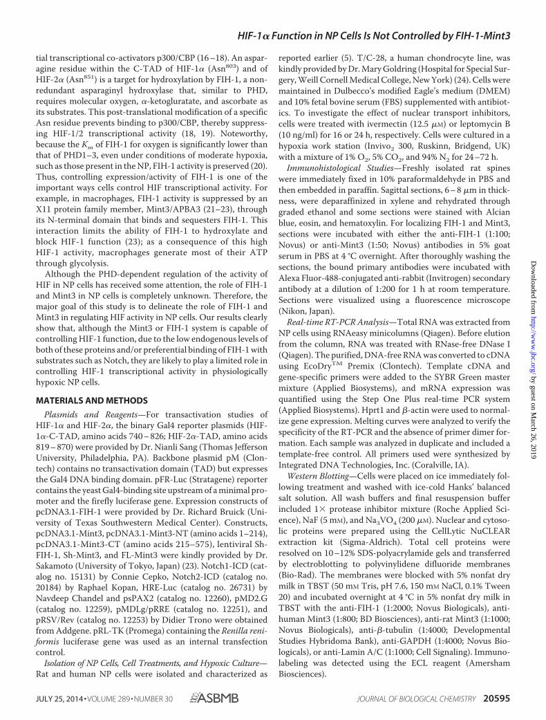
FIH-1 and Mint3 Are Expressed in Both Rat and Human NPTissues and Are Co-localized—To investigate expression ofFIH-1 and Mint3 in the intervertebral disc, we stained rat NPtissues with antibodies against FIH-1 and Mint3 (Fig. 1A). Fig.1A shows that FIH-1 and Mint3 proteins are expressed in bothNP and AF tissues. Real-time RT-PCR analysis shows lowerexpression of both Mint3 and Fih-1 mRNAs in AF tissue than inNP (Fig. 1B). We also measured expression of FIH-1 and Mint3protein in NP tissues isolated from three rats by Western blotanalysis (Fig. 1C). All three samples expressed both FIH-1 andMint3. Then to investigate the cellular localization of FIH-1 andMint3 in NP cells, we immunostained NP cells with anti-Mint3and anti-FIH-1 along with antibodies directed against RCAS1, aGolgi marker, or EEA1, an early endosome marker. Surpris-ingly, Mint3 was strongly co-localized with FIH-1 in the cellnucleus. On the other hand, although some localization ofMint3 was seen in Golgi, little or no staining was seen in earlyendosomes (Fig. 1D).
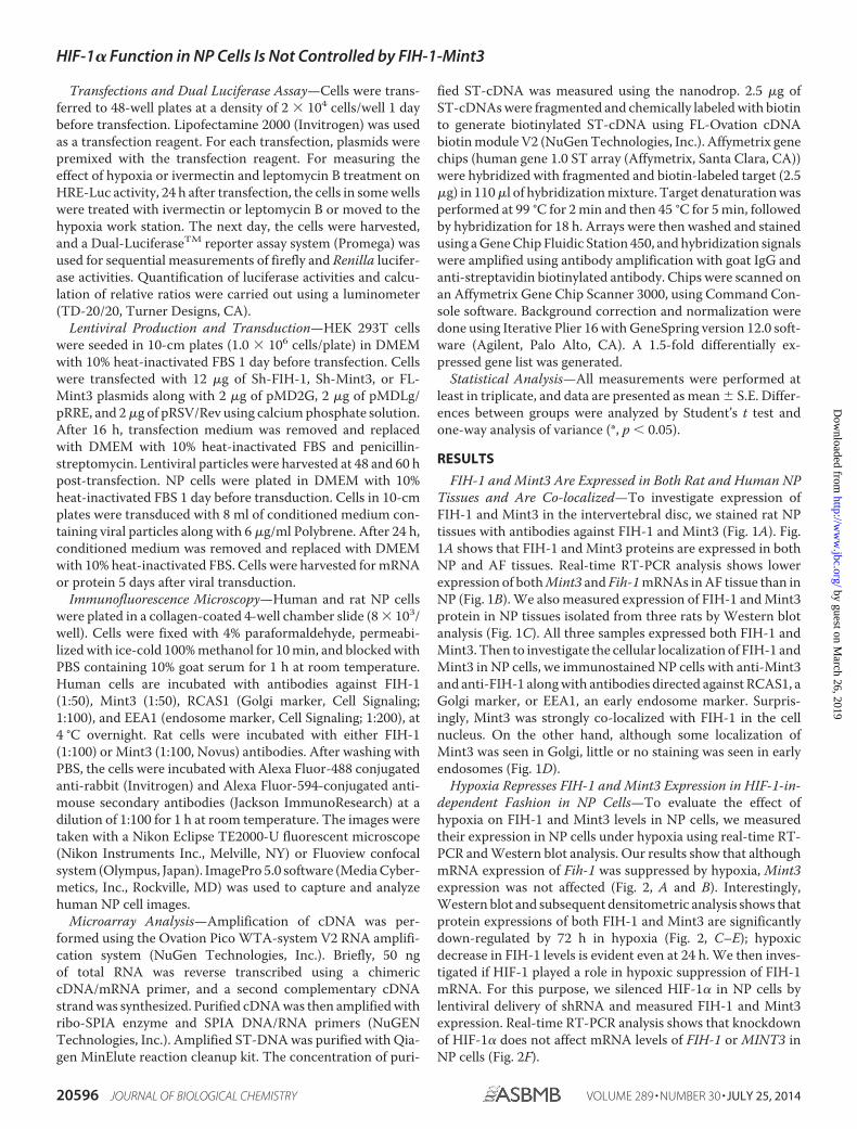
Hypoxia Represses FIH-1 and Mint3 Expression in HIF-1-in-dependent Fashion in NP Cells—To evaluate the effect ofhypoxia on FIH-1 and Mint3 levels in NP cells, we measuredtheir expression in NP cells under hypoxia using real-time RT-PCR and Western blot analysis. Our results show that althoughmRNA expression of Fih-1 was suppressed by hypoxia, Mint3expression was not affected (Fig. 2, A and B). Interestingly,Western blot and subsequent densitometric analysis shows thatprotein expressions of both FIH-1 and Mint3 are significantlydown-regulated by 72 h in hypoxia (Fig. 2, C–E); hypoxicdecrease in FIH-1 levels is evident even at 24 h. We then inves-tigated if HIF-1 played a role in hypoxic suppression of FIH-1mRNA. For this purpose, we silenced HIF-1� in NP cells bylentiviral delivery of shRNA and measured FIH-1 and Mint3expression. Real-time RT-PCR analysis shows that knockdownof HIF-1� does not affect mRNA levels of FIH-1 or MINT3 inNP cells (Fig. 2F).
HIF-1� Function in NP Cells Is Not Controlled by FIH-1-Mint3
20596 JOURNAL OF BIOLOGICAL CHEMISTRY VOLUME 289 • NUMBER 30 • JULY 25, 2014
by guest on March 26, 2019
http://ww
w.jbc.org/
Dow
nloaded from
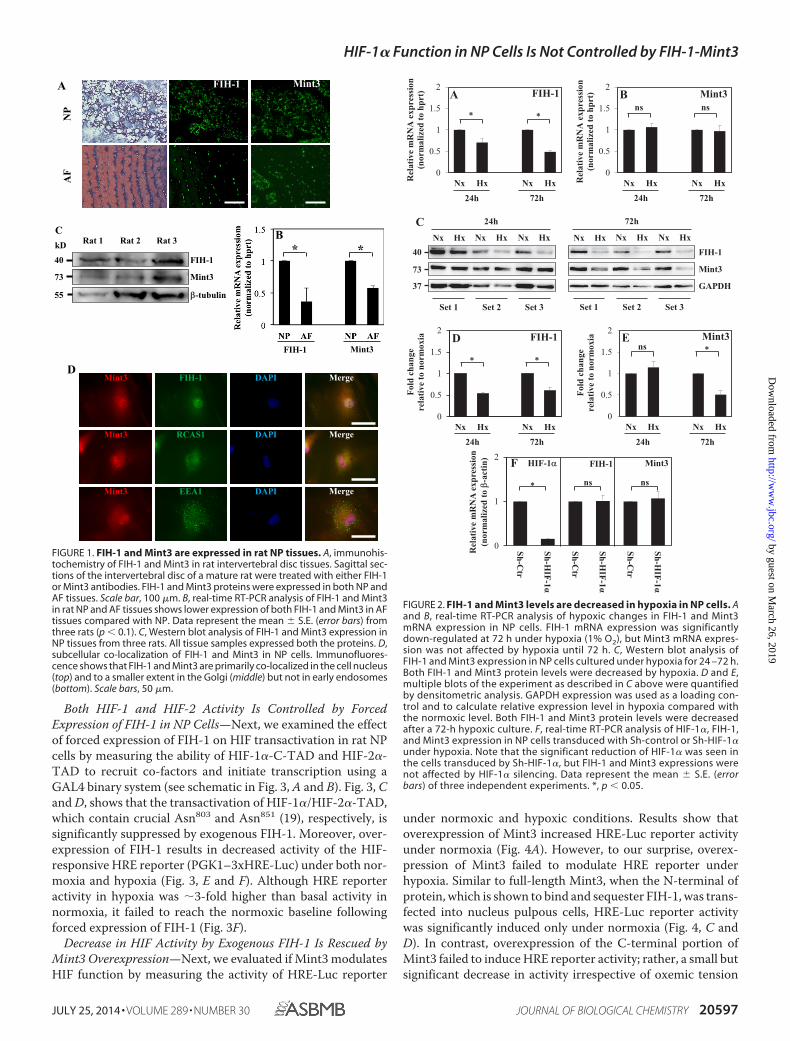
Both HIF-1 and HIF-2 Activity Is Controlled by ForcedExpression of FIH-1 in NP Cells—Next, we examined the effectof forced expression of FIH-1 on HIF transactivation in rat NPcells by measuring the ability of HIF-1�-C-TAD and HIF-2�-TAD to recruit co-factors and initiate transcription using aGAL4 binary system (see schematic in Fig. 3, A and B). Fig. 3, Cand D, shows that the transactivation of HIF-1�/HIF-2�-TAD,which contain crucial Asn803 and Asn851 (19), respectively, issignificantly suppressed by exogenous FIH-1. Moreover, over-expression of FIH-1 results in decreased activity of the HIF-responsive HRE reporter (PGK1–3xHRE-Luc) under both nor-moxia and hypoxia (Fig. 3, E and F). Although HRE reporteractivity in hypoxia was �3-fold higher than basal activity innormoxia, it failed to reach the normoxic baseline followingforced expression of FIH-1 (Fig. 3F).
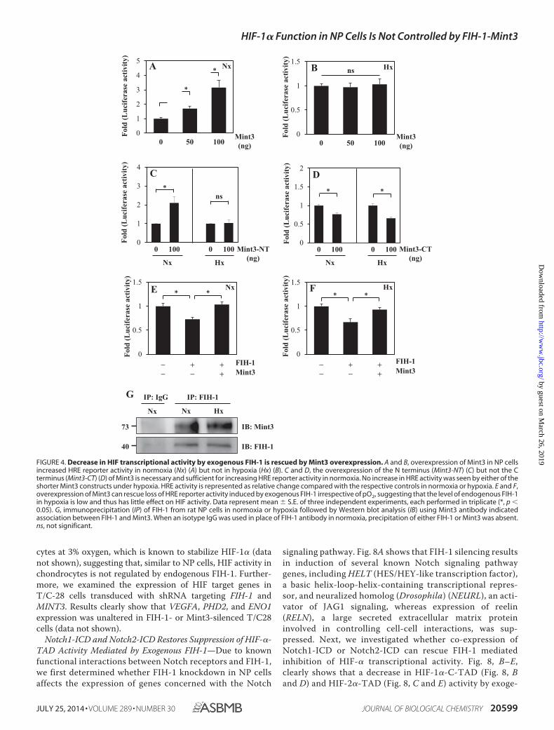
Decrease in HIF Activity by Exogenous FIH-1 Is Rescued byMint3 Overexpression—Next, we evaluated if Mint3 modulatesHIF function by measuring the activity of HRE-Luc reporter
under normoxic and hypoxic conditions. Results show thatoverexpression of Mint3 increased HRE-Luc reporter activityunder normoxia (Fig. 4A). However, to our surprise, overex-pression of Mint3 failed to modulate HRE reporter underhypoxia. Similar to full-length Mint3, when the N-terminal ofprotein, which is shown to bind and sequester FIH-1, was trans-fected into nucleus pulpous cells, HRE-Luc reporter activitywas significantly induced only under normoxia (Fig. 4, C andD). In contrast, overexpression of the C-terminal portion ofMint3 failed to induce HRE reporter activity; rather, a small butsignificant decrease in activity irrespective of oxemic tension
C
FIH-1
Mint3
β-tubulin
40
73
55
Rat 1 Rat 2 Rat 3kD
Mint3FIH-1
Merge
Merge
Merge
D
B
FIH-1 Mint3
* *
AN
PMint3FIH-1
AF
Mint3
Mint3
Mint3
FIH-1
RCAS1
EEA1
DAPI
DAPI
DAPI
Merge
Merge
Merge
FIGURE 1. FIH-1 and Mint3 are expressed in rat NP tissues. A, immunohis-tochemistry of FIH-1 and Mint3 in rat intervertebral disc tissues. Sagittal sec-tions of the intervertebral disc of a mature rat were treated with either FIH-1or Mint3 antibodies. FIH-1 and Mint3 proteins were expressed in both NP andAF tissues. Scale bar, 100 �m. B, real-time RT-PCR analysis of FIH-1 and Mint3in rat NP and AF tissues shows lower expression of both FIH-1 and Mint3 in AFtissues compared with NP. Data represent the mean � S.E. (error bars) fromthree rats (p � 0.1). C, Western blot analysis of FIH-1 and Mint3 expression inNP tissues from three rats. All tissue samples expressed both the proteins. D,subcellular co-localization of FIH-1 and Mint3 in NP cells. Immunofluores-cence shows that FIH-1 and Mint3 are primarily co-localized in the cell nucleus(top) and to a smaller extent in the Golgi (middle) but not in early endosomes(bottom). Scale bars, 50 �m.
E Mint3ns ∗
24h 72h
D FIH-1
∗∗
24h 72h
0
0.5
1
1.5
2
Nx Hx Nx HxRel
ativ
e m
RN
A e
xpre
ssio
n(n
orm
aliz
ed to
hpr
t) Mint3ns ns
24h 72h
0
0.5
1
1.5
2
Nx Hx Nx HxRel
ativ
e m
RN
A e
xpre
ssio
n(n
orm
aliz
ed to
hpr
t)
24h 72h
∗ ∗
FIH-1A B
C
Set 1 Set 2 Set 3 Set 1 Set 2 Set 3
Nx Hx
24h
FIH-1
Mint3
GAPDH
40
73
37
Nx Hx Nx Hx Nx Hx
72h
Nx Hx Nx Hx
0
1
2
Sh-Ctr
Sh-HIF-1α
Sh-Ctr
Sh-HIF-1α
Sh-Ctr
Sh-HIF-1α
Rel
ativ
e m
RN
A e
xpre
ssio
n (n
orm
aliz
ed to
β-a
ctin
)ns∗ ns
FIH-1 Mint3HIF-1αF
0
0.5
1
1.5
2
Nx Hx Nx Hx
Fold
cha
nge
rela
tive
to n
orm
oxia
0
0.5
1
1.5
2
Nx Hx Nx Hx
Fold
cha
nge
rela
tive
to n
orm
oxia
FIGURE 2. FIH-1 and Mint3 levels are decreased in hypoxia in NP cells. Aand B, real-time RT-PCR analysis of hypoxic changes in FIH-1 and Mint3mRNA expression in NP cells. FIH-1 mRNA expression was significantlydown-regulated at 72 h under hypoxia (1% O2), but Mint3 mRNA expres-sion was not affected by hypoxia until 72 h. C, Western blot analysis ofFIH-1 and Mint3 expression in NP cells cultured under hypoxia for 24 –72 h.Both FIH-1 and Mint3 protein levels were decreased by hypoxia. D and E,multiple blots of the experiment as described in C above were quantifiedby densitometric analysis. GAPDH expression was used as a loading con-trol and to calculate relative expression level in hypoxia compared withthe normoxic level. Both FIH-1 and Mint3 protein levels were decreasedafter a 72-h hypoxic culture. F, real-time RT-PCR analysis of HIF-1�, FIH-1,and Mint3 expression in NP cells transduced with Sh-control or Sh-HIF-1�under hypoxia. Note that the significant reduction of HIF-1� was seen inthe cells transduced by Sh-HIF-1�, but FIH-1 and Mint3 expressions werenot affected by HIF-1� silencing. Data represent the mean � S.E. (errorbars) of three independent experiments. *, p � 0.05.
HIF-1� Function in NP Cells Is Not Controlled by FIH-1-Mint3
JULY 25, 2014 • VOLUME 289 • NUMBER 30 JOURNAL OF BIOLOGICAL CHEMISTRY 20597
by guest on March 26, 2019
http://ww
w.jbc.org/
Dow
nloaded from
was seen. To delineate whether Mint3 mediated its action onHIF function through controlling FIH-1, we overexpressedFIH-1 in the presence or absence of exogenous Mint3 andmeasured HRE reporter activity. Fig. 4, E and F, clearly showsthat, irrespective of pO2, exogenous Mint3 can restore thedecrease in HRE reporter activity mediated by exogenousFIH-1, suggesting interaction between these two proteins(Fig. 4, E and F). To confirm whether FIH-1 binds to Mint3 inNP cells, we performed immunoprecipitation using FIH-1antibody. Fig. 4G shows that FIH-1 interacts with Mint3under both normoxia and hypoxia with similar affinity (Fig.4G).
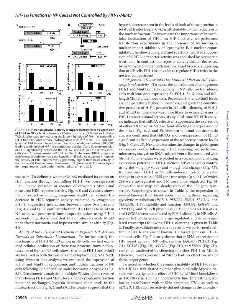
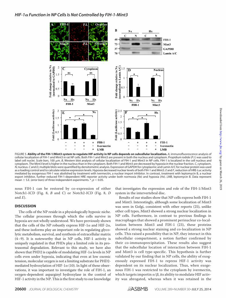
Ability of the FIH-1/Mint3 System to Regulate HIF ActivityDepends on Subcellular Localization—To further clarify themechanism of FIH-1/Mint3 action in NP cells, we first exam-ined cellular localization of these two proteins. Immunofluo-rescence of human NP cells shows that both FIH-1 and Mint3are localized in both the nucleus and cytoplasm (Fig. 5A). Next,using Western blot analysis, we evaluated the expression ofFIH-1 and Mint3 in cytoplasmic and nuclear fractions of NPcells following 72 h of culture under normoxia or hypoxia (Fig.5B). Densitometric analysis of multiple Western blots revealedthat whereas FIH-1 and Mint3 levels in the cytoplasmic fractionremained unchanged, hypoxia decreased their levels in thenuclear fraction (Fig. 5, C and D). This clearly suggests that the
hypoxic decrease seen in the levels of both of these proteins intotal cell lysates (Fig. 2, C–E) is attributable to their reduction inthe nuclear fraction. To investigate the importance of intracel-lular localization of FIH-1 on HIF-1 activity, we performedtransfection experiments in the presence of ivermectin, anuclear import inhibitor, or leptomycin B, a nuclear exportinhibitor. As shown in Fig. 5, E and F, FIH-1-mediated suppres-sion of HRE-Luc reporter activity was abolished by ivermectintreatment. In contrast, the reporter activity further decreasedby leptomycin B under both normoxia and hypoxia, suggestingthat in NP cells, FIH-1 is only able to regulate HIF activity in thenuclear compartment.
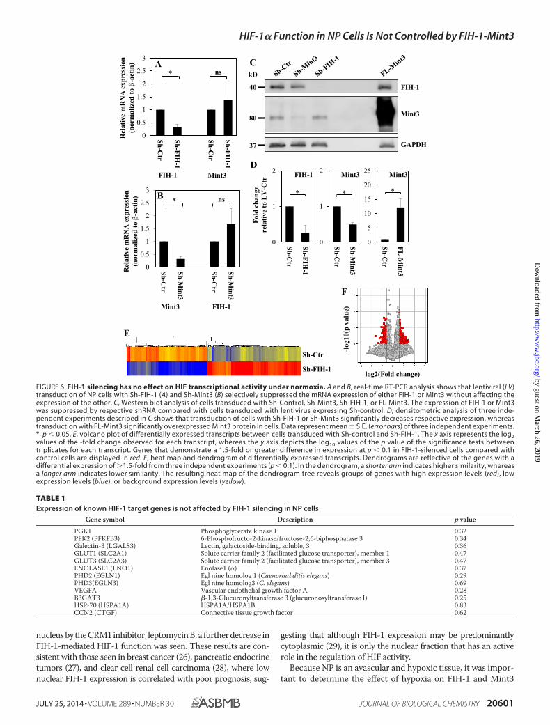
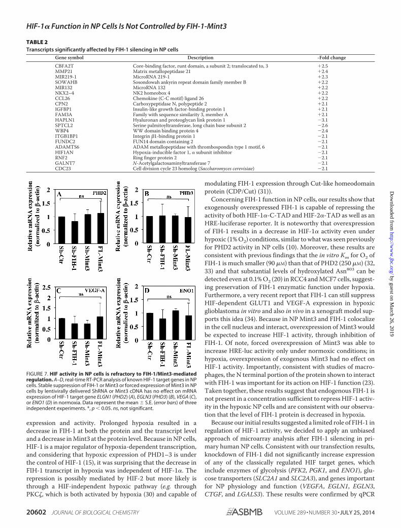
Endogenous FIH-1/Mint3 Has Minimal Effect on HIF Tran-scriptional Activity—To assess the contribution of endogenousFIH-1 and Mint3 on HIF-1 activity in NP cells, we transducedcells with lentivirus expressing Sh-FIH-1, Sh-Mint3, and full-length Mint3 under normoxia. Because FIH-1 and Mint3 levelsare comparatively higher in normoxia, and given the constitu-tive presence of HIF-1 protein in NP cells, silencing of FIH-1and Mint3 in normoxia was more likely to evince changes inHIF-1 transcriptional activity, if any. Real-time RT-PCR analy-sis indicates that shRNA selectively suppressed the expressionof either FIH-1 or MINT3 without affecting the expression ofthe other (Fig. 6, A and B). Western blot and densitometricanalysis confirmed that shRNAs and overexpression of Mint3significantly affected expression of the respective protein levels(Fig. 6, C and D). Next, to determine the changes in global geneexpression profile following FIH-1 silencing, we performedmicroarray analysis on RNA isolated from cells transduced withSh-FIH-1. The values were plotted in a volcano plot analyzingexpression patterns in FIH-1-silenced NP cells versus controlusing the �log10(p value) and �log2(-fold change) (Fig. 6E).Knockdown of FIH-1 in NP cells induced 1.5-fold or greaterchange in expression of 535 gene transcripts (p � 0.1), of which289 were up-regulated and 246 were down-regulated. Fig. 6Fshows the heat map and dendrogram of the 535 gene tran-scripts. Surprisingly, as shown in Table 1, the expression ofseveral known HIF-1 target genes, including those regulatingglycolytic metabolism (PGK-1, PFKFB2, ENO1, SLC2A1, andSLC2A3), HIF-1 stability and function (EGLN1, EGLN3, andHSPA1A), and NP cell physiology (CTGF, LGALS3, B3GAT3,and VEGFA), were not affected by FIH-1 silencing in NP cells. Apartial list of the maximally up-regulated and down-regu-lated transcripts following FIH-1 silencing is given in Table2. Finally, to validate microarray results, we performed real-time RT-PCR analysis of known HIF target genes in FIH-1-silenced cells. Fig. 7 clearly shows that mRNA expression ofHIF target genes in NP cells, such as EGLN1 (PHD2) (Fig.7A), EGLN3 (Fig. 7B), VEGFA (Fig. 7C), and ENO1 (Fig. 7D),remained unaffected by silencing of either FIH-1 or Mint3.Likewise, overexpression of Mint3 had no effect on any ofthese target genes.
To ascertain whether the seeming inability of FIH-1 to regu-late HIF is a trait shared by other physiologically hypoxic tis-sues, we investigated the effect of FIH-1 and Mint3 knockdownin T/C-28 cells, a human chondrocyte line. Interestingly, fol-lowing transfection with shRNA targeting FIH-1 as well asMINT3, HRE reporter activity did not change in the chondro-
FIGURE 3. HIF transcriptional activity is suppressed by forced expressionof FIH-1 in NP cells. A, schematic of Gal4 constructs of HIF-1�- and HIF-2�-TAD. B, schematic summarizing the known function of FIH-1 in controllingHIF-1 transcriptional activity. Hydroxylation of Asn803 in HIF-1�-C-TAD cata-lyzed by FIH-1 blocks interaction with transcriptional co-activators p300/CBP,leading to diminished HIF-1 transcriptional activity. C and D, overexpressionof FIH-1 significantly decreased the HIF-1�- and HIF-2�-TAD activity in NPcells. E and F, overexpression of FIH-1 resulted in decreased HRE-Luc reporteractivity under both normoxia and hypoxia in NP cells. As expected, in hypoxiathe activity of HRE reporter was significantly higher than basal activity innormoxia (NX). Data represent the mean � S.E. (error bars) of three indepen-dent experiments, each performed in triplicate. *, p � 0.05.
HIF-1� Function in NP Cells Is Not Controlled by FIH-1-Mint3
20598 JOURNAL OF BIOLOGICAL CHEMISTRY VOLUME 289 • NUMBER 30 • JULY 25, 2014
by guest on March 26, 2019
http://ww
w.jbc.org/
Dow
nloaded from
cytes at 3% oxygen, which is known to stabilize HIF-1� (datanot shown), suggesting that, similar to NP cells, HIF activity inchondrocytes is not regulated by endogenous FIH-1. Further-more, we examined the expression of HIF target genes inT/C-28 cells transduced with shRNA targeting FIH-1 andMINT3. Results clearly show that VEGFA, PHD2, and ENO1expression was unaltered in FIH-1- or Mint3-silenced T/C28cells (data not shown).
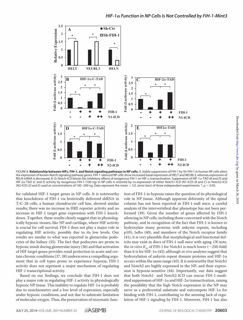
Notch1-ICD and Notch2-ICD Restores Suppression of HIF-�-TAD Activity Mediated by Exogenous FIH-1—Due to knownfunctional interactions between Notch receptors and FIH-1,we first determined whether FIH-1 knockdown in NP cellsaffects the expression of genes concerned with the Notch
signaling pathway. Fig. 8A shows that FIH-1 silencing resultsin induction of several known Notch signaling pathwaygenes, including HELT (HES/HEY-like transcription factor),a basic helix-loop-helix-containing transcriptional repres-sor, and neuralized homolog (Drosophila) (NEURL), an acti-vator of JAG1 signaling, whereas expression of reelin(RELN), a large secreted extracellular matrix proteininvolved in controlling cell-cell interactions, was sup-pressed. Next, we investigated whether co-expression ofNotch1-ICD or Notch2-ICD can rescue FIH-1 mediatedinhibition of HIF-� transcriptional activity. Fig. 8, B–E,clearly shows that a decrease in HIF-1�-C-TAD (Fig. 8, Band D) and HIF-2�-TAD (Fig. 8, C and E) activity by exoge-
A
C
E F
Mint3-NT (ng)
∗ns
Nx Hx
D
Mint3-CT (ng)
∗ ∗
Nx Hx
Mint3 (ng)
∗
∗ Nx B
Mint3 (ng)
ns Hx
∗ ∗
Mint3FIH-1
Nx∗ ∗
Mint3FIH-1
Hx
IB: Mint3
IB: FIH-1
Nx Nx Hx
IP: IgG IP: FIH-1
73
40
G
0
0.5
1
1.5
0 50 100
Fold
(Luc
ifera
se a
ctiv
ity)
0
0.5
1
1.5
2
0 100 0 100
Fold
(Luc
ifera
se a
ctiv
ity)
0
1
2
3
4
0 100 0 100
Fold
(Luc
ifera
se a
ctiv
ity)
0
0.5
1
1.5
−−
+−
++
Fold
(Luc
ifera
se a
ctiv
ity)
0
0.5
1
1.5
−−
+−
++
Fold
(Luc
ifera
se a
ctiv
ity)
0
1
2
3
4
5
0 50 100
Fold
(Luc
ifera
se a
ctiv
ity)
FIGURE 4. Decrease in HIF transcriptional activity by exogenous FIH-1 is rescued by Mint3 overexpression. A and B, overexpression of Mint3 in NP cellsincreased HRE reporter activity in normoxia (Nx) (A) but not in hypoxia (Hx) (B). C and D, the overexpression of the N terminus (Mint3-NT) (C) but not the Cterminus (Mint3-CT) (D) of Mint3 is necessary and sufficient for increasing HRE reporter activity in normoxia. No increase in HRE activity was seen by either of theshorter Mint3 constructs under hypoxia. HRE activity is represented as relative change compared with the respective controls in normoxia or hypoxia. E and F,overexpression of Mint3 can rescue loss of HRE reporter activity induced by exogenous FIH-1 irrespective of pO2, suggesting that the level of endogenous FIH-1in hypoxia is low and thus has little effect on HIF activity. Data represent mean � S.E. of three independent experiments, each performed in triplicate (*, p �0.05). G, immunoprecipitation (IP) of FIH-1 from rat NP cells in normoxia or hypoxia followed by Western blot analysis (IB) using Mint3 antibody indicatedassociation between FIH-1 and Mint3. When an isotype IgG was used in place of FIH-1 antibody in normoxia, precipitation of either FIH-1 or Mint3 was absent.ns, not significant.
HIF-1� Function in NP Cells Is Not Controlled by FIH-1-Mint3
JULY 25, 2014 • VOLUME 289 • NUMBER 30 JOURNAL OF BIOLOGICAL CHEMISTRY 20599
by guest on March 26, 2019
http://ww
w.jbc.org/
Dow
nloaded from
nous FIH-1 can be restored by co-expression of eitherNotch1-ICD (Fig. 8, B and C) or Notch2-ICD (Fig. 8, Dand E).
DISCUSSION
The cells of the NP reside in a physiologically hypoxic niche.The cellular processes through which the cells survive inhypoxia are not wholly understood. We have previously shownthat the cells of the NP robustly express HIF-1� and HIF-2�,and these isoforms play an important role in regulating glyco-lytic metabolism, survival, and synthesis of extracellular matrix(4 –9). It is noteworthy that in NP cells, HIF-1 activity isuniquely regulated in that PHDs play a limited role in its pro-teasomal degradation. Relevant to this study, we have alsoshown that PHD2 is capable of modulating HIF-1� levels in NPcells even under hypoxia, indicating that even at low oxemictension, molecular oxygen is not a limiting substrate for PHD2-mediated hydroxylation of HIF-1� (10). In light of these obser-vations, it was important to investigate the role of FIH-1, anoxygen-dependent asparginyl hydroxylase in the control ofHIF-1 activity in the NP. This is the first study to our knowledge
that investigates the expression and role of the FIH-1/Mint3system in the intervertebral disc.
Results of our studies show that NP cells express both FIH-1and Mint3. Interestingly, although some localization of Mint3was seen in Golgi, consistent with other reports (25), unlikeother cell types, Mint3 showed a strong nuclear localization inNP cells. Furthermore, in contrast to previous findings inmacrophages that showed a prominent perinuclear co-local-ization between Mint3 and FIH-1 (23), these proteinsshowed a strong nuclear staining and co-localization in NPcells. This raised a possibility that in NP, they interact in thissubcellular compartment, a notion further confirmed bytheir co-immunoprecipitation. These results also suggestthat the subcellular location of interaction between FIH-1and Mint3 is cell type-specific. This hypothesis is furthervalidated by our finding that in NP cells, the ability of exog-enously expressed FIH-1 to repress HIF-1 activity wasdependent on its nuclear localization. Thus, when exoge-nous FIH-1 was restricted to the cytoplasm by ivermectin,which targets importin �/�, its ability to modulate HIF activ-ity was abrogated, whereas when it was retained in the
FIGURE 5. Ability of the FIH-1/Mint3 system to regulate HIF activity in NP cells depends on subcellular localization. A, immunofluorescence analysis ofcellular localization of FIH-1 and Mint3 in rat NP cells. Both FIH-1 and Mint3 are present in both the nucleus and cytoplasm. Propidium iodide (P.I.) was used tolabel cell nuclei. Scale bars, 100 �m. B, Western blot analysis of cellular localization of FIH-1 and Mint3 in NP cells. FIH-1 is localized in the cell nucleus andcytoplasm. The Mint3 level is higher in the nucleus than in the cytoplasm. Both FIH-1 and Mint3 are decreased by hypoxia in the nuclear fraction. C, cytoplasm.N, nucleus. C and D, multiple blots were quantified by densitometric analysis. Expression of GAPDH for cytoplasmic and Lamin A/C for nuclear protein was usedas a loading control and to calculate relative expression levels. Hypoxia decreased nuclear levels of both FIH-1 and Mint3. E and F, reduction of HRE-Luc activitymediated by exogenous FIH-1 was abolished by treatment with ivermectin, a nuclear import inhibitor. In contrast, treatment with leptomycin B, a nuclearexport inhibitor, further reduced FIH-1-dependent HRE reporter activity under both normoxia (Nx) and hypoxia (Hx). LMB, leptomycin B. Data representmean � S.E. (error bars) of three independent experiments. *, p � 0.05.
HIF-1� Function in NP Cells Is Not Controlled by FIH-1-Mint3
20600 JOURNAL OF BIOLOGICAL CHEMISTRY VOLUME 289 • NUMBER 30 • JULY 25, 2014
by guest on March 26, 2019
http://ww
w.jbc.org/
Dow
nloaded from
nucleus by the CRM1 inhibitor, leptomycin B, a further decrease inFIH-1-mediated HIF-1 function was seen. These results are con-sistent with those seen in breast cancer (26), pancreatic endocrinetumors (27), and clear cell renal cell carcinoma (28), where lownuclear FIH-1 expression is correlated with poor prognosis, sug-
gesting that although FIH-1 expression may be predominantlycytoplasmic (29), it is only the nuclear fraction that has an activerole in the regulation of HIF activity.
Because NP is an avascular and hypoxic tissue, it was impor-tant to determine the effect of hypoxia on FIH-1 and Mint3
0
0.5
1
1.5
2
2.5
3
Sh-Ctr
Sh-FIH-1
Sh-Ctr
Sh-FIH-1
Rel
ativ
e m
RN
A ex
pres
sion
(nor
mal
ized
to β
-act
in)
FIH-1 Mint3
∗ ns
0
0.5
1
1.5
2
2.5
3
Sh-Ctr
Sh-Mint3
Sh-Ctr
Sh-Mint3
Rel
ativ
e m
RN
A e
xpre
ssio
n(n
orm
aliz
ed to
β-a
ctin
)
FIH-1
Mint3
GAPDH
40
80
37
kDA C
FIH-1Mint3
B ∗ ns
D
Sh-Ctr
Sh-FIH-1
-log1
0(p
valu
e)
log2(Fold change)
E
F
0
5
10
15
20
25Sh-C
tr
FL-Mint3
FIH-1
0
1
2
Sh-Ctr
Sh-Mint3
∗
Mint3Mint3
∗∗
0
1
2
Sh-Ctr
Sh-FIH-1
Fold
cha
nge
rela
tive
to L
V- C
tr
FIGURE 6. FIH-1 silencing has no effect on HIF transcriptional activity under normoxia. A and B, real-time RT-PCR analysis shows that lentiviral (LV)transduction of NP cells with Sh-FIH-1 (A) and Sh-Mint3 (B) selectively suppressed the mRNA expression of either FIH-1 or Mint3 without affecting theexpression of the other. C, Western blot analysis of cells transduced with Sh-Control, Sh-Mint3, Sh-FIH-1, or FL-Mint3. The expression of FIH-1 or Mint3was suppressed by respective shRNA compared with cells transduced with lentivirus expressing Sh-control. D, densitometric analysis of three inde-pendent experiments described in C shows that transduction of cells with Sh-FIH-1 or Sh-Mint3 significantly decreases respective expression, whereastransduction with FL-Mint3 significantly overexpressed Mint3 protein in cells. Data represent mean � S.E. (error bars) of three independent experiments.*, p � 0.05. E, volcano plot of differentially expressed transcripts between cells transduced with Sh-control and Sh-FIH-1. The x axis represents the log2values of the -fold change observed for each transcript, whereas the y axis depicts the log10 values of the p value of the significance tests betweentriplicates for each transcript. Genes that demonstrate a 1.5-fold or greater difference in expression at p � 0.1 in FIH-1-silenced cells compared withcontrol cells are displayed in red. F, heat map and dendrogram of differentially expressed transcripts. Dendrograms are reflective of the genes with adifferential expression of �1.5-fold from three independent experiments (p � 0.1). In the dendrogram, a shorter arm indicates higher similarity, whereasa longer arm indicates lower similarity. The resulting heat map of the dendrogram tree reveals groups of genes with high expression levels (red), lowexpression levels (blue), or background expression levels (yellow).
TABLE 1Expression of known HIF-1 target genes is not affected by FIH-1 silencing in NP cells
Gene symbol Description p value
PGK1 Phosphoglycerate kinase 1 0.32PFK2 (PFKFB3) 6-Phosphofructo-2-kinase/fructose-2,6-biphosphatase 3 0.34Galectin-3 (LGALS3) Lectin, galactoside-binding, soluble, 3 0.36GLUT1 (SLC2A1) Solute carrier family 2 (facilitated glucose transporter), member 1 0.47GLUT3 (SLC2A3) Solute carrier family 2 (facilitated glucose transporter), member 3 0.47ENOLASE1 (ENO1) Enolase1 (�) 0.37PHD2 (EGLN1) Egl nine homolog 1 (Caenorhabditis elegans) 0.29PHD3(EGLN3) Egl nine homolog3 (C. elegans) 0.69VEGFA Vascular endothelial growth factor A 0.28B3GAT3 �-1,3-Glucuronyltransferase 3 (glucuronosyltransferase I) 0.25HSP-70 (HSPA1A) HSPA1A/HSPA1B 0.83CCN2 (CTGF) Connective tissue growth factor 0.62
HIF-1� Function in NP Cells Is Not Controlled by FIH-1-Mint3
JULY 25, 2014 • VOLUME 289 • NUMBER 30 JOURNAL OF BIOLOGICAL CHEMISTRY 20601
by guest on March 26, 2019
http://ww
w.jbc.org/
Dow
nloaded from
expression and activity. Prolonged hypoxia resulted in adecrease in FIH-1 at both the protein and the transcript leveland a decrease in Mint3 at the protein level. Because in NP cells,HIF-1 is a major regulator of hypoxia-dependent transcription,and considering that hypoxic expression of PHD1–3 is underthe control of HIF-1 (15), it was surprising that the decrease inFIH-1 transcript in hypoxia was independent of HIF-1�. Therepression is possibly mediated by HIF-2 but more likely isthrough a HIF-independent hypoxic pathway (e.g. throughPKC�, which is both activated by hypoxia (30) and capable of
modulating FIH-1 expression through Cut-like homeodomainprotein (CDP/Cut) (31)).
Concerning FIH-1 function in NP cells, our results show thatexogenously overexpressed FIH-1 is capable of repressing theactivity of both HIF-1�-C-TAD and HIF-2�-TAD as well as anHRE-luciferase reporter. It is noteworthy that overexpressionof FIH-1 results in a decrease in HIF-1� activity even underhypoxic (1% O2) conditions, similar to what was seen previouslyfor PHD2 activity in NP cells (10). Moreover, these results areconsistent with previous findings that the in vitro Km for O2 ofFIH-1 is much smaller (90 �M) than that of PHD2 (250 �M) (32,33) and that substantial levels of hydroxylated Asn803 can bedetected even at 0.1% O2 (20) in RCC4 and MCF7 cells, suggest-ing preservation of FIH-1 enzymatic function under hypoxia.Furthermore, a very recent report that FIH-1 can still suppressHIF-dependent GLUT1 and VEGF-A expression in hypoxicglioblastoma in vitro and also in vivo in a xenograft model sup-ports this idea (34). Because in NP Mint3 and FIH-1 colocalizein the cell nucleus and interact, overexpression of Mint3 wouldbe expected to increase HIF-1 activity, through inhibition ofFIH-1. Of note, forced overexpression of Mint3 was able toincrease HRE-luc activity only under normoxic conditions; inhypoxia, overexpression of exogenous Mint3 had no effect onHIF-1 activity. Importantly, consistent with studies of macro-phages, the N terminal portion of the protein shown to interactwith FIH-1 was important for its action on HIF-1 function (23).Taken together, these results suggest that endogenous FIH-1 isnot present in a concentration sufficient to repress HIF-1 activ-ity in the hypoxic NP cells and are consistent with our observa-tion that the level of FIH-1 protein is decreased in hypoxia.
Because our initial results suggested a limited role of FIH-1 inregulation of HIF-1 activity, we decided to apply an unbiasedapproach of microarray analysis after FIH-1 silencing in pri-mary human NP cells. Consistent with our transfection results,knockdown of FIH-1 did not significantly increase expressionof any of the classically regulated HIF target genes, whichinclude enzymes of glycolysis (PFK2, PGK1, and ENO1), glu-cose transporters (SLC2A1 and SLC2A3), and genes importantfor NP physiology and function (VEGFA, EGLN1, EGLN3,CTGF, and LGALS3). These results were confirmed by qPCR
TABLE 2Transcripts significantly affected by FIH-1 silencing in NP cells
Gene symbol Description -Fold change
CBFA2T Core-binding factor, runt domain, a subunit 2; translocated to, 3 �2.5MMP21 Matrix metallopeptidase 21 �2.4MIR219-1 MicroRNA 219-1 �2.3SOWAHB Sosondowah ankyrin repeat domain family member B �2.2MIR132 MicroRNA 132 �2.2NKX2–4 NK2 homeobox 4 �2.2CCL26 Chemokine (C-C motif) ligand 26 �2.2CPN2 Carboxypeptidase N, polypeptide 2 �2.1IGFBP1 Insulin-like growth factor-binding protein 1 �2.1FAM3A Family with sequence similarity 3, member A �2.1HAPLN1 Hyaluronan and proteoglycan link protein 1 �3.1SPTCL2 Serine palmitoyltransferase, long chain base subunit 2 �2.6WBP4 WW domain binding protein 4 �2.4ITGB1BP1 Integrin �1-binding protein 1 �2.1FUNDC2 FUN14 domain containing 2 �2.1ADAMTS6 ADAM metallopeptidase with thrombospondin type 1 motif, 6 �2.1HIF1AN Hypoxia-inducible factor 1, � subunit inhibitor �2.1RNF2 Ring finger protein 2 �2.1GALNT7 N-Acetylgalactosaminyltransferase 7 �2.1CDC23 Cell division cycle 23 homolog (Saccharomyces cerevisiae) �2.1
FIGURE 7. HIF activity in NP cells is refractory to FIH-1/Mint3-mediatedregulation. A–D, real-time RT-PCR analysis of known HIF-1 target genes in NPcells. Stable suppression of FIH-1 or Mint3 or forced expression of Mint3 in NPcells by lentivirally delivered ShRNA or Mint3 cDNA has no effect on mRNAexpression of HIF-1 target gene ELGN1 (PHD2) (A), EGLN3 (PHD3) (B), VEGA (C),or ENO1 (D) in normoxia. Data represent the mean � S.E. (error bars) of threeindependent experiments. *, p � 0.05. ns, not significant.
HIF-1� Function in NP Cells Is Not Controlled by FIH-1-Mint3
20602 JOURNAL OF BIOLOGICAL CHEMISTRY VOLUME 289 • NUMBER 30 • JULY 25, 2014
by guest on March 26, 2019
http://ww
w.jbc.org/
Dow
nloaded from
for validated HIF-1 target genes in NP cells. It is noteworthythat knockdown of FIH-1 via lentivirally delivered shRNA inT/C-28 cells, a human chondrocyte cell line, showed similarresults; there was no increase in HRE reporter activity and noincrease in HIF-1 target gene expression with FIH-1 knock-down. Together, these results clearly suggest that in physiolog-ically hypoxic tissues, like NP and cartilage, where HIF activityis crucial for cell survival, FIH-1 does not play a major role inregulating HIF activity, possibly due to its low levels. Ourresults are similar to what was reported in glomerular podo-cytes of the kidney (35). The fact that podocytes are prone tohypoxic insult during glomerular injury (36) and that activationof HIF target genes provides renal protection in acute and cer-tain chronic conditions (37, 38) underscores a compelling argu-ment that in cell types prone to experience hypoxia, FIH-1activity does not represent a major mechanism of regulatingHIF-1 transcriptional activity.
Based on our findings, we conclude that FIH-1 does notplay a major role in regulating HIF-1 activity in physiologicallyhypoxic NP tissue. This inability to regulate HIF-1� is probablydue to stoichiometry and a low level of expression, especiallyunder hypoxic conditions, and not due to substrate limitationof molecular oxygen. Thus, the preservation of enzymatic func-
tion of FIH-1 in hypoxia raises the question of its physiologicalrole in NP tissue. Although apparent deformity of the spinalcolumn has not been reported in FIH-1-null mice, a carefulanalysis of the intervertebral disc phenotype has not been per-formed (39). Given the number of genes affected by FIH-1silencing in NP cells, including those concerned with the Notchpathway, and in recognition of the fact that FIH-1 is known tohydroxylate many proteins with ankyrin repeats, includingp105, I�B� (40), and members of the Notch receptor family(41), it is very plausible that morphological and functional def-icits may exist in discs of FIH-1 null mice with aging. Of note,the in vitro Km of FIH-1 for Notch1 is much lower (�250-fold)than it is for HIF-1� (42), although in vivo analyses suggest thathydroxylation of ankyrin repeat domain proteins and HIF-1�occurs within the same range (43). It is noteworthy that Notch1and Notch2 are highly expressed in the NP, and their expres-sion is hypoxia-sensitive (44). Importantly, our data suggestthat both Notch1- and Notch2-ICD can rescue FIH-1-medi-ated suppression of HIF-1� and HIF-2� transactivation, raisingthe possibility that the high Notch expression in the NP mayserve as a preferential substrate and outcompete HIF-1� forbinding with FIH-1, contributing to the seeming lack of regu-lation of HIF-1 signaling by FIH-1. Moreover, FIH-1 has also
00.5
11.5
22.5
3
1 2 3 4
Fold
(Luc
ifera
se a
ctiv
ity)
FIH-1N1-ICD
- + + +- - + ++
C HIF-2α-TAD
*
* *
E HIF2α-TAD
FIH-1N2-ICD
- + + +- - + ++
0
0.5
1
1.5
2Fo
ld (L
ucife
rase
act
ivity
)
** *
00.5
11.5
22.5
3
1 2 3 4
Fold
(Luc
ifera
se a
ctiv
ity)
HIF-1α-C-TAD
FIH-1N1-ICD
- + + +- - + ++
B
*
* *
FIH-1N2-ICD
- + + +- - + ++
D HIF1α-C-TAD
0
0.5
1
1.5
2
Fold
(Luc
ifera
se a
ctiv
ity)
** *
HELT NEURL3 RELN
* *
*
A
0
0.5
1
1.5
2
2.5
Rel
ativ
e E
xpre
ssio
n Sh-Ctr
Sh-FIH-1
FIGURE 8. Relationship between HIFs, FIH-1, and Notch signaling pathways in NP cells. A, stable suppression of FIH-1 by Sh-FIH-1 in human NP cells altersthe expression of known Notch signaling pathway genes. FIH-1-silenced NP cells show increased basal expression of HELT and NEURL3, whereas expression ofRELN mRNA is decreased. B–D, Notch-ICD blocks the inhibitory effects of exogenous FIH-1 on HIF-� transactivation. Suppression of HIF-1�-TAD (B and D) andHIF-2�-TAD (C and E) activity by exogenous FIH-1 (100 ng) in NP cells is restored by co-expression of either Notch1-ICD (N1-ICD) (B and C) or Notch2-ICD(N2-ICD) (D and E) used at concentrations of 100 –200 ng. Data represent the mean � S.E. (error bars) of three independent experiments. *, p � 0.05.
HIF-1� Function in NP Cells Is Not Controlled by FIH-1-Mint3
JULY 25, 2014 • VOLUME 289 • NUMBER 30 JOURNAL OF BIOLOGICAL CHEMISTRY 20603
by guest on March 26, 2019
http://ww
w.jbc.org/
Dow
nloaded from
recently been reported to interact with and inhibit the proapo-ptotic protein Bax, independent of its hydroxylase activity (45).In light of our recent finding of PHD3 activating p65 signalingin NP cells independent of proline hydroxylation (46), it isimportant to also consider the potential role of FIH-1 inde-pendent of asparagine hydroxylation in NP physiology.
Acknowledgments—We thank Renata Skubutyte (Thomas JeffersonUniversity) for technical assistance with the transactivation assaysand Dr. Motomi Enomoto-Iwamoto (Children’s Hospital of Philadel-phia) for kind assistance with fluorescence microscopy.
REFERENCES1. Feng, H., Danfelter, M., Stromqvist, B., and Heinegard, D. (2006) Extra-
cellular matrix in disc degeneration. J. Bone Joint Surg. Am. 88, 25–292. Setton L. A., and Chen, J. (2006) Mechanobiology of the intervertebral disc
and relevance to disc degeneration. J. Bone Joint Surg. Am. 88, 52–573. Kauppila L. I. (1995) Ingrowth of blood vessels in disc degeneration. An-
giographic and histological studies of cadaveric spines. J. Bone Joint Surg.Am. 77, 26 –31
4. Rajpurohit, R., Risbud, M. V., Ducheyne, P., Vresilovic, E. J., and Shapiro,I. M. (2002) Phenotypic characteristics of the nucleus pulposus. Expres-sion of hypoxia inducing factor-1, glucose transporter-1 and MMP-2. CellTissue Res. 308, 401– 407
5. Risbud, M. V., Guttapalli, A., Stokes, D. G., Hawkins, D., Danielson, K. G.,Schaer, T. P., Albert, T. J., and Shapiro, I. M. (2006) Nucleus pulposus cellsexpress HIF-1� under normoxic culture conditions. A metabolic adapta-tion to the intervertebral disc microenvironment. J. Cell Biochem. 98,152–159
6. Zeng, Y., Danielson, K. G., Albert, T. J., Shapiro, I. M., and Risbud, M. V.(2007) HIF-1� is a regulator of Galectin-3 expression in the intervertebraldisc. J. Bone Miner. Res. 22, 1851–1861
7. Skubutyte, R., Markova, D., Freeman, T. A., Anderson, D. G., Dion, A. S.,Williams, C. J., Shapiro, I. M., and Risbud, M. V. (2010) Hypoxia-induciblefactor regulation of ANK expression in nucleus pulposus cells. Possibleimplications in controlling dystrophic mineralization in the intervertebraldisc. Arthritis Rheum. 62, 2707–2715
8. Agrawal, A., Guttapalli, A., Narayan, S., Albert, T. J., Shapiro, I. M., andRisbud, M. V. (2007) Normoxic stabilization of HIF-1� drives glycolyticmetabolism and regulates aggrecan gene expression in nucleus pulposuscells of the rat intervertebral disk. Am. J. Physiol. Cell Physiol. 293,C621–C631
9. Gogate, S. S., Nasser, R., Shapiro, I. M., and Risbud, M. V. (2011) Hypoxicregulation of �-1,3-glucuronyltransferase 1 expression in nucleus pulpo-sus cells of the rat intervertebral disc. Role of hypoxia-inducible factorproteins. Arthritis Rheum. 63, 1950 –1960
10. Fujita, N., Chiba, K., Shapiro, I. M., and Risbud, M. V. (2012) HIF-1� andHIF-2� degradation is differentially regulated in nucleus pulposus cells ofthe intervertebral disc. J. Bone Miner. Res. 27, 401– 412
11. Gogate, S. S., Fujita, N., Skubutyte, R., Shapiro, I. M., and Risbud, M. V.(2012) Tonicity enhancer binding protein (TonEBP) and hypoxia-induc-ible factor (HIF) coordinate heat shock protein 70 (Hsp70) expression inhypoxic nucleus pulposus cells. Role of Hsp70 in HIF-1� degradation.J. Bone Miner. Res. 27, 1106 –1117
12. Chan, D. A., Sutphin, P. D., Yen, S. E., and Giaccia, A. J. (2005) Coordinateregulation of the oxygen-dependent degradation domains of hypoxia-in-ducible factor 1�. Mol. Cell Biol. 25, 6415– 6426
13. Berra, E., Benizri, E., Ginouvès, A., Volmat, V., Roux, D., and Pouysségur,J. (2003) HIF prolyl-hydroxylase 2 is the key oxygen sensor setting lowsteady-state levels of HIF-1� in normoxia. EMBO J. 22, 4082– 4090
14. Kaelin, W. G., Jr. (2008) The von Hippel-Lindau tumour suppressor pro-tein: O2 sensing and cancer. Nat. Rev. Cancer. 8, 865– 873
15. Fujita, N., Markova, D., Anderson, D. G., Chiba, K., Toyama Y., ShapiroI. M., and Risbud M. V. (2012) Expression of prolyl hydroxylases (PHDs) isselectively controlled by HIF-1 and HIF-2 proteins in nucleus pulposus
cells of the intervertebral disc. J. Biol. Chem. 287, 16975–1698616. Kasper, L. H., Boussouar, F., Boyd, K., Xu, W., Biesen, M., Rehg, J.,
Baudino, T. A., Cleveland, J. L., and Brindle, P. K. (2005) Two transactiva-tion mechanisms cooperate for the bulk of HIF-1-responsive gene expres-sion. EMBO J. 24, 3846 –3858
17. Arany, Z., Huang, L. E., Eckner, R., Bhattacharya, S., Jiang, C., Goldberg,M. A., Bunn, H. F., and Livingston, D. M. (1996) An essential role forp300/CBP in the cellular response to hypoxia. Proc. Natl. Acad. Sci. U.S.A.93, 12969 –12973
18. Lando, D., Peet, D. J., Gorman, J. J., Whelan, D. A., Whitelaw, M. L., andBruick, R. K. (2002) FIH-1 is an asparaginyl hydroxylase enzyme that reg-ulates the transcriptional activity of hypoxia-inducible factor. Genes Dev.16, 1466 –1471
19. Dann, C. E., 3rd, Bruick, R. K., and Deisenhofer, J. (2002) Structure offactor-inhibiting hypoxia-inducible factor 1: an asparaginyl hydroxylaseinvolved in the hypoxic response pathway. Proc. Natl. Acad. Sci. U.S.A. 99,15351–15356
20. Tian, Y. M., Yeoh, K. K., Lee, M. K., Eriksson, T., Kessler, B. M., Kramer,H. B., Edelmann, M. J., Willam, C., Pugh, C. W., Schofield, C. J., andRatcliffe, P. J. (2011) Differential sensitivity of hypoxia inducible factorhydroxylation sites to hypoxia and hydroxylase inhibitors. J. Biol. Chem.286, 13041–13051
21. Tanahashi, H., and Tabira, T. (1999) X11L2, a new member of the X11protein family, interacts with Alzheimer’s �-amyloid precursor protein.Biochem. Biophys. Res. Commun. 255, 663– 667
22. Okamoto, M., and Südhof, T. C. (1998) Mint 3: a ubiquitous mint isoformthat does not bind to munc18-1 or -2. Eur. J. Cell Biol. 77, 161–165
23. Sakamoto, T., and Seiki, M. (2009) Mint3 enhances the activity of hypoxia-inducible factor-1 (HIF-1) in macrophages by suppressing the activity offactor inhibiting HIF-1. J. Biol. Chem. 284, 30350 –30359
24. Goldring, M. B., Birkhead, J. R., Suen, L. F., Yamin, R., Mizuno, S.,Glowacki, J., Arbiser, J. L., and Apperley, J. F. (1994) Interleukin-1 �-mod-ulated gene expression in immortalized human chondrocytes. J. Clin. In-vest. 94, 2307–2316
25. Caster, A. H., and Kahn, R. A. (2013) Recruitment of the Mint3 adaptor isnecessary for export of the amyloid precursor protein (APP) from theGolgi complex. J. Biol. Chem. 288, 28567–28580
26. Hyseni, A., van der Groep, P., van der Wall, E., and van Diest, P. J. (2011)Subcellular FIH-1 expression patterns in invasive breast cancer in relationto HIF-1� expression. Cell Oncol. (Dordr.) 34, 565–570
27. Couvelard, A., Deschamps, L., Rebours, V., Sauvanet, A., Gatter, K., Pez-zella, F., Ruszniewski, P., and Bedossa, P. (2008) Overexpression of theoxygen sensors PHD-1, PHD-2, PHD-3, and FIH Is associated with tumoraggressiveness in pancreatic endocrine tumors. Clin. Cancer Res. 14,6634 – 6639
28. Kroeze, S. G., Vermaat, J. S., van Brussel, A., van Melick, H. H., Voest, E. E.,Jonges, T. G., van Diest, P. J., Hinrichs, J., Bosch, J. L., and Jans, J. J. (2010)Expression of nuclear FIH independently predicts overall survival of clearcell renal cell carcinoma patients. Eur. J. Cancer 46, 3375–3382
29. Metzen, E., Berchner-Pfannschmidt, U., Stengel, P., Marxsen, J. H., Stolze,I., Klinger, M., Huang, W. Q., Wotzlaw, C., Hellwig-Bürgel, T., Jelkmann,W., Acker, H., and Fandrey, J. (2003) Intracellular localisation of humanHIF-1� hydroxylases: implications for oxygen sensing. J. Cell Sci. 116,1319 –1326
30. Short, M. D., Fox, S. M., Lam, C. F., Stenmark, K. R., and Das, M. (2006)Protein kinase C� attenuates hypoxia-induced proliferation of fibroblastsby regulating MAP kinase phosphatase-1 expression. Mol. Biol. Cell. 17,1995–2008
31. Li, J., Wang, E., Dutta, S., Lau, J. S., Jiang, S. W., Datta, K., and Mukhopad-hyay, D. (2007) Protein kinase C-mediated modulation of FIH-1 expres-sion by the homeodomain protein CDP/Cut/Cux. Mol. Cell Biol. 27,7345–7353
32. Hirsilä, M., Koivunen, P., Günzler, V., Kivirikko, K. I., and Myllyharju, J.(2003) Characterization of the human prolyl 4-hydroxylases that modifythe hypoxia-inducible factor. J. Biol. Chem. 278, 30772–30780
33. Koivunen, P., Hirsilä, M., Günzler, V., Kivirikko, K. I., and Myllyharju, J.(2004) Catalytic properties of the asparaginyl hydroxylase (FIH) in theoxygen sensing pathway are distinct from those of its prolyl 4-hydroxy-
HIF-1� Function in NP Cells Is Not Controlled by FIH-1-Mint3
20604 JOURNAL OF BIOLOGICAL CHEMISTRY VOLUME 289 • NUMBER 30 • JULY 25, 2014
by guest on March 26, 2019
http://ww
w.jbc.org/
Dow
nloaded from
lases. J. Biol. Chem. 279, 9899 –990434. Wang, E., Zhang, C., Polavaram, N., Liu, F., Wu, G., Schroeder, M. A., Lau,
J. S., Mukhopadhyay, D., Jiang, S. W., O’Neill, B. P., Datta, K., and Li, J.(2014) The role of factor inhibiting HIF (FIH-1) in inhibiting HIF-1 tran-scriptional activity in glioblastoma multiforme. PLoS One. 9, e86102
35. Schödel, J., Bohr, D., Klanke, B., Schley, G., Schlötzer-Schrehardt, U., War-necke, C., Kurtz, A., Amann, K., Eckardt, K. U., and Willam, C. (2010)Factor inhibiting HIF limits the expression of hypoxia-inducible genes inpodocytes and distal tubular cells. Kidney Int. 78, 857– 867
36. Lu, H., Kapur, G., Mattoo, T. K., and Lyman, W. D. (2012) Hypoxia de-creases podocyte expression of slit diaphragm proteins. Int. J. Nephrol.Renovasc. Dis. 5, 101–107
37. Weidemann, A., Bernhardt, W. M., Klanke, B., Daniel, C., Buchholz, B.,Câmpean, V., Amann, K., Warnecke, C., Wiesener, M. S., Eckardt, K. U.,and Willam, C. (2008) HIF activation protects from acute kidney injury.J. Am. Soc. Nephrol. 19, 486 – 494
38. Deng, A., Arndt, M. A., Satriano, J., Singh, P., Rieg, T., Thomson, S., Tang,T., and Blantz, R. C. (2010) Renal protection in chronic kidney disease:hypoxia-inducible factor activation vs. angiotensin II blockade. Am. J.Physiol. Renal Physiol. 299, F1365–F1373
39. Zhang, N., Fu, Z., Linke, S., Chicher, J., Gorman, J. J., Visk, D., Haddad,G. G., Poellinger, L., Peet, D. J., Powell, F., and Johnson, R. S. (2010) Theasparaginyl hydroxylase factor inhibiting HIF-1� is an essential regulatorof metabolism. Cell Metab. 11, 364 –378
40. Cockman, M. E., Lancaster, D. E., Stolze, I. P., Hewitson, K. S., Mc-Donough, M. A., Coleman, M. L., Coles, C. H., Yu, X., Hay, R. T., Ley, S. C.,Pugh, C. W., Oldham, N. J., Masson, N., Schofield, C. J., and Ratcliffe, P. J.(2006) Posttranslational hydroxylation of ankyrin repeats in I�B proteins
by the hypoxia-inducible factor (HIF) asparaginyl hydroxylase, factor in-hibiting HIF (FIH). Proc. Natl. Acad. Sci. U.S.A. 103, 14767–14772
41. Coleman, M. L., McDonough, M. A., Hewitson, K. S., Coles, C., Meci-novic, J., Edelmann, M., Cook, K. M., Cockman, M. E., Lancaster, D. E.,Kessler, B. M., Oldham, N. J., Ratcliffe, P. J., and Schofield, C. J. (2007)Asparaginyl hydroxylation of the Notch ankyrin repeat domain by factorinhibiting hypoxia-inducible factor. J. Biol. Chem. 282, 24027–24038
42. Wilkins, S. E., Hyvärinen, J., Chicher, J., Gorman, J. J., Peet, D. J., Bilton,R. L., and Koivunen, P. (2009) Differences in hydroxylation and binding ofNotch and HIF-1� demonstrate substrate selectivity for factor inhibitingHIF-1 (FIH-1). Int. J. Biochem. Cell Biol. 41, 1563–1571
43. Singleton, R. S., Trudgian, D. C., Fischer, R., Kessler, B. M., Ratcliffe, P. J.,and Cockman, M. E. (2011) Quantitative mass spectrometry reveals dy-namics of factor-inhibiting hypoxia-inducible factor-catalyzed hydroxyla-tion. J. Biol. Chem. 286, 33784 –33794
44. Hiyama, A., Skubutyte, R., Markova, D., Anderson, D. G., Yadla, S., Sakai,D., Mochida, J., Albert, T. J., Shapiro, I. M., and Risbud, M. V. (2011)Hypoxia activates the notch signaling pathway in cells of the intervertebraldisc: implications in degenerative disc disease. Arthritis Rheum. 63,1355–1364
45. Yan, B., Kong, M., and Chen, Y. H. (2011) Prevention of apoptosis by theinteraction between FIH1 and Bax. Mol. Cell Biochem. 348, 1–9
46. Fujita, N., Gogate, S. S., Chiba, K., Toyama, Y., Shapiro, I. M., and Risbud,M. V. (2012) Prolyl hydroxylase 3 (PHD3) modulates catabolic effects oftumor necrosis factor-� (TNF-�) on cells of the nucleus pulposus throughco-activation of nuclear factor �B (NF-�B)/p65 signaling. J. Biol. Chem.287, 39942–39953
HIF-1� Function in NP Cells Is Not Controlled by FIH-1-Mint3
JULY 25, 2014 • VOLUME 289 • NUMBER 30 JOURNAL OF BIOLOGICAL CHEMISTRY 20605
by guest on March 26, 2019
http://ww
w.jbc.org/
Dow
nloaded from
Kazuhiro Chiba, Yoshiaki Toyama, Irving M. Shapiro and Makarand V. RisbudYuichiro Hirose, Zariel I. Johnson, Zachary R. Schoepflin, Dessislava Z. Markova,
Pulposus Cells Transcriptional Activity in NucleusαFIH-1-Mint3 Axis Does Not Control HIF-1
doi: 10.1074/jbc.M114.565101 originally published online May 27, 20142014, 289:20594-20605.J. Biol. Chem.
10.1074/jbc.M114.565101Access the most updated version of this article at doi:
Alerts:
When a correction for this article is posted•
When this article is cited•
to choose from all of JBC's e-mail alertsClick here
http://www.jbc.org/content/289/30/20594.full.html#ref-list-1
This article cites 46 references, 24 of which can be accessed free at
by guest on March 26, 2019
http://ww
w.jbc.org/
Dow
nloaded from