Embed Size (px)
Citation preview

Advanced Management in Shocks
ผศ.นพ.เรวตั ชุณหสุวรรณกลุ
สาขาวิชาศลัยศาสตร์อุบติัเหตุ
คณะแพทยศาสตร์ศิริราชพยาบาล

Shocks
“ Inadequate tissue perfusion and oxygenation “

Autoresuscitation
1. Peripheral and splanchnic vasoconstriction
(epinephrine, norepinephrine and vasopressin)
increases peripheral resistance and reduces intravascular plasma loss 2. Hormonal response
(vasopressin, Renin-Angiotensin II, Cortisol)
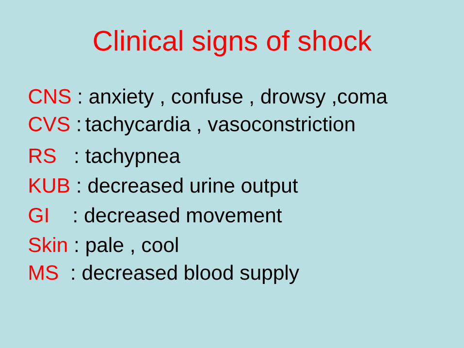
Clinical signs of shock
CNS : anxiety , confuse , drowsy ,coma CVS : tachycardia , vasoconstriction RS : tachypnea KUB : decreased urine output GI : decreased movement Skin : pale , cool MS : decreased blood supply
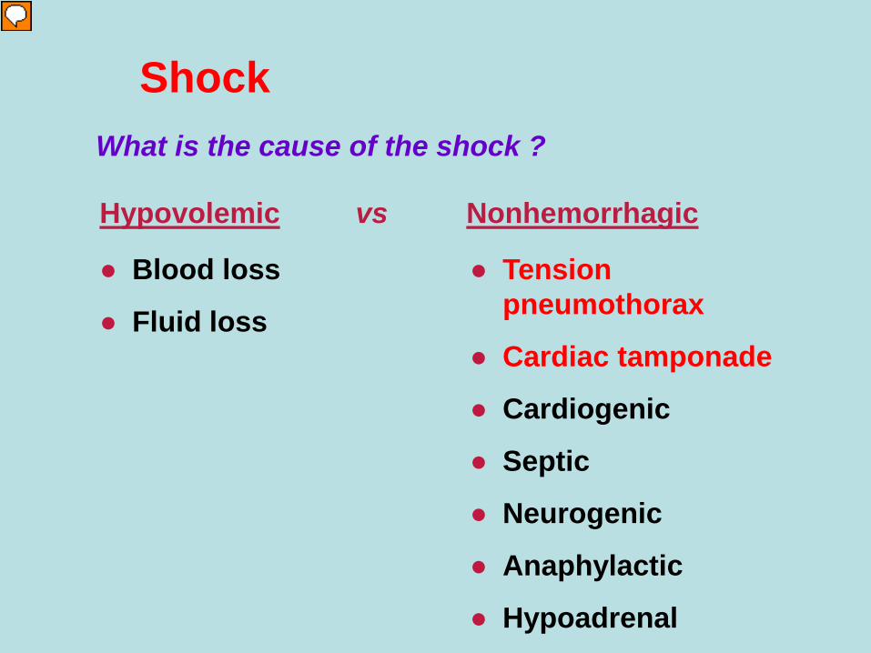
Shock What is the cause of the shock ?
● Blood loss
● Fluid loss
● Tension pneumothorax
● Cardiac tamponade
● Cardiogenic
● Septic
● Neurogenic
● Anaphylactic
● Hypoadrenal
Hypovolemic
Nonhemorrhagic
vs

Tension pneumothorax
High pressure pneumothorax causing cardiovascular compromised status
* chest injury * dyspnea & tachypnea * distended neck vein * deviated trachea * hypotension * tympanic on percussion * absent breath sound


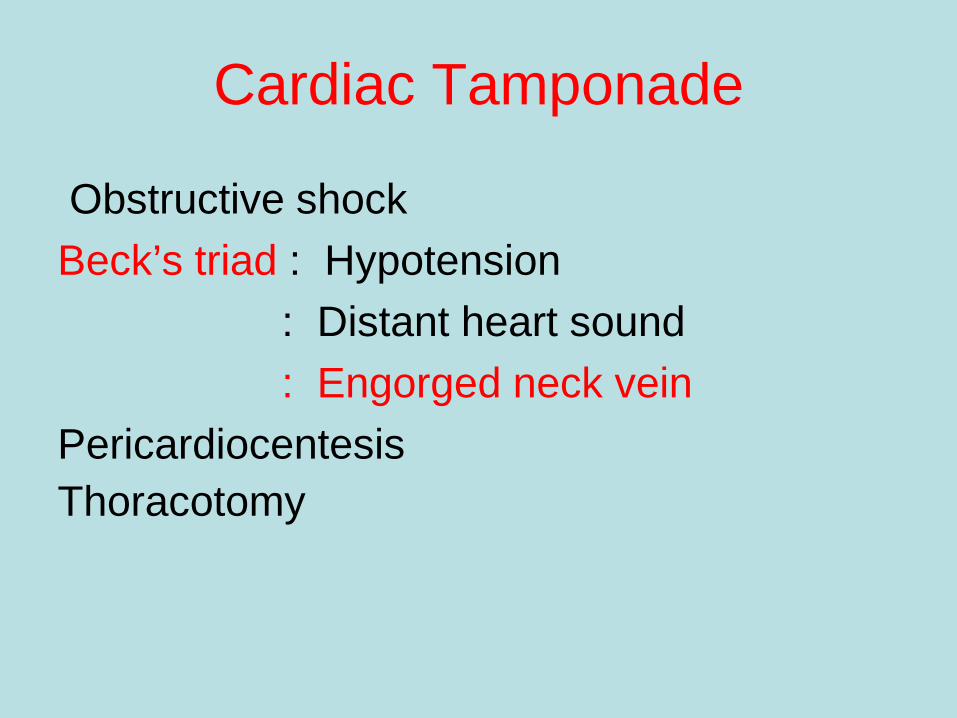
Cardiac Tamponade
Obstructive shock Beck’s triad : Hypotension : Distant heart sound : Engorged neck vein Pericardiocentesis Thoracotomy


Hemorrhagic shock Source of bleeding
External bleeding Internal bleeding
Chest
Abdomen
Pelvis
Long bone not intracranial hemorrhage

Save Life and Save Limb Stop bleeding
Direct pressure Tourniquet
Splinting
Rudge WBJ, Rudge BCJ, Rudge CJ. Ann R Coll Surg Engl. 2010 January;92(1):77-78

Classes of Shock
class I class II class III class IV Blood loss <15% 15-30% 30-40% >40% BP normal normal SBP<90 SBP<70 Pulse <100 100-120 120-140 >140 Mental anxiety anxiety confused lethargic Urine >30 20-30 <20 negligible Fluid crystalloid crystalloid+blood • Adult blood : 70ml/kg • Child blood : 80ml/kg

Class I Hemorrhage
● Slightly anxious ● Normal blood pressure ● Heart rate < 100 / min ● Respirations 14-20 / min ● Urinary output 30 ml / hour
BVL (15%) ; adult 70ml/kg , child 80mi/kg
Crystalloid

Class II Hemorrhage
● Anxious ● Normal blood pressure ● Heart rate > 100 / min ● Decreased pulse pressure ● Respirations 20-30 / min ● Urinary output 20-30 ml / hour
BVL (15-30%) ; adult 70ml/kg , child 80ml/kg
Crystalloid, ? blood

Class III Hemorrhage
● Confused, anxious ● Decreased blood pressure ● Heart rate > 120 / min ● Decreased pulse pressure ● Respirations 30-40 / min ● Urinary output 5-15 ml / hour
BVL (30-40%) ; adult 70ml/kg , child 80ml/kg
Crystalloid, blood components,
operation

Class IV Hemorrhage
● Confused, lethargic ● Profound hypotension ● Heart rate > 140 / min ● Decreased pulse pressure ● Respirations >35 / min ● Urinary output negligible
BVL (>40%) ; adult 70ml/kg , child 80ml/kg
Definitive control, blood
components

Fluid Resuscitation
Fluid challenge test : 2000ml I.V. in 15 min : 20ml/kg in 15 min Warm fluid and patient Blood for lab test Cross-match 2 x estimated blood loss Uncross matched blood : gr.O ,Rh +ve PRC

Response 1. Rapid response : <20% , cease 2. Transient response : 20-40% , on going 3. Unresponsive : >40% , active bleeding
Fluid Resuscitation

Adjuncts
• Monitors : V/S , O2 sat , EKG , urine output : GCS , ABG • Catheters : N-G , Foley catheter • Investigations : FAST , DPL , CT , Angiogram : CXR , Film pelvis & limb
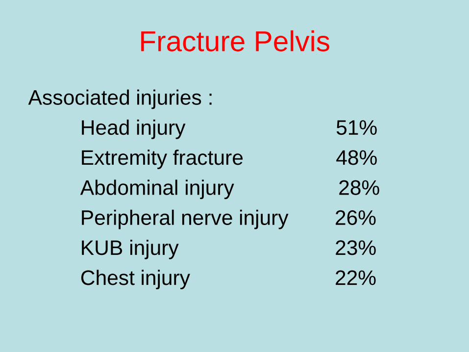
Fracture Pelvis
Associated injuries : Head injury 51% Extremity fracture 48% Abdominal injury 28% Peripheral nerve injury 26% KUB injury 23% Chest injury 22%
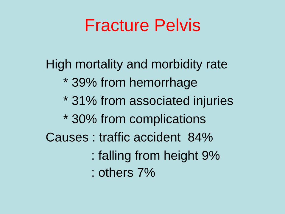
Fracture Pelvis
High mortality and morbidity rate * 39% from hemorrhage * 31% from associated injuries * 30% from complications Causes : traffic accident 84% : falling from height 9% : others 7%

Anatomy
Volume of Pelvis : ¶H( R² + 2Rr + r² )/3 R or r ↑ 2cm → vol. ↑ 1.3 litre R or r ↑ 5cm → vol. ↑ 5.0 litre

Diagnosis
1. History of injury * car accident * motorcycle accident * pedestrian accident * falling from height * crush injury

Diagnosis
2. Physical examination * marks at pelvis and perineum * leg deformity or length discrepancy * signs of ruptured urethra or bladder * anorectal or vaginal lacerations * pelvic compression test ???
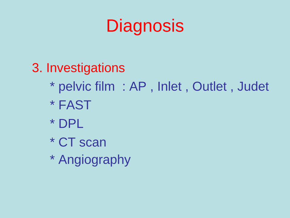
Diagnosis
3. Investigations * pelvic film : AP , Inlet , Outlet , Judet * FAST * DPL * CT scan * Angiography


Interventions
Direct pressure / tourniquet
STOP the
bleeding! Reduce pelvic volume
Angio-embolization
Splint fractures
Operation
What can I do about it?

Hypotensive Resuscitation
“ Delivery of limited volumes of intravenous fluids to sustain blood pressure lower than normal until control of hemorrhage has been established “
“ Rapid resuscitation can exacerbate bleeding by dislodging fragile clots , decreasing blood viscosity and creating compartment syndrome of cranial vault, abdomen, extremities and it also exacerbate the Lethal Triad of hypothermia, acidosis and coagulopathy “

Trauma Induced Coagulopathy
Lethal triad Dilutional coagulopathy Consumptive coagulopathy Hyperfibrinolysis Anemia Electrolyte imbalance
Hypocalcemia
Sorensen B, Fries D. British Journal of Surgery 2012; 99(Suppl 1): 40–50
Hypothermia
Acidosis
Coagulopathy

Hypotensive Resuscitation
“ Tissue injury from regional hypoperfusion is a risk “
“ Early control of hemorrhage was paramount and attempts at fluid resuscitation prior to this would result in increased bleeding and mortality “
Hypotensive Resuscitation
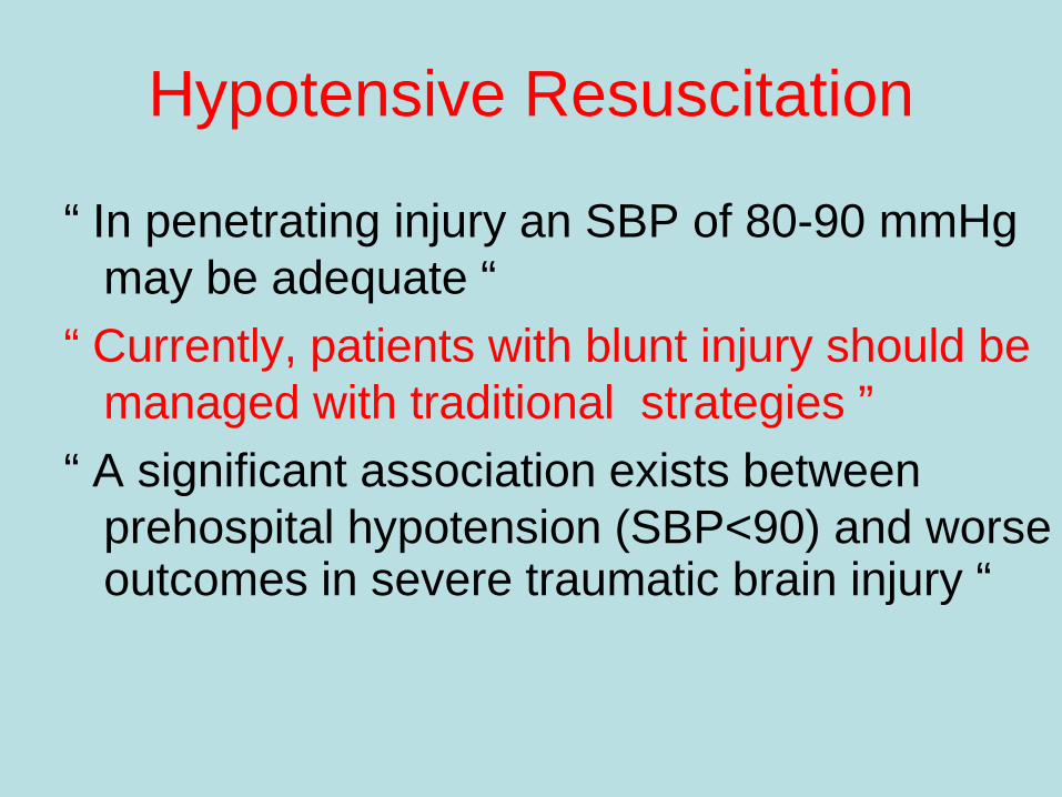
“ In penetrating injury an SBP of 80-90 mmHg may be adequate “
“ Currently, patients with blunt injury should be managed with traditional strategies ”
“ A significant association exists between prehospital hypotension (SBP<90) and worse outcomes in severe traumatic brain injury “

Hypotensive Resuscitation
“ Early identification of bleeding sources and control of hemorrhage will lead to more rapid replacement of intravascular volume and decreased morbidity and mortality “


Neurogenic Shock Circulatory shock
Spinal shock: neurological shock
Bradycardia
May not present if injury occur below T4
Vasopressor usually needed
Clinical recognition
Decreased sensation
Motor impairment
Loose sphincter tone

Choices of Fluids
“ Extracellular fluid redistributed into both intravascular and intracellular spaces during shock and rapid correction of this extracellular deficit required an infusion of a 3:1 ratio of crystalloid fluid to blood loss “

Crystalloids
: replace interstitial and intravascular fluid loss
: do not cause allergic reaction : inexpensive : limited intravascular expansion : tissue edema ( pulmonary edema , bowel
edema and compartment syndrome )

Colloids
: longer intravascular half-life : may improve organ perfusion and cause less tissue
edema in early phase : allergic reaction , impaired blood cross-matching ,
altered platelet function , hyperchloremic acidosis
: greater expense “ There is no clear basis to give colloid products over
crystalloid solutions for fluid resuscitation “

RLS VS NSS
: large volume of NSS can lead to hyperchloremic metabolic acidosis
: large volume of RLS can increase lactate level but not cause acidosis
: RLS does not increase clots when giving blood : no literature supporting the use of NSS over
RLS for the treatment of severe head injury to reduce intracerebral swelling

Hypertonic Saline
“ causes influx of fluid into intravascular space
with small volume” “ In head trauma patients, it can limit cerebral
edema , lower intracranial pressure and improve cerebral perfusion “
“ 3% hypertonic saline plus 6% dextran showed the greatest benefit in shock patients with concomitant severe closed head injury “

Artificial Oxygen-Carrying Blood Substitutes
“ improve oxygen-carrying capacity without the storage, availability, immune suppression, transfusion reaction, compatibility, disease transmission problems associated with standard transfusions”
“ fail to restore coagulation components causing hemorrhage “
Blood Transfusion
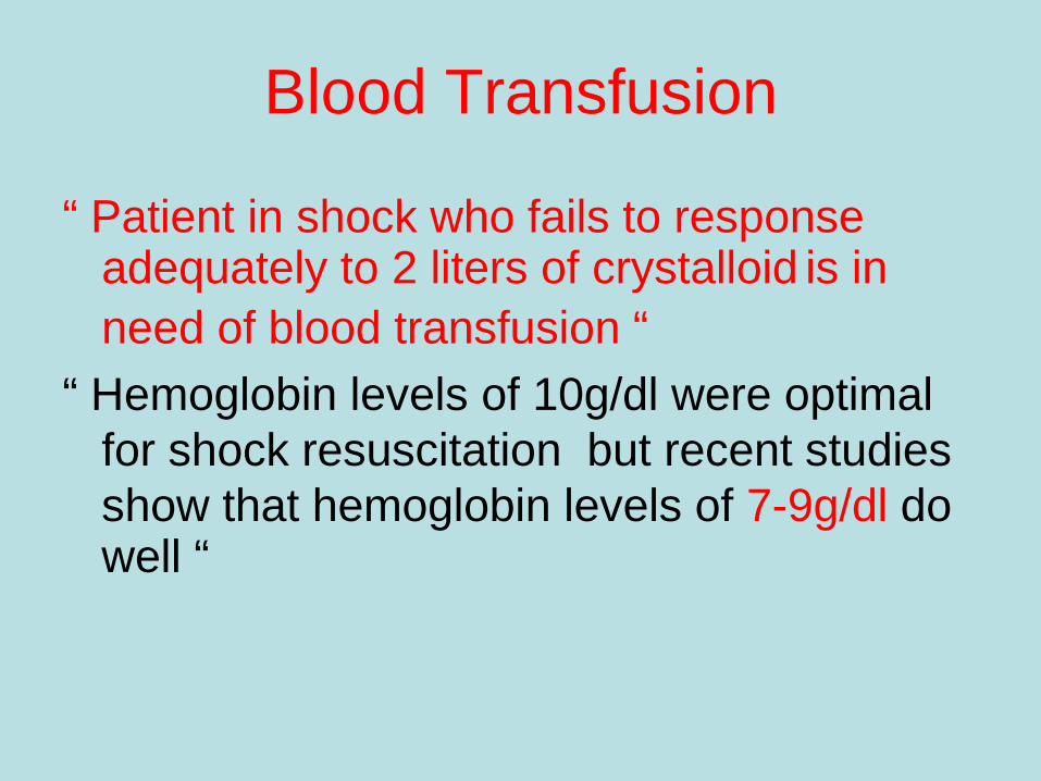
“ Patient in shock who fails to response adequately to 2 liters of crystalloid is in need of blood transfusion “
“ Hemoglobin levels of 10g/dl were optimal for shock resuscitation but recent studies show that hemoglobin levels of 7-9g/dl do well “

Massive Blood Transfusion
“ Blood transfusion of total blood volume in 24 hours or 50% of blood volume in 1 hours “
“ Bleeding > 150ml/min or > half of blood
volume in 20 minutes “ “ PRC : FFP : PLT = 1:1:1 “


Complications of Transfusion
Hypothermia : mild = 32-35 degrees Celsius : mod = 28-32 degrees Celsius : sev = < 28 degrees Celsius Trauma victims with core temperature < 32 degrees Celsius have 100% mortality

Complications of Resuscitation
Coagulopathy : dilutional ( one blood volume replacement ) : hypothermia : INR >2 : PTT >1.5 times : plt < 50000/mcl : fibrinogen level < 100mg/dl : head injury ( release of thromboplastin )

Complications of Resuscitation
Acidosis : NSS >> RLS : pH < 7.1 independently predicted coagulopathy
: decreases fibrinogen and platelet
: increases PTT and bleeding time

Complications of Resuscitation
Compartment Syndromes “ tissue edema is a frequent result of large
volume resuscitation , in restricted body compartments , the resulting increase in pressure can lead to ischemia and subsequent tissue necrosis “
“ The three affected areas are the extremities, abdomen and cranial vault “

Inotropes and Vasopressors
1. Dopamine 2-3ug/kg/min เพิ่ม urine output 3-5 เพิ่ม heart rate 5-10 เพิ่ม blood pressure 2. Dobutamine 5-20ug/kg/min เพิ่ม myocardial contractility vasodilatation เหมาะกบั heart failure จาก M.I.

3. Epinephrine 0.01-0.05 ug/kg/min เพิ่ม stroke volume และ heart rate ขนาดมากกวา่น้ี เพิ่ม BP และ vascular resistance 4. Norepinephrine เหมาะสาํหรับเพิ่ม BP จากการเพิ่ม
vascular resistance ในผูป่้วยท่ีได ้volume เพียงพอ
แลว้
Inotropes and Vasopressors

5. Phenylephrine เพิ่ม vascular resistance เหมาะเป็น first line drug ในผูป่้วย neurogenic
shock ท่ีได ้fluid resuscitation เพียงพอแลว้
6. Vasopressin เพิ่ม vascular resistance และ
arterial pressure ในผูป่้วย septic shock แต่อาจทาํ
ใหเ้กิด M.I. และ ischemic bowel ได ้
Inotropes and Vasopressors

7. Amrinone and Milrinone เพิ่ม cardiac contractility โดยไม่มีผลกบั oxygen consumption
เหมาะกบัผูป่้วย cardiogenic shock
Inotropes and Vasopressors