Embed Size (px)
Citation preview
Hepatic Flavin-Containing Monooxygenase 3 Enzyme
Suppressed by Type 1 Allergy-Produced Nitric Oxide
Tadatoshi Tanino, Toru Bando, Akira Komada, Yukie Nojiri, Yuna Okada,
Yukari Ueda, and Eiichi Sakurai
Faculty of Pharmaceutical Sciences, Tokushima Bunri University, Tokushima, Japan
This article has not been copyedited and formatted. The final version may differ from this version.DMD Fast Forward. Published on July 31, 2017 as DOI: 10.1124/dmd.117.076570
at ASPE
T Journals on February 27, 2020
dmd.aspetjournals.org
Dow
nloaded from
DMD/2017/076570
1
Running title: NO-Suppressed FMO3 Activity in Type 1 Allergic Mice
Corresponding Author: Eiichi Sakurai, Ph.D.
Professor, Laboratory of Pharmaceutics, Faculty of Pharmaceutical Sciences, Tokushima
Bunri University
180 Bouji Nishihama, Yamashiro-Cho, Tokushima, Tokushima 770-8514, Japan.
Phone +81-88-602-8468, FAX +81-88-655-3051
E-mail: [email protected]
Number of Text pages: 28
Tables: 0
Figures: 6
References: 63
Number of Words:
Abstracts: 249 words
Introduction: 639 words
Discussion: 1388 words
This article has not been copyedited and formatted. The final version may differ from this version.DMD Fast Forward. Published on July 31, 2017 as DOI: 10.1124/dmd.117.076570
at ASPE
T Journals on February 27, 2020
dmd.aspetjournals.org
Dow
nloaded from

DMD/2017/076570
2
ABBREVIATIONS: AS, L-ascorbate; AG, aminoguanidine; BDZ, benzydamine; CYP,
cytochrome P450; eNOS, endothelial nitric oxide synthase; FMO, flavin-containing
monooxygenase; HPLC, high-performance liquid chromatography; IMP, imipramine;
IgE, immunoglobulin E; IL, interleukin; iNOS, inducible nitric oxide synthase; IS,
internal standard; LPS, lipopolysaccharide; NADPH, nicotinamide adenine dinucleotide
phosphate; NO, nitric oxide; NOC7, 1-hydroxy-2-oxo-3-(N-methyl-3-aminopropyl)-3-
methyl-1-triazene; NOx, nitrate plus nitrite; nNOS, neuronal nitric oxide synthase; OVA,
ovalbumin; PCR, real-time polymerase chain reaction; PS7, 7 days after primary
sensitization; SS7, 7 days after secondary sensitization; Th, T helper; TNF-, tumor
necrosis factor alpha.
This article has not been copyedited and formatted. The final version may differ from this version.DMD Fast Forward. Published on July 31, 2017 as DOI: 10.1124/dmd.117.076570
at ASPE
T Journals on February 27, 2020
dmd.aspetjournals.org
Dow
nloaded from
DMD/2017/076570
3
ABSTRACT
Flavin-containing monooxygenases (FMOs) are major mammalian non-cytochrome P450
oxidative enzymes. T Helper2 cell-activated allergic diseases produce excess levels of
nitric oxide (NO) that modify the functions of proteins. However, it remains unclear
whether allergy-induced NO affects the pharmacokinetics of drugs metabolized by FMOs.
This study investigated alterations of hepatic microsomal FMO1 and FMO3 activities in
type 1 allergic mice, and further examined the interaction of FMO1 and FMO3 with
allergy-induced NO. Imipramine (IMP; FMO1 substrate) N-oxidation activity was not
altered in allergic mice with high serum NO and immunoglobulin E levels. At 7 days after
primary sensitization (PS7) or secondary sensitization (SS7), benzydamine (BDZ; FMO1
and FMO3 substrate) N-oxygenation was significantly decreased to 70% of individual
controls. The expression levels of FMO1 and FMO3 proteins were not significantly
changed in the sensitized mice. Hepatic inducible NO synthase (iNOS) mRNA level
increased 5-fold and 15-fold in PS7 and SS7 mice, respectively, and hepatic tumor
necrosis factor- levels were greatly enhanced. When a selective iNOS inhibitor was
injected into allergic mice, serum NO levels and BDZ N-oxygenation activity returned to
control levels. NO directly suppressed BDZ N-oxygenation, which was likely related to
FMO3-dependent metabolism in comparison with IMP N-oxidation. In hepatic
microsomes from PS7 and SS7 mice, the suppression of BDZ N-oxygenation was
restored by ascorbate. Therefore, type 1 allergic mice had differentially suppressed
FMO3-dependent BDZ N-oxygenation. The suppression of FMO3 metabolism related to
reversible S-nitrosyl modifications of iNOS-derived NO. NO is expected to alter FMO3-
metabolic capacity-limited drug pharmacokinetics in humans.
This article has not been copyedited and formatted. The final version may differ from this version.DMD Fast Forward. Published on July 31, 2017 as DOI: 10.1124/dmd.117.076570
at ASPE
T Journals on February 27, 2020
dmd.aspetjournals.org
Dow
nloaded from
DMD/2017/076570
4
Introduction
Flavin-containing monooxygenases (FMOs) are major mammalian non-
cytochrome P450 oxidative enzymes (Rettie et al., 1995), and commonly react to form
N- and S-oxides of nucleophilic heteroatom-containing drugs and xenobiotics. Five FMO
forms have been identified in humans and mice, and require nicotinamide adenine
dinucleotide phosphate (NADPH) and oxygen. FMO expression is species-, tissue-, age-,
and gender-dependent. Of interest, FMO1 form is found in the fetal human liver, however,
marked changes in enzyme expression at birth result in FMO3 becoming the predominant
human FMO (Dolphin et al., 1996; Koukouritaki et al., 2002). In male and female mice,
the FMO1 protein is expressed at a high level, and hepatic FMO3 protein is gender-
specific to females (Cherrington et al., 1998). Sex hormones in rats and mice regulate
FMO1 and FMO3 expression (Dannan et al., 1986; Lemoine et al., 1991; Falls et al.,
1997; Coecke et al., 1998), and glucocorticoids and 17-estradiol are considered to be
regulators of gene transcription in rodents and humans (Dixit and Roche 1984; Esposito
et al., 2014). FMO3 exists at levels similar to the major human liver cytochrome P450
(CYP) 3A4 (Haining et al., 1997). Loss-of-function mutations, nonsense mutations, and
missense mutations in the FMO3 gene produce phenotypes associated with the inherited
disorder trimethylaminuria (Treacy et al., 1998). FMO4 protein is expressed in liver of
humans and mice (Novick et al., 2009), however, FMO4 does not contribute significantly
to drug metabolism in humans (Krueger and Williams, 2005). Functional FMO2 is not
expressed in liver of most humans (Dolphin et al., 1998). FMO5 is highly expressed in
the liver of mice (Janmohamed et al., 2004) and humans (Cashman and Zhang, 2006).
Only a few chemicals are known to be metabolized by mouse FMO5 (Zhang et al., 2007)
and the contribution of FMO5 to liver xenobiotic metabolism is not clear. Therefore,
This article has not been copyedited and formatted. The final version may differ from this version.DMD Fast Forward. Published on July 31, 2017 as DOI: 10.1124/dmd.117.076570
at ASPE
T Journals on February 27, 2020
dmd.aspetjournals.org
Dow
nloaded from
DMD/2017/076570
5
FMO1 and FMO3 are considered to be the most important isoforms of the FMO family
with respect to the metabolism of foreign chemicals (Ziegler, 1993).
Type I allergic diseases, such as atopic dermatitis, asthma, and allergic rhinitis, are
a significant health problem. Approximately 20% of the world’s population suffers from
some kind of allergic disorder, with incidences continuing to rise (Warner et al., 2006).
Concerning the allergy-inducing mechanism, T helper (Th) 2 cytokines (IL-4, IL-5 and
IL-13) stimulate mast cells and eosinophils and increase allergen-specific
immunoglobulin (Ig)E production. Subsequently, activated mast cells release cytokines
and some inflammatory mediators (Zhu et al., 1999; Stassen et al., 2001). Patients with
bronchial asthma and allergic rhinitis show an increased level of nitric oxide (NO) in
exhaled air (Alving et al., 1993; Kharitonov et al., 1997). We also found that type 1 allergy
with high serum IgE concentrations enhance serum NO concentrations (Tanino et al.,
2016). Park et al. (1999) suggested that excessive activation of the L-arginine/NO
pathway (NO-dependent pathway) was responsible for the suppression of FMO activities.
However, their findings showed that NO did not react with the flavin adenine dinucleotide
prosthetic group of FMO enzymes. The result differs from the formation of iron-nitrosyl
complexes in CYP enzymes (Minamiyama et al. 1997). Since FMO3 contains 11
cysteines, one possibility remains that NO participates in S-nitrosylation and/or nitration
of FMO proteins, leading to consequent reversible and/or irreversible modulation(s) of
FMO activity. Concerning the contribution of Th2 cell-activated immune systems, little
information is available on the ability of cytokines to directly regulate FMO1 and 3
expression. We reported that CYP1A2, CYP2C, CYP2E1, and CYP3A activities were
markedly suppressed by allergy-induced NO production, but not the cytokine-dependent
pathway, resulting in marked changes in drug pharmacokinetics (Tanino et al., 2016). Our
This article has not been copyedited and formatted. The final version may differ from this version.DMD Fast Forward. Published on July 31, 2017 as DOI: 10.1124/dmd.117.076570
at ASPE
T Journals on February 27, 2020
dmd.aspetjournals.org
Dow
nloaded from

DMD/2017/076570
6
results differed from the significant contribution of Th1 cell-activated proinflammatory
cytokines (cytokine-dependent pathway) in lipopolysaccharide-stimulating inflammatory
diseases (Renton 2001, 2004; Morgan et al., 2008). Therefore, the main aim of this study
was to examine the interaction of hepatic FMO1 and FMO3 enzymes with IgE-mediated
allergy-induced NO.
Materials and Methods
Materials. Benzydamine (BDZ) was purchased from Tokyo Kasei Co., Ltd.
(Tokyo, Japan). L-Ascorbate (AS; effective on S-NO binding), Bordetella pertussis
inactive bacterial suspension and aminogunidine (AG) were purchased from Nacalai
Tesque Co. (Kyoto, Japan). Aluminum hydroxide gel (alhydrogelⓇ) was obtained from
InvivoGen (San Diego, CA, USA). Imipramine (IMP) and amitriptyline were obtained
from Wako Pure Chemicals (Osaka, Japan). Chicken egg albumin (OVA) was obtained
from Sigma-Aldrich (St Louis, MO, USA). Talinolol, IMP N-oxide and BDZ N-oxide
were purchased from Toronto Research Chemicals Inc. (North York, ON, Canada). 1-
Hydroxy-2-oxo-3-(N-methyl-3-aminopropyl)-3-methyl-1-triazene (NOC7) was obtained
from Dojin Co. (Kumamoto, Japan). -Nicotinamide adenine dinucleotide 2’-phosphate
reduced tetrasodium (-NADP+), glucose-6-phosphate dehydrogenase, and glucose-6-
phosphate were obtained from Oriental Yeast Co. Ltd. (Tokyo, Japan). All other
chemicals used were of analytical grade and commercially available.
Animals and Treatments. Female ICR mice (Japan SLC Co., Shizuoka, Japan),
weighing 15-20 g, were housed under standard conditions (light/dark cycle of 12 h; room
temperature of 23±1˚C) and provided with food and water ad libitum. For establishment
This article has not been copyedited and formatted. The final version may differ from this version.DMD Fast Forward. Published on July 31, 2017 as DOI: 10.1124/dmd.117.076570
at ASPE
T Journals on February 27, 2020
dmd.aspetjournals.org
Dow
nloaded from
DMD/2017/076570
7
of IgE-mediated allergic animals, mice (age, 6 weeks; weight range, 18-22 g) were
sensitized with an intraperitoneal (i.p.) injection of a mixture containing 5 g OVA, 2 mg
aluminum hydroxide gel, and Bordetella pertussis, inactive bacterial suspension (2×10⁹
cells) on day 0 according to the methods described by Pauwels et al. (1979) and Lebrec
et al. (1996). On day 8, the mice were challenged with 2.5 g OVA (250 g/ml of saline)
by intravenous (i.v.) injection. Control mice were given single i.p. and i.v. injections of
saline. At 7 days after the primary and secondary sensitization (PS7 and SS7,
respectively), mice were sacrificed by decapitation. Blood was collected in heparinized
tubes, and plasma was separated by centrifugation at 3000 rpm for 10 min. The liver was
excised and washed three times with ice-cold 50 mM Tris-HCl (pH 7.4). Further
treatments for high-performance liquid chromatography (HPLC) assay were detailed in
our recent study (Tanino et al., 2016). All animal experiments were approved by the
Tokushima Bunri University Faculty of Pharmaceutical Sciences’ Committee for the care
and use of laboratory animals.
In separate experiments, PS7 and SS7 mice were repeatedly injected with AG (100
mg/kg/day, i.p.) from day 2 to day 7 or to day 14 after the primary sensitization. The AG
dosage was determined by the in vivo data reported by Okuda et al. (1996). The body
weight of AG-treated mice did not differ from that of control mice receiving only saline
(data not shown).
Assay of Total Plasma IgE and Serum NO Levels. Total plasma IgE levels
were measured by enzyme-linked immunosorbent assay (ELISA) using a commercially
available mouse IgE ELISA Kit (Shibayagi, Gunma, Japan) according to the
manufacturer’s instructions. The absorbance was measured at 450 nm using a microplate
This article has not been copyedited and formatted. The final version may differ from this version.DMD Fast Forward. Published on July 31, 2017 as DOI: 10.1124/dmd.117.076570
at ASPE
T Journals on February 27, 2020
dmd.aspetjournals.org
Dow
nloaded from
DMD/2017/076570
8
reader.
Serum concentrations of NO2- were determined using a Nitrate/Nitrite Colorimetric
Assay kit (Cayman Chemical Company., An Arbor, MI) according to the manufacturer’s
instructions. The assay conditions were detailed in our recent report (Tanino et al., 2016).
Enzyme Assay. Liver microsomes from control and treated mice were prepared as
described previously (Tanino et al., 2016). Protein concentrations were determined with
a BCA Protein Assay Kit (Thermo Fisher Scientific Inc., Rockford, IL, USA) and
bovine serum albumin as a protein standard. Aliquots of microsomes were stored at -80
˚C until analysis.
Hepatic FMO1 and FMO3 activities were determined by measuring the N-oxide
formation of IMP and BDZ (Narimatsu et al., 1999; Ubeaud et al., 1999). The incubation
mixtures (400 l) contained liver microsomal protein (final protein concentration, 0.5
mg/ml for IMP metabolism, 0.1 mg/ml for BDZ metabolism) from control (saline) and
treated mice, a NADPH-generating system (0.2 mM NADP+, 28.6 mM MgCl2, 11.4 mM
glucose-6-phoshate and 20 units/ml glucose-6-phoshate dehydrogenase), and FMO
substrate (20 M IMP, or 0.5 M BDZ). All enzymatic reactions were initiated by the
addition of the NADPH-generating system and were allowed to proceed at 37˚C for
different times (5 min for IMP and 2 min for BDZ). Each reaction was stopped with ice-
cold acetonitrile containing amitriptyline and talinolol as internal standards (ISs) in a
HPLC assay of IMP and BDZ, respectively. Sodium hydroxide 0.1 N was added to the
mixture. Subsequently, N-oxides of IMP and BDZ were extracted in ethyl acetate
(Narimatsu et al., 1999) and chloroform (Ubeaud et al., 1999), respectively, and then
centrifuged at 3000 rpm for 10 min. After evaporating the organic layers, the residues
This article has not been copyedited and formatted. The final version may differ from this version.DMD Fast Forward. Published on July 31, 2017 as DOI: 10.1124/dmd.117.076570
at ASPE
T Journals on February 27, 2020
dmd.aspetjournals.org
Dow
nloaded from
DMD/2017/076570
9
were reconstituted in each mobile phase used for HPLC analysis and loaded on HPLC
columns. FMO activities were expressed as metabolic formation rates of IMP N-oxide
and BDZ N-oxide. Our preliminary study confirmed that the incubation time, substrate
concentration, and amount of microsomes were determined to be in the linear range for
metabolite formation rate.
To examine the direct interaction of hepatic microsomal FMO enzymes with NO,
in vitro experiments were performed according to the method reported by Minamiyama
et al (1997). Briefly, an efficient NO donor, NOC7, dissolved in 0.1 N NaOH was added
to the microsomes and NADPH-generating system at a final concentration of 1 mM. The
same volume of vehicle (final concentration, 0.025% NaOH for NOC7 treatment) was
added as the references. According to the method reported by Ryu et al. (2004), the pre-
treated microsomes were exposed to AS at a final concentration of 10 mM. After the AS
exposure, BDZ N-oxygenation was assayed under the conditions as described above.
In separate experiments, we examined S-nitrosylation of hepatic microsomal FMO1
and FMO3 enzymes in PS7 and SS7 mice. Hepatic microsomes from PS7 and SS7 mice
were pre-treated with 10 mM AS in the presence of the NADPH-generating system.
Subsequently, BDZ was added, and the reaction mixtures were incubated for 5 min. Our
preliminary study on S-nitrosylated FMO enzymes confirmed that NOC7 concentration,
AS concentration, exposure time, substrate concentration, and amount of microsomes
were determined to be in the linear range for the metabolite formation rate.
Immunoblotting. FMO1 and FMO3 protein levels in mouse liver microsomes
were determined by sodium dodecyl sulfate (SDS)-polyacrylamide gel electrophoresis
followed by Western blotting. In brief, hepatic microsomal proteins (50 g) were
This article has not been copyedited and formatted. The final version may differ from this version.DMD Fast Forward. Published on July 31, 2017 as DOI: 10.1124/dmd.117.076570
at ASPE
T Journals on February 27, 2020
dmd.aspetjournals.org
Dow
nloaded from
DMD/2017/076570
10
separated by 7.5% SDS-polyacrylamide gel and transferred electrophoretically. Proteins
were transferred onto Immobilon-P transfer membranes (Millipore Corporation, Billerica,
MA, USA). Membranes were blocked with Blocking One (Nacalai Tesque) and then
incubated with rabbit polyclonal antibody human FMO1 (sc-98502, 1:500 dilution, Santa
Cruz, California, USA) and goat polyclonal antibody human FMO3 (sc-51288, 1:500
dilution, Santa Cruz) (Sakurai et al., 2013). After washing the membrane with TBS (Tris-
buffered saline)-Tween (20 mM Tris-HCl, 137 mM NaCl, and 0.1% Tween 20; pH 7.6),
resulting membranes were incubated with horseradish peroxidase-conjugated anti-rabbit
IgG (NA934, 1:20000 dilution; GE Healthcare, Little Chalfont, Buckinghamshire, UK)
and anti-goat IgG (sc-2352, 1:20000 dilution; Santa Cruz) secondary antibodies for
FMO1 and FMO3, respectively. An enhanced chemiluminescence method was used to
visualize the protein bands. The changes in intensity of FMO1 and FMO3 proteins were
corrected using the intensity obtained in the internal control bands (glyceraldehyde-3-
phosphate dehydrogenase, GAPDH). We selected GAPDH primary antibody (ab181602,
1:2000 dilution; Abcam, Cambridge, United Kingdom) and horseradish peroxidase-
conjugated anti-rabbit IgG secondary antibody (NA934, 1:2000 dilution; GE Healthcare,
Little Chalfont, Buckinghamshire, United Kingdom). Using microsomal protein
preparations ranging from 10 to 60 g/lane, we confirmed a linear relationship between
the abundance of FMO1 and FMO3 and staining intensity. FMO3 protein was not
detectable in male mice 6 to 8 weeks of age. Protein content was determined by BCA
protein assay (Thermo Scientific, Rockford, USA).
HPLC Assays. HPLC analysis was performed on a system equipped with a
Shimadzu SPD-10A, a fluorescence detector, a UV detector, a Shimadzu LC10A pump
This article has not been copyedited and formatted. The final version may differ from this version.DMD Fast Forward. Published on July 31, 2017 as DOI: 10.1124/dmd.117.076570
at ASPE
T Journals on February 27, 2020
dmd.aspetjournals.org
Dow
nloaded from
DMD/2017/076570
11
and a Shimadzu CR-4A chromatopac integrator (Kyoto, Japan). BDZ and its N-oxide
were determined by the method of Ubeaud et al. (1999) with some modifications. Briefly,
they were separated using a Mightysil RP-18 column (particle size 5 m, 4.6 150 mm,
Kanto Kagaku, Tokyo, Japan). Detection was performed at an excitation wavelength of
305 nm and an emission wavelength of 375 nm. A mobile phase of acetonitrile and 10
mM sodium acetate (pH 4.3) (30:70, v/v) was used at a flow rate of 1 ml/min.
IMP and its N-oxide concentrations were determined using the method reported by
Narimatsu et al. (1999) with slight modifications. Briefly, IMP and its N-oxide were
eluted with acetonitrile-50 mM phosphate buffer (pH 5.5) (42:58, v/v) at a flow rate of
0.8 ml/min and detected by absorbance at 254 nm. They were separated using an Inertsil
ODS column (particle size 5 m, 4.6 150 mm, GL Sciences Inc., Osaka, Japan).
Assay for Hepatic Inducible NO Synthase (iNOS) and Tumor Necrosis Factor
(TNF)- mRNA. Total RNA was isolated from the liver using a high pure RNA tissue
kit (Roche Diagnostics, Mannheim, Germany) according to the manufacturer’s
instructions. The concentration of total RNA in the final eluates was determined by
spectrophotometry using a NanoDrop 2000 (Thermo Fisher Scientific, Waltham, MA,
USA). cDNA synthesis was performed using total RNA (2 g) in 20 l of reaction mixture
consisting of Transcriptor Universal Reverse Transcriptase (Roche Diagnostics) and
Transcriptor Universal Reaction buffer (Roche Diagnostics). cDNA product was
amplified by a GeneAmp PCR System 9700 (Applied Biosystems, Foster City, CA, USA).
Intron-spanning, FAM-labeled hydrolysis probe assays were designed for genes of
interest using the Universal ProbeLibrary (UPL) (Roche Diagnostics) and multiplexed
with a primer-limited, VIC-labeled UPL mouse ACTB gene assay. Primer sequences for
This article has not been copyedited and formatted. The final version may differ from this version.DMD Fast Forward. Published on July 31, 2017 as DOI: 10.1124/dmd.117.076570
at ASPE
T Journals on February 27, 2020
dmd.aspetjournals.org
Dow
nloaded from
DMD/2017/076570
12
mouse inducible NOS (iNOS; forward, 5’tggccaccaagctgaact3’; reverse,
5’ttcatgataacgtttctggctct3’) and mouse TNF- (forward, 5’tggccaccaagctgaact3’; reverse,
5’ttcatgataacgtttctggctct3’). Triplicate reactions per cDNA sample were prepared using
FastStar Universal Probe master (Roche Diagnostics), 500 nM forward primer, 500 nM
reverse primer, 250 nM FAM-label UPL probe (#68, Cat. No.04688678001 for iNOS;
#49, Cat. No.04688104001 for TNF-), 0.2 l VIC-labeled -actin assay, 5 l cDNA
(diluted 1:20 from stock), and DNase-free water in 20 l of reaction mixture.
Amplification and detection were performed using a StepOne Plus Real Time PCR system
(Thermo Fisher Scientific, Waltham, MA, USA). Target gene expression levels were
calculated after normalization to the standard housekeeping gene -actin (Universal
ProbeLibrary mouse ACTB gene assay, Roshe Diagnostics). Relative expression of the
gene of interest compared to -actin expression was calculated using a standard curve of
serially diluted cDNA to correct for PCR efficiency.
Statistical Analysis. Statistical analysis was performed by a one-way analysis of
variance test, and significance was assessed using Tukey’s post-hoc test.
Results
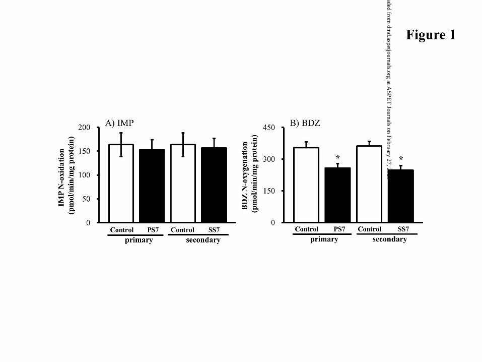
Hepatic Microsomal IMP and BDZ Metabolism. Imipramine is converted to
imipramine N-oxide solely by FMO1 (Hernandez et al., 2009), and BDZ is metabolized
to BDZ N-oxide by recombinant FMO1 and FMO3 isoforms (Störmer et al., 2000).
Störmer et al. (2000) showed that the rates of BDZ N-oxide formed by recombinant
CYP3A, 2C, 2D6, 1A2 and 2E1 were one order of magnitude lower than that determined
for recombinant FMO1 and FMO3 isoforms, implying a much minor contribution of CYP
This article has not been copyedited and formatted. The final version may differ from this version.DMD Fast Forward. Published on July 31, 2017 as DOI: 10.1124/dmd.117.076570
at ASPE
T Journals on February 27, 2020
dmd.aspetjournals.org
Dow
nloaded from
DMD/2017/076570
13
enzymes to BDZ N-oxygenation. In our study, PS7 (primary sensitization) and SS7
(secondary sensitization) mice did not have altered relative metabolic IMP activity (Fig.
1A). However, the relative BDZ activity was significantly decreased to 72.8±5.2% and
68.4±5.4% by PS7 and SS7, respectively (Fig. 1B). This finding on FMO1-dependent
IMP metabolism may help to clarify FMO3-dependent BDZ N-oxide metabolism in
hepatic microsomes.
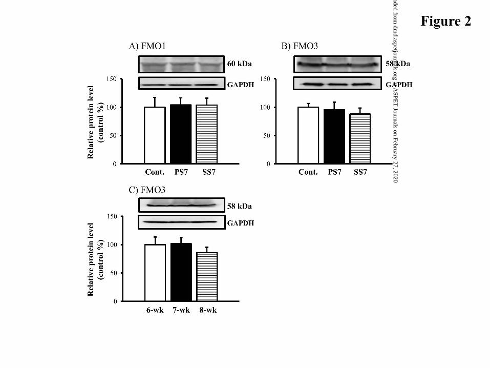
Western Blot Analysis of Hepatic FMO1 and FMO3. The expression levels of
hepatic microsomal FMO1 and FMO3 proteins were investigated by Western blot
analysis (Fig. 2). In PS7 mice, protein levels of FMO1 and FMO3 enzymes were similar
to those of control mice. SS7 mice had slightly reduced protein levels of FMO3, resulting
in an insignificant difference between SS7 and control mice (Fig. 2B).
FMO3 mRNA expression does not change in the liver of female mice aged 6-8 weeks
(Janmohamed et al., 2004). To determine if FMO3 protein changes at this age, we
evaluated the abundance of FMO3 protein in untreated 6-8-week old female mice by
Western blotting. As shown in Fig. 2C, hepatic FMO3 protein expression was not
significantly different over this time period.
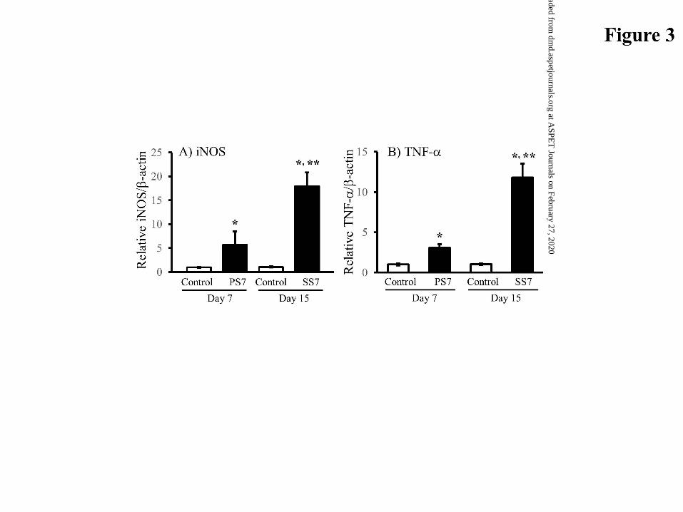
Hepatic iNOS and TNF- mRNA Levels. Hepatic iNOS mRNA expression
was determined by real-time polymerase chain reaction (PCR). The mRNA level of
hepatic iNOS was significantly increased in sensitized mice compared with the
corresponding control mice (Fig. 3A), resulting in an average 5-fold and 15-fold times
higher expression in the PS7 and SS7 mice, respectively. Further, hepatic TNF- mRNA
expression was significantly enhanced by type 1 allergic diseases (Fig. 3B).
This article has not been copyedited and formatted. The final version may differ from this version.DMD Fast Forward. Published on July 31, 2017 as DOI: 10.1124/dmd.117.076570
at ASPE
T Journals on February 27, 2020
dmd.aspetjournals.org
Dow
nloaded from
DMD/2017/076570
14
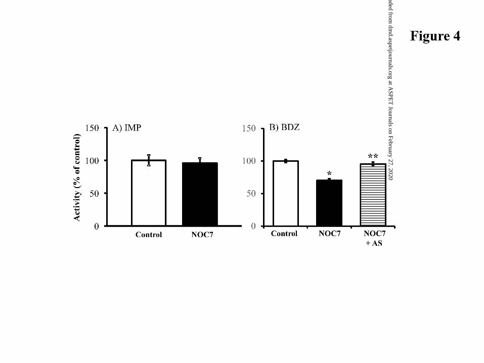
Direct Effect of NO and AS on Hepatic Microsomal FMO Enzymes. We
examined the direct effect of NO on hepatic microsomal IMP and BDZ metabolism (Fig.
4). When NOC7, an efficient NO donor, was incubated with hepatic microsomes prepared
from control mice, NO overproduction did not alter IMP N-oxidation activity. In contrast,
the inhibition degree of BDZ N-oxygenation activity was 36.8± 7.4%. To further
characterize the suppression of BDZ-metabolic activity, we examined the involvement of
cysteine thiols S-nitrosylation in FMO-NO interactions using AS, a sulfhydryl-reducing
reagent. After the NOC7 pre-incubation with hepatic microsomes, AS treatment restored
the suppression of BDZ metabolism to the control levels (Fig. 4B).
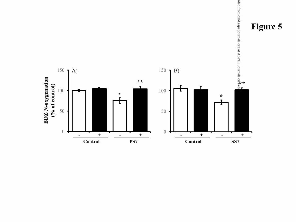
Preventable Effect of AS on Hepatic BDZ Metabolism in Sensitized Mice.
Figure 5 shows the preventable effect of AS on hepatic microsomal BDZ metabolism
suppressed by type 1 allergy. When hepatic microsomes from PS7 and SS7 mice were
treated with AS, BDZ N-oxygenation activity was completely restored to control levels.
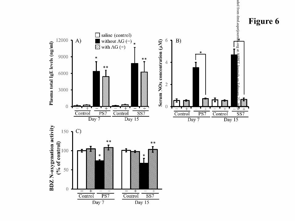
Effect of iNOS Inhibitor on Allergy-Induced Serum NO Levels. Plasma total
IgE levels at 7 days after primary or secondary sensitization were significantly elevated
to 6357 ± 1740 and 7799 ± 2893 ng/ml, respectively, showing 60-fold higher
concentrations compared with IgE values in control mice (Fig 6A). The plasma total IgE
level in SS7 mice did not differ from that in PS7 mice. Chronic treatment with a selective
iNOS inhibitor, AG, did not alter the high IgE levels in the two sensitized mice.
PS7 and SS7 mice showed higher levels of serum nitrate plus nitrite (NOx; 3.52±
0.48 M and 4.68±0.53 M, respectively) compared with corresponding control mice
This article has not been copyedited and formatted. The final version may differ from this version.DMD Fast Forward. Published on July 31, 2017 as DOI: 10.1124/dmd.117.076570
at ASPE
T Journals on February 27, 2020
dmd.aspetjournals.org
Dow
nloaded from
DMD/2017/076570
15
(0.55±0.15 M for PS7 mice and 0.61±0.17 M for SS7 mice) (Fig. 6B). PS7 mice had
a similar serum NOx concentration to SS7 mice. When AG was repeatedly injected into
two sensitized mice, the high serum NOx levels observed were markedly reduced to
control levels.
Hepatic Microsomal BDZ Activity in AG-Treated Sensitized Mice. We studied
participation of allergy-induced NO in microsomal BDZ N-oxygenation activity
suppressed by primary and secondary sensitization. Chronic AG treatment restored the
BDZ activity suppressed in PS7 and SS7 mice (Fig. 6C). When AG was injected in saline-
treated (control) mice, hepatic BDZ activity was 104.9±6.5% and 97.9±2.5% of saline-
treated mice. This result indicated that AG did not interact with FMO1 and FMO3
isoforms.
Discussion
FMO enzymes play significant roles in hepatic metabolism of drugs such as
nicotine, chlorpromazine, clozapine, cimetidine and tamoxifen (Cashman et al., 1992,
1993; Bhamre et al., 1995; Tugnait et al., 1997; Mani et al., 1993). In allergic diseases,
little is known about interactions between chemical mediators and FMO enzymes.
Concerning allergic disease-induced mediators, our recent study reported that the gas
messenger NO markedly altered pharmacokinetics of major CYP-metabolic capacity-
limited drugs without altering hepatic CYP protein levels, protein binding of drugs, and
blood flow rate (Tanino et al., 2016). The altered pharmacokinetics were responsible for
NO-derived posttranslational suppression of hepatic CYP-metabolic activities, except for
CYP2D. Therefore, our attention was focused on the interaction of hepatic microsomal
This article has not been copyedited and formatted. The final version may differ from this version.DMD Fast Forward. Published on July 31, 2017 as DOI: 10.1124/dmd.117.076570
at ASPE
T Journals on February 27, 2020
dmd.aspetjournals.org
Dow
nloaded from
DMD/2017/076570
16
FMO1 and FMO3 with type 1 allergy-induced NO, and further the changes in NO-
produced factors in liver.
Functional FMO1, FMO3, and FMO5 proteins are expressed in the mouse liver
(Cherrington et al., 1998). As shown in Fig. 2A and B, we also confirmed the expression
of hepatic FMO1 and FMO3 proteins in ICR female mice. Cherrington et al. (1998)
showed that IMP was a much better substrate for FMO1 than for FMO3 and FMO5 in
hepatic microsomes of mice. Furthermore, in FMO1 gene-deficient mice, the FMO1
isoform plays an important role in IMP metabolism (Hernandez et al., 2009). Therefore,
we selected IMP as a mouse FMO1 substrate. Concerning FMO3 substrates, BDZ is often
used as in vitro probe of human and mouse FMO3-dependent metabolism (Fisher et al.,
2002). The recombinant FMO3 enzyme has a one-half lower Vmax (maximal
velocity)/Km (Michaelis-Menten constant) value (intrinsic clearance) in BDZ N-oxide
formation than the recombinant FMO1 enzyme (Stömer et al., 2000). The recombinant
FMO5 enzyme does not participate in BDZ metabolism. In recombinant human FMO3,
trimethylamine, methimazole, and tozasertib strongly suppress BDZ N-oxygenation
activity by 40-50% among well-known 41 FMO3 substrates including cimetidine and
nicotine (Shimizu et al., 2015). However, Siddens et al. (2008) showed that methimazole
was metabolized by mouse FMO1, FMO2, and FMO3. Tozaserib, as well as BDZ, is
reported to be a probe for FMO1 and FMO3 in humans and experimental animals
(Yamazaki et al., 2014). As far as we know, FMO3-specific substrates, except for
endogenous trimethylamine, have not been reported in mice. Unfortunately, since we had
no instruments for trimethylamine analysis, FMO3 activity was estimated from the data
of BDZ N-oxygenation and specific IMP oxidation.
Unlike microsomal IMP-metabolic activity, PS7 and SS7 mice showed differential
This article has not been copyedited and formatted. The final version may differ from this version.DMD Fast Forward. Published on July 31, 2017 as DOI: 10.1124/dmd.117.076570
at ASPE
T Journals on February 27, 2020
dmd.aspetjournals.org
Dow
nloaded from
DMD/2017/076570
17
suppression of hepatic microsomal BDZ-metabolic activity, showing around 30%
inhibition (Fig. 1). In Western blot analysis, primary and secondary sensitization did not
significantly alter the expression level of the FMO1 and FMO3 proteins (Fig. 2). Park et
al (1999) showed that hepatic FMO1 enzyme activity and contents were greatly
suppressed by lipopolysaccharide (LPS), and this was at least partially prevented by
treatment with inducible NOS (iNOS) inhibitor. The targets of LPS in up-regulating
hepatic NO release are considered to be Kupffer and endothelial cells (Spolarics et al.,
1993). In Citrobacter rodentium-treated mice of a different inflammatory model, hepatic
FMO1 and FMO3 mRNA levels were markedly down-regulated at day 10 post-treatment
to 42% and 0.6% of controls, respectively (Zhang et al., 2009). These findings in Th1
cell-dependent inflammatory diseases differed from our data showing no changes in
protein expression and activity of microsomal FMO1 (Figs. 1 and 2). In this study, the
differential suppression of FMO3 activity may be type 1 allergic disease-dependent. In
differential suppression mechanism(s), it remains unclear whether direct cytokine
stimulation and/or NO-releasing pathway prefer to suppress hepatic BDZ metabolism
with irreversible and/or reversible inhibition.
The serum NO level was significantly enhanced by type 1 allergic diseases (Fig.
5B). In general, NO is released from the vascular endothelium, Kupffer cells, platelets,
mast cells, macrophages, nerve endings, and the brain (Garthwaite et al., 1988; Knowles
et al., 1990). NO is a small, unchanged free radical that diffuses freely in and out of cells
and between cellular compartments (Coleman 2001). Excess NO can modify the
functions of various proteins with haem-nitrosylation, S-nitrosylation (S-NO binding
with cysteine thiols), and tyrosine nitration, implying post-translational modifications.
When we evaluated the direct interaction of FMO1 and FMO3 with NO in hepatic
This article has not been copyedited and formatted. The final version may differ from this version.DMD Fast Forward. Published on July 31, 2017 as DOI: 10.1124/dmd.117.076570
at ASPE
T Journals on February 27, 2020
dmd.aspetjournals.org
Dow
nloaded from
DMD/2017/076570
18
microsomes from saline-treated mice, IMP N-oxidation activity was not suppressed by
NO overproduction, however, BDZ N-oxide activity was significantly suppressed (Fig.
4). The differential suppression seemed to cause FMO3-dependent BDZ metabolism
rather than FMO1-dependent BDZ metabolism. Additionally, the NO-derived inhibition
capability and differential suppression may explain the changes in BDZ metabolic activity
observed in PS7 and SS7 mice (Fig. 1). Ryu et al. (2004) showed that recombinant human
FMO3 activity (substrates: ranitidine, trimethylamine, and thiobenzamide) was directly
suppressed by NO donors (SNP, SNAP, and Sin-1), resulting in the FMO3 activity being
increased by incubation with AS. We were interested in reversible and/or irreversible
inhibition(s) of microsomal BDZ metabolism down-regulated by NO overproduction. As
shown in Fig. 4B, the direct NO-derived interaction was markedly reduced by AS
treatment at an effective dose. Another reducing regent for -S-S adduct, dithiothreitol, did
not restore NO-derived BDZ N-oxygenation activity to control level (data not shown).
Unlike CYP enzymes, FMO enzymes cannot produce iron-nitrosyl complexes in
molecules (Minamiyama et al., 1997). Since AS reduces S-nitroso adduct (S-NO adduct)
only (Xian et al., 2000), we speculated that the direct effect of NO was related to protein
S-nitrosyl cysteine, but not nitration of protein tyrosine residues (irreversible and stable
adducts known as a marker of NO-mediated tissue damage), possibly leading to reversible
suppression of FMO3-dependent BDZ activity. Protein S-nitrosylation is reported to be
enhanced by NOS activation (Iwakiri and Kim, 2015). In this study, we performed the AS
treatment to characterize a suppression mechanism of FMO3-metabolic BDZ activity.
Hepatic NOS family is classified as constitutively expressed endothelial NOS
(eNOS), neuronal NOS (nNOS), and iNOS (Knowles et al., 1990). Eum et al. (2006)
showed that iNOS-derived NO mediated hepatocellular damage. Hepatic eNOS is mainly
This article has not been copyedited and formatted. The final version may differ from this version.DMD Fast Forward. Published on July 31, 2017 as DOI: 10.1124/dmd.117.076570
at ASPE
T Journals on February 27, 2020
dmd.aspetjournals.org
Dow
nloaded from
DMD/2017/076570
19
expressed in sinusoidal endothelial cells and endothelial cells of the hepatic artery, portal
vein, and central vein (Iwakiri and Kim, 2015; McNaughton et al., 2002). Miyoshi et al.
(2005) reported that iNOS played an important role in the elevation of plasma NOx by
endotoxin, although the slight NOx elevation in plasma of TNF--/- mice was induced by
eNOS enzyme. During endotoxemic shock and LPS-stimulated inflammation (Th1-
dependent pathway), it is believed that Kupffer cells are the main source of NO
(Alexander, 1998). Of interest, Takagi et al. (2007) demonstrated that hepatic iNOS
expression was completely absent in TNF--/- mice. Bidri et al. (2001) showed that mast
cells elicited sustained NO production, possibly through participation of a positive
amplification loop via TNF- release, since FcRI-triggering induces TNF- release
(Gordon and Galli, 1991). Concerning other iNOS-stimulated cytokines, IL-1 is not
essential for the induction of iNOS in hepatocytes of IL-1/ double–knockout mice
(Takagi et al., 2007). IL-1, IL-6 and interferon- are well-known to suppress the protein
levels and activities of CTP1A2, CYP2C, CYP2E1, and CYP3A (Abdel-Razzak et al.,
1993), completely differing from our recent data (Tanino et al., 2016). Therefore, we
focused on hepatic TNF- activation among the iNOS-stimulated cytokines. As shown
in Fig. 3B, PS7 and SS7 mice showed a 4-fold and 10-fold increase in hepatic TNF-
mRNA levels, respectively. Although iNOS is not constitutively expressed under normal
conditions, PS7 and SS7 mice showed significantly enhanced hepatic iNOS mRNA levels
(Fig. 3A). These results may be helpful in understanding a NO-produced pathway of the
allergy-suppressed BDZ N-oxide activity. We further evaluated iNOS-dependent NO
production in type 1 allergic mice. In PS7 and SS7 mice, a selective iNOS inhibitor, AG,
completely suppressed the high serum NO levels (Fig. 6B), and restored BDZ N-oxide
activity to control levels (Fig 6C). After AS was incubated with hepatic microsomes from
This article has not been copyedited and formatted. The final version may differ from this version.DMD Fast Forward. Published on July 31, 2017 as DOI: 10.1124/dmd.117.076570
at ASPE
T Journals on February 27, 2020
dmd.aspetjournals.org
Dow
nloaded from
DMD/2017/076570
20
PS7 and SS7 mice, BDZ N-oxygenation returned to control levels (Fig. 5). Therefore, our
results suggest that iNOS-derived NO participates in the formation of reversible -S-NO
adducts, resulting in BDZ N-oxide activity suppressed in type 1 allergic mice.
In conclusion, we showed that the onset of IgE-mediated allergic diseases
differentially suppressed the metabolic activity of clinically significant FMO3 enzyme.
This differential suppression related to post-translational interaction (cysteine-thiols S-
nitrosylation) with NO overproduced by hepatic iNOS activation, and our findings may
suggest marked changes in hepatic FMO3-metabolic capacity-limited drug
pharmacokinetics. This study would provide a first step towards clarifying the
mechanisms of drug-disease interactions in allergic diseases via the Th2 cell-dependent
pathway.
Authorship Contributions
Participated in research design: Tanino, Sakurai
Conducted experiments: Tanino, Komada, Bando, Nojiri, Okada, Ueda
Performed data analysis: Tanino, Komada, Bando, Nojiri, Okada, Ueda
Conflict of Interest
The author(s) declare(s) that they have no conflicts of interest to disclose.
This article has not been copyedited and formatted. The final version may differ from this version.DMD Fast Forward. Published on July 31, 2017 as DOI: 10.1124/dmd.117.076570
at ASPE
T Journals on February 27, 2020
dmd.aspetjournals.org
Dow
nloaded from
DMD/2017/076570
21
References
Abdel-Razzak Z, Loyer P, Fautrel A, Gautier JC, Corcos L, Turlin B, Beaune P, and
Guillouzo A (1993) Cytokines down-regulated expression of major cytochrome P-
450 enzymes in adult human hepatocytes in primary culture. Mol Pharmacol
44:707-715.
Alexander B (1998) The role of nitric oxide in hepatic metabolism. Nutrition 14:376-
390.
Alving KE, Weitzberg E, and Lundberg JM (1993) Increased amount of nitric oxide in
exhaled air of asthmatics. Eur Respir J 6:1268-1370.
Bhamre S, Bhagwat SV, Shankar SK, Boyd MR, and Ravindranath V (1995) Flavin-
containing monooxygenase mediated metabolism of psychoactive drugs by human
brain microsomes. Brain Res 672:276-280.
Bidri M, Feger F, Varadaradjalou S, Ben Hamouda N, Guillosson JJ, and Arock M (2001)
Mast cells as a source and target for nitric oxide. Int Immunopharmacol 1:1543-
1558.
Cashman JR, Park SB, Yang ZC, Wrighton SA, Jacob P, and Benowitz NL (1992)
Metabolism of nicotine by human liver microsomes: stereoselective formation of
trans-nicotine N’-oxide. Chem Res Toxicol 5:639-646.
Cashman JR, and Zhang J (2006) Human Flavin-containing monooxygenases. Annu Rev
Pharmacol Toxicol 46:65-100.
Cherrington NJ, Cao Y, Cherrington JW, Rose RL, and Hodgson E (1998) Physiological
factors affecting protein expression of flavin-containing monooxygenases 1, 3,
and 5. Xenobiotica 28: 673-682.
Coecke S, Debast G, Phillips IR, Vercruysse A, Shephard EA, and Rogiers V (1998)
This article has not been copyedited and formatted. The final version may differ from this version.DMD Fast Forward. Published on July 31, 2017 as DOI: 10.1124/dmd.117.076570
at ASPE
T Journals on February 27, 2020
dmd.aspetjournals.org
Dow
nloaded from
DMD/2017/076570
22
Hormonal regulation of microsomal flavin-containing monooxygenase activity by
sex steroids and growth hormone in co-cultured adult male rat hepatocytes.
Biochem Pharmacol 56: 1047-1051.
Coleman JW (2001) Nitric oxide in immunity and inflammation. Int Immunopharmacol
1:1397-1406.
Dannan GA, Guengerich FP, and Waxman DJ (1986) Hormonal regulation of rat liver
microsomal enzymes. Role of gonadal steroidals in programming, maintenance,
and suppression of delta 4-steroid 5 alpha-reductase, flavin-containing
monooxygenase, and sex-specific cytochrome P450. J Biol Chem 261: 10728-
10735.
Dixit A, and Roche TE (1984) Spectrophotometric assay of the flavin-containing
monooxygenase and changes in its activity in female mouse liver with nutritional
and diurnal conditions. Arch Biochem Biophys 233:50-63.
Dolphin CT, Cullingford TE, Shephard EA, Smith RI, and Phillips IR (1996)
Differential developmental and tissue-specific regulation of expression of the
genes encoding three membranes of the flavin-containing monooxygenase family
of man, FMO1, FMO3 and FMO4. Eur J Biochem 235:683-689.
Dolphin CT, Beckett DJ, Janmohamed A, Cullingford TE, Smith RL, Shephard EA, et
al. (1998) The flavin-containing monooxygenase 2 gene (FMO2) of humans, but
not of other primates, encodes a truncated, nonfunctional protein. J Biol Chem
273:30599-30607.
Esposito T, Varriale B, D’Angelo R, Amato A, and Sidoti A (2014) Regulation of flavin-
containing mono-oxygenase (Fmo3) gene expression by steroids in mice and
human. Horm Mol Biol Clin Investig 20:99-109.
This article has not been copyedited and formatted. The final version may differ from this version.DMD Fast Forward. Published on July 31, 2017 as DOI: 10.1124/dmd.117.076570
at ASPE
T Journals on February 27, 2020
dmd.aspetjournals.org
Dow
nloaded from
DMD/2017/076570
23
Eum HA, Yeom DH, and Lee SM (2006) Role of nitric oxide in the inhibition of liver
cytochrome P450 during spsis. Nitric Oxide 15:423-431.
Falls JG, Ryu DY, Cao Y, Levi PE, and Hodgson E (1997) Regulation of mouse liver
flavin-containing monooxygenases 1 and 3 by sex steroids. Arch Biochem Biophys
342: 212-223.
Fisher MB, Yoon K, Vaughn ML, Strelevitz TJ, and Foti RS (2002) Flavin-containing
monooxygenase activity in hepatocytes and microsomes: in vitro characterization
and in vivo scaling of benzydamine clearance. Drug Metab Dispos 30:1087-1093.
Garthwaite J, Charles SL, and Chess-Williams R. (1988) Endothelium-derived relaxing
factor release on activation of NMDA receptors suggests role as intercellular
messenger in the brain. Nature 336:385-388.
Gordon JR, and Galli SJ (1991) Release of both preformed and newly synthesized
tumor necrosis factor alpha (TNF-alpha)/cachectin by mouse mast cells stimulated
via the Fc epsilon RI. A mechanism for the sustained action of mast cell-derived
TNF-alpha during IgE-dependent biological responses. J Exp Med 174:103-107.
Haining RL, Hunter AP, Sadeque AJM, Philpot RM, and Rettie AE (1997) Baculovirus-
mediated expression and purification of human FMO3: catalytic, immune-
chemical and structural characterization. Drug Metab Dispos 25: 790-797.
Hernandez D, Janmohamed A, Chandan P, Omar BA, Phillips IR, and Shephard EA
(2009) Deletion of the mouse Fmo1 gene results in enhanced pharmacological
behavioural responses to imipramine. Pharmacogenet Genomics 19; 288-299.
Iwakiri Y, and Kim MY (2015) Nitric oxide in liver diseases. Trends Pharmacol Sci
36:524-536.
Janmohamed A, Hernandez D, Phillips IR, and Shephard EA (2004) Cell-, tissue-, sex-
This article has not been copyedited and formatted. The final version may differ from this version.DMD Fast Forward. Published on July 31, 2017 as DOI: 10.1124/dmd.117.076570
at ASPE
T Journals on February 27, 2020
dmd.aspetjournals.org
Dow
nloaded from
DMD/2017/076570
24
and developmental stage-specific expression of mouse flavin-containing
monooxygenases (FMOs). Biochem Pharmacol 68: 73-83.
Kharitonov SA, Rajakulasingam K, O’Connor B, Durham SR, and Barnes PJ (1997)
Nasal nitric oxide is increased inpatients with asthma and allergic rhinitis and may
be modulated by nasal glucocorticoids. J Allergy Clin Immunol 99:58-64.
Knowles RG, Merrett M, Salter M, and Moncada S. (1990) Differential induction of
brain, lung and liver nitric oxide synthase by endotoxin in the rat. Biochem J
270:833-836.
Koukouritaki SB, Simpson P, Yeung CK, Rettie AE, and Hines RN (2002) Human
hepatic flavin-containing monooxygenases 1 (FMO1) and 3 (FMO3)
developmental expression. Pediatr Res 51:236-243.
Krueger SK, and Williams DE (2005) Mammalian Flavin-containing monooxygenases:
structure/function, genetic polymorphisms and role in drug metabolism.
Pharmacol Ther 106:357-387.
Lebrec H, Sarlo K, and Burleson GR, (1996) Effect of influenza virus infection on
ovalbumin-specific IgE responses to inhaled antigen in the rat. J Toxicol Environ
Health 49: 619-630.
Lemoine A, Williams DE, Cresteil T, and Leroux JP (1991) Hormonal regulation of
microsomal flavin-containing monooxygenase: tissue-dependent expression and
substrate specificity. Mol Pharmacol 40:211-217.
Mani C, Hodgson E, and Kupfer D (1993) Metabolism of the antimamary cancer
antiestrogenic agent tamoxifen. II. flavin-containing monooxygenase-mediated N-
oxidation. Drug Metab Dispos 21:657-661.
McNaughton L, Puttagunta L, Martinez-Cuesta MA, Kneteman N, Mayers I, Moqbel R,
This article has not been copyedited and formatted. The final version may differ from this version.DMD Fast Forward. Published on July 31, 2017 as DOI: 10.1124/dmd.117.076570
at ASPE
T Journals on February 27, 2020
dmd.aspetjournals.org
Dow
nloaded from
DMD/2017/076570
25
Hamid Q, and Radomski MW (2002) Distribution of nitric oxide synthase in normal
and cirrhotic human liver. Pro Natl Acad USA 99:17161-17167.
Minamiyama Y, Takemura S, Imaoka S, Funae Y, Tanimoto Y, and Inoue M (1997)
Irreversible inhibition of cytochrome P450 by nitric oxide. J Pharmacol Exp Ther
283:1479-1485.
Miyoshi M, Nadai M, Nitta A, Ueyama J, Shimizu A, Takagi K, Nabeshima T, Takagi K,
Saito K, and Hasegawa T (2005) Role of tumor necrosis factor-a in down-regulation
of hepatic cytochrome P450 and P-glycoprotein by endotoxin. Eur J Pharmacol
507:229-237.
Morgan ET, Goralski KB, Piquette-Miller M, Renton KW, Robertson GR, Chaluvadi
MR, Charles KA, Clarks SJ, Kacevska M, et al. (2008) Regulation of drug
metabolizing enzymes and transporters in infection, inflammation, and cancer.
Drug Metab Dispos 36:205-216.
Narimatsu S, Yamamoto S, Kato R, Masubuchi Y, and Horie T (1999) Contribution of
flavin-containing monooxygenase and cytochrome P450 to imipramine N-
oxidation in rat hepatic microsomes. Biol Pharm Bull 22:567-571.
Novick RM, Mitzey AM, Brownfield MS, and Elfarra AA (2009) Differential
localization of Flavin-containing monooxygenase (FMO) isoforms 1, 3, and 4 in
rat liver and kidney and evidence for expression of FMO4 in mouse, rat, and
human liver and kidney microsomes. J Pharmacol Exp Ther 329:1148-1155.
Okuda Y, Sakoda S, Fujimura H, and Yanagihara T (1996) Aminoguanidine, a selective
inhibitor of the inducible nitric oxide synthase, has different effects on
experimental allergic encephalomyelitis in the induction and progression phase. J
Neuroimmunol 81:201-210.
This article has not been copyedited and formatted. The final version may differ from this version.DMD Fast Forward. Published on July 31, 2017 as DOI: 10.1124/dmd.117.076570
at ASPE
T Journals on February 27, 2020
dmd.aspetjournals.org
Dow
nloaded from
DMD/2017/076570
26
Park CS, Baek HM, Chung WG, Lee KH, Ryu SD, and Cha YN (1999) Suppression of
flavin-containing monooxygenase by overproduced nitric oxide in rat live. Mol
Pharmacol 56:507-514.
Pauwels R, Bazin H, Platteau B, and Van Der Straeten M (1979) The influence of
different adjuvants on the production of IgD and IgE antibody. Ann Immunol
130c: 49-58.
Renton KW (2001) Alteration of drug biotransformation and elimination during
infection and inflammation. Pharmacol Ther 92:147-163.
Renton KW (2004) Cytochrome P450 regulation and drug biotransformation during
inflammation and infection. Curr Drug Metab 5:235-243.
Rettie AE, Meier GP, and Sadeque AJM (1995) Prochiral sulfides as in vitro probes for
multiple forms of the flavin-containing monooxygenase. Chem Biol Interact 96:3-
15.
Ryu SD, Yi HG, Cha YN, Kang JH, Kang JS, Jeon YC, Park HK, Yu TM, Lee JN, and
Park CS (2004) Flavin-containing monooxygenase activity can be inhibited by
nitric oxide-mediated S-nitrosylation. Life Sci 75:2559-2572.
Sakurai E, Ueda Y, Mori Y, Shinmyouzu Y, and Sakurai E (2013) Flavin-containing
monooxygenase (FMO) protein expression and its activity in rat brain
microvascular endothelial cells. Pharmacol Pharm 4:1-6.
Shimizu M, Shiraishi A, Sato A, Nagashima S, and Yamazaki H. (2015) Potential for drug
interactions mediated by polymorphic Flavin-containing monooxygenase 3 in
human livers. Drug Metab Pharmacokinet 30:70-74.
Siddens LK, Henderson MC, VanDyke JE, Williams D, and Krueger SK (2008)
Characterization of mouse Flavin-containing monooxygenase transcript levels in
This article has not been copyedited and formatted. The final version may differ from this version.DMD Fast Forward. Published on July 31, 2017 as DOI: 10.1124/dmd.117.076570
at ASPE
T Journals on February 27, 2020
dmd.aspetjournals.org
Dow
nloaded from
DMD/2017/076570
27
lung and liver, and activity of expressed isoforms. Biochem Pharmacol 75:570-579.
Spolarics Z, Spitzer JJ, Wang JF, Xie JF, Kolls J, and Greenberg S. (1993) Alcohol
administration attenuates LPS-induced expression of inducible nitric oxide
symthase in Kupffer and hepatic endothelial cells. Biochem Biophys Res Commun
197:606-611
Stassen M, Muller C, Arnold M, Hultne L, Klein-Hessling S, Neudorfl C, Reineke T,
Serfling E, and Schmitt E (2001) IL-9 and IL-13 production by activated mast
cells is strongly enhanced in the presence of lipopolysaccharide: NF-kappa B is
decisively involved in the expression of IL-9. J Immunol 166: 4391-4398.
Störmer E, Roots l, and Brockmöller J (2000) Benzydamine N-oxidation as an index
reaction reflecting FMO activity in human liver microsomes and impact of FMO3
polymorphisms on enzyme activity. Br J Clin Pharmacol 50:553-561.
Takagi K, Matsumura S, Okuda-Ashitaka E, Okuda K, Watanabe J, Takahashi H,
Iwakura Y, and Ito S (2007) Interleukin-1 is not essential for expression of
inducible NOS in hepatocytes induced by lipopolysaccharide in vivo. Nitric Oxide
16:433-441.
Tanino T, Komada A, Ueda K, Bando T, Nojiri Y, Ueda Y, and Sakurai E (2016)
Pharmacokinetics and defferential regulation of cytochrome P450 enzymes in type
1 allergic mice. Drug Metab Dispos 44:1950-1957.
Treacy EP, Akerman BR, Chow LML, Youil R, Bibeau C, Lin J, Bruce AG, Knight M,
Danks DM, Cashman JR, and Forrest SM (1998) Mutations of the Flavin-
containing monooxygenase gene (FMO3) cause trimethylaminuria, a defect in
detoxication. Hum Mol Genet 7:839-845.
Tugnait M, Hawes EM, McKay G, Rettie AE, Haining RL, and Midha KK (1997) N-
This article has not been copyedited and formatted. The final version may differ from this version.DMD Fast Forward. Published on July 31, 2017 as DOI: 10.1124/dmd.117.076570
at ASPE
T Journals on February 27, 2020
dmd.aspetjournals.org
Dow
nloaded from
DMD/2017/076570
28
Oxygenation of clozapine by flavin-containing monooxygenase. Drug Metab
Dispos 25:524-527.
Ubeaud G, Schiller CD, Hurbin F, Jaeck D, and Coassolo P (1999) Estimation of flavin-
containing monooxygenase activity in intact hepatocytes monolayers of rat,
hamster, rabbit, dog and human by using N-oxidation of benzydamine. Eur J
Pharm Sci 8:255-260.
Warner JO, Kaliner MA, Crisci CD, Del Giacco S, Frew AJ, Liu GH, Maspero J, Moon
HB, Nakagawa T, Potter PC, Rosenwasser LJ, Singh AB, Valovirta E, and
Cauwenberge P (2006) Allergy practice worldwide: a reported by the world
allergy organization specialty and training council. Int Arch Allergy Immunol
139:166-174.
Xian M, Chen X, Liu Z, Wang K, and Wang PG (2000) Inhibition of papain by S-
nitrosothiols. formation of mixed disulfides. J Biol Chem 275:20467-20473.
Yamazaki M, Shimizu M, Uno Y, and Yamazaki H (2014) Drug oxygenation activities
mediated by liver microsomal Flavin-containing monooxygenases 1 and 3 in
humans, monkeys, rats, and minipigs. Biochem Pharmacol 90:159-165.
Zhang J, Chaluvadi MR, Reddy R, Motika MS, Richardson TA, Cashman JR, and Morgan
ET (2009) Hepatic flavin-containing monooxygenase gene regulation in different
mouse inflammation models. Drug Metab Dispos 37:462-468.
Zhang J, Cerny MA, Lawson M, Mosadeghi R, and Cashman JR (2007) Functional
activity of the mouse flavin-containing monooxygenase forms 1, 3, and 5. J
Biochem Mol Toxicol 21:206-215.
Zhang J, and Cashman JR (2006) Quantitative analysis of FMO gene mRNA levels in
human tissue. Drug Metab Dispos 34:19-26.
This article has not been copyedited and formatted. The final version may differ from this version.DMD Fast Forward. Published on July 31, 2017 as DOI: 10.1124/dmd.117.076570
at ASPE
T Journals on February 27, 2020
dmd.aspetjournals.org
Dow
nloaded from
DMD/2017/076570
29
Zhu Z, Homer RJ, Wang Z, Chen Q, Geba GP, Wang J, Zhang Y, and Elias JA (1999)
Pulmonary expression of interleukin-13 causes inflammation, mucus
hypersecretion, subepithelial fibrosis, physiologic abnormalities, and eotaxin
production. J Clin Invest 103: 779-788.
Ziegler DM (1993) Recent studies on the structure and function of multi-substrate
flavin-containing monooxygenases. Ann Rev Pharmacol Toxicol 33:179-199.
This article has not been copyedited and formatted. The final version may differ from this version.DMD Fast Forward. Published on July 31, 2017 as DOI: 10.1124/dmd.117.076570
at ASPE
T Journals on February 27, 2020
dmd.aspetjournals.org
Dow
nloaded from
DMD/2017/076570
30
Figure captions
Figure 1. Change in hepatic microsomal IMP N-oxidation and BDZ N-oxygenation
activities in IgE-mediated allergic mice. (A) IMP N-oxide formed from 20 M
IMP after a 5 min-incubation with hepatic microsomes (0.5 mg/ml). (B) BDZ N-
oxide formed from 0.5 M BDZ after a 2 min-incubation with hepatic
microsomes (0.1 mg/ml). □, control mice for PS7 and SS7; ■, sensitized mice.
At 7 days after the primary and secondary sensitization (PS7 and SS7,
respectively), the mice were sacrificed. On day 8 after the primary sensitization,
the mice received the secondary sensitization to OVA. The mice were sacrificed
at SS7. Control mice were given a single i.p. and/or i.v. injection of saline. Data
are expressed as the mean±S.D. of 4-6 mice. *P<0.01 compared with each
control mouse.
Figure 2. Expression levels of hepatic FMO proteins. (A, B) Western blotting for
hepatic microsomal FMO1 and FMO3 isoforms. Data are normalized to GAPDH
bands, and are representative of 3 independent experiments. The protein
expression is expressed in terms of the percentage of the control mice (Cont) on
day 7 after i.p. injection of saline. (C) Expression levels of hepatic microsomal
FMO3 protein in 6-8-week-old mice. Data are normalized to GAPDH bands.
The protein expression is expressed in terms of percentage of 6-week-old mice
(6-wk) before primary sensitization. 7-wk: the control mice for PS7 mice. 8-wk:
the control mice for SS7 mice. All samples were loaded in triplicate.
Figure 3. Expression levels of hepatic iNOS and hepatic TNF- mRNA in sensitized
This article has not been copyedited and formatted. The final version may differ from this version.DMD Fast Forward. Published on July 31, 2017 as DOI: 10.1124/dmd.117.076570
at ASPE
T Journals on February 27, 2020
dmd.aspetjournals.org
Dow
nloaded from
DMD/2017/076570
31
mice. (A) The hepatic iNOS (A) and hepatic TNF- (B) levels were determined
using real-time PCR and normalized to the -actin mRNA level in the same
sample. Data are expressed as the mean±S.D. (n=3-6). *P<0.01, significant
difference from each control mouse. ** P<0.01, significant difference from PS7
mice.
Figure 4. Direct effect of NO and AS on IMP N-oxidation and BDZ N-oxygenation
activities in untreated hepatic microsomes. IMP (A) or BDZ (B) was incubated
with NOC7-pre-treated hepatic microsomes. Further, after the exposure of the
NOC7-treated microsomes to AS, BDZ N-oxygenation was determined.
*P<0.01, significant difference from controls without treating with NOC7.
**P<0.05, significant difference from NOC7 treatment.
Figure 5. Effect of AS on hepatic microsomal BDZ N-oxygenation activity in sensitized
mice. BDZ was incubated with hepatic microsomes prepared from PS7 (A) and
SS7 (B) mice. (–), incubation without AS; (+), incubation with AS. *P<0.01,
compared with each control without adding AS. **P<0.01, compared with
microsomes from PS7 and SS7 mice in the absence of AS. Data are expressed as
the mean±S.D. of 4 mice.
Figure 6. Change in the total plasma IgE level, serum NO concentration, and hepatic
metabolic activity in AG-treated PS7 and SS7 mice. Data are expressed as the
mean ± S.D. of 4-6 mice. (A) total plasma IgE concentration. *P<0.01,
significant difference from each control mouse without AG (-). **P<0.01,
This article has not been copyedited and formatted. The final version may differ from this version.DMD Fast Forward. Published on July 31, 2017 as DOI: 10.1124/dmd.117.076570
at ASPE
T Journals on February 27, 2020
dmd.aspetjournals.org
Dow
nloaded from
DMD/2017/076570
32
significant difference from each control mouse with AG (+). (B) serum NOx
level. *P<0.01, significant difference from PS7 or SS7 mice without AG (-). (C)
hepatic microsomal BDZ activity in PS7 and SS7 mice treated with AG (+) or
without AG (-). PS7 and SS7 mice were repeatedly injected with AG (100
mg/kg/day, i.p.) from day 2 to day 7 or to day 14 after the primary sensitization,
respectively. *P<0.01, significant difference from each control mouse without
AG (-). **P<0.01, significant difference from PS7 or SS7 mice without AG (-).
This article has not been copyedited and formatted. The final version may differ from this version.DMD Fast Forward. Published on July 31, 2017 as DOI: 10.1124/dmd.117.076570
at ASPE
T Journals on February 27, 2020
dmd.aspetjournals.org
Dow
nloaded from
This article has not been copyedited and formatted. The final version may differ from this version.DMD Fast Forward. Published on July 31, 2017 as DOI: 10.1124/dmd.117.076570
at ASPE
T Journals on February 27, 2020
dmd.aspetjournals.org
Dow
nloaded from
This article has not been copyedited and formatted. The final version may differ from this version.DMD Fast Forward. Published on July 31, 2017 as DOI: 10.1124/dmd.117.076570
at ASPE
T Journals on February 27, 2020
dmd.aspetjournals.org
Dow
nloaded from
This article has not been copyedited and formatted. The final version may differ from this version.DMD Fast Forward. Published on July 31, 2017 as DOI: 10.1124/dmd.117.076570
at ASPE
T Journals on February 27, 2020
dmd.aspetjournals.org
Dow
nloaded from
This article has not been copyedited and formatted. The final version may differ from this version.DMD Fast Forward. Published on July 31, 2017 as DOI: 10.1124/dmd.117.076570
at ASPE
T Journals on February 27, 2020
dmd.aspetjournals.org
Dow
nloaded from
This article has not been copyedited and formatted. The final version may differ from this version.DMD Fast Forward. Published on July 31, 2017 as DOI: 10.1124/dmd.117.076570
at ASPE
T Journals on February 27, 2020
dmd.aspetjournals.org
Dow
nloaded from
This article has not been copyedited and formatted. The final version may differ from this version.DMD Fast Forward. Published on July 31, 2017 as DOI: 10.1124/dmd.117.076570
at ASPE
T Journals on February 27, 2020
dmd.aspetjournals.org
Dow
nloaded from

![Hepatic cancer stem cell marker granulin-epithelin ... · 21645 ncotarget xenografts [14, 16]. Recently, we revealed that GEP was a hepatic oncofetal protein regulating hepatic cancer](https://img.pdfslide.tips/doc/110x75/6032aadad662762bd97dbde0/hepatic-cancer-stem-cell-marker-granulin-epithelin-21645-ncotarget-xenografts.jpg)