Embed Size (px)
Citation preview

RESEARCH Open Access
Histological study of chronic pulmonaryaspergillosisNaobumi Tochigi1†, Takao Ishiwatari1†, Yoichiro Okubo1, Tsunehiro Ando1, Minoru Shinozaki1, Kyoko Aki1,Kyoko Gocho2, Yoshinobu Hata3, Somay Y. Murayama4, Megumi Wakayama1, Tetsuo Nemoto1, Yasuhiro Hori1
and Kazutoshi Shibuya1*
Abstract
Background: Chronic pulmonary aspergillosis (CPA) has been accepted the criteria for the diagnosis ofpulmonary Aspergillus infection. Whereas, either pathophysiology or signs of CPA remains still controversial.
Methods: In this study, we histopathologically investigated 25 specimens of CPA, surgically resected.
Results: 21 (84 %) of that comprised male. There were 21 cases with mild impairment of the immune systemand/or a scar mostly due to old tuberculosis. There is a tendency for a negative correlation between peripheralblood white cell numbers and value level of beta-(1,3)-D-glucan. Four cases showed a granular fluorescent signalin granulation tissue surrounding the cavity without the fungal aspects itself.
Conclusions: In conclusion, acute inflammatory exudate along the terminal respiratory tract is most significantpathophysiolocial complication of the CPA, caused to organizing pneumonia, which derives fatal respiratoryfailure. In addition, the viability of fungus does not concern extension of exudative inflammation at the site oferosion along terminal airway.
BackgroundChronic pulmonary aspergillosis (CPA) has become an ac-cepted criterion for the diagnosis of pulmonary Aspergillusinfection, whereas the use of other aspects involvingpathophysiology or clinico-pathology remains controver-sial. CPA tends to occur in elderly and/or debilitated indi-viduals who might not otherwise be immunodeficient.The underlying chronic cavitary lung disease may be dueto prior tuberculosis, bullous lung disease, chronic inter-stitial disease, lung irradiation, surgical lung resection,lung infarction, or cystic fibrosis [1]. End-stage sarcoidosisis a common cause of the cystic remodel associated withCPA. Pathophysiology of CPA may be essentially definedby epithelial destruction and localized infiltration of fungiinduced by mild impairment of the immune system withairway anatomical reconstruction. Previously, semi-in-vasive pulmonary aspergillosis [2], chronic necrotizing pul-monary aspergillosis [3], and chronic cavitary pulmonary
aspergillosis [4] were suggested clinically. However, thereare few histopathological studies of CPA [5]. In this study,we investigated surgically resected CPA specimenshistopathologically and analyzed the structure of the pre-existing cavity found in most cases. Additionally, weattempted to clarify the pathogenesis of CPA by analyzinglaboratory data from CPA cases, and suggest effectivetools to monitor the pathogenesis of CPA.
Materials and methodsThis study was approved by the ethics committee of TohoUniversity (approval number: 2600524051). We reviewedthe medical records of Toho University Omori MedicalCenter from 1999 to 2013, and found 25 surgicallyresected CPA cases. Firstly, we analyzed the character ofthe cavity surrounding the fungus ball histopathologicallyusing hematoxylin-eosin double stain (H-E), Grocott’smethenamine silver stain (GMS), and elastic van Giesonstain (EVG). Immunohistochemical staining for cytokera-tin was done as a routine procedure. We measured theerosion ratio for each case using a representative slide.Secondly, we collected laboratory data including whiteblood cells in peripheral blood (WBC), c-reactive protein
* Correspondence: [email protected]†Equal contributors1Department of Surgical Pathology, Toho University School of Medicine,6-11-1 Omori-nishi, Ota-ku, Tokyo 143-8541, JapanFull list of author information is available at the end of the article
© 2015 Tochigi et al. Open Access This article is distributed under the terms of the Creative Commons Attribution 4.0International License (http://creativecommons.org/licenses/by/4.0/), which permits unrestricted use, distribution, andreproduction in any medium, provided you give appropriate credit to the original author(s) and the source, provide a link tothe Creative Commons license, and indicate if changes were made. The Creative Commons Public Domain Dedication waiver(http://creativecommons.org/publicdomain/zero/1.0/) applies to the data made available in this article, unless otherwise stated.
Tochigi et al. Diagnostic Pathology (2015) 10:153 DOI 10.1186/s13000-015-0388-8
(CRP), and beta-(1,3)-D-glucan (BD). In the BD assay, weused Fungitec® G test MK (Seikagaku Corporation, Tokyo)or MK-II (Nissui Pharmaceutical Co. Ltd., Tokyo).Thirdly, we analyzed the diffusion of fungal ingredientwhich can be detected as fine granules by glucan-spe-cific tissue fluorescent assay system (Fungiflora Y®, TrustMedical Co. Ltd., Kasai, Hyogo) at the eroded tissue con-sisting cavity wall.
ResultsTable 1 summarizes details of the 25 surgically resectedCPA cases. The age of patients varied from 28 to78 years (median 60), 21 cases (84 %) were male, andall cases involved an upper lobe lesion. There was nosevere impairment of the immune system, such as ma-lignant hematopoietic tumor or cytotoxic chemother-apy. However, mild impairment of the immune systemoccurred by a low dose of cortico-steroid administration
(SA) and/or diabetes mellitus (DM). Additionally, a scarmostly due to old tuberculosis (OT) was recorded in ninecases. There were 21 cases (84 %) with SA and/or DMand/or OT.Histopathological examination revealed erosion of the
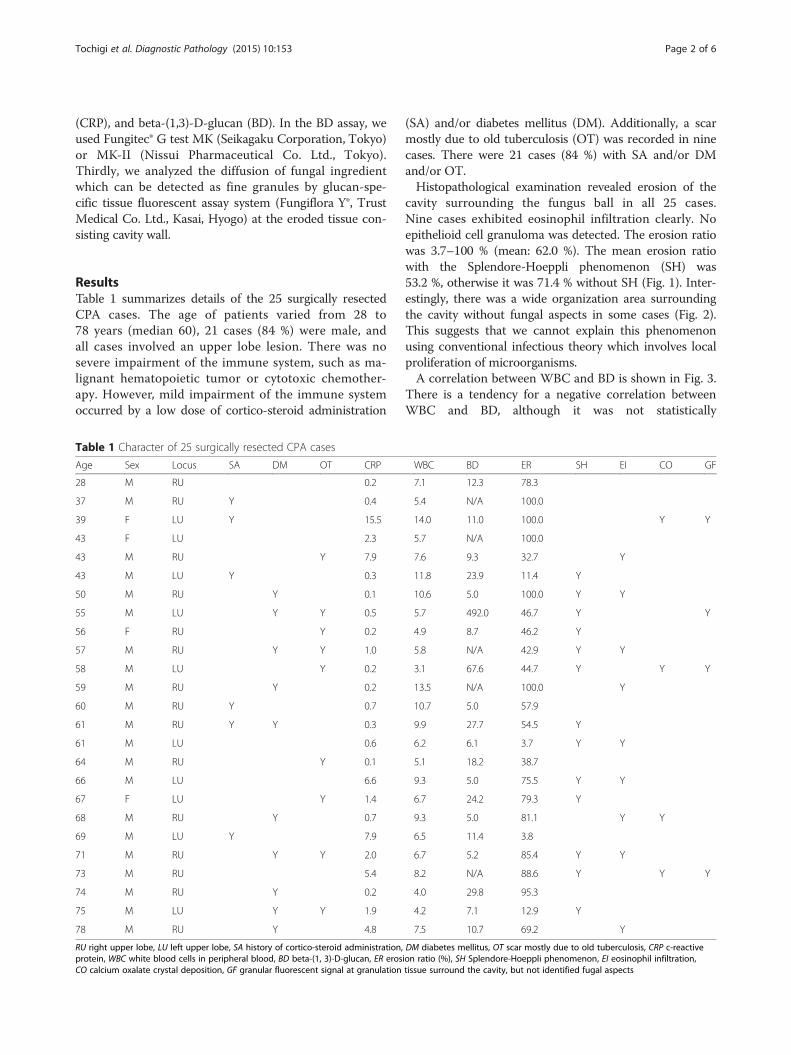
cavity surrounding the fungus ball in all 25 cases.Nine cases exhibited eosinophil infiltration clearly. Noepithelioid cell granuloma was detected. The erosion ratiowas 3.7–100 % (mean: 62.0 %). The mean erosion ratiowith the Splendore-Hoeppli phenomenon (SH) was53.2 %, otherwise it was 71.4 % without SH (Fig. 1). Inter-estingly, there was a wide organization area surroundingthe cavity without fungal aspects in some cases (Fig. 2).This suggests that we cannot explain this phenomenonusing conventional infectious theory which involves localproliferation of microorganisms.A correlation between WBC and BD is shown in Fig. 3.
There is a tendency for a negative correlation betweenWBC and BD, although it was not statistically
Table 1 Character of 25 surgically resected CPA cases
Age Sex Locus SA DM OT CRP WBC BD ER SH EI CO GF
28 M RU 0.2 7.1 12.3 78.3
37 M RU Y 0.4 5.4 N/A 100.0
39 F LU Y 15.5 14.0 11.0 100.0 Y Y
43 F LU 2.3 5.7 N/A 100.0
43 M RU Y 7.9 7.6 9.3 32.7 Y
43 M LU Y 0.3 11.8 23.9 11.4 Y
50 M RU Y 0.1 10.6 5.0 100.0 Y Y
55 M LU Y Y 0.5 5.7 492.0 46.7 Y Y
56 F RU Y 0.2 4.9 8.7 46.2 Y
57 M RU Y Y 1.0 5.8 N/A 42.9 Y Y
58 M LU Y 0.2 3.1 67.6 44.7 Y Y Y
59 M RU Y 0.2 13.5 N/A 100.0 Y
60 M RU Y 0.7 10.7 5.0 57.9
61 M RU Y Y 0.3 9.9 27.7 54.5 Y
61 M LU 0.6 6.2 6.1 3.7 Y Y
64 M RU Y 0.1 5.1 18.2 38.7
66 M LU 6.6 9.3 5.0 75.5 Y Y
67 F LU Y 1.4 6.7 24.2 79.3 Y
68 M RU Y 0.7 9.3 5.0 81.1 Y Y
69 M LU Y 7.9 6.5 11.4 3.8
71 M RU Y Y 2.0 6.7 5.2 85.4 Y Y
73 M RU 5.4 8.2 N/A 88.6 Y Y Y
74 M RU Y 0.2 4.0 29.8 95.3
75 M LU Y Y 1.9 4.2 7.1 12.9 Y
78 M RU Y 4.8 7.5 10.7 69.2 Y
RU right upper lobe, LU left upper lobe, SA history of cortico-steroid administration, DM diabetes mellitus, OT scar mostly due to old tuberculosis, CRP c-reactiveprotein, WBC white blood cells in peripheral blood, BD beta-(1, 3)-D-glucan, ER erosion ratio (%), SH Splendore-Hoeppli phenomenon, EI eosinophil infiltration,CO calcium oxalate crystal deposition, GF granular fluorescent signal at granulation tissue surround the cavity, but not identified fugal aspects
Tochigi et al. Diagnostic Pathology (2015) 10:153 Page 2 of 6

Fig. 1 Histopathology of erosion caused by CPA. a Cavity [§] is covered by ciliated epithelium without erosion (H-E). Note the Splendore-Hoeppliphenomenon (eosinophilic staining in fungus ball [†]). b Immounohistochemical staining for cytokeratin AE1/AE3 is done in case A. Erosion ratiois 3.7 %. c Epitheliums disappear and inflammatory granulation tissue exposes the surface of cavity. d Immounohistochemical staining for cytokeratinAE1/AE3 is done in case A. Erosion ratio is 95.3 %
Fig. 2 Histopathology of organization surrounding the cavity. a Macroscopic findings reveal the fungus ball (arrow head). Pleura with fibrousthickening is also noted. b Organization area can be seen around the fungus ball (H-E staining). c Alveolar spaces are filled with dense collagenoustissue (EVG staining)
Tochigi et al. Diagnostic Pathology (2015) 10:153 Page 3 of 6
significant. These data suggest that impairment of theimmune system worsens CPA pathogenesis.Four cases (16 %) showed a granular fluorescent signal in
granulation tissue surrounding the cavity using FungifloraY® stain, but we did not identify fungal aspects (GF) usingH-E or GMS (Fig. 4). Among the 19 cases with measuredBD before the surgical resection, the three GF-positivecases had a BD of 190.2 ± 201.2 pg/ml, while the 16 GF-negative cases had a BD of 12.8 ± 7.5 pg/ml. The detectedcorrelation between the presence of GF and the high levelof BD is statistically significant (one-sided test, p = 0.027,Mann–Whitney U test).
DiscussionGefter et al. defined semi-invasive pulmonary aspergillosisas a chronic cavitary form of pulmonary aspergillosis withmild immunosuppression or underlying lung disease [2].They indicated that this form of aspergillosis was part of aspectrum of disease ranging from saprophytic to franklyinvasive types. This was followed by report from Binder etal. which finally defined chronic necrotizing pulmonaryaspergillosis [3]. Within recent decade, Denning et al.described chronic cavitary pulmonary aspergillosis andchronic fibrosing pulmonary aspergillosis using radio-logical findings [4]. On the other hand, because of the dif-ficulty to distinguish chronic necrotizing pulmonaryaspergillosis and chronic cavitary pulmonary aspergillosis,
Izumikawa et al. proposed chronic progressive pulmonaryaspergillosis, which included chronic pulmonary aspergil-losis of both type; necrotizing and cavitation [6]. However,there are few reports conducting histological and patho-physiological analyses on this chronic form of aspergillosiswith reference to some representative monitoring system.Whereas our literature search for previous study couldhighlight the best-constructed histopathological study ofYousem reviewed 10 CPA cases and yielded a classifica-tion with three categories of granulomatous response [5],little were discussed on the pathophysiology of the disease.With comparison to Yousem’s subject group, there mightbe closer relationship between sequela of tuberculosis andCPA emerged from our study which can be supported bya result that our 25 CPA cases involved an upper lobe thatthe commonest area of the primary lesion of tuberculosis.However, since none of our patient showed active granu-loma that must be essentially induced by tuberculous in-fection, previous tuberculous infection might simplyplayed a role to re-construct airway, and an active tuber-culous infection has little contribution to develop the pul-monary lesion.Some our CPA cases showed pre-existing airway re-
modeling, such as cavitary formation, most of whichmight be sequel to old tuberculosis followed by emphy-sema. This anatomical reconstruction can promote in-festation or saprophytic proliferation of mold onto the
Fig. 3 Correlations between WBC and BD
Tochigi et al. Diagnostic Pathology (2015) 10:153 Page 4 of 6
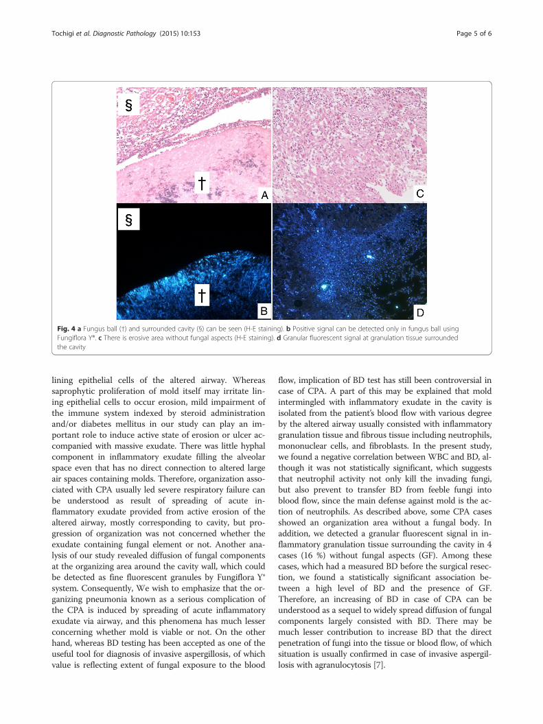
lining epithelial cells of the altered airway. Whereassaprophytic proliferation of mold itself may irritate lin-ing epithelial cells to occur erosion, mild impairment ofthe immune system indexed by steroid administrationand/or diabetes mellitus in our study can play an im-portant role to induce active state of erosion or ulcer ac-companied with massive exudate. There was little hyphalcomponent in inflammatory exudate filling the alveolarspace even that has no direct connection to altered largeair spaces containing molds. Therefore, organization asso-ciated with CPA usually led severe respiratory failure canbe understood as result of spreading of acute in-flammatory exudate provided from active erosion of thealtered airway, mostly corresponding to cavity, but pro-gression of organization was not concerned whether theexudate containing fungal element or not. Another ana-lysis of our study revealed diffusion of fungal componentsat the organizing area around the cavity wall, which couldbe detected as fine fluorescent granules by Fungiflora Y®system. Consequently, We wish to emphasize that the or-ganizing pneumonia known as a serious complication ofthe CPA is induced by spreading of acute inflammatoryexudate via airway, and this phenomena has much lesserconcerning whether mold is viable or not. On the otherhand, whereas BD testing has been accepted as one of theuseful tool for diagnosis of invasive aspergillosis, of whichvalue is reflecting extent of fungal exposure to the blood
flow, implication of BD test has still been controversial incase of CPA. A part of this may be explained that moldintermingled with inflammatory exudate in the cavity isisolated from the patient’s blood flow with various degreeby the altered airway usually consisted with inflammatorygranulation tissue and fibrous tissue including neutrophils,mononuclear cells, and fibroblasts. In the present study,we found a negative correlation between WBC and BD, al-though it was not statistically significant, which suggeststhat neutrophil activity not only kill the invading fungi,but also prevent to transfer BD from feeble fungi intoblood flow, since the main defense against mold is the ac-tion of neutrophils. As described above, some CPA casesshowed an organization area without a fungal body. Inaddition, we detected a granular fluorescent signal in in-flammatory granulation tissue surrounding the cavity in 4cases (16 %) without fungal aspects (GF). Among thesecases, which had a measured BD before the surgical resec-tion, we found a statistically significant association be-tween a high level of BD and the presence of GF.Therefore, an increasing of BD in case of CPA can beunderstood as a sequel to widely spread diffusion of fungalcomponents largely consisted with BD. There may bemuch lesser contribution to increase BD that the directpenetration of fungi into the tissue or blood flow, of whichsituation is usually confirmed in case of invasive aspergil-losis with agranulocytosis [7].
Fig. 4 a Fungus ball (†) and surrounded cavity (§) can be seen (H-E staining). b Positive signal can be detected only in fungus ball usingFungiflora Y®. c There is erosive area without fungal aspects (H-E staining). d Granular fluorescent signal at granulation tissue surroundedthe cavity
Tochigi et al. Diagnostic Pathology (2015) 10:153 Page 5 of 6
ConclusionsWe wish to conclude that extension of acute inflamma-tory exudate along the terminal respiratory tract is mostsignificant pathophysiological complication of the CPA,because the spreading of exudate filling alveolar spacemust cause organizing pneumonia, which derives fatalrespiratory failure. In addition, the viability commonlysuggesting the invasiveness of saprophytic Aspergilli doesnot concern progression of exudative inflammation at thesite of erosion.
Competing interestsDr. Shibuya reports receiving research grants from Pfizer Inc., JanssenPharmaceutical K.K., Dainippon Sumitomo Pharma Co., Astellas Pharma Inc.,Taiho Pharmaceutical Co., and POLA-Pharma Inc. Other authors declare thatthey have no competing interests.
Authors’ contributionsAll authors contributed towards the conceptualization, writing, reading, andapproval of the final manuscript. In particular, NT and TI jointly conceptualizedthis study, integrated the data, wrote the manuscript, and contributed equallyto this work.
AcknowledgmentThis work was supported by Health Science Research Grants for Researchon Emerging and Re-emerging Infectious Diseases (grant numbers: H25-shinkouippan-006 and H26-shinkoujitsuyouka-ippan-010) from the Ministry ofHealth, Labour and Welfare of Japan, a grant from the Strategic Basis on ResearchGrounds for Non-governmental Schools at Heisei 20th, the Strategic ResearchFoundation Grant-aided Project for Private Schools at Heisei 23rd, KAKENHI (grantnumbers: #24790364, 26860250, 26460460, and 26860774) from the Ministry ofEducation, Culture, Sports, Science, and Technology of Japan, Toho Universityproject grants (grant numbers: #23-19, 21, and 28, and #24-11, 16, and 28, and#25-33), a Yokohama Foundation for the Advancement of Medical Science grantto YO, KA, and MS, Dr. Yanase’s grant from Toho University Medical School to YO,and Kurozumi Medical Foundation grant to NT.
Author details1Department of Surgical Pathology, Toho University School of Medicine,6-11-1 Omori-nishi, Ota-ku, Tokyo 143-8541, Japan. 2Division of RespiratoryMedicine, Toho University School of Medicine, 6-11-1 Omori-nishi, Ota-ku,Tokyo 143-8541, Japan. 3Division of Chest Surgery, Toho University School ofMedicine, 6-11-1 Omori-nishi, Ota-ku, Tokyo 143-8541, Japan. 4Laboratory ofMolecular Cell Biology, School of Pharmacy, Nihon University, 7-7-1Narashinodai, Funabashi-shi, Chiba 274-8555, Japan.
Received: 8 January 2015 Accepted: 27 August 2015
References1. Grahame-Clarke CN, Roberts CM, Empey DW. Chronic necrotizing
pulmonary aspergillosis and pulmonary phycomycosis in cystic fibrosis.Respir Med. 1994;88:465–8.
2. Gefter WB, Weingrad TR, Epstein DM, Oches RH, Miller WT. “Semi-invasive”pulmonary aspergillosis: a new look at the spectrum of Aspergillus infectionsof the lung. Radiology. 1981;140:313–21.
3. Binder RE, Faling LJ, Pugatch RD, Masasaen C, Snider GL. Chronicnecrotizing pulmonary aspergillosis: a discrete clinical entity. Medicine(Baltimore). 1982;61:109–24.
4. Denning DW, Riniotis K, Dobrashian R, Sambatakou H. Chronic cavitaryand fibrosing pulmonary and pleural aspergillosis: case series, proposednomenclature change, and review. Clin Infect Dis. 2003;37 Suppl3:S265–80.
5. Yousem SA. The histological spectrum of chronic necrotizing forms ofpulmonary aspergillosis. Hum Pathol. 1997;28:650–6.
6. Izumikawa K, Tashiro T, Tashiro M, Takazono T, Kosai K, Morinaga Y, et al.Pathogenesis and clinical features of chronic pulmonary aspergillosis – Is it
possible to distinguish CNPA and CCPA clinically? J Infect Chemother.2014;20:208–12.
7. Shibuya K, Takaoka M, Uchida K, Wakayama M, Yamaguchi H, Takahashi K, etal. Histopathology of experimental invasive pulmonary aspergillosis in rats:pathological comparison of pulmonary lesions induced by specific virulentfactor deficient mutants. Microb Pathog. 1999;27:123–31.
Submit your next manuscript to BioMed Centraland take full advantage of:
• Convenient online submission
• Thorough peer review
• No space constraints or color figure charges
• Immediate publication on acceptance
• Inclusion in PubMed, CAS, Scopus and Google Scholar
• Research which is freely available for redistribution
Submit your manuscript at www.biomedcentral.com/submit
Tochigi et al. Diagnostic Pathology (2015) 10:153 Page 6 of 6


![Aspergillosis[1]- Katie Jacquie Qazi.pdf](https://img.pdfslide.tips/doc/110x75/577c7c161a28abe054993f8d/aspergillosis1-katie-jacquie-qazipdf.jpg)


![Aspergillosis [Dr. Ali Santoso Sp. PD]](https://img.pdfslide.tips/doc/110x75/563db7dc550346aa9a8e9f1a/aspergillosis-dr-ali-santoso-sp-pd.jpg)