Embed Size (px)
Citation preview

Kobe University Repository : Kernel
タイトルTit le
Structures and funct ions of insect arylalkylamine N-acetylt ransferase(iaaNAT); a key enzyme for physiological and behavioral switch inarthropods
著者Author(s)
Hiragaki, Susumu / Suzuki, Takeshi / Mohamed, Ahmed A. M. / Takeda,Makio
掲載誌・巻号・ページCitat ion Front iers in Physiology,6:113
刊行日Issue date 2015-04-13
資源タイプResource Type Journal Art icle / 学術雑誌論文
版区分Resource Version publisher
権利Rights
© 2015 Hiragaki, Suzuki, Mohamed and Takeda. This is an open-access art icle distributed under the terms of the Creat ive CommonsAttribut ion License (CC BY). The use, distribut ion or reproduct ion inother forums is permit ted, provided the original author(s) or licensor arecredited and that the original publicat ion in this journal is cited, inaccordance with accepted academic pract ice. No use, distribut ion orreproduct ion is permit ted which does not comply with these terms.
DOI 10.3389/fphys.2015.00113
JaLCDOI
URL http://www.lib.kobe-u.ac.jp/handle_kernel/90002868
PDF issue: 2020-08-13
HYPOTHESIS AND THEORYpublished: 13 April 2015
doi: 10.3389/fphys.2015.00113
Frontiers in Physiology | www.frontiersin.org 1 April 2015 | Volume 6 | Article 113
Edited by:
Raman Chandrasekar,
Kansas State University, USA
Reviewed by:
David Dolezel,
Biology Centre Academy of Sciences
of the Czech Republic,
Czech Republic
Xanthe Vafopoulou,
York University, Canada
Bharat Bhusan Patnaik,
Chonnam National University,
South Korea
*Correspondence:
Makio Takeda,
Graduate School of Agricultural
Science, Kobe University, 1-1
Rokkodai-cho, Nada-ku, Kobe
657-8501, Japan
Specialty section:
This article was submitted to
Integrative Physiology,
a section of the journal
Frontiers in Physiology
Received: 27 November 2014
Paper pending published:
17 January 2015
Accepted: 25 March 2015
Published: 13 April 2015
Citation:
Hiragaki S, Suzuki T, Mohamed AAM
and Takeda M (2015) Structures and
functions of insect arylalkylamine
N-acetyltransferase (iaaNAT); a key
enzyme for physiological and
behavioral switch in arthropods.
Front. Physiol. 6:113.
doi: 10.3389/fphys.2015.00113
Structures and functions of insectarylalkylamine N-acetyltransferase(iaaNAT); a key enzyme forphysiological and behavioral switchin arthropodsSusumu Hiragaki 1, Takeshi Suzuki 2, Ahmed A. M. Mohamed 1 and Makio Takeda 1*
1Graduate School of Agricultural Science, Kobe University, Kobe, Japan, 2Department of Biology, The University of Western
Ontario, London, ON, Canada
The evolution of N-acetyltransfeases (NATs) seems complex. Vertebrate arylalkylamine
N-acetyltransferase (aaNAT) has been extensively studied since it leads to the synthesis
of melatonin, a multifunctional neurohormone prevalent in photoreceptor cells, and is
known as a chemical token of the night. Melatonin also serves as a scavenger for
reactive oxygen species. This is also true with invertebrates. NAT therefore has distinct
functional implications in circadian function, as timezymes (aaNAT), and also xenobiotic
reactions (arylamine NAT or simply NAT). NATs belong to a broader enzyme group,
the GCN5-related N-acetyltransferase superfamily. Due to low sequence homology and
a seemingly fast rate of structural differentiation, the nomenclature for NATs can be
confusing. The advent of bioinformatics, however, has helped to classify this group of
enzymes; vertebrates have two distinct subgroups, the timezyme type and the xenobiotic
type, which has a wider substrate range including imidazolamine, pharmacological drugs,
environmental toxicants and even histone. Insect aaNAT (iaaNAT) form their own clade in
the phylogeny, distinct from vertebrate aaNATs. Arthropods are unique, since the phylum
has exoskeleton in which quinones derived from N-acetylated monoamines function
in coupling chitin and arthropodins. Monoamine oxidase (MAO) activity is limited in
insects, but NAT-mediated degradation prevails. However, unexpectedly iaaNAT occurs
not only among arthropods but also among basal deuterostomia, and is therefore more
apomorphic. Our analyses illustrate that iaaNATs has unique physiological roles but
at the same time it plays a role in a timezyme function, at least in photoperiodism.
Photoperiodism has been considered as a function of circadian system but the detailed
molecular mechanism is not well understood. We propose a molecular hypothesis
for photoperiodism in Antheraea pernyi based on the transcription regulation of NAT
interlocked by the circadian system. Therefore, the enzyme plays both unique and
universal roles in insects. The unique role of iaaNATs in physiological regulation urges
the targeting of this system for integrated pest management (IPM). We indeed showed
Hiragaki et al. Insect arylalkylamine N-acetyltransferase (iaaNAT)
a successful example of chemical compound screening with reconstituted enzyme and
further attempts seem promising.
Keywords: serotonin (5HT), melatonin (MEL), arylalkylamine N-acetyl transferase (aaNAT), arylamine
N-acetyltransferase, circadian rhythms, photoperiodism, UV adaptation
Historical Aspects and Reality in InsectaaNAT
Arylalkylamine N-acetyltransferase (aaNAT) has been mostextensively studied as a penultimate enzyme formelatonin (MEL)synthesis in vertebrates (Klein and Weller, 1970; Axelrod, 1974;Ebisawa and Deguchi, 1991; Chong et al., 2000; Iuvone et al.,2005; Falcón et al., 2014), particularly in the context of dailyand seasonal timing for activity, metabolism and physiologicalphenomena, thus commonly called timezyme (Klein, 2007). Thisenzyme is expressed in the pineal gland, retina, and parietaleyes that are photosensitive organs but the exocrine Hardeliangland also contains MEL synthesizing machinery (Dieridane andTouitou, 2001). A massive amount of MEL has been reportedin the gastroenteric tract in mammals (Bubenik, 2008). Thenumber of genes for aaNAT is one in higher vertebrates (Sab-bagh et al., 2013). However, multiple acetylases are known invertebrates that function as xenobiotic enzyme, ex. seven incephalochordate and three in human (Pavlicek et al., 2010; Sab-bagh et al., 2013), and human subject has polymorphism inthis gene to symptomatically separate forms commonly termedslow and fast-acetylators. This enzyme has been called arylamineN-acetyltransferase (Ebisawa and Deguchi, 1991; Deguchi, 1992).
In arthropods especially insects, more extensive use of thisenzyme is made for irrelevant functions to vertebrates such assclerotization where dopamine, tyramine, norepinephrine andoctopamine are N-acetylated to provide reactive quinones forlinking chitin and arthropodins forming a cuticular matrix (Karl-son et al., 1962; Sekeris and Karlson, 1962). The enzyme wasfirst called dopamine acetyltransferase (DAT). Also, metabolicpathways of neurotransmitter monoamines are different fromvertebrates; in vertebrates monoamines are metabolized mainlyby monoamine oxidases (MAO1 and 2) but in arthropods theactivity of MAOs is usually limited and these amines are metabo-lized mainly by NAT instead (Dewhurst et al., 1972; Sloley, 2004),though oxidized products are substantially detected during dia-pause in Antheraea pernyi at the pupal stage (Matsumoto andTakeda, 2002).
Since major neurotransmitter metabolism depends on thisenzyme, it serves as an important regulator in a variety ofphysiological functions, metabolism, developmental determina-tion, reproduction and behavior. Activities of aaNAT have been
Abbreviations: aaNAT, arylalkylamineN-acetyltransferase; iaaNAT, insect aaNAT;
5HT, serotonin; MEL, melatonin; DAT, dopamine acetyltransferase; MAO,
monoamine oxidase; aNAT, acidic aaNAT; bNAT, basic aaNAT; SOG, sube-
sophageal ganglion; SCN, suprachiasmatic nucleus; CPM, circadian pacemaker;
PT, pars tuberalis; MT, MEL receptor; GNAT, GCN5-related N-acetyltransferase;
VT-aaNAT, vertebrate aaNAT; NV-aaNAT, non-vertebrate aaNAT; PTTH, pro-
thoracicotropic hormone; GNA, glucosamine 6-phosphate N-acetyltransferases;
HAT, histone acetyltransferase; RGC, retinal ganglion cell; ipRGC, intrinsically
photosensitive RGC; SNAT, serotonin N-acetyltransferas.
observed in various organs not only the CNS but also in themidgut, reproductive glands of both sexes andMalpigian tublulesin the cockroach, Periplaneta americana, for example (Ichiharaet al., 1997, 2001; Asano and Takeda, 1998; Asano et al., 2003).Isolation and characterization of aaNAT revealed that the activ-ities are separated to different fractions, each showing a uniqueoptimal pH and kinetic character (Ichihara et al., 1997, 1998,2001; Asano and Takeda, 1998; Sakamoto et al., 1998), suggestingthat aaNAT activities consist of a family of enzymes. Two iso-forms have been isolated from testicular accessory gland andmidgut. Both forms had 28 kDa molecular mass in SDS-PAGEbut the former had an optimal pH at 5–6 whereas the latter 9–10and thus called acidic aaNAT (aNAT) and basic aaNAT (bNAT),respectively (Ichihara et al., 1997, 2001; Bembenek et al., 2005a,b).Vmax and Km-values estimated from a purified fraction of bothforms are listed in Table 1.
In Drosophila melanogaster, two aaNAT isoforms have beenpurified and aaNAT1 has been detected by in situ hybridizationin the brain, ventral cord, midgut and oenocyte (Hintermannet al., 1996). aaNAT2 had 30% identity to aaNAT1 and estimatedmolecular mass was 24.4 vs. 31.0 kDa of NAT1 and IEP, 5.02 vs.5.08 (Amherd et al., 2000). MEL rhythm had bimodal peaks butboth enzymes failed to show circadian fluctuation in transcriptamount (Hintermann et al., 1996; Amherd et al., 2000), whileaaNAT of cockroach and A. pernyi showed a circadian rhythm inactivity and in transcription (Bembenek et al., 2005a,b; Mohamedet al., 2014).
Kinetic Aspects: How Many Enzymes?
There was a practical reason for the difficulty to isolate aaNATin vertebrates. This enzyme is extremely sensitive to light in ver-tebrates even at second order. Therefore, the detection requiredBolton-Hunter labeling, apparently too small an amount forsequence determination (Namboodiri et al., 1987). However, dif-ferential cloning successfully identified aaNAT before purifica-tion was completed (Borjigin et al., 1995; Coon et al., 1995).However, iaaNAT was much more photorefractory and HPLC
TABLE 1 | Comparison in kinetic properties of two forms of isolated
aaNAT from P. americana. aNAT/bNAT.
Substrate Km (mM) Vmax (µmol/min/mg protein)
Tryptamine 0.025/1.33 11.2/276
5HT 0.05/0.325 9.33/110
Dopamine 0.025/1.52 6.22/172
Octopamine 0.02/0.385 9.33/276
Tyramine 0.0286/0.0157 18.7/420
Methoxy-TN 0.025/0.2 6.57/230
Norepinephrine 0.0286/- 12.4/-
Frontiers in Physiology | www.frontiersin.org 2 April 2015 | Volume 6 | Article 113

Hiragaki et al. Insect arylalkylamine N-acetyltransferase (iaaNAT)
resulted in a purified fraction. The protein sequence was deter-mined in cleavage products from cDNA cloned from the acces-sory gland of male gonad of P. americana (Ichihara et al.,1997, 1998; Bembenek et al., 2005b). Chromatographic isolationshowed that at least two types of NATs occurred at differentfractions that can be characterized by pH optima and substratespecificity. We tentatively called the two types, NATa and b thathad pH optima at acidic and basic side, respectively. Differentorgans contain different ratios of the mixture (Ichihara et al.,1997, 2001; Bembenek et al., 2005b). The two types show dif-ferent affinity to serotonin (5HT)/tryptamine and octopamine.The combination of amine substrate and pH using samples iso-lated from the accessory gland of cockroach at different agesshowed at least four distinct patterns of change along ovula-tion cycle (Asano et al., 2003). Since homology of amino acidsequence was low among aaNATs, the ordinary PCR approachwas not successful, but functional domains were relatively con-served and from the Bombyx EST database, cDNA was amplifiedfrom Bombyx mori brain-subesophageal ganglion (SOG) (Tsug-ehara et al., 2007). The cDNA encodes a 261 amino acid pro-tein and Baculovirus-mediated reconstituted enzyme had a widerange of substrate affinity including tryptamine, 5HT, dopamine,octopamine and norepinephrine (Tsugehara et al., 2007). Thetranscript was expressed in different tissues such as head, ovary,testes and even flight muscles and in eggs, larvae, and adults.It showed a substrate inhibition by a high concentration oftryptamine. We also obtained cDNA for aaNAT from A. pernyi(Tsugehara et al., 2013), expressing this enzyme in Sf9 cell linesimilarly and confirmed its enzymatic activity (Tsugehara et al.,2013). It had the optimal pH at 8.0 and apparent Km and Vmax
1.42µM and 154 nmol/min/mg protein with acetyl CoA and3.31µM and 190 nmol/min/mg with tryptamine. Substrate inhi-bition was observed above 0.03mM with 0.11mM acetyl CoA.Since the enzyme employs two substrates, arylalkylamines andacetyl CoA, we made a bisubstrate kinetic analysis employingbrain-SOG and collaterial gland homogenates of P. americana.The former followed ping pong Bi-Bi mechanism and the lat-ter sequential Bi-Bi mechanism, respectively (Asano and Takeda,1998; Sakamoto et al., 1998).
Functions of aaNAT in Vertebrates
aaNAT transfers acetyl moiety to 5HT and controls daily changesin circulating MEL in boney vertebrates (Ganguly et al., 2002).In mammals, only the suprachiasmatic nucleus (SCN) serves asa circadian pacemaker (CPM) that receives light signals from theretina, and directly regulates pineal MEL biosynthesis as an out-put of the clock (Bell-Pedersen et al., 2005). The rhythmic patternof activity in the MEL pathway is conserved among vertebrates,consistent with the role of MEL as the vertebrate hormone oftime, i.e., high level signal at night and low level signal day. MELis involved in numerous physiological processes including circa-dian entrainment, blood pressure regulation, oncogenesis, reti-nal physiology, ovarian physiology, and immune function (Altunand Ugur-Altun, 2007). As another notable example, cyclingexpression of the clock gene Period1 in rodent pars tuberalis (PT)completely depends on the nocturnal activation of MEL receptor
type-1 (MT1; von Gall et al., 2002). Cyclic Period1 expressionin PT is noteworthy because PT is where MEL acts as a media-tor between CPM and output of photoperiodism (Hoffman andReiter, 1965; Messager et al., 1999; Masumoto et al., 2010). Theremarkable role that aaNAT plays in vertebrate time keepingmonikered the aaNAT as “the timezyme” (Klein, 2007).
The aaNAT family, belonging to the GCN5 acetyltransferases(GNAT) superfamily (Dyda et al., 2000) is classified as vertebrate(VT) aaNAT and non-vertebrate (NV) aaNAT based on sequencesimilarity. Striking features of VT-aaNAT are found in regula-tory and catalytic regions of the encoded proteins (Ganguly et al.,2001; Coon and Klein, 2006; Pavlicek et al., 2010). VT-aaNATexists exclusively in two organs, the pineal gland and retina, bothphotosensitive (Coon et al., 1995). NV-aaNAT performs a detoxi-fication function through acetylation of a broad range of arylalky-lamines and polyamines in throughout body segment (Gangulyet al., 2001; Pavlicek et al., 2010), in contrast to VT-aaNAT whichplays as a timezyme (Klein, 2007).
According to similarity to VT-aaNAT and NV-aaNAT, theseaaNATs have been found in Gram-positive bacteria, fungi, somelower plants, algae, Placozoa, Annelida, Cephalochordates, andVertebrates (Coon and Klein, 2006; Falcón et al., 2014) but notin other published genomes, including higher plants, nematodesand arthropods (Coon and Klein, 2006). In addition, both VT-and NV-aaNATs are not found in Echinodermata, Hemichor-dates, and Urochordates, which leaves an open question of whenand how NV-aaNAT first appeared in Deuterostomia evolution(Pavlicek et al., 2010). Iyer et al. (2004) have developed a hypoth-esis that a horizontal gene transfer from bacteria to an ancestor ofvertebrate caused the evolution of VT-aaNAT. The homologs inamphioxus, unicellular green algae, fungi and bacteria lack regu-latory sequences found in VT-aaNAT. Also, all these are intron-less. These features are consistent with a horizontal gene trans-fer of the aaNAT ancestor from bacteria to green algae, fungiand Cephalochordates (Coon and Klein, 2006). Very recently,two types of aaNAT were cloned from Chondrichthyes (Falcónet al., 2014). One was found in the pineal gland and retina,structurally, biochemically, and kinetically similar to VT-aaNAT.The other occurs throughout the body like NV-aaNAT. Calcula-tion of the evolutionary rates revealed an acceleration of evolu-tion by approximately an order of magnitude in the stem of theVT-aaNAT subfamily (Falcón et al., 2014). Therefore, the emer-gence of VT-aaNAT timezyme apparently dramatically acceler-ated evolution, accompanied with neofunctionalization after theNV-aaNAT gene duplication.
Function of Insect-type AaNAT
aaNATs play multiple roles in behavioral and physiological regu-lations in insects. Insect aaNAT (iaaNAT) is involved in cuticularsclerotization and suppression of black pigmentation (Dai et al.,2010; Zhan et al., 2010; Osanai-Futahashi et al., 2012). iaaNATinactivates biogenic amines, such as octopamine, dopamine, and5HT (Hintermann et al., 1995; Tsugehara et al., 2007), since thereis little or no MAO activity within the insect nervous tissue (Slo-ley, 2004) unlike mammals (Bortolato et al., 2008). Followingevidence indicates that iaaNATs regulate physiological events via
Frontiers in Physiology | www.frontiersin.org 3 April 2015 | Volume 6 | Article 113
Hiragaki et al. Insect arylalkylamine N-acetyltransferase (iaaNAT)
MEL in insects; orally administrated MEL synchronizes circa-dian locomotor rhythms of the house cricket Acheta domesticus(Yamano et al., 2001) and MEL stimulates the release of a pro-thoracicotropic hormone (PTTH) in the brain of P. americana(Richter et al., 2000).
Very recently, we demonstrated that iaaNAT is the criticaljoint between the circadian system and photoperiodism in A.pernyi (Mohamed et al., 2014). It is noteworthy that the functionof iaaNAT has several striking similarities to that of VT-aaNATin the endocrine switch, as it is supported by the following evi-dence. (1) iaanat is highly likely controlled by a negative feedbackloop via E-box element. In the iaanat promoter region (1596bp upstream region), there were 2 perfect E-boxes (CACGTG),4 canonical E-boxes (CANNTG). qRT-PCR revealed rhythmi-cal expression of iaanat in LD cycle. RNAi against per, putativenegative regulator of E-box, resulted in up-regulation of nat tran-scription. In contrast, RNAi knockdown of Clk and cyc, posi-tive regulator of E-box, suppressed the expression level of nat.In chicken, the abundance of VT-aaNAT mRNA in the pinealgland exhibits a circadian rhythm (Bernard et al., 1997), which isregulated by BMAL1/CLOCK and BMAL1/MOP4 heterodimersvia E-box (Chong et al., 2000). (2) iaaNAT regulates photoperi-odism. dsRNAiaaNAT-injected pupae failed to emerge even underlong day, whereas pupae injected with dsRNAper showed a highproportion of emergence even under short day. In mammals,the nocturnal MEL secretion provides an endocrine signal ofphotoperiod to the PT to regulate thyroid-stimulating hormonewhich regulates seasonal reproduction (Yoshimura, 2013). (3)MEL content is regulated by the expression level of iaanat.Injection of dsRNAiaaNAT induced a great decline of MEL in48 h. In contrast, MEL level was significantly higher than con-trol pupae after knocking down of per by 24 h. In higher ver-tebrates, the switch between day and night profiles of pinealMEL is driven by changes in the activity and expression levelof VT-aaNAT, which increases at night 10- to 100-fold (Gangulyet al., 2002). (4) iaaNAT located at a unique from neuroendocrinecells which control photoperiodisum. iaaNAT-like immunoreac-tivity (-ir) was observed in the PER/CYC/CLK-ir cells, putative“clock neurons.” MT2-ir and PTTH-ir were co-localized in a pairof neurosecretory neurons juxtaposing the putative clock neu-rons. In mammals, VT-aaNAT is highly expressed in the pinealgland, which is near MT1-expressing tissue, PT (Nakane andYoshimura, 2014).
According to the significance and similarity to the originaltimezyme, we considered that iaaNAT in A. pernyi serves as aswitch of PTTH secretion despite no universally accepted theoryfor the function of iaaNAT as timezyme to date in insects sinceiaaNAT family has primary structure distinct from VT-aaNATand NV-aaNAT. These families are only distantly related to eachother, although both belong to the GNAT superfamily (Falcónet al., 2014).
Phylogenetic Relationship of aaNATsDepicted from Metazoan Genome Study
Insects do not have VT-aaNAT or NV-aaNAT homologs, basedon sequence similarity searches of public genomic sequences
(Pavlicek et al., 2010); however, insects do have multipleaaNAT enzymes that show very low sequence identity withVT-aaNAT/NV-aaNAT. According to enzymatic properties ofaaNAT, insects seems to have more than 4 aaNAT-like enzymesin a single species as described before. The biochemical charac-terizations of iaaNATs were followed up with molecular isolationinD. melanogaster (Hintermann et al., 1995, 1996; Amherd et al.,2000), B. mori (Tsugehara et al., 2007), A. pernyi (Tsugehara,2006), and P. americana (Ichihara et al., 1997, 2001; Bembeneket al., 2005a,b). Primary structure of iaaNATs show very lowsequence identity between each other (Hintermann et al., 1996;Amherd et al., 2000; Bembenek et al., 2005b; Tsugehara et al.,2007). How many iaaNATs exist in one species? How is therelationship between each iaaNAT? To answer these questions,exhaustive analysis of iaaNAT is required.
A first remarkable genome-wide characterization of iaaNATswas held on several dipteran species, Anopheles gambiae, Aedesaegypti, Culex quinquefasciatus, and D. melanogaster (Mehereet al., 2011; Han et al., 2012). They studied three proteins in detail:iaaNAT2, iaaNAT5b, and putative iaaNAT7 (piaaNAT7) from A.aegypti, each from a different cluster of phylogenetic trees. Allthree iaaNAT and sheep VT-aaNAT structures have a commonfold core of GNAT superfamily proteins, along with a uniquestructural feature: helix/helices between β3 and β4 strands.
Subsequently, genome-wide characterization of iaaNATs washeld on the pea aphid,Acyrthosiphon pisum (Barberà et al., 2013).They yielded five predicted sequences with E-values lower than1.0× 10−4 in BLASTP search on the A. pisum genomic databaseusing D. melanogaster (Dm) DAT as a query. Following screen-ing using conserved domain (CD)-search tool available at theNational Center for Biotechnology Information identified fouriaaNATs (iaaNAT1, iaaNAT2, iaaNAT3, and iaaNAT4). Amongthem, two (iaaNAT1 and iaaNAT3) showed highly significantvariation in transcription levels depending on photoperiods.
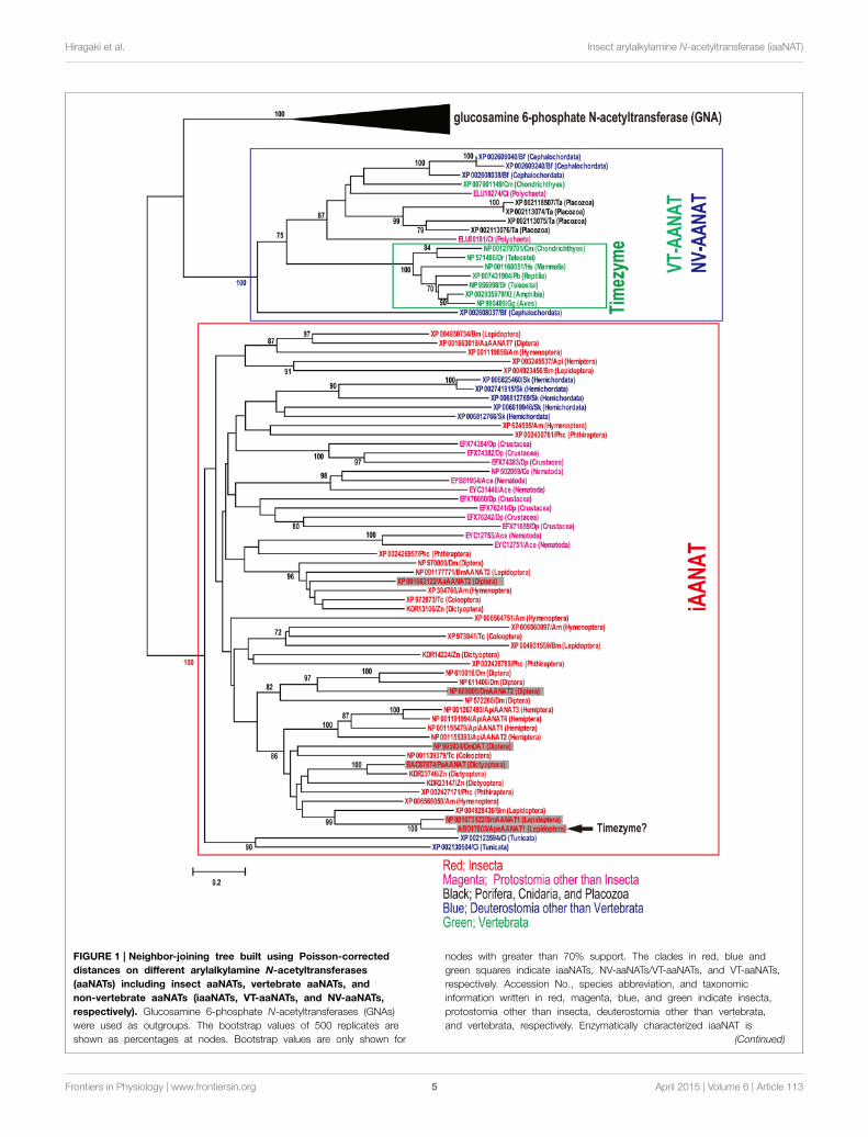
According to a series of genome-wide identification ofiaaNAT, we try comprehensive phylogenetic analysis of VT-aaNAT, NV-aaNAT, and iaaNAT among metazoan species forbetter understanding of potential functions of iaaNAT. Combina-tion of similarity search and CD-search revealed 57 iaaNATs, 11NV-aaNATs, and 7VT-aaNATs from the public genome databasedescribed above (Table S1). To confirm the orthology of theiaaNATs, NV-aaNATs, and VT-aaNATs genes screened, we car-ried out a phylogenetic analysis that included all 75 sequencesand glucosamine 6-phosphate N-acetyltransferases (GNAs) fromthe public genome database described above (Figure 1). VT-aaNAT/NV-aaNAT and iaaNAT genes clearly clustered into dis-tinct groups (mean pairwise value 100%). As expected, onlyiaaNATs were isolated from insect species among differentorders. In addition to insect, iaaNATs were detected fromDaphnia pulex (Crustacea), Caenorhabditis elegans (Nematoda),and Ancylostoma ceylanicum (Nematoda) from protostomespices. As described previously, Capitella teleta (Annelida; Poly-chaeta), Trichoplax adhaerens (Placozoa), and Branchiostomafloridae (Cephalochordata) have NV-aaNATs, whereas mostvertebrates have only VT-aaNATs except Callorhinchus milii(Chondrichthyes) that had both NV-aaNAT and VT-aaNAT(Coon and Klein, 2006; Falcón et al., 2014). Surprisingly, 4
Frontiers in Physiology | www.frontiersin.org 4 April 2015 | Volume 6 | Article 113
Hiragaki et al. Insect arylalkylamine N-acetyltransferase (iaaNAT)
FIGURE 1 | Neighbor-joining tree built using Poisson-corrected
distances on different arylalkylamine N-acetyltransferases
(aaNATs) including insect aaNATs, vertebrate aaNATs, and
non-vertebrate aaNATs (iaaNATs, VT-aaNATs, and NV-aaNATs,
respectively). Glucosamine 6-phosphate N-acetyltransferases (GNAs)
were used as outgroups. The bootstrap values of 500 replicates are
shown as percentages at nodes. Bootstrap values are only shown for
nodes with greater than 70% support. The clades in red, blue and
green squares indicate iaaNATs, NV-aaNATs/VT-aaNATs, and VT-aaNATs,
respectively. Accession No., species abbreviation, and taxonomic
information written in red, magenta, blue, and green indicate insecta,
protostomia other than insecta, deuterostomia other than vertebrata,
and vertebrata, respectively. Enzymatically characterized iaaNAT is
(Continued)
Frontiers in Physiology | www.frontiersin.org 5 April 2015 | Volume 6 | Article 113

Hiragaki et al. Insect arylalkylamine N-acetyltransferase (iaaNAT)
FIGURE 1 | Continued
indicated in shade. Arrow shows potential iaaNAT possess a function as
a timezyme (Mohamed et al., 2014). Species abbreviations; Dm,
Drosophila melanogaster; Aa, Aedes aegypti; Am, Apis mellifera, Bm,
Bombyx mori; Ape, Antheraea pernyi; Tc, Tribolium castaneum; Api,
Acyrthosiphon pisum; Zn, Zootermopsis nevadensis; Pa, Periplaneta
americana; Phc, Pediculus humanus corporis; Dp, Daphnia pulex; Ce,
Caenorhabditis elegans; Ace, Ancylostoma ceylanicum; Ct, Capitella
teleta; Ta, Trichoplax adhaerens; Sk, Saccoglossus kowalevskii; Ci, Ciona
intestinalis; Bf, Branchiostoma floridae; Cm, Callorhinchus milii; Dr, Danio
rerio; Xt, Xenopus tropicalis; Gg, Gallus gallus; Pb, Python bivittatus; Hs,
Homo sapiens.
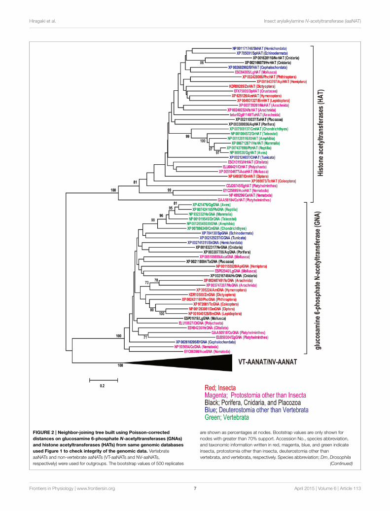
iaaNATs and 2 iaaNATs were retrieved from genomic databaseof deuterostome species, Saccoglossus kowalevskii (Hemichor-data) and Ciona intestinalis (Urochordata), respectively. NeitheriaaNAT, VT-aaNAT, nor NV-aaNAT have been detected in Ixodesscapularis (Arachnida), Metaseiulus occidentalis (Arachnida),Tetranychus urticae (Arachnida), Aplysia californica (Mollusca),Lottia gigantea (Mollusca), Helobdella robusta (Annelida; Clitel-lata), Clonorchis sinensis (Platyhelminthes), Echinococcus granu-losus (Platyhelminthes), Hydra vulgaris (Cnidaria), Nematostellavectensis (Cnidaria), Amphimedon queenslandica (Porifera),and Strongylocentrotus purpuratus (Echinodermata). To checkintegrity of the genomic data lacking known aaNAT, sequencesimilarity searches was conducted using other acetyltransferasesbelonging to GNAT superfamily, HsGNA (NP_932332) and Hshistone acetyltransferase (HAT; XP_006712871) as queries. Aphylogenetic analysis was carried out in all isolated GNAs, HATs,and VT-aaNAT/NV-aaNAT to confirm the orthology (Figure 2).Expectedly, GNAs, HATs, and VT-aaNATs/NV-aaNATs clusterinto three groups, all strongly supported (mean pairwise value100%) by neighbor-joining algorithm on Poisson-corrected dis-tances. This strongly suggests unique distributions of aaNATswithin phyla unlike other GNAT superfamily.
The distribution of timezyme, VT-aaNAT and its ancestralform, NV-aaNAT is understood by gene duplications, deletion,and a horizontal gene transfer (Coon and Klein, 2006), followedby VT-aaNAT evolution from the NV-aaNAT associated withdrastic neofunctionalization of aaNAT in the Cephalochordate-Vertebrate split (Falcón et al., 2014). However, according todiversity of aaNATs and current view of phylogenetic relationshipof Metazoa (Figure 3), the evolution of aaNAT looks much morecomplex than expected. Controversial phylogenetic positions ofUrochordata, Cephalochordata, and Vertebrata were accessed byBayesian and maximum likelihood methods on a 1090 ortholo-gous genes (Putnam et al., 2008). Both molecular phylogeneticanalyses support that Cephalochordata represents the most basalextant chordate lineage in relation to Urochordata and Verte-brate. Also, the relationship of Placozoa to other animals wasanalyzed using Bayesian, maximum likelihood, and parsimonyanalyses of a concatenation of 104 slowly evolving single-copynuclear genes (Srivastava et al., 2008). With 100% Bayesian sup-port and 92% likelihood bootstrap support that Porifera divergedbefore the Placozoa-Cnidaria-Bilaterian clade. We found iaaNATin Deuterostomia, such as Urochordata, a sister group betweenthe Cephalochordate and Vertebrate. There are NV-aaNATs inPlacozoa and Polychaeta but no NV-aaNATs in closely relatedspecies. iaaNATs are distributed in known genomic databases ofEcdysozoa but not in Arachnida in spite of distinct activity ofaaNAT (Suzuki et al., 2008). No phyla have both iaaNAT andNV-aaNAT/VT-aaNAT among metazoan species so far. It is very
difficult to explain in distribution of aaNATs if iaaNAT and NV-aaNAT/VT-aaNAT are not related at all. Comprehensive analysesof aaNATs are required.
Spectral Responses and Non-VisualPhotoreceptors
The activity of aaNAT shows a nocturnal increase and thisenhances the biosynthesis of MEL at night (Baler et al., 1997).Deguchi and Axelrod (1972a) first developed a radioenzymaticmeasurement of aaNAT activity with [14C]-acetyl CoA. Thishighly-sensitivemethod clearly showed that aaNAT activity in therat pineal organ increased 50-fold at night (Deguchi and Axel-rod, 1972b). According to Korf (1994), aaNAT activity and sub-sequent secretion of MEL are suppressed by light signals fromphotoreceptors through endogenous oscillators, i.e., circadianclock. In mammals, the locations of photoreception, endogenousoscillation, and MEL secretion are the retina, SCN, and pinealorgan, respectively. In contrast, pineal organ equips these threecomponents in non-mammalian vertebrates (Falcón, 1999). First,researchers focused on the non-visual photoreceptors expressedin the pineal organs in non-mammalian vertebrates. Deguchi(1981) revealed that the isolated chicken pineal is sensitive tolight (<700 nm) and that green light (λmax 500 nm) is the mosteffective in suppressing the aaNAT activity. From the actionspectrum, a rhodopsin-like molecule was suggested to functionas photoreceptors in the pineal organ. However, the isolatedphotoreceptor was a blue sensitive pigment “pinopsin” (λmax
468 nm) which is specifically expressed only in the pineal organ(Okano et al., 1994). Since the red-sensitive pigment “iodopsin”(λmax 571 nm) was isolated from chicken pineal (Okano et al.,1989), cooperative photoreception by pinopsin and iodopsin isconsidered to suppress the aaNAT activity and results in thegreen-sensitive broad action spectrum (Deguchi, 1981). In addi-tion, Blackshaw and Snyder (1997) identified an ultraviolet (UV)-sensitive pigment “parapinopsin” (λmax 370 nm) in the catfishpineal and it was also isolated from the pineal organ of lampreys,fishes, and frogs (Koyanagi et al., 2004; Kawano-Yamashita et al.,2007). Furthermore, another opsin-based non-visual photopig-ment “melanopsin” (λmax 480 nm) (Qiu et al., 2005) was iden-tified from the photosensitive dermal melanophores of X. laevis(Provencio et al., 1998) and the homologs have been identified invaried vertebrates (Koyanagi and Terakita, 2008). In mammals,melanopsin is localized in retinal ganglion cells (RGCs) whichproject to SCN and suggested as a photoreceptor that sets the cir-cadian clock (Berson et al., 2002; Hattar et al., 2002). Since bluelight (446–477 nm) is most effective in suppressing human MEL(Brainard et al., 2001), light signals relayed from melanopsin
Frontiers in Physiology | www.frontiersin.org 6 April 2015 | Volume 6 | Article 113
Hiragaki et al. Insect arylalkylamine N-acetyltransferase (iaaNAT)
FIGURE 2 | Neighbor-joining tree built using Poisson-corrected
distances on glucosamine 6-phosphate N-acetyltransferases (GNAs)
and histone acetyltransferases (HATs) from same genomic databases
used Figure 1 to check integrity of the genomic data. Vertebrate
aaNATs and non-vertebrate aaNATs (VT-aaNATs and NV-aaNATs,
respectively) were used for outgroups. The bootstrap values of 500 replicates
are shown as percentages at nodes. Bootstrap values are only shown for
nodes with greater than 70% support. Accession No., species abbreviation,
and taxonomic information written in red, magenta, blue, and green indicate
insecta, protostomia other than insecta, deuterostomia other than
vertebrata, and vertebrata, respectively. Species abbreviation; Dm, Drosophila
(Continued)
Frontiers in Physiology | www.frontiersin.org 7 April 2015 | Volume 6 | Article 113
Hiragaki et al. Insect arylalkylamine N-acetyltransferase (iaaNAT)
FIGURE 2 | Continued
melanogaster; Am, Apis mellifera, Bm, Bombyx mori; Tc, Tribolium
castaneum; Api, Acyrthosiphon pisum; Zn, Zootermopsis nevadensis;
Phc,Pediculus humanus corporis; Dp, Daphnia pulex; Is, Ixodes
scapularis; Mo, Metaseiulus occidentalis; Tu, Tetranychus urticae; Ce,
Caenorhabditis elegans; Ace, Ancylostoma ceylanicum; Aca, Aplysia
californica; Lg, Lottia gigantea; Ct, Capitella teleta; Hr, Helobdella
robusta; Cs, Clonorchis sinensis; Eg, Echinococcus granulosus; Nv,
Nematostella vectensis; Hv, Hydra vulgaris; Aq, Amphimedon
queenslandica; Ta, Trichoplax adhaerens; Sp, Strongylocentrotus
purpuratus; Sk, Saccoglossus kowalevskii; Ci, Ciona intestinalis; Bf,
Branchiostoma floridae; Cm, Callorhinchus milii; Dr, Danio rerio; Xt,
Xenopus tropicalis; Gg, Gallus gallus; Pb, Python bivittatus; Hs, Homo
sapiens.
expressed in the intrinsically photosensitive RGCs (ipRGCs) maysuppress aaNAT activity in mammals. Interestingly, melanopsinanalysis in the cephalochordate, the closest living invertebrateto the vertebrates showed an evolutionary link between ver-tebrate ipRGCs and invertebrate visual cells (Koyanagi et al.,2005). Similarities in structure, phylogenic position, and photo-chemical properties between vertebrate melanopsin expressed inipRGCs and invertebrate visual pigments (opsin-based photopig-ments) expressed in rhabdomeric photoreceptor cells suggest ascenario that both ipRGCs and rhabdomeric photoreceptor cellsare evolved from an ancestral photoreceptor cell and the for-mer cells lost visual function (Provencio et al., 1998; Koyanagiet al., 2005). Therefore, visual pigments emerge as a strong candi-date that mediates light-induced suppression of aaNAT activity ininvertebrates as melanopsin in vertebrates (Foster and Hankins,2007; Hankins et al., 2008).
In several invertebrates, spectral responses of aaNAT wererecently reported. In C. elegans, blue light (450-500 nm) is themost effective in suppressing aaNAT activity (Migliori et al.,2012). In the band-legged cricketDianemobius nigrofasciatus, theaction spectrum for light-induced suppression of aaNAT activityalso shows a highest peak at a blue light region (λmax 450 nm)(Izawa et al., 2009). In the mite T. urticae, the main and secondmaxima of the action spectrum are at UV-A (350 nm) and bluelight (450 nm) regions, respectively (Suzuki et al., 2009a). Thesespectral responses suggest that short-wave photopigments play arole as the input system for phototransduction for aaNAT regu-lation in invertebrates. From the spectral characteristics, not onlyopsin-based photopigments but also flavin-based photopigment“cryptochrome” which is a short-wave photopigment and func-tions as a photoreceptor for circadian entrainment in Drosophila(Hall, 2000; Van Gelder, 2002) may emerge as a candidate thatmediates light-induced suppression of aaNAT activity in inver-tebrates. Actually, the action spectrum for light-induced sup-pression of aaNAT activity in T. urticae (Suzuki et al., 2009a)is similar to the absorption spectra of Drosophila cryptochrome(Berndt et al., 2007). Functional analysis of these photoreceptorcandidates are needed to reveal the phototransduction for aaNATregulation in invertebrates.
Antioxidative Protection
In addition to the involvement in circadian system,MEL has beendiscovered to be a potent free radical scavenger and antioxidant(Reiter et al., 1994, 1997, 2000, 2007, 2009, 2010, 2013; Tan et al.,2000, 2002, 2007, 2013; Galano et al., 2011). The antioxidativeproperties ofMEL are found in almost all living organisms (Hard-eland and Poeggeler, 2003) and the capability to reduce oxidativedamage is higher than the classic antioxidants such as vitamin C,
vitamin E, and glutathione (Tan et al., 2013). VT-aaNAT and NV-aaNAT have been found in bacteria, unicellular chlorophyceans,fungi, and some of animals (Coon and Klein, 2006; Pavlicek et al.,2010), but not in higher plants until 2013 (Tan et al., 2012). In2013, a NAT cDNA was cloned from GNAT cDNAs from riceand it showed high activity of N-acetylation of serotonin (Kanget al., 2013). The serotoninN-acetyltransferase (SNAT) homologsare found in cyanobacteria to higher plants (Byeon et al., 2013;Kang et al., 2013; Lei et al., 2013). Animal aaNAT is localized inmitochondria (Kerenyi et al., 1979) and plant SNAT is localized inbothmitochondria and chloroplasts (Tan et al., 2013; Byeon et al.,2014). Since these organelles are the main source of free radicalsgenerated in the courses of respiration and photosynthesis, theyacquired theMEL synthetic function to reduce oxidative damages(Tan et al., 2013). It is considered that mitochondria and chloro-plasts were originally transferred from bacterial species endosym-biosed with the eukaryotic host cells (Sagan, 1967); thus, thehighly-conserved and widely-distributed function for MEL syn-thesis might be supported by these organelles even in the courseof evolution. HigherMEL levels in plants than animals can also beexplained by the presence of both mitochondria and chloroplastsin plant cells (Tan et al., 2013).
Physiological and Behavioral FunctionsRegulated by MEL and aaNAT in thePhylum Arthropoda
InsectsNAT has first been studied in conjunction with sclerotizationin insects as described before (Karlson et al., 1962; Sekeris andKarlson, 1962), since the coupling between chitin and arthro-podin is made using monoamine quinones that are provided byN-acetylated amines such as N-acetyl dopamine. This is one ofthe two major metabolic pathways of sclerotization in insects(Hopkins and Krames, 1992). Sclerotization is a unique need forarthropod where other catecolamines and phenolamines suchas norepinephrine, tyramine, and octopamine are used as sub-strates. A periodical flood of these monoamines in molting cyclemust have affected the usage of these amines as neurotransmit-ters. In vertebrates, these amines are metabolized by MOAs butthese enzymes have limited activity in insects (Dewhurst et al.,1972). NAT took over this role. Arylalkylamines are not restrictedin the nervous system unlike in vertebrates where aaNAT’s majorrole is a MEL synthesis. We have investigated the possibilitythat iaaNAT also plays a similar role in arthropods. First weasked whether MEL content fluctuates in a circadian fashion. Itdid indeed in the brain-SOG in P. americana (Bembenek et al.,2005a) and A. pernyi (Mohamed et al., 2014). MEL in drinking
Frontiers in Physiology | www.frontiersin.org 8 April 2015 | Volume 6 | Article 113

Hiragaki et al. Insect arylalkylamine N-acetyltransferase (iaaNAT)
FIGURE 3 | Schematic phylogenetic time tree showing the phyla
used in this study and divergence time. The phylogenetic positions of
tunicata, cephalochordata, and vertebrata are according to the results of
molecular phylogenetic analysis of 1090 orthologous genes from whole
genomic sequences (Putnam et al., 2008). Similarly, the phylogenetic
position of placozoans relative to other metazoans is according to the
results of Bayesian, maximum likelihood, and parsimony analyses
concatenation of 104 slowly evolving singlecopy nuclear genes from fully
sequenced genomes (Srivastava et al., 2008). The phyla in red, blue and
light green squares indicate possession of iaaNATs, NV-aaNATs, and
VT-aaNAT(s), respectively. The phyla in green square indicate possession
of both NV-aaNATs and VT-aaNATs (Falcón et al., 2014). There are no
phyla which have both iaaNAT and NV-aaNAT/VT-aaNAT among
metazoan species so far.
water synchronized both entrained and free-running rhythms inlocomotor activity of A. domestica (Yamano et al., 2001). NATactivity and NAT transcript showed a circadian fluctuation in the
cockroach CNS. Circadian fluctuations in NAT activity, mRNAcontent, and MEL have been documented also in many otherspecies (Itoh et al., 1995a,b, 1997; Itoh and Sumi, 1998a,b). We
Frontiers in Physiology | www.frontiersin.org 9 April 2015 | Volume 6 | Article 113

Hiragaki et al. Insect arylalkylamine N-acetyltransferase (iaaNAT)
then investigated the neuronal localization of dNAT-ir in thebrain-SOG complex of B. mori (Sehadová et al., 2004). The NAT-ir was colocalized in PaPer-, dCyc-, BmDBT- and dCRY-ir neu-rons of dorsolateral protocerebrum and SOG (Sehadová et al.,2004). The results showed that NAT is produced in some of thecircadian neurons. In P. americana the colocalization of NAT-irwith Per- and DBT-ir was observed in the protocerebral neu-rons but NAT-ir was missing in the circadian neurons in theoptic lobe (unpublished data). In A. pernyi, NAT-, HIOMT- andMEL-ir were found in Per-, Cyc-, and CLK-ir neurons of thedorsolateral protocerebrum (Mohamed et al., 2014). These clockneurons juxtapose PTTH-ir neurons that express theMEL recep-tor (MT)- and two serotonin receptors (5HTRs)-ir (Wang et al.,2013). The regulation of PTTH release depends on several neu-rotransmitters such as 5HT, glutamate, and acetylcholine in B.mori (Aizono et al., 1997) in in vitro cultures containing the brainand prothoracic glands. In P. americana, 5HT inhibited PTTHrelease while MEL stimulated its release (Richter et al., 2000).However, how these neurotransmitters themselves are controlledremained unanswered, although the gated release of PTTH hasbeen documented in many species (ex., Vafopoulou and Steele,1996). We have identified the critical conjunction between circa-dian clock and gating device. We cloned cDNA encoding aaNATfrom A. pernyi (Tsugehara et al., 2007, 2013). This sequencehad multiple E-boxes that are CLK/CYC binding enhancer ele-ments. DsRNAcyc and dsRNAclk both knocked out NAT tran-scription and moreover dsRNAper enhanced NAT transcriptionwhich confers that a simpleDrosophila type circadian system hav-ing a negative feed-back loop operates. When dsRNAiaaNAT wasinjected into diapause pupae under a long day, the long day failedto break diapause (Mohamed et al., 2014). Photoperiodism wasindeed dysfunctioned. It was concluded that the circadian systemcontrols NAT transcription that leads to a circadian fluctuationin NAT activity which regulates a relative balance between 5HTand MEL both daily and seasonally. One of the 5HTRs expressedin PTTH neurons locks the release of PTTH, which leads tothe induction and maintenance of diapause (Hiragaki et al.,2008). The level of NAT activity depends on the circadian systemaccording to seasonal changes in day-length (Wang et al., 2013;Mohamed et al., 2014). The NAT activity level is up-regulated bylong day conditions whereas down-regulated by short day condi-tions. DsRNA5HTRB led to diapause termination even under shortday (Wang et al., 2013). Therefore, photoperiodism depends ona dual control mechanism, diapause induction/ maintenance vs.diapause termination/ avoidance as a Yin/Yang system via tran-scriptional regulation of NAT as a clock controlled gene (ccg) asdepicted in Figure 4.
CrustaceansA wide range of physiological events are regulated by 5HT/MELmetabolic pathways in Decapoda including neuritogenesis andneuroprotective effects on X-organ neurosecretory neurons(Cary et al., 2012), induction of precocious molting in the crab,Oziotelphusa senex senex (Sainath and Reddy, 2010), and mod-ification and amplitude of photoreceptor potential and ERG inthe crayfish, Procambarus clarkia (Solis-Chagoyan et al., 2008). Inthe esterine crab, Neohelice granulate, MEL increased the oxygen
consumption, glutamate cysteine ligase activity and glutathionecontents, while decreasing MEL content in the antioxidantdefense system of locomotor muscles via mitochondrial mech-anisms (Geihs et al., 2010). Correlations between hemolymphMEL and day/night and depth differences have been observed inthe Norwegian lobster, Nephrops norvegicus (Aguzzi et al., 2009).MEL rhythm has been observed in the optic lobes of the crab,N. grabulata (Maciel et al., 2008) and NAT from the giant tigershrimp has been characterized (Withyachumnarnkul et al., 2007);Km = 22µM, Vmax = 100 pmole/h/µg protein).
MitesEver since life first appeared on the earth, it has been periodi-cally exposed to solar radiation containing UV radiation whichdirectly damages DNA and produces harmful reactive oxygenspecies (ROS) including free radicals and other reactive oxy-gen intermediates such as superoxide radicals, hydroxyl radicals,hydrogen peroxide and singlet oxygen as a result of electron orenergy transfer to oxygen, and these cause lipid peroxidation andDNA damage (Jurkiewicz and Buettner, 1994; Shindo et al., 1994;He and Häder, 2002).
At the earliest times when the protective ozone layer wasinsufficient, organisms would be required to avoid and repairthe damages caused by intense UV radiation, particularly UV-C (100–280 nm) and UV-B (280–315 nm). Even with the devel-oped ozone layer today, a part of UV-B can penetrate throughit and most of organisms still need the defenses against UV-Bdamages. Actually, most of spider mites died or escaped fromUV-B at a dose equivalent to field levels (Suzuki et al., 2009b,2013, 2014). Therefore, in addition to free radicals associated withown respiration and photosynthesis, organisms require to equipa protection system against oxidative damages caused by UV-B-induced ROS. MEL may also function as a scavenger of UV-B-induced ROS. Actually, theMEL concentration in the roots of themedicinal plant Glycyrrhiza uralensis is elevated by UV-B irra-diation (Afreen et al., 2006). In T. urticae, the aaNAT activityand MEL content were suppressed at a low intensity of UV-B aswell as visible light, but elevated only at a high intensity of UV-B(Suzuki et al., 2008). Although the antioxidant cascade of MELvia electron donation has been well investigated (Tan et al., 2002;Reiter et al., 2007), the upstream mechanisms of the activationof aaNAT and SNAT by UV-B irradiation or UV-B-induced ROSstill remain unclarified.
Non-vertebrate Animals other than ArthropodsIn the gastropod, Helix asperrsa, aaNAT and HIOMT activitiesare known but the data suggest that 5 metoxytryptophol insteadof MEL is released into the general circulation (Blanc et al., 2003).In C. elegans, MEL exogenously applied decreased locomotoractivities via MT-1 receptor because luzindol, a MT-1/2 antag-onist, blocked the effect of MEL, while 4P-PDOT, a MT2 specificantagonist and Prazoin, a MT-3 antagonist had no effect (Tanakaet al., 2007).
Fungi and Higher PlantsA homolog of aaNAT to vertebrate timezyme has been clonedfrom Sacchromyces cerevisiae (Ganguly et al., 2001) despite the
Frontiers in Physiology | www.frontiersin.org 10 April 2015 | Volume 6 | Article 113

Hiragaki et al. Insect arylalkylamine N-acetyltransferase (iaaNAT)
FIGURE 4 | An illustration of the coupling between the circadian
neurons and PTTH-secreting neurons that control photoperiodism
in A. pernyi. The site of coupling is at aaNAT transcription. aaNAT gene,
a clock controlled gene, has several E-box enhancer elements where
Cyc/Clk heterodimer binds to stimulate nat transcription. Up-regulation of
this process leads to NAT translation that changes a balance between
5HT and MEL in favor of the latter. MEL stimulates PTTH
synthesis/secretion in A. pernyi as Richter et al. (2000) and Huybrechts
et al. (2005) demonstrated in P. americana (PTTH) and Locusta migratoria
(AKH-related peptide precursor peptides, neuroparsins), respectively. All
instrumentation for MEL synthesis is co-localized in circadian neurons in
the dorsolateral protocerebrum. They juxtapose the PTTH neurons that
have both MT (Mohamed et al., 2014) and two 5HTRs (Wang et al.,
2013). The MEL receptor seems to be the type two-like because
anti-hMT2 antibody reacted to these neurons and luzindol was effective to
block MEL action. Since the same cells were reactive to anti-Rab8, PKC
and PTTH antisera in P. americana (Hiragaki et al., 2009), the most likely
process of stimulation by MEL binding is coupling to the Gq/PLC/Ca2+
pathway as in mouse photoreceptor cells (Baba et al., 2013) then PKC
phosphorylates Rab8 required for exocytotic release of PTTH.
FIGURE 5 | Screening using chemical library on the expressed
apaaNAT by Baculovirus expression system revealed several
compounds that had inhibitor activity. These were effective also on
bmaaNAT expressed in the same way (Tsugehara et al., 2007, 2013).
broader substrate specificity and higher pH optimum. scaaNAThas 47% similarity to ovine aaNAT but lacks the regulatoryN- and C-terminal flanking regions conserved in all the ver-tebrate type aaNATs. The most critical difference from verte-brate type seems that scaaNAT lacks the regulatory domain thatmost vertebrate type enzymes have, which may be essential forphotochemical transduction.
cDNA encoding SNAT has been cloned from rice, thoughvertebrate type aaNAT is absent from the plant genome (Kanget al., 2013). GCN5-related enzyme (GNAT) is identified as SNATin plants. Km and Vmax were 385µM and 282 pmol/min/mgprotein, respectively, while peak activity occurred at pH 8.8.Substrate inhibition was observed. The rate limiting process is atN-acetylserotoninmethyltransferase but not at SNAT because the
latter is constitutively expressed. In rice endogenous MEL peakis observed at night as in vertebrates (Park et al., 2013a). MEL-rich transgenic rice plants became resistant to herbicide-inducedoxidative stress (Park et al., 2013b). Similarly, MEL promoteswater-stress tolerance, lateral root formation, and seed germi-nation in cucumber (Zhang et al., 2013). MEL may be involvedin preservation of chlorophyll, promotion of photosynthesis, andstimulation of root development (Tan et al., 2012).
aaNAT as a Potential Target for PestManagement
Since aaNAT regulates a wide variety of functions and the enzymein insects are differentiated from human counterparts, distur-bance of this system should provide the ideal measures for pestmanagement.We therefore cloned cDNAs encoding Bombyx andAntheraea NATs, and expressed by means of the Baculovirusrapid expression system (Tsugehara et al., 2007, 2013). Usinga reconstructed enzyme, a compound library was screened andcompounds of high inhibitory activity were identified (Figure 5).The structure of these inhibitors is not related to any knowninhibitors of sheep aaNAT. These compounds were alreadyscreened as antifungal agents. Related compounds are registeredas herbicidal agents. The mode of action is known as acetyl CoAcarboxylase inhibition. These compounds are reported and reg-istered as miticidal, which indicates that these compounds pene-trate the cuticle. The more systematic approach should gain morepotent and compounds with species specificity in the future.
Frontiers in Physiology | www.frontiersin.org 11 April 2015 | Volume 6 | Article 113
Hiragaki et al. Insect arylalkylamine N-acetyltransferase (iaaNAT)
As has been shown above, the two spotted spider mite avoidsUV-blue light. Therefore, UV-blue light can be employed to repelthe mite from the attack site. The illumination of the mite toshort-wavelength light not only affects photoperiodic determina-tion and suppresses their settlement but affects mortality and therate of oviposition. NAT synthesizing pathway is sensitive to thisrange of light and this enzyme induces MEL, a scavenger of reac-tive oxygen species (ROS), to rescue the mite from ROS attack.Disruption of this enzyme system should bring (1) disruptionof their photoperiodism, (2) disruption of settlement, (3) reduc-tion in oviposition and (4) reduction of defense against ROS (5)inhibition of detoxification of monoamine neurotransmitters, (6)disruption of cuticle synthesis and suppression of reproductivematuration. Integrated approach wisely combining UV exposureand inhibitor of NAT may provide most effective measures forcontrol phytophagous mites.
Conclusions
NAT superfamily of enzyme (GNAT) regulates a diverse phys-iological, morphological and behavioral functions and evolveddistinct lineages of substrate specificities including arylamines,arylalkylamines, hydrazines, histone etc. even in organismsoutside animal kingdom. Phylogenetic analysis of sequence sim-ilarity depicted each phylum has discrete clade but cladistic con-tinuity is often disrupted. For example iaaNAT is not restrictedto protostomial lineage but occurs in deuterostomia. Possible
explanation could be horizontal transfer or opportunistic selec-tion of a particular type of enzyme depending on phylogeneticconstraint such as derivation of eyes, exoskeleton vs. endoskele-ton, UV stress etc. Good correspondence of each phylum to aparticular type of NAT, instead of complete randomness, favorsthe latter interpretation.
iaaNAT as a timezyme has been demonstrated in the photope-riodic regulation of pupal diapause in A. pernyi (Wang et al.,2013; Mohamed et al., 2014). These findings demonstrate theimportance of indolamine metabolic pathways in understandinginsect physiology and behavior. This in turn suggests that chem-ical manupulation of this system is an effective maneuver forintegrated pest management as shown in Tsugehara et al. (2013).
Acknowledgments
We are thankful to Dr. Wang Xueyin of Shenyang AgriculturalUniversity for continuous support of our important experimen-tal material, Antheraea pernyi. We would like to dedicate thispiece of work to Prof. Hachiro Nakagawa of Osaka University forintroducing and guiding us to this fascinating field of science.
Supplementary Material
The Supplementary Material for this article can be foundonline at: http://www.frontiersin.org/journal/10.3389/fphys.2015.00113/abstract
References
Afreen, F., Zobayed, S. M. A., and Kozai, T. (2006). Melatonin in Glycyrrhiza
uralensis: response of plant roots to spectral quality of light and UV-B radiation.
J. Pineal Res. 41, 108–115. doi: 10.1111/j.1600-079X.2006.00337.x
Aguzzi, J., Sanchez-Pardo, J., Garcia, J. A., and Sarda, F. (2009). Day-night and
depth differences in haemolymph melatonin of the Norway lobster, Nephrops
norvegicus (L.). Deep Sea Res. I 56, 1894–1905. doi: 10.1016/j.dsr.2009.06.001
Aizono, Y., Yamada, T., Hirooka, K., Takeda, M., Shirai, Y., and Matsubara, F.
(1997). Cholinergic and serotonergic control of prothoracic hormonr release
during the pupal stage of the silkworm, Bombyx mori (Lepidoptera: Bombici-
dae). Appl. Entomol. Zool. 32, 508–511.
Altun, A., and Ugur-Altun, B. (2007). Melatonin: therapeutic and clinical utiliza-
tion. Int. J. Clin. Pract. 61, 835–845. doi: 10.1111/j.1742-1241.2006.01191.x
Amherd, R., Hintermann, E., Walz, D., AVolter, M., and Meyer, U. A. (2000).
Purification, cloning and characterization of a second arylalkylamine N-
acetyltransferase from Drosophila melanogaster. DNA Cell Biol. 19, 697–705.
doi: 10.1089/10445490050199081
Asano, H., Bembenek, J., and Takeda, M. (2003). Multiple forms of arylalkylamine
N-acetyltransferase (NAT) from cockroach female colleterial glands and activ-
ity changes during oocyte maturation. Comp. Biochem. Physiol. A 134, 795–803.
doi: 10.1016/S1095-6433(03)00013-8
Asano, H., and Takeda, M. (1998). Characterization and kinetic analysis of
indolamine N-acetyltransferase from the collaterial glands of the American
cockroach, Periplaneta americana. Appl. Entomol. Zool. 33, 127–132.
Axelrod, J. (1974). The pineal gland: a neurochemical transducer. Science 184,
1341–1348. doi: 10.1126/science.184.4144.1341
Baba, K., Benleulmi-Chaachoua, A., Journé, A. S., Kamal, M., Guillaume, J. L., Dus-
saud, S., et al. (2013). Heteromeric MT1/MT2 melatonin receptors modulate
photoreceptor function. Sci. Signal. 6, ra89. doi: 10.1126/scisignal.2004302
Baler, R., Covington, S., and Klein, D. C. (1997). The rat arylalky-
lamine N-acetyltransferase gene promoter cAMP activation via a
cAMP-responsive element-CCAAT complex. J. Biol. Chem. 272, 6979–6985.
doi: 10.1074/jbc.272.11.6979
Barberà, M., Mengual, B., Collantes-Alegre, J. M., Cortés, T., González, A.,
and Martínez-Torres, D. (2013). Identification, characterization and analy-
sis of expression of genes encoding arylalkylamine N-acetyltransferases in
the pea aphid Acyrthosiphon pisum. Insect Mol. Biol. 22, 623–634. doi:
10.1111/imb.12050
Bell-Pedersen, D., Cassone, V. M., Earnest, D. J., Golden, S. S., Hardin, P.
E., Thomas, T. L., et al. (2005). Circadian rhythms from multiple oscil-
lators: lessons from diverse organisms. Nat. Rev. Genet. 26, 544–556. doi:
10.1038/nrg1633
Bembenek, J., Sakamoto, K., and Takeda, M. (2005b). Molecular cloning of a cDNA
encoding arylalkylamineN-acetyltransferase from the testicular system of Peri-
planeta american: primary protein structure and expression analysis. Arch.
Insect Biochem. Physiol. 59, 219–229. doi: 10.1002/arch.20070
Bembenek, J., Sehadova, H., Ichihara, N., and Takeda, M. (2005a). Day/night fluc-
tuation in melatonin content, arylalkylamine N-acetyltransferase activity and
nat mRNA expression in the CNS, peripheral tissues and hemolymph of the
cockroach, Periplaneta americana. Comp. Biochem. Physiol. B 140, 27–36. doi:
10.1016/j.cbpc.2004.03.017
Bernard, M., Iuvone, P. M., Cassone, V. M., Roseboom, P. H., Coon, S. L.,
and Klein, D. C. (1997). Avian melatonin synthesis: photic and circadian
regulation of serotonin N-acetyltransferase mRNA in the chicken pineal
gland and retina. J. Neurochem. 68, 213–224. doi: 10.1046/j.1471-4159.1997.
68010213.x
Berndt, A., Kottke, T., Breitkreuz, H., Dvorsky, R., Hennig, S., Alexander, M.,
et al. (2007). A novel photoreaction mechanism for the circadian blue light-
photoreceptor Drosophila cryptochrome. J. Biol. Chem. 282, 13011–13021. doi:
10.1074/jbc.M608872200
Berson, D. M., Dunn, F. A., and Takao, M. (2002). Phototransduction by reti-
nal ganglion cells that set the circadian clock. Science 295, 1070–1073. doi:
10.1126/science.1067262
Frontiers in Physiology | www.frontiersin.org 12 April 2015 | Volume 6 | Article 113
Hiragaki et al. Insect arylalkylamine N-acetyltransferase (iaaNAT)
Blackshaw, S., and Snyder, S. H. (1997). Parapinopsin, a novel catfish opsin
localized to the parapineal organ, defines a new gene family. J. Neurosci. 17,
8083–8092.
Blanc, A., Vivien-Roels, B., Pevet, P., Attia, J., and Buisson, B. (2003). Mela-
tonin and 5-methoxytryptophol (5-ML) in nervous and/or neurosensory struc-
tures of a gastropod mollusk (Helix aspersa maxima): synthesis and diurnal
rhythms. Gen. Comp. Endocrinol. 131, 168–175. doi: 10.1016/S0016-6480(03)
00008-X
Borjigin, J., Wang, M. M., and Snyder, S. H. (1995). Diurnal variation in mRNA
encoding serotonin N-acetyltransferase in pineal gland. Nature 378, 783–785.
doi: 10.1038/378783a0
Bortolato, M., Chen, K., and Shih, J. C. (2008). Monoamine oxidase inactivation:
from pathophysiology to therapeutics. Adv. Drug Deliv. Rev. 60, 1527–1533.
doi: 10.1016/j.addr.2008.06.002
Brainard, G. C., Hanifin, J. P., Greeson, J. M., Byrne, B., Glickman, G., Gerner, E.,
et al. (2001). Action spectrum for melatonin regulation in humans: evidence for
a novel circadian photoreceptor. J. Neurosci. 21, 6405–6412.
Bubenik, G. A. (2008). Thirty four years since discovery of gastrointestinal mela-
tonin. J. Physiol. Pharmacol. Suppl. 2, 33–51.
Byeon, Y., Lee, H. Y., Lee, K., Park, S., and Back, K. (2014). Cellular localization and
kinetics of the rice melatonin biosynthetic enzymes SNAT and ASMT. J. Pineal
Res. 56, 107–114. doi: 10.1111/jpi.12103
Byeon, Y., Lee, K., Park, Y. I., Park, S., and Back, K. (2013). Molecular cloning and
functional analysis of serotonin N-acetyltransferase from the cyanobacterium
Synechocystis sp. PCC 6803. J. Pineal Res. 55, 371–376.
Cary, G. A., Cuttler, A. S., Duda, K. A., Kusema, T., Myers, J. A., and
Tilden, A. R. (2012), Malatonin: neuritogenesis and neuroprotective effects
in crustacean x-organ cells. Comp. Biochem. Physiol. A 161, 355–360. doi:
10.1016/j.cbpa.2011.12.005
Chong, N. W., Bernard, M., and Klein, D. C. (2000). Characterization of
the chicken serotonin N-acetyltransferase gene. Activation via clock gene
heterodimers/E box interaction. J. Biol. Chem. 275, 32991–32998. doi:
10.1074/jbc.M005671200
Coon, S. L., and Klein, D. C. (2006). Evolution of arylalkylamine N-
acetyltransferase: emergence and divergence. Mol. Cell. Endocrinol. 252, 2–10.
doi: 10.1016/j.mce.2006.03.039
Coon, S. L., Roseboom, P. H., Baker, R., Weller, J. L., Namboodiri, M. A. A.,
Koonin, E. V., et al. (1995). Pineal serotonin N-acetyltransferase; expres-
sion cloning and molecular analysis. Science 270, 1681–1683. doi: 10.1126/sci-
ence.270.5242.1681
Dai, F. Y., Qiao, L., Tong, X. L., Cao, C., Chen, P., Chen, J., et al. (2010).
Mutations of an arylalkylamine-N-acetyltransferase, Bm-iAANAT, are respon-
sible for silkworm melanism mutant. J. Biol. Chem. 285, 19553–19560. doi:
10.1074/jbc.M109.096743
Deguchi, T. (1981). Rhodopsin-like photosensitivity of isolated chicken pineal
gland. Nature 290, 706–707. doi: 10.1038/290706a0
Deguchi, T. (1992). Sequences and expression of alleles of polymorphic arylamine
N-acetyltransferase of human liver. J. Biol. Chem. 267, 18140–18147.
Deguchi, T., and Axelrod, J. (1972a). Sensitive assay for serotonin N-
acetyltransferase activity in rat pineal. Anal. Biochem. 50, 174–179.
Deguchi, T., and Axelrod, J. (1972b). Control of circadian change of serotonin N-
acetyltransferase activity in the pineal organ by the β-adrenergic receptor. Proc.
Natl. Acad. Sci. U.S.A. 69, 2547–2550.
Dewhurst, S. A., Croker, S. G., Ikeda, K., and McCaman, R. E. (1972). Metabolism
of biogenic amines in Drosophila nervous tissue. Comp. Biochem. Physiol. B 43,
975–981.
Dieridane, Y., and Touitou, Y. (2001). Melatonin synthesis in the rat hard-
elian gland:age-and time related effect. Exp. Eye Res. 72, 487–492. doi:
10.1006/exer.2000.0973
Dyda, F., Klein, D. C., and Hickman, A. B. (2000). GCN5-related N-
acetyltransferases: a structural overview.Annu. Rev. Biophys. Biomol. Struct. 29,
81–103. doi: 10.1146/annurev.biophys.29.1.81
Ebisawa, T., and Deguchi, T. (1991). Structure and restriction fragment length
polymorphism of genes for human liver arylamine N-acetyltransferases.
Biochem. Biophys. Res. Commun. 177, 1252–1257. doi: 10.1016/0006-
291X(91)90676-X
Falcón, J. (1999). Cellular circadian clocks in the pineal. Prog. Neurobiol. 58,
121–162. doi: 10.1016/S0301-0082(98)00078-1
Falcón, J., Coon, S. L., Besseau, L., Cazamea-Catalan, D., Fuentes, M., Magnanou,
E., et al. (2014). Drastic neofunctionalization associated with evolution of the
timezyme AANAT 500 Mya. Proc. Natl. Acad. Sci. U.S.A. 111, 314–319. doi:
10.1073/pnas.1312634110
Foster, R. G., and Hankins, M. W. (2007). Circadian vision. Curr. Biol. 17,
R746–R751. doi: 10.1016/j.cub.2007.07.007
Galano, A., Tan, D. X., and Reiter, R. J. (2011). Melatonin as a natural ally against
oxidative stress: a physicochemical examination. J. Pineal Res. 51, 1–16. doi:
10.1111/j.1600-079X.2011.00916.x
Ganguly, S., Coon, S. L., and Klein, D. C. (2002). Control of melatonin synthesis
in the mammalian pineal gland: the critical role of serotonin acetylation. Cell
Tissue Res. 309, 127–137. doi: 10.1007/s00441-002-0579-y
Ganguly, S., Mummanenit, P., Steinbach, P. J., Klein, D. C., and Coon, S.
L. (2001). Characterization of the Saccharomyces cerevisiae Homolog
of the melatonin rhythm enzyme arylalkylamine N-acetyltransferease
(EC 2.3.1.87). J. Biol. Chem. 276, 47239–47247. doi: 10.1074/jbc.
M107222200
Geihs, M. A., Vargas, M. A., Maciel, F. E., Caldas, S. S., Cruz, B. P.,
Primel, E. G., et al. (2010). Effect of melatonin in the antioxidant defense
system in the locomotor muscles of the estuarine crab Neohelice gran-
ulata (Decapoda, Brachyura). Gen. Comp. Endocrinol. 166, 72–82. doi:
10.1016/j.ygcen.2009.09.018
Hall, J. C. (2000). Cryptochromes: sensory reception, transduction, and clock func-
tions subserving circadian systems. Curr. Opin. Neurobiol. 10, 456–466. doi:
10.1016/S0959-4388(00)00117-3
Han, Q., Robinson, H., Ding, H., Christensen, B. M., and Li, J. (2012). Evolution of
insect arylalkylamine N-acetyltransferases: structural evidence from the yellow
fever mosquito, Aedes aegypti. Proc. Natl. Acad. Sci. U.S.A. 109, 11669–11674.
doi: 10.1073/pnas.1206828109
Hankins, M. W., Peirson, S. N., and Foster, R. G. (2008). Melanopsin: an
exciting photopigment. Trends Neurosci. 31, 27–36. doi: 10.1016/j.tins.2007.
11.002
Hardeland, R., and Poeggeler, B. (2003). Non-vertebrate melatonin. J. Pineal Res.
34, 233–241. doi: 10.1034/j.1600-079X.2003.00040.x
Hattar, S., Liao, H. W., Takao, M., Berson, D. M., and Yau, K. W.
(2002). Melanopsin-containing retinal ganglion cells: architecture, projec-
tions, and intrinsic photosensitivity. Science 295, 1065–1070. doi: 10.1126/sci-
ence.1069609
He, Y. Y., and Häder, D. P. (2002). UV-B-induced formation of reactive oxygen
species and oxidative damage of the cyanobacterium Anabaena sp.: protective
effects of ascorbic acid andN-acetyl-L-cysteine. J. Photochem. Photobiol. B Biol.
66, 115–124. doi: 10.1016/S1011-1344(02)00231-2
Hintermann, E., Grieder, N. C., Amherd, R., Brodbeck, D., andMayer, U. A. (1996).
Cloning of an arylalkylamine N-acetyltransferase (aaNAT1) from Drosophila
melanogaster expressed in the nervous system and gut. Proc. Natl. Acad. Sci.
U.S.A. 93, 12315–12320. doi: 10.1073/pnas.93.22.12315
Hintermann, E., Jeno, P., and Meyer, U. A. (1995). Isolation and characterization
of an arylalkylamine N-acetyltransferase from Drosophila melanogaster. FEBS
Lett. 375, 148–150. doi: 10.1016/0014-5793(95)01198-N
Hiragaki, S., Kawabe, Y., and Takeda, M. (2008). Molecular cloning and expression
analysis of two putative serotonin receptors in the brain of Antheraea pernyi
pupa. Int. J. Wild Silkmoth Silk 13, 1–14.
Hiragaki, S., Uno, T., and Takeda, M. (2009). Putative regulatory mechanism of
prothoracicotropic hormone (PTTH) secretion in the American cockroach,
Periplaneta americana as inferred from co-locatization of Rab8, PTTH and
protein kinase C in neurosectretory cells. Cell Tiss Res. 335, 607–615. doi:
10.1007/s00441-008-0747-9
Hoffman, R. A., and Reiter, R. J. (1965). Pineal grand: influence on gonads
of male hamsters. Science 148, 1609–1611. doi: 10.1126/science.148.3677.
1609
Hopkins, T. L., and Krames, K. J. (1992). Insect cuticle sclerotization. Annu. Rev.
Entomol. 37, 273–302. doi: 10.1146/annurev.en.37.010192.001421
Huybrechts, J., De Loof, A., and Schoofs, L. (2005). Melatonin-induced neuropep-
tide release from isolated locust corpora cardiaca. Peptides 26, 73–80. doi:
10.1016/j.peptides.2004.07.012
Ichihara, N., Okada, M., Nakagawa, H., and Takeda, M. (1997). Purification of
erotonin N-acetyltransferase from cockroach testicular glands. Insect Biochem.
Mol. Biol. 27, 241–246. doi: 10.1016/S0965-1748(96)00091-4
Frontiers in Physiology | www.frontiersin.org 13 April 2015 | Volume 6 | Article 113

Hiragaki et al. Insect arylalkylamine N-acetyltransferase (iaaNAT)
Ichihara, N., Okada, M., and Takeda, M. (1998). “Purification of arylalkylamine
N-acetyltransferases from three organs of the American cockroach,” in Insects:
Chemical Physiological and Environmental Aspects, eds D. Konopinska, G.
Goldsworthy, J. Nawrot, I. Orchard, and G. Rozinski (Poland: University of
Wroclow Press), 70–80.
Ichihara, N., Okada, M., and Takeda, M. (2001). Characterization and purifica-
tion of polymorphic arylalkylamine N-acetyltransferase from the American
cockroach, Periplaneta americana. Insect Biochem. Mol. Biol. 32, 15–22. doi:
10.1016/S0965-1748(01)00075-3
Itoh, M. T., Hattori, A., Nomura, T., Sumi, Y., and Suzuki, T. (1995b). Melatonin
and arylalkylamine N-acetyltransferase activity in the silkworm, Bombyx mori.
Moll. Cell. Endocrinol. 115, 59–64.
Itoh, M. T., Hattori, A., Nomura, T., Sumi, Y., and Suzuki, T. (1997).
Hydroxyindole-O-methyltransferaseactivity in the silkworm (Bombyx mori).
Brain Res. 765, 61–66. doi: 10.1016/S0006-8993(97)00482-4
Itoh, M. T., Hattori, A., Sumi, Y., and Suzuki, T. (1995a). Day-night changes
in melatonin levels in different organs of the cricket (Gryllus bimaculatus).
J. Pineal Res. 18, 165–169.
Itoh, M. T., and Sumi, Y. (1998a). Circadian clock controlling arylalkylamine N-
acetyltransferase like activity in cricket (Gryllus bimaculatus) egg. Brain. Res.
799, 172–175.
Itoh, M. T., and Sumi, Y. (1998b). Melatonin and serotonin N-acetyltransferase
activity in developing eggs of the cricket Gryllus bimaculatus. Brain Res. 781,
91–99.
Iuvone, P. M., Tosinis, G., Pozdeyev, N., Haque, R., Klein, D. C., and Chaurasia, S.
S. (2005). Circadian clocks, clock networks, arylalkylamineN-acetyltransferase,
and melatonin in the retina. Prog. Retin. Eye Res. 24, 433–456. doi:
10.1016/j.preteyeres.2005.01.003
Iyer, L. M., Aravind, L., Coon, S. L., Klein, D. C., and Koonin, E. V. (2004).
Evolution of cell-cell signaling in animals: did late horizontal gene transfer
from bacteria have a role? Trends Genet. 20, 292–299. doi: 10.1016/j.tig.2004.
05.007
Izawa, N., Suzuki, T., Watanabe, M., and Takeda, M. (2009). Characteriza-
tion of arylalkylamine N-acetyltransferase (NAT) activities and action spec-
trum for suppression in the band-legged cricket, Dianemobius nigrofascia-
tus (Orthoptera: Grillidae). Comp. Biochem. Physiol. B 152, 346–351. doi:
10.1016/j.cbpb.2008.12.016
Jurkiewicz, B. A., and Buettner, G. R. (1994). Ultraviolet light-induced
free radical formation in skin: an electron paramagnetic resonance
study. Photochem. Photobiol. 59, 1–4. doi: 10.1111/j.1751-1097.1994.
tb04993.x
Kang, K., Lee, K., Park, S., Byeon, Y., and Back, K. (2013). Molecular cloning of
rice serotonin N-acetyltransferase, the penultimate gene in plant melatonin
biosynthesis. J. Pineal Res. 55, 7–13. doi: 10.1111/jpi.12011
Karlson, P., Sekeris, C. E., and Sekeri, K. E. (1962). Zum Tyrosinstoffwechsel der
Insecten: VI. Identifizierung von N-acetyl 3, 4-dixydroxy-h-phena..thylamin
(N-acetyldopamine) als Tyrosinmetabolit. Z. Phys. Chem. 327, 86–94. doi:
10.1515/bchm2.1962.327.1.86
Kawano-Yamashita, E., Terakita, A., Koyanagi, M., Shichida, Y., Oishi, T., and
Tamotsu, S. (2007). Immunohistochemical characterization of a parapinopsin-
containing photoreceptor cell involved in the ultraviolet/green discrimination
in the pineal organ of the river lamprey Lethenteron japonicum. J. Exp. Biol. 210,
3821–3829. doi: 10.1242/jeb.007161
Kerenyi, N. A., Balogh, I., Somogyi, E., and Sotonyi, P. (1979). Cytochemical inves-
tigation of acetyl-serotonin-transferase activity in the pineal gland. Cell Mol.
Biol. Incl. Cyto Enzymol. 25, 259–262.
Klein, D. C. (2007). Arylalkylamine N-acetyltransferase: “the Timezyme”. J. Biol.
Chem. 282, 4233–4237. doi: 10.1074/jbc.R600036200
Klein, D. C., and Weller, J. L. (1970). Indole metabolism in the pineal gland: a cir-
cadian rhythm inN-acetyltransferase. Science 169, 1093–1095. doi: 10.1126/sci-
ence.169.3950.1093
Korf, H. W. (1994). The pineal organ as a component of the biological clock. Ann.
N.Y. Acad. Sci. 719, 13–42. doi: 10.1111/j.1749-6632.1994.tb56818.x
Koyanagi, M., Kawano, E., Kinugawa, Y., Oishi, T., Shichida, Y., Tamotsu, S., et al.
(2004). Bistable UV pigment in the lamprey pineal. Proc. Natl. Acad. Sci. U.S.A.
101, 6687–6691. doi: 10.1073/pnas.0400819101
Koyanagi, M., Kubokawa, K., Tsukamoto, H., Shichida, Y., and Terakita, A. (2005).
Cephalochordate melanopsin: evolutionary linkage between invertebrate visual
cells and vertebrate photosensitive retinal ganglion cells. Curr. Biol. 15,
1065–1069. doi: 10.1016/j.cub.2005.04.063
Koyanagi, M., and Terakita, A. (2008). Gq-coupled rhodopsin subfamily com-
posed of invertebrate visual pigment and melanopsin. Photochem. Photobiol.
84, 1024–1030. doi: 10.1111/j.1751-1097.2008.00369.x
Lei, Q., Wang, L., Tan, D. X., Zhao, Y., Zheng, X. D., Chen, H., et al. (2013).
Identification of genes for melatonin synthetic enzymes in ‘Red Fuji’ apple
(Malus domestica Borkh. cv. Red) and their expression and melatonin pro-
duction during fruit development. J. Pineal Res. 55, 443–451. doi: 10.1111/jpi.
12096
Maciel, F. E., Geihs, M. A., Vargas, M. A., Cruz, B. P., Ramos, B. P., Vakkuri,
O., et al. (2008). Daily variation of melatonin content in the optic lobes of
the crab Neohelice granulata. Comp. Biochem. Physiol. A 149, 162–166. doi:
10.1016/j.cbpa.2007.11.007
Masumoto, K. H., Ukai-Tadenuma, M., Kasukawa, T., Nagano, M., Uno, K. D.,
Tsujino, K., et al. (2010). Acute induction of Eya3 by late-night light stimula-
tion triggers TSHβ expression in photoperiodism. Curr. Biol. 20, 2199–2206.
doi: 10.1016/j.cub.2010.11.038
Matsumoto, M., and Takeda, M. (2002). Changes in brain monoamine contents
in diapause pupae of Antheraea pernyi when activated under long-day and by
chilling. J. Insect Physiol. 48, 265–771. doi: 10.1016/S0022-1910(02)00102-6
Mehere, P., Han, Q., Christensen, B. M., and Li, J. (2011). Identification and
characterization of two arylalkylamine N-acetyltransferases in the yellow
fever mosquito, Aedes aegypti. Insect Biochem. Mol. Biol. 41, 707–714. doi:
10.1016/j.ibmb.2011.05.002
Messager, S., Ross, A. W., Barrett, P., and Morgan, P. J. (1999). Decoding photope-
riodic time through Per1 and ICER gene amplitude. Proc. Natl. Acad. Sci. U.S.A.
96, 9938–9943. doi: 10.1073/pnas.96.17.9938
Migliori, M. L., Romanowski, A., Simonetta, S. H., Valdez, D., Guido, M., and
Golombek, D. A. (2012). Daily variation in melatonin synthesis and arylalky-
lamine N-acetyltransferase activity in the nematode Caenorhabditis elegans.
J. Pineal Res. 53, 38–46. doi: 10.1111/j.1600-079X.2011.00969.x
Mohamed, A. A., Wang, Q.-S., Bembenek, J., Ichihara, N., Hiragaki, S., Suzuki,
T., et al. (2014). N-acetyltransferase (nat) is a critical conjunct of photoperi-
odism between the circadian system and endocrine axis in Antheraea pernyi.
PLoS ONE 9:e92680. doi: 10.1371/journal.pone.0092680
Nakane, Y., and Yoshimura, T. (2014). Universality and diversity in the sig-
nal transduction pathway that regulates seasonal reproduction in vertebrates.
Front. Neurosci. 8:115. doi: 10.3389/fnins.2014.00115
Namboodiri, M. A. A., Brownstein, M. J., Voisin, P., Weller, J. L., and Klein, D.
(1987). A simple and rapid method for the purification of ovine pineal arylalky-
lamine N-acetyltransferase. J. Neurochem. 48, 580–585. doi: 10.1111/j.1471-
4159.1987.tb04132.x
Okano, T., Fukada, Y., Artamonov, I. D., and Yoshizawa, T. (1989). Purification
of cone visual pigments from chicken retina. Biochemistry 28, 8848–8856. doi:
10.1021/bi00448a025
Okano, T., Yoshizawa, T., and Fukada, Y. (1994). Pinopsin is a chicken pineal
photoreceptive molecule. Nature 372, 94–97. doi: 10.1038/372094a0
Osanai-Futahashi, M., Ohde, T., Hirata, J., Uchino, K., Futahashi, R., Tamura, T.,
et al. (2012). A visible dominant marker for insect transgenesis. Nat. Commun.
3, 1295. doi: 10.1038/ncomms2312
Park, S., Byeon, Y., Kim, Y. S., and Back, K. (2013a). Kinetic analysis of puri-
fied recombinant rice N-acetylserotonin methyltransferase and peak mela-
tonin production in etiolated rice shoots. J. Pineal Res. 54, 139–144. doi:
10.1111/j.1600-079X.2012.01019.x
Park, S., Lee, D. E., Jang, H., Byeon, Y., Kim, Y. S., and Back, K. (2013b).
Melatonin-rich transgenic rice plants exhibit resistance to herbicide-induced
oxidative stress. J. Pineal Res. 54, 258–263. doi: 10.1111/j.1600-079X.2012.
01029.x
Pavlicek, J., Sauzet, S., Besseau, L., Coon, S. L., Weller, J. L., Boeuf, G., et al. (2010).
Evolution of AANAT: expansion of the gene family in the cephalochordate
amphioxus. BMC Evol. Biol. 10:154. doi: 10.1186/1471-2148-10-154
Provencio, I., Jiang, G., DeGrip, W. J., Hayes, W. P., and Rollag, M. D. (1998).
Melanopsin: an opsin in melanophores, brain, and eye. Proc. Natl. Acad. Sci.
U.S.A. 95, 340–345. doi: 10.1073/pnas.95.1.340
Putnam, N. H., Butts, T., Ferrier, D. E., Furlong, R. F., Hellsten, U., Kawashima,
T., et al. (2008). The amphioxus genome and the evolution of the chordate
karyotype. Nature 453, 1064–1071. doi: 10.1038/nature06967
Frontiers in Physiology | www.frontiersin.org 14 April 2015 | Volume 6 | Article 113

Hiragaki et al. Insect arylalkylamine N-acetyltransferase (iaaNAT)
Qiu, X., Kumbalasiri, T., Carlson, S. M., Wong, K. Y., Krishna, V., Provencio,
I., et al. (2005). Induction of photosensitivity by heterologous expression of
melanopsin. Nature 433, 745–749. doi: 10.1038/nature03345
Reiter, R. J., Paredes, S. D., Manchester, L. C., and Tan, D. X. (2009). Reducing
oxidative/nitrosative stress: a newly-discovered genre for melatonin. Crit. Rev.
Biochem. Mol. Biol. 44, 175–200. doi: 10.1080/10409230903044914
Reiter, R. J., Tan, D. X., and Fuentes-Broto, L. (2010). Melatonin: a multitasking
molecule. Prog. Brain Res. 181, 127–151. doi: 10.1016/S0079-6123(08)81008-4
Reiter, R. J., Tan, D. X., Osuna, C., and Gitto, E. (2000). Actions of mela-
tonin in the reduction of oxidative stress. J. Biomed. Sci. 7, 444–458. doi:
10.1007/BF02253360
Reiter, R. J., Tan, D. X., Poeggeler, B., Menendez-Pelaez, A. R. M. A. N. D. O.,
Chen, L. D., and Saarela, S. (1994). Melatonin as a free radical scavenger: impli-
cations for aging and age-related diseases. Ann. N.Y. Acad. Sci. 719, 1–12. doi:
10.1111/j.1749-6632.1994.tb56817.x
Reiter, R. J., Tan, D. X., Rosales-Corral, S., and Manchester, L. C. (2013). The uni-
versal nature, unequal distribution and antioxidant functions of melatonin and
its derivatives. Mini Rev. Med. Chem. 13, 373–384. doi: 10.2174/138955751131
3030006
Reiter, R. J., Tan, D. X., Terron, M. P., Flores, L. J., and Czarnocki, Z. (2007). Mela-
tonin and its metabolites: new findings regarding their production and their
radical scavenging actions. Acta Biochim. Pol. 54, 1–9.
Reiter, R. J., Tang, L., Garcia, J. J., and Muñoz-Hoyos, A. (1997). Pharmacological
actions of melatonin in oxygen radical pathophysiology. Life Sci. 60, 2255–2271.
Richter, K., Peschke, E., and Peschke, D. (2000). A neuroendocrine releasing effect
of melatonin in the brain of an insect, Periplaneta americana (L.). J. Pineal Res.
28, 129–135. doi: 10.1034/j.1600-079X.2001.280301.x
Sabbagh, A., Marin, J., Veyssière, C., Lecompte, E., Boukouvala, S., Poloni, E., et al.
(2013). Rapid birth-and-death evolution of the xenobiotic metabolizing NAT
gene family in vertebrates with evidence of adaptive selection. BMC Evol. Biol.
13:62. doi: 10.1186/1471-2148-13-62
Sagan, L. (1967). On the origin of mitosing cells. J. Theor. Biol. 14, 225–274. doi:
10.1016/0022-5193(67)90079-3
Sainath, S. B., and Reddy, P. S. (2010). Evidence for the involvement of selected
biogenic amines (serotonin and melatonin) in the regulation of molting of the
edible crab, Oziotelphusa senex senex Fabricius. Aquaculture 302, 261–164. doi:
10.1016/j.aquaculture.2010.02.025
Sakamoto, T., Ichihara, N., and Takeda, M. (1998). Characterization of indolamine
N-acetyltransferase activity from the head ganglia of the American cockroach,
Periplaneta americana. Appl. Entomol. Zool. 33, 97–104.
Sehadová, H.,Markova, E. P., Sehnal, F., and Takeda,M. (2004). Distribution of cir-
cadian clock-related proteins in the cephalic nervous system of the silkworm,
Bombyx mori. J. Biol. Rhythms 19, 466–482. doi: 10.1177/0748730404269153
Sekeris, C. E., and Karlson, P. (1962). Zum tyrosinstoffwechsel der insekten, VII.
Der katabolische abbau des tyrosins und die biogenese der sklerotisierungs-
substanz, N-acetyl-dopamin. Biochim. Biophys. Acta 62, 103–113. doi:
10.1016/0006-3002(62)90495-X
Shindo, Y., Witt, E., Han, D., and Packer, L. (1994). Dose-response effects of
acute ultraviolet irradiation on antioxidants and molecular markers of oxida-
tion in murine epidermis and dermis. J. Invest. Dermatol. 102, 470–475. doi:
10.1111/1523-1747.ep12373027
Sloley, B. D. (2004). Metabolism of monoamines in invertebrates: the relative
importance of monoamine oxidase in different phyla. Neurotoxicology 25,
175–183. doi: 10.1016/S0161-813X(03)00096-2
Solis-Chagoyan, H., Mendoza-Vargas, L., and Fuentes-Pardo, B. (2008). Melatonin
modulates the ERG circadian rhythm in crayfish. Comp. Biochem. Physiol. A
149, 373–379. doi: 10.1016/j.cbpa.2008.01.040
Srivastava, M., Begovic, E., Chapman, J., Putnam, N. H., Hellsten, U., Kawashima,
T., et al. (2008). The Trichoplax genome and the nature of placozoans. Nature
454, 955–960. doi: 10.1038/nature07191
Suzuki, T., Izawa, N., Takashima, T.,Watanabe,M., and Takeda,M. (2009a). Action
spectrum for the suppression of arylalkylamine N-acetyltransferase activity in
the two-spotted spider mite Tetranychus Urticae. Photochem. Photobiol. 85,
214–219. doi: 10.1111/j.1751-1097.2008.00419.x
Suzuki, T., Kojima, T., Takeda, M., and Sakuma, M. (2013). Photo-orientation
may regulate seasonal habitat selection in the two-spotted spider mite Tetrany-
chus urticae. J. Exp. Biol. 216, 977–983. 10.1242/jeb.079582. doi: 10.1242/jeb.
079582
Suzuki, T., Takashima, T., Izawa, N., Watanabe, M., and Takeda, M. (2008). UV
radiation elevates arylalkylamine N-acetyltransferase activity and melatonin
content in the two-spotted spider mite, Tetranychus urticae. J. Insect Physiol.
54, 1168–1174. doi: 10.1016/j.jinsphys.2008.06.005
Suzuki, T., Watanabe, M., and Takeda, M. (2009b). UV tolerance in the two-
spotted spider mite, Tetranychus urticae (accepted for publication) J. Insect
Physiol. 55, 649–654. doi: 10.1016/j.jinsphys.2009.04.005
Suzuki, T., Yoshioka, Y., Tsarsitalidou, O., Ntalia, V., Ohno, S., Ohyama, K., et al.
(2014). An LED-based UV-B irradiation system for tiny organisms: system
description and demonstration experiment to determine the hatchability of
eggs from four Tetranychus spider mite species from Okinawa. J. Insect Physiol.
62, 1–10. doi: 10.1016/j.jinsphys.2014.01.005
Tan, D., Hardeland, R., Manchester, L. C., Korkmaz, A., Ma, S., Rosales-Corral,
S., et al. (2012). Functional roles of melatonin in plants, and perspectives in
nutritional and agricultural science. J. Exp. Bot. 63, 577–597. doi: 10.1093/jxb/
err256
Tan, D. X., Manchester, L. C., Liu, X., Rosales-Corral, S. A., Acuna-Castroviejo, D.,
and Reiter, R. J. (2013). Mitochondria and chloroplasts as the original sites of
melatonin synthesis: a hypothesis related to melatonin’s primary function and
evolution in eukaryotes. J. Pineal Res. 54, 127–138. doi: 10.1111/jpi.12026
Tan, D. X., Manchester, L. C., Reiter, R. J., Qi, W. B., Karbownik, M., and Calvo, J.
R. (2000). Significance of melatonin in antioxidative defense system: reactions
and products. Neurosignals 9, 137–159. doi: 10.1159/000014635
Tan, D. X., Manchester, L. C., Terron, M. P., Flores, L. J., and Reiter, R. J.
(2007). One molecule, many derivatives: a never-ending interaction of mela-
tonin with reactive oxygen and nitrogen species? J. Pineal Res. 42, 28–42. doi:
10.1111/j.1600-079X.2006.00407.x
Tan, D. X., Reiter, R. J., Manchester, L. C., Yan,M. T., El-Sawi,M., Sainz, R.M., et al.
(2002). Chemical and physical properties and potential mechanisms: melatonin
as a broad spectrum antioxidant and free radical scavenger. Curr. Top. Med.
Chem. 2, 181–197. doi: 10.2174/1568026023394443
Tanaka, D., Furusawa, K., Kameyama, K., Okamoto, H., and Doi, M. (2007), Mela-
tonin signaling regulates locomotion behavior and homeostatic states through
distinct receptor pathways in Caenorhabditis ellegans. Neuropharmacology 53,
157–168. doi: 10.1016/j.neuropharm.2007.04.017
Tsugehara, T. (2006). Molecular, Pharmacological and Spectroscopic Approaches
to Mechanism of Diapause Termination in Antheraea pernyi with a Special
Reference to Arylalkylamine N-acetyltransferase. Doctoral dissertation, Kobe
University, Kobe.
Tsugehara, T., Imai, T., and Takeda, M. (2013). Characterization of arylalkylamine
N-acetyltransferase from Antheraea pernyi and pesticidal drug design based on
the baculovirus-expressed enzyme.Comp. Biochem. Physiol. C 157, 93–102. doi:
10.1016/j.cbpc.2012.10.003
Tsugehara, T., Iwai, S., Fujiwara, Y., Mita, K., and Takeda, M. (2007). Cloning and
characterization of insect arylalkylamine N-acetyltransferase for Bombyx mori.
Comp. Biochem. Physiol. B 147, 358–366. doi: 10.1016/j.cbpb.2006.10.112
Vafopoulou, X., and Steele, C. G. H. (1996). The insect neuropeptide pro-
thoracicptropic hormone is released with a daily rhythm: re-evaluation of
its role in development. Proc. Natl. Acad. Sci. U.S.A. 93, 3368–3372. doi:
10.1073/pnas.93.8.3368
Van Gelder, R. N. (2002). Tales from the crypt (ochromes). J. Biol. Rhythms 17,
110–120. doi: 10.1177/074873002129002401
von Gall, C., Garabette, M. L., Kell, C. A., Frenzel, S., Dehghani, F., Schumm-
Draeger, P. M., et al. (2002). Rhythmic gene expression in pituitary depends
on heterologous sensitization by the neurohormone melatonin. Nat. Neurosci.
5, 234–238. doi: 10.1038/nn806
Wang, Q. S., Mohamed, A. A., and Takeda, M. (2013). Serotonin receptor B may
lock the gate of PTTH release/synthesis in the Chinese silk moth, Antheraea
pernyi; a diapause initiation/maintenance mechanism? PLoS ONE 8:e79381.
doi: 10.1371/journal.pone.0079381
Withyachumnarnkul, B., Pongtippatee, P., and Ajpru, S. (2007). N-
acetyltransferase, hydroxyindole-O-methyltransferase and melatonin in
the optic lobes of the giant tiger shrimp Penaeus monodon. J. Pineal Res. 18,
217–221. doi: 10.1111/j.1600-079X.1995.tb00162.x
Yamano, H., Watari, Y., Arai, T., and Takeda, M. (2001). Melatonin in drink-
ing water synchronizes a circadian rhythm of locomotor activity in the house
cricket, Acheta domesticus. J. Insect Physiol. 47, 943–949. doi: 10.1016/S0022-
1910(01)00067-1
Frontiers in Physiology | www.frontiersin.org 15 April 2015 | Volume 6 | Article 113

Hiragaki et al. Insect arylalkylamine N-acetyltransferase (iaaNAT)
Yoshimura, T. (2013). Thyroid hormone and seasonal regulation of repro-
duction. Front. Neuroendocrinol. 34, 157–166. doi: 10.1016/j.yfrne.2013.
04.002
Zhan, S., Guo, Q., Li, M., Li, M., Li, J., Miao, X., et al. (2010). Disruption of an
N-acetyltransferase gene in the silkworm reveals a novel role in pigmentation.
Development 137, 4083–4090. doi: 10.1242/dev.053678
Zhang, N., Zhao, B., Zhang, H. J., Weeda, S., Yang, C., Yang, Z. C., et al. (2013).
Melatonin promotes water-stress tolerance, lateral root formation, and seed
germination in cucumber (Cucumis sativus L.). J. Pneal Res. 54, 15–23. doi:
10.1111/j.1600-079X.2012.01015.x
Conflict of Interest Statement: The authors declare that the research was con-
ducted in the absence of any commercial or financial relationships that could be
construed as a potential conflict of interest.
Copyright © 2015 Hiragaki, Suzuki, Mohamed and Takeda. This is an open-access
article distributed under the terms of the Creative Commons Attribution License (CC
BY). The use, distribution or reproduction in other forums is permitted, provided the
original author(s) or licensor are credited and that the original publication in this
journal is cited, in accordance with accepted academic practice. No use, distribution
or reproduction is permitted which does not comply with these terms.
Frontiers in Physiology | www.frontiersin.org 16 April 2015 | Volume 6 | Article 113


![Kobe University Repository : Kernelin NPQ, a reduction of the plastoquinone pool, and decreases in the quantum e ciency of PSI [Y(I)] [1,18,20–22,34–36]. These results suggest](https://img.pdfslide.tips/doc/110x75/5e2c3d80c2b50239464c9f0e/kobe-university-repository-in-npq-a-reduction-of-the-plastoquinone-pool-and.jpg)


![[ TDC SP 2017] Carreiras em tech além da programação - Jacqueline Asano](https://img.pdfslide.tips/doc/110x75/5a6722247f8b9a453d8b48d1/-tdc-sp-2017-carreiras-em-tech-alem-da-programacao-jacqueline-asano.jpg)