Embed Size (px)
Citation preview
Annals of Agricultural and Environmental Medicine 2012, Vol 19, No 1, 45-49
www.aaem.plOrIgInal arTIClE
Molecular evidence of Anaplasma phagocytophilum and Babesia microti co-infections in Ixodes ricinus ticks in central-eastern region of PolandHubert Sytykiewicz1, Grzegorz Karbowiak2, Joanna Hapunik2, Adam Szpechciński3,
Marta Supergan-Marwicz4, Sylwia Goławska1, Iwona Sprawka1, Paweł Czerniewicz1
1 University of Natural Sciences and Humanities, Siedlce, Poland2 W. Stefański Institute of Parasitology of the Polish Academy of Sciences, Warsaw, Poland3 Institute of Tuberculosis and Lung Disease, Warsaw, Poland4 Medical University, Warsaw, Poland
Sytykiewicz H, Karbowiak G, Hapunik J, Szpechciński A, Supergan-Marwicz M, Goławska S, Sprawka I, Czerniewicz P. Molecular evidence of Anaplasma phagocytophilum and Babesia microti co-infections in Ixodes ricinus ticks in central-eastern region of Poland. Ann Agric Environ Med. 2012; 19(1): 45-49.
AbstractThe aim of the study was to elucidate the distribution of Anaplasma phagocytophilum and Babesia microti co-infection in Ixodes ricinus populations within the central-eastern region of Poland. The prevalence of analysed tick-borne human pathogens in single and polymicrobial infections in I. ricinus ticks were analysed using the conventional and nested PCR techniques. A total number of 1,123 questing tick individuals (291 females, 267 males and 565 nymphs) were collected at different ecosystems (municipal parks, suburban forests, and woodlands). In the presented study, 95 samples of ticks (8.5%) were infected with A. phagocytophilum, 3.1% (n=35) with B. microti, whereas the co-existence status of these human pathogens was detected in 1.8% (n=20) of all tested samples. It has been demonstrated that the prevalence of co-infection status was the highest among females of I. ricinus (11 samples, 3.8%), whereas the lowest within tested nymphs (5 samples, 0.9%). Ticks collected at city parks in Warsaw and suburban areas of this town characterized the highest prevalence of co-infections (3.3 and 4.8%, respectively). Furthermore, it was established that co-infection rates of ticks inhabiting woodlands within Kampinos National Park and Nadbużański Landscape Park were similar and reached the levels of 1.4% (n=5) and 1.1% (n=4), respectively.
Key wordsAnaplasma phagocytophilum, Babesia microti, Ixodes ricinus, co-infection, molecular diagnostics
INTRODUCTION
Hard ticks represent a significant group of ectoparasites involved in transmitting many contagious and invasive agents (viruses, bacteria and protozoans) pathogenic for human and animals [1-6]. In Europe, the common tick (Ixodes ricinus L.) is considered as a clinically important vector of Anaplasma phagocytophilum, the etiological agent of granulocytic anaplasmosis [7, 8, 9], and some species of Babesia genus, known to be responsible for human babesiosis [10, 11, 12, 13]. The co-existence of several pathogens within individual ticks is perceived as an extremely important phenomenon associated with a broad spectrum of ecological variables [14]. It has been increasingly recognized that many factors affect the frequency and geographic distribution of I. ricinus-borne pathogens occurring in multiple infections, such as: global climate warming [15], rising density of I. ricinus populations, increased tick exposure to pathogens [1], vector competence [16], habitat quality [15, 17], differences in life cycles, reproduction levels and transmission efficiency [17,
18, 19, 20]. Furthermore, some researchers postulate that host co-infection status may determine the transmission of pathogens to vectors [21]. Tokarz et al. [6] claim that a single tick bite may lead to polymicrobial infections. Therefore, molecular investigations of specific patterns of mixed infections within individual ticks inhabiting different ecosystems may provide valuable epidemiological data during formulating and implementation of prevention strategies for human health.
The primary objective of the performed analyses was to assess the distribution of A. phagocytophilum and Babesia microti mixed infections in active developmental stages of I. ricinus ticks (nymphs, adult females and males) inhabiting different environments throughout the central-eastern region of Poland. The additional purpose was to evaluate the potential human exposure to these agents within tested ecosystems (municipal parks, suburban areas and woodlands). The distribution of these tick-borne human pathogens in mixed infections may be involved with habitat specificity. In this context, it is hypothesized that I. ricinus ticks occurring in the investigated habitats may possess different prevalence levels of A. phagocytophilum and B. microti co-infections. Verifying the hypothesis has been performed in 3 subsequent phases: 1) molecular detecting of genomic DNA (gDNA) of analysed
pathogens within collected tick samples;
Address for correspondence: Hubert Sytykiewicz, Department of Biochemistry and Molecular Biology, University of Natural Sciences and Humanities, Prusa 12, 08-110 Siedlce, Poland. E-mail: [email protected]
Received: 16 March 2011; accepted: 26 December 2011
-
-
-
-
-
46 Annals of Agricultural and Environmental Medicine 2012, Vol 19, No 1
Hubert Sytykiewicz, Grzegorz Karbowiak, Joanna Hapunik, Adam Szpechciński, Marta Supergan-Marwicz, Sylwia Goławska et al. Molecular evidence of Anaplasma…
2) estimation of prevalence levels of tested human pathogens in ticks;
3) assessment of frequency of A. phagocytophilum and B. microti co-infections in I. ricinus populations living within various ecosystems in central and eastern Poland.
MATERIALS AND METHODS
Collection of ticks. Individuals of Ixodes ricinus L. (Acari: Ixodidae) were assembled in 2007-2008. Sampling of host-seeking ticks was carried out by dragging a white woollen flag (1.0 m2) over the lower vegetation at different habitats (municipal parks, suburban forests and woodland locations) in central and eastern Poland. Collected ticks were immersed in 70% ethanol and stored at 4°C for further investigation. Tested specimens were taxonomically identified by their morphological characteristics.
DNA extraction. The collected ticks were rinsed with sterile de-ionized water. Genomic DNA was extracted from lysates of analysed ticks with the application of Genomic Mini kit (A&A Biotechnology, Gdynia, Poland), following the manufacturer’s protocol. Nymphs of I. ricinus were analysed in pools of 5 specimens, whereas adult ticks were processed individually.
Molecular detecting of A. phagocytophilum. Detection of A. phagocytophilum DNA was performed using a conventional PCR technique, in accordance with the method described by Grzeszczuk et al. [22]. A set of primers was used: EHR521 (5’-TGTAGGCGGTTCGGTAAGTTAAAG-3’) and EHR747 (5’-GCACTCATCGTTTACAGCGTG-3’), amplifying a fragment of the 16S rDNA (247 bp) of A. phagocytophilum.
Molecular detecting of B. microti. Molecular screening of B. microti DNA was based on the application of a nested PCR assay according to Stańczak et al. [23]. Primary PCR reactions were performed using Bab1 (5’-CTTAGTATAAGATTTTATACAGC-3’) and Bab4 (5’-ATAGGTCAGAAACTTGAATGATACA-3’) primers. Nested amplifications were conducted with the use of Bab2 (5’-GTTATAGTTTATTTGATGTTCGTT T-3’) and Bab3 (5’-AAGCCATGCGATTCGCTAAT-3’) primers [24]. Primers complementary to the fragment of a gene encoding the nuclear small subunit ribosomal RNA (18S rDNA) were used. The lengths of DNA amplicons were 238 and 154 bp, respectively. Negative and positive controls were included in each set of PCR reactions.
The products of PCR reactions were separated electrophoretically in 2.0% agarose gels under standard conditions. Visualisation of amplicons was carried out by ethidium bromide staining and UV transillumination. Evaluation of the molecular mass of obtained products was conducted by using DNA Molecular Weight Markers 100-1000 bp (DNA-Gdańsk II, Poland).
DNA sequencing of amplicons and species identification. The selected PCR products were purified with MontageTM
PCR Centrifugal Filter Devices (Millipore). Sequencing reactions were performed using a BigDYE Terminator v.3.1 Cycle Sequencing Kit (Applied Biosystems). Nucleotide terminators of sequencing reactions were removed with the
use of an ExTerminator kit (A&A Biotechnology, Gdynia, Poland). Direct cycle sequencing analysis of both strands of amplicons was performed using capillary electrophoresis on an automatic 3130xl Genetic Analyzer (Applied Biosystems). For species identification, DNA sequences were further analysed using a nucleotide database (BLAST), provided by the NCBI (National Center for Biotechnology Information, Bethesda, MD, USA).
Statistical analyses. The data were analysed by χ2 test using Yates’ correction. All calculations were performed with the use of STATISTICA 9.0 software (StatSoft Poland).
RESULTS
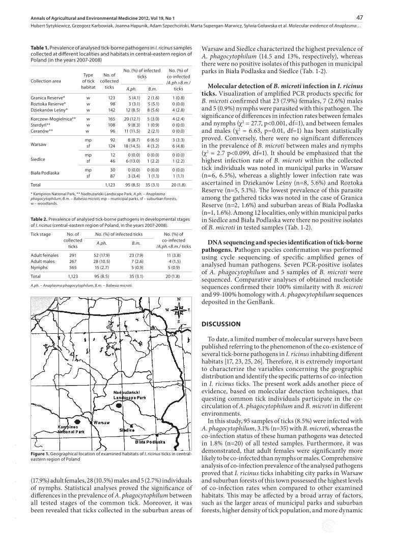
Density of I. ricinus populations in examined ecosystems. A total number of 1,123 questing tick individuals (291 females, 267 males and 565 nymphs) were collected at different ecosystems (municipal parks, suburban forests, and woodlands) in the central-eastern region of Poland (Tab. 1-2, Fig.1). It was observed that the highest density of I. ricinus populations occurred in woodlands within the Nadbużański Landscape Park (96-165 collected ticks, depending on locality) and Kampinos National Park (98-142 ticks). On the other hand, the lowest number of ticks was gathered within municipal parks in Warsaw (n=92), Biała Podlaska (n=30) and Siedlce (n=12) when compared to suburban areas of these cities (124, 87 and 46 collected ticks, respectively) (Tab. 1).
Molecular detecting of co-infection status in I. ricinus populations in the central-eastern region of Poland. The prevalence of tick-borne human pathogens A. phagocytophilum and B. microti in single and polymicrobial infections in I.ricinus ticks was analysed using conventional and nested PCR techniques. Overall, 1.8% of ticks (n=20) were found to be co-infected with analysed human pathogens (A. phagocytophilum and B. microti). Statistical analyses proved that adult females were significantly (c2 = 7.3, p=0.007, df=1) more likely to be co-infected than nymphs. On the other hand, no statistically significant difference was detected in co-infection rates between adult males and nymphs (χ2 = 0.2, p=0.661, df=1). It has been demonstrated that the prevalence of co-infection status was the highest among females of I. ricinus (11 samples, 3.8%), whereas the lowest within tested nymphs (5 samples, 0.9%). A moderate level of mixed infections was detected in adult males (4 individuals, 1.5%). It should be underlined that there were no co-infected samples of analysed human pathogens in ticks that were gathered at 4 localities (woodlands in Roztoka Reserve and Sterdyń; municipal parks in Siedlce and Biała Podlaska). On the other hand, ticks collected in city parks in Warsaw and suburban areas of the city characterized the highest prevalence of co-infections (3.3 and 4.8%, respectively). Furthermore, it was established that co-infection rates of ticks inhabiting woodlands within Kampinos National Park and Nadbużański Landscape Park were similar and reached the levels of 1.37% (n=5) and 1.10% (n=4), respectively.
Molecular detection of A. phagocytophilum infection in I. ricinus ticks. The PCR results revealed that A. phagocytophilum-positive samples were obtained for 52
-
-
-
-
-
47Annals of Agricultural and Environmental Medicine 2012, Vol 19, No 1
Hubert Sytykiewicz, Grzegorz Karbowiak, Joanna Hapunik, Adam Szpechciński, Marta Supergan-Marwicz, Sylwia Goławska et al. Molecular evidence of Anaplasma…
Warsaw and Siedlce characterized the highest prevalence of A. phagocytophilum (14.5 and 13%, respectively), whereas there were no positive isolates of this pathogen in municipal parks in Biała Podlaska and Siedlce (Tab. 1-2).
Molecular detection of B. microti infection in I. ricinus ticks. Visualization of amplified PCR products specific for B. microti confirmed that 23 (7.9%) females, 7 (2.6%) males and 5 (0.9%) nymphs were parasited with this pathogen. The significance of differences in infection rates between females and nymphs (c2 = 27.7, p<0.001, df=1), and between females and males (χ2 = 6.63, p=0.01, df=1) has been statistically proved. Conversely, there were no significant differences in the prevalence of B. microti between males and nymphs (c2 = 2.7 p<0.099, df=1). It should be emphasized that the highest infection rate of B. microti within the collected tick individuals was noted in municipal parks in Warsaw (n=6, 6.5%), whereas a slightly lower infection rate was ascertained in Dziekanów Leśny (n=8, 5.6%) and Roztoka Reserve (n=5, 5.1%). The lowest prevalence of this parasite among the gathered ticks was noted in the case of Granica Reserve (n=2, 1.6%) and suburban areas of Biała Podlaska (n=1, 1.6%). Among 12 localities, only within municipal parks in Siedlce and Biała Podlaska were there no positive isolates of B. microti in tested samples (Tab. 1-2).
DNA sequencing and species identification of tick-borne pathogens. Pathogen species confirmation was performed using cycle sequencing of specific amplified genes of analysed human pathogens. Seven PCR-positive isolates of A. phagocytophilum and 5 samples of B. microti were sequenced. Comparative analyses of obtained nucleotide sequences confirmed their 100% similarity with B. microti and 99-100% homology with A. phagocytophilum sequences deposited in the GenBank.
DISCUSSION
To date, a limited number of molecular surveys have been published referring to the phenomenon of the co-existence of several tick-borne pathogens in I. ricinus inhabiting different habitats [17, 23, 25, 26]. Therefore, it is extremely important to characterize the variables concerning the geographic distribution and identify the specific patterns of co-infection in I. ricinus ticks. The present work adds another piece of evidence, based on molecular detection techniques, that questing common tick individuals participate in the co-circulation of A. phagocytophilum and B. microti in different environments.
In this study, 95 samples of ticks (8.5%) were infected with A. phagocytophilum, 3.1% (n=35) with B. microti, whereas the co-infection status of these human pathogens was detected in 1.8% (n=20) of all tested samples. Furthermore, it was demonstrated, that adult females were significantly more likely to be co-infected than nymphs or males. Comprehensive analysis of co-infection prevalence of the analysed pathogens proved that I. ricinus ticks inhabiting city parks in Warsaw and suburban forests of this town possessed the highest levels of co-infection rates when compared to other examined habitats. This may be affected by a broad array of factors, such as the larger areas of municipal parks and suburban forests, higher density of tick population, and more dynamic
(17.9%) adult females, 28 (10.5%) males and 5 (2.7%) individuals of nymphs. Statistical analyses proved the significance of differences in the prevalence of A. phagocytophilum between all tested stages of the common tick. Moreover, it was been revealed that ticks collected in the suburban areas of
Table 2. Prevalence of analysed tick-borne pathogens in developmental stages of I. ricinus (central-eastern region of Poland, in the years 2007-2008).
Tick stage No. of collected
ticks
No. (%) of infected ticks No. (%) of co-infected
/A.ph.+B.m./ ticksA.ph. B.m.
Adult females 291 52 (17.9) 23 (7.9) 11 (3.8)Adult males 267 28 (10.5) 7 (2.6) 4 (1.5)Nymphs 565 15 (2.7) 5 (0.9) 5 (0.9)
Total 1,123 95 (8.5) 35 (3.1) 20 (1.8)
A.ph. – Anaplasma phagocytophilum, B.m. – Babesia microti.
Table 1. Prevalence of analysed tick-borne pathogens in I. ricinus samples collected at different localities and habitats in central-eastern region of Poland (in the years 2007-2008)
Collection areaType
of tick habitat
No. of collected
ticks
No. (%) of infected ticks
No. (%) of co-infected /A.ph.+B.m./
ticksA.ph. B.m.
Granica Reserve* w 123 5 (4.1) 2 (1.6) 1 (0.8)Roztoka Reserve* w 98 3 (3.1) 5 (5.1) 0 (0.0)Dziekanów Leśny* w 142 12 (8.5) 8 (5.6) 4 (2.8)
Korczew-Mogielnica** w 165 20 (12.1) 5 (3.0) 4 (2.4)Sterdyń** w 108 9 (8.3) 1 (0.9) 0 (0.0)Ceranów** w 96 11 (11.5) 2 (2.1) 0 (0.0)
Warsaw mp 92 8 (8.7) 6 (6.5) 3 (3.3)sf 124 18 (14.5) 4 (3.2) 6 (4.8)
Siedlcemp 12 0 (0.0) 0 (0.0) 0 (0.0)sf 46 6 (13.0) 1 (2.2) 1 (2.2)
Biała Podlaskamp 30 0 (0.0) 0 (0.0) 0 (0.0)sf 87 3 (3.4) 1 (1.1) 1 (1.1)
Total 1,123 95 (8.5) 35 (3.1) 20 (1.8)
* Kampinos National Park, ** Nadbużański Landscape Park, A.ph. – Anaplasma phagocytophilum, B.m. – Babesia microti; mp – municipal parks, sf – suburban forests, w – woodlands.
Figure 1. Geographical location of examined habitats of I. ricinus ticks in central-eastern region of Poland
NadbużańskiLandscape Park
Biała Podlaska
Siedlce
WarsawKampinos National Park
N
S
W E
-
-
-
-
-
48 Annals of Agricultural and Environmental Medicine 2012, Vol 19, No 1
Hubert Sytykiewicz, Grzegorz Karbowiak, Joanna Hapunik, Adam Szpechciński, Marta Supergan-Marwicz, Sylwia Goławska et al. Molecular evidence of Anaplasma…
circulation of the targeted pathogens. Conversely, there were no co-infected tick individuals among samples collected within the city parks in Siedlce and Biała Podlaska, though co-infected samples were detected in suburban areas of these towns. Interestingly, it was revealed that co-infection rates of ticks inhabiting woodlands within Kampinos National Park and Nadbużański Landscape Park reached similar levels (1.37% and 1.10%, respectively). It is noteworthy that the presented study confirmed significant differences in co-infection rates in I. ricinus ticks living within examined habitats.
It has been reported recently that the co-existence of different tick-borne pathogens is not a rare event in questing and engorged common tick individuals in Europe [18, 21, 25, 27-34]. According to Wójcik-Fatla et al. [26], comparing the levels of co-infection rates in ticks inhabiting different countries, or parts of the same country, is quite difficult. It is involved with biotope specificity, number of collected ticks and differences in methodology. In Poland, the co-infection prevalence of A. phagocytophilum and B. microti in I. ricinus vary from 1.1% in the Lublin macroregion to 2.0% in the Tri-City agglomeration of Gdańsk-Gdynia-Sopot [23, 26]. The results obtained there are in accordance with those presented by cited authors. Furthermore, it should be underlined that Zygner et al. [35] proved the occurrence of B. microti DNA in lysates of I. ricinus females (11/193, 5.7%) removed from dogs in the Warsaw area (central Poland). Additionally, these authors revealed that DNA sequences of targeted pathogen showed 100% similarity with the Gray strain of B. microti isolated from human. Our results strengthen the concept that I. ricinus may play an important role in the circulation of B. microti in the Warsaw agglomeration. Hildebrandt et al. [36] in 2007 provided the evidence that confirmed the first autochthonous case of human B. microti infection in Europe. However, it still remains a matter of debate whether strains of B. microti occurring in Europe possess a sufficient level of pathogenic potential to cause human babesiosis. The another possibility involves an underestimation of the prevalence of this disease in European populations exposed to B. microti infections. On one hand, it may be due to sub-clinically expressed babesiosis, and on the other, to the lack of sensitive and reliable diagnostic tools, [4, 37, 38, 39, 40, 41].
In conclusion, the concurrent presence of various tick-borne pathogens in I. ricinus individuals, especially when they reach high levels of co-infection rates, may increase the likelihood of the acquisition of multiple infections by humans. Considering the rising abundance of I. ricinus populations in Poland, and increasing number of reports emphasizing the co-infection status of this ectoparasite, more detailed studies should be undertaken to evaluate the risk of acquisition of human pathogens transmitted by questing ticks within different geographical regions of Poland. The application of more advanced molecular and epidemiological surveys of polymicrobial infections in I. ricinus may be useful for constructing detailed maps showing endemic areas with specific co-infection patterns of tick-borne pathogens. Furthermore, it is of great medical importance to implement new molecular methods for rapid and reliable identification of human cases of tick-borne diseases (TBDs), as well as the co-infection status in I. ricinus ticks.
CONCLUSIONS
The performed molecular studies proved that I. ricinus can play an important role in the co-circulation of A. phagocytophilum and B. microti in different habitats (municipal parks, suburban forests and woodlands). Obtained results support the hypothesis that the distribution of these tick-borne human pathogens in mixed infections in ticks may be involved with habitat specificity.
REFERENCES
1. Angelakis E, Billeter SA, Breitschwerdt EB, Chomel BB, Raoult D. Potential for tick-borne bartonelloses. Emerg Infect Dis. 2010; 16: 385-391.
2. Castellaw AH, Chenney EF, Varela-Stokes AS. Tick-borne disease agents in various wildlife from Mississippi. Vector Borne Zoonotic Dis. 2011, 11(4): 439-442.
3. Daniel M, Materna J, Hőnig V, Metelka L, Danielová V, Harčarik J, Kligrová S, Grubhoffer L. Vertical distribution of the tick Ixodes ricinus and tick-borne pathogens in the northern Moravian mountains correlated with climate warming (Jeseníky Mts., Czech Republic). Cent Eur J Public Health. 2009; 17: 139-145.
4. Lempereur L, De Cat A, Caron Y, Madder M, Claerebout E, Saegerman C, Losson B. First molecular evidence of potentially zoonotic Babesia microti and Babesia sp. EU1 in Ixodes ricinus ticks in Belgium. Vector Borne Zoonotic Dis. 2011; 11: 125-130.
5. Severinsson K, Jaenson TG, Pettersson J, Falk K, Nilsson K. Detection and prevalence of Anaplasma phagocytophilum and Rickettsia helvetica in Ixodes ricinus ticks in seven study areas in Sweden. Parasit Vectors 2010, 3:66, DOI: 10.1186/1756-3305-3-66.
6. Tokarz R, Jain K, Bennett A, Briese T, Lipkin WI. Assessment of polymicrobial infections in ticks in New York State. Vector Borne Zoonotic Dis. 2010; 10: 217-221.
7. Doudier B, Olano J, Parola P, Brouqui P. Factors contributing to emergence of Ehrlichia and Anaplasma spp. as human pathogens. Vet Parasitol. 2010; 167: 149-159.
8. Heyman P, Cochez C, Hofhuis A, van der Giessen J, Sprong H, Porter SR, Losson B, Saegerman C, Donoso-Mantke O, Niedrig M, Papa A. A clear and present danger: tick-borne diseases in Europe. Expert Rev Anti Infect Ther. 2010; 8: 33-50.
9. Reye AL, Hübschen JM, Sausy A, Muller CP. Prevalence and seasonality of tick-borne pathogens in questing Ixodes ricinus ticks from Luxembourg. Appl Environ Microbiol. 2010; 76: 2923-2931.
10. Blaschitz M, Narodoslavsky-Gfoller M, Kanzler M, Stanek G, Walochnik J. Babesia species occurring in Austrian Ixodes ricinus ticks. Appl Environ Microbiol. 2008; 74: 4841-4846.
11. Hildebrandt A, Pauliks K, Sachse S, Straube E. Coexistence of Borrelia spp. and Babesia spp. in Ixodes ricinus ticks in middle Germany. Vector Borne Zoonotic Dis. 2010; 10: 831-837.
12. Karbowiak G. Zoonotic reservoir of Babesia microti in Poland. Pol J Microbiol. 2004; 53(Suppl.): 61-65.
13. Sytykiewicz H, Pucyk A. Detection of Babesia sp. DNA in common tick population occuring at Kampinos National Park. In: Buczek A, Błaszak C (Eds): Stawonogi – środowisko, patogeny i żywiciele. Wyd. Koliber, Lublin 2007; 147-152.
14. Siński E. Wpływ konfekcji u kleszczy (Ixodidae) na transmisję mikropasożytów krwi. Wiad Parazytol. 2009; 55: 341-347.
15. Halos L, Bord S, Cotté V, Gasqui P, Abrial D, Barnouin J, Boulouis H-J, Vayssier-Taussat M, Vourc’h G. Ecological factors characterizing the prevalence of bacterial tick-borne pathogens in Ixodes ricinus ticks in pastures and woodlands. Appl Environ Microbiol. 2010; 76: 4413-4420.
16. Dietrich F, Schmidgen T, Maggi RG, Richter D, Matuschka F-R, Vonthein R, Breitschwerdt EB, Kempf VAJ. Prevalence of Bartonella henselae and Borrelia burgdorferi sensu lato DNA in Ixodes ricinus ticks in Europe. Appl Environ Microbiol. 2010; 76: 1395-1398.
17. Aktas M, Vatansever Z, Altay K, Aydin MF, Dumanli N. Molecular evidence for Anaplasma phagocytophilum in Ixodes ricinus from Turkey. Trans R Soc Trop Med Hyg. 2010; 104: 10-15.
18. Franke J, Fritzsch J, Tomaso H, Straube E, Dorn W, Hildebrandt A. Coexistence of pathogens in host-seeking and feeding ticks within a single natural habitat in central Germany. Appl Environ Microbiol. 2010; 76: 6829-6836.
-
-
-
-
-
49Annals of Agricultural and Environmental Medicine 2012, Vol 19, No 1
Hubert Sytykiewicz, Grzegorz Karbowiak, Joanna Hapunik, Adam Szpechciński, Marta Supergan-Marwicz, Sylwia Goławska et al. Molecular evidence of Anaplasma…
19. Nonaka E, Ebel GD, Wearing HJ. Persistence of pathogens with short infectious periods in seasonal tick populations: the relative importance of three transmission routes. PLoS ONE 2010; 5: e11745.
20. Torina A, Alongi A, Scimeca S, Vicente J, Caracappa S, de la Fuente J. Prevalence of tick-borne pathogens in ticks in Sicily. Transbound Emerg Dis. 2010; 57: 46-48.
21. Václav R, Ficová M, Prokop P, Betáková T. Associations between coinfection prevalence of Borrelia lusitaniae, Anaplasma sp., and Rickettsia sp. in hard ticks feeding on reptile hosts. Microb Ecol. 2011; 61: 245-253.
22. Grzeszczuk A, Stańczak J, Kubica-Biernat B. Serological and molecular evidence of human granulocytic ehrlichiosis focus in the Białowieża Primeval Forest (Puszcza Białowieska), northeastern Poland. Eur J Clin Microbiol Infect Dis. 2002; 21: 6-11.
23. Stańczak J, Gable RM, Kruminis-Łozowska W, Carewicz M, Kubica-Biernat B. Ixodes ricinus as a vector of Borrelia burgdorferi sensu lato, Anaplasma phagocytophilum and Babesia microti in urban and suburban forests. Ann Agric Environ Med. 2004; 11: 109-114.
24. Persing DH, Mathiesen D, Marshall WF, Telford SR, Spielman A, Thomford JW, Conrad PA. Detection of Babesia microti by polymerase chain reaction. J Clin Microbiol. 1992; 30: 2097-2103.
25. Masuzawa T, Kharitonenkov IG, Okamoto Y, Fukui T, Ohashi N. Prevalence of Anaplasma phagocytophilum and its coinfection with Borrelia afzelii in Ixodes ricinus and Ixodes persulcatus ticks inhabiting Tver Province (Russia) – a sympatric region for both tick species. J Med Microbiol. 2008; 57: 986-991.
26. Wójcik-Fatla A, Szamańska J, Wdowiak L, Buczek A, Dutkiewicz J. Coincidence of three pathogens (Borrelia burgdorferi sensu lato, Anaplasma phagocytophilum and Babesia microti) in Ixodes ricinus ticks in the Lublin macroregion. Ann Agric Environ Med. 2009; 16: 151-158.
27. Halos L, Jamal T, Maillard R, Beugnet F, Le Menach A, Boulouis H-J, Vayssier-Taussat M. Evidence of Bartonella sp. in questing adult and nymphal Ixodes ricinus ticks from France and co-infection with Borrelia burgdorferi sensu lato and Babesia sp. Vet Res. 2005; 36: 79-87.
28. Hildebrandt A, Straube E, Neubauer H, Schmoock G. Coxiella burnetii and co-infections in Ixodes ricinus ticks in central Germany. Vector Borne Zoonotic Dis. 2010, DOI:10.1089/vbz.2010.0180 [ahead of print].
29. Koči J, Movila A, Taragel’ová V, Toderas I, Uspenskaia I, Derdáková M, Labuda M. First report of Anaplasma phagocytophilum and its coinfections with Borrelia burgdorferi sensu lato in Ixodes ricinus ticks (Acari: Ixodidae) from Republic of Moldova. Exp Appl Acarol. 2007; 41: 147-152.
30. Nieto NC, Foley JE. Meta-analysis of coinfection and coexposure with Borrelia burgdorferi and Anaplasma phagocytophilum in humans, domestic animals, wildlife, and Ixodes ricinus-complex ticks. Vector Borne Zoonotic Dis. 2009; 9: 93-101.
31. Swanson SJ, Neitzel D, Reed KD, Belongia EA. Coinfections acquired from Ixodes ticks. Clin Microbiol Rev. 2006; 19: 708-727.
32. Toledo Á, Olmeda AS, Escudero R, Jado I, Valcárcel F, Casado-Nistal MA, Rodríguez-Vargas M , Gil H, Anda P. Tick-borne zoonotic bacteria in ticks collected from central Spain. Am J Trop Med Hyg. 2009; 81: 67-74.
33. Vennestrøm J, Egholm H, Jensen PM. Occurrence of multiple infections with different Borrelia burgdorferi genospecies in Danish Ixodes ricinus nymphs. Parasitol Int. 2008; 57: 32-37.
34. Vorou RM, Papavassiliou VG, Tsiodras S. Emerging zoonoses and vector-borne infections affecting humans in Europe. Epidemiol Infect. 2007; 135: 1231-1247.
35. Zygner W, Bąska P, Wiśniewski M, Wędrychowicz H. The molecular evidence of Babesia microti in hard ticks removed from dogs in Warsaw (central Poland). Pol J Microbiol. 2010; 59: 95-97.
36. Hildebrandt A, Hunfeld KP, Baier M, Krumbholz A, Sachse S, Lorenzen T, Kiehntopf M, Fricke HJ, Straube E. First confimed autochtonous case of human Babesia microti infection in Europe. Eur J Clin Microbiol Infect Dis. 2007; 26: 595-601.
37. Becker CA, Bouju-Albert A, Jouglin M, Chauvin A, Malandrin L. Natural transmission of zoonotic Babesia spp. by Ixodes ricinus ticks. Emerg Infect Dis. 2009; 15; 320-322.
38. Bonnet S, Brisseau N, Hermouet A, Jouglin M, Chauvin A. Experimental in vitro transmission of Babesia sp. (EU1) by Ixodes ricinus. Vet Res. 2009; 40: 21-28.
39. Casati S, Sager H, Gern L, Piffaretti JC. Presence of potentially pathogenic Babesia sp. for human in Ixodes ricinus in Switzerland. Ann Agric Environ Med. 2006; 13: 65-70.
40. Häselbarth K, Tenter AM, Brade V, Krieger G, Hunfeld KP. First case of human babesiosis in Germany-clinical presentation and molecular characterisation of the pathogen. Int J Med Microbiol. 2007; 297: 197-204.
41. Hunfeld KP, Brade V. Zoonotic Babesia: possibly emerging pathogens to be considered for tick-infested humans in Central Europe. Int J Med Microbiol. 2004; 293(Suppl. 37): 93-103.
-
-
-
-
-