Embed Size (px)
Citation preview

Neurophysiology of Nerve
신형철 교수 간호학과


http://www.youtube.com/watch?v=Of0GrgxypuI

BCI-Hyung-Cheul Shin


http://www.youtube.com/watch?v=JNThQJ9dtdI


환경 (자극, 정보 )---> peripheral receptors---> brain (인식 , 기억 , 명령 )---> 근육조절 , 행동
http://www.youtube.com/watch?v=sX87g3AHIbc

신경계내에는 2 종류의 세포가 있다 : 1. Nerve cells (neuron) & 2. glial cells (glia)
4 elements : Cell body (Soma), dendrites, axon, presynaptic terminal, 1011 neurons, 10,000 different types, *examples: based on the number of neurites, based on dendritic tree structure
1. Cell body (soma): 기능 :- metabolic center
구조 :- triangular or pyramidal shape- pyramidal type 은 apical & basal dendrites 를 보임 - dendrites 와 axon 이 붙어 있다
2. Dendrites: 기능 : - 정보의 input area
3. Axon: 구조 : 0.2 - 20 um 의 직경 , ramification, up to 1 m굵은것은 myelin sheath 에 둘러 쌓임 (fatty insulation 역할 ).Nodes of Ranvier ( 일정간격으로 , myelin sheath 가 없다 )기능 : - axon potential (AP) 을 all-or-none way 로 먼거리 전달 (axon hillock에서 AP 를 생성 )- axoplasmic transport - 굵은 신경섬유가 더 빠른 전도 속도
4. Presynaptic terminal:기능 : 다른 neuron 의 dendrites, cell body, axon 과 contact 하여 synapse를 형성한다 .
Neuron 의 기능적 3 구분 :1) sensory (afferent): 정보를 외부로 부터 받아들이는 neurons2) motor (efferent): 근육과 glands 로 command 를 보냄3) interneuron: local infoamtion processing & transfer


http://www.indiana.edu/~phys215/lecture/lecnotes/lecgraphics/channel.swf

외부의자극에 의해 흥분되지 않은 resting 상태에서 그대로 일정한membrane potential(MP) 을 유지 ---> resting potential(RP)
•nerve, muscle 에서 항상 negative, constant, cell type 에 특징적e.g.: warm-blooded animal: -55 to -100 mV; smooth muscle: -30 mV
* 이러한 RP 은 막의 채널을 통해 ion 들이 passive 하게 이동함으로서 발생한다 .Resting 시 대부분의 열린 채널은 K+ ion, 따라서 RP 은 주로 K+ ion 에 대한 tansmembrane concentration gradient 에 의해 주로 결정된다 .

Nernst Equation
By the end of the 19th century, it was known that the cytoplasm was high in K+ and that [Na+] was very low--and that this relationship was reversed outside the cell.
The assumption was made that the cell membrane was permiable to K+ but not to Na+.

Goldman equation was derived to solve for transmembrane potential using all ions involved simultaneously.

http://www.sensory-systems.ethz.ch/MultiMedia/actionp.swf
http://instruct.uwo.ca/biology/022/unit3/48-09-ActionPotential.mov
http://schools.tdsb.on.ca/rhking/departments/science/bio/homeostasis/nervous_system/action.mov
http://instruct.uwo.ca/biology/022/unit3/48-07-RestingPotential.mov
http://www.youtube.com/watch?v=jgtD3AfTXE4


http://www.youtube.com/watch?v=JBnz3tPjbeA


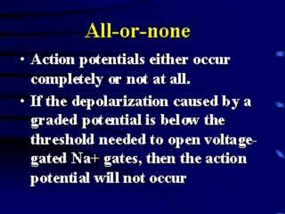
1. an axon terminal on the left and a dendritic process on the right seperated by a synaptic cleft.
2. When an impulse reaches the end of an axon, synaptic vessicles transport the neurotransmitters to the end of the axon and release them into the synaptic cleft.
3. These neurotransmitters attach to receptor sites on the cell membrane of the receiving neuron. When enough receptor sites are filled, the firing threshold of the receiving neuron is reached and a depolarization event is triggered.
4. As neurotransmitters attach to the receptor sites and overcome the firing threshold, small molecular gates open along the cell membrane allowing the sodium ions to rapidly flood the neuron. This sudden change in polarity from the influx of positive ions triggers an action potential that moves like a wave down the axon triggering another nerve, muscle cell, etc.
5. At this point, a different series of molecular gates open which allows potassium ions to rush out of the neuron. The potassium ions, which have a positive charge as well, create a negatively charged cell interior by their absence. This event stops the depolarization process. The sodium ions are pumped more slowly to the cell exterior by active transport, resulting in the fully restored resting potential once again.

A close-up view of the recording dish under the dissecting scope. The forceps are touching the proximal portion of leg (the part that was stuck to the blue tape, above), which will be opened to expose the nerve bundle.
COMPOUND ACTION POTENTIAL: NERVE CONDUCTION


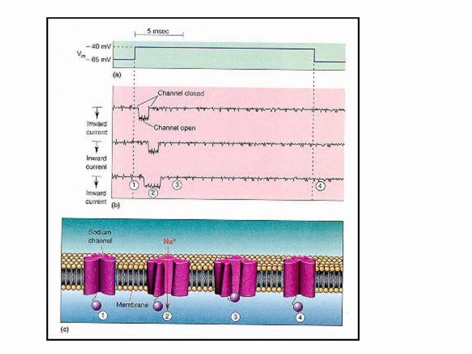


single ACh channels 의 patch clamp 실험 (Erwin Neher, Bert Sakmann).A: patch clamp setup.
B1: in frog muscle fiber, in the presence of 100 nM ACh, at RP -90 mV.opening of channel---> pulse of inward current (downward deflection).all-or-none fashionamplitude fixed, but duration of the opening varies (mean 1 ms)single channel open---> 17,000 Na+ ions flow into the cellsomewhat smaller number of K+ ions flow out
B2: amplitudes: average ca. 2.69 pA (elementary current)
C: in the presence of 100 nM ACh, at -130 mV MP.individual channel currents---> all-or-none increments of -3.9 pA각개 채널이 동시에 열리면 ---> amplitude linearly added up (up to 3 channels).

Membrane potential 을 voltage clmap 로 고정시키면서 2 uM ACh 투여에 의한single channel 의 크기와 direction 을 조사 .
reversal potential: at 0 mVnegtiave potential 에서는 inward current flowpostive potential: outward current flow
current magnitude 의 변화 : driving force 의 변화 때문
Currents for the end-plate potential depend on four factors1) total number of end-plate channels ( 보통 200,000 channels)2) channel open probability (receptor 주위의 transmitter 농도 )3) conductance of each open channel4) driving force

Total end-plate potential: summed average of the currents in thousands of individual ion channels
A: ACh 투여 : all channels open rapidly in response to ACh but closing times are varying
B: Summed average of the currents of individual ion channels

in CNS, at most synapses: individual synaptic potentials are far below threshold, often smaller than 1 mV.(motor neuron: 0.2-0.4 mV, threshold about 10 mV)
postsynaptic cell: many synapses---> summation
presynaptic elements usually come from many different cells-->converging (inhibitory, excitatory) on the postsynaptic cell (a motor neuron: as many as 10,000 different presynaptic endings)
: integrative action of the nervous system: brain's ability to choose between competing alternatives: decision-making capability


http://instruct.uwo.ca/biology/022/unit3/48-12-Synapse.mov

1) Synapses on cell body are often Inhibitory2) synapses on dendritic spines are often excitatory3) synapses on axon terminals are often modulatory--->controlling the amount of transmitter released4)excitatory and inhibitory synapses have distintive ultrastructure


1) 3.5 nm between presynaptic & postsynaptic cell membranes2) Gap junction, 3) cytoplasmic continuity, 4) synaptic delay virtually absent, 5) ionic current flow6) direction of transmission: usually bidirectional
*not unique to nerve cells*present in other cells: heart, smooth muscle, epithelial liver cells*in the brain electrical synapse is less common*electrical synapses do not readily allow inhibitory actions orlong-lasting changes in effectiveness.


Changes in membrane potentials of the presynaptic terminal affect the amount of transmitter released as assayed by measuring the amplitude of the postsynatpic potential.
When the membrane potential of the presynaptic terminal is at the normal resting potential, an action potential produces a postsynaptic potential of about 8 mV.
When it is hyperpolarized by 10 mV, the steady-state (leakage) Ca++ influx is decreased and the presynaptic spike produces a postsynaptic potential of only 5 mV.
When the presynaptic neuron is returned to the resting level and then depolarized by 10 mV, the steady-state Ca++ influx is increased and the resulting presunaptic action potential produces a synaptic potential of 15 mV, which triggers an action potential in the postsynaptic cell.

B: TTX 로 presynaptic Na+ channel 을 완전히 block 하고 current pulse injection 을 증가 시키면 presynaptic terminal 이 점점 크게 depolarize 되면서 postsynaptic potential 이 점진적으로 증가한다 .(Na+ influx 가 transmitter release 와는 무관하다 )
C: TTX 로 Na+ channel 을 block 한후 다시 TEA 로 voltage-dependnet K+ channel 을 block.presynaptic cell 에 current pulse injection--->presynaptic depolarization--->postsynaptic potential 이 current pulse injection 에 비례적으로 증가(neither Na+ nor K+ is required for effective transmitter release)
D: input-output curve

squid giant axon, Na+ and K+ channels blocked by TTX and TEAdepolarize presynaptic cell---> increase in Ca2+ current--->increase in postsynaptic potentialCa2+ chan

squid giant axon, Na+ and K+ channels blocked by TTX and TEAdepolarize presynaptic cell---> increase in Ca2+ current--->increase in postsynaptic potentialCa2+ chan

Tetrodotoxin (TTX): Na+ channel blocker, no effects on K+ channel따라서 , at cholinergic synapse: TTX blocks the presynaptic Na+ spike, 그러나 postsynaptic receptor 에 직접 투여한 ACh 은 EPSP 생성
A: recording electrodes in both pre- and post-synaptic membrane of thesquid giant axon
B: Presynaptic cell: AP 약 110 mV---> transmitter release---> large EPSPif TTX 투여 ---> presynaptic AP 이 점점 작아짐 ---> EPSP 감소presynaptic AP 이 40 mV 정도 되면 --->EPSP 소멸
C: TTX 로 presynaptic Na+ channel 을 block 했을 때 transmitter release 의input-output curve 를 볼 수 있다 .presynaptic potential 이 40 mV 의 threshold level 이하에서는 EPSP 불능40 mV 를 조금 넘어서면 steep increase in EPSP

Four types of Glutamate Receptor*Second-messenger linked receptor:Quisqualate-B: phosphoinositide-linked second messenger system 을activation 시켜서 channel 을 Na+ 과 K+ 에 permeable 하게 한다 .
*Directly gated receptors:AMPA (alpha-amino-3 hydroxy-5 methyl-4 isoxazole proprionic acid (Na+, K+ ions)Kinate Quisqualate-A NMDA (N-methyl D-asprtate), (permeable to Na+, K+, Ca2+)(resting 시 Mg2+ 에 의해 blocked, 막이 20-30 mV depolarize되면 Mg2+ 가 빠지면서 glutamate 가 binding 한다 .)

Channel opening by GABA (10 uM) and Glycine (10 uM)
---> produce similar elementary pulses of outward currents(transmitter-gated Cl- inhbitory channels in a mouse spinal neuron)

rat hippocampal neuron 에서Glutamate single channel current VS. GABA single channel current: different reversal potentials
A: Glutamate currentMP---> depolarizing direction---> Glutamate (inward) current smallerat 0 mV MP: current pulses are nullified(averaged Ek + ENa at 0 mV MP)at 30 mV MP: reversed
B: GABA currentat -60 mV MP: reversal potential for IPSP(near the ECl-)at more depolarizing levels: current pulses are outward

five action potentials of different sizes. Each size AP is from a different motor axon.

AstrocytesAstrocytes are star-shaped glial cells of the CNS. The end-feet processes line blood vessels and make up part of the blood-brain barrer. Bar = 30 Microns
Microglia are the smallest of the glial cells. Some act as phagocytes cleaning up CNS debris. Most serve as representatives of the immune system in the brain. Microglia protect the brain from invading microorganisms and are thought to be si
milar in nature to microphages in the blood system.

http://www.youtube.com/watch?v=ysDGX6bOgAw


![NEURurofisiologia - neurophysiology[1]](https://img.pdfslide.tips/doc/110x75/5571f1d449795947648bb940/neururofisiologia-neurophysiology1.jpg)