Embed Size (px)
Citation preview
Contents
Research on IP3R
Protein & Protein Structure
NMR/X-ray crystallography
Research on IP3R
Protein & Protein Structure
NMR/X-ray crystallography
The Structures of Life (2007, NIH Publication No. 07-2778)
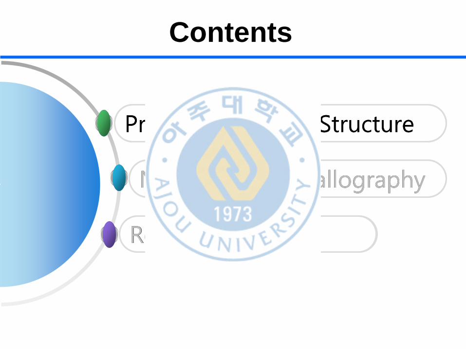
Proteins are the Body’s Worker Molecules
The Structures of Life
(2007, NIH Publication No. 07-2778 http://www.nigms.nih.gov)
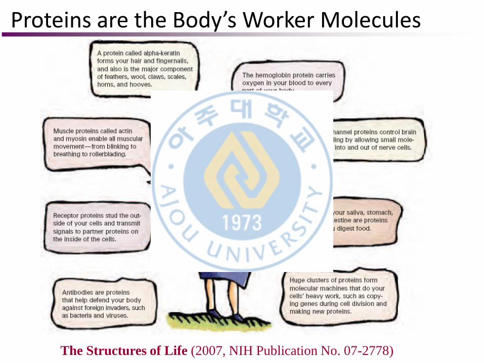
Sometimes, an error in just one amino
acid can cause disease.
Sickle cell disease, which most often
affects those of African descent, is
caused by a single error in the gene for
hemoglobin, the oxygen-carrying
protein in red blood cells. This error, or
mutation, results in an incorrect amino
acid at one position in the molecule.
Hemoglobin molecules with this
incorrect amino acid stick together and
distort the normally smooth, lozenge-
shaped red blood cells into jagged
sickle shapes.
Normal Red
Blood Cells
Sickled Red
Blood Cells
Small Errors in Proteins can cause Disease
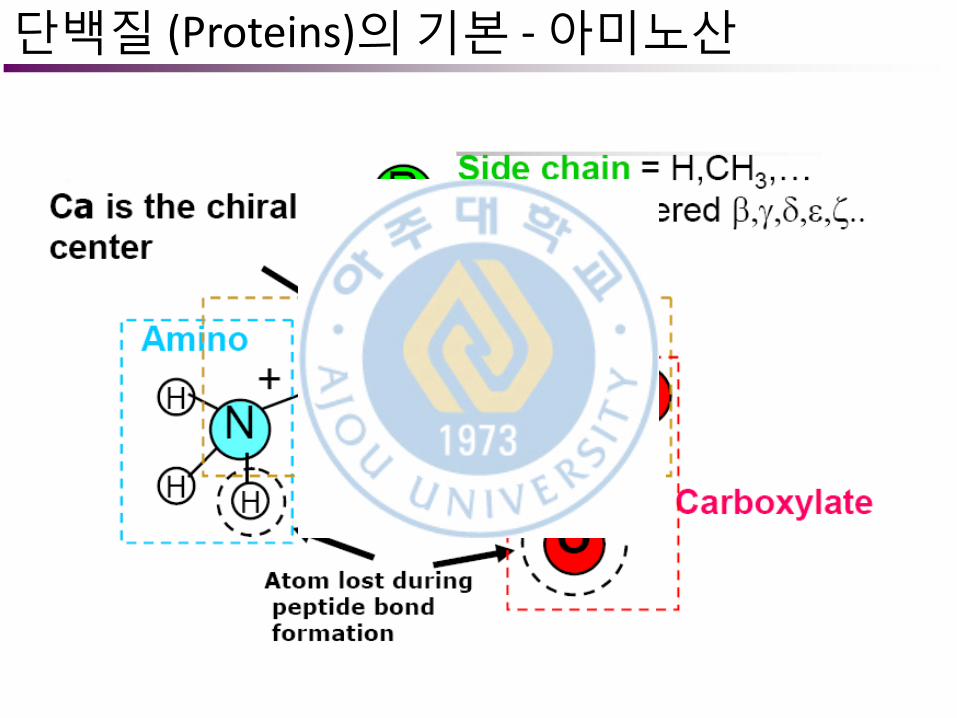
단백질 (Proteins)의 기본 - 아미노산
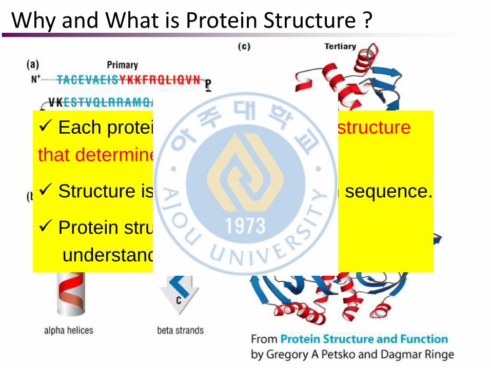
Each protein has a particular 3D structure
that determines its function.
Structure is more conserved than sequence.
Protein structure is central for
understanding protein functions.
Why and What is Protein Structure ?
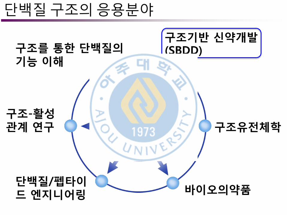
단백질 구조의 응용분야
구조유전체학
단백질/펩타이드 엔지니어링
구조기반 신약개발 (SBDD)
바이오의약품
구조를 통한 단백질의 기능 이해
구조-활성 관계 연구
V.S.
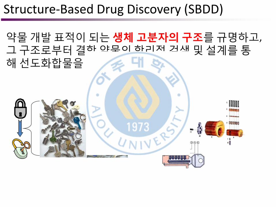
Structure-Based Drug Discovery (SBDD)
약물 개발 표적이 되는 생체 고분자의 구조를 규명하고, 그 구조로부터 결합 약물의 합리적 검색 및 설계를 통해 선도화합물을 발굴하는 방법
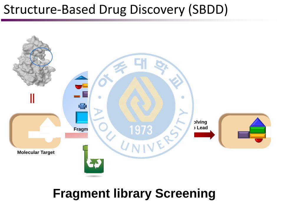
Molecular Target
Fragment library
Evolving
Into Lead
Fragment library Screening
Structure-Based Drug Discovery (SBDD)
Nature Review Drug Discovery (2012) 463, 873-886
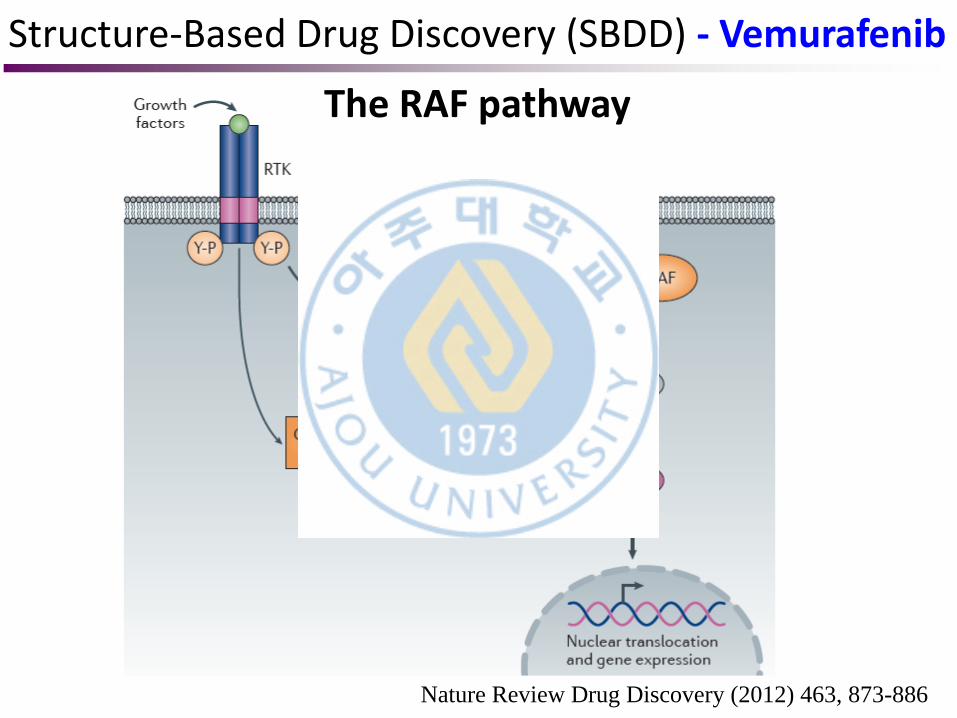
The RAF pathway
Structure-Based Drug Discovery (SBDD) - Vemurafenib
Nature Review Drug Discovery (2012) 463, 873-886
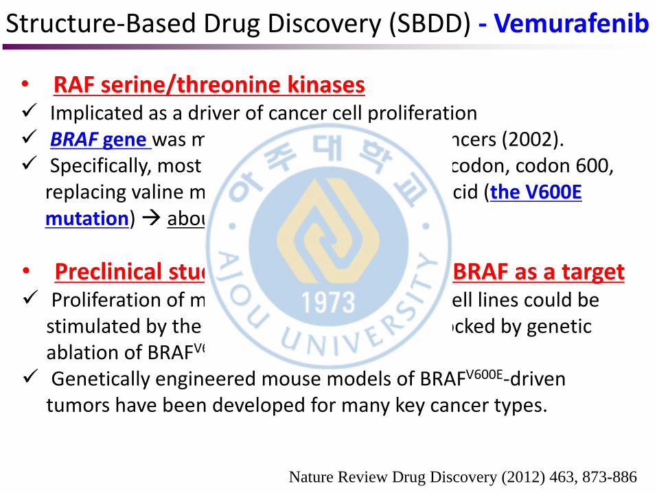
• RAF serine/threonine kinases Implicated as a driver of cancer cell proliferation BRAF gene was mutated in many different cancers (2002). Specifically, most mutations occur at a single codon, codon 600,
replacing valine most typically with glutamic acid (the V600E mutation) about half of all melanomas
• Preclinical studies to validate mutant BRAF as a target Proliferation of melanoma and other tumor cell lines could be
stimulated by the BRAFV600E oncogene and blocked by genetic ablation of BRAFV600E expression.
Genetically engineered mouse models of BRAFV600E-driven tumors have been developed for many key cancer types.
Structure-Based Drug Discovery (SBDD) - Vemurafenib
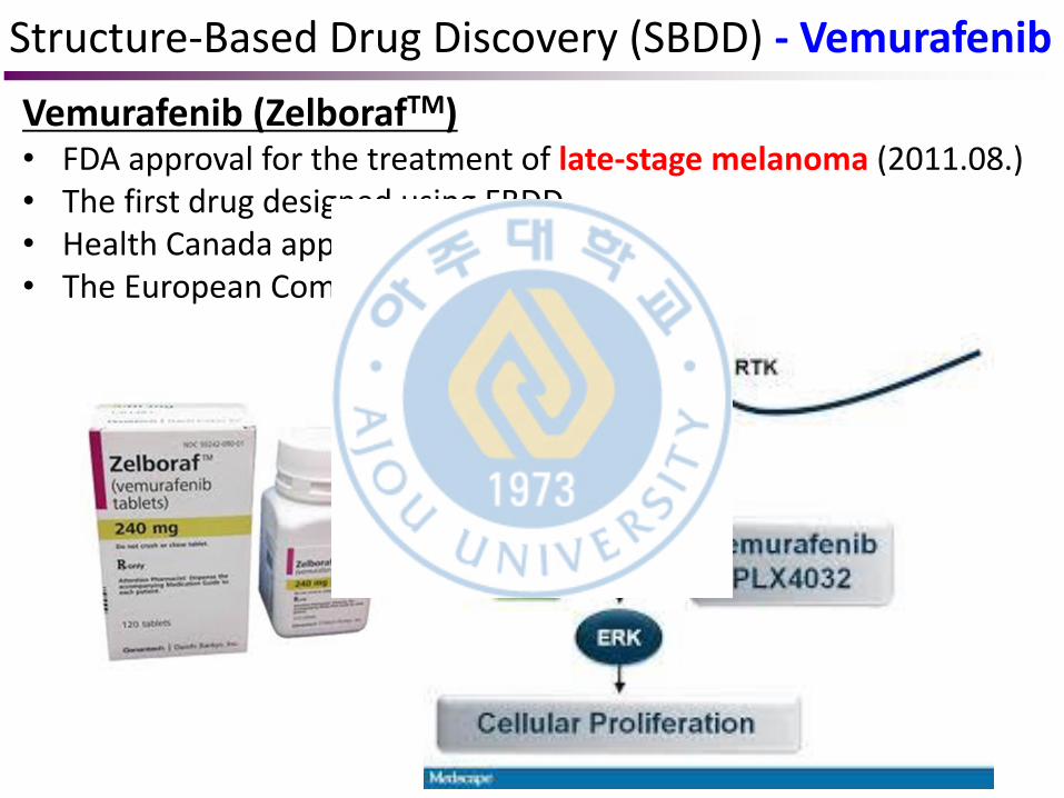
Vemurafenib (ZelborafTM) • FDA approval for the treatment of late-stage melanoma (2011.08.) • The first drug designed using FBDD. • Health Canada approval (2012.02.) • The European Commission approval (2012.02.)
Structure-Based Drug Discovery (SBDD) - Vemurafenib
Nature Review Drug Discovery (2012) 463, 873-886
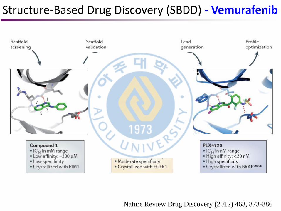
Scaffold-based discovery of kinase inhibitors
1. Library of ‘scaffold-like’ compounds • Screened small molecules (molecular mass 150–350 Da, fewer than eight hydrogen bond donors and acceptors, few rotatable bonds and relatively high aqueous solubility) • As these smaller compounds have limited compositional variabilities, a library of 20,000 compounds covers a relatively large swath of chemical space in the specified molecular mass range.
2. Biochemical assays were then developed • Five different kinases were screened through the library at a concentration of 200 μM. • Compounds that inhibited at least three of the five kinases were selected for follow-up studies.
Structure-Based Drug Discovery (SBDD) - Vemurafenib
Nature Review Drug Discovery (2012) 463, 873-886
Scaffold-based discovery of kinase inhibitors
3. Co-crystallography • A co-crystal structure of the kinase and the hit compound unambiguously identified true binding interactions. • From the initial screen, 238 compounds were selected for co-crystal analysis. • Over 100 structures of kinases co-crystallized with bound compounds were successfully determined. • In particular, the serine/threonine kinase PIM1 provided a robust system to identify novel scaffold candidates67; initially, PIM1 and a second kinase, fibroblast growth factor receptor 1 (FGFR1), provided more reliable crystallization systems while BRAF crystallization conditions were in optimization.
Structure-Based Drug Discovery (SBDD) - Vemurafenib
Nature Review Drug Discovery (2012) 463, 873-886
Scaffold-based discovery of kinase inhibitors
4. Hits screening & Rational design • With the structural information at hand, chemists, computational chemists and structural biologists could select the best screening hits and rationally design a next iteration of compounds for synthesis.
5. Optimization • Based on this approach, a 3-substituted 7-azaindole was selected for further optimization based on the structure of the compound co-crystallized with PIM1.
Structure-Based Drug Discovery (SBDD) - Vemurafenib
Nature Review Drug Discovery (2012) 463, 873-886
Scaffold-based discovery of kinase inhibitors
6. Expression & Crystallization of BRAF • In order to generate a robust expression and crystallization system for BRAF, a highly soluble form of the truncated kinase domain was engineered by mutating surface hydrophobic residues into relatively isosteric hydrophilic amino acids. • Determine the co-crystal structures of over 100 different compounds bound to BRAF.
7. Drug candidates • Vemurafenib and PLX4720 were identified within a year of initiating BRAF-specific improvements. • These compounds were optimized for binding affinity, selectivity and pharmacokinetic properties.
Structure-Based Drug Discovery (SBDD) - Vemurafenib
Nature Review Drug Discovery (2012) 463, 873-886
Structure-Based Drug Discovery (SBDD) - Vemurafenib
Nature Review Drug Discovery (2012) 463, 873-886
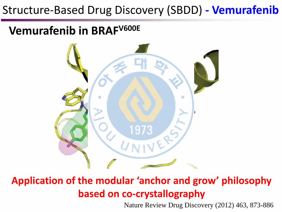
Vemurafenib in BRAFV600E
Application of the modular ‘anchor and grow’ philosophy based on co-crystallography
Structure-Based Drug Discovery (SBDD) - Vemurafenib
Contents
Research on IP3R
Protein & Protein Structure
NMR/X-ray crystallography
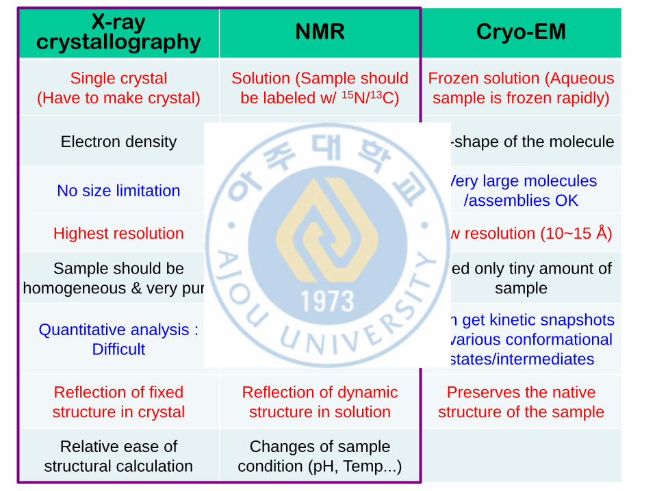
X-ray crystallography NMR Cryo-EM
Single crystal
(Have to make crystal)
Solution (Sample should
be labeled w/ 15N/13C)
Frozen solution (Aqueous
sample is frozen rapidly)
Electron density Atomic nuclei, Chemical
bonds 3D-shape of the molecule
No size limitation Size limitation (< 25kD) Very large molecules
/assemblies OK
Highest resolution High resolution Low resolution (10~15 Å )
Sample should be
homogeneous & very pure
Required concentrated
sample
Need only tiny amount of
sample
Quantitative analysis :
Difficult
Dynamic, Quantitative
analysis : Possible
Can get kinetic snapshots
of various conformational
states/intermediates
Reflection of fixed
structure in crystal
Reflection of dynamic
structure in solution
Preserves the native
structure of the sample
Relative ease of
structural calculation
Changes of sample
condition (pH, Temp...)
X-ray crystallography
Google image
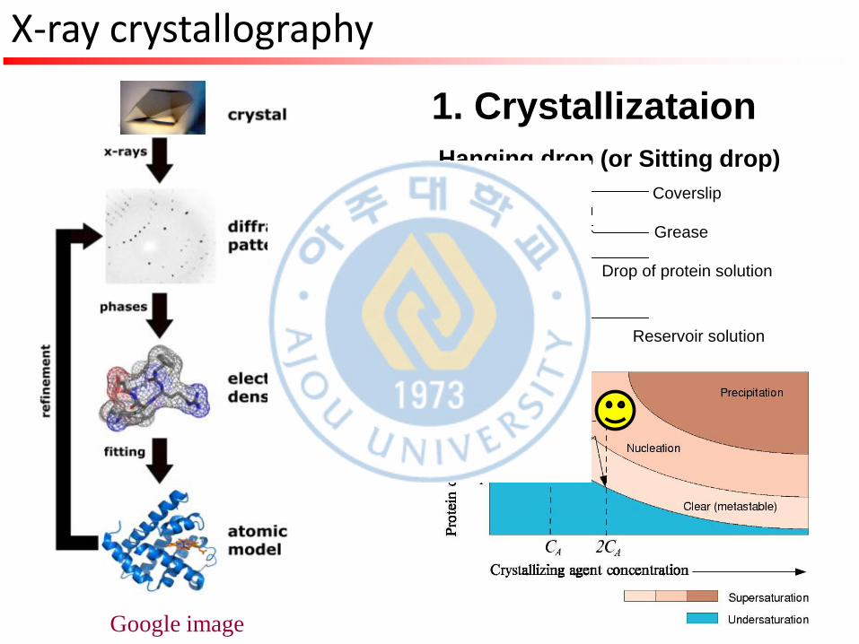
1. Crystallizataion
Coverslip
Grease
Drop of protein solution
Reservoir solution
Hanging drop (or Sitting drop)
X-ray crystallography
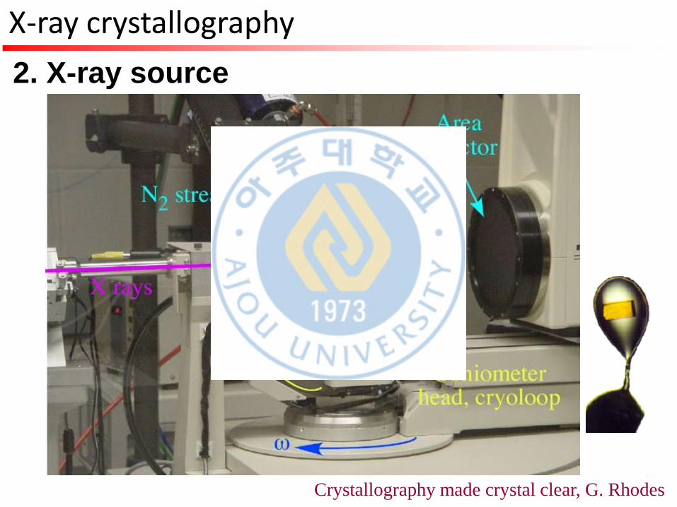
2. X-ray source
Crystallography made crystal clear, G. Rhodes
X-ray crystallography
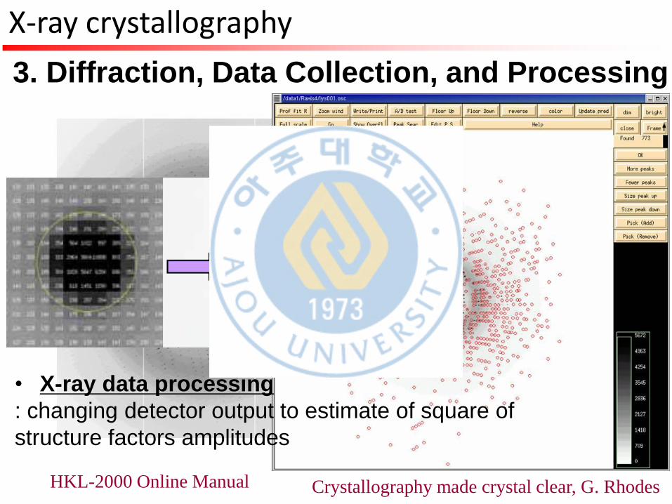
3. Diffraction, Data Collection, and Processing
Crystallography made crystal clear, G. Rhodes HKL-2000 Online Manual
• X-ray data processing
: changing detector output to estimate of square of
structure factors amplitudes
X-ray crystallography
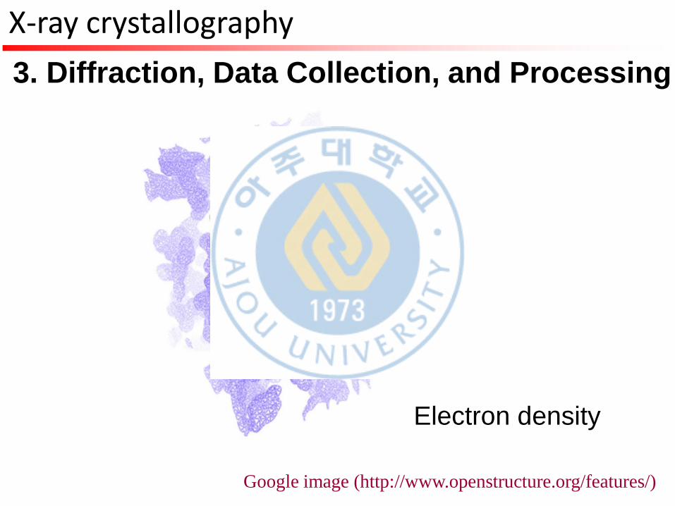
3. Diffraction, Data Collection, and Processing
Google image (http://www.openstructure.org/features/)
Electron density
X-ray crystallography
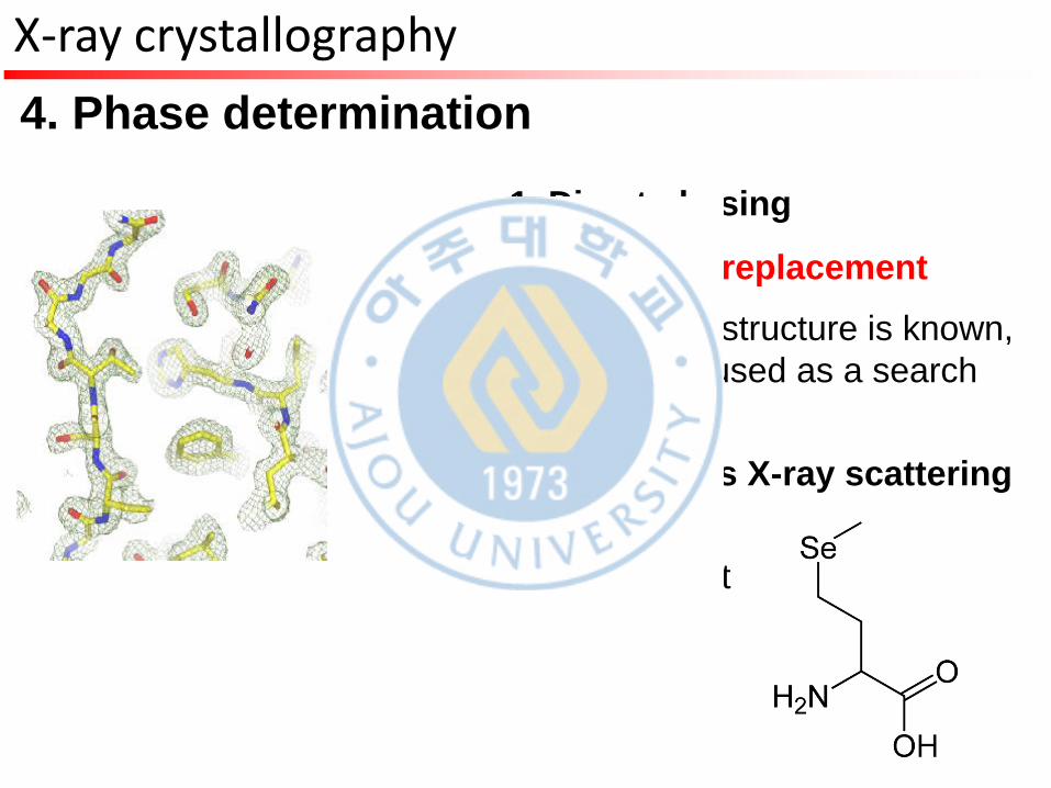
4. Phase determination
1. Direct phasing
2. Molecular replacement
: if a related structure is known,
it can be used as a search
model
3. Anomalous X-ray scattering
(MAD)
: seleno-Met
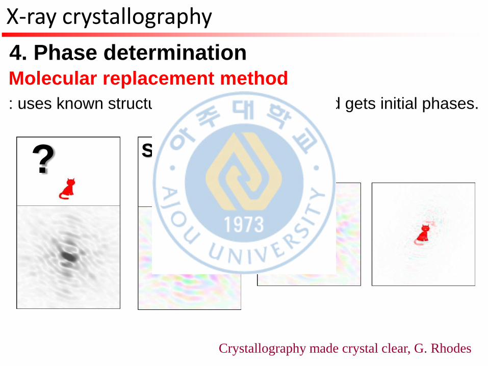
X-ray crystallography
4. Phase determination Molecular replacement method
: uses known structure as a search model and gets initial phases.
? Search model
Crystallography made crystal clear, G. Rhodes
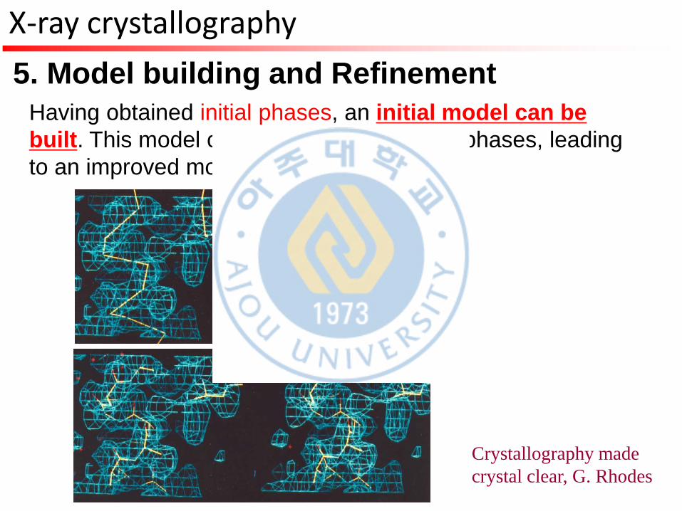
X-ray crystallography
5. Model building and Refinement
Having obtained initial phases, an initial model can be
built. This model can be used to refine the phases, leading
to an improved model.
Crystallography made
crystal clear, G. Rhodes
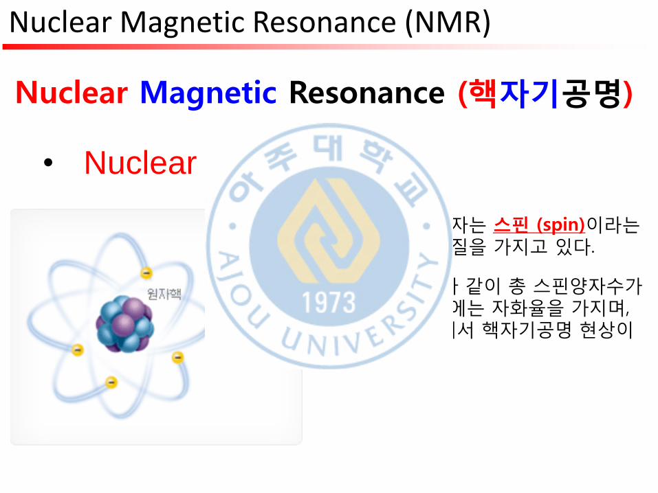
Nuclear Magnetic Resonance (NMR)
Nuclear Magnetic Resonance (핵자기공명)
• Nuclear (원자핵)
• 양성자와 중성자는 스핀 (spin)이라는 양자역학적 성질을 가지고 있다.
• 1H이나 13C등과 같이 총 스핀양자수가 0이 아닌 경우에는 자화율을 가지며, 바로 이 성질에서 핵자기공명 현상이 비롯된다.
• Magnetic : 자석의, 자기의
Nuclear Magnetic Resonance (NMR)
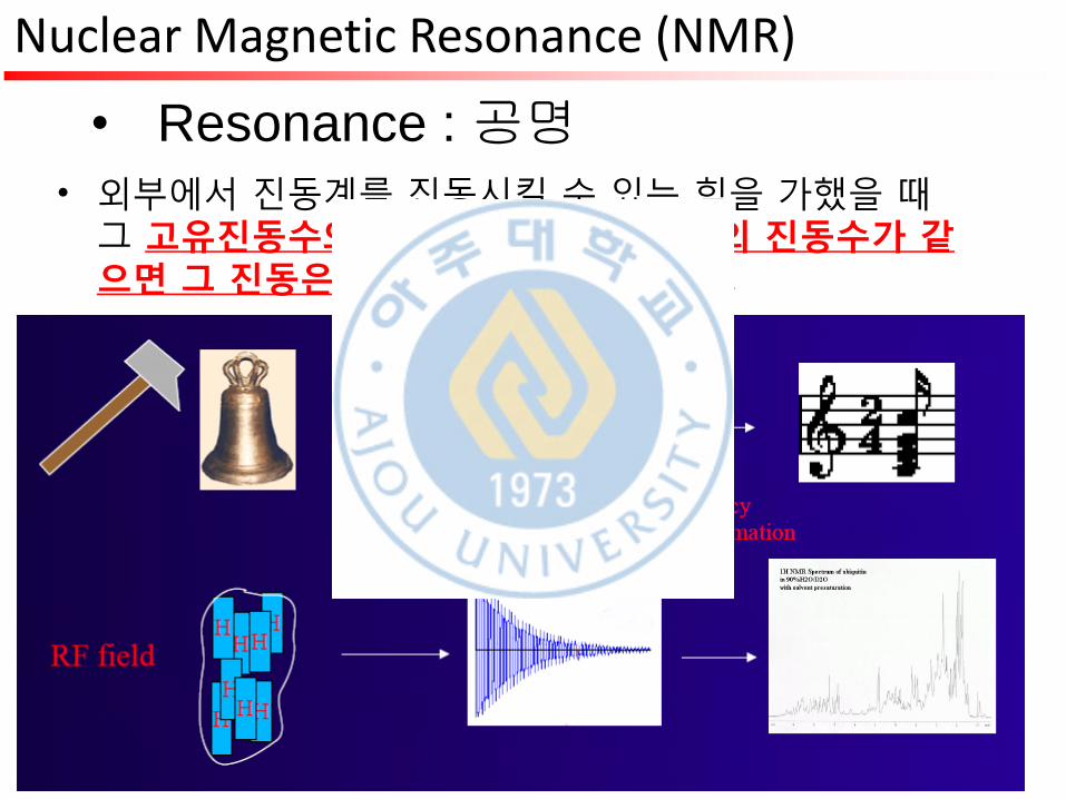
• Resonance : 공명
• 외부에서 진동계를 진동시킬 수 있는 힘을 가했을 때 그 고유진동수와 외부에서 가해주는 힘의 진동수가 같으면 그 진동은 심해지고 진폭도 커진다.
Nuclear Magnetic Resonance (NMR)
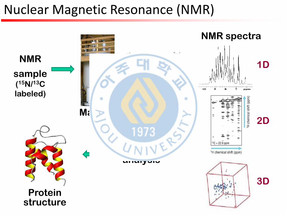
NMR
sample (15N/13C
labeled)
Magnet
Pulse sequence
NMR spectra
1D
2D
3D
Data analysis
Protein structure
Nuclear Magnetic Resonance (NMR)
Contents
Research on IP3R
Protein & Protein Structure
NMR/X-ray crystallography
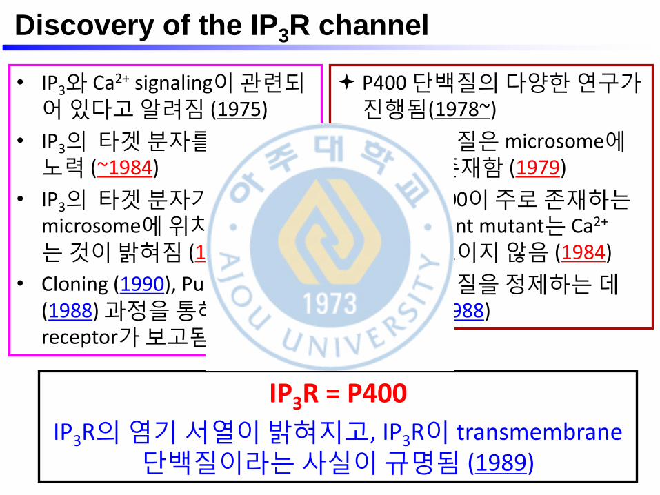
• IP3와 Ca2+ signaling이 관련되어 있다고 알려짐 (1975)
• IP3의 타겟 분자를 찾기 위한 노력 (~1984)
• IP3의 타겟 분자가 세포의 microsome에 위치하고 있다는 것이 밝혀짐 (1984)
• Cloning (1990), Purification (1988) 과정을 통해 IP3 receptor가 보고됨
P400 단백질의 다양한 연구가 진행됨(1978~)
P400 단백질은 microsome에도 다량 존재함 (1979)
Spine (P400이 주로 존재하는 곳) deficient mutant는 Ca2+
spike를 보이지 않음 (1984)
P400 단백질을 정제하는 데 성공함 (1988)
IP3R = P400
IP3R의 염기 서열이 밝혀지고, IP3R이 transmembrane 단백질이라는 사실이 규명됨 (1989)
Discovery of the IP3R channel
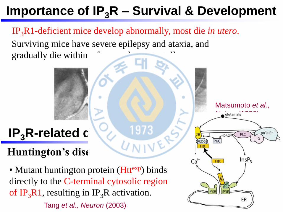
Matsumoto et al.,
Nature (1996)
IP3R1-deficient mice develop abnormally, most die in utero.
Surviving mice have severe epilepsy and ataxia, and
gradually die within a few weeks postnatally.
IP3R-related diseases
Tang et al., Neuron (2003)
Huntington’s disease (HD)
• Mutant huntington protein (Httexp) binds
directly to the C-terminal cytosolic region
of IP3R1, resulting in IP3R activation.
Importance of IP3R – Survival & Development
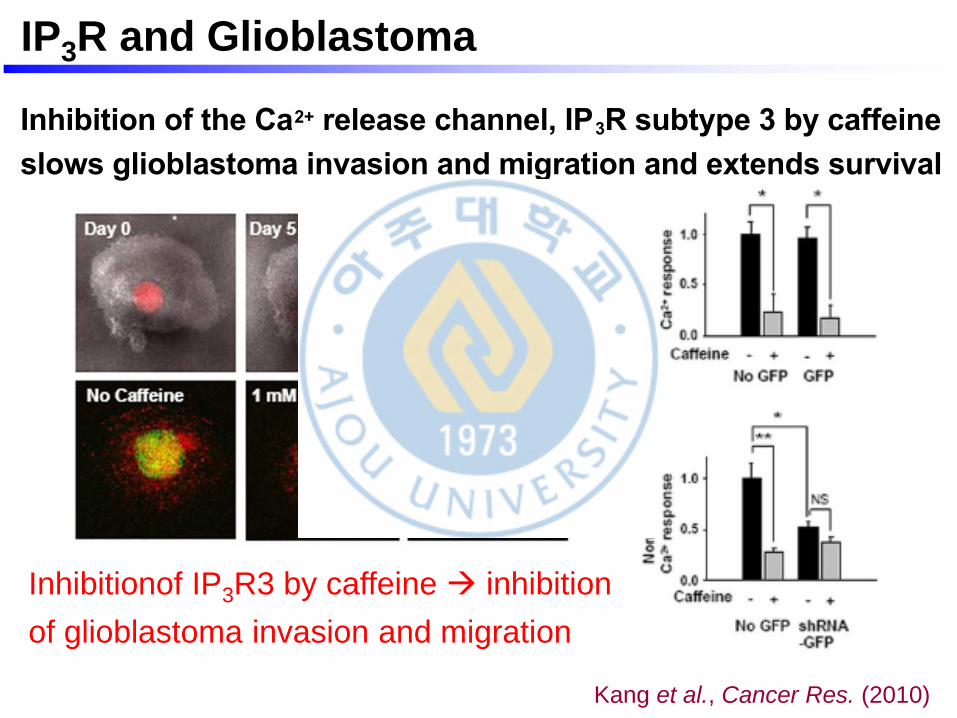
Inhibition of the Ca2+ release channel, IP3R subtype 3 by caffeine
slows glioblastoma invasion and migration and extends survival
Sang Soo Kang3,*, Kyung-Seok Han1,2,*, Bo Mi Ku3, Yeon Kyung Lee3, Jinpyo Hong8, Hye
Young Shin6, Antoine G. Almonte4, Dong Ho Woo1,2, Daniel J. Brat4, Eun Mi Hwang3,
Seung Hyun Yoo9, Chun Kee Chung6, Sung-Hye Park7, Sun Ha Paek6, Eun Joo Roh5, Sung
joong Lee8, Jae-Yong Park3, Stephen F. Traynelis4, and C. Justin Lee1,2
1 Center for Neural Science, Future Fusion Technology Laboratory, Republic of Korea
5 Division of Life Sciences, Korea Institute of Science and Technology (KIST), Republic of Korea
2 Neuroscience Program, Univ of Science and Technology (UST), Republic of Korea
3 Dept of Anatomy and Neurobiology, Dept of Physiology, Institute of Health Sciences, School of
Medicine, Gyeongsang National Univ, Republic of Korea
4 Dept of Pharmacology, Emory Univ, Atlanta, GA 30322, USA
6 Dept of Neurosurgery, Cancer Research Institute, Clinical Research Institute, Seoul National
Univ College of Medicine, Seoul, Republic of Korea
7 Dept of Pathology, Cancer Research Institute, Clinical Research Institute, Seoul National Univ
College of Medicine, Seoul, Republic of Korea
8 Program in Molecular and Cellular Neuroscience, Dept of Oral Physiology, School of Dentistry,
Seoul National Univ, Seoul, Republic of Korea
9 Dept of Biochemistry, Inha Univ. School of Medicine, Republic of Korea
Abstract
Ca2+ signaling is an important determining factor in many cellular processes, especially in cancer
cell proliferation, motility and invasion. Glioblastoma is the deadliest brain cancer with its average
survival time of less than a year, with the most prominent cellular feature being the ability of these
cells to migrate to and invade the neighboring tissue. We hypothesized that disturbing the Ca 2+
signaling pathway would decrease the propensity for these cells to migrate. Thus, we investigated
the detailed Ca2+ signaling pathway of the glioblastoma cells in response to various receptor
tyrosine kinases (RTK) and G-protein coupled receptor (GPCR) agonists. Here we report that
caffeine, which is a well-known activator of ryanodine receptors (RyRs), paradoxically inhibits
inositol-1, 4, 5-triphospate receptor(IP3R)-mediated Ca2+ increase by selectively targeting IP3R
subtype 3(IP3R3), whose mRNA expression is significantly increased in glioblastoma cells.
Consequently, by inhibiting IP3R3-mediated Ca2+ release, caffeine was found to inhibit the
invasion and migration of various glioblastoma cell lines in scrape motility, Matrigel invasion, soft
agar, and brain slice implantation assays. In a mouse xenograft model of glioblastoma, caffeine
intake via drinking water greatly increased mean survival duration of subject animals. These
findings propose IP3R3 as a novel target for glioblastoma treatment and that caffeine may be a
useful adjunct therapy that slows glioblastoma invasion and migration by selectively targeting
IP3R3.
Corresponding Author: C. Justin Lee, Korea Institute of Science and Technology, Center for Neural Science, 39-1 Hawolkok-dong,Seongbuk-gu, Seoul, Korea 136-791, Tel: 82-2-958-6940, Fax: 82-2-958-6937, [email protected].*These authors contributed equally.
NIH Public AccessAuthor ManuscriptCancer Res. Author manuscript; available in PMC 2012 February 7.
Published in final edited form as:
Cancer Res. 2010 February 1; 70(3): 1173–1183. doi:10.1158/0008-5472.CAN-09-2886.
NIH
-PA
Auth
or M
anuscrip
tN
IH-P
A A
uth
or M
anuscrip
tN
IH-P
A A
uth
or M
anuscrip
t
Inhibitionof IP3R3 by caffeine inhibition
of glioblastoma invasion and migration
IP3R and Glioblastoma
Kang et al., Cancer Res. (2010)
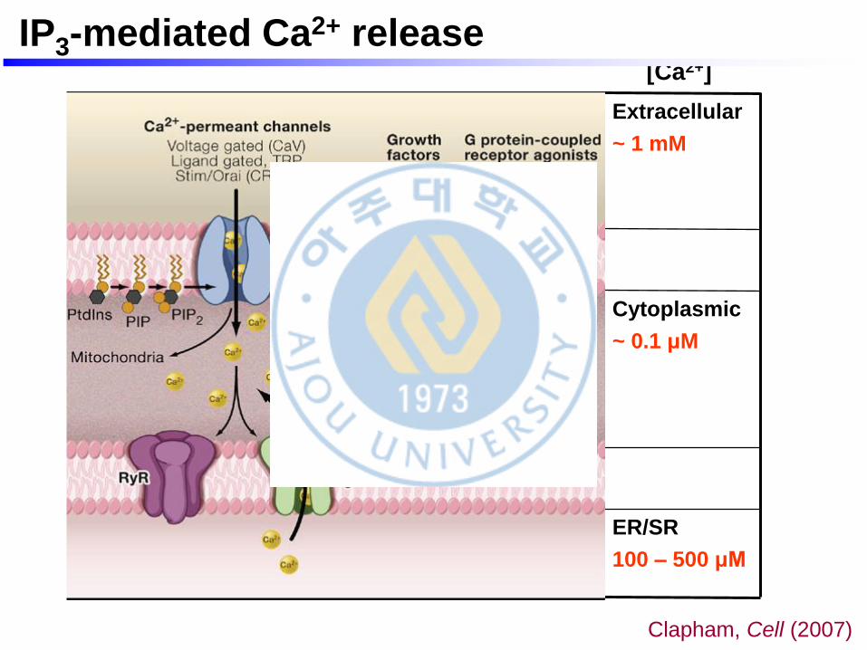
Extracellular
~ 1 mM
Cytoplasmic
~ 0.1 μM
ER/SR
100 – 500 μM
[Ca2+]
Clapham, Cell (2007)
IP3-mediated Ca2+ release
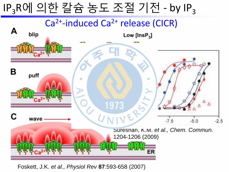
Ca2+-induced Ca2+ release (CICR)
Foskett, J.K. et al., Physiol Rev 87:593-658 (2007)
IP3R에 의한 칼슘 농도 조절 기전 - by IP3
Sureshan, K.M. et al., Chem. Commun.
1204-1206 (2009)
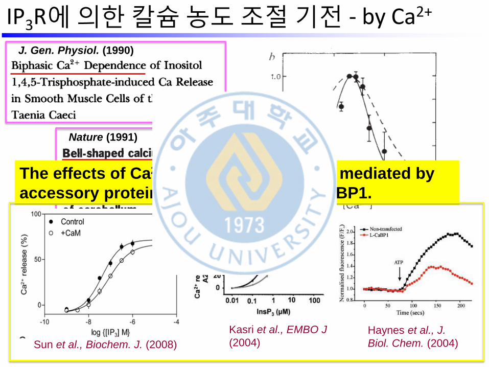
IP3R에 의한 칼슘 농도 조절 기전 - by Ca2+
Op
en
pro
ba
bili
ty
J. Gen. Physiol. (1990)
Nature (1991)
Kasri et al., EMBO J
(2004) Haynes et al., J.
Biol. Chem. (2004) Sun et al., Biochem. J. (2008)
The effects of Ca2+ on IP3R might also be mediated by
accessory proteins, such as CaM and CaBP1.
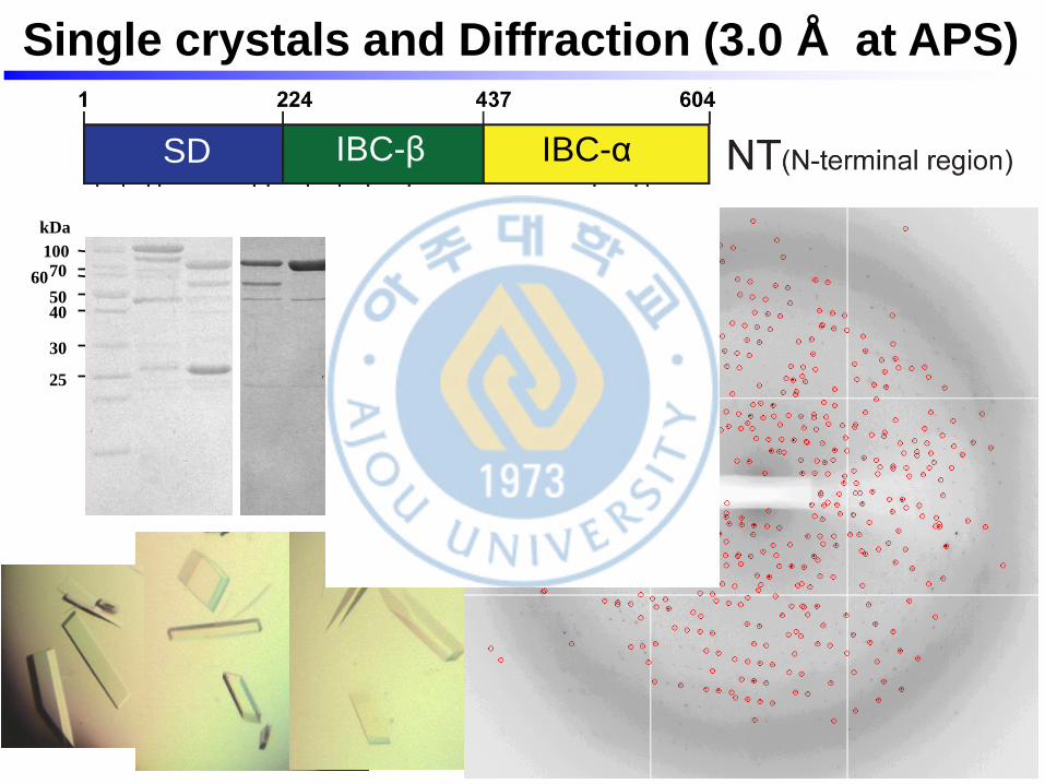
Single crystals and Diffraction (3.0 Å at APS)
100 70 60
50 40
30
25
kDa
SD IBC-β IBC-α
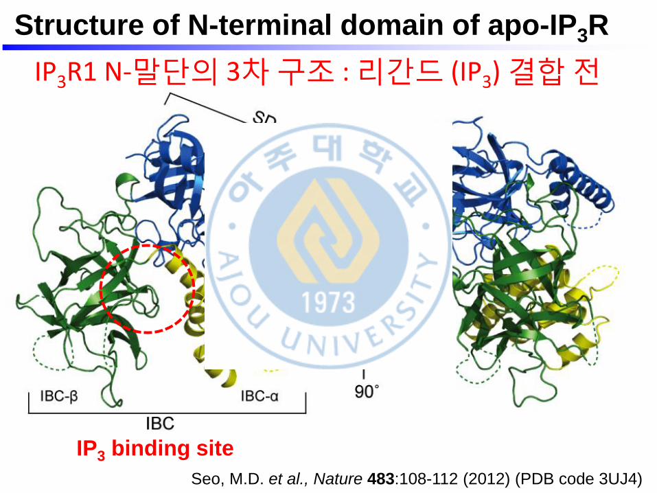
Structure of N-terminal domain of apo-IP3R
IP3 binding site
Seo, M.D. et al., Nature 483:108-112 (2012) (PDB code 3UJ4)
IP3R1 N-말단의 3차 구조 : 리간드 (IP3) 결합 전
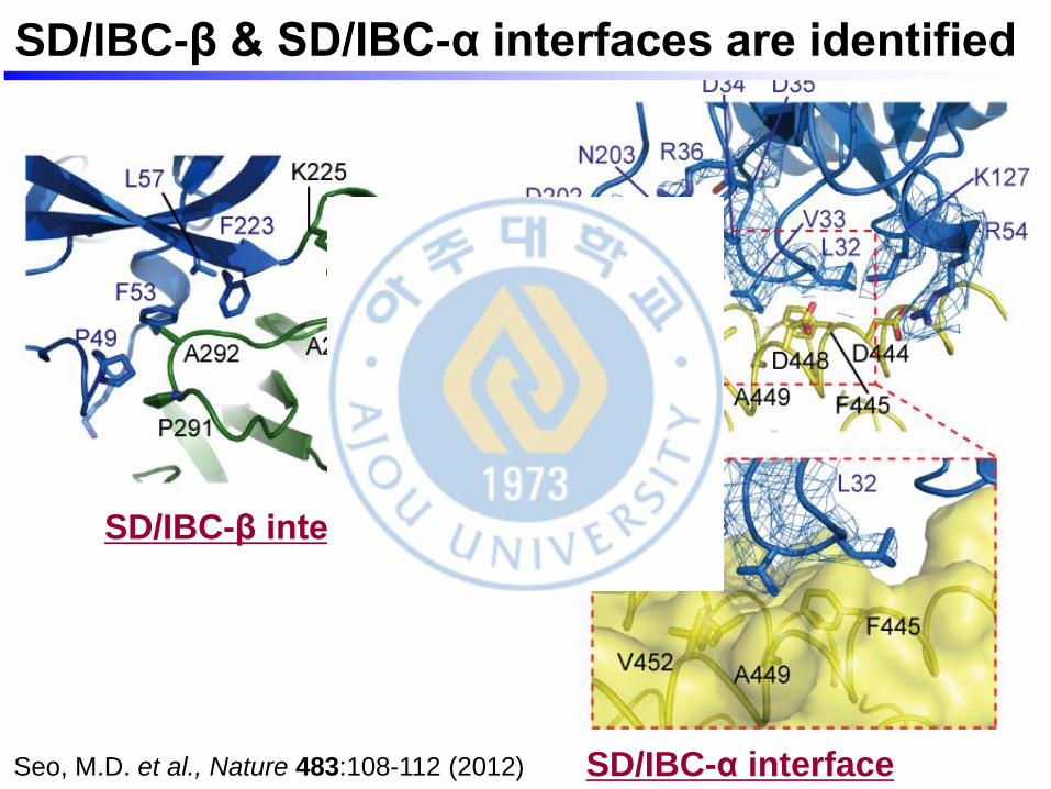
SD/IBC-β & SD/IBC-α interfaces are identified
SD/IBC-α interface
SD/IBC-β interface
Seo, M.D. et al., Nature 483:108-112 (2012)
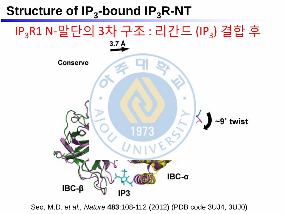
Structure of IP3-bound IP3R-NT
Seo, M.D. et al., Nature 483:108-112 (2012) (PDB code 3UJ4, 3UJ0)
IP3R1 N-말단의 3차 구조 : 리간드 (IP3) 결합 후
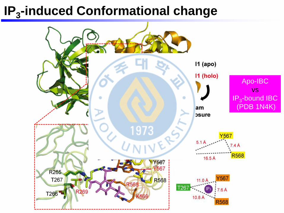
IP3-induced Conformational change
Apo-IBC
vs
IP3-bound IBC
(PDB 1N4K)
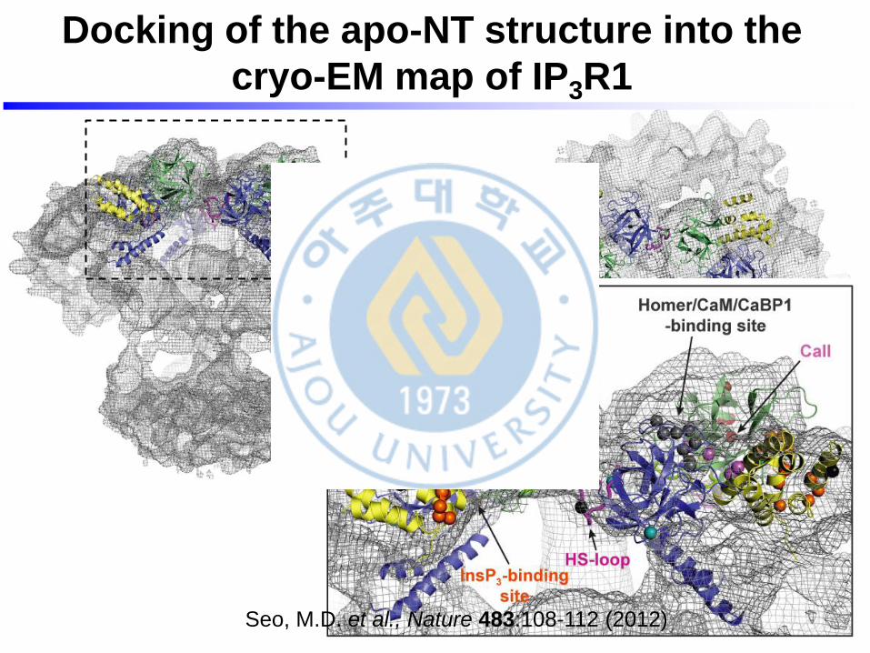
Docking of the apo-NT structure into the
cryo-EM map of IP3R1
Seo, M.D. et al., Nature 483:108-112 (2012)
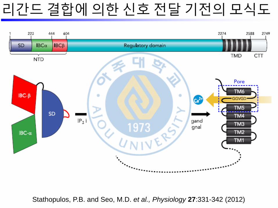
Stathopulos, P.B. and Seo, M.D. et al., Physiology 27:331-342 (2012)
리간드 결합에 의한 신호 전달 기전의 모식도
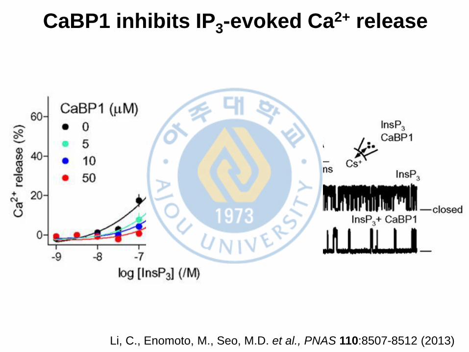
CaBP1 inhibits IP3-evoked Ca2+ release
Li, C., Enomoto, M., Seo, M.D. et al., PNAS 110:8507-8512 (2013)
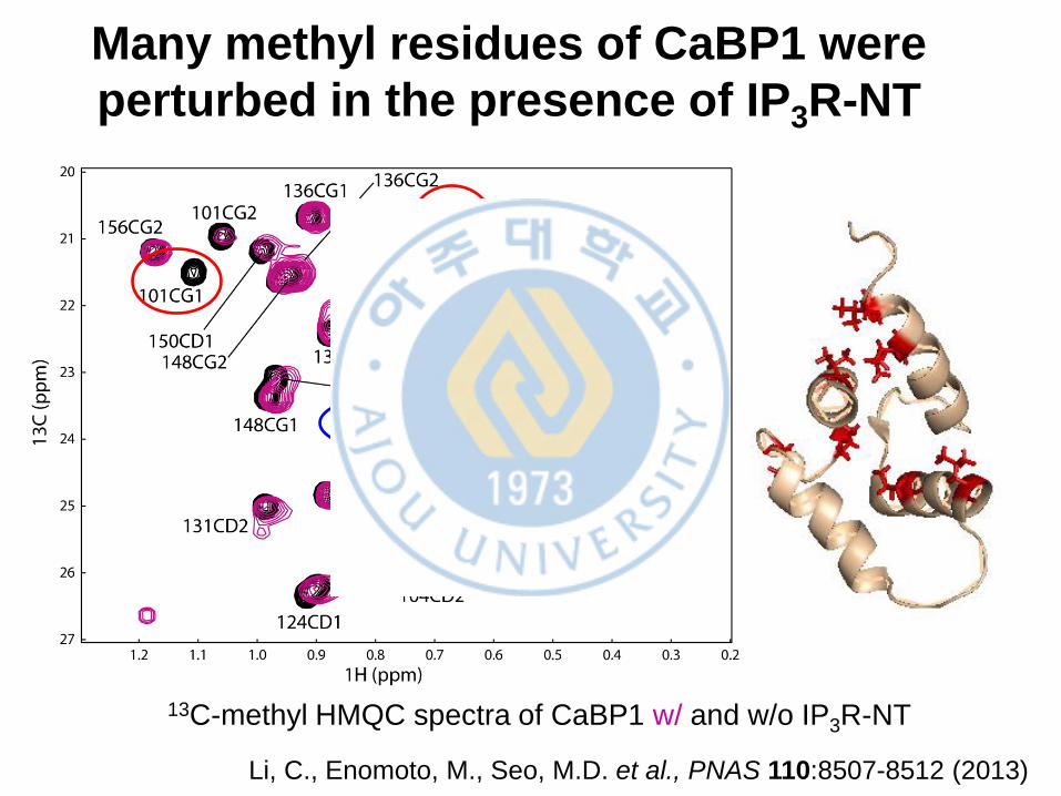
Many methyl residues of CaBP1 were
perturbed in the presence of IP3R-NT
13C-methyl HMQC spectra of CaBP1 w/ and w/o IP3R-NT
Li, C., Enomoto, M., Seo, M.D. et al., PNAS 110:8507-8512 (2013)
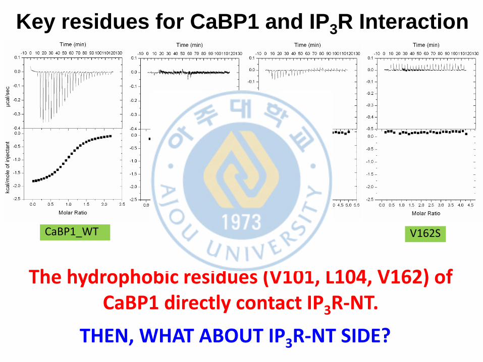
CaBP1_WT V101S L104S V162S
Key residues for CaBP1 and IP3R Interaction
The hydrophobic residues (V101, L104, V162) of CaBP1 directly contact IP3R-NT.
THEN, WHAT ABOUT IP3R-NT SIDE?
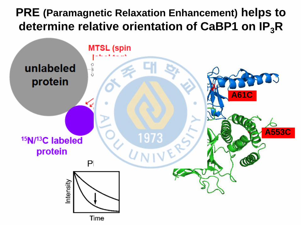
PRE (Paramagnetic Relaxation Enhancement) helps to
determine relative orientation of CaBP1 on IP3R
A61C
A553C
A394C
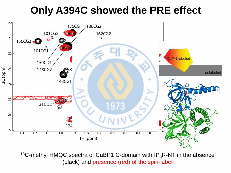
Only A394C showed the PRE effect
13C-methyl HMQC spectra of CaBP1 C-domain with IP3R-NT in the absence
(black) and presence (red) of the spin-label
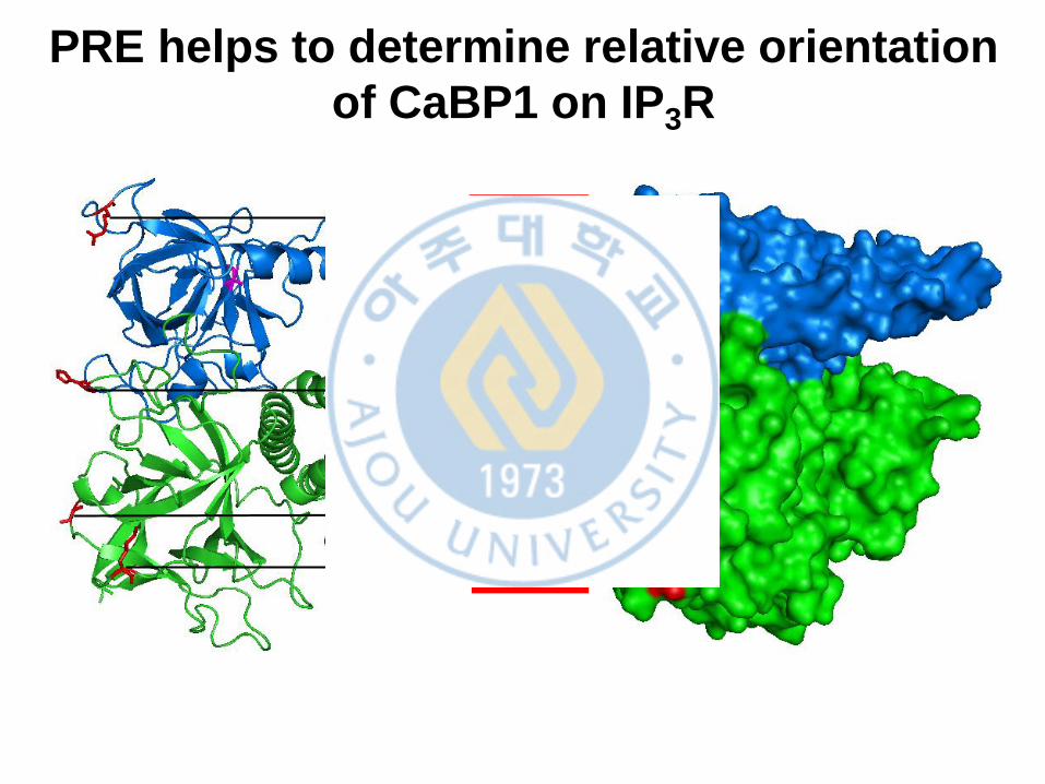
PRE helps to determine relative orientation
of CaBP1 on IP3R
R170C
H289C
K424C
A394C
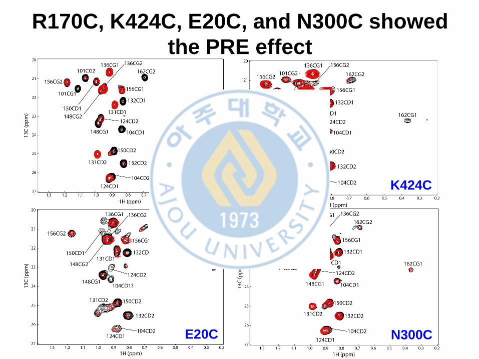
R170C, K424C, E20C, and N300C showed
the PRE effect
R170C K424C
E20C N300C
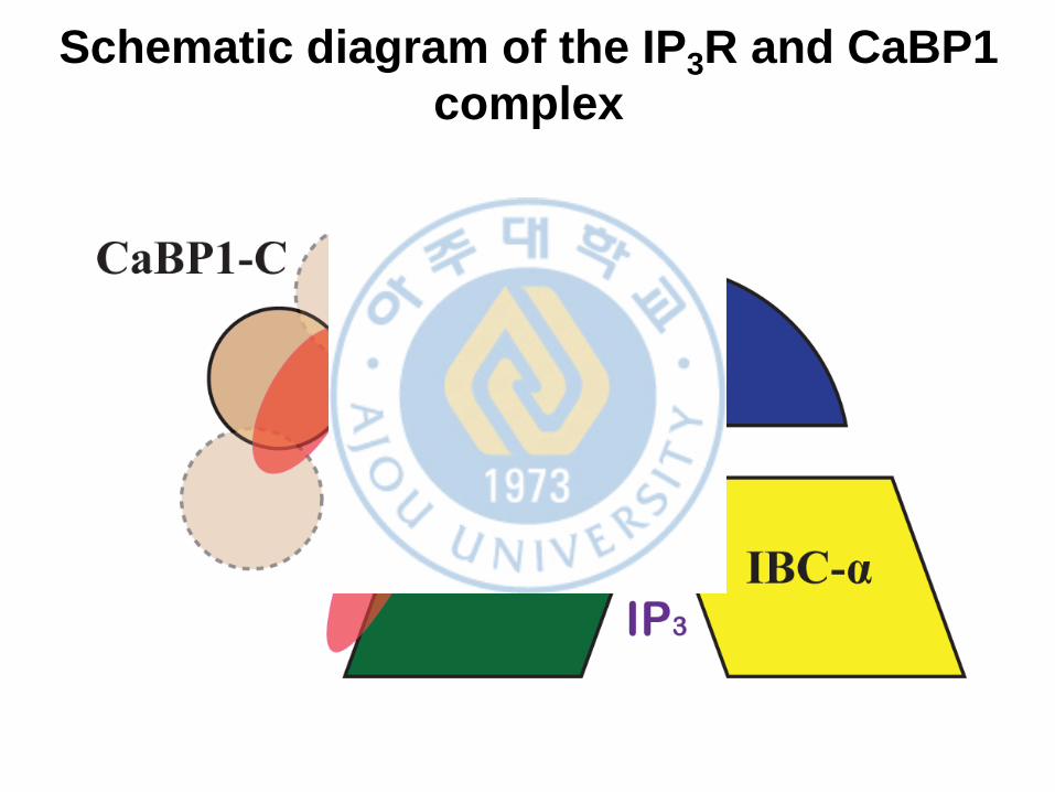
Schematic diagram of the IP3R and CaBP1
complex
V162
V101
L104
L302
I364
L393
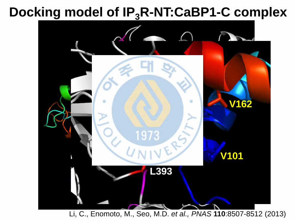
Docking model of IP3R-NT:CaBP1-C complex
Li, C., Enomoto, M., Seo, M.D. et al., PNAS 110:8507-8512 (2013)
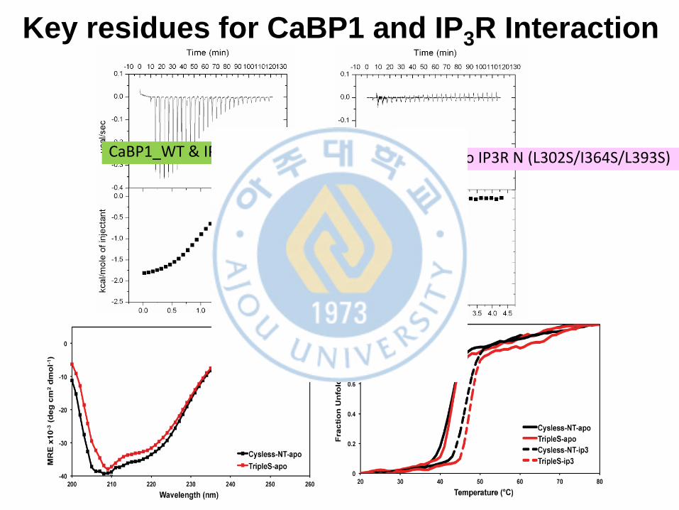
CaBP1_WT & IP3R N _WT CaBP1_WT to IP3R N (L302S/I364S/L393S)
Key residues for CaBP1 and IP3R Interaction
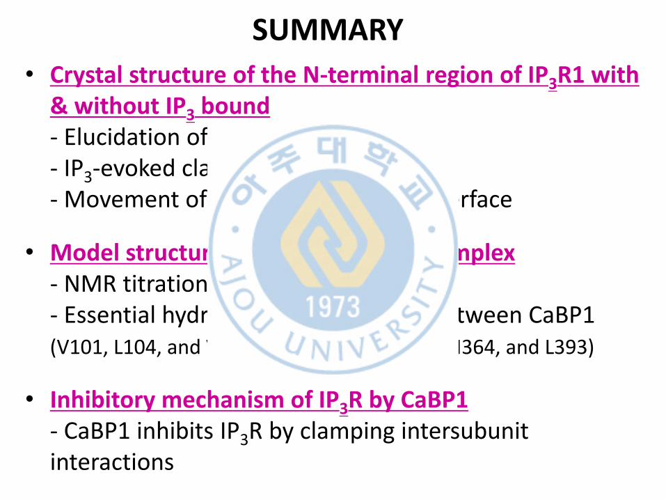
SUMMARY
• Crystal structure of the N-terminal region of IP3R1 with & without IP3 bound - Elucidation of two discrete interface - IP3-evoked clam closure - Movement of SD & retaining of α-interface
• Model structure of the IP3R:CaBP1 complex - NMR titration & Molecular docking - Essential hydrophobic interactions between CaBP1 (V101, L104, and V162) and IP3R-NT (L302, I364, and L393)
• Inhibitory mechanism of IP3R by CaBP1 - CaBP1 inhibits IP3R by clamping intersubunit interactions

![[ 구조 엔지니어링을 위한 기능 안내 ]wemadeinc.co.kr/newsletter/2014/04/images/BDS_U_revit.pdf · (4) 구조 분석 모델 3. 구조 해석 프로그램 안내 (Revit](https://img.pdfslide.tips/doc/110x75/5e1f932686dd854e2c479b08/-e-ee-oeoe-ee-e-4-e-e-ee-3-e.jpg)