Embed Size (px)
Citation preview

NPHS2 Gene Mutation Screening in
Palestinian Children With Steroid-Resistant
Nephrotic Syndrome
لدى األطفال الفمسطينيين المصابين NPHS2الكشف عن طفرات في جين
بمتالزمة الكالئية المقاومة لعالج الستيرويد
Ata J. Deif Allah
Supervised by
Prof. Dr. Fadel A. Sharif
Molecular Biology
A Proposal Submitted in Partial Fulfillment for the Degree of Master of Science
in Biological Sciences- Medical Technology
OCT/2017
زةــــــــغب تــالميـــــاإلسـت ـــــــــامعـالج
والدراساااث العليااا العلماا البحاا عمااا ة
ــــــــومـــالعـلــــــــــــت ــــــــــــــــــــليـك
حـياتـــــيتـــوم الــــالعلــ ماجستيـــــــر
طبيـــــــــــــــــــــت التحــــــــــــــــــاليل
The Islamic University of Gaza
Deanship of Research and graduated Studies
Faculty of Science
Master of Biological Sciences
Medical Technology

I

I
إقــــــــــــــرار
:أنا الموقع أدناه مقدم الرسالة التي تحمل العنوان
NPHS2 Gene Mutation Screening in Palestinian Children
with Steroid-Resistant Nephrotic Syndrome
لدى األطفال الفمسطينيين المصابين NPHS2 الكشف عن طفرات في جين بمتالزمة الكالئية المقاومة لعالج الستيرويد
تمت اإلشارة إليو حيثما ورد، أقر بأن ما اشتممت عميو ىذه الرسالة إنما ىو نتاج جيدي الخاص، باستثناء ما
وأن ىذه الرسالة ككل أو أي جزء منيا لم يقدم من قبل االخرين لنيل درجة أو لقب عممي أو بحثي لدى أي
.مؤسسة تعميمية أو بحثية أخرى
Declaration
I understand the nature of plagiarism, and I am aware of the University’s policy on
this.
The work provided in this thesis, unless otherwise referenced, is the researcher's own
work and has not been submitted by others elsewhere for any other degree or
qualification.
:Student's name عطا جابر محمد ضيف اهلل اسم الطالب:
التوقيع:Signature:
22/11/2017 اريخ:التDate:

II
Abstract
Background: Nephrotic syndrome (NS), an uncommon childhood disorder, is
characterized by edema, massive proteinuria, hypoalbuminemia, hyperlipidemia, and
may progress to end stage renal disease. Based on the response to steroid therapy, NS
is divided into steroid-sensitive (SSNS) and steroid-resistant (SRNS). SRNS is where
the patient does not respond to prednisone treatment.
SRNS inherited, as an autosomal recessive disorder with NPHS2 being the most
frequently mutated gene. NPHS2 gene encodes podocin protein, which has an
important role in glomerular ultrafiltration and controlling slit membrane
permeability.
Objective: The spectrum and frequency of NPHS2 mutations in the Palestinian
population have not been explored before. The aim of this study is to screen 20
SRNS Palestinian patients for NPHS2 mutations and to compare our findings with
those reported in other populations.
Methods: Twenty SRNS patients were recruited from the Ranteesy pediatric
hospital. All the eight exons of the NPHS2 gene were PCR-amplified from patients
genomic DNA using appropriate primers. Direct sequencing of the purified PCR
fragments was then ensued by automated Sanger sequencing method. Nucleotide
changes were verified by comparing obtained sequences with the reference gene
sequence stored in the NCBI database.
Results: Analysis of the obtained sequences identified previously known mutations
in 3 (15%) of the patients. Two of the mutations, G130K and R229Q were missense
mutations and the third, R138X was a nonsense mutation. All mutations were present
in homozygous form.
Conclusions: The current study reports the identification of G130K, R138X, and
R229Q NPHS2 mutation in SRNS patients in Gaza-Palestinian. The mutations thus
identified would spare patients from the unnecessary and harmful
immunosuppressive steroids and help physicians and patients' families take proper
decisions regarding patient management and their future offspring.
Keywords: SRNS, NPHS2, Sequencing, Gaza-Palestine.

III
الممخص( اضطرابا غير شائع يصيب األطفال، ومن أعراضو االستسقاء، البول NS: تعتبر المتالزمة الكالئية )لمقدمةا
البروتيني، نقص ألبومين الدم، ارتفاع الدىون في الدم، ويمكن أن يؤدي الى فشل الكموي. ويمكن تقسيمو إلى قسمين
( والمتالزمة الكالئية SSNSة الكالئية المستجيبة لمستيرويد )بالنظر إلى مدى استجابتو لمعالج بالستيرويدات: المتالزم
(، حيث ال يستجيب المريض في النوع الثاني لمعالج بالبريدنيزون.SRNSالمقاومة لمستيرويد )( كاضطرابات صبغية جسدية متنحية )صفة متنحية(، حيث SRNSوتورث المتالزمة الكالئية المقاومة لمستيرويد )
( والذي لو دور ميم في ترشيح podocin(، حيث يتم تشفير بروتين )NPHS2الجينية غالبا في جين ) تتشكل الطفرات
التميف الكبيبي والتحكم بنفاذية الغشاء الفمعي.
( بين السكان الفمسطينيين، وذلك من خالل NPHS2: تيدف الدراسة إلى استكشاف مدى انتشار طفرات )هدف الدراسة
(، ومقارنة النتائج التي تم التوصل إلييا مع الطفرات SRNSمة الكالئية المقاومة لمستيرويد )مصابا بالمتالز 02فحص
الموثقة دوليا في مجموعات أخرى من السكان.
: تم اختيار عشرين مريضا بالمتالزمة الكالئية من مستشفى الرنتيسي لألطفال، حيث تم فحص منهجية الدراسة
( لفحص PCR(، حيث تم مضاعفتيا باستخدام تقنية البممرة المتسمسمة )NPHS2) االكسونات الثمانية الخاصة بجين
البادئات المناسبة . وتم بعد ذلك تحميل التتابعات المباشر لألجزاء المنقاة بواسطة الحمض النووي لممرضى باستخدام
تيدية من خالل مقارنة (، وذلك لمالحظة مدى وجود أية تغيرات نوكميو Sanger( باستخدام طريقة )PCRتقنية )
(. NCBIالتتابعات التي تمت مالحظتيا مع تمك الموجودة في الجين المرجعي، والمخزنة في قاعدة بيانات )
% 51مرضى ) 3: أكدت نتائج تحميل التتابعات التي تم الحصول عمييا وجود طفرات جينية معروفة مسبقا في النتائج
، بينما المغمطةمن ضمن الطفرات ( R229Qو G130Kان منيا )وىما من العينة( من المرضى، حيث كانت اثنت
. وكانت جميع الطفرات موجودة في شكل متماثل.ىرائية( طفرة R138Xكانت الثالثة )
( لدى NPHS2( في جين )R138Xو R229Qو G130K: توصمت الدراسة إلى تحديد وجود طفرات )االستنتاجات
فمسطين. ومن شأن الطفرات التي تم تحديدىا أن -( في غزة SRNSاومة لمستيرويد )مرضى المتالزمة الكالئية المق
توفر عمى المرضى عناء المجوء إلى استرويدات المثبطة لممناعة كونيا غير ضرورية، مما يساعد األطباء وعائالت
تقبل.المرضى عمى اتخاذ القرارات المناسبة فيما يتعمق بإدارة حياة المرضى وأبنائيم في المس
فمسطين. -، التسمسل، غزة SRNS ،NPHS2: الكممات المفتاحية

IV
Dedication
I dedicate my modest work to most dear people, my parents who bestowed me
courage and support.
To my beloved wife who gave me all the chance to achieve this work, to my
daughter and all my brothers and sisters. Be proud!

V
Acknowledgments
I am grateful to Allah, who always offer me life, power, peace of mind, and courage
to accomplish this study.
Foremost, I would like to express my sincere gratitude to my advisor Prof. Dr. Fadel
A. Sharif for the continuous support of my Master study and research, for his
patience, motivation, enthusiasm, and immense knowledge. His guidance helped me
in all the time of research and writing of this thesis. I could not have imagined having
a better advisor and mentor for my Master study.
I want to express my deep thanks to the Faculty of Health Sciences and the
Department of Medical Technology in the Islamic University.
I want to express my deep thanks to the Rantessay Hospital and the Department of
Nephrology, doctors, nursing staff and special thanks to the Dr. Mostafa Elaila for
helping me in the completing this study.
I want to express my deep thanks to Trans-Orient Company, especially the head of
the company Hammam EL-rayes for donating all materials used in this study.
Special thanks to the IUG Genetics lab, in particular, Mr. Mohammed Ashour and
Mr. Shadi El-Ashi, who offered the support and equipment I have needed to
produce and complete my thesis.

VI
Table of Contents
I ..........................................................................................إقــــــــــــــرار
Abstract ............................................................................................ II
III .............................................................................................. الملخص
Dedication ....................................................................................... IV
Acknowledgments ............................................................................ V
Table of Contents ............................................................................ VI
List of Figures .................................................................................. X
List of Abbreviations ...................................................................... XI
Chapter 1 Introduction ...................................................................... 1
1.1 Overview .................................................................................. 2
1.2 Objectives of the study ............................................................ 3
1.3 Significance .............................................................................. 4
Chapter 2 Literature Review ............................................................. 5
2.1 Nephrotic Syndrome ............................................................... 6
2.1.1 Congenital nephrotic syndrome ......................................... 6
2.1.2 Infantile nephrotic syndrome ........................................... 7
2.1.4 Steroid-sensitive nephrotic syndrome ............................... 7
2.1.5 Steroid-resistant nephrotic syndrome ................................ 8
2.2 Nephron Structure .................................................................... 9
2.2.1 The Glomerulus ............................................................... 10
2.2.2 The glomerular filtration barrier: ..................................... 11
2.3 Podocin .................................................................................. 13
2.4 NPHS2 Gene .......................................................................... 14
2.4.1 Normal Function .............................................................. 14
2.4.2 NPHS2 mutations ............................................................ 15
2.5 Genetics of SRNS ................................................................ 15
VII
2.6 Previous Molecular Studies ................................................. 17
Chapter 3 Materials and Methods ................................................... 19
3.1 Study design ......................................................................... 20
3.2 Study Sample: ..................................................................... 20
3.3 Study location ..................................................................... 20
3.4 Inclusion Criteria ................................................................ 20
3.5 Ethical considerations ......................................................... 20
3.6 Specimen collection and processing .................................... 20
3.7 Materials ............................................................................. 21
3.7.1 Equipment ...................................................................... 21
3.7.2 Chemicals, Kits, and Disposables .................................. 22
3.7.3 PCR primers ................................................................... 23
3.8 Methods ................................................................................ 24
3.8.1 DNA extraction .............................................................. 24
3.8.1.1 DNA purification ........................................................ 24
3.8.1.2 Quality and quantity of extracted DNA..................... 24
3.8.2 NPHS2 gene mutation screening ................................... 24
3.8.2.1 Primers reconstitution ................................................. 24
3.8.2.2 Polymerase Chain Reaction (PCR) ............................ 24
3.8.2.3 Purification of PCR products ........................................ 25
3.8.3. DNA sequencing ............................................................. 26
3.8.3.1. Automated Sanger sequencing .................................... 26
3.8.3.2 BigDye® terminator cycle sequencing....................... 26
3.8.3.3. Purification ................................................................ 27
3.8.3.4 Sample preparation for injection ................................ 27
3.8.3.3.4. Principle of Capillary Electrophoresis ...................... 27
3.9 NPHS2 Gene Sequence Analysis .......................................... 28
Chapter 4 Results ............................................................................ 29

VIII
4.1 Isolated Genomic DNA ....................................................... 30
4.2 NPHS2 PCR-amplified exons .............................................. 31
4.3 DNA Sequencing results ...................................................... 31
4.3.1 Detected Mutations ........................................................ 31
4.3.1.1 Exon 3 (c.388G>A) mutation ..................................... 31
4.3.1.2 Exon 3 (c.412C>T) mutation ...................................... 32
4.3.1.3 Exon 5 (c.686G>A) mutation ..................................... 33
4.4 Summary of the identified mutations .................................. 33
4.5 Age of onset of proteinuria and NPHS2 mutations .............. 33
4.6 NPHS2 nucleotide variants ................................................... 34
4.7 Renal Histologic Findings ................................................... 34
Chapter 5 Discussion ...................................................................... 35
Chapter 6 Conclusion & Rrecommendations ................................. 40
6.1 Conclusion ........................................................................... 41
6. 2 Recommendations ............................................................... 41
References .................................................................................... 43
Appendix ......................................................................................... 48

IX
List of Tables
Table (2.1): Genes implicated in SRNS ..................................................................... 16
Table (3.1): The major equipment's used in the study ............................................... 21
Table (3.2): Chemicals, kits, and disposables ............................................................ 22
Table (3.3): Primers and expected PCR product size. ............................................... 23
Table (3.4): PCR components .................................................................................... 25
Table (3.5): Thermal cycling program for PCR amplification of the NPHS2 gene. . 25
Table (3.6): Exons annealing temperatures. .............................................................. 25
Table (3.7): To prepare the reaction mixtures. .......................................................... 26
Table (3.8): The thermal cycler program. .................................................................. 26
Table (4.1): NPHS2 sequence mutations identified in Palestinian SRNS patients .... 33
Table (4.2): NPHS2 detected variants. ....................................................................... 34
Table (4.3): Renal Histologic findings ...................................................................... 34
Table (5.1): NPHS2 nucleotide variants .................................................................... 38

X
List of Figures
Figure (2.1) : the nephron structure. Adopted from: http://www. buzzle.com. ........... 9
Figure (2.2): Structure of the renal corpuscle ............................................................ 10
Figure (2.3): A cross-section (electron microscopy, original magnification ×30,000)
of the glomerular filtration barrier. ............................................................................ 12
Figure (2.4): podocyte foot process cross section. ..................................................... 13
Figure (2.5): A schematic diagram of NPHS2 gene. ................................................. 14
Figure (2.6): NPHS2 reported mutations ................................................................... 15
Figure (2.7) Molecular analysis of a cohort of 49 families with CNS and SRNS..18
Figure (4.1): A photograph of ethidium bromide stained 3% agarose gel showing the
genomic DNA samples ...............................................................................................30
Figure (4.2): A photograph of ethidium bromide stained 3% agarose gel showing the
PCR products ..............................................................................................................31
Figure (4.3): Partial electropherograms for exon 3 (c.388 G>A) mutation. ...............32
Figure (4.4): Partial electropherograms for exon 3 (c.412C>T) mutation. ................32
Figure (4.5): Partial electropherograms for exon 5 (c.686G>A) mutation. ................33

XI
List of Abbreviations
CD2AP CD2 Associated Protein
CKD Chronic Kidney Disease
CNS Congenital Nephrotic Syndrome
Cs Capillary Space
DCT Distal Convoluted Tubule
DdNTPs Dideoxynucleotides
DNA Deoxyribonucleic Acid
E Endothelium Cell
EDTA Ethylenediaminetetraacetic Acid
FSGS Focal Segmental Glomerulosclerosis
GC Glucocorticoid
GEC Glomerular Endothelial Cell
GFR Glomerular Filtration Rate
GMB Glomerular Basement Membrane
GN Glomerulonephritis
IL Interleukin
INS Idiopathic Nephrotic Syndrome
IVF In Vitro Fertilization
KDIGO Kidney Disease: Improving Global Outcomes
NCBI National Center for Biotechnology Information
NS Nephrotic Syndrome
P Podocyte
PCR Polymerase Chain Reaction
PCT Proximal Convoluted Tubule
PGD Pre-implantation Genetic Diagnosis
PHB Prohibitin Homolology
RSPH Ranteesy Specialized Pediatric Hospital
SNP Single Nucleotide Polymorphism
SRNS Steroid-Resistant Nephrotic Syndrome
SSNS Steroid-Sensitive Nephrotic Syndrome
Us Urinary Space
WHO World Health Organization
WT1 Wilms Tumor

1
Chapter 1
Introduction
2
Chapter 1
Introduction
1.1 Overview
Nephrotic syndrome (NS) is considered as one of the main kidney diseases in
children and is characterized by proteinuria, edema, hypoalbuminemia, and
hyperlipidemia. The idiopathic nephrotic syndrome affects about 2 children per
100,000 aged 15 years and younger. About 10% to 20% of these children are
resistant to steroids treatment (Otukesh, Otukesh, et al., 2009; Patrakka et al.,
2002).
The generally used definition of steroid-resistant nephrotic syndrome (SRNS)
is lack of remission after exposure of 8 weeks of prednisone 2mg/kg/d or 60
mg/m2/d for 4 weeks followed by 1.5mg/kg or 40 mg/m2 per dose alternate-day for
4 weeks (Gipson et al., 2009).
Genetic forms of SRNS do not respond to strengthened immunosuppressive
treatment and progress inevitably to end-stage kidney disease but on the other hand,
They have a minimal risk of post-transplant recurrence. Hence, genetic testing is of
importance of clinical relevance in this complex group of patients (Jungraithmayr
et al., 2011; Santín et al., 2010) Genetically, SRNS is caused by mutations in a
number of different genes, the most common being NPHS2 (Lipska et al., 2013).
NPHS2 gene encodes podocin, a member of the stomatin protein family that is
localized at the slit membrane in Bowman's capsules in the nephrons of the kidney.
(Guaragna et al., 2015). The gene contains 8 exons which code for the 383 amino
acid long podocin protein. Podocin localizes at the slit diaphragm and interacts
directly with nephrin and CD2-associated protein.
Nephrin and podocin are key components of the slit diaphragm of the
glomerular epithelial cell and play a necessary role in the normal function of the
glomerular filtration barrier. Podocin deficiencies alter slit diaphragm permeability
and can also alter the processing and localization of nephrin (Otukesh, Ghazanfari,
et al., 2009).

3
So far, 126 NPHS2 pathogenic mutations have been identified worldwide. The
mutations are distributed along the entire coding region of the gene and cause all
kinds of alterations, including 53 missense, 17 nonsense, 11 small insertions, 26
small deletions, 16 splicing, two insertion/deletion mutations and one mutation in the
stop codon (Bouchireb et al., 2014).
In the present study, 20 Palestinian patients suffering from SRNS were tested
with the aim of detecting the spectrum of, and the common NPHS2 mutations in our
population. Mutation detection was performed through automated Sanger sequencing
of the eight exons and exon-intron boundaries of the NPHS2 gene.
Knowledge of the NPHS2 mutations in our population will improve the
diagnosis and medical care of SRNS patients and could also be used as a basis for
patient counseling, carrier screening and prenatal/ preimplantation diagnosis
programs in Gaza Strip.
1.2 Objectives of the study
1. The aim of the present study is to identify the spectrum of NPHS2
mutations in Palestinian patients suffering from SRNS.
2. Developing PCR-based techniques for rapid detection of the identified
NPHS2 mutations in Gaza Strip.
3. Correlating severity of SRNS and disease onset age with the identified
mutations.

4
1.3 Significance
The significance of this study can be summarized as follows:
1. Determining, for the first time in Gaza strip, the NPHS2 mutations
prevalent in our population.
2. Mutational analysis in SRNS would help in preventing unnecessary
exposure to immunosuppressants and their adverse effects, besides
helping in prognostication.
3. The results may help physicians in Gaza Strip to manage SRNS
associated with NPHS2 gene alterations and to reducing birth of
affected children by employing prenatal diagnosis or in vitro
fertilization (IVF) with preimplantation genetic diagnosis (PGD)
techniques.

5
Chapter 2
Literature Review
6
Chapter 2
Literature Review
2.1 Nephrotic Syndrome
Nephrotic syndrome (NS) is one of the most common causes of chronic kidney
disease (CKD) in the pediatric population. The cardinal features are edema, massive
proteinuria, and hypoalbuminemia. Children with NS have a decreased quality of life
(McCaffrey, Lennon, & Webb, 2016), They are at the risk of a wide range of
complications associated with significant morbidity and experience mortality rates of
up to 2.7 % (Kawaguchi et al., 2014).
NS can be divide into congenital NS (presentation before 3 months of age),
infantile NS (presentation between 3 months to 1 year of age) and idiopathic
nephrotic syndrome (INS). The largest of these groups is INS, and initial
management involves an 8-12-week course of oral glucocorticoid (Gc) therapy
(Ehrich & Brodehl, 1993). Approximately, 92 % of children with INS will enter
remission during this initial treatment and are subsequently classified as having a
steroid-sensitive nephrotic syndrome (SSNS), while 8 % fail to enter remission and
are classified as having a steroid-resistant nephrotic syndrome (SRNS)
(Nephrologie, 1979).
2.1.1 Congenital nephrotic syndrome
At least 170 mutations in the NPHS2 gene have been found to cause the
congenital nephrotic syndrome. This condition is a kidney disorder that begins in
infancy and typically leads to irreversible kidney failure (end-stage renal disease) by
early childhood. Mutations in this gene appear to be the most frequent cause of the
congenital nephrotic syndrome. Most NPHS2 gene mutations change single protein
building blocks (amino acids) in the podocin protein. These mutations result in a
reduction or absence of functional protein, which impairs the formation of normal slit
diaphragms. Without a functional slit diaphragm, molecules pass through the kidneys
abnormally and are excreted in urine. The filtering ability of the kidneys worsens
from birth, eventually leading to end-stage renal disease (Machuca et al., 2010).
7
2.1.2 Infantile nephrotic syndrome
NPHS2 gene mutations can cause other forms of nephrotic syndrome that
develop later in life. In one form, called infantile nephrotic syndrome, signs and
symptoms of the condition appear between 4 and 12 months of age. The features of
this condition are similar to congenital nephrotic syndrome (described above), but
they are often less severe. It is likely that NPHS2 gene mutations that cause infantile
nephrotic syndrome have less effect on podocin function than those that cause
congenital nephrotic syndrome, accounting for the later onset of the disorder
(Sadowski et al., 2015).
2.1.4 Steroid-sensitive nephrotic syndrome
Steroid sensitive nephrotic syndrome, SSNS, is the most common form of NS
in children. It usually first manifests at 2-6 years of age and is twice as likely in boys
than in girls. It is characterized clinically by a rapid onset of edema. Most often
urinalysis reveals the only proteinuria but some patients may have hyaline or waxy
casts and hematuria. While SSNS is usually associated with minimal changes in the
glomerular structure in light microscopy. (Patrakka et al., 2002).
In current clinical practice, children who enter disease remission during the
initial course of Gc therapy do not require a kidney biopsy (unless there are atypical
features), as initial clinical response to Gc therapy in children is of greater prognostic
significance than histological findings (Vivarelli, Moscaritolo, Tsalkidis, Massella,
& Emma, 2010).
Children with SSNS generally have a favorable outcome, and the results of
older studies suggested they achieve long-term remission during teenage years
(Trompeter, Hicks, Lloyd, White, & Cameron, 1985). However, recent data
suggest that over 30 % of SSNS children relapse in adulthood (Rüth, Kemper,
Leumann, Laube, & Neuhaus, 2005).
The etiology of SSNS is still not well understood although several avenues
have been researched. The popular proposition of the SSNS is that it has an
immunological component as it is associated with abnormal T-cell response and

8
often appears together with disorders with the immunological bases, such as atopy,
autoimmune diseases, and lymphomas (WEENING, 2004; Eddy & Symons, 2003).
It also often first appears after an infection although the mechanism of is this
not known. Recently, especially respiratory tract virus infections are implicated
(Eddy & Symons, 2003). T2-helper cell activation and production of cytokines such
as IL4 and -13 (IL4 and IL13) that occurs in allergy, also occurs in animal models of
SSNS (Ikeuchi et al., 2009; Kausman & Kitching, 2007).
However, a direct link between immune system and SSNS is yet to find. On the
glomerular level, loss of Glomerular Basement Membrane (GBM) charge has been
reported in some patients (Chugh, Clement, & Macé, 2012).
2.1.5 Steroid-resistant nephrotic syndrome
The most common histological diagnosis in SRNS is focal segmental
glomerulosclerosis, a term which in practice (though not strictly correct) is used
interchangeably with SRNS (Saleem, 2013).
Children with SRNS are treated with a range of other (non-Gc)
immunomodulatory agents, including mycophenolate mofetil, cyclophosphamide,
ciclosporin and rituximab (Lombel, Gipson, & Hodson, 2013).
More than 60 % of children with SRNS who fail to achieve remission with
pharmacological intervention will progress to end-stage renal disease
(Abeyagunawardena et al., 2007). There are also differences in mortality rates
according to the initial response to Gc therapy: 2.7 % in the INS population overall,
18.5 % in children with SRNS, 6.3 % in children with early relapse following initial
remission and 0.4 % in children without early relapse (McCaffrey et al., 2016).
Over the past 15 years, genetic discoveries have vastly improved our
understanding of the molecular basis of NS. The PodoNet consortium has recently
reported data from a heterogeneous population of 1655 children with SRNS with a
median follow-up time of 3.7 years. In this series, SRNS was either congenital (6 %),
infantile (7 %), adolescent onset (13 %) or childhood onset (74 %). At the time of the
last follow-up, 11.7 % of the children required dialysis, 14.2 % had received a kidney
transplant and 2.3 % were deceased (Trautmann et al., 2015). Remarkably, 23.6 %

9
of children from this heterogeneous SRNS cohort had a genetic mutation, in contrast
with another series where mutations were found in 66 % of NS cases presenting
under the age of 1 year (Hinkes et al., 2007).
The most common mutations in childhood NS are found in genes encoding
nephrin (NPHS1), podocin (NPHS2) and Wilms tumour 1 (WT1) (Saleem, 2013).
The increasing availability of genetic testing in SRNS will help clinicians minimize
exposure to immunosuppressive agents since genetic mutations are associated with a
poor response to immunosuppression (Bullich et al., 2015).
2.2 Nephron Structure
The nephron is the functional unit of the kidney. A nephron consists of a
twisted tubule closed at one end, open at the other with a network of associated blood
vessels. Each kidney of man is formed of about one million nephrons. It is in the
nephrons that urine is separated from the blood, and some of the water and salts in it
are reabsorbed. It is responsible for maintaining the pH and temperature of the
bloodstream.
Each nephron has a length of about 3 cm. It is differentiated into four regions
having different anatomical features and different physiological roles (Kriz &
Kaissling, 1992). The four regions are Bowman's capsule, proximal convoluted
tubule (PCT), a loop of Henle, and distal convoluted tubule (DCT). These four
regions are illustrated in Figure 2.1.
Figure (2.1) : the nephron structure. (Adopted from: http://www. buzzle.com, 20015).
10
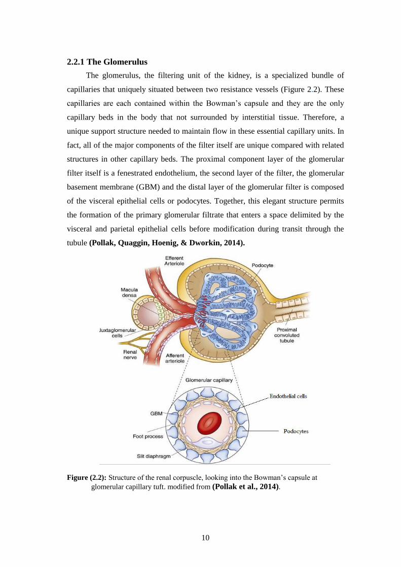
2.2.1 The Glomerulus
The glomerulus, the filtering unit of the kidney, is a specialized bundle of
capillaries that uniquely situated between two resistance vessels (Figure 2.2). These
capillaries are each contained within the Bowman’s capsule and they are the only
capillary beds in the body that not surrounded by interstitial tissue. Therefore, a
unique support structure needed to maintain flow in these essential capillary units. In
fact, all of the major components of the filter itself are unique compared with related
structures in other capillary beds. The proximal component layer of the glomerular
filter itself is a fenestrated endothelium, the second layer of the filter, the glomerular
basement membrane (GBM) and the distal layer of the glomerular filter is composed
of the visceral epithelial cells or podocytes. Together, this elegant structure permits
the formation of the primary glomerular filtrate that enters a space delimited by the
visceral and parietal epithelial cells before modification during transit through the
tubule (Pollak, Quaggin, Hoenig, & Dworkin, 2014).
Figure (2.2): Structure of the renal corpuscle, looking into the Bowman’s capsule at
glomerular capillary tuft. modified from (Pollak et al., 2014).
11
2.2.2 The glomerular filtration barrier:
The glomerulus consists of a cluster of capillaries appearing in a looped
formation supported by mesangial cells. While blood plasma passes the glomerular
capillary loops, the local intra-capillary pressure drives plasma through the
glomerular filtration barrier, which consists of three layers that each contributes to its
perm-selective properties: the capillary endothelium, the glomerular basement
membrane (GBM) and the single-celled layer of glomerular epithelial cells (GEC) or
podocytes (Figure 2.3) (Tanaka, 2008).
The first layer, the glomerular endothelial cells separate the blood and tissue
compartments. The endothelial cells are highly flattened cells and regulate vasomotor
tone and hemostasis, characterized by the presence of individual fenestrae on the
order of 70–100 nm in diameter. These cells drape the luminal aspect of the capillary
and permit filtration. (Ballermann, 2005).
The second layer, the glomerular basement membrane (GBM). Interposed
between the endothelial layer and the podocytes. It is composed of three layers
identified by electron density. These layers are named the lamina rara internal,
lamina densa, and lamina rara external. It is described as a dynamic gel-like acellular
meshwork of glycoproteins and proteoglycans (Levidiotis & Power, 2005).
The third layer, the podocytes. They are highly specialized, terminally
differentiated cells with cytoplasmic extensions, the so-called foot processes.
Podocytes have an important role in size and charge selective permeability, but also
in synthesizing and maintaining the GBM (Pavenstädt, Kriz, & Kretzler, 2003).
Furthermore, the fenestration of endothelial cells depends most notably on vascular
endothelial growth factor A secreted from differentiated podocytes. These
remarkable cells help to create the filtration slit diaphragm and serve as support to
help sustain the integrity of the free-standing capillary loops. (Ballermann, 2005).

12
Figure (2.3): A cross-section (electron microscopy, original magnification ×30,000) of the
glomerular filtration barrier with the capillary space (Cs), urinary space (Us), endothelial
cells (E), glomerular basement membrane (BM), and podocytes (P) (Löwik, Groenen,
Levtchenko, Monnens, & Van Den Heuvel, 2009a).
The foot processes of the podocytes attach to the outer surface of the GBM
through cell membrane receptors (α3β1 integrins linked to talin, vinculin, and
paxillin, and α- and β-dystroglycans) linked to utrophin; see Figure 2.4 (Kretzler,
2002). Adjacent foot processes interdigitate, forming a pore of about 25–40 nm in
width. This pore, or slit, is covered by a membrane with a “zipper-like” structure
(Rodewald & Karnovsky, 1974).
Recently, the structure of the slit membrane became the focus of many studies,
and although the complete structure has not elucidated yet, several new proteins were
found to be important for its function. Many components of the slit membrane are
involved in the pathogenesis of (nephrotic range) proteinuria (Löwik, Groenen,
Levtchenko, Monnens, & Van Den Heuvel, 2009b).

13
Figure (2.4): podocyte foot process cross section, depicting important components involved
in hereditary nephrotic syndrome (Geary & Schaefer, 2008).
2.3 Podocin
Podocin (NPHS2, OMIM 604766): It is a member of the stomatin protein
family is exclusively expressed in the podocytes and localizes at the insertion of the
slit membrane. Due to its similarity to stomatin, it is believed that podocin forms a
hairpin-like structure with intracellular NH2- termini and COOH-termini (Roselli et
al., 2002).
Podocin, like nephrin, associates with lipid rafts and recruits nephrin and
CD2AP in these rafts ensuring a stable and proper functioning filtration barrier. The
COOH terminal cytoplasmic tail of podocin interacts with nephrin and CD2AP (the
mouse homolog) (Schwarz et al., 2001). This protein interaction greatly enhances
nephrin-induced signaling in vitro (Huber, Köttgen, Schilling, Walz, & Benzing,
2001).
The COOH-terminal domain of podocin also binds NEPH-1; a podocyte slit
membrane protein structurally related to nephrin (Sellin et al., 2003) NEPH-1 is
involved in maintaining the structure of the filtration barrier and also interacts with
nephrin (Liu et al., 2004).
Podocin dysfunction leads to alterations of the slit membrane assembly and to
proteinuria in experimental models. NPHS2−/− mice develop proteinuria and

14
massive mesangial sclerosis (different from focal segmental glomerulosclerosis
"FSGS" seen in humans), the podocytes are enlarged and focally vacuolized.
Sclerosis rapidly progresses with age. Besides the absence of podocin, no nephrin
found in the foot processes as well. The podocin-deficient mice die a few days after
birth (Roselli et al., 2004).
Early reports showed that patients with NPHS2 mutations had no recurrence of
FSGS after renal transplantation. Now it is believed that patients with NPHS2
mutations have a lower risk for recurrent FSGS after renal transplantation compared
to patients with idiopathic FSGS (Ruf et al., 2004; Weber et al., 2004).
2.4 NPHS2 Gene
Mutations in NPHS2 gene, located at 1q25-31, are the most common cause of
SRNS in childhood. The coding region of the gene encompasses 1,149 bp, which 8
exons (Figure 2.5) and encodes a 383-amino acid protein with 42 kD, called podocin,
which is expressed in fetal and mature kidney (Guaragna et al., 2017).
Figure (2.5): A schematic diagram of NPHS2 gene. Exons represented by black boxes with
corresponding numbers above and introns by lines between the exons. Gray boxes represent
5` and 3` untranslated regions. Modified from (Franceschini, North, Kopp, Mckenzie, &
Winkler, 2006)
2.4.1 Normal Function
The NPHS2 gene provides instructions for making a protein called
podocin. Podocin primarily found in the kidneys, which are organs that filter waste
products from the blood and remove them in urine. Specifically, podocin found in
cells called podocytes, which are located in specialized kidney structures called
glomeruli. Podocin is located at the cell surface in the area between two podocytes
called the slit diaphragm. The slit diaphragm is known as a filtration barrier because
it captures proteins in blood so that they remain in the body while allowing other
molecules like sugars and salts to be excreted in urine. Podocin likely helps bring
other proteins that are needed for a functional slit diaphragm to the podocyte cell

15
surface. The protein also is involved with podocyte cell signaling and helping the cell
adapt to changes that occur during the filtration process (Grahammer, Schell, &
Huber, 2013).
2.4.2 NPHS2 mutations
NPHS2 mutations spread across the entire gene and lead to all kinds of
alterations including missense, nonsense, frameshift, and splice-site mutations
(Figure 2.6).
Figure (2.6): NPHS2 reported mutations. A: Exon structure of the NPHS2 gene with
geometric shapes indicating relative positions of different types of mutations. B: Podocin
domain positions,PHB (Bouchireb et al., 2014).
2.5 Genetics of SRNS
Steroid-resistant nephrotic syndrome (SRNS) occurs at an estimated incidence
of 1 per 200,000 children. Genetic causes can be identified in nearly 50 % of affected
children with this highly heterogeneous disorder. Genetic forms of SRNS do not
respond to intensified immunosuppressive treatment and progress inevitably to end-
stage kidney disease, but, on the other hand, have a minimal risk of post-transplant
recurrence. Hence, genetic testing is of eminent clinical relevance in this complex
group of patients (Lipska et al., 2013).
Mutations in the NPHS2 gene, encoding podocin, are a major cause of
autosomal-recessive steroid-resistant nephrotic syndrome (SRNS) in childhood,

16
accounting for up to 30% of sporadic and 20-40% of familial cases (Megremis, 2009
#83).
Molecular studies have implicated many genes in the pathogenesis of SRNS
(Table 2.1). The table indicates the results of an international cohort of 526 patients
of 1783 families, in whom a single-gene cause of SRNS was detected in 1 of 21
monogenic causes of SRNS (27 genes have been examined).
Table (2.1): Genes implicated in SRNS
Gene Causing
SRNS
Mode of
Inheritance
SRNS Families
Molecularly
Diagnosed by
Sanger Sequencing
(n)
SRNS Families
Molecularly
Diagnosed by
Multiplex PCR
(n)
Total SRNS
Families with
Molecular
Diagnosis (% of
Families)
NPHS2 AR 170 (42) 7 177 (9.93)
NPHS1 AR 93 (61) 38 131 (7.34)
WT1 AD 78 (50) 7 85 (4.77)
PLCE1 AR 23 (16) 14 37 (2.17)
LAMB2 AR 10 (6) 10 20 (1.12)
SMARCAL1 AR 1 (0) 15 16 (0.89)
INF2 AD 2 (0) 7 9 (0.5)
TRPC6 AD 1 (1) 8 9 (0.53)
COQ6 AR 6 (5) 2 8 (0.45)
ITGA3 AR 3 (3) 2 5 (0.28)
MYO1E AR 0 (0) 5 5 (0.28)
CUBN AR 1 (1) 4 5 (0.28)
COQ2 AR 0 (0) 4 4 (0.22)
LMX1B AD 0 (0) 4 4 (0.22)
ADCK4 AR 3 (3) 0 3 (0.17)
DGKE1 AR 0 (0) 2 2 (0.11)
PDSS2 AR 0 (0) 2 2 (0.11)
ARHGAP24 AD 0 (0) 1 1 (0.06)
ARHGDIA AR 1 (1) 0 1 (0.06)
CFH AR 0 (0) 1 1 (0.06)
ITGB4 AR 0 (0) 1 1 (0.06)
Total 392 (189) 134 526 (29.5)
Table (2.2): AD, autosomal dominant; AR, autosomal recessive. the number in parenthesis
show “molecularly solved” families with causative mutation the detected that was published
before our cohort (Sadowski et al., 2015).

17
The main conclusions which can be drawn from molecular genetic studies on
early onset nephrotic syndrome: First, two-thirds of nephrotic syndrome manifesting
in the first year of life can be explained by mutations in 4 genes (NPHS1, NPHS2,
WT1, or LAMB2). Second, NPHS1 mutations occur in congenital nephrotic syndrome
only. Third, infants with causative mutations in any of the 4 genes do not respond to
steroid treatment; therefore, unnecessary treatment attempts can be avoided. Fourth,
there are most likely additional unknown genes mutated in early-onset nephrotic
syndrome (Hinkes et al., 2007).
2.6 Previous Molecular Studies
A study on Brazilian children, analysis of the NPHS1, WT1 and NPHS2 genes
in 27 steroid-resistant revealed four missense mutations and one frameshift mutation.
All encountered mutations were in the NPHS2 gene. WT1 and NPHS1 gene analysis
did not reveal any mutation (Guaragna et al., 2015).
A study on Saudi Arabia children to determine the frequency of inherited NS,
62 cases (representing 49 families with NS) were screened for mutations in NPHS1,
NPHS2, LAMB2, PLCE1, CD2AP, MYO1E, WT1, PTPRO and NEIL1. Potential
causative mutations were detected in 25 out of the 49 families studied (51%). The
most common genetic cause of NS in the investigated cohort was a homozygous
mutation in the NPHS2 gene, found in 11 of the 49 families (22%). Mutations in the
NPHS1 and PLCE1 genes allowed a molecular genetic diagnosis in 12% and 8% of
families, respectively. New MYO1E mutations were evident in three families (6%).
No mutations were found in WT1, PTPRO or NEIL1 (Figure 2.7) (Al-Hamed et al.,
2013).

18
Figure (2.7): Molecular analysis of a cohort of 49 families with CNS and SRNS (Al-
Hamed et al., 2013).
A study on Indian children for evaluating the frequency of NPHS2 mutations in
25 with sporadic SRNS, Sun et al. found a homozygous mutation at nucleotide
position 211, C>T in only one patient, resulting in a stop codon i.e., p.R71X. This
mutation in exon 1 leads to the formation of a truncated protein, rendering it non-
functional (Sun et al., 2009).
A study on Israeli-Arab children from the 27 patients tested (familial and
nonfamilies), 15 patients (55%) were homozygous for the mutation C412T
(p.R138X) in NPHS2 is the cause of SRNS. (Frishberg et al., 2002).

19
Chapter 3
Materials and Methods

20
Chapter 3
Materials and Methods
3.1 Study design
The current study is a mutation analysis study, in which the NPHS2 gene from
SRNS patients was screened for potential pathogenic mutations.
3.2 Study Sample:
The target population of this study consisted of 20 Palestinian children residing
in Gaza strip. All participants were ≤ 12 years old.
3.3 Study location
All experimental work was done at the Genetics laboratory of the Islamic
University, Gaza strip.
3.4 Inclusion Criteria
Patients greater than 12 years, diagnosed with primary SRNS.
3.5 Ethical considerations
Informed consent was taken from all the subjects who agreed to participate in
the study. The objective of the study was fully explained to all participants. The
study was approved by the Helsinki ethics committee in Gaza Strip.
3.6 Specimen collection and processing
About 2.0 ml of venous blood drawn into sterile EDTA tubes and mixed
gently, under quality control and safety procedures. The blood samples were used for
DNA extraction and purification.

21
3.7 Materials
3.7.1 Equipment
The present work was carried out in the Genetics lab at the Islamic University
of Gaza. The major equipments used in the study are listed in Table 3.1.
Table (3.1): The major equipment's used in the study
# Instrument Manufacturer
1. Thermal Cycler Biometra, Germany
2. Electrophoresis chambers and tanks (horizontal) BioRad, USA
3. Electrophoresis power supply BioRad, USA
4. Microcentrifuge Sanyo, UK
5. Microwave Oven L.G, Korea
6. Digital balance AE adam, USA
7. Freezer, refrigerator ORSO, pharml-spain
8. Micropipettes (0.1-2.5μl / 0.5-10μl / 5-50μl / 20-200μl /
100-1000μl) Dragon-lab, USA
9. Safety cabinet N-Biotek,Inc
10. Gel documentation system Vision, Scie-Plas Ltd, UK
11. Nano-drop spectrophotometer Implen, Germany
12. ABI PRISM 310 Genetic Analyzer Applied Biosystems, USA
22
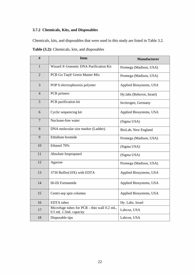
3.7.2 Chemicals, Kits, and Disposables
Chemicals, kits, and disposables that were used in this study are listed in Table 3.2.
Table (3.2): Chemicals, kits, and disposables
# Item Manufacturer
1 Wizard ® Genomic DNA Purification Kit Promega (Madison, USA)
2 PCR Go Taq® Green Master Mix Promega (Madison, USA)
3 POP 6 electrophoresis polymer Applied Biosystems, USA
4 PCR primers Hy.labs (Rehovot, Israel)
5 PCR purification kit Invitrogen, Germany
6 Cyclic sequencing kit Applied Biosystems, USA
7 Nuclease-free water (Sigma USA)
8 DNA molecular size marker (Ladder). BioLab, New England
9 Ethidium bromide Promega (Madison, USA)
10 Ethanol 70% (Sigma USA)
11 Absolute Isopropanol (Sigma USA)
12 Agarose Promega (Madison, USA)
13 3730 Buffer(10X) with EDTA Applied Biosystems, USA
14 Hi-Di Formamide Applied Biosystems, USA
15 Centri-sep spin columns Applied Biosystems, USA
16 EDTA tubes Hy. Labs. Israel
17 Microfuge tubes for PCR - thin wall 0.2 mL,
0.5 mL 1.5mL capacity Labcon, USA
18 Disposable tips Labcon, USA

23
3.7.3 PCR primers
The PCR primers used for amplifying the NPHS2 gene exons and the expected
product size are indicated (Table 3.3). Primers sequences were adopted from (Boute
et al., 2000b).
Table (3.3): Primers and expected PCR product size.
Exon `Forward Reverse
PCR
product
size
Exon 1 A 5′-GCAGCGACTCCACAGGGACT-3′ 5′-GGACCTCATCCACGTCCAC-3′ 289 bp
Exon 1 B 5′-GGTGGACGTGGATGAGGTC-3′ 5′-TCAGTGGGTCTCGTGGGGAT-3′ 177 bp
Exon 2 5′-AGGCAGTGAATACAGTGAAG-3′ 5′-GGCCTCAGGAAATTACCTA-3′ 203 bp
Exon 3 5′-TTCTGGGAGTGATTTGAAAG-3′ 5′-TGAAGAAATTGGCAAGTCAG-3′ 168 bp
Exon 4 5′-AAGGTGAAACCCAAACAGC-3′ 5′-CGGTAGGTAGACCATGGAAA-3′ 204 bp
Exon 5 5′-CATAGGAAAGGAGCCCAAGA-3′ 5′-TTTCAGCATATTGGCCATTA-3′ 292 bp
Exon 6 5′-CTCCCACTGACATCTGA-3′ 5′-AATTTAAAATGAAACCAGAA-3′ 155 bp
Exon 7 5′-CTAAATCATGGCTGCACACC-3′ 5′-CTTCCTAAAGGGCAGTCTGG-3′ 167 bp
Exon 8 5′-GGTGAAGCCTTCAGGGAATG-3′ 5′-TTCTATGGCAGGCCCCTTTA-3′ 380 bp

24
3.8 Methods
3.8.1 DNA extraction
3.8.1.1 DNA purification
Genomic DNA extracted from peripheral blood leukocytes using a commercial
kit (Wizard gDNA Purification Kit; Promega-USA) following the manufacturer
instructions. The isolated DNA was stored at -20C◦ until analysis.
3.8.1.2 Quality and quantity of extracted DNA
The quality of the isolated DNA was determined by running 5 µl of each
sample on ethidium bromide stained 1.0% agarose gel. The DNA samples then
visualized on a Gel documentation system. Additionally, the DNA concentration of
each sample measured by the use of Nano-drop spectrophotometer.
3.8.2 NPHS2 gene mutation screening
3.8.2.1 Primers reconstitution
Primers were received in a lyophilized state. Primer containers first
centrifuged at 14,500 rpm for 3 minutes and then reconstituted with ultrapure
nuclease-free water to create a stock solution of each primer with a final
concentration of 100pmol/µl. The stock primer solution was then vortex
mixed. Thirty-microliter aliquot taken from the stock primer and diluted with
270-µl nuclease free water to make 10pmol/µl working solution.
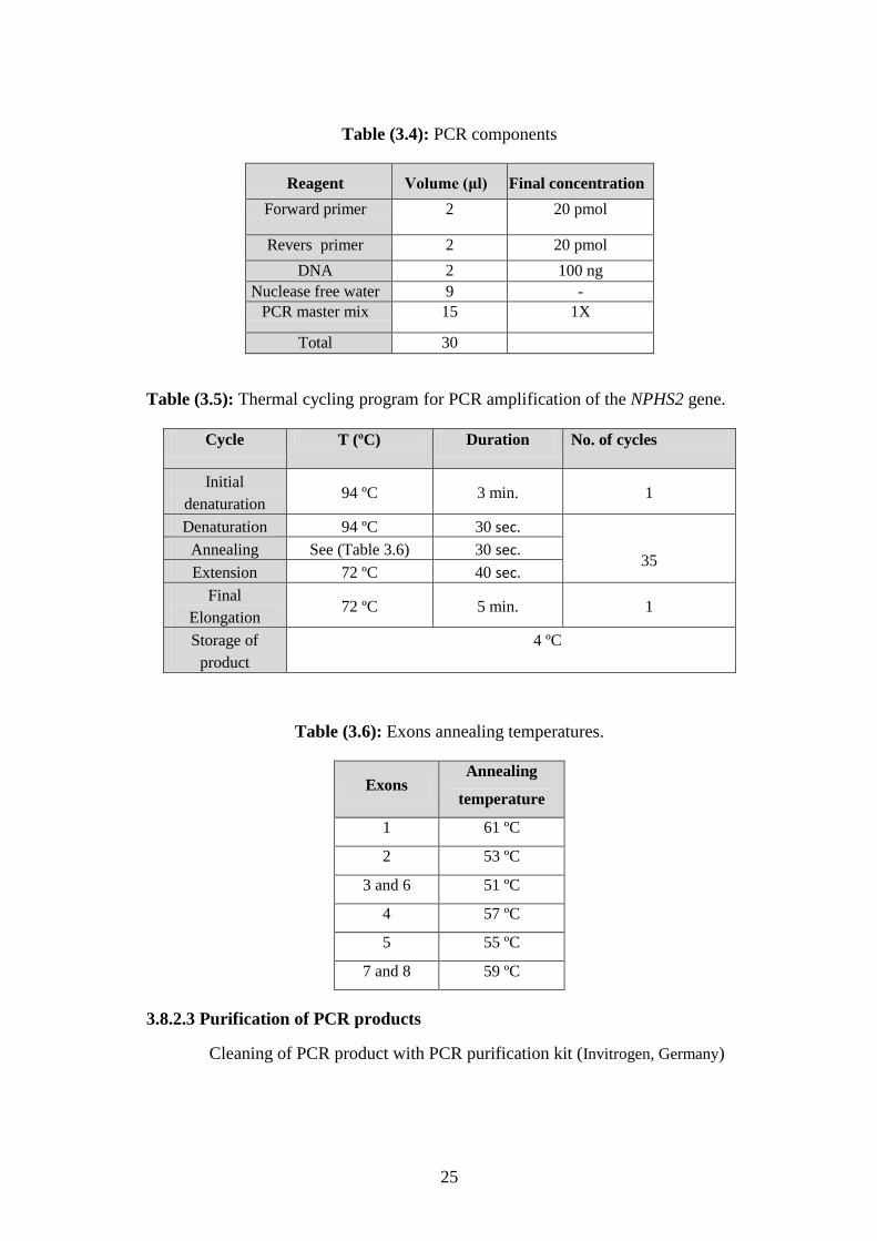
3.8.2.2 Polymerase Chain Reaction (PCR)
Polymerase chain reaction (PCR) was done with a total volume of 30μl. The
components of the (PCR) for each exon are shown in Table 3.4. PCR tubes were then
placed in the thermal cycler and PCR amplification was done according to the
program provided in Table 3.5. The PCR annealing temperatures for the various
NPHS2 exons are given in Table 3.6.
25
Table (3.4): PCR components
Reagent Volume (μl) Final concentration
Forward primer 2 20 pmol
Revers primer 2 20 pmol
DNA 2 100 ng
Nuclease free water 9 -
PCR master mix 15 1X
Total 30
Table (3.5): Thermal cycling program for PCR amplification of the NPHS2 gene.
Cycle T (ºC) Duration No. of cycles
Initial
denaturation 94 ºC 3 min. 1
Denaturation 94 ºC 30 sec.
35 Annealing See (Table 3.6) 30 sec.
Extension 72 ºC 40 sec.
Final
Elongation 72 ºC 5 min. 1
Storage of
product
4 ºC
Table (3.6): Exons annealing temperatures.
Exons Annealing
temperature
1 61 ºC
2 53 ºC
3 and 6 51 ºC
4 57 ºC
5 55 ºC
7 and 8 59 ºC
3.8.2.3 Purification of PCR products
Cleaning of PCR product with PCR purification kit (Invitrogen, Germany)
26
3.8.3. DNA sequencing
3.8.3.1. Automated Sanger sequencing
Automated Sanger sequencing was employed for sequencing the nucleotides of
the eight exons of the NPHS2 gene. The amount of the DNA template of the
amplified and purified exons was in the range of 3-20 ng.
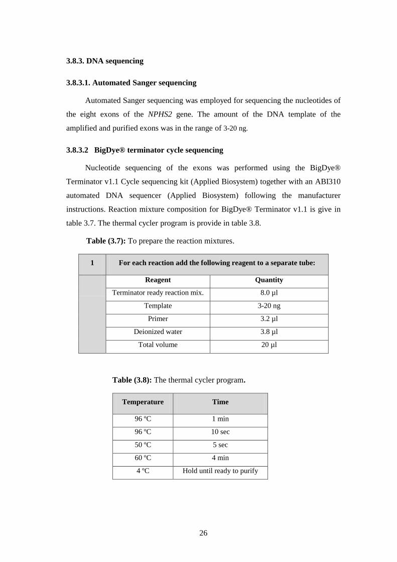
3.8.3.2 BigDye® terminator cycle sequencing
Nucleotide sequencing of the exons was performed using the BigDye®
Terminator v1.1 Cycle sequencing kit (Applied Biosystem) together with an ABI310
automated DNA sequencer (Applied Biosystem) following the manufacturer
instructions. Reaction mixture composition for BigDye® Terminator v1.1 is give in
table 3.7. The thermal cycler program is provide in table 3.8.
Table (3.7): To prepare the reaction mixtures.
1 For each reaction add the following reagent to a separate tube:
Reagent Quantity
Terminator ready reaction mix. 8.0 µl
Template 3-20 ng
Primer 3.2 µl
Deionized water 3.8 µl
Total volume 20 µl
Table (3.8): The thermal cycler program.
Temperature Time
96 ºC 1 min
96 ºC 10 sec
50 ºC 5 sec
60 ºC 4 min
4 ºC Hold until ready to purify

27
3.8.3.3. Purification
Desalting (salt interferes with electrokinetic injection) and elimination of
remaining labeled ddNTPs. Gel filtration Centri-sep columns (Princeton/ Applied
Biosystem) used for purifying the obtained BigDye® Terminator labeled products
following the manufacturer protocol.
3.8.3.4 Sample preparation for injection
BigDye® Terminator labeled products were mixed with Hi Di formamid (1:1
v/v) heated at 95 ºC for 2 min and then put on ice for 1 min. Samples are now ready
for injection into the ABI310 automated DNA sequencer (Applied Biosystem).
3.8.3.3.4. Principle of Capillary Electrophoresis
During capillary electrophoresis, the extension products of the cycle
sequencing reaction enter the capillary as a result of electrokinetic injection. A high
voltage charge applied to the buffered sequencing reaction forces the negatively
charged fragments into the capillaries. The extension products are separate by size
based on their total charge. Shortly before reaching the positive electrode, the
fluorescently labeled DNA fragments, separated by size, move across the path of a
laser beam. The laser beam causes the dyes on the fragments to fluoresce. An optical
detection device on the genetic analyzers detects the fluorescence. The Data
Collection Software converts the fluorescence signal to digital data, then records the
data in a *.ab1 file. Because each dye emits light at a different wavelength when
excited by the laser, all four colors, and therefore all four bases, can be detect and
distinguished in one capillary injection. The results are then display as exemplified in
(Figure 3.1).
Figure (3.1). An illustrative diagram of fluorescent sequencing result.
28
3.9 NPHS2 Gene Sequence Analysis
Obtained NPHS2 exons sequences were analyzed for nucleotide changes by
comparing them to the reference sequences in the NCBI database (NCBI human
genome build 38.7) using the NCBI BLAST (basic local alignment search tool) tool.
Detected sequence changes then searched for novelty using the NPHS2 variation
table stored in the Ensembl (http://www.ensembl.org) database.

29
Chapter 4
Results

30
Chapter 4
Results
The study cohort consisted of 20 children suffering from SRNS (10 girls and
10 boys). The patients were recruited from the Ranteesy Specialized Pediatric
Hospital, Gaza. The mean age at onset of proteinuria was 3.57 years (range, 2 month
to 6 years). Three (15.0%) patients had familial NS and 17 (85.0%) had sporadic
NS. All patients were tested for the presence of NPHS2 gene mutations by
sequencing the eight coding exons and their intron boundaries.
4.1 Isolated Genomic DNA
The following figure 4.1 represents samples of patients genomic DNA. The
bands in the figure shows that the isolated DNA is of good quality since it is intact
and of high molecular weight.
Figure (4.1): A photograph of ethidium bromide stained 3% agarose gel showing the
genomic DNA samples of different patients (lanes 2 to 8). The first lane contains a size
marker.
31
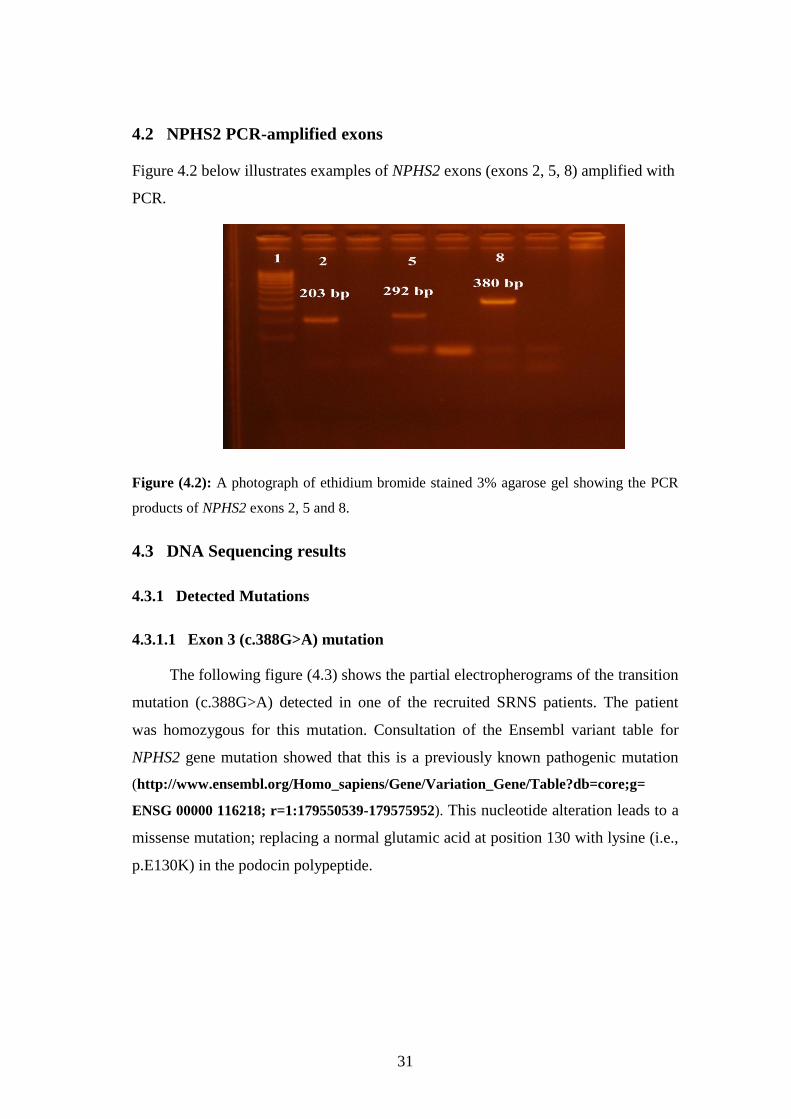
4.2 NPHS2 PCR-amplified exons
Figure 4.2 below illustrates examples of NPHS2 exons (exons 2, 5, 8) amplified with
PCR.
Figure (4.2): A photograph of ethidium bromide stained 3% agarose gel showing the PCR
products of NPHS2 exons 2, 5 and 8.
4.3 DNA Sequencing results
4.3.1 Detected Mutations
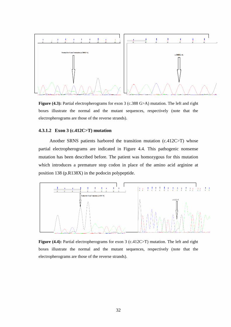
4.3.1.1 Exon 3 (c.388G>A) mutation
The following figure (4.3) shows the partial electropherograms of the transition
mutation (c.388G>A) detected in one of the recruited SRNS patients. The patient
was homozygous for this mutation. Consultation of the Ensembl variant table for
NPHS2 gene mutation showed that this is a previously known pathogenic mutation
(http://www.ensembl.org/Homo_sapiens/Gene/Variation_Gene/Table?db=core;g=
ENSG 00000 116218; r=1:179550539-179575952). This nucleotide alteration leads to a
missense mutation; replacing a normal glutamic acid at position 130 with lysine (i.e.,
p.E130K) in the podocin polypeptide.
32
Figure (4.3): Partial electropherograms for exon 3 (c.388 G>A) mutation. The left and right
boxes illustrate the normal and the mutant sequences, respectively (note that the
electropherograms are those of the reverse strands).
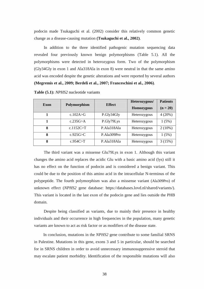
4.3.1.2 Exon 3 (c.412C>T) mutation
Another SRNS patients harbored the transition mutation (c.412C>T) whose
partial electropherograms are indicated in Figure 4.4. This pathogenic nonsense
mutation has been described before. The patient was homozygous for this mutation
which introduces a premature stop codon in place of the amino acid arginine at
position 138 (p.R138X) in the podocin polypeptide.
Figure (4.4): Partial electropherograms for exon 3 (c.412C>T) mutation. The left and right
boxes illustrate the normal and the mutant sequences, respectively (note that the
electropherograms are those of the reverse strands).

33
4.3.1.3 Exon 5 (c.686G>A) mutation
The third mutation detected in our cohort of SRNS patients was the transition
(c.686G>A) in exon 5. The partial electropherograms illustrating this mutation are
indicated in Figure 4.5. This previously reported missense mutation replaces the
amino acid arginine at position 229 by glutamine (p.R229Q). The mutation was
evident in homozygous form.
Figure (4.5): Partial electropherograms for exon 5 (c.686G>A) mutation. The left and right
boxes illustrate the normal and the mutant sequences, respectively.
4.4 Summary of the identified mutations
Three patients comprising 15 % of the study group harbored NPHS2 gene
mutations. Table 4.1 below summarizes the detected mutations.
Table (4.1): NPHS2 sequence mutations identified in Palestinian SRNS patients
Patient
number Exon
Nucleotide
change Effect on protein Genotype
Proteinuria onset
(months)
7 3 c.388G>A p. Glu130Lys Homozygous 2
17 5 c.686G>A p. Arg229Gln Homozygous 45
21 3 c.412C>T p. Arg138X Homozygous 60
4.5 Age of onset of proteinuria and NPHS2 mutations
The disease-causing mutation in NPHS2 was detected in SRNS patients whose
age of onset of proteinuria was <5 years (Table 4.1).

34
4.6 NPHS2 nucleotide variants
Analyses of the obtained exons sequences revealed several nucleotide variants
that proved to be benign as reported in the literature and Ensembl database. The
detected variants and their pertinent characteristics are illustrated in Table 4.2.
Table (4.2): NPHS2 detected variants.
4.7 Renal Histologic Findings
Renal biopsy performed previously on 80% of the recruited patients were as
follows: Focal segmental glomerulosclerosis (FSGS) was seen in 7 (35%), Minimal-
change disease (MCD) was a frequent finding in 6 (30%) and Membrano-
proliferative glomerulonephritis (MPGN) was seen in 3 (15%). These findings were
adapted for patients records and are summarized in Table 4.3.
Table (4.3): Renal Histologic findings
Renal Biopsy Patients (n = 16)
FSGS 7 (35%)
MCD 6 (30%)
MPGN 3 (15%)
Exon Polymorphism Effect Heterozygous/
Homozygous
Patients
(n = 20)
1 c.102A>G P.Gly34Gly Heterozygous 4 (20%)
1 c.235G>A P.Gly79Lys Heterozygous 1 (5%)
8 c.1152C>T P.Ala318Ala Heterozygous 2 (10%)
8 c.925G>C P.Ala309Pro Heterozygous 1 (5%)
8 c.954C>T P.Ala318Ala Heterozygous 3 (15%)

35
Chapter 5
Discussion

36
Chapter 5
Discussion
Identification of mutations associated with genetic disorders is important
because it clarifies the cause of the disease, influences physicians’ decision on
patient treatment and management, and helps the patient's family take very important
decisions for their future progeny. Moreover, as in the case of SRNS, patients
harboring pathogenic mutations can be spared the side effects of useless
immunosuppressive therapy and could be considered for kidney transplantation.
This study, the first of its kind in Palestine, was conducted in order to identify
NPHS2 gene mutations in 20 SRNS patients residing in Gaza strip. Based on our
results, the incidence of podocin mutations in the examined cohort approached 15%.
This figure is comparable to the NPHS2 mutation detection rate reported from
studies on Turkish and Brazilian SRNS children (Guaragna et al., 2015; Kitamura
et al., 2006) but lower than that documented in European and American children
(Kitamura et al., 2006; Caridi, Perfumo, & Ghiggeri, 2005; Weber et al., 2004;
Caridi et al., 2003). Interestingly, no pathogenic NPHS2 mutations were found in
Japanese and Korean SRNS children (Kitamura et al., 2006). This controversy
could be due to the high heterogeneity in the nephrotic syndrome and the associated
genetic loci (so far more than 40 loci have been implicated in childhood nephrotic
syndrome (Ha, 2017)) and reflects ethnic differences in disease genes.
In the SRNS patients examined in this study we could identify three pathogenic
genotypes (all in homozygous form) comprising three different NPHS2 mutations
(E130K, R138X, and R229Q) in 3 out of the 20 investigated familial SRNS patients.
It is important to indicate here that all the three affected individuals were the
offspring of consanguineous parents.
The first mutation (E130K) was encountered in homozygous form in male
patient #7. This missense mutation results from a G to A transition in the coding
region of exon 3 at position 388. The mutation was also evident in the affected
brother of the patient. The mutation replaces an acidic amino acid (glutamic acid)
with a basic amino acid (lysine) in the podocin polypeptide. Obviously, the two

37
amino acids have different characteristics and is expected to impair the function of
the podocin. Moreover, this amino acid is among the amino acids located in the PHB
(prohibitin homology). PHB encompasses the amino acids 125 to 284 and is required
for the binding of podocin to nephrin (Huber et al., 2003). This mutation was
reported before by Kari et al. (2015) who identified this mutation in homozygous
form in two patients of Arabic origin (Sadowski et al., 2015). The familial and
autosomal recessive nature of this mutation was confirmed by detecting the same
genotype in the affected brother of the patient.
The second mutation (R138X) was detected in homozygous form in female
patient #21. This nonsense mutation is due to a C to T transition at nucleotide 412 in
exon 3. The mutation was also evident in the affected sister of the patient. The
genetic alteration in this mutation introduces a premature (TGA) stop codon in place
of the amino acid arginine at position 138 in the podocin sequence thus leading to the
production of a non-functional truncated polypeptide. This amino acid is also located
in the PHB domain of podocin and was shown to be crucial for the function of
podocin (Huber et al., 2003). R138X mutation was first described by Boute et al.
(2000) (Boute et al., 2000a). Interestingly, this mutation has been shown by
Frishberg et al. (2002) to be prevalent among Arab SRNS patients (where 15 out of
their 27 patients i.e., 55% harbored this mutation in homozygous form) a result
which made the authors raise the possibility of this mutation as being a founder
mutation among Arabs (Frishberg et al., 2002). This same mutation was also
reported by Becker-Cohen et al. (2007) in homozygous form, again in a girl of
Arabic descent (Becker‐Cohen et al., 2007), further potentiating the importance of
this mutation in patients of Arab ethnicity.
The third mutation R229Q was documented in homozygous form in meal
patient # 17 who also has an affected sister. This missense mutation occurs as a result
of G to A transition at nucleotide 686 in exon 5 and leads to replacing the amino acid
arginine by glutamine. Exon 5 also constitutes part of the PHB domain and the
missense mutation is expected to influence the function of podocin. R229Q is the
most frequently encountered mutation in diverse populations (Tsukaguchi et al.,
2002). Based on the conservation of Arg 229 residue in podocin homologs,
segregation of the mutation with the disease, and functional study of the mutated
38
podocin made Tsukaguchi et al. (2002) consider this relatively common genetic
change as a disease-causing mutation (Tsukaguchi et al., 2002).
In addition to the three identified pathogenic mutation sequencing data
revealed four previously known benign polymorphisms (Table 5.1). All the
polymorphisms were detected in heterozygous form. Two of the polymorphism
(Gly34Gly in exon 1 and Ala318Ala in exon 8) were neutral in that the same amino
acid was encoded despite the genetic alterations and were reported by several authors
(Megremis et al., 2009; Berdeli et al., 2007; Franceschini et al., 2006).
Table (5.1): NPHS2 nucleotide variants
Exon Polymorphism Effect Heterozygous/
Homozygous
Patients
(n = 20)
1 c.102A>G P.Gly34Gly Heterozygous 4 (20%)
1 c.235G>A P.Gly79Lys Heterozygous 1 (5%)
8 c.1152C>T P.Ala318Ala Heterozygous 2 (10%)
8 c.925G>C P.Ala309Pro Heterozygous 1 (5%)
8 c.954C>T P.Ala318Ala Heterozygous 3 (15%)
The third variant was a missense Glu79Lys in exon 1. Although this variant
changes the amino acid replaces the acidic Glu with a basic amino acid (lys) still it
has no effect on the function of podocin and is considered a benign variant. This
could be due to the position of this amino acid in the intracellular N-terminus of the
polypeptide. The fourth polymorphism was also a missense variant (Ala309Pro) of
unknown effect (NPHS2 gene database: https://databases.lovd.nl/shared/variants/).
This variant is located in the last exon of the podocin gene and lies outside the PHB
domain.
Despite being classified as variants, due to mainly their presence in healthy
individuals and their occurrence in high frequencies in the population, many genetic
variants are known to act as risk factor or as modifiers of the disease state.
In conclusion, mutations in the NPHS2 gene contribute to some familial SRNS
in Palestine. Mutations in this gene, exons 3 and 5 in particular, should be searched
for in SRNS children in order to avoid unnecessary immunosuppressive steroid that
may escalate patient morbidity. Identification of the responsible mutations will also

39
help the affected families in planning for carrier screening, prenatal and even pre-
implantation genetic diagnosis. Further genetic work should be done for NPHS2-
negative families in order to identify the mutations in other genes (e.g., NPHS1,
WT1, and PLCE1) strongly implicated in the pathogenesis of SRNS.

40
Chapter 6
Conclusion &
Rrecommendations

41
Chapter 6
Conclusion & Recommendations
6.1 Conclusion
The current study reports the identification of NPHS2 mutations in 3 out of 20
(15%) familial SRNS patients in Palestinian cohort. The mutations were identified
through nucleotide sequencing of the coding exons and the exon-intron boundaries of
the NPHS2 gene. Analysis of DNA sequences revealed the following, previously
known, pathogenic mutations: G130K, R138X, and R229Q. All detected mutations
were encountered in homozygous form, thus confirming the autosomal recessive
inheritance pattern of the disease. Knowledge of gene mutations associated with
SRNS spares patients from unnecessary and harmful immunosuppressive steroids
and helps physicians and patients' families take proper decisions regarding patient
management and their future offspring.
6. 2 Recommendations
1. Starting genetic studies on nephrotic syndrome patients with screening the
identified mutations, and sequencing of NPHS2 exon 3 and 5.
2. Searching for pathogenic mutations in other SRNS-related genes, such as NPHS1,
PLCE1 and WT1.
3. Providing awareness for patients' families, in whom mutations were identified,
about the available options to avoid having affected children such as prenatal
diagnosis and pre-implantation genetic diagnosis.

42
References
43
References
Abeyagunawardena, A. S., Sebire, N. J., Risdon, R. A., Dillon, M. J., Rees, L., van’t Hoff, W., . . . Trompeter, R. S. (2007). Predictors of long-term outcome of children with
idiopathic focal segmental glomerulosclerosis. Pediatric Nephrology, 22(2), 215-221.
Al-Hamed, M. H., Al-Sabban, E., Al-Mojalli, H., Al-Harbi, N., Faqeih, E., Al Shaya, H., . . . Edwards, N. (2013). A molecular genetic analysis of childhood nephrotic syndrome
in a cohort of Saudi Arabian families. J Hum Genet, 58(7), 480-489.
Ballermann, B. J. (2005). Glomerular endothelial cell differentiation. Kidney international, 67(5), 1668-1671.
Becker‐Cohen, R., Bruschi, M., Rinat, C., Feinstein, S., Zennaro, C., Ghiggeri, G., & Frishberg, Y. (2007). Recurrent Nephrotic Syndrome in Homozygous Truncating NPHS2
Mutation Is Not Due to Anti‐Podocin Antibodies. American journal of transplantation, 7(1), 256-260.
Berdeli, A., Mir, S., Yavascan, O., Serdaroglu, E., Bak, M., Aksu, N., . . . Yildiz, N. (2007). NPHS2 (podicin) mutations in Turkish children with idiopathic nephrotic syndrome.
Pediatric Nephrology, 22(12), 2031-2040.
Bouchireb, K., Boyer, O., Gribouval, O., Nevo, F., Huynh‐Cong, E., Morinière, V., . . . Dantal, J. (2014). NPHS2 Mutations in Steroid‐Resistant Nephrotic Syndrome: A Mutation
Update and the Associated Phenotypic Spectrum. Human mutation, 35(2), 178-186.
Boute, N., Gribouval, O., Roselli, S., Benessy, F., Lee, H., Fuchshuber, A., . . . Antignac, C. (2000a). NPHS2, encoding the glomerular protein podocin, is mutated in autosomal
recessive steroid-resistant nephrotic syndrome. Nature genetics, 24(4).
Boute, N., Gribouval, O., Roselli, S., Benessy, F., Lee, H., Fuchshuber, A., . . . Antignac, C. (2000b). NPHS2, encoding the glomerular protein podocin, is mutated in autosomal
recessive steroid-resistant nephrotic syndrome. Nature genetics, 24(4), 349.
Bullich, G., Trujillano, D., Santín, S., Ossowski, S., Mendizábal, S., Fraga, G., . . . Torra, R. (2015). Targeted next-generation sequencing in steroid-resistant nephrotic
syndrome: mutations in multiple glomerular genes may influence disease severity. European Journal of Human Genetics, 23(9), 1192.
Caridi, G., Bertelli, R., Di Duca, M., Dagnino, M., Emma, F., Muda, A. O., . . . Murer, L. (2003). Broadening the spectrum of diseases related to podocin mutations. Journal of the
American Society of Nephrology, 14(5), 1278-1286.
Caridi, G., Perfumo, F., & Ghiggeri, G. M. (2005). NPHS 2(Podocin) Mutations in Nephrotic Syndrome. Clinical Spectrum and Fine Mechanisms. Pediatric research, 57(5), 54R.
Chugh, S. S., Clement, L. C., & Macé, C. (2012). New insights into human minimal change disease: lessons from animal models. American Journal of Kidney Diseases, 59(2),
284-292.
Eddy, A. A., & Symons, J. M. (2003). Nephrotic syndrome in childhood. The Lancet, 362(9384), 629-639.

44
Ehrich, J., & Brodehl, J. (1993). Long versus standard prednisone therapy for initial treatment of idiopathic nephrotic syndrome in children. European journal of
pediatrics, 152(4), 357-361.
Franceschini, N., North, K. E., Kopp, J. B., Mckenzie, L., & Winkler, C. (2006). NPHS2 gene, nephrotic syndrome and focal segmental glomerulosclerosis: a HuGE review.
Genetics in Medicine, 8(2), 63-75.
Frishberg, Y., Rinat, C., Megged, O., Shapira, E., Feinstein, S., & Raas-Rothschild, A. (2002). Mutations in NPHS2 encoding podocin are a prevalent cause of steroid-resistant
nephrotic syndrome among Israeli-Arab children. Journal of the American Society of Nephrology, 13(2), 400-405.
Geary, D. F., & Schaefer, F. (2008). Comprehensive Pediatric Nephrology E-Book: Text with CD-ROM: Elsevier Health Sciences.
Gipson, D. S., Massengill, S. F., Yao, L., Nagaraj, S., Smoyer, W. E., Mahan, J. D., . . . Lin, J.-J. (2009). Management of childhood onset nephrotic syndrome. Pediatrics, 124(2),
747-757.
Grahammer, F., Schell, C., & Huber, T. B. (2013). The podocyte slit diaphragm—from a thin grey line to a complex signalling hub. Nature Reviews Nephrology, 9(10), 587-598.
Guaragna, M. S., Lutaif, A. C. G., Maciel-Guerra, A. T., Belangero, V., Guerra-Júnior, G., & De Mello, M. P. (2017). NPHS2 Mutations: A Closer Look to Latin American Countries.
BioMed research international, 2017.
Guaragna, M. S., Lutaif, A. C. G., Piveta, C. S., Souza, M. L., de Souza, S. R., Henriques, T. B., . . . De Mello, M. P. (2015). NPHS2 mutations account for only 15% of nephrotic
syndrome cases. BMC medical genetics, 16(1), 88.
Ha, T.-S. (2017). Genetics of hereditary nephrotic syndrome: a clinical review. Korean journal of pediatrics, 60(3), 55-63.
Hinkes, B. G., Mucha, B., Vlangos, C. N., Gbadegesin, R., Liu, J., Hasselbacher, K., . . . Hildebrandt, F. (2007). Nephrotic syndrome in the first year of life: two thirds of
cases are caused by mutations in 4 genes (NPHS1, NPHS2, WT1, and LAMB2). Pediatrics, 119(4), e907-e919.
http://www. buzzle.com, 2015
http://www.ensembl.org
https://www.ncbi.nlm.nih.gov/blast/
http://www.ensembl.org/Homo_sapiens/Gene/Variation_Gene/Table?db=core;g= ENSG 00000 116218; r=1:179550539-179575952
Huber, T. B., Köttgen, M., Schilling, B., Walz, G., & Benzing, T. (2001). Interaction with podocin facilitates nephrin signaling. Journal of Biological Chemistry, 276(45),
41543-41546.
Huber, T. B., Simons, M., Hartleben, B., Sernetz, L., Schmidts, M., Gundlach, E., . . . Benzing, T. (2003). Molecular basis of the functional podocin–nephrin complex: mutations in
the NPHS2 gene disrupt nephrin targeting to lipid raft microdomains. Human molecular genetics, 12(24), 3397-3405.

45
Ikeuchi, Y., Kobayashi, Y., Arakawa, H., Suzuki, M., Tamra, K., & Morikawa, A. (2009). Polymorphisms in interleukin-4-related genes in patients with minimal change
nephrotic syndrome. Pediatric Nephrology, 24(3), 489.
Jungraithmayr, T. C., Hofer, K., Cochat, P., Chernin, G., Cortina, G., Fargue, S., . . . Neuhaus, T. (2011). Screening for NPHS2 mutations may help predict FSGS recurrence after transplantation. Journal of the American Society of Nephrology, 22(3), 579-585.
Kausman, J. Y., & Kitching, A. R. (2007). A new approach to idiopathic nephrotic syndrome. Journal of the American Society of Nephrology, 18(10), 2621-2622.
Kawaguchi, E., Ishikura, K., Hamada, R., Nagaoka, Y., Morikawa, Y., Sakai, T., . . . Miura, M. (2014). Early and frequent development of ocular hypertension in children with
nephrotic syndrome. Pediatric Nephrology, 29(11), 2165-2171.
Kitamura, A., Tsukaguchi, H., Iijima, K., Araki, J., Hattori, M., Ikeda, M., . . . Yoshikawa, N. (2006). Genetics and clinical features of 15 Asian families with steroid-resistant nephrotic syndrome. Nephrology Dialysis Transplantation, 21(11), 3133-3138.
Kretzler, M. (2002). Regulation of adhesive interaction between podocytes and glomerular basement membrane. Microscopy research and technique, 57(4), 247-253.
Kriz, W., & Kaissling, B. (1992). Structural organization of the mammalian kidney. The kidney: physiology and pathophysiology, 3, 587-654.
Levidiotis, V., & Power, D. A. (2005). New insights into the molecular biology of the glomerular filtration barrier and associated disease. Nephrology, 10(2), 157-166.
Lipska, B. S., Balasz-Chmielewska, I., Morzuch, L., Wasielewski, K., Vetter, D., Borzecka, H., . . . Jarmolinski, T. (2013). Mutational analysis in podocin-associated hereditary
nephrotic syndrome in Polish patients: founder effect in the Kashubian population. Journal of applied genetics, 54(3), 327-333.
Liu, X. L., Doné, S. C., Yan, K., Kilpeläinen, P., Pikkarainen, T., & Tryggvason, K. (2004). Defective trafficking of nephrin missense mutants rescued by a chemical
chaperone. Journal of the American Society of Nephrology, 15(7), 1731-1738.
Lombel, R. M., Gipson, D. S., & Hodson, E. M. (2013). Treatment of steroid-sensitive nephrotic syndrome: new guidelines from KDIGO. Pediatric Nephrology, 28(3), 415-
426.
Löwik, M., Groenen, P., Levtchenko, E., Monnens, L., & Van Den Heuvel, L. (2009a). Molecular genetic analysis of podocyte genes in focal segmental
glomerulosclerosis—a review. European journal of pediatrics, 168(11), 1291-1304.
Löwik, M., Groenen, P., Levtchenko, E., Monnens, L., & Van Den Heuvel, L. (2009b). Molecular genetic analysis of podocyte genes in focal segmental
glomerulosclerosis—a review. European journal of pediatrics, 168(11), 1291.
Machuca, E., Benoit, G., Nevo, F., Tête, M.-J., Gribouval, O., Pawtowski, A., . . . Gubler, M.-C. (2010). Genotype–phenotype correlations in non-Finnish congenital nephrotic syndrome. Journal of the American Society of Nephrology, 21(7), 1209-1217.
McCaffrey, J., Lennon, R., & Webb, N. J. (2016). The non-immunosuppressive management of childhood nephrotic syndrome. Pediatric Nephrology, 31(9), 1383-1402.
Megremis, S., Mitsioni, A., Mitsioni, A. G., Fylaktou, I., Kitsiou-Tzelli, S., Stefanidis, C. J., . . . Traeger-Synodinos, J. (2009). Nucleotide variations in the NPHS2 gene in Greek
46
children with steroid-resistant nephrotic syndrome. Genetic testing and molecular biomarkers, 13(2), 249-256.
Nephrologie, A. F. P. (1979). Alternate day versus intermittent prednisone in frequently relapsing nephrotic syndrome. Lancet, 1, 401-403.
Otukesh, H., Ghazanfari, B., Fereshtehnejad, S.-M., Bakhshayesh, M., Hashemi, M., Hoseini, R., . . . Rahiminia, A. (2009). NPHS2 mutations in children with steroid-resistant
nephrotic syndrome. Iranian journal of kidney diseases, 3(2).
Otukesh, H., Otukesh, S., Mojtahedzadeh, M., Hoseini, R., Fereshtehnejad, S.-M., Riahi Fard, A., . . . Hooman, N. (2009). Management and outcome of steroid-resistant
nephrotic syndrome in children. Iran J Kidney Dis, 3(4), 210-217.
Patrakka, J., Lahdenkari, A.-T., Koskimies, O., Holmberg, C., Wartiovaara, J., & Jalanko, H. (2002). The number of podocyte slit diaphragms is decreased in minimal change
nephrotic syndrome. Pediatric research, 52(3), 349-355.
Pavenstädt, H., Kriz, W., & Kretzler, M. (2003). Cell biology of the glomerular podocyte. Physiological reviews, 83(1), 253-307.
Pollak, M. R., Quaggin, S. E., Hoenig, M. P., & Dworkin, L. D. (2014). The Glomerulus: The Sphere of Influence. Clinical Journal of the American Society of Nephrology : CJASN,
9(8), 1461-1469. doi: 10.2215/CJN.09400913
Rodewald, R., & Karnovsky, M. J. (1974). Porous substructure of the glomerular slit diaphragm in the rat and mouse. The Journal of Cell Biology, 60(2), 423.
Roselli, S., Gribouval, O., Boute, N., Sich, M., Benessy, F., Attié, T., . . . Antignac, C. (2002). Podocin localizes in the kidney to the slit diaphragm area. The American journal of
pathology, 160(1), 131-139.
Roselli, S., Heidet, L., Sich, M., Henger, A., Kretzler, M., Gubler, M.-C., & Antignac, C. (2004). Early glomerular filtration defect and severe renal disease in podocin-deficient
mice. Molecular and cellular biology, 24(2), 550-560.
Ruf, R. G., Lichtenberger, A., Karle, S. M., Haas, J. P., Anacleto, F. E., Schultheiss, M., . . . Mucha, B. (2004). Patients with mutations in NPHS2 (podocin) do not respond to
standard steroid treatment of nephrotic syndrome. Journal of the American Society of Nephrology, 15(3), 722-732.
Rüth, E.-M., Kemper, M. J., Leumann, E. P., Laube, G. F., & Neuhaus, T. J. (2005). Children with steroid-sensitive nephrotic syndrome come of age: long-term outcome. The
Journal of pediatrics, 147(2), 202-207.
Sadowski, C. E., Lovric, S., Ashraf, S., Pabst, W. L., Gee, H. Y., Kohl, S., . . . Halbritter, J. (2015). A single-gene cause in 29.5% of cases of steroid-resistant nephrotic
syndrome. Journal of the American Society of Nephrology, 26(6), 1279-1289.
Saleem, M. A. (2013). New developments in steroid-resistant nephrotic syndrome. Pediatric Nephrology, 28(5), 699-709.
Santín, S., Tazón-Vega, B., Silva, I., Cobo, M. Á., Giménez, I., Ruíz, P., . . . Ars, E. (2010). Clinical value of NPHS2 analysis in early-and adult-onset steroid-resistant nephrotic syndrome. Clinical Journal of the American Society of Nephrology, CJN. 03770410.
47
Schwarz, K., Simons, M., Reiser, J., Saleem, M. A., Faul, C., Kriz, W., . . . Mundel, P. (2001). Podocin, a raft-associated component of the glomerular slit diaphragm, interacts
with CD2AP and nephrin. The Journal of clinical investigation, 108(11), 1621-1629.
Sellin, L., Huber, T. B., Gerke, P., Quack, I., Pavenstädt, H., & Walz, G. (2003). NEPH1 defines a novel family of podocin interacting proteins. The FASEB Journal, 17(1), 115-117.
Sun, H., Zhou, W., Wang, J., Yin, L., Lu, Y., & Fu, Q. (2009). A novel mutation in NPHS2 gene identified in a Chinese pedigree with autosomal recessive steroid-resistant
nephrotic syndrome. Pathology, 41(7), 661-665.
Tanaka, M. (2008). The twelve large colour prints of William Blake: a study on techniques, materials and context: Nottingham Trent University (United Kingdom).
Trautmann, A., Bodria, M., Ozaltin, F., Gheisari, A., Melk, A., Azocar, M., . . . Gellermann, J. (2015). Spectrum of steroid-resistant and congenital nephrotic syndrome in
children: the PodoNet registry cohort. Clinical Journal of the American Society of Nephrology, CJN. 06260614.
Trompeter, R., Hicks, J., Lloyd, B., White, R., & Cameron, J. (1985). Long-term outcome for children with minimal-change nephrotic syndrome. The Lancet, 325(8425), 368-
370.
Tsukaguchi, H., Sudhakar, A., Le, T. C., Nguyen, T., Yao, J., Schwimmer, J. A., . . . Appel, G. B. (2002). NPHS2 mutations in late-onset focal segmental glomerulosclerosis: R229Q is
a common disease-associated allele. The Journal of clinical investigation, 110(11), 1659.
Vivarelli, M., Moscaritolo, E., Tsalkidis, A., Massella, L., & Emma, F. (2010). Time for initial response to steroids is a major prognostic factor in idiopathic nephrotic syndrome.
The Journal of pediatrics, 156(6), 965-971.
Weber, S., Gribouval, O., Esquivel, E. L., Morinière, V., Tête, M.-J., Legendre, C., . . . Antignac, C. (2004). NPHS2 mutation analysis shows genetic heterogeneityof
steroid-resistant nephrotic syndrome and lowpost-transplant recurrence. Kidney international, 66(2), 571-579.
WEENING, J. J. (2004). Role of the immune system in the pathogenesis of idiopathic nephrotic syndrome. Clinical Science, 107(2), 125-136.

48
Appendix