Embed Size (px)
Citation preview

Pathophysiology of
Type 1 Diabetes
2010. 10.
가톨릭대학교 의과대학 소아과학교실
정 민 호

Introduction
� Type 1A diabetes
� one of >80 diseases with autoimmune etiology
� organ-specific immune destruction of β-cells in the islets of
Langerhans within the pancreas
� The β-cells: elegant glucose ‘thermostat’� The β-cells: elegant glucose ‘thermostat’
� sensing glucose
� releasing insulin
� Pathogenesis
� Genetic predisposition
� Environmental factors

Schematic Representation of Glucose Metabolism

HIGH PLASMA INSULIN
(POSTPRANDIAL STATE)
LOW PLASMA INSULIN
(FASTED STATE)
Liver Glucose uptake Glucose production
Glycogen synthesis Glycogenolysis
Absence of gluconeogenesis Gluconeogenesis
Lipogenesis Absence of lipogenesis
Absence of ketogenesis Ketogenesis
Influence of Feeding (High Insulin) or of Fasting (Low Insulin) on some Metabolic Processes in Liver, Muscle, and Adipose Tissue
Absence of ketogenesis Ketogenesis
Muscle Glucose uptake Absence of glucose uptake
Glucose oxidation Fatty acid and ketone oxidation
Glycogen synthesis Glycogenolysis
Protein synthesis Proteolysis and amino acid release
Adipose tissue Glucose uptake Absence of glucose uptake
Lipid synthesis Lipolysis and fatty acid release
Triglyceride uptake Absence of triglyceride uptake

Histopathology of Type 1 Diabetes
� Selective destruction of β-cells within islets
� Heterogeneity of islet lesions
� a normal islet with no infiltrate
� an islet containing β-cells with intense infiltrate
� a pseudoatrophic islet� a pseudoatrophic islet
� Islets of patients with type 1A diabetes
� overexpress class I HLA antigens
� relatively rarely express class II HLA on β-cells
� express IFN-α, up-regulate Fas molecules on all islet cells

Islet invasion by lymphocytes of NOD mice is asynchronous during progression of diabetes.
Bluestone JA, et al, Nature 2010

Incidence Rates of Type 1 DM

Descriptive Genetics
Proband with DM % Childhood DM Islet AutoAb
General population, US 0.3% 3%, single Ab
(15-25/100,000) 0.3% multiple Ab
Risk of Type 1A Diabetes
(15-25/100,000) 0.3% multiple Ab
Offspring 1% 4.1%
Sibling 3.2% 7.4%
6% lifetime
Dizygotic twin 6% 10%
Mother 2% 5%
Father 4.6% 6.5%
Both parents 10%? ?
Monozygotic twin 50%, variable 50%

Progression to Diabetes of Initially Discordant Monozygotic Twins of Patients with T1D
Diabetes risk
Environmental factors� Environmental factors
� Non-germ line-inherited variations
• imprinting
• T-cell receptor polymorphisms
• somatic mutations
Redondo MJ, et al, Diabetologia 2001

MHC class II molecules are requiredfor the education of thymic precursors and for the restriction of CD4 T cells responses.
Casares S, et al, Curr Molec Med 2001
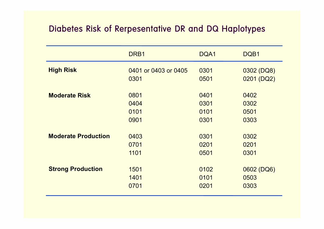
Diabetes Risk of Rerpesentative DR and DQ Haplotypes
DRB1 DQA1 DQB1
0401 or 0403 or 0405 0301 0302 (DQ8)
0301 0501 0201 (DQ2)
0801 0401 0402
0404 0301 0302
0101 0101 0501
High Risk
Moderate Risk
0101 0101 0501
0901 0301 0303
0403 0301 0302
0701 0201 0201
1101 0501 0301
1501 0102 0602 (DQ6)
1401 0101 0503
0701 0201 0303
Moderate Production
Strong Production

Percentage of Specific HLA Alleles or Haplotypes in the Patients with Type 1 Diabetes and Normal Controls in Korea
Yu J, et al, Clin Immunol 2004

Association between HLA Class II and Type 1 Diabetes
� A correlation between the binding affinity of the
peptide to MHC class II and antigenicity� Protective class II molecules
: bind self-peptides with high affinity � delete thymic precursors
� Susceptible class II molecules
: bind self-peptides with low affinity � a failure of central tolerance
(escape of self-reactive T cells to periphery)
� Peptide processing and stability of HLA-DR-peptide
complexes

Putative Functions of Non-HLA-Associated Loci in Type 1 Diabetes
Concannon P, et al, N Engl J Med 2009

Differential Roles of Risk Loci in the Pathogenesis of Type 1 Diabetes
Concannon P, et al,
N Engl J Med 2009

Environmental Factors
� Infections
� Congenital rubella infection
� Enteroviruses
� Rotavirus
Vaccination (?)� Vaccination (?)
� Diet
� Early introduction of bovine milk/cereals
� Vitamin D and its analogues, omega-3 fatty acids
� Meat preservatives/N-nitroso compounds
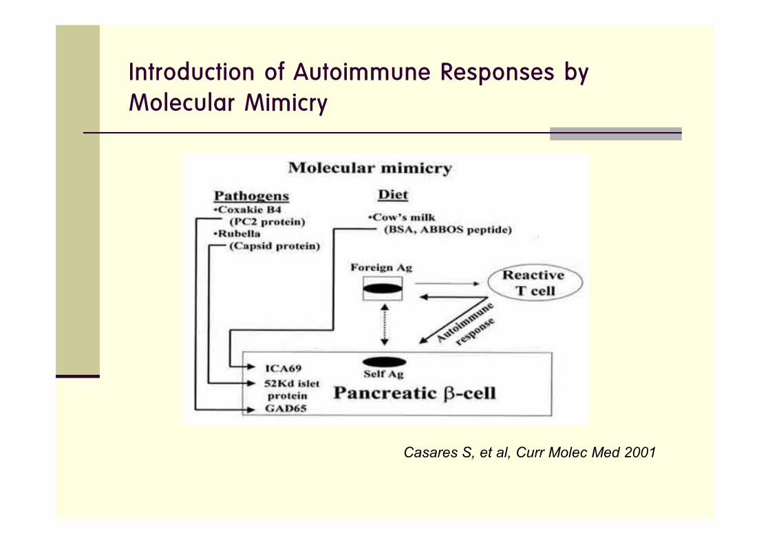
Introduction of Autoimmune Responses by Molecular Mimicry
Casares S, et al, Curr Molec Med 2001

Pathophysiology of Type 1 Diabetes
Genetic background Environmental factorsHuman IDDM1-IDDM15 susceptibility genes Dietary factors Drugs
Mouse Idd1-19 susceptibility genes Viruses
Breakdown of self-tolerance
Immune attack on pancreatic β-cellsAutoantigens: GAD65, proinsulin, insulin hsp60,
Tyrosine phosphatase IA-2, ICA69
DIABETES
Casares S, et al, Curr Molec Med 2001

Hypothetical Stages in the Development of Type 1A Diabetes
Eisenbarth GS, N Engl J Med 1986
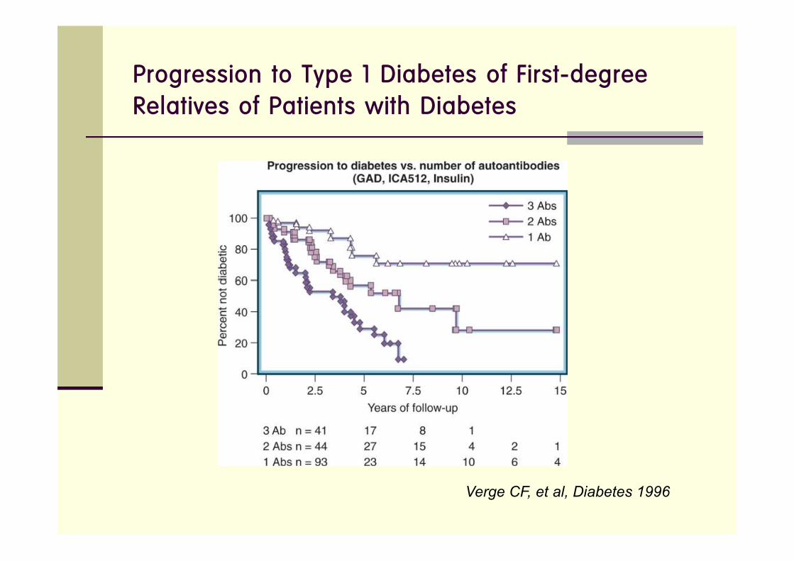
Progression to Type 1 Diabetes of First-degree Relatives of Patients with Diabetes
Verge CF, et al, Diabetes 1996

Reversal of β-Cell Suppression In Vitro in Pancreatic Islets Isolated from Nonobese Diabetic Mice during the Phase Preceding Insulin-depedent Diabetes Mellitus
Strandell E, et al, J Clin Invest 1990

The Role of T Cells in the Pathogenesis of Type 1 DMIn animal models: NOD mice, BB rats, transgenic mice
� Neonatal thymectomy prevents the disease.
� Transplantation of isolated isles into the thymus induce T cell
tolerance and prevention of disease.
� The disease can be transferred by autoreactive bone marrow-
derived cells, splenic T cells, or T cell clones isolated from derived cells, splenic T cells, or T cell clones isolated from
infiltrated islets.
� Adoptive transfer of protective T cells prevent diabetes.
� NOD nu/nu (lacking of T cells) do not develop diabetes.
� NOD nu/nu mice expressing monoclonal diabetogenic TCRs
develop inulitis and diabetes.

The Autoimmune Response against the Pancreatic β-cells
1. Environmental insult
2. Priming of T cells
3. T cell differentiation
4. β-cell destruction
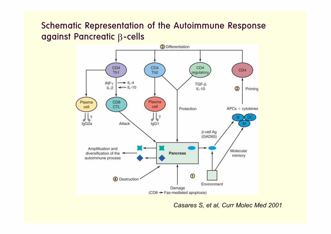
Schematic Representation of the Autoimmune Response against Pancreatic β-cells
Casares S, et al, Curr Molec Med 2001

Antigen and specific cytokines signals induce differentiation of naive T
cells into various subsets of T helper cells (Th1, Th2, Th17).

Activation of IL-17 immunity in circulating memory cells in T1D
Honkanen J, et al,
J Immunol 2010
Martin-Orozco N, et al,
Eur J Immunol 2009

The Fine Balance of Immune Regulation versus Pathogenesis
Bluestone JA, et al, Nature 2010

Critical Pathways Known to Contribute to the Pathogenesis of Type 1 Diabetes and Relevant Drugs for Intervention
Matthews JB, et al, Clin Exp Immunol 2010
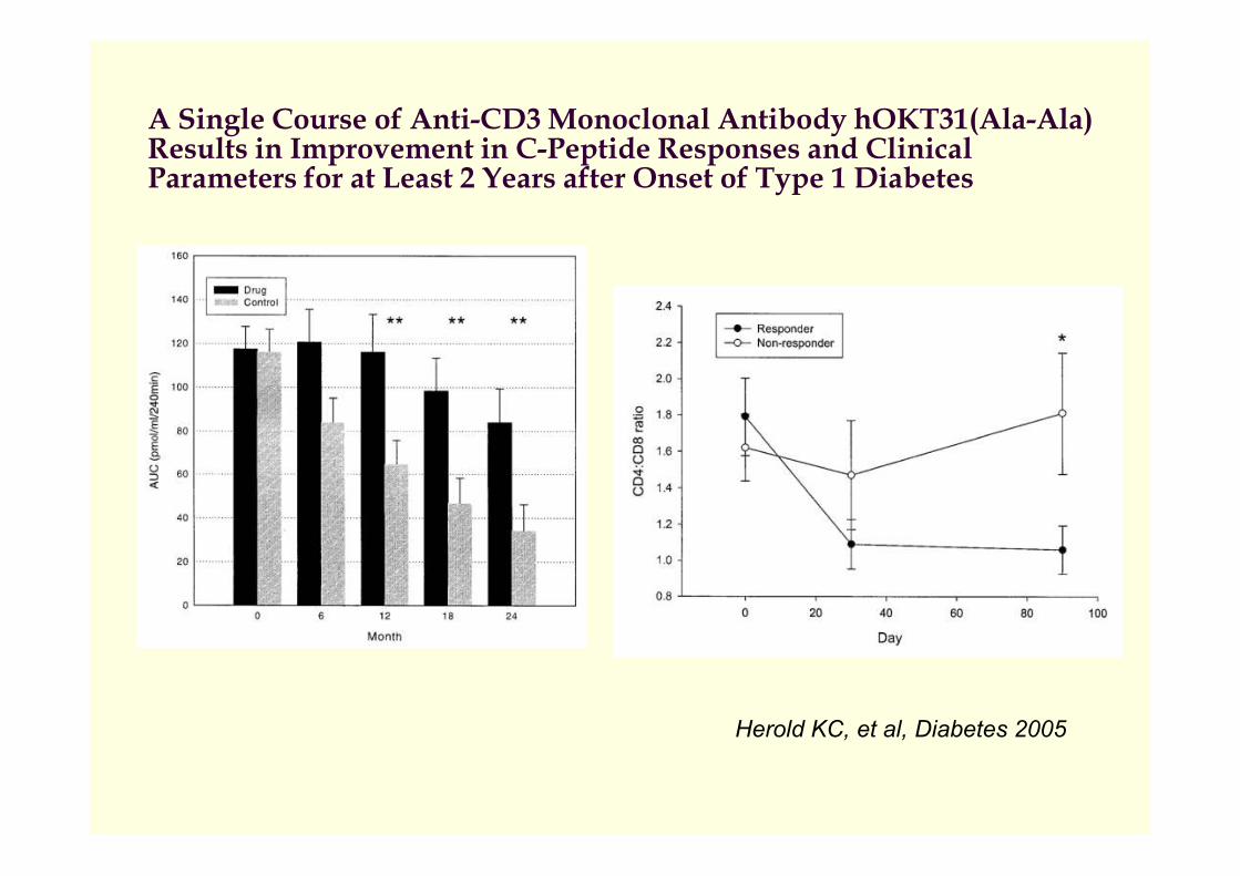
A Single Course of Anti-CD3 Monoclonal Antibody hOKT31(Ala-Ala) Results in Improvement in C-Peptide Responses and Clinical Parameters for at Least 2 Years after Onset of Type 1 Diabetes
Herold KC, et al, Diabetes 2005

Biological Effects of Anti-CD3 Antibodies on T Cells : Physical Elimination and Antigenic Modulation
Chatenoud L, Nature Rev Endocrinol 2010

Summary
� The genetic background (HLA and non-HLA genes) and
environmental factors (pathogens, drugs and diet) are critical for
the initiation of the autoimmune response against the pancreatic
β-cells.
� The role of T cells in the pathogenesis of type 1 diabetes has � The role of T cells in the pathogenesis of type 1 diabetes has
been demonstrated in humans and animal models.
� Increased understanding of the pathogenesis and the
identification of genes and environmental factors that control
disease incidence will provide a wealth of potential targets of
disease intervention.


![Prefabricated Ducts - alnor.com.pl · Galvanisation - type of metal, poliuretan - type of fabric type fabric width A [mm] ... Galvanized pipe RURA and bolts WKD create a set of elements](https://img.pdfslide.tips/doc/110x75/5ad517ad7f8b9a1a028ca967/prefabricated-ducts-alnorcompl-type-of-metal-poliuretan-type-of-fabric.jpg)
![Enhancement of ceramide formation increases endocytosis of ......Cytokine production differs in both type and magnitude dependent on the type of microbial stimulation [1,2]. The type](https://img.pdfslide.tips/doc/110x75/5f33e885a4573a2325398318/enhancement-of-ceramide-formation-increases-endocytosis-of-cytokine-production.jpg)