Embed Size (px)
Citation preview
Pneumocystis carinii Cell Wall β-Glucan Induces Release of Macrophage
Inflammatory Protein-2 from Alveolar Epithelial Cells via a Lactosylceramide
Mediated Mechanism1
Peter Y. Hahn, MD*
Scott E. Evans, MD*
Theodore J. Kottom, MS*
Joseph E. Standing, BS*
Richard E. Pagano, PhD*,†
Andrew H. Limper, MD*,†,†--
*Thoracic Diseases Research Unit, Division of Pulmonary, Critical Care, and Internal Medicine,
and the †Department of Biochemistry and Molecular Biology, Mayo Clinic and Foundation,
Rochester, Minnesota, 55905, USA.
†--Address reprint requests and correspondence to Dr. Andrew Limper, Thoracic Diseases Research
Unit, 8-24 Stabile Building, Mayo Clinic and Foundation, Rochester, MN 55905,
Copyright 2002 by The American Society for Biochemistry and Molecular Biology, Inc.
JBC Papers in Press. Published on November 4, 2002 as Manuscript M209715200 by guest on February 27, 2019
http://ww
w.jbc.org/
Dow
nloaded from
Phone (507) 284-2964, Fax (507) 284-4521; E-mail: [email protected].
Running Title: P. carinii β-glucan induces alveolar epithelial cell MIP-2
Keywords: Pneumocystis, β-Glucans, Lipid Mediators, Inflammation, Lung, AIDS
Hahn/Limper-2
by guest on February 27, 2019http://w
ww
.jbc.org/D
ownloaded from
ABSTRACT
Infiltration of the lungs with neutrophils promotes respiratory failure during severe
Pneumocystis carinii (PC) pneumonia. Recent studies have shown that alveolar epithelial cells
(AECs), in addition to promoting PC attachment, also participate in lung inflammation by the
release of cytokines and chemokines. Herein, we demonstrate that a PC β-glucan rich cell wall
isolate (PCBG) stimulates the release of macrophage inflammatory protein-2 (MIP-2) from
isolated AEC’s through a lactosylceramide dependent mechanism. The results demonstrate that
MIP-2 mRNA and protein production is significantly increased at both early and late time points
after PCBG challenge. Although CD11b/CD18 (Mac-1, CR3) is the most widely studied β-
glucan receptor, we demonstrate that CD11b/CD18 is not present on AECs. This study instead
demonstrates that pre-incubation of AECs with an antibody directed against the membrane
glycosphingolipid lactosylceramide (CDw17) results in a significant decrease in MIP-2
secretion. Preincubation of the anti-CDw17 antibody with solubilized lactosylceramide reverses
this effect. Furthermore, incubation of AEC’s with inhibitors of glycosphingolipid biosynthesis,
including N-butyldeoxynojirimycin (NB-DNJ) and D-threo-1-phenyl-2-decanoylamino-3-
morpholino-1-propanol-HCl (PDMP), also results in a significant decrease in AEC MIP-2
production following challenge with PCBG. These data demonstrate that PC β-glucan induces
significant production of MIP-2 from AEC’s and that CDw17 participates in the glucan-induced
inflammatory signaling in lung epithelial cells during PC infection.
Hahn/Limper-3
by guest on February 27, 2019http://w
ww
.jbc.org/D
ownloaded from
INTRODUCTION
Pneumocystis carinii (PC) pneumonia remains a life threatening opportunistic infection in
immunocompromised hosts.2 Despite availability of effective medications, the mortality from
PC pneumonia ranges between 15% and 40% (1). Neutrophilic lung inflammation promotes
diffuse alveolar damage and contributes significantly to the respiratory failure, which is
characteristic of severe PC pneumonia (2). The exact mechanisms by which PC induce this lung
inflammation are not fully understood, though recent studies indicate that β-glucan components
of the organism cell wall drive neutrophil accumulation during this pneumonia (3).
P. carinii has recently been classified as a fungal organism based on RNA homology and
evidence that PC assembles a cell wall rich in β-glucans (4,5). Fungal β-glucans consisting of
1,3-linked β-D-glucopyranosyl residues with varying amounts of 1,6-linked β-D-pyranosyl
side chains represent essential structural components of many fungal cell walls (6,7). Purified β-
glucans from various fungal species strongly induce the production of proinflammatory
cytokines and chemokines from inflammatory cells (8-10). Previous studies demonstrate that
the PC cyst cell wall contains complex carbohydrates rich in β-glucans, chitin and a mannose-
rich major surface glycoprotein complex termed gpA (5,7). We have recently purified
particulate β-glucan from P. carinii and have demonstrated that alveolar macrophages produce
tumor necrosis factor-alpha (TNFα) and macrophage inflammatory protein-2 (MIP-2) in
response to this PC cell wall component (3,11).
Hahn/Limper-4
by guest on February 27, 2019http://w
ww
.jbc.org/D
ownloaded from
Fungal glucans have been shown to bind several different receptors. In particular, the β2
integrin CD11b/CD18 (CR3, Mac-1) has been extensively studied as a major leukocyte
membrane receptor for β-glucans (12,13). CD11b/CD18 possesses one or multiple lectin
binding sites on the C-terminal domain of the CD11b/CD18 α subunit capable of interacting
with complex carbohydrates (14). Additional studies using leukocytes have further demonstrated
that the cell membrane glycosphingolipid, lactosylceramide (CDw17), also interacts with β-
glucans (15). Interaction of glucans with CDw17 has also been reported to promote
enhancement of the oxidative burst response and NF-kB activation of neutrophils (16).
Histological studies consistently identify P. carinii organisms closely associated with the
alveolar epithelium in infected human and animal tissues. Furthermore, the binding of P. carinii
to alveolar epithelial cells is believed to be essential for establishment of infection (17).
However, accumulating evidence demonstrates that pulmonary alveolar epithelial cells not only
function as a passive barrier mediating gas exchange, but also actively participate in the host
immune response. Alveolar epithelial cells are capable of processing and presenting antigen to T
lymphocytes (18,19). Furthermore, isolated alveolar epithelial cells produce and secrete various
cytokines and chemokines including TNFα and MIP-2 when stimulated with bacterial
components like lipopolysaccharide (LPS) or whole organisms such as Mycobacterium
tuberculosis (20-23).
MIP-2 is of particular interest in the pathogenesis of P. carinii pneumonia since it
Hahn/Limper-5
by guest on February 27, 2019http://w
ww
.jbc.org/D
ownloaded from
represents the rodent homologue of the human C-X-C chemokine IL-8 and is thus a potent
stimulant of neutrophil accumulation and activation (24-26). Although chemokine expression is
an important component of the inflammatory response against PC pneumonia, exuberant
expression can cause deleterious effects on respiratory system function. Bronchoalveolar lavage
studies in humans demonstrate that greater degrees of lung inflammation, as evidenced by
increased neutrophil burdens, are associated with worse oxygenation and increased mortality
during P. carinii pneumonia (2).
The current study was therefore undertaken to determine the extent to which
Pneumocystis carinii cell wall β-glucans, induce the expression of MIP-2 from primary AEC’s.
We further sought to determine whether the mechanisms of AEC activation by PC β-glucans
was mediated through CD11b/CD18, or through the alternate lactosylceramide (CDw17)
associated β-glucan receptor. Enhanced local production of MIP-2 by AEC’s may significantly
promote neutrophilic lung inflammation that characterizes severe Pneumocystis carinii
pneumonia and contributes to respiratory failure in this infection.
Hahn/Limper-6
by guest on February 27, 2019http://w
ww
.jbc.org/D
ownloaded from
MATERIALS AND METHODS
Materials. Unless otherwise noted, general reagents were from Sigma Chemical Co. (St.
Louis, MO). P. carinii f. sp. (formae specialis) carinii was originally obtained through the
American Type Culture Collection (ATCC) and maintained in our immunosuppressed rat colony.
The following antibodies were used: Anti-CD11b (M-19, Santa Cruz Inc., Santa Cruz, CA.),
FITC anti-CD45 (OX-1, Pharmingen, San Diego, CA.), Anti-CDw17 (Huly-M13, Ancell,
Bayport, MN), Anti-asialoGM1 (Matreya, State College, PA.). We recently described the
purification and characterization of a β-glucan rich cell wall isolate from P. carinii (3). This
isolate was found to contain 95.7% carbohydrate and 4.3% protein by weight using the orcinol-
sulfuric acid method and BCA protein determinations, respectively. A sample of the P. carinii
cell wall isolate was largely, though not completely, hydrolyzed with 2M HCl thereby releasing
82% of its content as D-glucose measured by the glucose oxidase method. Thus, the majority of
this material represents P. carinii derived glucose polymer, namely glucans (3). This material
was previously reported to be active in stimulating alveolar macrophages to release TNFα and
was markedly degraded by β-1,3-glucanases, losing most of its stimulatory activity following
digestion (3). The β-glucan rich P. carinii cell wall isolate was exhaustively washed with SDS
(1%) and polymyxin (10 µg/ml) to remove any contaminating endotoxin. The final wash was
assayed for endotoxin using a modified Limulus amebocyte lysate assay (Whittaker M.A.
Bioproducts, Walkersville, MD) and consistently found to contain <0.125 U/ml of endotoxin (3).
Hahn/Limper-7
by guest on February 27, 2019http://w
ww
.jbc.org/D
ownloaded from
Primary Alveolar Epithelial Cell Isolation. Rat alveolar epithelial cells were isolated
according to the method of Dobbs et al. (27). Briefly, Sprague-Dawley rats (~250g) were
anesthetized by an intraperitoneal injection of pentobarbital (100 mg/kg) and exsanguinated by
transection of the inferior vena cava. Blood was evacuated from the pulmonary vasculature by
perfusion with normal saline. The trachea was isolated and the lungs lavaged multiple times to
remove alveolar macrophages. AEC’s were separated from the basement membrane by filling
and incubating the lungs with porcine elastase (1500 units/animal) for 20 minutes in situ. Next,
the lungs were minced, filtered, and centrifuged (130 x g). The collected cells were resuspended
in DMEM and incubated for one additional hour on petri dishes coated with rat IgG to remove
macrophages by Fc-mediated adherence. The non-adherent cells were collected, centrifuged
(400 x g for 10 min), and resuspended in DMEM with 10% bovine calf serum, penicillin (50,000
U/L) and streptomycin (50 mg/L) solution. Cells were counted using a standard hemocytometer.
Freshly isolated alveolar epithelial cells (3x10⁵ cells/well) were plated on 96 well tissue culture
plates, and incubated over 48 hrs (37o C, 5% CO2), with the media being changed after the
initial 24 hours. A sample of cells was also stained with FITC anti-CD45 (OX-1, Pharmingen)
and viewed under fluorescence, to ensure minimal contamination by macrophages (<5%). These
AEC monolayers derived after 48 hours of culture were used to study epithelial cell MIP-2
release following β-glucan stimulation.
MIP-2 Protein Quantification. To characterize AEC responses to the PC β-glucan-rich
Hahn/Limper-8
by guest on February 27, 2019http://w
ww
.jbc.org/D
ownloaded from
cell wall fraction, varying concentrations (1-10 x 106 particles/ml) of the PCBG isolate were
used to stimulate the alveolar epithelial cell monolayers. In preliminary studies, a concentration
of (5x10⁶ particles/ml) was found to cause optimal cell stimulation. AEC’s were then stimulated
and supernatants were collected at 0, 2, 6, 12, 16 or 24 hrs. A MIP-2 sandwich ELISA
(Biosource, Camarrilo, CA.) was used to determine MIP-2 protein concentrations released into
the media at the different time points. To further determine the relative potency of MIP-2
release from AEC’s compared to alveolar macrophages (AMS), and to exclude the possibility
that residual macrophages in the AEC preparations significantly contributed to the observed
MIP-2 release, identical numbers of AEC’s and alveolar macrophages (3 x 10⁵ cells/well), as
well as the maximal number of residual alveolar macrophages in the AEC isolates (5% AMS,
equal to 1.5 x 10⁴ cells) were stimulated with PCBG (5 x 10⁶ particles/ml) for 24 hours. MIP-2
release into the medium was again determined by ELISA.
MIP-2 mRNA Determination. To further characterize AEC expression of MIP-2,
ribonuclease protection assays (RPA) were performed to evaluate steady-state MIP-2 mRNA
levels following stimulation with the P. carinii β-glucan. Rat alveolar epithelial cell monolayers
(5 x 10⁶ cells/well) were stimulated with P. carinii β-glucan (5 x 10⁶ particles/well) for 0, 2, 6,
and 24 hours and MIP-2 RNA was measured by ribonuclease protection assay. To confirm our
findings in primary AEC, and to further exclude the possibility that MIP-2 was derived from
residual alveolar macrophages in the AEC preparations, MLE-12 cells (ATCC, Rockville, MD),
Hahn/Limper-9
by guest on February 27, 2019http://w
ww
.jbc.org/D
ownloaded from
a transformed alveolar type II cell line, were similarly cultured with identical concentrations of
P. carinii β-glucan at parallel time points. Total RNA was isolated at the end of each time point
using the RNeasy Mini Kit (Qiagen Inc., Valencia, CA). Ribonuclease protection assays was
then performed according to the manufacturers instructions (Pharmingen Inc., San Diego, CA).
Briefly, 15 µg of RNA was precipitated using ammonium acetate and ethanol. A custom
cytokine template set and the T7 polymerase were used to transcribe [P32]-radiolabeled,
antisense probes for rat MIP-2, MCP-1, IL-1α, TNFα, TNFβ, IL-3, IL-4, IL-5, Il-10, IL-2,
IFNγ, L32, and GAPDH. The radioprobes were hybridized in excess of the target RNA. At the
end of the hybridization, free probe and other single-stranded RNA were digested with RNAse.
The RNase protected probes were purified using chloroform/isoamyl alcohol/phenol and
precipitated with ethanol. The probes were then resuspended in Laemmli loading buffer,
separated on 6% polyacrylamide gels, dried, and exposed to X-ray film.
In Vivo Determination of MIP-2 mRNA levels. Intratracheal instillation of PC β-glucan was used to
evaluate the in vivo expression of MIP-2 by AEC’s. Sprague-Dawley rats were briefly
anesthetized, the trachea isolated through a neck incision and 0.5 ml of Pneumocystis carinii β-
glucan rich cell wall isolate (2x10⁷particles/ml) injected into the lungs. After 24 hours, alveolar
epithelial cells were isolated from the inoculated rats as described above. A sample of cells was
also stained with FITC anti-CD45 (OX-1, Pharmingen) and viewed under fluorescence, to
ensure minimal contamination by macrophages (<5%). RNA was immediately extracted from
the freshly isolated AECs and RPA performed to determine MIP-2 mRNA levels.
Hahn/Limper-10
by guest on February 27, 2019http://w
ww
.jbc.org/D
ownloaded from
Immunoprecipitation and Immunoblot Analysis for CD11b. Since the integrin
CD11b/CD18 represents a major well-characterized β-glucan receptor, we evaluated whether it
was present on cultured AECs. AEC’s (5 x 10⁶) were cultured over 48 hours. Rat macrophages
(2 x 10⁶) were also isolated from the same animals to serve as a reference source for the CD11b
integrin. The cells were extracted with octyl-thioglucoside (100 mM) containing a protease
inhibitor cocktail (Complete Mini, Roche, Mannheim, Germany). Lysates were precleared with
1 µg of goat IgG and 20 µl of Protein G-Sepharose for 30 minutes. Two µg of Anti-CD11b
(M-19, Santa Cruz) or goat IgG was added to lysates containing 200 µg of cellular protein, and
incubated for two hours. Twenty µl of Protein-G Sepharose was then added to the lysates and
incubated an additional hour. Immunoprecipitates were collected by centrifugation 1,000 x g for
5 minutes, washed and resuspended in Laemmli buffer. The immunoprecipitated products were
separated over a 10% Tris-HCL SDS-PAGE gel and CD11b detected by immunoblotting with
anti-CD11b using streptavidin-biotinylated alkaline phosphatase complex detection.
Effect of anti-CD11b, anti-asialoGM1, and anti-CDw17 on AEC MIP-2 Release.
Recent studies indicate that lactosylceramide (CDw17) may function as an alternate β-glucan
receptor mediating cellular activation (16). To test this, freshly isolated AECs (3x10⁵) were
plated on 96 well plates and cultured for 48 hr. Cells were next pre-incubated for 20 minutes
with anti-CD11b, anti-CDw17, anti-asialoGM1 or isotype control mouse IgM (Sigma, St.
Louis, MO), and stimulated with the PCBG (5x10⁶ particles/ml) for 16 hours in the presence of
Hahn/Limper-11
by guest on February 27, 2019http://w
ww
.jbc.org/D
ownloaded from
these antibodies at the indicated concentrations. In an identical fashion, AEC’s were also
cultured with LPS (1.0 µg/ml), an independent potent stimulant of epithelial cells. Supernatants
were collected after 16 hours and MIP-2 concentrations determined by ELISA. Cell viability
was confirmed using the Cell Proliferation Kit II (Roche, Mannheim, Germany). This assay
measures the conversion of sodium-3’-[1-(phenylamino-carbonyl)-3,4-tetrazolium]-bis(4-
methoxy-6-nitro) benzene sulfonic acid hydrate (XTT) to a formazan dye through electron-
coupling in metabolically active mitochondria using the coupling reagent N-methyl
dibenzopyrazine methyl sulfate. Only metabolically active cells are capable of mediating this
reaction, which is detected by absorbance of the dye at 450-500 nm.
To further confirm specificity of the antibody, in parallel experiments, anti-CDw17 was
also preincubated with solubilized lactosylceramide (LacCer, 1 mg/ml) for 20 minutes to
neutralize the antibody prior to addition to the cells. Immunohistochemistry (IHC) was
performed to verify that both the anti-CDw17 and the anti-asialoGM1 antibodies specifically
interacted with the cultured AEC’s. Immunohistochemistry was also performed on AEC
monolayers which had been digested with endoglycosidase Hf (New England Biolabs,
1000U/ml, 24 hours), to remove sugar modifications of the cellular surfaces. Finally, to further
exclude steric hindrance effects of the bulky IgM anti-CDw17 antibody, in additional
experiments, the IgM was digested into IgG and rIgG fragments prior to use (ImmunoPure IgM
Fragmentation Kit; Pierce Inc., Rockford, IL).
Effect of Lactosylceramide Synthesis Inhibition on Glucan-Induced Release of MIP-2
Hahn/Limper-12
by guest on February 27, 2019http://w
ww
.jbc.org/D
ownloaded from
by AEC’s. We further investigated whether reduction of AEC membrane lactosylceramide
would reduce MIP-2 production following β-glucan challenge. AEC’s (3x10⁵) were cultured in
DMEM/F12 containing lipid free FCS (Intracel, Issaquah, WA.), in the presence of the
glycosphingolipid biosynthesis inhibitors, N-butyldeoxynojirimycin (NB-DNJ; 200 µM, 400
µM) or D-threo-1-phenyl-2-decanoylamino-3-morpholino-1-propanol-HCl (PDMP; 20 µM)
(28-30). Cells were incubated with NB-DNJ, PDMP, or lipid free media alone for 72 hours and
subsequently stimulated with PC β-glucan (5x10⁶ particles/ml) for 16 hours. MIP-2 release
was determined by ELISA. Toxicity to the AEC’s induced by either NB-DNJ and PDMP was
again determined using the XTT Viability Assay (Roche). To further study the effect of an
independent stimulus, AECs were cultured with LPS (1.0 µg/ml), and MIP 2 release determined
in the presence or absence of NB-DNJ (400 µM)
Statistical Analysis. All data are expressed as the mean ± SEM. Differences between
groups were determined using two-tailed Student’s t test. Statistical testing was performed
using SPSS software program, with statistical differences considered significant if P<0.05.
Hahn/Limper-13
by guest on February 27, 2019http://w
ww
.jbc.org/D
ownloaded from
RESULTS
P. carinii β-glucan Cell Wall Isolate Induces the Release of MIP-2 from Alveolar
Epithelial Cells. AEC’s responded to stimulation with P. carinii β-glucan by release of the
chemokine MIP-2. After six hours of stimulation MIP-2 production was significantly increased
with concentrations of PCBG as low as 1x10⁶ particles/ml, which caused 165 ± 95 fold more
MIP-2 release compared with unstimulated controls (P=0.003). The response of AEC’s
occurred fairly rapidly after PCBG stimulation (Figure 1A). MIP-2 levels after PC β-glucan
(5x10 particles/ml) stimulation were significantly increased as early as 2 hours post stimulation.
MIP-2 secretion was further enhanced at 6 through 24 hours with maximal accumulation of
MIP-2 in the media observed at 16 and 24 hours. Thus, AEC’s respond to PCBG stimulation
by release of MIP-2 in a graded time-dependent fashion.
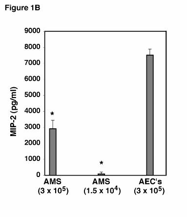
To further determine the relative potency of the AEC response to PC β-glucan compared
to alveolar macrophages, equal numbers of AEC’s and macrophages (3x10⁵ cells/well) were
cultured with PC β-glucan for 24 hours under identical conditions and the level of MIP-2 in the
medium determined by ELISA (Figure 1B). Under these conditions, the AEC’s release 7510 ±
376 pg/ml of MIP-2. In contrast, equal numbers of macrophages released only 2911 ± 532
pg/ml of MIP-2 (P<0.01). Thus, alveolar epithelial cells are a potent source of MIP-2 following
challenge with this P. carinii cell wall component. To further exclude the possibility that residual
macrophages present in the AEC isolates were responsible for the MIP-2 release in response to
PC β-glucan, we determined that the AEC preparations maximally contained 5% residual
Hahn/Limper-14
by guest on February 27, 2019http://w
ww
.jbc.org/D
ownloaded from
macrophages at the time of isolation. This number of macrophages (1.5x10⁴ cells/well), were
also concurrently cultured with PC β-glucan, and found to release only 89 ± 117 pg/ml of MIP-
2 (P<0.001 compared to the AEC isolates challenged with PCBG) (Figure 1B). Thus, it is
unlikely that residual macrophages contributed significantly to the observed MIP-2 release in the
AEC preparations.
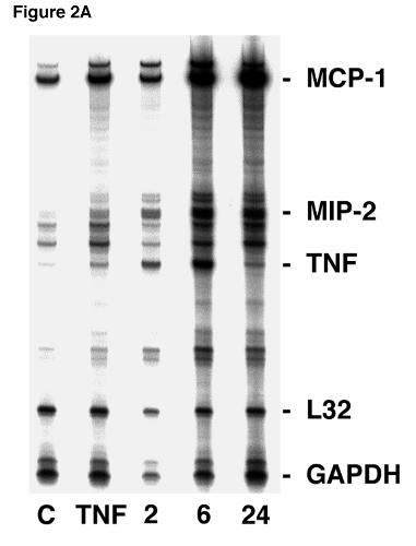
P. carinii β-glucan Stimulates Enhanced Steady-State MIP-2 mRNA in Alveolar
Epithelial Cells. Next, ribonuclease protection assays (RPA’s) were performed to determine
whether glucan stimulation similarly increased MIP-2 RNA expression in primary alveolar
epithelial cells (Figure 2A). Steady state MIP-2 mRNA was significantly increased following
two hours of P. carinii β-glucan stimulation compared to unstimulated control. A doublet
hybridization pattern was observed for MIP-2. Further increases in MIP-2 mRNA were also
noted after 6 and 24 hours of stimulation. The total achieved level of MIP-2 RNA expression in
AEC’s was comparable and exceeded that elicited by six hours of challenge with TNFα (100
ng/ml), an established stimulant of MIP-2 expression. RNA loading was verified by
hybridization to the constituitively expressed L32 and GAPDH genes. In addition, consistent
with our prior studies, RPA on primary AECs further demonstrated an increased level of TNFα
and MCP-1 mRNA at both 6 and 24 hours after stimulation with P. carinii β-glucan (3,11).
Although the AEC preparations consistently contained >95% epithelial cells, with few
remaining macrophages, we further confirmed that P. carinii β-glucan increase MIP-2
expression in MLE-12 cells, an immortalized alveolar epithelial cell line, free of any
Hahn/Limper-15
by guest on February 27, 2019http://w
ww
.jbc.org/D
ownloaded from
macrophage contamination (Figure 2B). RPA using MLE-12 cells demonstrated that MIP-2
mRNA was increased 2 and 4 hours after PC β-glucan stimulation compared to controls.
Interestingly, an increase in the murine chemokine TCA3 mRNA was also observed following 2
and 4 hours of PCBG stimulation in MLE12 cells. The MLE12 cell line exhibited MIP-2
expression following challenge with PCBG, providing further confirmation that alveolar
epithelial cells can directly respond to PCBG with release of the MIP-2 chemokine in the
complete absence of macrophages.
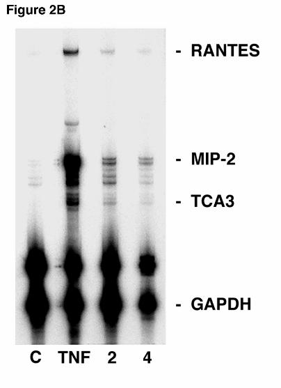
P. carinii β-glucan Stimulates MIP-2 mRNA Expression in Alveolar Epithelial Cells in
Vivo. We have previously reported that PC β-glucan induces substantial neutrophilic lung
inflammation and enhanced lung MIP-2 levels. Thus, we next sought to determine whether PC
β-glucan similarly enhanced MIP-2 gene expression in alveolar epithelial cells following
intratracheal instillation in vivo (0.5 ml containing 1 x 10⁷ PCBG particles). Twenty-four hours
following challenge, alveolar epithelial cells were isolated from the rats, and MIP-2 expression
analyzed by RPA (Figure 3). The 24-hour time point was chosen as a steady state time point at
which neutrophil recruitment and cytokine expression are well established following PC β-
glucan challenge in our previous studies (3). MIP-2 mRNA expression was enhanced in the PC
β-glucan stimulated animals compared with control animals. Minimal basal MIP-2 RNA
expression was found in the unstimulated controls. As described above, RNA was immediately
isolated after the alveolar cell isolation procedure and the AEC’s were not maintained in culture.
Again, CD45 fluorescence immunostaining of the AEC preparations consistently confirmed less
Hahn/Limper-16
by guest on February 27, 2019http://w
ww
.jbc.org/D
ownloaded from
than 5% contamination with alveolar macrophages. These data demonstrate that AEC’s express
MIP-2 mRNA following challenges in the intact host. Under such in vivo conditions epithelial
cells may be acting in direct responses to PC β-glucan, but MIP-2 responses may also be related
to macrophage activity, including the release of factors such as TNFα, which are known to
stimulate AECs.
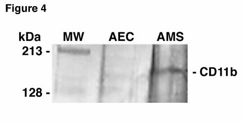
Alveolar Epithelial Cells Lack the CD11b/CD18 β-Glucan Receptor. The integrin
CD11b/CD18 is present on leukocytes and initiates cell activation in response to β-glucans (12-
14). Although no prior reports of CD11b/CD18’s presence on AEC’s could be identified, we
sought to independently confirm that this β-glucan receptor was absent from primary AEC
cultures (Figure 4). Sequential immunoprecipitation and immunoblot analysis clearly
demonstrated that CD11b, while present on alveolar macrophages, was lacking from the alveolar
epithelial cells, indicating that an alternate receptor must be mediating MIP-2 activation in
response to PC β-glucan. This sequential immunoprecipitation and immunoblot analysis further
verified that viable alveolar macrophages represented only a negligible component of the
primary AEC isolates.
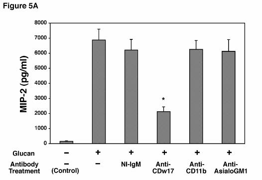
Antibodies to Lactosylceramide (CDw17) Strongly Inhibit MIP-2 Release by AECs
Challenged with P. carinii β-Glucan. As anticipated from the preceding experiments, anti-
CD11b did not significantly inhibit MIP-2 production following stimulation with PC β-glucan,
further indicating that AECs must utilize an alternate β-glucan receptor (Figure 5A).
Hahn/Limper-17
by guest on February 27, 2019http://w
ww
.jbc.org/D
ownloaded from
Lactosylceramide has recently been shown to function as such an alternate β-glucan receptor
(16). In contrast to anti-CD11b, incubation of AECs with anti-CDw17, a mouse monoclonal
IgM antibody, markedly suppressed MIP-2 production as compared to isotype matched IgM
non-immune control (Figure 5A). The maximum degree of inhibition was found with an
antibody concentration of 200 µg/ml. In contrast, anti-asialo-GM1, an antibody recognizing
another glycosphingolipid found in mammalian membranes, had no discernable effect on MIP-2
release. Immunohistochemical staining confirmed strong binding of both the anti-CDw17 and
the anti-asialo-GM1 antibodies to AEC cultures. Staining for both CDw17 and asialo-GM1
was eliminated following digestion of the cells with endoglycosidase Hf (data not shown).
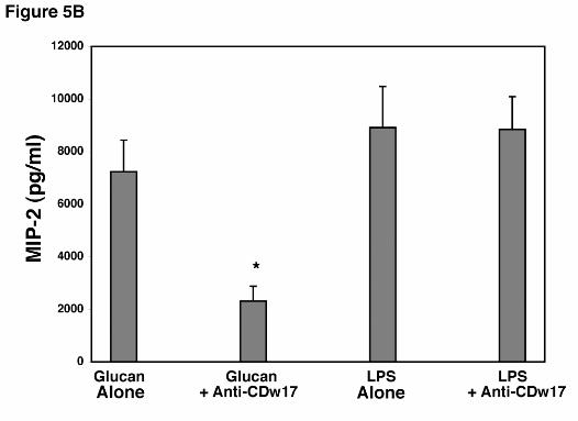
To further verify the specificity of suppression induced by anti-CDw17 on MIP-2
release by AECs in response to PCBG, additional experiments were performed to assess the
ability of AECs to release MIP-2 following stimulation with LPS (1.0 µg/ml) in the presence of
this antibody (Figure 5B). LPS stimulates cells using receptor systems distinct from those know
to interact with β-glucans (3). Notably, AECs were well stimulated to release MIP-2 following
challenge with LPS, and this response was not altered by the presence of anti-CDw17. Thus,
antibodies to lactosylceramide (CDw17) specifically inhibit the responses of AEC’s to PC β-
glucan, yet the cells remain viable and able to respond normally to an alternative stimulant such
as LPS.
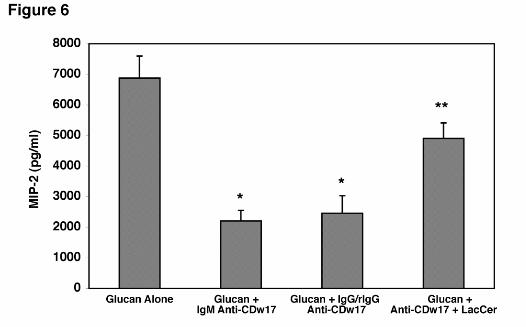
The ability of the anti-CDw17 antibody was significantly, though partially, reversed by
Hahn/Limper-18
by guest on February 27, 2019http://w
ww
.jbc.org/D
ownloaded from
pre-incubation of the antibody with solubilized lactosylceramide before being added to the
AEC’s (Figure 6). To exclude the possibility that the inhibitory effects were solely related to
steric hindrance imparted by the bulky IgM anti-CDw17 binding to the membrane, the anti-
CDw17 (IgM) was digested into IgG and rIgG fragments. Again, when AEC’s were incubated
with fragmented anti-CDw17 (IgG and rIgG), significant decreases in MIP-2 still occurred
following β-glucan stimulation, comparable to the effects of the whole IgM anti-CDw17
antibody (Figure 6). Taken together, these data strongly implicate that lactosylceramide
(CDw17) participates in the membrane complex mediating AEC’s activation to release MIP-2
following PCBG stimulation.
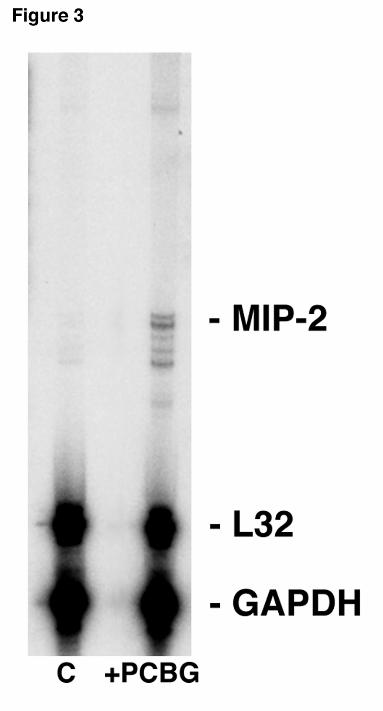
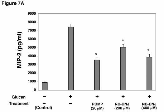
Inhibition of Lactosylceramide Production in AEC’s Decreased MIP-2 Production
Following Stimulation with PC β-Glucan. A number of agents have been produced which
inhibit the synthesis of glycosphinogolipids (28-30). Such compounds may represent novel
means to alter P. carinii-induced lung inflammation. To test this, cellular generation of
lactosylceramide (CDw17) in AEC’s was inhibited with NB-DNJ and PDMP, two potent
inhibitors of glycosphingolipid synthesis (Figure 7A). After 72 hr incubation with NB-DNJ
(400 µM), there was significantly reduced MIP-2 release following β-glucan stimulation
compared to control cells incubated in lipid-free media alone. Similarly, incubation of AEC’s
with PDMP (20 µM) also resulted in significantly decreased levels of MIP-2 production from
AEC’s following P. carinii glucan challenge. Neither NB-DNJ and PDMP altered cellular
viability as measured by the XTT viability assay.
Hahn/Limper-19
by guest on February 27, 2019http://w
ww
.jbc.org/D
ownloaded from
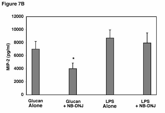
Additional experiments we performed to assess the ability of AEC’s to release MIP-2
following stimulation with a potent alternative agent, namely LPS (1.0 µg/ml), in the presence of
the glycosphinogolipid synthesis inhibitor (Figure 7B). Again, AECs were potently stimulated
to release MIP-2 following stimulation with LPS, and this release was not significantly altered
by the presence of NB-DNJ. Thus, inhibition of glycosphinogolipid synthesis specifically
inhibits AEC release of MIP-2 in response to PCBG, yet the cells remain fully viable and are
able to respond normally to an alternative stimulant such as LPS, which acts through other
receptor pathways.
Hahn/Limper-20
by guest on February 27, 2019http://w
ww
.jbc.org/D
ownloaded from
DISCUSSION
Despite effective antimicrobial agents, Pneumocystis carinii pneumonia continues to
inflict a high rate of respiratory failure and death. Severe PC pneumonia is characterized by
intense neutrophilic lung inflammation leading to impaired gas exchange, diffuse alveolar
damage, and respiratory failure. In fact, neutrophil infiltration in the lungs of patients with PC
pneumonia is a stronger predictor of respiratory failure and death than the organism burden itself
(2). Additional investigations indicate that P. carinii cell wall β-glucans strongly stimulate
neutrophilic lung inflammation (3,11). Non-specific anti-inflammatory therapies, such as
adjunctive corticosteroids, have significantly decreased mortality during severe PC pneumonia
(31). Thus, a better understanding of glucan-induced inflammation may provide novel means by
which lung inflammation can be selectively modulated during this infection.
It is well established that alveolar macrophages produce and secrete cytokines and
chemokines such as TNFα and MIP-2, which are potent inducers of neutrophil migration and
activation (3). It is, however, becoming more evident that alveolar epithelial cells are also active
participants in the host’s inflammatory response (20,21,32). For instance, Standiford reported
that A549 alveolar epithelial cells produce significant quantities of IL-8, the human functional
homologue of MIP-2, when stimulated with TNFα (32). However, A549s did not express IL-8
in response to LPS alone, and these authors postulated the existence of a cytokine network model
whereby alveolar epithelial cells are stimulated by activated alveolar macrophages (32). In
Hahn/Limper-21
by guest on February 27, 2019http://w
ww
.jbc.org/D
ownloaded from
contrast, primary alveolar epithelial cell cultures have been shown to produce both TNFα and
MIP-2 in direct response to LPS (14). The current study is the first to demonstrate that the β-
glucan cell wall fraction from Pneumocystis carinii can induce production of significant
quantities of the chemokine, MIP-2, from alveolar epithelial cells, further implicating the
alveolar epithelium as an important participant in host inflammation during lung infection.
It is important to note that strict precautions were taken to minimize alveolar macrophage contamination in
the AEC isolates used in these studies. Anti-CD45 immunofluorescence consistently demonstrated fewer than
5% macrophages in these preparations, and negligible CD11b, a prominent macrophage marker,
was detected by sequential immunoprecipitation and immunoblotting. Our data further
demonstrate that, on a cell-by-cell basis, AEC’s secrete greater amounts of MIP-2 than alveolar
macrophages. In addition, assuming a 5% contamination of the AEC with macrophages, that
number of macrophages would have only generated minimal MIP-2 (89 pg/ml) in these cultures
(3,11). Thus, it is highly unlikely that the substantial levels of MIP-2 produced by P. carinii β-
glucan stimulation of the AEC cultures were related to the few residual macrophages in these
isolates. Furthermore, the murine alveolar epithelial cell line, MLE-12, which is free of any
other cell population also demonstrated a significant increase in MIP-2 mRNA after PC β-
glucan stimulation. In light of the greater numbers of alveolar epithelial cells in the lung
contrasted to macrophages and other cells, the direct response of AECs to β-glucan components
of the PC cell wall likely represents a significant source of MIP-2 during this infection. It does,
however, remain possible that macrophages may also release factors that augment epithelial cell
release of chemokines such as MIP-2 during in vivo challenge.
Hahn/Limper-22
by guest on February 27, 2019http://w
ww
.jbc.org/D
ownloaded from
Although MIP-2 is an important chemokine involved in lung inflammation, it is only
one of a number of neutrophil directed cytokines. Our RPA analysis indicates that AEC also
increase expression of TNFα and MCP-1 following PCBG stimulation. Prior studies by our
group and others have documented that TNFα is essential for optimal clearance of P. carinii
infection (3,11,33-35). Additional studies indicate that MCP-1 is also significantly expressed in
rodent lungs during P. carinii infection and by A549 cells stimulated with P. carinii major surface
glycoprotein (36,37). We instead have focused on the stimulatory effects of P. carinii β-glucan
because our prior studies have indicated this component to principally mediate lung
inflammatory responses during P. carinii infection (3,11). In addition to observing enhanced
expression of MIP-2, we also observed that TCA3 mRNA expression was increased after PC β-
glucan stimulation in MLE12 cells. TCA3 is an activation specific cytokine product previously
described to be produced by T cells and Mast cells and to function as a potent chemotactic agent
for neutrophils and macrophages (38,39). We believe this is the first demonstration that
epithelial cells express TCA3. These studies strongly implicate lung epithelial cells as an
integral component in the recognition and response of the host to P. carinii, with epithelial cell-
derived MIP-2 serving to promote neutrophil recruitment during infection. Moreover, since β-
glucans are common throughout the pathogenic fungi, this innate response may represent a
generalized response of the epithelium to fungal infection.
Several potential β-glucan receptors have been reported in the literature. The most
widely studied is the β2 integrin CD11b/CD18, which possesses a lectin-binding site on CD11b
Hahn/Limper-23
by guest on February 27, 2019http://w
ww
.jbc.org/D
ownloaded from
that has specificity for β-glucan. This receptor is cross-linked by high molecular weight soluble
and particulate β-glucans leading to the activation of phagocytic cells (40,41). The current study
demonstrates, however, that CD11b/CD18 is not present on epithelial cells to any significant
extent and does not function as a glucan receptor mediating AEC activation. Very recent studies
have also documented an additional β-glucan receptor, namely dectin-1, which is largely
present on macrophages and dendritic cells (42,43). Its presence in epithelial and other cells has
not yet been established. Instead, our study instead implicates lactosylceramide as an epithelial
cell receptor participating in AEC activation to release MIP-2 in response to P. carinii β-glucan
components.
The glycosphingolipid, lactosylceramide (LacCer), is widely distributed in the plasma
membranes of many different cell types including alveolar epithelial cells. LacCer has been
shown to bind several different bacteria and yeast (41). Recently, LacCer has been demonstrated
to bind PGG-glucan, a purified β-glucan derived from the cell wall of Saccharomyces
cerevisiae (15). The binding of PGG-glucan to LacCer activates a NF-κB-like nuclear
transcription factor in human polymorphonuclear cells (16). Herein, we demonstrate that
antibodies to cell membrane LacCer (CDw17) and inhibitors of LacCer synthesis significantly
attenuate the secretion of MIP-2 after PC β-glucan challenge in alveolar epithelial cells. The
glycosphingolipid biosynthesis inhibitors N-butyldeoxynojirimycin (NB-DNJ) inhibits the first
step in glycosphingolipid biosynthesis, where glucose is transferred to ceramide. Culture of cells
with NB-DNJ causes a depletion of glycosphingolipids including LacCer (28,29). In contrast,
Hahn/Limper-24
by guest on February 27, 2019http://w
ww
.jbc.org/D
ownloaded from
D-threo-1-phenyl-2-decanoylamino-3-morpholino-1-propanol (PDMP) is an inhibitor of
glycosyl transferase (29). Incubation of AEC’s with either NB-DNJ or PDMP causes a
significant decrease in MIP-2 release after stimulation with PC glucan. Such compounds might
represent a novel means to similarly modulate P. carinii induced inflammation in vivo.
How CDw17 transmits its signal has yet to be elucidated. Accumulating evidence
indicates that glycosphingolipids are concentrated in microdomains on the plasma membrane
surface (44,45). These microdomains or “rafts” are thought to represent platforms where
signaling molecules are concentrated. Interestingly, antibodies to asialoGM1, another
glycosphingolipid on AEC’s and a known Pseudomonas pilin receptor, did not result in any
attenuation of MIP-2 production. It might be postulated that ligation of β-glucan in
microdomains may result in concerted uptake of β-glucan particles and recruitment and
activation of signaling kinases within these microdomains.
In summary, our study demonstrates that β-glucan components of the Pneumocystis
carinii cell wall are able to stimulate alveolar epithelial cells to produce MIP-2. These
investigations further demonstrate that the membrane glycosphingolipid lactosylceramide
participates in the receptor complex mediating PC β-glucan activation of AEC’s. MIP-2
produced by AEC’s and macrophages significantly promotes neutrophilic lung inflammation
during severe Pneumocystis pneumonia. Emerging strategies to attenuate glucan mediated
inflammatory signaling, including inhibition of lactosylceramide-based signaling, may hold
Hahn/Limper-25
by guest on February 27, 2019http://w
ww
.jbc.org/D
ownloaded from
therapeutic benefit to reduce lung injury during P. carinii pneumonia.
Hahn/Limper-26
by guest on February 27, 2019http://w
ww
.jbc.org/D
ownloaded from
AKNOWLEDGEMENTS
We thank Drs. Zvezdana Vuk-Pavlovic, Robert Vassallo, and David Marks for many
helpful discussions. We also thank Kathy Streich for her assistance during the final preparation
of the manuscript.
Hahn/Limper-27
by guest on February 27, 2019http://w
ww
.jbc.org/D
ownloaded from
FOOTNOTES
1 This work was supported by NIH Grants R01’s HL55934HL57125 and HL62150 to AHL and
GM 22942 and GM60934 to REP, and a grant from the Robert N. Brewer Family Foundation to
Thoracic Diseases Research Unit. REP was also supported by a grant from the Ara Parsegian
Foundation.
2Abbreviations used: AEC, alveolar epithelial cell; MIP-2, macrophage inflammatory protein-
2; TNF, Tumor necrosis factor α; PC, Pneumocystis carinii; PCBG, Pneumocystis carinii β-
glucan-rich cell wall fraction; RPA, ribonuclease protection assay; MCP-1, monocyte
chemoattractant protein-1; GAPDH, glyceraldehyde-3-Phosphate dehydrogenase; TCA3, T cell
activation-3 cytokine; RANTES, regulated on activation normal T-cell expressed chemokine.
Hahn/Limper-28
by guest on February 27, 2019http://w
ww
.jbc.org/D
ownloaded from
FIGURE LEGENDS
Figure 1. MIP-2 production from alveolar epithelial cells following stimulation with
Pneumocystis carinii β-glucan cell wall isolate. A. Isolated AECs (3 x 10⁵ cells/well in 200 µl)
were stimulated with PCBG (5 x 10⁶ particles/ml) for 0 hours (control), and 2, 6, 12, 16, or 24
hours, and MIP-2 release into the medium determined by ELISA. Data points are expressed as
mean ± SEM (* Denotes P<0.05 compared to control). B. To assess the relative potency of
MIP-2 release from AEC’s compared to alveolar macrophages (AMS), and to exclude the
possibility that residual macrophages in the AEC preparations significantly contributed to the
observed MIP-2 release, identical numbers of AEC’s and alveolar macrophages (3 x 10⁵
cells/well), as well as the maximal number of residual alveolar macrophages in the AEC isolates
(equal to 1.5 x 10⁴) were stimulated with PCBG (5 x 10⁶ particles/ml) for 24 hours. MIP-2
release into the medium was determined by ELISA. (* Denotes P<0.01 compared to 3 x 10⁵
AEC’s).
Figure 2. MIP-2 RNA expression in lung epithelial cells after stimulation with Pneumocystis
carinii β-glucan. A. Ribonuclease protection assay of primary AEC cultures (5 x 10⁶ cells/well
in 3 ml) were performed following stimulation with PC β-glucan rich cell wall isolate (5 x 10⁶
particles/well) for 2, 6, or 24 hours. Control (“C”) denotes unstimulated cultured AEC’s. In
Hahn/Limper-29
by guest on February 27, 2019http://w
ww
.jbc.org/D
ownloaded from
parallel cultures, AEC’s were also cultured with TNFα (100 ng/ml), a known stimulant of MIP-2
expression for six hours. B. In a parallel fashion, the MLE-12 murine alveolar epithelial cell
line was assessed for chemokine RNA expression following PCBG challenge for 2 or 4 hours
compared to unstimulated control AECs. Again, AECs were also stimulated with TNFα (100
ng/ml), for 4 hours. RNA loading was determined by assessing GAPDH and L32 expression.
Figure 3. MIP-2 mRNA expression in alveolar epithelial cells isolated from mice challenged
intratracheally with PC β-glucan cell wall isolate. Twenty-four hours after intratracheal
injection, lungs were removed and alveolar epithelial cells immediately isolated. MIP-2 mRNA
expression was measured using RPA. Shown is a representative blot of three separate
experiments. The first lane contains control unstimulated mouse lung RNA, the second lane
contains +PCBG stimulated mouse lung RNA
Figure 4. Alveolar epithelial cells lack the CD11b/CD18 β-glucan receptor. Primary
alveolar epithelial cells and alveolar macrophages were isolated, membrane proteins extracted,
and immunoprecipitated with anti-CD11b antibodies. The precipitated products were separated
by SDS-PAGE, transferred and analyzed with immunoblot using an antibody recognizing the
CD-11b integrin component receptor.
Figure 5. Effect of antibodies to lactosylceramide on MIP-2 release by alveolar epithelial
cells incubated with P. carinii β-glucan. A. Isolated AECs were incubated with media alone,
Hahn/Limper-30
by guest on February 27, 2019http://w
ww
.jbc.org/D
ownloaded from
non-immune mouse IgM, anti-CDw17, anti-CD11b, or anti-asialoGM1 (200 µg/ml each) for
30 minutes before and throughout a subsequent 16 hour challenge with the PC β-glucan rich cell
wall isolate (5 x 10⁶ particles/ml). MIP-2 release under these conditions was determined by
ELISA. B. To further verify the specificity of anti-CDw17 on suppression of MIP-2 release by
AEC’s in response to P. carinii β-glucan, epithelial cells were stimulated in parallel with either
PC β-glucan (5 x 10⁶ particles/ml) or LPS (1.0 µg/ml) in the presence or absence of anti-
CDw17 (200 µg/ml), and MIP-2 release assessed by ELISA. Data points are expressed as
mean ± SEM (* Denotes P<0.05, compared to control AECs incubated with glucan alone)
Figure 6. Incubation of AEC’s with either whole IgM or IgG/rIgG anti-CDw17 both
significantly decrease MIP-2 production following PCBG stimulation. AEC’s were incubated
with 200 µg/ml of either whole IgM anti-CDw17 or digested anti-CDw17 IgM (IgG and rIgG)
during challenge with PC β-glucan and MIP-2 release determined by ELISA. In parallel, anti-
CDw17 (IgM) was pre-incubated with solubilized lactosylceramide. Data points are expressed
as mean ± SEM (* Denotes P<0.05, compared to control AEC’s incubated with glucan alone; **
Denotes P<0.05 comparing IgM anti-CDw17 and anti-CDw17 preincubated with
lactosylceramide).
Figure 7. Effect of glycosphingolipid biosynthesis inhibitors on β-glucan induced MIP-2
generation from alveolar epithelial cells. A. AECs were incubated with either PDMP (20 µM)
or NB-DNJ (200-400 µM) for 72 hours, challenged with PC β-glucan for a subsequent 18
Hahn/Limper-31
by guest on February 27, 2019http://w
ww
.jbc.org/D
ownloaded from
hours, and MIP-2 release determined. B. To further verify specificity of the effect of inhibiting
glycosphingolipid synthesis on suppression of MIP-2 release by AEC’s, epithelial cells were
stimulated in parallel with either PC β-glucan (5 x 10⁶ particles/ml) or LPS (1.0 µg/ml) in the
presence or absence of NB-DNJ (400 µM). Data points are expressed as mean ± SEM.
(* Denotes P<0.05 compared to AECs incubated with PC β-glucan alone)
Hahn/Limper-32
by guest on February 27, 2019http://w
ww
.jbc.org/D
ownloaded from
REFERENCES
1. Thomas, C. F., Jr., and Limper, A. H. (1998) Semin. Respir. Infect. 13:289-95.
2. Limper, A. H., Offord, K. P., Smith, T. F., and Martin, W. J. 2nd. (1989). Am. Rev. Respir. Dis.
140:1204-09.
3. Vassallo, R., Standing, J. E., and Limper, A. H. (2000) J. Immunol. 164:3755-63.
4. Edman, J. C., Kovacs, J. A., Masur, H., Santi, D. V., Elwood, H. J., and Sogin, M. L. (1988) Nature
334:519-22.
5. Matsumoto, Y., Matsuda, S., and Tegoshi, T. (1989) J. Protozool. 36:21S-22S.
6. Kollar, R., Petrakova, E., Ashwell, G., Robbins, P. W., and Cabib, E. (1995) J. Biol. Chem. 270:1170-8.
7. Nollstadt, K. H., Powles, M. A., Fujioka, H., Aikawa, M. and Schmatz. D. M. (1994) Antimicrob. Agents
Chemother. 38:2258-65.
8. Abel, G., and Czop, J. K. (1992) Int. J. Immunopharmacol. 14:1363-73.
9. Castro, M., Morgenthaler, T. I., Hoffman, O. A., Standing, J. E., Rohrbach, M. S., and Limper, A. H (1993)
Am. J. Respir. Cell. Mol. Biol. 9:73-81.
10. Olson, E. J., Standing, J. E., Griego-Harper, N., Hoffman, O. A., and Limper, A. H. (1996) Infect. Immun.
64:3548-54.
11. Hoffman, O.A., Standing, J.E., and Limper, A.H. (1993) J. Immunol. 150:3932-3940.
12. Thornton, B.P., Vetvicka, V., Pitman, M., Goldman, R.C., and Ross, G.D. (1996) J Immunol. 156:1235-
46.
13. Ross, GD. (2000) Crit. Rev. Immunol. 20:197-222.
14. Vetvicka, V., Thornton, B. P., and Ross, G. D. (1996) J. Clin. Invest. 98:50-61.
15. Zimmerman, J. W., Lindermuth, J., Fish, P. A., Palace, G. P., Stevenson, T. T., and DeMong, D. E. (1998)
J. Biol. Chem. 273:22014-20.
16. Wakshull, E., Brunke-Reese, D., Lindermuth, J., Fisette, L., Nathans, R. S., Crowley, J. J., Tufts, J. C.,
Hahn/Limper-33
by guest on February 27, 2019http://w
ww
.jbc.org/D
ownloaded from
Zimmerman, J., Mackin, W., and Adams, D. S. (1999) Immunopharmacology 41:89-107.
17. Limper, A.H., Edens, M., Anders, R.A., and Leof, E.B. (1998) J. Clin. Invest. 101: 1148-1155.
18. Kallenberg, C. G., Schilizzi, B. M., Beaumont, F., Poppema, S., De Leij, L., and The, T. H. (1987) Clin.
Exp. Immunol. 67:182-90.
19. Schneeberger, E. E., DeFerrari, M., Skoskiewicz, M. J., Russell, P. S., and Colvin, R. B. (1986) Lab.
Invest. 55:138-44.
20. Standiford, T. J., Kunkel, S. L., Phan, S. H., Rollins, B. J., and Strieter, R. M. (1991) J. Biol. Chem
266:9912-8.
21. Xavier, A. M., Isowa, N., Cai, L., Dziak, E., Opas, M., McRitchie, D. I., Slutsky, A. S., Keshavjee, S. H.,
and Liu, M. (1999) Am. J. Respir. Cell. Mol. Biol. 21:510-20.
22. Lin, Y., Zhang, M., and Barnes, P. F. (1998) Infect. Immun. 66:1121-6.
23. Wickremasinghe, M. I., Thomas, L. H., and Friedland, J. S. (1999) J. Immunol. 163:3936.
24. Wolpe, S.D., and Cerami, A. (1989) Faseb J. 3:2565-73.
25. Schmal, H., Shanley, T. P., Jones, M. L., Friedl, H. P., and Ward, P. A. (1996) J. Immunol. 156:1963-72.
26. Driscoll, K. E., Hassenbein, D. G., Howard, B. W., Isfort, R. J., Cody, D., Tindal, M. H., Suchanek, M.,
and Carter, J. M. (1995) J Leukoc Biol 58:359-64.
27. Dobbs, L. G., Gonzalez, R. and Williams, M. C. (1986) Am. Rev. Respir. Dis. 134:141-5.
28. Andersson, U., Butters, T.D., Dwek, R.A., and Platt, F.M. (2000) Bioche. Pharmacol. 59:821-9.
29. Platt, F. M., Reinkensmeier, G., Dwek, R.A., and Butters, T.D. (1997) J. Biol. Chem. 272:19365-72.
30. Kovacs, P., Pinter M., and Csaba G. (2000) Cell. Biochem. Funct. 18:269-80.
31. Bozzette, S.A., Sattler, F.R., Chiu, J., Wu, A.W., Gluckstein, D., Kemper, C., Bartok, A., Niosi, J.,
Abramson, I., and Coffman, J. (1991) N. Engl. J. Med. 323:1451-7.
32. Standiford, T. J., Kunkel, S. L., Basha, M. A., Chensue, S. W., Lynch, J. P. 3rd, Toews, G. B., Westwick,
J., and Strieter, R. M. (1990) J. Clin. Invest. 86:1945-53.
33. Chen, W., Havell, E.A., and Harmsen, A.G. (1992) Infect. Immun. 60:1279-84.
Hahn/Limper-34
by guest on February 27, 2019http://w
ww
.jbc.org/D
ownloaded from
34. Kolls, J.K., Lei, D., Vazquez, C., Odom, G., Summer, W.R., Nelson, S., and Shellito, J. (1997) Am. J.
Respir. Cell. Mol. Biol. 16:112-18.
35. Rudmann, D.G., Preston, A.M., Moore, M.W., and Beck, J.M. (1998) J. Immunol. 161:360-6.
36. Wright, T.W., Johnston, C.J., Harmsen, A.G., and Finkelstein J.N. (1999) Infect. Immun. 67:3452-60.
37. Benfield, T.L., Lundgren, B., Shelhamer, J.H., and Lundgren, J.D. (1999) Eur. J. Clin. Invest. 29:717-22.
38. Luo, Y., Laning, J., and Dorf, M. E. (1993) J. Immunol. 150:971-9.
39. Luo, Y., Laning, J., Devi, S., Mak, J., Schall, T. J., and Dorf, M. E. (1994) J. Immunol. 153:4616-24.
40. Yan, J., Vetvicka, V., Xia, Y., Coxon, A., Carroll, M. C., Mayadas, T. N., and Ross, G. D. (1999) J.
Immunol. 163:3045-52.
41. Jimenez-Lucho, V., Ginsburg, V., and Krivan, H. C. (1990) Infect. Immun. 58:2085-90.
42. Brown, G.D. and Gordon, S. (2001) Nature 413:36-7.
43. Willment, J.A., Gordon, S., and Brown, G. D. (2001) J. Biol. Chem. 276: 43818-23.
44. Simons, K., and Ikonen, E. (1997) Nature 387:569-572.
45. Varma, R., and Mayor, S. (1998) Nature 394:798-801.
Hahn/Limper-35
by guest on February 27, 2019http://w
ww
.jbc.org/D
ownloaded from
and Andrew H. LimperPeter Y. Hahn, Scott E. Evans, Theodore J. Kottom, Joseph E. Standing, Richard E. Pagano
protein-2 from alveolar epithelial cells via a lactosylceramide mediated mechanism-glucan induces release of macrophage inflammatoryβPneumocystis carinii cell wall
published online November 4, 2002J. Biol. Chem.
10.1074/jbc.M209715200Access the most updated version of this article at doi:
Alerts:
When a correction for this article is posted•
When this article is cited•
to choose from all of JBC's e-mail alertsClick here
by guest on February 27, 2019http://w
ww
.jbc.org/D
ownloaded from