Embed Size (px)
Citation preview
1
PowerPoint Slides English Text Brazilian Portuguese Translation Treatment Modalities: Radiation Therapy
VideoTranscript Modalidades de Tratamento: Radioterapia Transcrição do vídeo
Professional Oncology Education Treatment Modalities: Radiation Therapy Time: 23:50
Educação Profissional em Oncologia
Modalidades de Tratamento: Radioterapia Duração: 23:50
Prajnan Das, M.D., M.P.H. Assistant Professor Radiation Oncology The University of Texas MD Anderson Cancer Center
Prajnan Das, M.D., M.P.H. Professor Assistente Radiação Oncológica MD Anderson Cancer Center, Universidade do Texas
Treatment Modalities: Treatment Modalities: Treatment Modalities: Treatment Modalities:
Radiation Therapy Radiation Therapy Radiation Therapy Radiation Therapy
Treatment Modalities: Treatment Modalities:
Radiation Therapy Radiation Therapy
Prajnan Das, M.D., M.P.H.
Assistant Professor
Radiation Oncology
Hello, I am Prajnan Das, Faculty Member in the Department of Radiation Oncology at The University of Texas MD Anderson Cancer Center. We are going to talk today about some of the basic principles regarding radiation therapy.
Olá, sou Prajnan Das, professor no Departamento de Radiação Oncológica do MD Anderson Cancer Center da Universidade do Texas. Hoje falaremos sobre alguns dos princípios básicos da radioterapia.
2
Treatment Modalities: Treatment Modalities: Treatment Modalities: Treatment Modalities:
Radiation Therapy Radiation Therapy Radiation Therapy Radiation Therapy
ObjectivesObjectivesObjectivesObjectives
• Upon completion of this lesson, participants will
be able to:
– Understand the biologic effects of radiation
– Understand the steps involved in radiation
therapy planning
– Identify selected clinical applications and
complications associated with radiation therapy
We will talk about the biologic effects of radiation, how radiation works. We will discuss the steps involving radiation therapy planning, and we will talk about some clinical applications and complications associated with radiation therapy.
Falaremos sobre os efeitos biológicos da radiação, como a radiação funciona. Discutiremos os passos para o planejamento da radioterapia e falaremos sobre algumas das implicações complicações clínicas associadas à radioterapia.
Treatment Modalities: Treatment Modalities: Treatment Modalities: Treatment Modalities:
Radiation Therapy Radiation Therapy Radiation Therapy Radiation Therapy
How Does Radiation Work?
First, how does radiation work?
Primeiro, como funciona a radioterapia?
3
Treatment Modalities: Treatment Modalities: Treatment Modalities: Treatment Modalities:
Radiation Therapy Radiation Therapy Radiation Therapy Radiation Therapy
Types of RadiationTypes of RadiationTypes of RadiationTypes of Radiation
• Non-ionizing
– Do not produce ions in matter
• Microwaves, ultrasound, radio waves
• Ionizing
– Eject orbital electrons and produce ions
• Directly ionizing – Charged particles (electrons, protons,
alpha particles)
• Indirectly ionizing – Uncharged particles (neutrons, gamma rays,
x-rays)
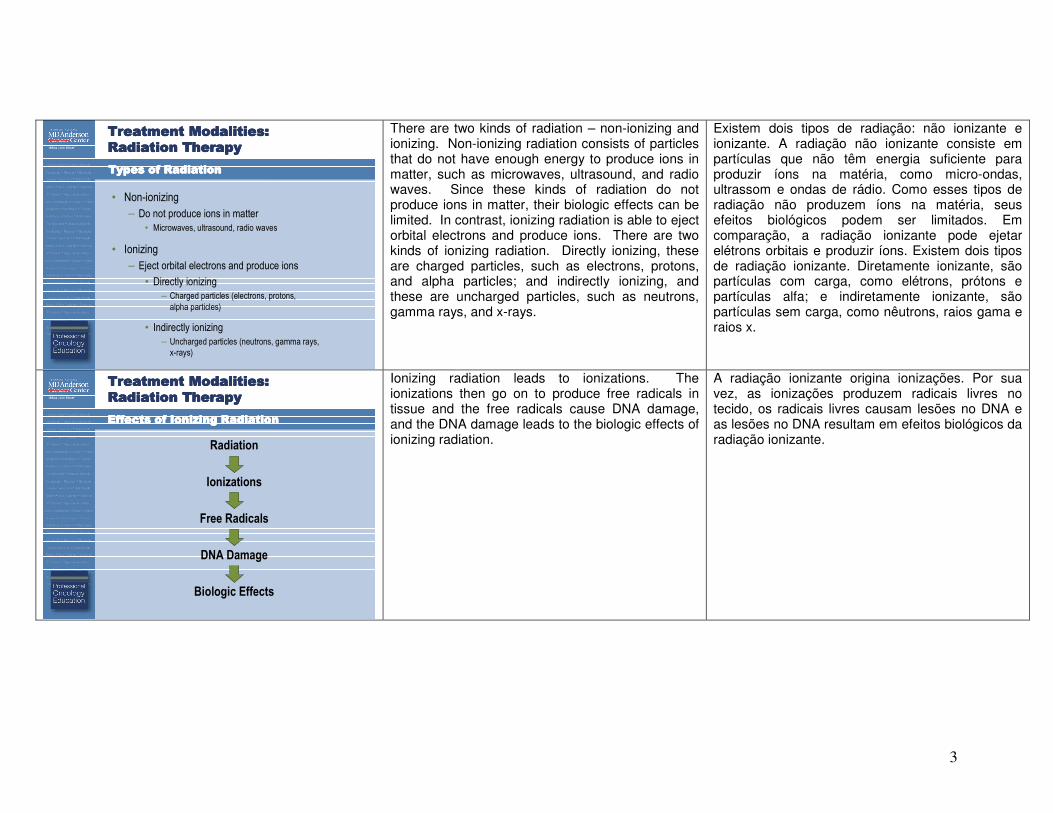
There are two kinds of radiation – non-ionizing and ionizing. Non-ionizing radiation consists of particles that do not have enough energy to produce ions in matter, such as microwaves, ultrasound, and radio waves. Since these kinds of radiation do not produce ions in matter, their biologic effects can be limited. In contrast, ionizing radiation is able to eject orbital electrons and produce ions. There are two kinds of ionizing radiation. Directly ionizing, these are charged particles, such as electrons, protons, and alpha particles; and indirectly ionizing, and these are uncharged particles, such as neutrons, gamma rays, and x-rays.
Existem dois tipos de radiação: não ionizante e ionizante. A radiação não ionizante consiste em partículas que não têm energia suficiente para produzir íons na matéria, como micro-ondas, ultrassom e ondas de rádio. Como esses tipos de radiação não produzem íons na matéria, seus efeitos biológicos podem ser limitados. Em comparação, a radiação ionizante pode ejetar elétrons orbitais e produzir íons. Existem dois tipos de radiação ionizante. Diretamente ionizante, são partículas com carga, como elétrons, prótons e partículas alfa; e indiretamente ionizante, são partículas sem carga, como nêutrons, raios gama e raios x.
Treatment Modalities: Treatment Modalities: Treatment Modalities: Treatment Modalities:
Radiation Therapy Radiation Therapy Radiation Therapy Radiation Therapy
Effects of Ionizing RadiationEffects of Ionizing RadiationEffects of Ionizing RadiationEffects of Ionizing Radiation
Radiation
Ionizations
Free Radicals
DNA Damage
Biologic Effects
Ionizing radiation leads to ionizations. The ionizations then go on to produce free radicals in tissue and the free radicals cause DNA damage, and the DNA damage leads to the biologic effects of ionizing radiation.
A radiação ionizante origina ionizações. Por sua vez, as ionizações produzem radicais livres no tecido, os radicais livres causam lesões no DNA e as lesões no DNA resultam em efeitos biológicos da radiação ionizante.
4
Treatment Modalities: Treatment Modalities: Treatment Modalities: Treatment Modalities:
Radiation Therapy Radiation Therapy Radiation Therapy Radiation Therapy
Radiation and DNA
• Free radicals produce DNA strand breaks
• Single strand breaks are repaired easily
• Double strand breaks difficult to repair
– Most important lesion produced
by radiation
– Can lead to cell death, mutation
or carcinogenesis
– Affects tumor and normal tissue
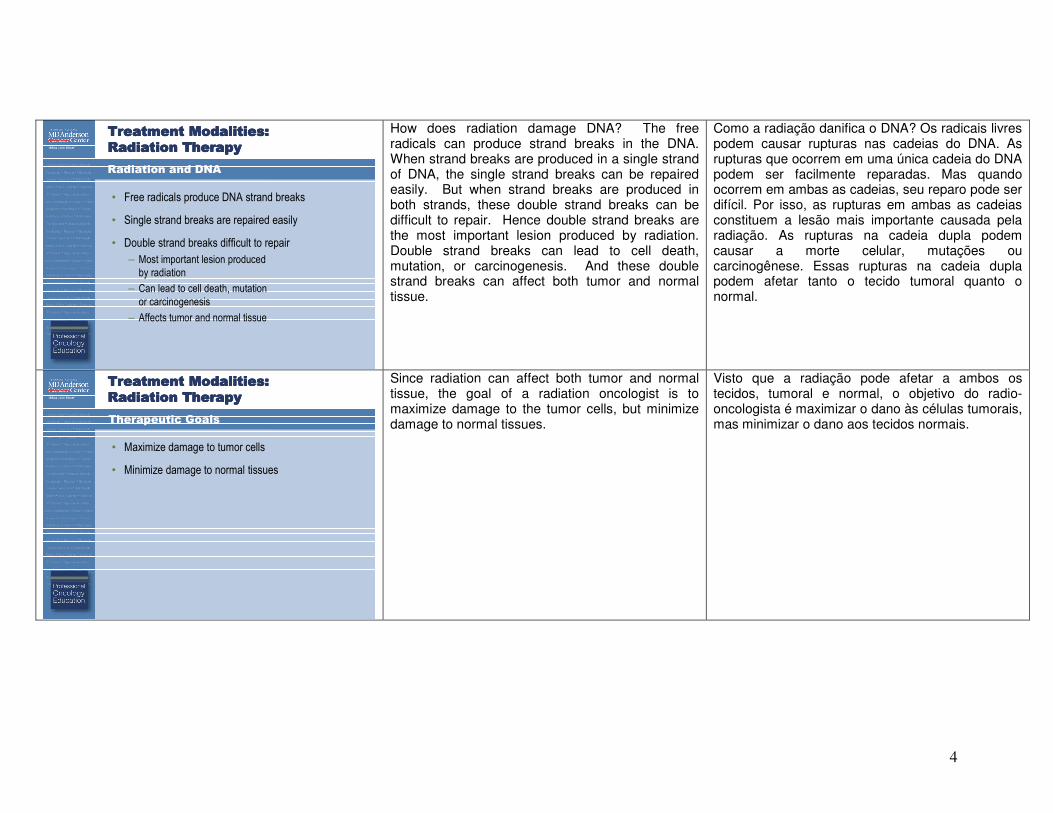
How does radiation damage DNA? The free radicals can produce strand breaks in the DNA. When strand breaks are produced in a single strand of DNA, the single strand breaks can be repaired easily. But when strand breaks are produced in both strands, these double strand breaks can be difficult to repair. Hence double strand breaks are the most important lesion produced by radiation. Double strand breaks can lead to cell death, mutation, or carcinogenesis. And these double strand breaks can affect both tumor and normal tissue.
Como a radiação danifica o DNA? Os radicais livres podem causar rupturas nas cadeias do DNA. As rupturas que ocorrem em uma única cadeia do DNA podem ser facilmente reparadas. Mas quando ocorrem em ambas as cadeias, seu reparo pode ser difícil. Por isso, as rupturas em ambas as cadeias constituem a lesão mais importante causada pela radiação. As rupturas na cadeia dupla podem causar a morte celular, mutações ou carcinogênese. Essas rupturas na cadeia dupla podem afetar tanto o tecido tumoral quanto o normal.
Treatment Modalities: Treatment Modalities: Treatment Modalities: Treatment Modalities:
Radiation Therapy Radiation Therapy Radiation Therapy Radiation Therapy
Therapeutic Goals
• Maximize damage to tumor cells
• Minimize damage to normal tissues
Since radiation can affect both tumor and normal tissue, the goal of a radiation oncologist is to maximize damage to the tumor cells, but minimize damage to normal tissues.
Visto que a radiação pode afetar a ambos os tecidos, tumoral e normal, o objetivo do radio-oncologista é maximizar o dano às células tumorais, mas minimizar o dano aos tecidos normais.
5
Treatment Modalities: Treatment Modalities: Treatment Modalities: Treatment Modalities:
Radiation Therapy Radiation Therapy Radiation Therapy Radiation Therapy
Radiation Dose
• Measured in terms of energy imparted by
ionizing radiation per unit mass of matter
• 1 Gray (Gy) = 1 Joule/kg
• 1 Gy = 100 cGy
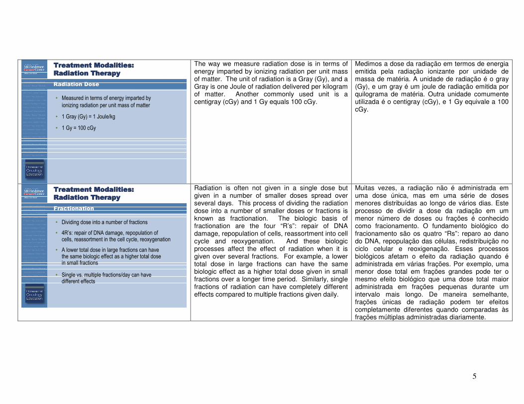
The way we measure radiation dose is in terms of energy imparted by ionizing radiation per unit mass of matter. The unit of radiation is a Gray (Gy), and a Gray is one Joule of radiation delivered per kilogram of matter. Another commonly used unit is a centigray (cGy) and 1 Gy equals 100 cGy.
Medimos a dose da radiação em termos de energia emitida pela radiação ionizante por unidade de massa de matéria. A unidade de radiação é o gray (Gy), e um gray é um joule de radiação emitida por quilograma de matéria. Outra unidade comumente utilizada é o centigray (cGy), e 1 Gy equivale a 100 cGy.
Treatment Modalities: Treatment Modalities: Treatment Modalities: Treatment Modalities:
Radiation Therapy Radiation Therapy Radiation Therapy Radiation Therapy
Fractionation
• Dividing dose into a number of fractions
• 4R’s: repair of DNA damage, repopulation of cells, reassortment in the cell cycle, reoxygenation
• A lower total dose in large fractions can have the same biologic effect as a higher total dose in small fractions
• Single vs. multiple fractions/day can have different effects
Radiation is often not given in a single dose but given in a number of smaller doses spread over several days. This process of dividing the radiation dose into a number of smaller doses or fractions is known as fractionation. The biologic basis of fractionation are the four “R’s”: repair of DNA damage, repopulation of cells, reassortment into cell cycle and reoxygenation. And these biologic processes affect the effect of radiation when it is given over several fractions. For example, a lower total dose in large fractions can have the same biologic effect as a higher total dose given in small fractions over a longer time period. Similarly, single fractions of radiation can have completely different effects compared to multiple fractions given daily.
Muitas vezes, a radiação não é administrada em uma dose única, mas em uma série de doses menores distribuídas ao longo de vários dias. Este processo de dividir a dose da radiação em um menor número de doses ou frações é conhecido como fracionamento. O fundamento biológico do fracionamento são os quatro “Rs”: reparo ao dano do DNA, repopulação das células, redistribuição no ciclo celular e reoxigenação. Esses processos biológicos afetam o efeito da radiação quando é administrada em várias frações. Por exemplo, uma menor dose total em frações grandes pode ter o mesmo efeito biológico que uma dose total maior administrada em frações pequenas durante um intervalo mais longo. De maneira semelhante, frações únicas de radiação podem ter efeitos completamente diferentes quando comparadas às frações múltiplas administradas diariamente.
6
Treatment Modalities: Treatment Modalities: Treatment Modalities: Treatment Modalities:
Radiation Therapy Radiation Therapy Radiation Therapy Radiation Therapy
Factors Affecting Biologic Effects of Radiation
• Type of radiation
– Depends on the density of ionizations produced by radiation
• Dose rate
• Type of tissue
– Early responding vs. late responding tissues
• Oxygenation
• Radiosensitization
– Chemotherapy (5-FU, cisplatin) and biologics (cetuximab)
• Overall treatment time
There are a number of factors that can affect the biologic effects of radiation. The type of radiation is important, and this can depend on the density of ionizations produced by that kind of radiation. The dose rate of radiation is important, whether you are giving radiation at a slower rate or at a faster rate. The type of tissue is important. In general, you can divide tissues into early responding tissues and late responding tissues. Early responding tissues typically include rapidly proliferating cells, such as skin and gastrointestinal mucosa. And these tend to be more sensitive to radiation and also exhibit damage from radiation early in the time course. Late responding tissues typically include cells that do not proliferate at a fast rate, such as neurons or renal cells. And these tend to be more resistant to radiation and exhibit damage from radiation at a later point in time. As we discussed earlier, for DNA damage to take place from radiation, you need free radicals, and free radical production requires oxygen. Hence, the level of oxygenation in a tissue can affect the role of radiation. And hypoxic cells tend to be less sensitive to radiation. A number of agents can be used for radiosensitization, such as chemotherapeutic agents, 5-FU, cisplatin, and biologic agents such as cetuximab. And these have been shown to enhance the effects of radiation in randomized trials. The overall treatment time is also important. Hence, if gaps are introduced in the middle of a patient’s radiation therapy course, the therapeutic effects of radiation may be diminished.
Existem vários fatores que podem afetar os efeitos biológicos da radiação. O tipo de radiação é importante e isto pode depender da densidade da ionização produzida por aquele tipo de radiação. A taxa da dose de radiação é importante, seja ela administrada a uma taxa mais lenta ou mais rápida. O tipo de tecido é importante. Em geral, os tecidos podem ser subdivididos em tecidos que respondem precocemente e os que respondem tardiamente. Os tecidos que respondem precocemente compreendem aqueles com células de rápida proliferação, como as da pele e da mucosa gastrointestinal. Esses tendem a ser mais sensíveis à radiação e também exibem danos de radiação no início. Normalmente, os tecidos que respondem tardiamente compreendem aqueles com células que não se proliferam a uma taxa rápida, como os neurônios ou células renais. Esses tendem a ser mais resistentes à radiação e exibem danos de radiação mais tarde. Como havíamos discutido antes, para que ocorra dano do DNA pela radiação, deve haver radicais livres e para a produção de radicais livres, é necessário oxigênio. Por conseguinte, o nível de oxigenação de um tecido pode afetar o papel da radiação. E as células hipóxicas tendem a ser menos sensíveis à radiação. Diversos agentes podem ser utilizados para radiossensibilidade, como os agentes quimioterapêuticos, 5-FU, cisplatina e agentes biológicos, como o cetuximabe. Foi demonstrado em estudos randomizados que esses agentes melhoram o efeito da radiação. O tempo global do tratamento também é importante. Por isso, se houver lapsos no decurso da radioterapia de um paciente, os efeitos terapêuticos da radiação poderão ser menores.

7
Treatment Modalities: Treatment Modalities: Treatment Modalities: Treatment Modalities:
Radiation Therapy Radiation Therapy Radiation Therapy Radiation Therapy
How is Radiation Given?
So far, we have been talking about how radiation works. Next, we are going to be talking about how radiation is given.
Até agora estivemos falando sobre como funciona a radiação. A seguir, falaremos sobre como a radiação é administrada.
Treatment Modalities: Treatment Modalities: Treatment Modalities: Treatment Modalities:
Radiation Therapy Radiation Therapy Radiation Therapy Radiation Therapy
SimulationSimulationSimulationSimulation
• Position patient
– Immobilization with masks,
body cradles
• Image patient
– Fluoroscopy → CT scan
– Newer techniques: image
fusion, 4-D CT
• Reference marks
– Ink marks, tattoos
The first step in the radiation planning process is simulation. For radiation simulation, the patient has to be positioned. We have to decide whether to position the patient prone or supine, what position the arms or legs have to be in. And the patient then gets immobilized with devices, such as thermoplastic masks for the head and neck, cradles for the body, and these allow us to place the patient in the same position every day in a reliable and reproducible manner. Once the patient is positioned, the patient gets imaged. In the past, fluoroscopy was used, but this has largely been replaced by CT scans now. The photo here shows an example of a CT scan used for radiation simulation. A number of newer techniques have been introduced in the simulation process. Image fusion can be used to fuse head images and MR images with CT images so that the target can be delineated more accurately. 4-D CT images can be used to track motion of a tumor and normal structures over time while the patient breathes. And
O primeiro passo no processo de planejamento da radiação é a simulação. Na simulação da radiação, o paciente tem que ser posicionado. Decidimos se a posição do paciente deve ser prona ou supina, a posição dos braços e das pernas. E o paciente é imobilizado por meio de dispositivos, como máscaras termoplásticas para a cabeça e pescoço, suportes para o corpo, que permitem manter o paciente na mesma posição todos os dias de maneira confiável e reproduzível. Após posicionado, o paciente recebe um exame imagiológico. No passado, usava-se a fluoroscopia, mas, hoje, foi substituída em grande parte por tomografias computadorizadas. Esta foto ilustra um exemplo de uma tomografia computadorizada utilizada na simulação de radiação. Diversas técnicas mais modernas foram introduzidas no processo de simulação. A fusão de imagens pode ser utilizada para unir imagens da cabeça e imagens de RM e imagens de TC para que o alvo possa ser delineado com maior precisão. Imagens 4-D de tomografia
8
this can be important for organs such as the lungs and the liver which move when a patient breathes. Once a patient is positioned and imaged, reference marks are placed on the patient using ink marks or tattoos, and the patient can be lined up to these every day for treatment.
computadorizadas podem ser utilizadas para acompanhar o movimento de tumores e as estruturas normais ao longo do tempo enquanto o paciente respira. Isso pode ser importante para órgãos, como pulmões e fígado, que se mexem quando o paciente respira. Depois de posicionar o paciente e tirar as imagens, marcam-se pontos de referência ou tatuagens no paciente, que pode ser alinhado com relação a essas marcas para o tratamento todos os dias.
Treatment Modalities: Treatment Modalities: Treatment Modalities: Treatment Modalities:
Radiation Therapy Radiation Therapy Radiation Therapy Radiation Therapy
Treatment PlanningTreatment PlanningTreatment PlanningTreatment Planning
• Contour radiation targets, other organs
• Design beam arrangement
– Number, size, orientation
• Design blocks to protect normal structures
– Cerrobend, multi-leaf collimators (MLC)
The next step in the radiation planning process is treatment planning. This involves the radiation oncologist going through the CT images slice by slice and outlining the radiation targets and other organs on every slice. This process is called contouring. Once the targets are delineated through the contouring process, we have to design the beam arrangement. We have to decide how many beams we will use, what size the beams are going to be, and what their orientation or angles are going to be. Then each beam can be shaped using blocks so that the target is treated, but normal structures are protected. Blocks can be made with a lead-containing material called Cerrobend®. Nowadays, fields are typically blocked using multi-leaf collimators which are thick leaves in the head of a radiation treatment machine that can move in and out of the field, thus shaping the field.
O seguinte passo no processo de planejamento da radiação é o planejamento terapêutico. Nele, o radio-oncologista analisar as imagens de TC fatia por fatia e delinear os alvos de radiação e outros órgãos em cada fatia. Esse processo chama-se contorno. Depois de os alvos serem delineados pelo processo de contorno, temos que traçar o arranjo dos feixes de radiação. Temos que decidir quantos feixes de radiação serão usados, qual será seu tamanho e sua orientação ou seus ângulos. Depois, utilizando blocos, cada feixe de radiação é conformado para tratar o alvo, mas protegendo as estruturas normais. Os blocos podem ser feitos com Cerrobend®, um produto que contém chumbo. Hoje em dia, os campos são normalmente bloqueados utilizando colimadores de múltiplas lâminas, que são lâminas grossas localizadas no cabeçote do aparelho de radioterapia que podem entrar e sair do campo, conformando-o.

9
Treatment Modalities: Treatment Modalities: Treatment Modalities: Treatment Modalities:
Radiation Therapy Radiation Therapy Radiation Therapy Radiation Therapy
Treatment MachinesTreatment MachinesTreatment MachinesTreatment Machines
• Cobalt-60
• Linear accelerators
– Photons
– Electrons
Once a patient goes through simulation and treatment planning, we are now ready to treat the patient. A variety of treatment machines can be used. In the past, radiation was most commonly delivered using Cobalt-60, which is an isotope that generates radiation. Cobalt-60 is still widely used in developing countries. In the developed world, Cobalt-60 is commonly used in gamma ray machines for stereotactic treatments. However, the most common method of delivering external beam radiation therapy is by using linear accelerators. The photo here shows an example of a linear accelerator used to deliver radiation therapy. Linear accelerators can generate two different kinds of radiation, photons and electrons, and these have different clinical applications.
Depois de haver passado pela simulação e pelo planejamento terapêutico, o paciente poderá receber o tratamento. Podem ser utilizados diversos aparelhos para o tratamento. Antes, a radiação mais comum era a do cobalto-60, que é um isótopo que gera radiação. O cobalto-60 ainda é muito utilizado em países em desenvolvimento. Nos países desenvolvidos, o cobalto-60 é comumente utilizado nos aparelhos de raios gama para tratamentos estereostáticos. No entanto, o método mais comum de administração de radioterapia com feixe externo utiliza aceleradores lineares. Esta foto ilustra um exemplo de um acelerador linear utilizado para administrar a radioterapia. Os aceleradores lineares podem gerar dois tipos de radiação diferentes: fótons e elétrons, e estes têm diferentes aplicações clínicas.
Treatment Modalities: Treatment Modalities: Treatment Modalities: Treatment Modalities:
Radiation Therapy Radiation Therapy Radiation Therapy Radiation Therapy
Case StudyCase StudyCase StudyCase Study
• 66 year old man, resectable pancreatic cancer
• Plan: Pre-op chemo-RT → Surgery
• CT simulation
– Supine, arms up, body cradle
• Contouring
– Tumor, LN, kidneys, liver, spinal cord
• 4-field technique
Next, we will go through a case study that illustrates the steps of radiation planning that we have been talking about. The patient here is a 66-year-old man who had resectable pancreatic cancer. After multidisciplinary discussion, we recommended pre-operative chemoradiation followed by surgery. We used a CT-based simulation technique. The patient was placed in a supine position with his arms up. A body cradle was used for immobilization, and then CT images of the abdomen were obtained. We then went through the CT images slice-by-slice and outlined the tumor, surrounding lymph nodes regions, and normal structures, such as the kidneys, liver and spinal cord. We decided to treat this patient with a 4-field technique: anterior, posterior, and two lateral fields.
A seguir, analisaremos um estudo de caso que ilustra os passos no planejamento da radiação do qual temos falado. O paciente é um homem de 66 anos de idade que apresenta câncer pancreático ressecável. Após discussão multidisciplinar, recomendamos quimiorradiação pré-cirúrgica seguida de cirurgia. Utilizamos uma técnica de simulação com TC. O paciente foi colocado em posição supina com os braços para cima. Foi utilizada uma mesa de tratamento tipo berço para imobilizar o corpo e foram tiradas imagens por TC do abdome. Depois, examinamos as imagens seccionadas da TC e delineamos o tumor, as regiões circundantes com linfonodos e estruturas normais, como rins, fígado e medula espinhal. Decidimos tratar o paciente com a técnica de 4 campos: anterior, posterior e dois laterais.
10
Treatment Modalities: Treatment Modalities: Treatment Modalities: Treatment Modalities:
Radiation Therapy Radiation Therapy Radiation Therapy Radiation Therapy
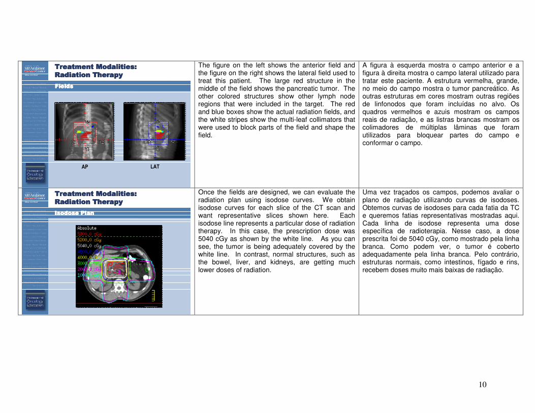
FieldsFieldsFieldsFields
AP LAT
The figure on the left shows the anterior field and the figure on the right shows the lateral field used to treat this patient. The large red structure in the middle of the field shows the pancreatic tumor. The other colored structures show other lymph node regions that were included in the target. The red and blue boxes show the actual radiation fields, and the white stripes show the multi-leaf collimators that were used to block parts of the field and shape the field.
A figura à esquerda mostra o campo anterior e a figura à direita mostra o campo lateral utilizado para tratar este paciente. A estrutura vermelha, grande, no meio do campo mostra o tumor pancreático. As outras estruturas em cores mostram outras regiões de linfonodos que foram incluídas no alvo. Os quadros vermelhos e azuis mostram os campos reais de radiação, e as listras brancas mostram os colimadores de múltiplas lâminas que foram utilizados para bloquear partes do campo e conformar o campo.
Treatment Modalities: Treatment Modalities: Treatment Modalities: Treatment Modalities:
Radiation Therapy Radiation Therapy Radiation Therapy Radiation Therapy
Isodose PlanIsodose PlanIsodose PlanIsodose Plan
Once the fields are designed, we can evaluate the radiation plan using isodose curves. We obtain isodose curves for each slice of the CT scan and want representative slices shown here. Each isodose line represents a particular dose of radiation therapy. In this case, the prescription dose was 5040 cGy as shown by the white line. As you can see, the tumor is being adequately covered by the white line. In contrast, normal structures, such as the bowel, liver, and kidneys, are getting much lower doses of radiation.
Uma vez traçados os campos, podemos avaliar o plano de radiação utilizando curvas de isodoses. Obtemos curvas de isodoses para cada fatia da TC e queremos fatias representativas mostradas aqui. Cada linha de isodose representa uma dose específica de radioterapia. Nesse caso, a dose prescrita foi de 5040 cGy, como mostrado pela linha branca. Como podem ver, o tumor é coberto adequadamente pela linha branca. Pelo contrário, estruturas normais, como intestinos, fígado e rins, recebem doses muito mais baixas de radiação.
11
Treatment Modalities: Treatment Modalities: Treatment Modalities: Treatment Modalities:
Radiation Therapy Radiation Therapy Radiation Therapy Radiation Therapy
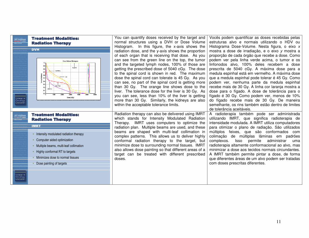
DVH
You can quantify doses received by the target and normal structures using a DVH or Dose Volume Histogram. In this figure, the x-axis shows the radiation dose, and the y-axis shows the proportion of each organ that is receiving that dose. As you can see from the green line on the top, the tumor and the targeted lymph nodes, 100% of those are getting the prescribed dose of 5040 cGy. The dose to the spinal cord is shown in red. The maximum dose the spinal cord can tolerate is 45 Gy. As you can see, no part of the spinal cord is getting more than 30 Gy. The orange line shows dose to the liver. The tolerance dose for the liver is 30 Gy. As you can see, less than 10% of the liver is getting more than 30 Gy. Similarly, the kidneys are also within the acceptable tolerance limits.
Vocês podem quantificar as doses recebidas pelas estruturas alvo e normais utilizando o HDV ou Histograma Dose-Volume. Nesta figura, o eixo x mostra a dose de irradiação, e o eixo y mostra a proporção de cada órgão que recebe a dose. Como podem ver pela linha verde acima, o tumor e os linfonodos alvo, 100% deles recebem a dose prescrita de 5040 cGy. A máxima dose para a medula espinhal está em vermelho. A máxima dose que a medula espinhal pode tolerar é 45 Gy. Como podem ver, nenhuma parte da medula espinhal recebe mais de 30 Gy. A linha cor laranja mostra a dose para o fígado. A dose de tolerância para o fígado é 30 Gy. Como podem ver, menos de 10% do fígado recebe mais de 30 Gy. De maneira semelhante, os rins também estão dentro de limites de tolerância aceitáveis.
Treatment Modalities: Treatment Modalities: Treatment Modalities: Treatment Modalities:
Radiation Therapy Radiation Therapy Radiation Therapy Radiation Therapy
IMRT
• Intensity modulated radiation therapy
• Computer aided optimization
• Multiple beams, multi-leaf collimation
• Highly conformal RT to targets
• Minimizes dose to normal tissues
• Dose painting of targets
Radiation therapy can also be delivered using IMRT which stands for Intensity Modulated Radiation Therapy. IMRT uses computers to optimize the radiation plan. Multiple beams are used, and these beams are shaped with multi-leaf collimation in complex patterns. This allows us to deliver highly conformal radiation therapy to the target, but minimize dose to surrounding normal tissues. IMRT also allows dose painting so that different areas of a target can be treated with different prescribed doses.
A radioterapia também pode ser administrada utilizando IMRT, que significa radioterapia de intensidade modulada. A IMRT utiliza computadores para otimizar o plano de radiação. São utilizados múltiplos feixes, que são conformados com colimação de múltiplas lâminas em padrões complexos. Isso permite administrar uma radioterapia altamente conformacional ao alvo, mas minimizar a dose aos tecidos normais circundantes. A IMRT também permite pintar a dose, de forma que diferentes áreas de um alvo podem ser tratadas com doses prescritas diferentes.
12
Treatment Modalities: Treatment Modalities: Treatment Modalities: Treatment Modalities:
Radiation Therapy Radiation Therapy Radiation Therapy Radiation Therapy
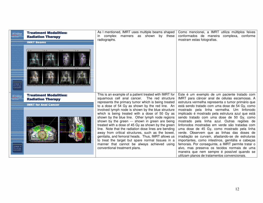
IMRT Beams
As I mentioned, IMRT uses multiple beams shaped in complex manners as shown by these radiographs.
Como mencionei, a IMRT utiliza múltiplos feixes conformados de maneira complexa, conforme mostram estas fotografias.
Treatment Modalities: Treatment Modalities: Treatment Modalities: Treatment Modalities:
Radiation Therapy Radiation Therapy Radiation Therapy Radiation Therapy
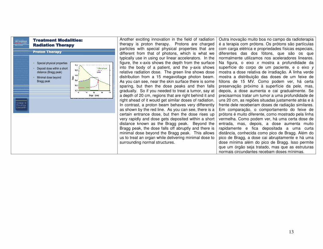
IMRT for Anal Cancer
This is an example of a patient treated with IMRT for squamous cell anal cancer. The red structure represents the primary tumor which is being treated to a dose of 54 Gy as shown by the red line. An involved lymph node is shown by the blue structure which is being treated with a dose of 50 Gy as shown by the blue line. Other lymph node regions shown by the green --- shown in green are being treated with a dose of 45 Gy as shown by the green line. Note that the radiation dose lines are bending away from critical structures, such as the bowel, genitalia, and femoral heads. Thus, IMRT allows us to treat the target but spare normal tissues in a manner that cannot be always achieved using conventional treatment plans.
Este é um exemplo de um paciente tratado com IMRT para câncer anal de células escamosas. A estrutura vermelha representa o tumor primário que está sendo tratado com uma dose de 54 Gy, como mostrado pela linha vermelha. Um linfonodo implicado é mostrado pela estrutura azul que está sendo tratado com uma dose de 50 Gy, como mostrado pela linha azul. Outras regiões de linfonodos mostradas em verde são tratadas com uma dose de 45 Gy, como mostrado pela linha verde. Observem que as linhas das doses de irradiação se curvam, afastando-se de estruturas importantes, como intestinos, genitália e cabeças femorais. Por conseguinte, a IMRT permite tratar o alvo, mas preserva os tecidos normais de uma maneira que nem sempre é possível quando se utilizam planos de tratamentos convencionais.
13
Treatment Modalities: Treatment Modalities: Treatment Modalities: Treatment Modalities:
Radiation Therapy Radiation Therapy Radiation Therapy Radiation Therapy
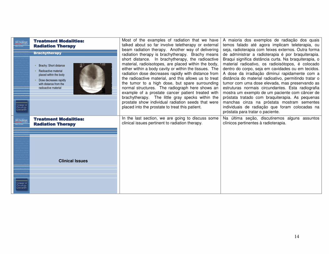
Proton Therapy
• Special physical properties
• Deposit dose within a short
distance (Bragg peak)
• Minimal dose beyond
Bragg peak
Another exciting innovation in the field of radiation therapy is proton therapy. Protons are charged particles with special physical properties that are different from that of photons, which is what we typically use in using our linear accelerators. In the figure, the x-axis shows the depth from the surface into the body of a patient, and the y-axis shows relative radiation dose. The green line shows dose distribution from a 15 megavoltage photon beam. As you can see, near the skin surface there is some sparing, but then the dose peaks and then falls gradually. So if you needed to treat a tumor, say at a depth of 20 cm, regions that are right behind it and right ahead of it would get similar doses of radiation. In contrast, a proton beam behaves very differently as shown by the red line. As you can see, there is a certain entrance dose, but then the dose rises up very rapidly and dose gets deposited within a short distance known as the Bragg peak. Beyond the Bragg peak, the dose falls off abruptly and there is minimal dose beyond the Bragg peak. This allows us to treat an organ while delivering minimal dose to surrounding normal structures.
Outra inovação muito boa no campo da radioterapia é a terapia com prótons. Os prótons são partículas com carga elétrica e propriedades físicas especiais, diferentes das dos fótons, que são os que normalmente utilizamos nos aceleradores lineares. Na figura, o eixo x mostra a profundidade da superfície do corpo de um paciente, e o eixo y mostra a dose relativa de irradiação. A linha verde mostra a distribuição das doses de um feixe de fótons de 15 MV. Como podem ver, há certa preservação próximo à superfície da pele, mas, depois, a dose aumenta e cai gradualmente. Se precisarmos tratar um tumor a uma profundidade de uns 20 cm, as regiões situadas justamente atrás e à frente dele receberiam doses de radiação similares. Em comparação, o comportamento do feixe de prótons é muito diferente, como mostrado pela linha vermelha. Como podem ver, há uma certa dose de entrada, mas, depois, a dose aumenta muito rapidamente e fica depositada a uma curta distância, conhecida como pico de Bragg. Além do pico de Bragg, a dose cai abruptamente e há uma dose mínima além do pico de Bragg. Isso permite que um órgão seja tratado, mas que as estruturas normais circundantes recebam doses mínimas.
14
Treatment Modalities: Treatment Modalities: Treatment Modalities: Treatment Modalities:
Radiation Therapy Radiation Therapy Radiation Therapy Radiation Therapy
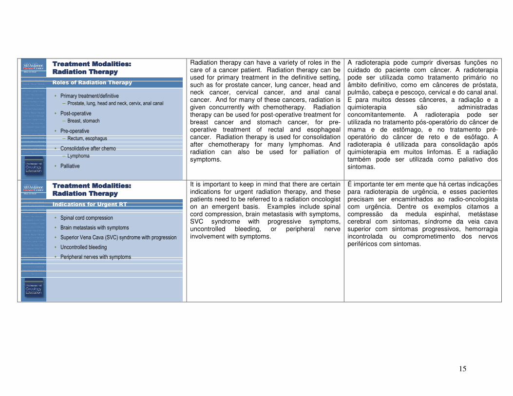
Brachytherapy
• Brachy: Short distance
• Radioactive material
placed within the body
• Dose decreases rapidly
with distance from the
radioactive material
Most of the examples of radiation that we have talked about so far involve teletherapy or external beam radiation therapy. Another way of delivering radiation therapy is brachytherapy. Brachy means short distance. In brachytherapy, the radioactive material, radioisotopes, are placed within the body, either within a body cavity or within the tissues. The radiation dose decreases rapidly with distance from the radioactive material, and this allows us to treat the tumor to a high dose, but spare surrounding normal structures. The radiograph here shows an example of a prostate cancer patient treated with brachytherapy. The little gray specks within the prostate show individual radiation seeds that were placed into the prostate to treat this patient.
A maioria dos exemplos de radiação dos quais temos falado até agora implicam teleterapia, ou seja, radioterapia com feixes externos. Outra forma de administrar a radioterapia é por braquiterapia. Braqui significa distância curta. Na braquiterapia, o material radioativo, os radioisótopos, é colocado dentro do corpo, seja em cavidades ou em tecidos. A dose da irradiação diminui rapidamente com a distância do material radioativo, permitindo tratar o tumor com uma dose elevada, mas preservando as estruturas normais circundantes. Esta radiografia mostra um exemplo de um paciente com câncer de próstata tratado com braquiterapia. As pequenas manchas cinza na próstata mostram sementes individuais de radiação que foram colocadas na próstata para tratar o paciente.
Treatment Modalities: Treatment Modalities: Treatment Modalities: Treatment Modalities:
Radiation Therapy Radiation Therapy Radiation Therapy Radiation Therapy
Clinical Issues
In the last section, we are going to discuss some clinical issues pertinent to radiation therapy.
Na última seção, discutiremos alguns assuntos clínicos pertinentes à radioterapia.
15
Treatment Modalities: Treatment Modalities: Treatment Modalities: Treatment Modalities:
Radiation Therapy Radiation Therapy Radiation Therapy Radiation Therapy
Roles of Radiation Therapy
• Primary treatment/definitive
– Prostate, lung, head and neck, cervix, anal canal
• Post-operative
– Breast, stomach
• Pre-operative
– Rectum, esophagus
• Consolidative after chemo
– Lymphoma
• Palliative
Radiation therapy can have a variety of roles in the care of a cancer patient. Radiation therapy can be used for primary treatment in the definitive setting, such as for prostate cancer, lung cancer, head and neck cancer, cervical cancer, and anal canal cancer. And for many of these cancers, radiation is given concurrently with chemotherapy. Radiation therapy can be used for post-operative treatment for breast cancer and stomach cancer, for pre-operative treatment of rectal and esophageal cancer. Radiation therapy is used for consolidation after chemotherapy for many lymphomas. And radiation can also be used for palliation of symptoms.
A radioterapia pode cumprir diversas funções no cuidado do paciente com câncer. A radioterapia pode ser utilizada como tratamento primário no âmbito definitivo, como em cânceres de próstata, pulmão, cabeça e pescoço, cervical e do canal anal. E para muitos desses cânceres, a radiação e a quimioterapia são administradas concomitantemente. A radioterapia pode ser utilizada no tratamento pós-operatório do câncer de mama e de estômago, e no tratamento pré-operatório do câncer de reto e de esôfago. A radioterapia é utilizada para consolidação após quimioterapia em muitos linfomas. E a radiação também pode ser utilizada como paliativo dos sintomas.
Treatment Modalities: Treatment Modalities: Treatment Modalities: Treatment Modalities:
Radiation Therapy Radiation Therapy Radiation Therapy Radiation Therapy
Indications for Urgent RT
• Spinal cord compression
• Brain metastasis with symptoms
• Superior Vena Cava (SVC) syndrome with progression
• Uncontrolled bleeding
• Peripheral nerves with symptoms
It is important to keep in mind that there are certain indications for urgent radiation therapy, and these patients need to be referred to a radiation oncologist on an emergent basis. Examples include spinal cord compression, brain metastasis with symptoms, SVC syndrome with progressive symptoms, uncontrolled bleeding, or peripheral nerve involvement with symptoms.
É importante ter em mente que há certas indicações para radioterapia de urgência, e esses pacientes precisam ser encaminhados ao radio-oncologista com urgência. Dentre os exemplos citamos a compressão da medula espinhal, metástase cerebral com sintomas, síndrome da veia cava superior com sintomas progressivos, hemorragia incontrolada ou comprometimento dos nervos periféricos com sintomas.
16
Treatment Modalities: Treatment Modalities: Treatment Modalities: Treatment Modalities:
Radiation Therapy Radiation Therapy Radiation Therapy Radiation Therapy
Complications from RT
• Complications mainly occur locally, in organs
that are in the RT field
• Some complications occur during treatment or
within days or weeks
• Some complications appear after months or years
Radiation can cause a variety of complications or side effects. These complications mainly occur locally in organs that are in the radiation field. Some complications can occur during treatment or within days or weeks while some complications can be long term and can appear after months or years.
A radiação pode causar várias complicações ou efeitos colaterais. Essas complicações ocorrem sobretudo localmente nos órgãos que se encontram no campo de irradiação. Algumas complicações podem ocorrer durante o tratamento ou após alguns dias ou semanas, enquanto que outras podem ser de longa duração e aparecer somente depois de meses ou anos.
Treatment Modalities: Treatment Modalities: Treatment Modalities: Treatment Modalities:
Radiation Therapy Radiation Therapy Radiation Therapy Radiation Therapy
Examples of Complications
• Fatigue
• Skin: erythema, desquamation
• CNS: somnolence, cognitive loss
• Head and Neck: mucositis, xerostomia
• Lung: pneumonitis
• Upper GI: nausea, esophagitis
• Lower GI: diarrhea, proctitis
• GU: cystitis, urethritis
• Hem: myelosuppression
• Cardiovascular toxicity
• Second malignancies
Examples of complications include fatigue, skin toxicity such as erythema and desquamation. Radiation to the brain can lead to somnolence or cognitive loss. That to the head and neck area could potentially lead to mucositis and xerostomia. Radiation to the lung can cause pneumonitis; to the upper GI can lead to nausea and esophagitis; and to the lower GI can lead to diarrhea and proctitis. Radiation therapy to the genitourinary system can cause cystitis, urethritis or sexual dysfunction. Radiation can also cause myelosuppression. Two of the most important long-term side effects of radiation include cardiovascular toxicity and second malignancies.
Exemplos de complicações são, entre outros: fadiga, toxicidade cutânea, como eritema e descamação. A radiação do cérebro pode causar sonolência ou perda da cognição. Daí à área da cabeça e do pescoço poderia resultar em mucosite e xerostomia. A radiação do pulmão pode causar pneumonite, a do trato gastrointestinal superior pode causar náuseas e esofagite e a do trato gastrointestinal inferior pode causar diarreia e proctite. A radioterapia do sistema geniturinário pode causar cistite, uretrite ou disfunção sexual. A radiação também pode causar mielossupressão. Dois dos efeitos colaterais de longo prazo mais importantes da radiação são a toxicidade cardiovascular e neoplasias malignas secundárias.
17
Treatment Modalities: Treatment Modalities: Treatment Modalities: Treatment Modalities:
Radiation Therapy Radiation Therapy Radiation Therapy Radiation Therapy
Role of the Radiation Oncologist
• Evaluates whether patient is appropriate for RT
– Integral part of multidisciplinary team
• Plans and delivers radiation therapy
– Aided by physicists, dosimetrists, therapists
• Treats symptoms and side-effects
• Monitors for relapses and long-term toxicity
• Specialized procedures
– Brachytherapy, Intraoperative radiotherapy
What is the role of a radiation oncologist? A radiation oncologist evaluates whether a particular patient is appropriate for radiation. And in doing so, the radiation oncologist functions as an integral part of the multidisciplinary oncology team. Next, the radiation oncologist plans and delivers radiation therapy. This is a team effort and the radiation oncologist is aided by physicists, dosimetrists, and radiation therapists. Radiation oncologists help treat symptoms and side effects of radiation and also monitors these patients in the long term for relapses and long-term toxicity. Finally, the radiation oncologist performs specialized procedures, such as brachytherapy and intraoperative radiation therapy.
Qual o papel do radio-oncologista? O radio-oncologista avalia o paciente para saber se pode receber radiação. Com isso, o radio-oncologista atua como parte integrante da equipe multidisciplinar de oncologia. A seguir, o radio-oncologista planeja e administra a radioterapia. Esse é um trabalho de grupo e o radio-oncologista conta com o apoio de médicos, dosimetristas e radioterapeutas. Os radio-oncologistas auxiliam no tratamento de sintomas e efeitos colaterais da radiação, bem como monitoram os pacientes quanto a recaídas e toxicidades em longo prazo. Finalmente, o radio-oncologista executa procedimentos especializados, como braquiterapia e radioterapia intraoperatória.
Treatment Modalities: Treatment Modalities: Treatment Modalities: Treatment Modalities:
Radiation Therapy Radiation Therapy Radiation Therapy Radiation Therapy
Conclusions
• Radiation is an important treatment modality for
the management of cancer
• Treatment planning includes simulation, contouring
targets and designing beam arrangements
• Specialized radiation techniques include
intensity modulated radiation therapy, proton
therapy and brachytherapy
In conclusion, we have discussed today that radiation has an --- is an important treatment modality for the management of cancer. The treatment planning process includes simulation, contouring targets, and designing beam arrangements. Specialized radiation techniques include Intensity Modulated Radiation Therapy, Proton Therapy, and Brachytherapy. We have also discussed some clinical issues relevant to radiation therapy. Thank you for your attention.
Em conclusão, hoje discutimos que a radiação é uma modalidade de tratamento importante para o manejo do câncer. O processo do planejamento terapêutico inclui simulação, contornos dos alvos e traçado dos arranjos dos feixes. As técnicas especializadas de radiação são a radioterapia de intensidade modulada, a terapia com prótons e a braquiterapia. Além disso, discutimos alguns assuntos clínicos relevantes à radioterapia. Obrigado pela atenção.