Embed Size (px)
Citation preview
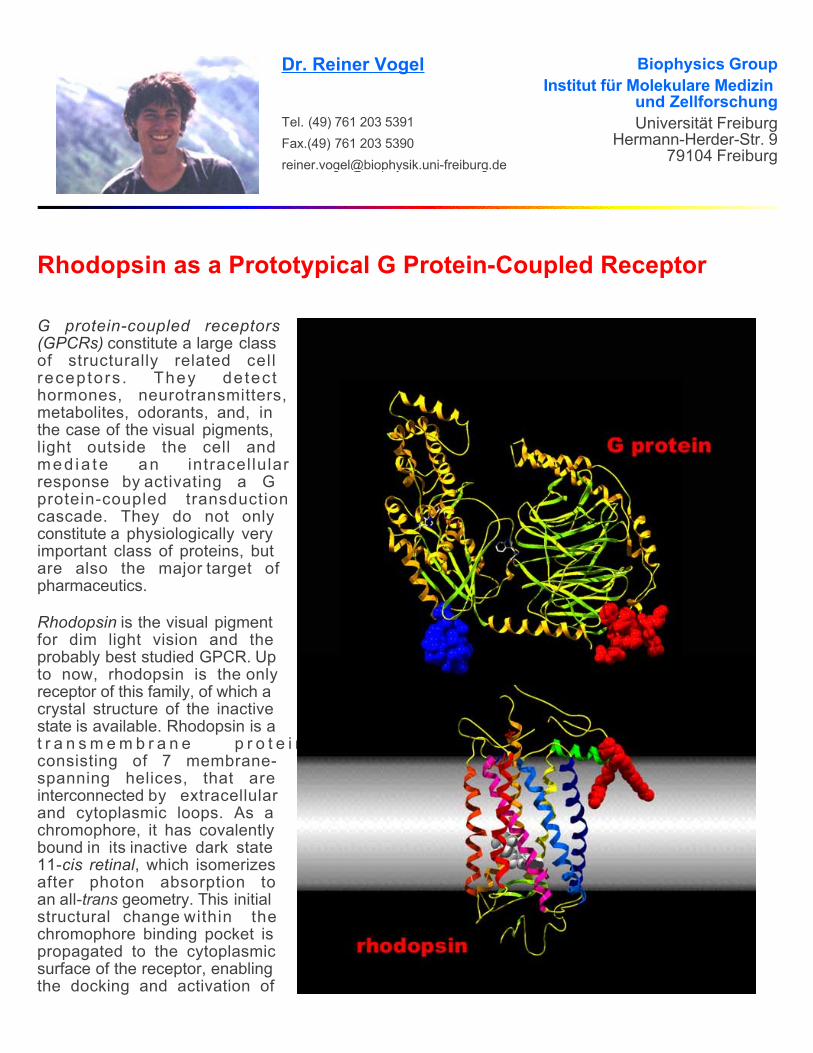
Dr. Reiner Vogel
Tel. (49) 761 203 5391Fax.(49) 761 203 [email protected]
Biophysics GroupInstitut für Molekulare Medizin
und ZellforschungUniversität Freiburg
Hermann-Herder-Str. 979104 Freiburg
Rhodopsin as a Prototypical G Protein-Coupled Receptor
G protein-coupled receptors (GPCRs) constitute a large class of structurally related cell receptors . They detec thormones, neurotransmitters, metabolites, odorants, and, in the case of the visual pigments, light outside the cell and med ia te an intracel lular response by activating a G protein-coupled transduction cascade. They do not only constitute a physiologically very important class of proteins, but are also the major target of pharmaceutics.
Rhodopsin is the visual pigment for dim light vision and the probably best studied GPCR. Up to now, rhodopsin is the only receptor of this family, of which a crystal structure of the inactive state is available. Rhodopsin is at r a n s m e m b r a n e p r o t e i n consisting of 7 membrane-spanning helices, that are interconnected by extracellular and cytoplasmic loops. As a chromophore, it has covalently bound in its inactive dark state 11-cis retinal, which isomerizesafter photon absorption toan all- geometry. This initial structural change within the chromophore binding pocket is propagated to the cytoplasmicsurface of the receptor, enabling the docking and activation of
trans
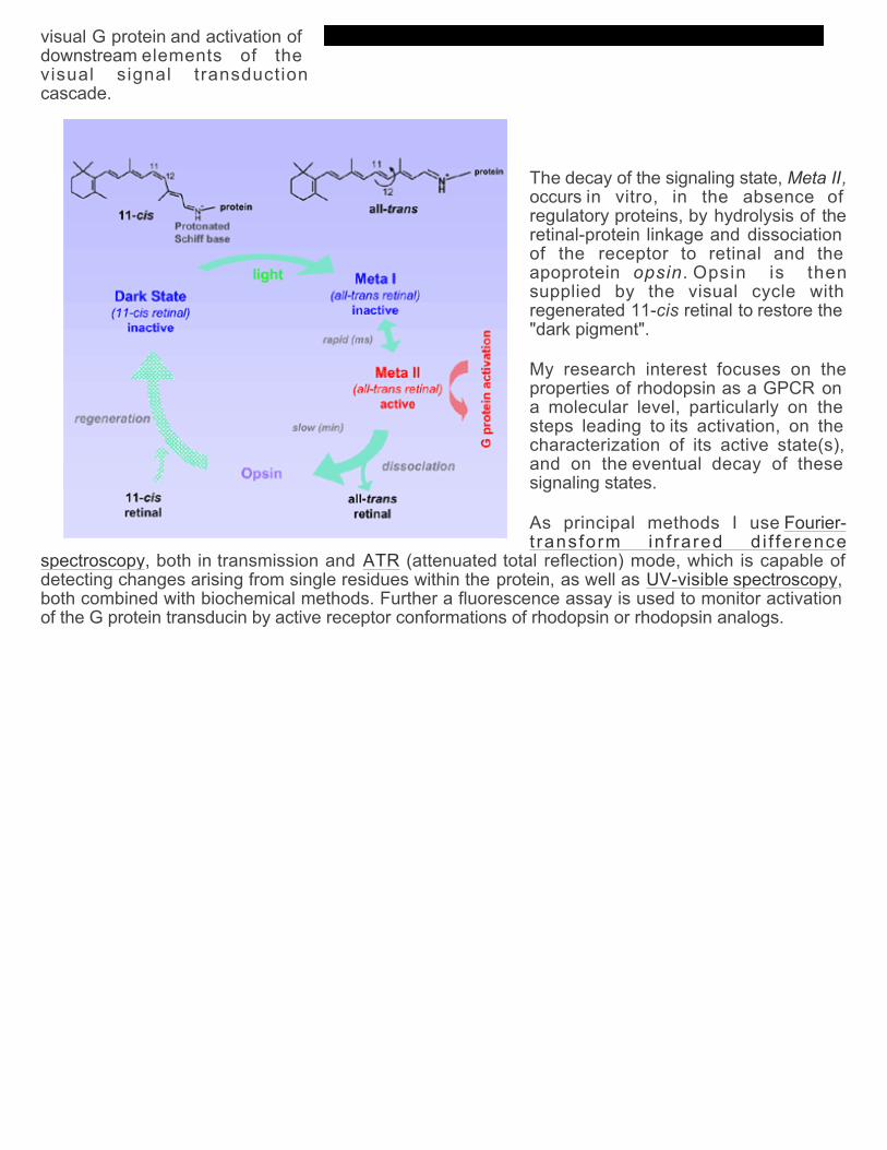
visual G protein and activation of downstream elements of the visual signal transduction cascade.
The decay of the signaling state, Meta II,occurs in vitro, in the absence of regulatory proteins, by hydrolysis of theretinal-protein linkage and dissociation of the receptor to retinal and the apoprotein opsin. Opsin is then supplied by the visual cycle with regenerated 11- retinal to restore the "dark pigment".
cis
My research interest focuses on theproperties of rhodopsin as a GPCR on a molecular level, particularly on the steps leading to its activation, on the characterization of its active state(s), and on the eventual decay of these signaling states.
As principal methods I use
, both in transmission and (attenuated total reflection) mode, which is capable ofdetecting changes arising from single residues within the protein, as well as , both combined with biochemical methods. Further a fluorescence assay is used to monitor activation of the G protein transducin by active receptor conformations of rhodopsin or rhodopsin analogs.
Fourier-t ransform in f rared d i f ference
spectroscopy ATRUV-visible spectroscopy
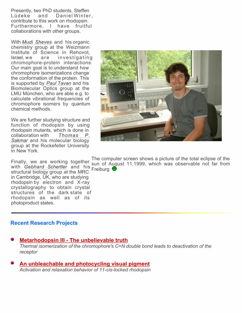
Presently, two PhD students, Steffen Lüdeke and Dan ie l Win te r , contribute to this work on rhodopsin. Furthermore, I have fruitful collaborations with other groups.
With and his organic chemistry group at the Weizmann Institute of Science in Rehovot, Israel, we a re inves t iga t ing chromophore-protein interactions. Our main goal is to understand how chromophore isomerizations change the conformation of the protein. This is supported by and his Biomolecular Optics group at the LMU München, who are able e.g. to calculate vibrational frequencies of chromophore isomers by quantumchemical methods.
Mudi Sheves
Paul Tavan
We are further studying structure andfunction of rhodopsin by using rhodopsin mutants, which is done in collaboration with
and his molecular biology group at the Rockefeller University in New York.
Thomas P.Sakmar
Finally, we are working together with and hisstructural biology group at the MRC in Cambridge, UK, who are studying rhodopsin by electron and X-ray crystallography to obtain crystal structures of the dark state of rhodopsin as well as of its photoproduct states.
Gebhard Schertler
The computer screen shows a picture of the total eclipse of the sun of August 11,1999, which was observable not far from Freiburg
Recent Research Projects
Metarhodopsin III - The unbelievable truthThermal isomerization of the chromophore's C=N double bond leads to deactivation of the receptor An unbleachable and photocycling visual pigmentActivation and relaxation behavior of 11-cis-locked rhodopsin
Membrane protein conformation and stabilityInfluence of salts on protein unfolding and conformational transitions of rhodopsin Active and inactive conformations of opsinConformational equilibrium of the receptor protein in the absence of ligands The protonation state of the Schiff base in the active receptor stateAnion binding mediates formation of an active Meta II state with protonated Schiff base Influence of protein-ligand interaction on receptor activationRemoval of the 9-methyl group changes all-trans retinal from a full to an only partial agonist
See also: Rhodopsin Mutants
Publications
Rhodopsin and G Protein-Coupled Receptors
2004
(2004) Electron crystallography reveals the structure of Metarhodopsin I. , in pressRuprecht J, Mielke T, Vogel R, Villa C, Schertler GFX
EMBO J
(2004) Photoreactions of Metarhodopsin III. 43: 10255-10264 Vogel R, Lüdeke S, Radu I, Siebert F, Sheves M Biochemistry
Medline URL
(2004) Formation of Meta III during the decay of activated rhodopsin proceeds via Meta I and not via Meta II. 43: 9457-9466 Vogel R, Siebert F, Zhang XY, Fan GB, Sheves M
Biochemistry Medline URL
(2004) Vogel R Influence of salts on rhodopsin photoproduct equilibria and protein stability. , in press
Curr Opin Colloid Interf Sci
(2004) Rhodopsin photoproducts in 2D crystals. 338:597-609
Vogel R, Ruprecht J, Villa C, Mielke T, Schertler GFX, Siebert F J Mol Biol Medline URL
2003
(2003) Deactivation of rhodopsin in the transition from the signaling state Meta II to Meta III involves a thermal isomerization of the retinal chromophore C=N double bond. 42: 9863-9874
Vogel R, Siebert F, Mathias G, Tavan P, Fan GB, and Sheves MBiochemistry
Medline URL
(2003) New insights from FTIR spectroscopy into molecular properties and activation mechanisms of the visual pigment rhodopsin. 72: 133-148 Vogel R, Siebert F
Biospectroscopy Medline URL
2002
(2002) Rhodopsin with 11- -locked chromophore is capable of forming an active state photoproduct. 277: 40229-40234 Fan GB, Siebert F, Sheves M, Vogel R cis
J Biol Chem Medline URL
(2002) A non-bleachable rhodopsin analogue with a slow photocycle. 277: 40222-40228
Vogel R, Fan GB, Lüdeke S, Siebert F, Sheves M J Biol Chem Medline URL
(2002) Conformation and stability of alpha-helical membrane proteins: I. Influence of salts on conformational equilibria between active and inactive states of rhodopsin. 41: 3529-3535 Vogel R, Siebert F
Biochemistry Medline URL
(2002) Conformation and stability of alpha-helical membrane Proteins: II. Influence of pH and salts on Vogel R, Siebert F
stability and unfolding of rhodopsin. 41: 3536-3445 Biochemistry Medline URL
2001
(2001) Conformations of the active and the inactive states of opsin. 276: 38487-38493 Vogel R, Siebert F J Biol Chem
Medline URL
(2001) Anions specifically stabilize a Metarhodopsin II-like photoproduct with a protonated Schiff base. 40: 13342-13352 Vogel R, Fan GB, Siebert F, Sheves M
Biochemistry Medline URL
(2001) Salt Dependence of the formation and stability of the signaling state in G protein-coupled receptors: Evidence for the involvement of the Hofmeister effect. 40: 483-493 Vogel R, Fan GB, Sheves M, Siebert F
Biochemistry Medline URL
(2001) Conformation and stability of rhodopsin photoproducts are influenced by salts: General implications of the Hofmeister effect on membrane proteins. 80: 47A Siebert F, Vogel R, Fan GB, Sheves M
Biophys J abstract
2000
(2000) Vibrational spectroscopy as a tool for probing protein function. 4: 518-523 Vogel R, Siebert F Curr Opin Chem Biol
Medline URL
(2000) The molecular origin of the inhibition of transducin activation in rhodopsin lacking the 9-methyl group of the retinal chromophore: A UV-vis and FTIR spectroscopic study. 39: 8895-8908
Vogel R, Fan GB, Sheves M, Siebert FBiochemistry
Medline URL
(2000) Bacterial expression of G-protein coupled receptors: Prediction of expression levels fromsequence. 7: 109-119 Kiefer H, Vogel R, Maier K
Receptors Channels (Medline URL)
1999
(1999) Refolding of G protein-coupled receptors from inclusion bodies produced in . 27: 908-912 Kiefer H, Maier K, Vogel R
E.coli Biochem Soc Trans Medline
Chemiluminescence and Reactive Oxygen Species
(1999) A model for the generation of low level chemiluminescence from microbiological growth media and its depletion by bacterial cells. 48: 375-383 Vogel R, Süssmuth R
Bioelectrochem Bioenerg Medline URL
(1999) Low level chemiluminescence from liquid culture media. 86: 999-1007 Vogel R, Süssmuth R J Appl Microbiol
Medline URL
(1999) Weak light emission patterns from lactic acid bacteria. 14: 99-105 Vogel R, Süssmuth R Luminescence Medline URL
1998
(1998) Involvement of the cell membrane in chemiluminescence patterns from bacterialcultures. 46: 65-69 Vogel R, Süssmuth R
Bioelectrochem Bioenerg abstract URL
(1998) Chemiluminescence patterns from bacterial cultures undergoing bacteriophage-induced mass lysis. 46: 59-64 Vogel R, Guo X, Süssmuth R
Bioelectrochem Bioenerg abstract URL
(1998) Interaction of bacterial cells with weak light emission from culture media. 45: 93-101
Vogel R, Süssmuth R Bioelectrochem Bioenerg abstract URL
(1998) About the quenching of the chemiluminescence from liquid culture media by microorganisms and weak light emission patterns from bacterial cultures. PhD Thesis, University of Hohenheim, GermanyVogel R
abstract pdf
(1998) Weak light emission from bacteria and their interaction with culture media. In , eds. Chang JJ, Fisch J, Popp FA. Kluwer Academic Publishers, Dordrecht, 19-44 Vogel R, Süssmuth R Biophotons
abstract URL
Theoretical Polymer Physics
(1995) Continuous-time random walk of a rigid triangle. 28: 6645-6653 Sokolov IM, Vogel R, Alemany PA, Blumen A J Phys A
abstract URL
(1994) A dumbell's random walk in continuous time. 27: 7733-7738 Alemany PA, Vogel R, Sokolov IM, Blumen A J Phys A
abstract URL
Links
SMA MeteoSchweiz Building Telescopes
Glen Canyon Without the Lake Die Maus
Molecular graphics on this site were generated with andray tracing.
Deep View Pov-Ray
last update: 04-Aug-04 Webmaster