Embed Size (px)
Citation preview
RESEARCH ARTICLE Open Access
Sequencing and curation strategies foridentifying candidate glioblastomatreatmentsMayu O. Frank1,2†, Takahiko Koyama3†, Kahn Rhrissorrakrai3†, Nicolas Robine1, Filippo Utro3, Anne-Katrin Emde1,Bo-Juen Chen1,12, Kanika Arora1, Minita Shah1, Heather Geiger1, Vanessa Felice1, Esra Dikoglu1,13, Sadia Rahman1,Alice Fang1, Vladimir Vacic1,14, Ewa A. Bergmann1,15, Julia L. Moore Vogel1,2,16, Catherine Reeves1, Depinder Khaira1,Anthony Calabro1,17, Duyang Kim1, Michelle F. Lamendola-Essel1,18, Cecilia Esteves1,19, Phaedra Agius1,Christian Stolte1, John Boockvar4, Alexis Demopoulos5, Dimitris G. Placantonakis6, John G. Golfinos6,Cameron Brennan7, Jeffrey Bruce8, Andrew B. Lassman8, Peter Canoll8, Christian Grommes7, Mariza Daras7,Eli Diamond7, Antonio Omuro7,20, Elena Pentsova7, Dana E. Orange2,9, Stephen J. Harvey10, Jerome B. Posner7,Vanessa V. Michelini10, Vaidehi Jobanputra1,8, Michael C. Zody1, John Kelly3, Laxmi Parida3,Kazimierz O. Wrzeszczynski1, Ajay K. Royyuru3 and Robert B. Darnell1,2,11*
Abstract
Background: Prompted by the revolution in high-throughput sequencing and its potential impact for treatingcancer patients, we initiated a clinical research study to compare the ability of different sequencing assays andanalysis methods to analyze glioblastoma tumors and generate real-time potential treatment options for physicians.
Methods: A consortium of seven institutions in New York City enrolled 30 patients with glioblastoma andperformed tumor whole genome sequencing (WGS) and RNA sequencing (RNA-seq; collectively WGS/RNA-seq); 20of these patients were also analyzed with independent targeted panel sequencing. We also compared results ofexpert manual annotations with those from an automated annotation system, Watson Genomic Analysis (WGA), toassess the reliability and time required to identify potentially relevant pharmacologic interventions.
Results: WGS/RNAseq identified more potentially actionable clinical results than targeted panels in 90% of cases,with an average of 16-fold more unique potentially actionable variants identified per individual; 84 clinicallyactionable calls were made using WGS/RNA-seq that were not identified by panels. Expert annotation and WGAhad good agreement on identifying variants [mean sensitivity = 0.71, SD = 0.18 and positive predictive value (PPV) =0.80, SD = 0.20] and drug targets when the same variants were called (mean sensitivity = 0.74, SD = 0.34 and PPV =0.79, SD = 0.23) across patients. Clinicians used the information to modify their treatment plan 10% of the time.
Conclusion: These results present the first comprehensive comparison of technical and machine augmentedanalysis of targeted panel and WGS/RNA-seq to identify potential cancer treatments.
© The Author(s). 2019 Open Access This article is distributed under the terms of the Creative Commons Attribution 4.0International License (http://creativecommons.org/licenses/by/4.0/), which permits unrestricted use, distribution, andreproduction in any medium, provided you give appropriate credit to the original author(s) and the source, provide a link tothe Creative Commons license, and indicate if changes were made. The Creative Commons Public Domain Dedication waiver(http://creativecommons.org/publicdomain/zero/1.0/) applies to the data made available in this article, unless otherwise stated.
* Correspondence: [email protected]†Mayu O. Frank, Takahiko Koyama and Kahn Rhrissorrakrai contributedequally to this work.1New York Genome Center, 101 Avenue of the Americas, New York, NY10013, USA2Laboratory of Molecular Neuro-Oncology, The Rockefeller University, 1230York Avenue, New York, NY 10065, USAFull list of author information is available at the end of the article
Frank et al. BMC Medical Genomics (2019) 12:56 https://doi.org/10.1186/s12920-019-0500-0
BackgroundOncology is one of the first areas where next-generationsequencing is being applied [1–3]. Sequencing is used toidentify genetic variants that could be pharmacologicallytargeted, allowing identification of drug effects in strati-fied populations that may otherwise be missed [2, 4].Panel-based sequencing, using hybridization and captureof specific regions of key genes or of all genes (wholeexomes; WES), versus whole genome sequencing (WGS)are different technologies with different costs that havenot previously been directly compared. 10,000 cancerpatients sequenced with the MSK-IMPACT panel identi-fied potentially clinically actionable calls in 36.7% ofindividuals sequenced [5]. Deep sequencing coverage in-creases sensitivity for rare variants in heterogeneous tu-mors. WGS, however, does not rely on hybridization andcapture, a source of potential bias, and is able to identifynon-coding variants such as enhancer bindings sites [6]and increases sensitivity for small copy number variants(CNVs) and missense mutations, indels, [7] intronic var-iants, [8] and gene fusions [9]. The relative impact of ofthese technologies on making clinically actionable vari-ant calls is unknown.Panel-based sequencing is used to identify treatment tar-
gets in tumors including glioblastoma (GBM) [10–13], themost common adult brain malignancy with a median sur-vival of 14.2months [14]. The Cancer Genome Atlas(TCGA) analyzed GBM and established four molecular sub-types [15] defined by IDH1 mutation and methylation sta-tus, [16] and more recently three subtypes which takes intoconsideration tumor purity and heterogeneity, [17] but hasnot yet led to new therapies. Panel sequencing and WGS/RNA-sequencing (WGS/RNA-seq) provide logical paths for-ward to identify variants. However, the process of sequenceanalysis and prioritizing variants is laborious, requiringhighly trained experts, particularly in WGS/RNA-seq; thisprompted prompting assessment of automated analyses.The New York Glioblastoma Genome Consortium
(NY-GGC) was organized in 2013 at the RockefellerUniversity and New York Genome Center (NYGC) toconduct a feasibility study of using WGS/RNA-seq toidentify tumor-specific variants and potential drug tar-gets, to compare WGS/RNA-seq to panels, and to assessthe reliability of automated versus manual analyses.Here, we describe an integrated analysis of 30 GBM pa-tients recruited through seven participating institutions.
MethodsStudy designThe NY-GGC was formed in 2013 through a collaborationinitiated at Rockefeller University, and included MemorialSloan Kettering Cancer Center, New York University Med-ical School, Northwell Health (Lenox Hill Hospital andNorth Shore University Hospital), Columbia University
Medical Center and New York Genome Center, sharingtumor and blood samples for sequencing, relevant clinicalhistories, and raw sequence data (BAM files). Variant callfiles (VCFs) were analyzed by NY-GGC and by WGA.
PatientsEntry criteria for this study were: minimum age of three,histologically confirmed GBM at referring institutionwith no requirement for central pathology review, Kar-nofsky score of at least 60, life expectancy of at least 6months, and potential interest in further treatment.Clinical data was collected including results of any otherclinical sequencing.All participants provided written informed consent. Pro-
tocols were approved by local or central Institutional Re-view Boards at: Rockefeller University, Biomedical ResearchAlliance of New York (on behalf of Northwell Health), Me-morial Sloan Kettering Cancer Center, New York UniversitySchool of Medicine, and Weill Cornell Medicine.
WGS and RNA-seqPaired tumor and normal (blood) samples were sequencedfrom each individual and analyzed by WGS at 80X tumorand 40X normal coverage as previously described [18].Ploidy values were used to estimate chromosome, gene,and allele copy number. We analyzed TERT promotervariants, intronic splice site variants (annotated bySnpEff), and exonic variants. Single nucleotide variants(SNVs) were classified by Tiers. Tier 1 variants are definedas variants with known clinical significance in GBM as de-fined by CIViC (v.alpha, 3/2015). Tier 2 variants areknown to be clinically significant in another tumor type asdefined by CIViC. Tier 3 are variants of unknown signifi-cance (VUS) in known actionable cancer genes with asso-ciated drugs. Tier 4 are VUS mutations in Cosmic CancerCensus Genes (v.75) [18].Where RNA was available, RNA-seq was performed as
previously described, [18] with the addition that whenthe RNA integrity number (RIN) score [19] was less than7, we used the KAPA Stranded RNA-seq with RiboErase(P/N: KK8483) with Agilent SureSelectXT v6 + Cosmic(P/N: 5190–9308). We used RNA-seq data to annotatevariants discovered in WGS according to the level of ex-pression of each variant, estimated by identifying thenumber of supporting reads and allelic fraction. Geneexpression in the tumor samples was assessed and com-pared with 169 GBM samples from TCGA and a modi-fied z-score was calculated as previously described [18].Modified z-scores of RNA-seq normalized expressiondata per gene was used as proxy for differential gene ex-pression. Modified z-score per gene was calculated bysubtracting the median transcripts per million (TPM)value (over the TCGA GBM cohort) from each sample’sTPM and dividing by the TCGA median absolute
Frank et al. BMC Medical Genomics (2019) 12:56 Page 2 of 16
deviation. The z-score therefore represents the numberof standard deviations each sample is from the medianexpression value of a specific gene of the TCGA GBMcancer cohort. For tumor-normal comparisons of splicesite variants, percent spliced in (PSI) was calculated asthe number of reads supporting the unannotated alter-native splicing event divided by the number of canonicalreads supporting the annotated event [20] and fusiontranscript discovery was performed using RNA-seq dataand FusionCatcher [21] as previously described [9].When associating variants with potential therapies, we
prioritized variants of high copy number focal gains andtwo copy homozygous loss over lower copy numberwhole arm gains or heterozygous losses. However, attimes, lower copy number changes (< 5 copy gains orheterozygous losses) were also reported as others havedone [22].
Comparison with Watson Genomic Analytics (WGA) andtargeted tumor panelsWGA is an IBM Research proof-of-concept environment ofWatson for Genomics described previously, [18, 23, 24] withthe addition of algorithmic updates that include basic pro-cessing of structural variants (SVs) (version 11/2016). WGAis a cloud-based cognitive system capable of analyzing muta-tion, gene expression, copy number alteration (CNA) andSV data provided as VCFs by leveraging 20+ structured andunstructured data sources. WGA first performs a MolecularProfile Analysis (MPA) to identify possible driver mutationsand drug response biomarkers in a disease-specific manner.MPA evaluates mutations using data from structured data-bases such as COSMIC and ExAC to search for known vari-ants and remove possible benign germline mutations. It alsouses evidence extracted from literature using bothmachine-based and expert-based manual curation. Thesedata sources are used to create a system capable of categor-izing alterations as pathogenic, benign, or VUS in a diseasespecific manner. WGA performs a similar analysis for CNAsand gene expression changes and takes into the account thefunctional annotation of a gene when assessing the rele-vance of a CNA or differential expression. WGA also pro-cesses SVs using DNA breakpoints data output bythe structural event detection program Delly [25].From the MPA results, pathogenic and likely patho-
genic alterations are assessed for potential direct and in-direct (i.e. via pathway mechanisms) therapeutic options.WGA’s pathway and drug analysis identifies which ther-apies are most applicable and categorizes therapies bydifferent levels of evidence from strongest, which in-cludes FDA-recognized marker or mutations predictiveof response, to the weakest, which can represent a nor-mally appropriate therapy with a clinically supported re-sistance marker present in the patient. For eachpotential treatment option that WGA identifies, it
provides all supporting evidence for the use of that ther-apy, mechanism of action and information on eligibleclinical trials if available. WGA currently limits availabletreatments to molecularly targeted therapies.To access unstructured data, particularly from literature,
clinical trial information and drug label data, WGA ap-plies Natural Language Processing (NLP). In brief, NLPrequires a training phase on a high-quality corpus of pa-pers and text that have been manually annotated by sub-ject matter experts for terms and relationships relevant forWGA to understand, including genes, proteins, mutations,drugs, and effect. After this training phase, the NLP modelis applied to the full unstructured data set, and after valid-ation by automated methods and subject matter experts,the information is integrated into WGA.NY-GGC provided VCFs, including CNV and gene ex-
pression files as input to WGA. Over the course of thisstudy, IBM and NY-GGC periodically reviewed resultsand discussed differences between NY-GGC and WGA.Insights and feedback from these sessions along withsimilar sessions for other studies [24] were used to im-prove the process of automated curation, for example byidentifying additional data sources for consideration andadjusting parameter weights. As new GBM samples be-came available, the entire cohort to that date was reana-lyzed by the then current version of WGA.Using NY-GGC calls as a truth set, we compared
mean sensitivity and positive predictive value (PPV)across patients to determine similarity between manualand automated systems for reported variants and drugtargets based on VCFs.
Comparison with targeted panelsPrior targeted NGS panel testing results were available for20 patients and were compared with WGS results. The per-centage of variants that were in common, that were uniquelycalled by WGS, or that were uniquely called by the targetedpanel were calculated and shown in a heatmap. TargetedNGS panel testing was done by Memorial Sloan KetteringCancer Center IMPACT (Panel 1), FoundationOne (Panel2), New York University Next Generation SequencingTumor 50 Panel (Panel 3), Weill Cornell Medical CenterPrecision Medicine’s whole exome sequencing assay(Panel 4), University of San Francisco’s 500 Cancer GenePanel (Panel 5) and Caris Molecular Intel (Panel 6).
Therapeutic targets and drug recommendationsAfter annotating the tumor-specific gene variants, rela-tive to normal germline DNA, based on SNV, CNV, SV,and RNA-seq data, [18] variants were associated withdrugs in the NYGC database.NYGC database was assembled by manual curation of
publically available data from the National ComprehensiveCancer Network, (https://www.nccn.org/), US Food and
Frank et al. BMC Medical Genomics (2019) 12:56 Page 3 of 16
Drug Administration (https://www.fda.gov/Drugs/Infor-mationOnDrugs/ApprovedDrugs), CIViC - Clinical Inter-pretations of Variants in Cancer (civic.genome.wustl.edu),Precision Cancer Therapy-MD Anderson (https://pct.mdanderson.org/), OncoKB (oncokb.org), canSar (https://cansar.icr.ac.uk), Pharmacogenomics Knowledgebase -PharmGKB (www.pharmgkb.org), Clinical Trials.gov (clin-icaltrials.gov) and from directed literature searches. Thecurrent NYGC drug to gene database contains 260 genesassociated with a least one drug.Prioritization and rationale of drug recommenda-
tions was based on further manual assessment by theNY-GGC including but not limited to: variant Tier,quality of data supporting variant call, interpretationof the consequences of VUS in light of literature re-search (structural and functional analysis of proteininteractions, prior knowledge of analogous variants),including analysis of X-Ray crystallographic structures,drug FDA approval status, drug identification in acurrent GBM trial, and record of drug success in thetreatment of GBM and/or other cancer types specificto the variant.Each individual’s results from NY-GGC were first
discussed in an internal tumor board comprised ofthose involved in development of the analytic pipe-line, bioinformaticians, project managers, pathway an-alysts, and clinical experts, and subsequently at aNY-GGC tumor board with that same team togetherwith referring physicians and collaborating physiciansand scientists. Further clinical use of the results pro-vided were at the sole discretion of the referringphysician.
Statistical analysisMean tumor purity and ploidy were calculated withstandard deviations (SD). Median number of SNVs arereported with interquartile range (IQR). Correlation be-tween RNA-seq and exonic SNV variant allele frequency(VAF) was assessed using Pearson’s correlation coeffi-cient. Mean number of alternative splice variants wascalculated with SD. Mean sensitivity and PPV were cal-culated to compare agreement between the number ofcalls made between WGS and WGA.
ResultsPatients and study processBetween March 2015 and July 2016, 36 patients werescreened and 30 were enrolled (Table 1). Four were ex-cluded due to final pathology indicating diagnosis otherthan GBM and 2 others died before sequencing began.Three participants had two separate tumor samples se-quenced; one from tumors resected from two distinctbrain areas, another from an enhancing andnon-enhancing region on MRI, and a third from samples
representing different histological characteristics.Throughout the study, we refined our processes for sam-ple collection, squencing, analysis, interpretation, and dis-semination of information to referring physicians. Theaverage time from sample receipt to the tumor board meet-ings was 4.5 months (SD = 2.1) and the average time frompost bioinformatic pipeline analysis to the tumor boardmeetings was 1.9months (SD= 1.1).
Tumor purity and ploidyTumor purity ranged from 15 to 95% (mean= 71%, SD=16%) for all samples. Two samples had tumor purity < 20%(15 and 19%). The average estimated ploidy was 2.03 (range1.59 to 4.06, SD= 1.235). Four samples were hyperploid andthree were hypoploid.
Table 1 Patient characteristics
n = 30
Age, median (range) 63 (25–81)
Female, no. (%) 12 (40%)
Resections, no. (%)
Initial 19 (63%)
Biopsy only 2 (7%)
Re-resection 7 (23%)
Re-resection of subtotal 2 (7%)
Prior treatment of 7 recurrent tumors, no.
RT/TMZ 6
Bevacizumab 3
Cetuximab 2
RT/Nivolimab 1
RT/Rindopepimut 1
CCNU 1
Gamma knife 1
Prior cranial RT, unrelated to GBM 1
Days from initial resection to sample submission,median (IQR)
67 (266)
Sample preservation, no. (%) n = 33
Fresh frozen 13 (40%)
OCT embedded 12 (36%)
Formalin-fixed paraffin-embedded 8 (24%)
Tumor biomarkers, no./no. assessed
EGFR amplification 8/20
MGMT methylation 12/30
IDH1 R132H mutation 2/30
All tumors were histologically confirmed as WHO grade IV gliomas. However,one had focal sarcomatous features, another was initially reviewed aspleomorphic xanthoastrocytoma with anaplastic features then as an epitheliodGBM upon re-revew, and a third had a PNET-like component. SD = standarddeviation, IQR = interquartile range, RT = radiation therapy,TMZ = temozolomide, CCNU = Lomustine
Frank et al. BMC Medical Genomics (2019) 12:56 Page 4 of 16
WGS analysisSamples from only three patients contained a Tier 1SNV, two with the IDH1 R132H variant and one withBRAF V600E, similar to the incidence of these variantsin 591 GBM patient samples in MSKCC’s cBioPortal (2and 1% respectively). Samples from three patients con-tained one Tier 2 variant: TP53 R175H, TP53 R273C,and PIK3CA E542K. In contrast, there were 163 Tier 3variants in 90 genes across 29 samples. There were 143variants in Tier 4 genes, but none were deemed target-able. Only one sample had no SNVs in targetable genes.In total, 44 genes and 32 samples contained targetableSNVs, the vast majority of which were Tier 3 variants.Among clinically actionable SNVs, PIK3R1 and RB1
were mutually exclusive, consistent with previous obser-vations [26, 27]. Ten SNVs were identified in PIK3CAand PIK3R1. Of the three variants identified in PIK3CA,one was assigned to Tier 2 (E542, within the catalyticsubunit), and the other two were assigned to Tier 3.Seven Tier 3 variants were identified in PIK3R1, all ofwhich were in the iSH2 regulatory domain.All but one sample had CNVs that were considered
targetable. We observed 24 arm-scale and nine focallosses of chromosome 10 that included PTEN, consistentwith previous observations in GBM [28–30]. Of those24, we observed 10 cases containing a secondary SNV.EGFR focal amplifications were observed containing theEGFRvIII, EGFRvV and A289V variants. Six patientswere previously treated with temozolomide; two hadcopy number losses of CDKN2A, and one also had acopy number loss of RB1 [26].TERT promoter mutations, which can have prognostic
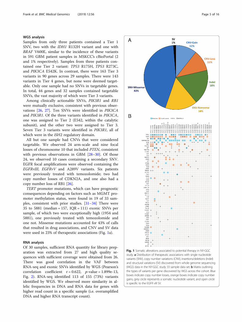
consequences depending on factors such as MGMT pro-moter methylation status, were found in 19 of 33 sam-ples, consistent with prior studies. [31–34] There were35 to 5881 (median = 157, IQR = 111) exonic SNVs persample, of which two were exceptionally high (1954 and5881), one previously treated with temozolomide andone not. Missense mutations accounted for 43% of callsthat resulted in drug associations, and CNV and SV datawere used in 23% of therapeutic associations (Fig. 1a).
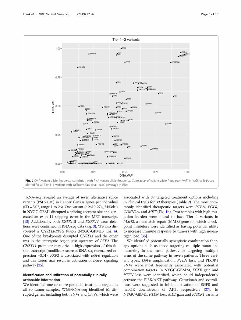
RNA analysisOf 30 samples, sufficient RNA quantity for library prep-aration was extracted from 27 and high quality se-quences with sufficient coverage were obtained from 26.There was good correlation in the VAF betweenRNA-seq and exonic SNVs identified by WGS (Pearson’scorrelation coefficient r = 0.622, p-value = 1.899e-13,Fig. 2). RNA-seq identified 113 of 155 (73%) variantsidentified by WGS. We observed more similarity in al-lelic frequencies in DNA and RNA data for genes withhigher read count in a specific sample (i.e. overamplifiedDNA and higher RNA transcript count).
Fig. 1 Somatic alterations associated to potential therapy in NY-GGCstudy. a Distribution of therapeutic associations with single nucleotidevariants (SNV), copy number variations (CNV), insertions/deletions (Indel)and structural variations (SV) discovered from whole genome sequencing(WGS) data in the NY-GGC study 33 sample data set. b Matrix outliningthe types of variants per gene discovered by WGS across the cohort. Blueboxes indicate copy number losses, orange boxes indicate copy numbergains, grey circle represents a somatic nucleotide variant, and open circleis specific to the EGFR vIII SV.
Frank et al. BMC Medical Genomics (2019) 12:56 Page 5 of 16
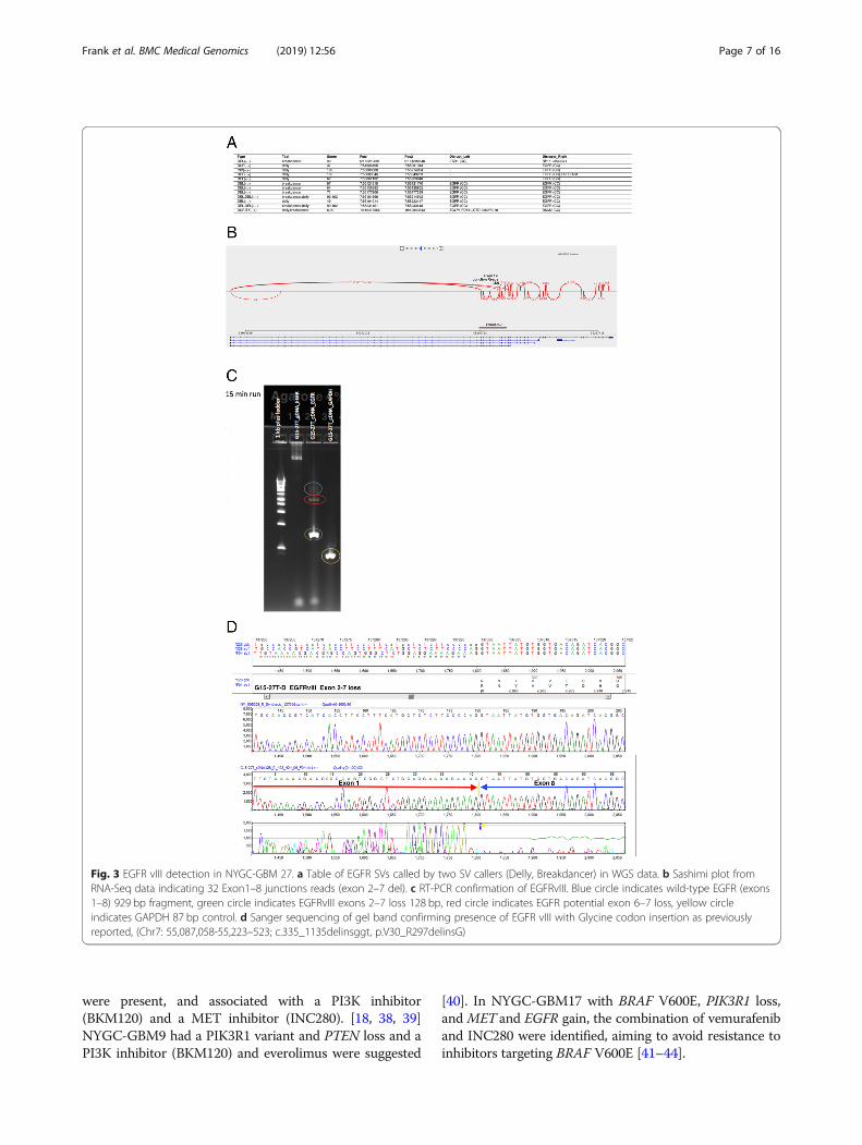
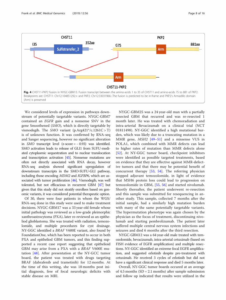
RNA-seq revealed an average of seven alternative splicevariants (PSI > 10%) in Cancer Census genes per individual(SD = 5.02, range 1 to 26). One variant (c.2419-274_2443del)in NYGC-GBM1 disrupted a splicing acceptor site and gen-erated an exon 11 skipping event in the MET transcript.[18] Additionally, both EGFRvIII and EGFRvV exon dele-tions were confirmed in RNA-seq data (Fig. 3). We also dis-covered a CHST11-PKP2 fusion (NYGC-GBM13, Fig. 4).One of the breakpoints disrupted CHST11 and the otherwas in the intergenic region just upstream of PKP2. TheCHST11 promoter may drive a high expression of this fu-sion transcript (modified z-score of RNA-seq normalized ex-pression =3.01). PKP2 is associated with EGFR regulationand this fusion may result in activation of EGFR signalingpathway [35].
Identification and utilization of potentially clinicallyactionable informationWe identified one or more potential treatment targets inall 30 tumor samples. WGS/RNA-seq identified 61 dis-rupted genes, including both SNVs and CNVs, which were
associated with 87 targeted treatment options including62 clinical trials for 39 therapies (Table 2). The most com-monly identified therapeutic targets were PTEN, EGFR,CDKN2A, and MET (Fig. 1b). Two samples with high mu-tation burden were found to have Tier 4 variants inMSH2, a mismatch repair (MMR) gene for which check-point inhibitors were identified as having potential utilityto increase immune response to tumors with high neoan-tigen load [36].We identified potentially synergistic combination ther-
apy options such as those targeting multiple mutationsoccurring in the same pathway or targeting multiplearms of the same pathway in seven patients. Three vari-ant types, EGFR amplification, PTEN loss, and PIK3R1SNVs were most frequently associated with potentialcombination targets. In NYGC-GBM24, EGFR gain andPTEN loss were identified, which could independentlyactivate the PI3K/AKT pathway. Cetuximab and everoli-mus were suggested to inhibit activation of EGFR andmTOR downstream of AKT, respectively [37]. InNYGC-GBM1, PTEN loss, MET gain and PI3KR1 variants
Fig. 2 DNA variant allele frequency correlation with RNA variant allele frequency. Correlation of variant allele frequency (VAF) in WGS vs RNA-seqplotted for all Tier 1–3 variants with sufficient (≥5 total reads) coverage in RNA
Frank et al. BMC Medical Genomics (2019) 12:56 Page 6 of 16
were present, and associated with a PI3K inhibitor(BKM120) and a MET inhibitor (INC280). [18, 38, 39]NYGC-GBM9 had a PIK3R1 variant and PTEN loss and aPI3K inhibitor (BKM120) and everolimus were suggested
[40]. In NYGC-GBM17 with BRAF V600E, PIK3R1 loss,and MET and EGFR gain, the combination of vemurafeniband INC280 were identified, aiming to avoid resistance toinhibitors targeting BRAF V600E [41–44].
Fig. 3 EGFR vIII detection in NYGC-GBM 27. a Table of EGFR SVs called by two SV callers (Delly, Breakdancer) in WGS data. b Sashimi plot fromRNA-Seq data indicating 32 Exon1–8 junctions reads (exon 2–7 del). c RT-PCR confirmation of EGFRvIII. Blue circle indicates wild-type EGFR (exons1–8) 929 bp fragment, green circle indicates EGFRvIII exons 2–7 loss 128 bp, red circle indicates EGFR potential exon 6–7 loss, yellow circleindicates GAPDH 87 bp control. d Sanger sequencing of gel band confirming presence of EGFR vIII with Glycine codon insertion as previouslyreported, (Chr7: 55,087,058-55,223–523; c.335_1135delinsggt, p.V30_R297delinsG)
Frank et al. BMC Medical Genomics (2019) 12:56 Page 7 of 16
We considered levels of expression in pathways down-stream of potentially targetable variants. NYGC-GBM7contained an EGFR gain and a nonsense SNV in thegene Smoothened (SMO), which is directly targetable byvismodegib. The SMO variant (p.Arg421*/c.1261C > T)is of unknown function. It was confirmed by RNA-seqand Sanger sequencing, however no significant alterationin SMO transcript level (z-score =− 0.93) was identified.SMO activation leads to release of GLI1 from SUFU-medi-ated cytoplasmic sequestration and to nuclear translocationand transcription activation [45]. Nonsense mutations areoften not directly associated with RNA decay, howeverRNA-seq analysis showed significant upregulation ofdownstream transcripts in the SMO-SUFU-GLI pathway,including those encoding MDM2 and IGFBP6, which are as-sociated with tumor proliferation [46]. Vismodegib, was welltolerated, but not efficacious in recurrent GBM [47] butgiven that this study did not stratify enrollees based on gen-omic variants, it was considered potential therapeutic option.Of 30, there were four patients in whom the WGS/
RNA-seq done in this study were used to make treatmentdecisions. NYGC-GBM17 was a 33 year-old female whoseinitial pathology was reviewed as a low-grade pleimorphicxanthroastrocytoma (PXA), later re-reviewed as an epithe-liod glioblastoma. She was treated with radiation, temozo-lomide, and multiple procedures for cyst drainage.NY-GGC identified a BRAF V600E variant, also found byFoundationOne, which has been reported to occur in bothPXA and epitheliod GBM tumors, and this finding sup-ported a recent case report suggesting that epithelioidGBM may arise from a PXA with a BRAF V600E mu-tation [48]. After presentation at the NY-GCC tumorboard, the patient was treated with drugs targetingBRAF (dabrafenib and trametinib) for 12 months. Atthe time of this writing, she was 18 months post ini-tial diagnosis, free of focal neurologic deficits withstable disease on MRI.
NYGC-GBM25 was a 24 year-old man with a partiallyresected GBM that recurred and was re-resected 1month later. He was treated with chemoradiation andintra-arterial Bevacizumab on a clinical trial (NCT01811498). NY-GGC identified a high mutational bur-den, which was likely due to a truncating mutation in aMMR gene, MSH2 [49–51] and a missense VUS inPOLA1, which combined with MMR defects can leadto higher rates of mutation than MMR defects alone[52]. At NY-GGC tumor board, checkpoint inhibitorswere identified as possible targeted treatments, basedon evidence that they are effective against MMR-defect-ive tumors and that there may be potential benefit ofconcurrent therapy [53, 54]. The referring physicianstopped adjuvant temozolomide, in light of evidencethat MSH6 protein loss could lead to progression ontemozolomide in GBM, [55, 56] and started nivolumab.Shortly thereafter, the patient underwent re-resectionand this sample was submitted for resequencing in an-other study. This sample, collected 7 months after theinitial sample, had a similarly high mutation burdenwith many of the same potentially targetable variants.The hypermutation phenotype was again chosen by thephysician as the focus of treatment, discontinuing nivo-lumab and starting pembrolizumab. The patient latersuffered multiple central nervous system infections andseizures and died 6 months after the third resection.NYGC-GBM12 was a 64 year-old male treated with tem-
ozolomide, bevacizumab, intra-arterial cetuximab (based onFISH evidence of EGFR amplification) and multiple resec-tions. NY-GGC identified an extreme focal EGFR amplifica-tion, and suggested erlotinib despite pre-treatment withcetuximab. He received 3 cycles of erlotinib but did nothave a significant clinical response and died 5 months later.Overall, NY-GGC tumor boards occured at an average
of 4.5 months (SD = 2.1 months) after sample submissionand follow up indicated that results were utilized in the
Fig. 4 CHST11-PKP2 fusion in NYGC-GBM13. Fusion transcript between the amino-acids 1 to 35 of CHST11 and amino-acids 75 to 881 of PKP2.Breakpoints are: CHST11: Chr12:104851292:+ and PKP2: Chr12:33031966:-.The fusion is predicted to be in-frame and PKP2’s Armadillo domain(Arm) is preserved
Frank et al. BMC Medical Genomics (2019) 12:56 Page 8 of 16
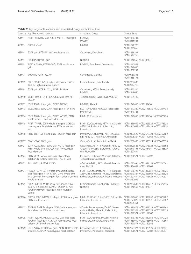
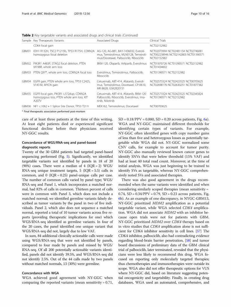
Table 2 Key targetable variants and associated drugs and clinical trials
Sample Key Therapeutic Variants Associated Drugs Clinical Trials
GBM1 PIK3R1 R562del, MET R755fs MET 11, focal gain BKM120,INC280
NCT01870726NCT02386826
GBM3 PIK3CA V344G BKM120 NCT01870726NCT01349660
GBM4 EGFR gain, PTEN W111C, whole arm loss Cetuximab, Everolimus NCT01238237NCT01870726
GBM5 PDGFRA/KIT/KDR gain Nilotinib NCT01140568 NCT01871311
GBM6 PIK3CA G542K, PTEN N59 fs, EGFR whole armgain
BKM120, Everolimus, Cetuximab NCT02142803NCT01349660NCT01238237
GBM7 SMO R421*, NF1 Q270* Vismodegib, MEK162 NCT00980343NCT01885195
GBM8* POLE P1505S, MSH2 splice site donor c.366 +1G > A, High mutation burden
Pembrolizumab, Nivolumab NCT02337686NCT02017717
GBM9 EGFR gain, KDR R1022*, PIK3R1 E443del Cetuximab, ABT41, Bevacizumab,BKM120
NCT02573324NCT01349660
GBM10 MGMT loss, PTEN R130*, whole arm loss NF1E1722*
Temozolomide, Everolimus, MEK162 NCT01885195
GBM12 EGFR A289V, focal gain, PIK3R1 D560G BKM120, Afatanib NCT01349660 NCT01934361
GBM13 MDM2 focal gain, CDK4 focal gain, PTEN R47S RG7112/RG7388, AMG232, Palbociclib,Everolimus
NCT01877382 NCT02143635 NCT01227434NCT01870726
GBM14 EGFR A289V, focal gain, PIK3R1 W597G, PTENwhole arm loss and focal deletion
BKM-120, Everolimus NCT01349660 NCT01934361 NCT01870726
GBM15 PIK3R1 T473P, EGFR whole arm gain, CDKN2Ahomozygous focal deletion, PTEN whole armloss
BKM-120, Cetuximab, ABT-414, Afatanib,ABBV-221, Palbociclib, Ribociclib,Everolimus
NCT01339052 NCT02423525 NCT02573324NCT02365662 NCT01227434 NCT02345824
GBM16 PTEN Y16*, EGFR focal gain, PDGFRA focal gain Everolimus, Cetuximab, ABT-414, Afata-nib, ABBV-221, Nilotinib, Crenolanib
NCT02423525 NCT02573324 NCT02365662NCT02626364 NCT01140568 NCT01871311
GBM17 BRAF V600E, EGFR gain Vemurafanib, Cobimetinib, ABT414 NCT02537600 NCT02573324 NCT02423525
GBM21 EGFR R222C, focal gain, MET P791L, focal gain,PTEN whole arm loss, CDKN2A homozygousfocal deletion
Cetuximab, ABT-414, Afatanib, ABBV-221Crizotinib, INC280, Everolimus, Palboci-clib, Ribociclib
NCT02423525 NCT02573324 NCT02365662NCT02540161 NCT02034981 NCT02386826NCT01227434
GBM22 PTEN V119F, whole arm loss, STAG2 focaldeletion, NF1283fs, focal loss, TP53 R158H
Everolimus, Olaparib, Veliparib, MEK162,Temsirolimus/Docetaxel
NCT01390571 NCT02152892
GBM23 IDH1 R132H, RPTOR A578G AG-120, AG-881, BAY-1436032, Everoli-mus, INK128
NCT02073994 NCT02481154 NCT02746081NCT01434602 NCT02142803
GBM24 PIK3CA R93W, EGFR whole arm amplification,MET focal gain, PTEN R335*, T277I, whole armloss, CDKN2A homozygous focal deletion, PALB2whole arm loss
BKM-120, Cetuximab, ABT-414, Afatanib,ABBV-221, Crizotinib, INC280, Everolimus,Palbociclib, Ribociclib, Olaparib, Veliparib
NCT01870726 NCT01339052 NCT02423525NCT02573324 NCT02365662 NCT02386826NCT01227434 NCT02345824 NCT01390571NCT02152982
GBM25 POLA1 G1178, MSH2 splice site donor c.366 +1G > A, TP53 R175H, G245S, PDGFRA Y375H,PDGFRA/KIT/KDR focal gain, High mutationburden
Pembrolizumab, Nivolumab, Paclitaxel,Nilotinib
NCT02337686 NCT02017717 NCT02379416NCT01140568 NCT01871311
GBM26 PIK3CA R88Q, MDM2 focal gain, CDK4 focal gain,PTEN whole arm loss
BKM-120, RG-7112, AMG-232, Palbociclib,Ribociclib, Everolimus
NCT01249660 NCT01339052 NCT01877282NCT01723020 NCT01390571 NCT02152982NCT02255461
GBM27 EGFRvIII, EGFR focal gain, CDKN2A homozygousfocal deletion, PTEN whole arm loss
Afatnib, Rindopepimut, CAR-T, Cetuxi-mab, ABT-414, Afatanib, Palbociclib,Ribociclib, Everolimus
NCT01480479 NCT02423525 NCT02664363NCT02573324 NCT02423525 NCT00703625NCT01390571 NCT02152982
GBM28 PIK3R1 Q579fs, PIK3CA D939G, MET focal gain,PDGFRA focal gain, CDKN2A homozygous focaldeletion, PTEN whole arm loss
BKM-120, Crizotinib, INC280, NilotinibPalbociclib, Ribociclib, Everolimus
NCT01870726 NCT01339052 NCT01870726NCT01339052 NCT02365662 NCT01140568NCT01390571 NCT02152982
GBM29 EGFR A289V, EGFR focal gain, PTEN R130*, wholearm loss, CDKN2A homozygous focal deletion,
Cetuximab, ABT-414, Afatanib,Everolimus, Palbociclib, Ribociclib
NCT02573324 NCT02423525 NCT0070362NCT01390571 NCT02152982 NCT01390571
Frank et al. BMC Medical Genomics (2019) 12:56 Page 9 of 16
care of at least three patients at the time of this writing.At least eight patients died or experienced significantfunctional decline before their physicians receivedNY-GGC results.
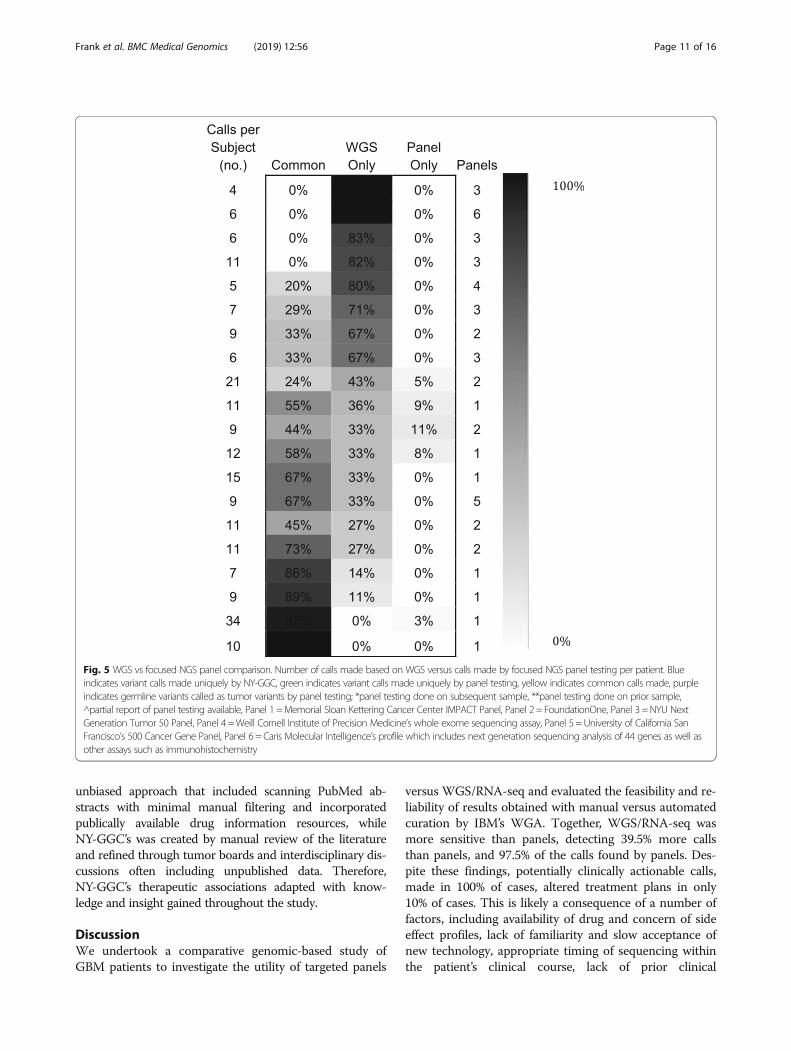
Concordance of WGS/RNA-seq and panel-baseddiagnostic reportsTwenty of the 30 GBM patients had targeted panel-basedsequencing performed (Fig. 5). Significantly, we identifiedtargetable variants not identified by panels in 18 of 20(90%) cases. There were a median of 4 (IQR = 2) WGS/RNA-seq unique treatment targets, 5 (IQR = 5.5) calls incommon, and 0 (IQR = 0.25) panel-unique calls per case.The number of common calls varied by panel type; WGS/RNA-seq and Panel 1, which incorporates a matched nor-mal, had 82% of calls in common. Thirteen percent of callswere in common with Panel 3, which does not include amatched normal; we identified germline variants falsely de-scribed as tumor variants by the panel in two of five indi-viduals. Panel 2, which also does not sequence a matchednormal, reported a total of 10 tumor variants across five re-ports (providing therapeutic implications for one) whichWGS/RNA-seq identified as germline variants. In four ofthe 20 cases, the panel identified one unique variant thatWGS/RNA-seq did not, largely due to low VAF.In sum, 84 additional clinically actionable calls were made
using WGS/RNA-seq that were not identified by panels,compared to four made by panels and missed by WGS/RNA-seq. Of all 200 potentially actionable variants identi-fied, panels did not identify 39.5%, and WGS/RNA-seq didnot identify 2.5%. Out of the 44 calls made by two panelswithout matched normals, 13 (30%) were germline.
Concordance with WGAWGA achieved good agreement with NY-GGC whencomparing the reported variants (mean sensitivity = 0.71,
SD = 0.18/PPV = 0.880, SD = 0.20 across patients, Fig. 6a).WGA and NY-GGC maintained different thresholds foridentifying certain types of variants. For example,NY-GGC often identified genes with copy number gainsof less than five and heterozygous losses as potentially tar-getable while WGA did not. NY-GGC normalized someCNV calls, for example to account for tumor purity.NY-GGC also manually reviewed known cancer genes toidentify SNVs that were below threshold (15% VAF) andhad at least 40 total read count. Moreover, at the time ofinitial analysis, WGA was just beginning to be trained toidentify SVs as targetable, whereas NY-GGC comprehen-sively noted SVs and associated therapies.There was also good agreement in the drugs recom-
mended when the same variants were identified and whenconsidering similarly scoped therapies (mean sensitivity =0.74, SD = 0.34/PPV = 0.79, SD = 0.23 across patients, Fig.6b). As an example of one discrepancy, in NYGC-GBM13,NY-GGC prioritized MDM2 amplification as a potentialtargetable variant, while WGA selected CDK4 amplifica-tion. WGA did not associate MDM2 with an inhibitor be-cause open trials were not for patients with GBM.NY-GGC prioritized MDM2 over CDK4 therapy based onin vitro studies that CDK4 amplification alone is not suffi-cient for CDK4 inhibitor sensitivity in cell lines. [57] TheCDK4 inhibitor, palbociclib, also had contradicting evidenceregarding blood-brain barrier penetration, [58] and tumorboard discussions of preliminary data of the GBM clinicaltrial of palbociclib, later terminated, revealed that the physi-cians were less likely to recommend this drug. WGA fo-cused on reporting only molecularly targeted therapies;thus chemotherapies and immunotherapies were outside itsscope. WGA also did not offer therapeutic options for VUSwhere NY-GGC did, based on literature suggesting poten-tial oncogenicity and targetability. Finally, in creating drugdatabases, WGA used an automated, comprehensive, and
Table 2 Key targetable variants and associated drugs and clinical trials (Continued)
Sample Key Therapeutic Variants Associated Drugs Clinical Trials
CDK4 focal gain NCT02152982
GBM31 IDH1 R132H, TSC2 P1215fs, TP53 R175H, CDKN2Ahomozygous focal deletion
AG-120, AG-881, BAY-1436032, Everoli-mus, Temsirolimus, MLN0128, Temsiroli-mus/Docetaxel, Palbociclib, Ribociclib
NCT02073994 NCT02481154 NCT02746081NCT002238946 NCT02142803 NCT01390571NCT02152982
GBM32 PIK3R1 A483P, STAG2 focal deletion, PTENM198R, whole arm loss
BKM-120, Olaparib, Veliparib, Everolimus NCT01870726 NCT01390571 NCT02152982NCT01434602
GBM33 PTEN Q97*, whole arm loss, CDKN2A focal loss Everolimus, Temsirolimus, Palbociclib,Ribociclib
NCT01390571 NCT02152982
GBM34 EGFR gain, PTEN whole arm loss, TP53 C242S,V143 M, MYCN gain
Cetuximab, ABT-414, Afatanib, Everoli-mus, Temsirolimus, Docetaxel, CP-0610,MK-8628, GSK2820151
NCT02573324 NCT02423525 NCT00703625NCT02698176 NCT02630251 NCT01877382
GBM35 EGFR focal gain, PIK3R1 L372dup, CDKN2Ahomozygous loss, PTEN whole arm loss, KITA207V
Cetuximab, ABT-414, Afatanib, BKM-120Palbociclib, Ribociclib, Everolimus, Ima-tinib, Nilotinib
NCT02573324 NCT02423525 NCT02345824NCT01390571 NCT02152982
GBM36 NF1 c.1062 + 1 Splice Site Donor, TP53 T211I MEK162, Temsirolimus, Docetaxel NCT00703625
* Final therapeutic association performed post-mortem
Frank et al. BMC Medical Genomics (2019) 12:56 Page 10 of 16
unbiased approach that included scanning PubMed ab-stracts with minimal manual filtering and incorporatedpublically available drug information resources, whileNY-GGC’s was created by manual review of the literatureand refined through tumor boards and interdisciplinary dis-cussions often including unpublished data. Therefore,NY-GGC’s therapeutic associations adapted with know-ledge and insight gained throughout the study.
DiscussionWe undertook a comparative genomic-based study ofGBM patients to investigate the utility of targeted panels
versus WGS/RNA-seq and evaluated the feasibility and re-liability of results obtained with manual versus automatedcuration by IBM’s WGA. Together, WGS/RNA-seq wasmore sensitive than panels, detecting 39.5% more callsthan panels, and 97.5% of the calls found by panels. Des-pite these findings, potentially clinically actionable calls,made in 100% of cases, altered treatment plans in only10% of cases. This is likely a consequence of a number offactors, including availability of drug and concern of sideeffect profiles, lack of familiarity and slow acceptance ofnew technology, appropriate timing of sequencing withinthe patient’s clinical course, lack of prior clinical
Fig. 5 WGS vs focused NGS panel comparison. Number of calls made based on WGS versus calls made by focused NGS panel testing per patient. Blueindicates variant calls made uniquely by NY-GGC, green indicates variant calls made uniquely by panel testing, yellow indicates common calls made, purpleindicates germline variants called as tumor variants by panel testing; *panel testing done on subsequent sample, **panel testing done on prior sample,^partial report of panel testing available, Panel 1 =Memorial Sloan Kettering Cancer Center IMPACT Panel, Panel 2 = FoundationOne, Panel 3 =NYU NextGeneration Tumor 50 Panel, Panel 4 =Weill Cornell Institute of Precision Medicine’s whole exome sequencing assay, Panel 5 =University of California SanFrancisco’s 500 Cancer Gene Panel, Panel 6 = Caris Molecular Intelligence’s profile which includes next generation sequencing analysis of 44 genes as well asother assays such as immunohistochemistry
Frank et al. BMC Medical Genomics (2019) 12:56 Page 11 of 16

information to rigorously assess risk-vs-benefits concerns,and clinician perception of the usefulness and relevance ofthe sequencing data based on time elapsed between resec-tion and sample submission; further research is warrantedto identify the biggest barriers to implementation andways to overcome them.The majority of potentially therapeutic associations
were identified in targetable genes (Tier 3) but with avariant unknown to be actionable. Such variants wereprioritized when there was evidence that they wouldlikely affect protein function, similarly to Tier 1–2 vari-ants. These findings were typically complex and requiredmanual curation of information from the literature, ne-cessitating many person-hours per case. [18] The inter-pretation of splicing variants and structural variantsresulting in splicing aberrations, only detected by
combining WGS [59] and RNA-Seq remains challenging,especially in the context of individual sample or smallcohort analyses. There may also be additional connec-tions between variants and pathways that we did notidentify. Furthermore, the therapeutic associations madeare based on what was available in the literature and inclinical trials at the time of analysis and interpretation ofthat individual sample and will evolve with new drugdevelopment.Hence a critical component of improving scalability of
data interpretation will be with automation. Here we ex-plored this approach through a comparative analysis ofmanual expert with automated curation. The time re-quired for WGA to match VCF calls with drug optionsimproved to within 6 min over the course of the study,while manual expert curation was an average of 1.9
A B
Fig. 6 Expert manual versus automated treatment target curation comparison. a Variants identified by expert manual versus automatedtreatment target curation. b Drug targets identified by NY-GGC’s expert manual versus WGA’s automated processing when the same variantswere identified
Frank et al. BMC Medical Genomics (2019) 12:56 Page 12 of 16
months. While WGA requires further development, forexample, to consider SVs as targets routinely or to ex-pand the drug database beyond molecularly targetedtherapies, the potential usefulness of this timeframe inthe clinical setting is clear.The per-patient cost of targerted panels at cancer cen-
ters are generally about $1000 and FoundationOne is cur-rently ~$5800 [60]. By comparison, the all-in cost ofclinical WGS/RNAseq and analysis by all platforms in thecurrent study was ~$10,000. While financial cost-benefitanalysis may currently favor panels or WES, our resultssuggest an imminent future in which the technical advan-tages and breadth of WGS/RNA-seq will increasinglyprovide improved cost-benefits by returning the mostcomprehensive analysis of tumor mutations. As costs andefficiencies improve, it may become reasonable to con-sider how to routinely apply WGS/RNA-seq to benefitcancer patients.In addition to lowered cost and timely interpretation
of sequencing data, other strategies towards improvedimplementation of WGS/RNA-seq include submissionof samples as soon as possible after resection so that re-sults are available for consideration at the time treat-ment decisions are made. This requires both clinicianeducation about the utility of such an assay as well asthe availability of clinically approved tests. NYGC has re-cently obtained such regulatory approval from the NewYork State Department of Health. One of the biggestchallenges in doing so in WGS was in demonstrating re-producibility at far less depth (80X/40X for tumor nor-mal pair) than depths of 100-200X for exomes and 500Xfor panels. Specifically, true negatives remain elusiveeven with very high depths because oncology variant cal-lers look for variants and not for correct base calls, andremain a limit of this assay. It will be interesting to seehow this influences adoption of WGS/RNA-seq for som-atic variants in the coming months.Limitations of this study include the inclusion of mul-
tiple sample preservation methods. This may have affectedthe variant calls, although previous studies have shownthat 98% of actionable calls made in fresh frozen samplescan also be made in FFPE samples. [61] Although action-able calls were made for all samples, some calls may havebeen missed in those samples that had tumor purity of lessthan 20%. [62] Another limitation is the small sample size.This sample may not have been representative of other pa-tients with GBM. Furthermore, only 20 of the 30 patientsenrolled had prior targeted panel with which the WGSanalysis could be compared. To address this, we are con-ducting a follow-up study of 200 patients.
ConclusionIn sum, when we compared manual and automatedsearches for therapeutic options, WGA offered a broader
array of options with a much faster processing time whilemaintaining significant sensitivity. Meanwhile, NY-GGC’smanual curation associated clinical significance to abroader array of variants, suggested combination therap-ies, and incorporated feedback regarding individual pa-tient clinical data as well as physician concerns discussedat tumor board meetings. Taken together, this study pointsto the potential of WGS/RNA-seq, combined with auto-mated curation, to maximize therapeutic options whichcan be used in clinical decision making for the benefit ofcancer patients.
AbbreviationsCNV: copy number variant; GBM: glioblastoma; IQR: interquartile range;MMR: mis-match repair; MPA: molecular profile analysis; NLP: naturallanguage processing; NYGC: New York Genome Center; NY-GGC: NewYork Glioblastoma Genome Consortium; PPV: positive predictive value;PSI: percent spliced in; PXA: pleiomorphic xanthroastrocytoma; RIN: RNAintegrity number; RNA-seq: RNA sequencing; SD: standard deviation;SNV: single nucleotide variant; SV: structural variant; TCGA: The CancerGenome Atlas; TPM: transcript per million; VAF: variant allele frequency;VCF: variant call file; VUS: variant of unknown significance; WES: wholeexome sequencing; WGA: Watson Genomic Analysis; WGS: whole genomesequencing
AcknowledgmentsWe are grateful for critical input from many scientific members of NYGC andour numerous collaborators at IBM, Rockefeller University, Memorial SloanKettering Cancer Center, Lenox Hill Hospital, New York University School ofMedicine, and Columbia University. We would like to thank the RockefellerUniversity IRB, and all treating physicians in the study for their time andparticipation in our tumor boards. Finally, we greatly thank all patients fortheir consent and participation in the study and for the research use of theirtissue specimens.
FundingSupported by a grant from IBM to NYGC, NYGC philanthropic funds,Rockefeller University grant #UL1TR000043 from the National Center forAdvancing Translational Sciences, National Institutes of Health (NIH) Clinicaland Translational Science Award, the Emerald Foundation to RBD and NIHgrant P30CA008748 to MSKCC. RBD is a Howard Hughes Medical InstituteInvestigator.
Availability of data and materialsThe datasets generated and/or analysed during the current study areavailable from the corresponding author until it becomes available from apublic repository.
Author’s contributionsMOF, AKR, and RBD contributed to the conception and design of the work.MOF, VF, DKhaira, AC, DKim, MFLE, CE, JB, AD, DGP, JGG, CB, JF, ABL, PC, CG,MD, ED, AO, and EP contributed to the acquisition of data. MOF, TK, KH, NR,FU, AKE, BJC, KA, MS, HG, VF, ED, SR, AF, VV, EAB, JLMV, CR, DKhaira, AC,DKim, MFLE, CE, PA, CS, JB, AD, DGP, JGG, CB, JF, ABL, PC, CG, MD, ED, AO,EP, DEO, SJH, JBP, VVM, VJ, MCZ, JK, LP, KOW, AKR and RBD contributed tothe analysis and interpretation of the data and revision of manuscript. MOF,TK, KH, NR, JLMV, KOW, and RBD drafted the manuscript. All authors readand approved the manuscript.
Ethics approval and consent to participateAll participants provided written informed consent to participate. Protocolswere approved by local or central Institutional Review Boards at: RockefellerUniversity, Biomedical Research Alliance of New York (on behalf of NorthwellHealth), Memorial Sloan Kettering Cancer Center, New York University Schoolof Medicine, and Weill Cornell Medicine.
Consent for publicationNot applicable.
Frank et al. BMC Medical Genomics (2019) 12:56 Page 13 of 16
Competing interestsMOF and RBD received consultant fees from New York Genome Center. TK, KH,and FU have patent applicatons related to Watson for Genomics, no. 14/745616. VV owns stock and options in 23andMe, Inc. MFLE received consultantfees from Rockefeller University. ABL in the past 12months, received personalcompensation (consulting fees/honoraria) from Northwest Biotherapeutics,AbbVie, Agios, Bioclinica, Sapience, WebMD, NCI; travel support fromKaropharm, Northwest Biotherapeutics, Oncoceutis, Global Coalition forAdaptive Research, New York University, Agios, Abbvie, Celgene, Novocure, NRGOncology Foundation, Tocagen, and Yale University; and research support (tothe institution) from NCI, Oncoceutics, AbbVie, Karyopharm, Beigene, VBIVaccines, Kadmon, Pfizer, RTOG-Foundation, Aeterna Zentaris, NorthwesternUniversity, Novartis, Pfizer, Celldex, Millenium, UCLA, and Amgen. NR, AKE, BJC,KA, MS, HG, VF, ED, SR, AF, EAB, JLMV, CR, DKhaira, AC, DKim, CE, PA, CS, JB, AD,DGP, JGG, CB, JF, PC, CG, MD, ED, AO, EP, DEO, SJH, JBP, VVM, VJ, MCZ, JK, LP,KOW and AKR declare no competing interests.
Publisher’s NoteSpringer Nature remains neutral with regard to jurisdictional claims inpublished maps and institutional affiliations.
Author details1New York Genome Center, 101 Avenue of the Americas, New York, NY10013, USA. 2Laboratory of Molecular Neuro-Oncology, The RockefellerUniversity, 1230 York Avenue, New York, NY 10065, USA. 3IBM Thomas J.Watson Research Center, Yorktown Heights, NY 10598, USA. 4NorthwellHealth, Lenox Hill Hospital, 100 E. 77th Street, New York, NY 10075, USA.5Northwell Health, The Brain Tumor Center, 450 Lakeville Road, Lake Success,Lakeville, NY 11042, USA. 6New York University, School of Medicine, 550 FirstAvenue, New York, NY 10016, USA. 7Memorial Sloan-Kettering Cancer Center,1275 York Avenue, New York, NY 10065, USA. 8Columbia University MedicalCenter, 710 West 168th Street, New York, NY 10032, USA. 9Hospital forSpecial Surgery, 535 E. 70th Street, New York, NY 10021, USA. 10IBM WatsonHealth, NW Broken Sound Bkwy, Boca Raton, FL 33487, USA. 11HowardHughes Medical Institute, The Rockefeller University, 1230 York Avenue, NewYork, NY 10065, USA. 12Present address: Google, 76 9th Avenue, New York,NY 10011, USA. 13Present address: Rockefeller University, 1230 York Avenue,New York, NY 10065, USA. 14Present address: 23&Me, 899 W Evelyn Ave,Mountain View, CA 94041, USA. 15Present address: Max Planck Institute ofImmunobiology and Epigenetics, Stübeweg 51 D-79108, Freiburg, Germany.16Present address: The Scripps Research Institute, 10550 N. Torrey Pines Road,La Jolla, CA 92037, USA. 17Present address: The Tisch Cancer Institute, 1470Madison Avenue, New York, NY 10029, USA. 18Present address: MemorialSloan-Kettering Cancer Center, 1275 York Avenue, New York, NY 10065, USA.19Present address: Harvard Medical School, 10 Shattuck Street, Boston, MA02115, USA. 20Present address: Yale School of Medicine, 333 Cedar Street,New Haven, CT 06510, USA.
Received: 11 September 2018 Accepted: 28 March 2019
References1. Griffith M, Miller CA, Griffith OL, Krysiak K, Skidmore ZL, Ramu A, et al.
Optimizing cancer genome sequencing and analysis. Cell Syst 2015;1(3):210–223. doi: https://doi.org/10.1016/j.cels.2015.08.015. PubMed PMID:26645048; PubMed Central PMCID: PMCPMC4669575.
2. Hyman DM, Solit DB, Arcila ME, Cheng DT, Sabbatini P, Baselga J, et al.Precision medicine at memorial Sloan Kettering Cancer center: clinical next-generation sequencing enabling next-generation targeted therapy trials.Drug Discov Today 2015;20(12):1422–1428. doi: https://doi.org/10.1016/j.drudis.2015.08.005. PubMed PMID: 26320725; PubMed Central PMCID:PMCPMC4940024.
3. Consortium APG. AACR project GENIE: powering precision medicinethrough an international consortium. Cancer Discov 2017. doi: https://doi.org/10.1158/2159-8290.CD-17-0151. PubMed PMID: 28572459.
4. Schmidt KT, Chau CH, Price DK, Figg WD. Precision oncology medicine: theclinical relevance of patient-specific biomarkers used to optimize Cancertreatment. J Clin Pharmacol 2016;56(12):1484–1499. doi: https://doi.org/10.1002/jcph.765. PubMed PMID: 27197880; PubMed Central PMCID:PMCPMC5112148.
5. Zehir A, Benayed R, Shah RH, Syed A, Middha S, Kim HR, et al. Mutationallandscape of metastatic cancer revealed from prospective clinicalsequencing of 10,000 patients. Nat Med 2017;23(6):703–713. doi: https://doi.org/10.1038/nm.4333. PubMed PMID: 28481359; PubMed Central PMCID:PMCPMC5461196.
6. Turner TN, Hormozdiari F, Duyzend MH, McClymont SA, Hook PW, Iossifov I,et al. Genome sequencing of autism-affected families reveals disruption ofputative noncoding regulatory DNA. Am J Hum Genet 2016;98(1):58–74.doi: https://doi.org/10.1016/j.ajhg.2015.11.023. PubMed PMID: 26749308;PubMed Central PMCID: PMCPMC4716689.
7. Belkadi A, Bolze A, Itan Y, Cobat A, Vincent QB, Antipenko A, et al. Whole-genome sequencing is more powerful than whole-exome sequencing fordetecting exome variants. Proc Natl Acad Sci U S A 2015;112(17):5473–5478.doi: https://doi.org/10.1073/pnas.1418631112. PubMed PMID: 25827230;PubMed Central PMCID: PMCPMC4418901.
8. Cummings BB, Marshall JL, Tukiainen T, Lek M, Donkervoort S, Foley AR, etal. Improving genetic diagnosis in Mendelian disease with transcriptomesequencing. Sci Transl Med 2017;9(386). doi: https://doi.org/10.1126/scitranslmed.aal5209. PubMed PMID: 28424332.
9. Honeyman JN, Simon EP, Robine N, Chiaroni-Clarke R, Darcy DG, Lim, II, etal. Detection of a recurrent DNAJB1-PRKACA chimeric transcript infibrolamellar hepatocellular carcinoma. Science. 2014;343(6174):1010–1014.doi: https://doi.org/10.1126/science.1249484. PubMed PMID: 24578576;PubMed Central PMCID: PMCPMC4286414.
10. Cheng DT, Mitchell TN, Zehir A, Shah RH, Benayed R, Syed A, et al. MemorialSloan Kettering-integrated mutation profiling of actionable Cancer targets (MSK-IMPACT): A Hybridization Capture-Based Next-Generation Sequencing ClinicalAssay for Solid Tumor Molecular Oncology J Mol Diagn 2015;17(3):251–264. doi:https://doi.org/10.1016/j.jmoldx.2014.12.006. PubMed PMID: 25801821.
11. Jones S, Anagnostou V, Lytle K, Parpart-Li S, Nesselbush M, Riley DR, et al.Personalized genomic analyses for cancer mutation discovery andinterpretation. Sci Transl Med 2015;7(283):283ra53. doi: https://doi.org/10.1126/scitranslmed.aaa7161. PubMed PMID: 25877891; PubMed CentralPMCID: PMCPMC4442685.
12. Prados MD, Byron SA, Tran NL, Phillips JJ, Molinaro AM, Ligon KL, et al.Toward precision medicine in glioblastoma: the promise and thechallenges. Neuro-Oncology 2015;17(8):1051–1063. doi: https://doi.org/10.1093/neuonc/nov031. PubMed PMID: 25934816; PubMed Central PMCID:PMCPMC4490873.
13. Xiu J, Piccioni D, Juarez T, Pingle SC, Hu J, Rudnick J, et al. Multi-platformmolecular profiling of a large cohort of glioblastomas reveals potentialtherapeutic strategies. Oncotarget. 2016. doi: https://doi.org/10.18632/oncotarget.7722. PubMed PMID: 26933808.
14. Johnson DR, O'Neill BP. Glioblastoma survival in the United States beforeand during the temozolomide era. J Neuro-Oncol 2012;107(2):359–364. doi:https://doi.org/10.1007/s11060-011-0749-4. PubMed PMID: 22045118.
15. Verhaak RG, Hoadley KA, Purdom E, Wang V, Qi Y, Wilkerson MD, et al.Integrated genomic analysis identifies clinically relevant subtypes ofglioblastoma characterized by abnormalities in PDGFRA, IDH1, EGFR, andNF1. Cancer Cell 2010;17(1):98–110. doi: https://doi.org/10.1016/j.ccr.2009.12.020. PubMed PMID: 20129251; PubMed Central PMCID: PMCPMC2818769.
16. Ceccarelli M, Barthel FP, Malta TM, Sabedot TS, Salama SR, Murray BA, et al.Molecular profiling reveals biologically discrete subsets and pathways ofprogression in diffuse glioma. Cell. 2016;164(3):550–563. doi: https://doi.org/10.1016/j.cell.2015.12.028. PubMed PMID: 26824661; PubMed Central PMCID:PMCPMC4754110.
17. Wang Q, Hu B, Hu X, Kim H, Squatrito M, Scarpace L, et al. Tumor evolutionof glioma-intrinsic gene expression subtypes associates with immunologicalchanges in the microenvironment. Cancer Cell 2017;32(1):42–56 e6. doi:https://doi.org/10.1016/j.ccell.2017.06.003. PubMed PMID: 28697342;PubMed Central PMCID: PMCPMC5599156.
18. Wrzeszczynski KO, Frank MO, Koyama T, Rhrissorrakrai K, Robine N, Utro F, etal. Comparing sequencing assays and human-machine analyses inactionable genomics for glioblastoma. Neurol Genet 2017;3(4):e164. doi:https://doi.org/10.1212/NXG.0000000000000164. PubMed PMID: 28740869;PubMed Central PMCID: PMCPMC5506390.
19. Schroeder A, Mueller O, Stocker S, Salowsky R, Leiber M, Gassmann M, et al.The RIN: an RNA integrity number for assigning integrity values to RNAmeasurements. BMC Mol Biol 2006;7:3. doi: https://doi.org/10.1186/1471-2199-7-3. PubMed PMID: 16448564; PubMed Central PMCID:PMCPMC1413964.
Frank et al. BMC Medical Genomics (2019) 12:56 Page 14 of 16
20. Wang ET, Sandberg R, Luo S, Khrebtukova I, Zhang L, Mayr C, et al.Alternative isoform regulation in human tissue transcriptomes. Nature. 2008;456(7221):470–476. doi: https://doi.org/10.1038/nature07509. PubMed PMID:18978772; PubMed Central PMCID: PMCPMC2593745.
21. Nicorici D, Satalan M, Edgren H, Kangaspeska S, Murumagi A, Kallioniemi O,et al. FusionCatcher - a tool for finding somatic fusion genes in paired-endRNA-sequencing data. bioRxiv. 2014. https://doi.org/10.1101/011650.
22. Xi R, Lee S, Xia Y, Kim TM, Park PJ. Copy number analysis of whole-genomedata using BIC-seq2 and its application to detection of cancer susceptibilityvariants. Nucleic Acids Res 2016;44(13):6274–6286. doi: https://doi.org/10.1093/nar/gkw491. PubMed PMID: 27260798.
23. Rhrissorrakrai KK, T.; Parida, L. Watson for genomics: moving personalizedmedicine forward. Trends in Cancer 2016;2(8):392–395.
24. Patel NM, Michelini VV, Snell JM, Balu S, Hoyle AP, Parker JS, et al. EnhancingNext-Generation Sequencing-Guided Cancer Care Through CognitiveComputing. Oncologist. 2018;23(2):179–85. Epub 2017/11/22. doi: https://doi.org/10.1634/theoncologist.2017-0170. PubMed PMID: 29158372; PubMedCentral PMCID: PMCPMC5813753.
25. Rausch T, Zichner T, Schlattl A, Stutz AM, Benes V, Korbel JO. DELLY: structuralvariant discovery by integrated paired-end and split-read analysis.Bioinformatics. 2012;28(18):i333-i3i9. doi: https://doi.org/10.1093/bioinformatics/bts378. PubMed PMID: 22962449; PubMed Central PMCID: PMCPMC3436805.
26. Johnson BE, Mazor T, Hong C, Barnes M, Aihara K, McLean CY, et al.Mutational analysis reveals the origin and therapy-driven evolution ofrecurrent glioma. Science. 2014;343(6167):189–193. doi: https://doi.org/10.1126/science.1239947. PubMed PMID: 24336570; PubMed Central PMCID:PMCPMC3998672.
27. McGranahan N, Swanton C. Biological and therapeutic impact of intratumorheterogeneity in cancer evolution. Cancer Cell 2015;27(1):15–26. doi: https://doi.org/10.1016/j.ccell.2014.12.001. PubMed PMID: 25584892.
28. Smith JS, Tachibana I, Passe SM, Huntley BK, Borell TJ, Iturria N, et al. PTENmutation, EGFR amplification, and outcome in patients with anaplasticastrocytoma and glioblastoma multiforme. J Natl Cancer Inst 2001;93(16):1246–1256. PubMed PMID: 11504770.
29. Wang SI, Puc J, Li J, Bruce JN, Cairns P, Sidransky D, et al. Somatic mutationsof PTEN in glioblastoma multiforme. Cancer Res 1997;57(19):4183–4186.PubMed PMID: 9331071.
30. Carico C, Nuno M, Mukherjee D, Elramsisy A, Dantis J, Hu J, et al. Loss ofPTEN is not associated with poor survival in newly diagnosed glioblastomapatients of the temozolomide era. PLoS One 2012;7(3):e33684. doi: https://doi.org/10.1371/journal.pone.0033684. PubMed PMID: 22479427; PubMedCentral PMCID: PMCPMC3315579.
31. Vinagre J, Almeida A, Populo H, Batista R, Lyra J, Pinto V, et al. Frequency ofTERT promoter mutations in human cancers. Nat Commun 2013;4:2185. doi:https://doi.org/10.1038/ncomms3185. PubMed PMID: 23887589.
32. Arita H, Yamasaki K, Matsushita Y, Nakamura T, Shimokawa A, Takami H, etal. A combination of TERT promoter mutation and MGMT methylationstatus predicts clinically relevant subgroups of newly diagnosedglioblastomas. Acta Neuropathol Commun 2016;4(1):79. doi: https://doi.org/10.1186/s40478-016-0351-2. PubMed PMID: 27503138; PubMed CentralPMCID: PMCPMC4977715.
33. Nguyen HN, Lie A, Li T, Chowdhury R, Liu F, Ozer B, et al. Human TERTpromoter mutation enables survival advantage from MGMT promotermethylation in IDH1 wild-type primary glioblastoma treated by standardchemoradiotherapy. Neuro-Oncology 2017;19(3):394–404. doi: https://doi.org/10.1093/neuonc/now189. PubMed PMID: 27571882; PubMed CentralPMCID: PMCPMC5464302.
34. Batista R, Cruvinel-Carloni A, Vinagre J, Peixoto J, Catarino TA, CampanellaNC, et al. The prognostic impact of TERT promoter mutations inglioblastomas is modified by the rs2853669 single nucleotidepolymorphism. Int J Cancer 2016;139(2):414–423. doi: https://doi.org/10.1002/ijc.30057. PubMed PMID: 26914704.
35. Arimoto K, Burkart C, Yan M, Ran D, Weng S, Zhang DE. Plakophilin-2promotes tumor development by enhancing ligand-dependent and-independent epidermal growth factor receptor dimerization and activation.Mol Cell Biol 2014;34(20):3843–3854. doi: https://doi.org/10.1128/MCB.00758-14. PubMed PMID: 25113560; PubMed Central PMCID: PMCPMC4187709.
36. Le DT, Uram JN, Wang H, Bartlett BR, Kemberling H, Eyring AD, et al. PD-1blockade in tumors with mismatch-repair deficiency. N Engl J Med 2015;372(26):2509–2520. doi: https://doi.org/10.1056/NEJMoa1500596. PubMedPMID: 26028255; PubMed Central PMCID: PMCPMC4481136.
37. Ciunci CA, Perini RF, Avadhani AN, Kang HC, Sun W, Redlinger M, et al.Phase 1 and pharmacodynamic trial of everolimus in combination withcetuximab in patients with advanced cancer. Cancer. 2014;120(1):77–85. doi:https://doi.org/10.1002/cncr.28294. PubMed PMID: 24108668.
38. Kanteti R, Riehm JJ, Dhanasingh I, Lennon FE, Mirzapoiazova T,Mambetsariev B, et al. PI3 kinase pathway and MET inhibition is efficaciousin malignant pleural mesothelioma. Sci Rep 2016;6:32992. doi: https://doi.org/10.1038/srep32992. PubMed PMID: 27623107; PubMed Central PMCID:PMCPMC5021085.
39. Maira S-M, Pecchi S, Huang A, Burger M, Knapp M, Sterker D, et al.Identification and characterization of NVP-BKM120, an orally available pan-class I PI3-kinase inhibitor. Mol Cancer Ther. 2012;11(2):317–28. https://doi.org/10.1158/1535-7163.mct-11-0474.
40. Chalhoub N, Baker SJ. PTEN and the PI3-kinase pathway in cancer. AnnuRev Pathol 2009;4:127–150. doi: https://doi.org/10.1146/annurev.pathol.4.110807.092311. PubMed PMID: 18767981; PubMed Central PMCID:PMCPMC2710138.
41. Deuker MM, Marsh Durban V, Phillips WA, McMahon M. PI3'-kinaseinhibition forestalls the onset of MEK1/2 inhibitor resistance in BRAF-mutated melanoma. Cancer Discov. 2015;5(2):143–153. doi: https://doi.org/10.1158/2159-8290.CD-14-0856. PubMed PMID: 25472943; PubMed CentralPMCID: PMCPMC4320669.
42. Sun C, Wang L, Huang S, Heynen GJ, Prahallad A, Robert C, et al. Reversibleand adaptive resistance to BRAF(V600E) inhibition in melanoma. Nature.2014;508(7494):118–122. doi: https://doi.org/10.1038/nature13121. PubMedPMID: 24670642.
43. Straussman R, Morikawa T, Shee K, Barzily-Rokni M, Qian ZR, Du J, et al.Tumour micro-environment elicits innate resistance to RAF inhibitorsthrough HGF secretion. Nature. 2012;487(7408):500–504. doi: https://doi.org/10.1038/nature11183. PubMed PMID: 22763439; PubMed Central PMCID:PMCPMC3711467.
44. Pietrantonio F, Oddo D, Gloghini A, Valtorta E, Berenato R, Barault L, et al.MET-Driven Resistance to Dual EGFR and BRAF Blockade May Be Overcomeby Switching from EGFR to MET Inhibition in BRAF-Mutated ColorectalCancer. Cancer Discov. 2016;6(9):963–71. Epub 2016/06/22. doi: https://doi.org/10.1158/2159-8290.CD-16-0297. PubMed PMID: 27325282.
45. Rudin CM. Vismodegib. Clin Cancer Res 2012;18(12):3218–3222. doi: https://doi.org/10.1158/1078-0432.CCR-12-0568. PubMed PMID: 22679179; PubMedCentral PMCID: PMCPMC3715061.
46. Zheng X, Zeng W, Gai X, Xu Q, Li C, Liang Z, et al. Role of the hedgehogpathway in hepatocellular carcinoma (review). Oncol Rep 2013;30(5):2020–2026. doi: https://doi.org/10.3892/or.2013.2690. PubMed PMID: 23970376.
47. Sloan AE, Nock CJ, Ye X, Kerstetter A, Supko J, Lamborn K, et al. Targetingglioma-initiating cells in GBM: ABTC-0904, a randomized phase 0/II studytargeting the sonic hedgehog-signaling pathway. J Clin Oncol. 2014;32(15_suppl):2026. https://doi.org/10.1200/jco.2014.32.15_suppl.2026.
48. Tanaka S, Nakada M, Nobusawa S, Suzuki SO, Sabit H, Miyashita K, et al.Epithelioid glioblastoma arising from pleomorphic xanthoastrocytoma withthe BRAF V600E mutation. Brain Tumor Pathol 2014;31(3):172–176. doi:https://doi.org/10.1007/s10014-014-0192-2. PubMed PMID: 24894018.
49. Alexandrov LB, Nik-Zainal S, Wedge DC, Aparicio SA, Behjati S, Biankin AV, etal. Signatures of mutational processes in human cancer. Nature. 2013;500(7463):415–421. doi: https://doi.org/10.1038/nature12477. PubMed PMID:23945592; PubMed Central PMCID: PMCPMC3776390.
50. Hunter C, Smith R, Cahill DP, Stephens P, Stevens C, Teague J, et al. Ahypermutation phenotype and somatic MSH6 mutations in recurrenthuman malignant gliomas after alkylator chemotherapy. Cancer Res 2006;66(8):3987–3991. doi: https://doi.org/10.1158/0008-5472.CAN-06-0127.PubMed PMID: 16618716.
51. Wang J, Cazzato E, Ladewig E, Frattini V, Rosenbloom DI, Zairis S, et al.Clonal evolution of glioblastoma under therapy. Nat Genet 2016;48(7):768–776. doi: https://doi.org/10.1038/ng.3590. PubMed PMID: 27270107.
52. Shlien A, Campbell BB, de Borja R, Alexandrov LB, Merico D, Wedge D, et al.Combined hereditary and somatic mutations of replication error repairgenes result in rapid onset of ultra-hypermutated cancers. Nat Genet 2015;47(3):257–262. doi: https://doi.org/10.1038/ng.3202. PubMed PMID:25642631.
53. Sampson JH, Vlahovic G, Sahebjam S, Omuro AMP, Baehring JM, Hafler DA,et al. Preliminary safety and activity of nivolumab and its combination withipilimumab in recurrent glioblastoma (GBM): CHECKMATE-143. J Clin Oncol.2015;33(15_suppl):3010. https://doi.org/10.1200/jco.2015.33.15_suppl.3010.
Frank et al. BMC Medical Genomics (2019) 12:56 Page 15 of 16
54. Vlahovic G, Fecci PE, Reardon D, Sampson JH. Programmed death ligand 1(PD-L1) as an immunotherapy target in patients with glioblastoma. Neuro-Oncology 2015;17(8):1043–1045. doi: https://doi.org/10.1093/neuonc/nov071. PubMed PMID: 25964311; PubMed Central PMCID:PMCPMC4490880.
55. Yip S, Miao J, Cahill DP, Iafrate AJ, Aldape K, Nutt CL, et al. MSH6 mutationsarise in glioblastomas during temozolomide therapy and mediatetemozolomide resistance. Clin Cancer Res 2009;15(14):4622–4629. doi:https://doi.org/10.1158/1078-0432.CCR-08-3012. PubMed PMID: 19584161;PubMed Central PMCID: PMCPMC2737355.
56. Cahill DP, Levine KK, Betensky RA, Codd PJ, Romany CA, Reavie LB, et al.Loss of the mismatch repair protein MSH6 in human glioblastomas isassociated with tumor progression during temozolomide treatment. ClinCancer Res 2007;13(7):2038–2045. doi: https://doi.org/10.1158/1078-0432.CCR-06-2149. PubMed PMID: 17404084; PubMed Central PMCID:PMCPMC2873832.
57. Wiedemeyer WR, Dunn IF, Quayle SN, Zhang J, Chheda MG, Dunn GP, et al.Pattern of retinoblastoma pathway inactivation dictates response to CDK4/6inhibition in GBM. Proc Natl Acad Sci U S A 2010;107(25):11501–11506. doi:https://doi.org/10.1073/pnas.1001613107. PubMed PMID: 20534551; PubMedCentral PMCID: PMCPMC2895056.
58. Schroder LB, McDonald KL. CDK4/6 inhibitor PD0332991 in glioblastomatreatment: does it have a future? Front Oncol 2015;5:259. doi: https://doi.org/10.3389/fonc.2015.00259. PubMed PMID: 26649278; PubMed CentralPMCID: PMCPMC4663246.
59. Wrzeszczynski KO, Felice V, Shah M, Rahman S, Emde AK, Jobanputra V, etal. Whole genome sequencing-based discovery of structural variants inglioblastoma. Methods Mol Biol 2018;1741:1–29. Epub 2018/02/03. doi:https://doi.org/10.1007/978-1-4939-7659-1_1. PubMed PMID: 29392687.
60. Chakradhar S. Tumor sequencing takes off, but insurance reimbursementlags. Nat Med 2014;20(11):1220–1221. doi: https://doi.org/10.1038/nm1114-1220. PubMed PMID: 25375911.
61. Robbe P, Popitsch N, Knight SJL, Antoniou P, Becq J, He M, et al. Clinicalwhole-genome sequencing from routine formalin-fixed, paraffin-embeddedspecimens: pilot study for the 100,000 genomes project. Genet Med 2018;20(10):1196–1205. Epub 2018/02/02. doi: https://doi.org/10.1038/gim.2017.241. PubMed PMID: 29388947.
62. Wrzeszczynski KO, Felice V, Abhyankar A, Kozon L, Geiger H, Manaa D, et al.Analytical validation of clinical whole-genome and transcriptomesequencing of patient-derived tumors for reporting targetable variants inCancer. J Mol Diagn 2018;20(6):822–835. Epub 2018/08/24. doi: https://doi.org/10.1016/j.jmoldx.2018.06.007. PubMed PMID: 30138725; PubMed CentralPMCID: PMCPMC6198246.
Frank et al. BMC Medical Genomics (2019) 12:56 Page 16 of 16