Embed Size (px)
Citation preview
Structural Determinants of Transmembrane â-Barrels
Themis Lazaridis*
Department of Chemistry, City College of New York/CUNY, 138th Street & ConVentAVenue, New York, New York 10031
Received March 6, 2005
Abstract: The recognition of â-barrel membrane proteins based on their sequence is more
challenging than the recognition of R-helical membrane proteins. This goal could benefit from a
better understanding of the physical determinants of transmembrane â-barrel structure. To that
end, we first extend the IMM1 implicit membrane model in a way that allows the modeling of
membrane proteins with an internal aqueous pore. The new model (IMM1-pore) gives stable
molecular dynamics trajectories for three â-barrel membrane proteins of different sizes and
negative water-to-membrane transfer energies of reasonable magnitude. It also discriminates
the correct fold for a pair of 10-stranded and 12-stranded transmembrane â-barrels. We then
consider a pair of â-barrel proteins: OmpA, which is a membrane â-barrel with hydrophobic
residues on the exterior and polar residues in the interior, and retinol binding protein, which is
a water soluble protein with polar residues on the exterior and hydrophobic residues in the
interior. By threading the sequence of one onto the structure of the other we make two pairs of
structures for each sequence, one native and the other a decoy, and evaluate their energy.
The energy function discriminates the correct structure. By decomposing the energy into residue
contributions we examine which features of each sequence make it fold into one or the other
structure. It is found that for the OmpA sequence the largest contribution to stability comes
from interactions between polar residues in the interior of the barrel. The major factor that
prevents the retinol binding protein sequence from adopting a transmembrane fold is the presence
of polar/charged residues at the edges of the putative transmembrane â-strands as well as the
less favorable interior polar residue interactions. These results could help design simplified scoring
functions for fold recognition and structure prediction of transmembrane â-barrels.
IntroductionThe known membrane protein structures belong to twocategories: allR-helical and transmembraneâ-barrels(TMBB). TMBBs occur in the outer membrane of Gramnegative bacteria and presumably also of mitochondria andchloroplasts. They have from 8 to 22â-strands (always aneven number). Their N and C termini are on the periplasmicside and the loops on that side are short while those on theextracellular side can be very long.1 These proteins performa wide range of functions, such as allowing passive diffusionof ions and hydrophilic molecules, specific import of
nutrients, energy dependent export of toxins, and celladhesion.
Discriminating TMBBs from solubleâ-proteins based onsequence is more challenging than discriminating helicalmembrane proteins because TMBBs lack the 20-residuehydrophobic stretches that characterize the latter. Early workutilized the alternate hydrophobicity of TMâ strands;2,3
however, this approach gives a lot of false positives. Overthe past few years a number of bioinformatic approacheshave been developed for the prediction and discriminationof TMBBs based on either neural networks4-6,7 or HiddenMarkov models.8-11 The accuracy of these methods istypically around 80%. Wimley12 developed an empiricalscore based on the relative abundance of amino acid types
* Corresponding author phone: (212)650-8364; fax: (212)650-6107; e-mail: [email protected].
716 J. Chem. Theory Comput.2005,1, 716-722
10.1021/ct050055x CCC: $30.25 © 2005 American Chemical SocietyPublished on Web 05/27/2005

in TMBBs. Liu et al. attempted to discriminate TMBBs fromsolubleâ sheet proteins based on their amino acid composi-tion.13 Other methods utilize a wider variety of properties,14
including the presence of a signal peptide.15
While the above methods can be successful in recognizinga sequence as a TMBB or predicting the number of strandsand the topology, they do not produce an atomic-level three-dimensional model. The methods available for doing so arehomology modeling and fold recognition. Application ofhomology modeling requires significant sequence identitybetween the sequence under investigation and a protein ofknown structure, which is not often the case in TMBBs. Foldrecognition appears more promising for TMBBs becausethese proteins have a limited number of folds.
The first goal of the present work was to develop anatomic-level energy function that can discriminate the nativestate of TMBBs. We do this by modification of IMM1, animplicit membrane effective energy function. IMM1 repre-sents the membrane as a slab of nonpolar solvent. Byembedding a cylinder of aqueous solvent into this slab wecreated a function that can be applied to TMBBs. The secondgoal was to use this energy function to obtain insights intothe determinants of TMBB structure, i.e., investigate thefeatures that make a certain sequence fold into a TMBB asopposed to a solubleâ-protein. We do this by considering apair of â-barrels of the same number of strands, one that isa TMBB (OmpA) and another that is a solubleâ-barrel(retinol binding protein). We thread each sequence on thestructure of the other sequence, evaluate the energy, andexamine which residues contribute most to the energydifference between the correct and the incorrect structure.
MethodsIMM1-Pore. The origin of the energy function developedhere is EEF1,16 an effective energy function for solubleproteins
whereE is the intramolecular energy of the protein, givenby a modified form of the CHARMM polar hydrogen forcefield, and∆Gslv is the solvation free energy, given by
where∆Gislv is the solvation free energy of atomi, rij is
the distance betweeni and j, gi is the solvation free energydensity ofi (a Gaussian function ofrij), Vj is the volume ofatomj, and∆Gi
ref is the solvation free energy of an isolatedatom. In addition, EEF1 employs a distance-dependentdielectric constant (ε)r in Å) and a neutralized form of theionic side chains. A more recent version with updatedparameters for the charged and polar side chains is referredto as EEF1.1.17
IMM1 is a generalization of EEF1 appropriate for model-ing proteins in lipid bilayers.17 In IMM1 ∆Gi
ref is a linear
combination of values for water and for cyclohexane (whichmodels the nonpolar core of the membrane)
wherez′ ) |z|/(T/2) andT is the thickness of the nonpolarcore of the membrane. The functionf(z′) describes thetransition from one phase to the other:
The exponentn controls the steepness of the transition. Avalue of 10 gives a region of 6 Å over which the environmentgoes from 90% nonpolar to 90% polar. The midpoint of thetransition (f)0.5) corresponds to the hydrocarbon-polarheadgroup interface (roughly the level of the ester carbonylsin phospholipids). The strengthening of electrostatic interac-tions in the membrane is effected by a modified dielectricscreening function
wherer is the distance in Å (butε is dimensionless), andfijdepends on the position of the interacting atoms with respectto the membrane. Far from the membrane,fij is equal to 1,and we recover the linear distance-dependent dielectricmodel. Forfij we employed the empirical model
with a being an adjustable parameter. The valuea ) 0.85was found to give membrane insertion or adsorption energiesof the expected order of magnitude.17
To make IMM1 applicable to membrane proteins withaqueous channels we replacef in eq 3 and eq 6 byF
where
whereR is the radius of the aqueous pore. The definition ofF ensures thatF ) 0 whenf andh are zero andF ) 1 whenf, or h, or both of them are equal to 1. Figure 1 shows thecontour of the lineF ) 0.5 on thexzplane. The same valueof n is used for bothf andh, but that could be changed. Thecode was implemented in the program CHARMM, versionc32a1.
To test this energy function (IMM1-pore) we performedmolecular dynamics (MD) simulations on the followingproteins: OmpA (pdb code 1BXW18), the translocatordomain of NalP (pdb code 1UYN19), and FhuA (pdb code1BY320). The extra Met residue at the N terminus of OmpAwas omitted. The missing loop of NalP was built in anarbitrary conformation. The parameterR in eq 9 was set equalto the radius of the barrel at the level of CR atoms. In onecase it was varied to identify the value that gives the lowest
∆Giref(z′)) f(z′)∆Gi
ref,wat + (1 - f(z′))∆Giref,chex (3)
f(z′) ) z′n
1 + z′n(4)
ε ) rfij (5)
fij ) a + (1 - a) xf(zi) f(zj) (6)
∆Giref (z′, r′) ) F(z′, r′)∆Gi
ref,wat + (1 - F(z′, r′) )∆Giref,chex
(7)
F(z′, r′) ) f(z′) + h(r′) - f(z′)h(r′) (8)
h(r′) ) 1 - r′n
1 + r′n, r′ ) r/R , r ) xx2 + y2 (9)
WEEF1) E + ∆Gslv (1)
∆Gslv) ∑i
∆Gislv ) ∑
i
∆Giref - ∑
i∑j*i
gi(rij)Vj (2)
Structural Determinants of Transmembraneâ-Barrels J. Chem. Theory Comput., Vol. 1, No. 4, 2005717

energy. All simulations proceeded with (a) generation ofhydrogen coordinates using HBUILD, (b) a 300-step ABNRenergy minimization, and (c) 1 ns MD simulation at 300 Kusing SHAKE, the Verlet algorithm, and 2 fs time step. Inall cases the width of the membrane (T) was set to 26 Å.
Generation of the Decoys.To investigate the features thatdistinguish TMBBs from solubleâ-barrels we first identifiedthe closest possible water soluble analogue of OmpA, whichseems to be retinol binding protein (RBP, pdb code 1AQB21).This protein has 8â-strands and an additional helix at the Cterminus (Figure 3). The structure is less regular than OmpA(the strands are shorter and more bent). TheR helix wasomitted in this study, i.e., only residues 1-141 weresimulated. No disulfide bridges were built, to avoid havingdifferent numbers of bonds in the native structure and thedecoy.
The decoys were generated using the program MOD-ELLER 7v7.22 The first hydrophobic residue of eachâ strandof RBP was aligned with the same from OmpA. Theplacement of the TM strands on RBP was adjusted slightlyto maximize the hydrophobicity of the lipid-exposed residues.The final alignment is OMPA/RBP: 8/20, 38/39, 50/53, 78/67, 92/82, 120/103, 136/116, and 161/131. The maximalpossible number of residues was aligned, and all insertionsand deletions were placed in the middle of the loops. TheMODELLER alignment files are given as SupportingInformation.
ResultsMD Simulations and Membrane Insertion Energies ofTMBBs. Two criteria are used for testing the IMM1-poreenergy function. First, MD simulations of TMBBs shouldgive stable structures with small root-mean-square deviations(RMSDs) from the experimental structure. Second, theenergy of insertion of a TMBB structure into the membrane(W in the membrane- W of the same conformation inwater) should be negative.
The smallest TMBBs are 8-stranded. The best studied ofthese is OmpA.18 The insertion energy of OmpA calculatedby the standard IMM1 is positive, due to the unfavorablechange in solvation free energy of the polar and chargedresidues in the interior of the barrel, which IMM1 treats asnonpolar. As a result, upon MD with the standard IMM1the protein moves out of the membrane. Using IMM1-pore,the protein remains stably in the membrane. The energy inthe membrane depends on the value ofR and is lowest forR ) 11 Å, which is close to radius of the cylinder formedby the Câ atoms of the lipid-exposed residues. Using thisvalue, the membrane insertion energy of the minimized
Figure 1. The boundary between nonpolar and aqueousregions in IMM1-pore (where F of eq 7 is equal to 0.5).
Figure 2. Native states and decoys for the fold recognition test: (a) native 1K24 (10 strands), (b) native 1UYN (12 strands), (c)the sequence of 1K24 threaded onto the structure of 1UYN, and (d) the sequence of 1UYN threaded onto the structure of 1K24.The structures shown are after 50 ps dynamics and minimization. Wmin are energies after minimization and Wdmin are energiesafter dynamics and minimization.
718 J. Chem. Theory Comput., Vol. 1, No. 4, 2005 Lazaridis

OmpA structure is-14 kcal/mol. A 1 ns MD simulationwith the same value ofR gave a backbone RMSD of 2.9 Å(0.63 Å for the TM region only). These are comparable toprevious explicit solvent results. Simulations of OmpA inan explicit lipid bilayer gave a CR RMSD of about 2 Å forall residues and 0.75 Å for the TM region.23 Somewhat largerdeviations (3.8 Å loops, 1.5 Å TM region) were found in amore recent simulation of spontaneous DPC micelle forma-tion around OmpA.24 A simulation of OmpX, another8-stranded TMBB, in water gave about 2 Å RMSD for allCR atoms and about 1 Å for the TM region.25 The 1 nssimulation of OmpA with IMM1-pore took 12.2 CPU h ona 3GHz Xeon processor. This is about 20 times faster thanthe explicit solvent simulations of OmpX.25
The translocator domain of the bacterial autotransporterNalP is a TMBB with 12â-strands. It has been crystallizedwith an R-helix inside the lumen of the barrel.19 The helixwas omitted from this study. A 1 ns MD simulation of thisTMBB gave a backbone RMSD of 1.9 Å for the TM region.The RMSD including the mobile loops was 4.2 Å (3.4 Åexcluding the missing loop that was built in an arbitraryconformation). The membrane insertion energy for theminimized structure after dynamics was-15 kcal/mol.
A much larger TMBB is the FhuA receptor (pdb code1BY3), which has 695 residues, 22 strands, and an N-terminal domain inside theâ-barrel. A 1 ns MD simulationof this system withR ) 25 Å gave overall backbone RMSD3.9 Å. The backbone RMSD of the N-terminal domain alonewas 3.4 Å and that of the TMâ-barrel region 2.1 Å. For thefinal minimized structure, the optimalR is 28 Å and for thatvalue ofR the membrane insertion energy is-4 kcal/mol.
In summary, IMM1-pore shows the desired behavior:stable MD simulations, modest RMSDs, and favorablemembrane insertion energies for TMBBs of different sizes.
Discrimination of TMBB Folds. Another important testof an effective energy function is whether it can discriminatethe correct fold of a protein. For this test to be meaningful,one has to be able to create good-quality decoys. In the caseof TMBBs, the sequence and the template must be of similarlength, the lipid-exposed sides of allâ-strands must behydrophobic, and the extramembranous portions must beplausible. One pair where these conditions are met is the10-stranded OpcA adhesin (pdb code 1K24) and the 12-stranded NalP (pdb code 1UYN, without the internal helix).
To convert OpcA into a 12-stranded TMBB we lookedfor two additional plausible TMâ-strands, i.e., 10-residuestretches with good one-sided hydrophobicity. The best werefound in the long loop between strands 3 and 4. The lipid-exposed residues on these two additional TM strands wereN, I, L, T, E and K, V, L, T, and V. The Lys and Glu fallon the edge of the membrane, which is commonly observed.The two threonines are unfavorable in the hydrophobic coreof a membrane but not excessively so. To convert NalP intoa 10-stranded TMBB we removed the last two TM strandsand built them into an extramembranousR-helix (when leftunfolded, the effective energy was somewhat higher). Three-dimensional models for the decoys were built using MOD-ELLER. The decoys and the native structures were energy-minimized and then subjected to a short (50-ps) MDsimulation.R was 11 Å for the 10-stranded structures and12 Å for the 12-stranded structures.
Figure 2 shows the native and decoy structures after MDand minimization together with their energies after minimi-zation or after MD+minimization. They all remained foldedin the membrane. Both the minimized and MD+minimizedenergies clearly discriminate the correct fold.
OmpA vs Retinol Binding Protein. Understanding of thesequence features that dictate the formation of a TMBB could
Figure 3. Native states and decoys: (a) native OmpA, (b) native RBP, (c) the sequence of OmpA threaded onto the structureof RBP, and (d) the sequence of RBP threaded onto the structure of OmpA. Wmin are energies after minimization.
Structural Determinants of Transmembraneâ-Barrels J. Chem. Theory Comput., Vol. 1, No. 4, 2005719
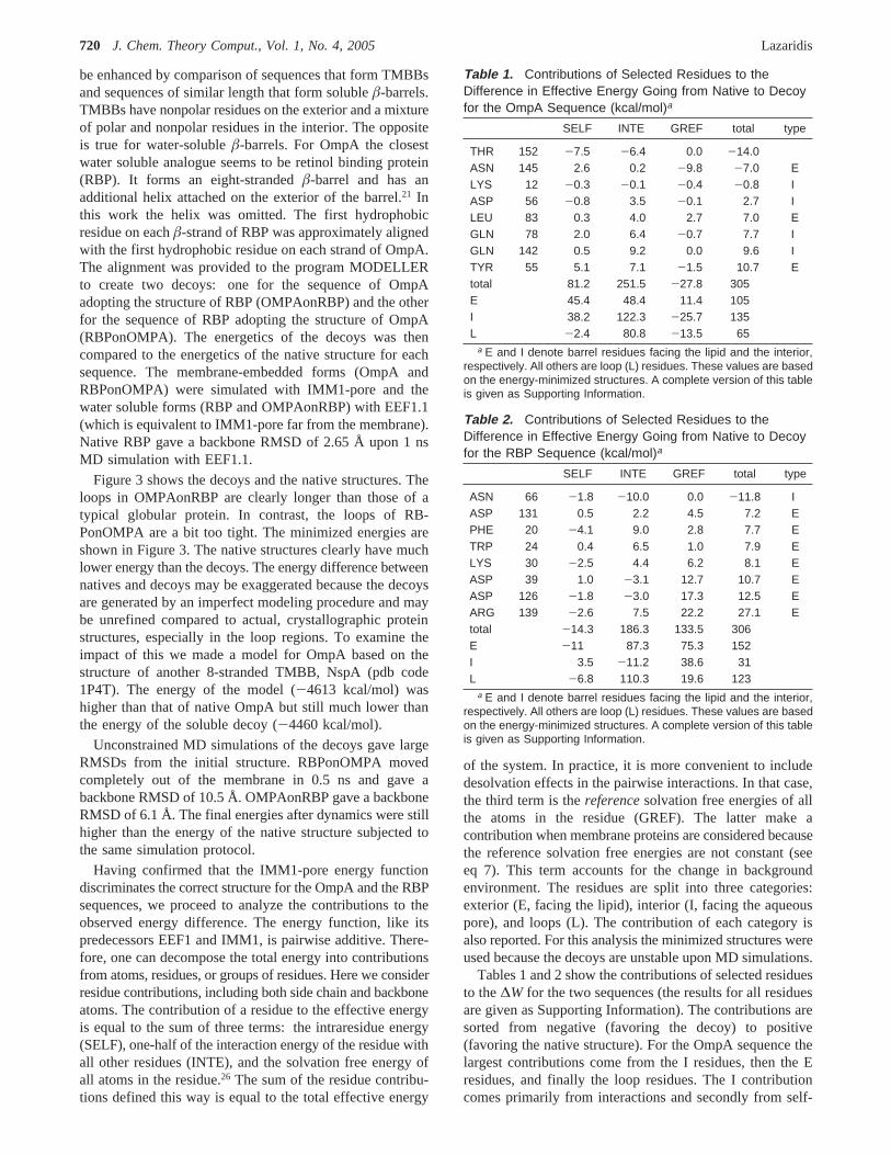
be enhanced by comparison of sequences that form TMBBsand sequences of similar length that form solubleâ-barrels.TMBBs have nonpolar residues on the exterior and a mixtureof polar and nonpolar residues in the interior. The oppositeis true for water-solubleâ-barrels. For OmpA the closestwater soluble analogue seems to be retinol binding protein(RBP). It forms an eight-strandedâ-barrel and has anadditional helix attached on the exterior of the barrel.21 Inthis work the helix was omitted. The first hydrophobicresidue on eachâ-strand of RBP was approximately alignedwith the first hydrophobic residue on each strand of OmpA.The alignment was provided to the program MODELLERto create two decoys: one for the sequence of OmpAadopting the structure of RBP (OMPAonRBP) and the otherfor the sequence of RBP adopting the structure of OmpA(RBPonOMPA). The energetics of the decoys was thencompared to the energetics of the native structure for eachsequence. The membrane-embedded forms (OmpA andRBPonOMPA) were simulated with IMM1-pore and thewater soluble forms (RBP and OMPAonRBP) with EEF1.1(which is equivalent to IMM1-pore far from the membrane).Native RBP gave a backbone RMSD of 2.65 Å upon 1 nsMD simulation with EEF1.1.
Figure 3 shows the decoys and the native structures. Theloops in OMPAonRBP are clearly longer than those of atypical globular protein. In contrast, the loops of RB-PonOMPA are a bit too tight. The minimized energies areshown in Figure 3. The native structures clearly have muchlower energy than the decoys. The energy difference betweennatives and decoys may be exaggerated because the decoysare generated by an imperfect modeling procedure and maybe unrefined compared to actual, crystallographic proteinstructures, especially in the loop regions. To examine theimpact of this we made a model for OmpA based on thestructure of another 8-stranded TMBB, NspA (pdb code1P4T). The energy of the model (-4613 kcal/mol) washigher than that of native OmpA but still much lower thanthe energy of the soluble decoy (-4460 kcal/mol).
Unconstrained MD simulations of the decoys gave largeRMSDs from the initial structure. RBPonOMPA movedcompletely out of the membrane in 0.5 ns and gave abackbone RMSD of 10.5 Å. OMPAonRBP gave a backboneRMSD of 6.1 Å. The final energies after dynamics were stillhigher than the energy of the native structure subjected tothe same simulation protocol.
Having confirmed that the IMM1-pore energy functiondiscriminates the correct structure for the OmpA and the RBPsequences, we proceed to analyze the contributions to theobserved energy difference. The energy function, like itspredecessors EEF1 and IMM1, is pairwise additive. There-fore, one can decompose the total energy into contributionsfrom atoms, residues, or groups of residues. Here we considerresidue contributions, including both side chain and backboneatoms. The contribution of a residue to the effective energyis equal to the sum of three terms: the intraresidue energy(SELF), one-half of the interaction energy of the residue withall other residues (INTE), and the solvation free energy ofall atoms in the residue.26 The sum of the residue contribu-tions defined this way is equal to the total effective energy
of the system. In practice, it is more convenient to includedesolvation effects in the pairwise interactions. In that case,the third term is thereferencesolvation free energies of allthe atoms in the residue (GREF). The latter make acontribution when membrane proteins are considered becausethe reference solvation free energies are not constant (seeeq 7). This term accounts for the change in backgroundenvironment. The residues are split into three categories:exterior (E, facing the lipid), interior (I, facing the aqueouspore), and loops (L). The contribution of each category isalso reported. For this analysis the minimized structures wereused because the decoys are unstable upon MD simulations.
Tables 1 and 2 show the contributions of selected residuesto the∆W for the two sequences (the results for all residuesare given as Supporting Information). The contributions aresorted from negative (favoring the decoy) to positive(favoring the native structure). For the OmpA sequence thelargest contributions come from the I residues, then the Eresidues, and finally the loop residues. The I contributioncomes primarily from interactions and secondly from self-
Table 1. Contributions of Selected Residues to theDifference in Effective Energy Going from Native to Decoyfor the OmpA Sequence (kcal/mol)a
SELF INTE GREF total type
THR 152 -7.5 -6.4 0.0 -14.0ASN 145 2.6 0.2 -9.8 -7.0 ELYS 12 -0.3 -0.1 -0.4 -0.8 IASP 56 -0.8 3.5 -0.1 2.7 ILEU 83 0.3 4.0 2.7 7.0 EGLN 78 2.0 6.4 -0.7 7.7 IGLN 142 0.5 9.2 0.0 9.6 ITYR 55 5.1 7.1 -1.5 10.7 Etotal 81.2 251.5 -27.8 305E 45.4 48.4 11.4 105I 38.2 122.3 -25.7 135L -2.4 80.8 -13.5 65
a E and I denote barrel residues facing the lipid and the interior,respectively. All others are loop (L) residues. These values are basedon the energy-minimized structures. A complete version of this tableis given as Supporting Information.
Table 2. Contributions of Selected Residues to theDifference in Effective Energy Going from Native to Decoyfor the RBP Sequence (kcal/mol)a
SELF INTE GREF total type
ASN 66 -1.8 -10.0 0.0 -11.8 IASP 131 0.5 2.2 4.5 7.2 EPHE 20 -4.1 9.0 2.8 7.7 ETRP 24 0.4 6.5 1.0 7.9 ELYS 30 -2.5 4.4 6.2 8.1 EASP 39 1.0 -3.1 12.7 10.7 EASP 126 -1.8 -3.0 17.3 12.5 EARG 139 -2.6 7.5 22.2 27.1 Etotal -14.3 186.3 133.5 306E -11 87.3 75.3 152I 3.5 -11.2 38.6 31L -6.8 110.3 19.6 123
a E and I denote barrel residues facing the lipid and the interior,respectively. All others are loop (L) residues. These values are basedon the energy-minimized structures. A complete version of this tableis given as Supporting Information.
720 J. Chem. Theory Comput., Vol. 1, No. 4, 2005 Lazaridis

energy. The L contribution also comes primarily frominteractions. Interactions and self-energy make equal con-tributions for E residues, with a smaller contribution comingfrom the change in environment (GREF). Overall, interac-tions make the largest contribution to∆W, followed by self-energy, while the change in environment favors the watersoluble decoy (polar residues are much “happier” in water).
For the 1AQB sequence the largest contributions todiscrimination come from the E residues, followed by the Lresidues, with only a small contribution from the I residues.The E contribution comes from interactions and change inenvironment. The L contribution comes primarily frominteractions. The I contribution comes primarily from changein environment.
For a more in-depth analysis we can look at individualresidue contributions. For the OmpA sequence, Tyr55 makesthe largest positive contribution, primarily because it hydro-gen bonds to Gln75 in the native (N) structure and makesno such interactions in the decoy. The value of thiscontribution is probably exaggerated by the use of a single,energy-minimized conformation. A number of I residuesmake positive contributions because they lose favorableinteractions from N to decoy. For example, Gln142 interactswith Gln78, Lys12, and Asp56 inside the pore in the N statebut lacks such interactions in the decoy. Several E residuesmake positive contributions too, which come from a mixtureof SELF, INTE, and GREF terms. For example, Leu83 losessome backbone interactions because the RBPâ-sheet is lessregular at the edges. It also loses some GREF energy becauseit goes from a position exposed to lipid to a position that ispartly exposed to water (there is a cavity in the interior ofthe barrel in RBP which is presumably filled with water).The largest negative contributions are from loop residuesthat happen to form better interactions in the decoy. Forexample, Thr152 has a chance to make a hydrogen bondwith Asn145 in the decoy but not in the N structure.Asn145’s contribution is also negative because of change inenvironment; it goes from the edge of the membrane to anaqueous environment.
For the RBP sequence, the largest positive contributioncomes from Arg139, primarily due to change in environment.In the decoy Arg139 is close to the edge of the membrane,whereas in the N structure it is exposed to water. Largepositive contributions are made by other charged E residuesthat are on the edge of the membrane (e.g. Asp126, Asp39,Asp131, and Lys30). Some nonpolar E residues, such asTrp24 and Phe20, also make positive contributions becauseof favorable interactions in the soluble N state. Many of thelarge negative contributions are from I residues that happento make good interactions in the TMBB decoy structure.
DiscussionA simple extension of the IMM1 implicit membrane modelled to an energy function that can be used to study membraneproteins with an aqueous pore. MD simulations of a rangeof TMBBs with this function gave stable trajectories andfavorable membrane insertion energies. In principle thefunction could be used with non-â-barrel MPs, such as ionchannels. However, the simple cylindrical shape of the pore
makes it best for cylindrical molecules. Other pore shapescould be accommodated by changing the definition of thefunction h(r′) in eq 9. For example, the pore cross sectioncould be made elliptical, or it could be made to vary alongthe z axis.
One limitation is that the aqueous pore is static. Therefore,the energy function cannot be used to study, for example,concerted protein insertion and aqueous pore formation. Thisproblem could conceivably be addressed by making the poreshape and size a dynamic variable in the MD simulation, inmuch the same way as the piston in constant pressuresimulations27 or the titration variables in constant-pH simula-tions.28
One important application of IMM1-pore stems from itsability to discriminate the native state of TMBBs. Becauseit is based on physics, it can be used to obtain insights intothe features that drive or do not drive protein sequences intoTMBB conformations. A first attempt to do so was madehere by comparing OmpA with RBP. The major conclusionsfrom this comparison are the following:
(1) The interior residues make a significant contributionto TMBB stability by engaging in favorable interactions withother interior residues. This effect may be largest for smallbarrels, where the interior residues are in contact withresidues across the pore.
(2) Polar and charged residues at the edges of the exteriorface of theâ-strands destabilize the putative TMBB fold.For the positively charged residues this effect will dependon the nature of the lipids (zwitterionic or anionic).
The results have implications for bioinformatic approachesto TMBB structure prediction. Neural networks and HiddenMarkov Models recognize amino acid composition andperhaps certain sequence patterns. Wimley’s analysis ofamino acid abundance at exterior and interior positions isessentially a one-body term. None of these approachesincludes the effect of specific interactions between residues.It appears that inclusion of a pairwise score (as, for example,in soluble protein structure prediction29,30) should improvethe discrimination.
Acknowledgment. This work was supported by theNational Science Foundation (MCB-0316667). Some com-putational resources were provided by an RCMI grant fromNIH (5G12RR003060).
Supporting Information Available: MODELLERalignment files and the complete versions of Tables 1 and2. This material is available free of charge via the Internetat http://pubs.acs.org.
References
(1) Schulz, G. E.Curr. Opin. Struct. Biol.2000, 10, 443.
(2) Vogel, H.; Jahnig, F.J. Mol. Biol. 1986, 190, 191.
(3) Schirmer, T.; Cowan, S. W.Protein Sci.1993, 2, 1361.
(4) Jacoboni, I.; Martelli, P. L.; Fariselli, P.; De Pinto, V.;Casadio, R.Protein Sci.2001, 10, 779.
(5) Gromiha, M. M.; Ahmad, S.; Suwa, M.J. Comput. Chem.2004, 25, 762.
Structural Determinants of Transmembraneâ-Barrels J. Chem. Theory Comput., Vol. 1, No. 4, 2005721
(6) Diederichs, K.; Freigang, J.; Umhau, S.; Zeth, K.; Breed, J.Protein Sci.1998, 7, 2413.
(7) Natt, N. K.; Kaur, H.; Raghava, G. P. S.Proteins: Struct.,Funct., Bioinformatics2004, 56, 11.
(8) Martelli, P. L.; Fariselli, P.; Krogh, A.; Casadio, R.Bioin-formatics2003, 18, S46.
(9) Bagos, P. G.; Liakopoulos, T. D.; Spyropoulos, I. C.;Hamodrakas, S. J.Bmc Bioinformatics2004, 5, 29.
(10) Bigelow, H. R.; Petrey, D. S.; Liu, J.; Przybylski, D.; Rost,B. Nucleic Acids Res.2004, 32, 2566.
(11) Liu, Q.; Zhu, Y. S.; Wang, B. H.; Li, Y. X.Comput. Biol.Chem. 2003, 27, 69.
(12) Wimley, W. C.Protein Sci. 2002, 11, 301.
(13) Liu, Q.; Zhu, Y.; Wang, B.; Li, Y.Comput. Biol. Chem.2003, 27, 355.
(14) Berven, F. S.; Flikka, K.; Jensen, H. B.; Eidhammer, I.Nucleic Acids Res.2004, 32, W394.
(15) Zhai, Y.; Saier, M. H., Jr.Protein Sci. 2002, 11, 2196.
(16) Lazaridis, T.; Karplus, M.Proteins1999, 35, 133.
(17) Lazaridis, T.Proteins2003, 52, 176.
(18) Pautsch, A.; Schulz, G. E.Nature Struct. Biol.1998, 5, 1013.
(19) Oomen, C. J.; Van Ulsen, P.; Van Gelder, P.; Feijen, M.;Tommassen, J.; Gros, P.Embo J. 2004, 23, 1257.
(20) Locher, K. P.; Rees, B.; Koebnik, R.; Mitschler, A.;Moulinier, L.; Rosenbusch, J. P.; Moras, D.Cell 1998, 95,771.
(21) Zanotti, G.; Panzalorto, M.; Marcato, A.; Malpeli, G.; Folli,C.; Berni, R.Acta Crystallogr. D Biol. Crystallogr.1998,54 (Pt 5), 1049.
(22) Marti-Renom, M. A.; Stuart, A. C.; Fiser, A.; Sanchez, R.;Melo, F.; Sali, A.Annu. ReV. Biophys. Biomol. Struct.2000,29, 291.
(23) Bond, P. J.; Sansom, M. S. P.J. Mol. Biol.2003, 329, 1035.
(24) Bond, P. J.; Cuthbertson, J. M.; Deol, S. S.; Sansom, M. S.P. J. Am. Chem. Soc.2004, 126, 15948.
(25) Bockmann, R. A.; Caflisch, A.Biophys J. 2005, 88, 3191.
(26) Lazaridis, T.; Karplus, M. Microscopic basis of macromo-lecular thermodynamics. InThermodynamics in biology;DiCera, E., Ed.; Oxford University Press: Oxford, 2001; p3.
(27) Feller, S. E.; Zhang, Y. H.; Pastor, R. W.; Brooks, B. R.J.Chem. Phys.1995, 103, 4613.
(28) Lee, M. S.; Salsbury, F. R., Jr.; Brooks, C. L., III.Proteins2004, 56, 738.
(29) Sippl, M. J.Curr. Opin. Struct. Biol.1995, 5, 229.
(30) Simons, K. T.; Kooperberg, C.; Huang, E.; Baker, D.J. Mol.Biol. 1997, 268, 209.
CT050055X
722 J. Chem. Theory Comput., Vol. 1, No. 4, 2005 Lazaridis