Embed Size (px)
Citation preview
DMD #31989
1
Superior Plasma Retention of A Cross-Linked Human Serum Albumin Dimer in
Nephrotic Rats as a New Type of Plasma Expander
Kazuaki Taguchi, Yukino Urata, Makoto Anraku, Hiroshi Watanabe, Keiichi Kawai, Teruyuki
Komatsu, Eishun Tsuchida, Toru Maruyama, Masaki Otagiri
Department of Biopharmaceutics (K.T., Y.U., M.A., H.W., T.M., M.O.), Center for Clinical
Pharmaceutical Sciences (H.W., T.M.), Graduate School of Pharmaceutical Sciences,
Kumamoto University, 5-1 Oe-honmachi, 862-0973 Kumamoto, Japan; Faculty of
Pharmaceutical Sciences (M.O.), Sojo University, 4-22-1 Ikeda, 860-0082 Kumamoto, Japan;
School of Health Sciences, Faculty of Medicine (K.K.), Kanazawa University, 5-11-80
Kodatsuno, Kanazawa 920-1192 Ishikawa, Japan; Research Institute for Science and
Engineering (T.K., E.T.), Waseda University, 3-4-1 Okubo, Shinjuku, 169-8555 Tokyo, Japan
DMD Fast Forward. Published on September 20, 2010 as doi:10.1124/dmd.109.031989
Copyright 2010 by the American Society for Pharmacology and Experimental Therapeutics.
This article has not been copyedited and formatted. The final version may differ from this version.DMD Fast Forward. Published on September 20, 2010 as DOI: 10.1124/dmd.109.031989
at ASPE
T Journals on M
ay 2, 2019dm
d.aspetjournals.orgD
ownloaded from
DMD #31989
2
Running title; Pharmacokinetics of Human Serum Albumin Dimer in Nephrosis
Corresponding author: Masaki Otagiri, Ph.D., Department of Biopharmaceutics,
Graduate School of Pharmaceutical Sciences, Kumamoto University, 5-1 Oe-honmachi,
Kumamoto 862-0973, Japan.
Phone.: +81-96-371-4150
Fax: +81-96-362-7690
E-mail: [email protected]
The number of text pages: 26
The number of tables: 2
The number of figures: 6
The number of references: 25
The number of words in the Abstract: 250
The number of words in the Introduction: 749
The number of words in the Discussion: 1499
Abbreviations: HSA, Human serum albumin; t1/2, half-life; AUC, area under the
concentration-time curve; CL, clearance; Vd, distribution volumes; % of ID, % of injection
of dose; HSA-FeP, albumin-heme; FcRn, Fc receptor
This article has not been copyedited and formatted. The final version may differ from this version.DMD Fast Forward. Published on September 20, 2010 as DOI: 10.1124/dmd.109.031989
at ASPE
T Journals on M
ay 2, 2019dm
d.aspetjournals.orgD
ownloaded from
DMD #31989
3
ABSTRACT
Human serum albumin (HSA) is clinically used as a plasma expander in patients with
hypoalbuminemia, and also can function as a drug carrier. However, the administered HSA is
readily eliminated from the blood circulation under pathological conditions, especially the
nephrotic syndrome. Here we present data on the pharmacokinetics of a structurally defined
HSA dimer, 2 HSA molecules that are crosslinked by reaction with 1,6-bis(maleimido)hexane
via Cys34, in nephrotic rats, and its superior circulation persistence, owing to the molecular
size-effect. The half-life (t1/2) of the HSA dimer persisted in the circulation 1.3-times longer
than that of monomeric HSA in normal rats, primarily because of the suppression of the
accumulation of the HSA dimer in the skin and muscle. In nephrotic rats, the t1/2 of the HSA
monomer decreased considerably, whereas the HSA dimer remained unaltered in the blood
stream, similar to that for normal rats. As a result, the t1/2 of the HSA dimer was two-fold
longer than that of the HSA monomer. This can be attributed to the fact that accumulation in
the kidney and urinary excretion of the HSA dimer were significantly suppressed. The
cross-linked HSA dimer shows a longer blood circulation than native HSA monomer in
nephrotic rats, which can be attributed to the suppression of renal filtration and leakage into
the extravascular space. This HSA dimer has the potential for use as a new plasma expander
and a useful drug carrier, but also as an artificial albumin-based oxygen carrier under high
glomerular permeability condition such as nephrosis.
This article has not been copyedited and formatted. The final version may differ from this version.DMD Fast Forward. Published on September 20, 2010 as DOI: 10.1124/dmd.109.031989
at ASPE
T Journals on M
ay 2, 2019dm
d.aspetjournals.orgD
ownloaded from
DMD #31989
4
INTRODUCTION
In clinical settings, a critically ill patient is typically given a plasma expander to
maintain colloid osmotic pressure and to increase the plasma volume. As of this writing,
dextran, hydroxylethyl starch and albumin have all been developed as plasma expanders, and
all are frequently used in critical situations.
As mentioned above, human serum albumin (HSA) is used as a plasma expander,
and is particularly useful when it is given as an infusion to patients with hypoalbuminemia,
such as the nephrotic syndrome. For many years, it was generally thought that an albumin
infusion improved life expectancy (Wilkes and Navickis, 2001; Vincent et al., 2004).
However, there has been little hard evidence to support its widespread clinical use. In fact, the
meta-analysis report concluded that an HSA infusion can be potentially harmful to critically
ill patients, and evidence in support of the administration of HSA reducing mortality in
critically ill patients with hypoalbuminaemia is lacking (Robert I, 1998). In such clinical
conditions, infused HSA does not appear to play a primary role as a plasma expander, because
the blood retention of HSA would likely be decreased. It is well known that capillary protein
permeability is increased in many pathological and physiological conditions, and that this is
accompanied by an increased HSA flux to the extravascular compartment. Under such
conditions, the administered HSA is transported to organs or extravasated, causing a
formation of edema, and hence, a worsening of the disease. This is especially true in the case
of the nephrotic syndrome, where the infused HSA is not only rapidly eliminated from the
intravascular to extravascular compartment but is also excreted in the urine (Pulimood and
Park, 2000). Therefore, it is necessary to administer a conventional HSA preparation
frequently, in order to maintain albumin concentration, and this leads to a dramatic increase in
the cost of treatment.
To overcome this issue, some investigators have attempted to increase the molecular
This article has not been copyedited and formatted. The final version may differ from this version.DMD Fast Forward. Published on September 20, 2010 as DOI: 10.1124/dmd.109.031989
at ASPE
T Journals on M
ay 2, 2019dm
d.aspetjournals.orgD
ownloaded from
DMD #31989
5
size of HSA by genetic or chemical dimerization (Andersson, 1970; Matsushita et al., 2006)
to prevent its leakage into the extravascular space. The reason for this is that it is widely
recognized that increasing the molecular size is the best strategy for inhibiting the capillary
permeability of a plasma expander, such as dextran or hydroxylethyl starch, in a clinical
setting. However, the blood retention of an HSA dimer is comparable or only slightly
increased compared to an HSA monomer, and the dimer has not been tested under high
permeability conditions (Matsushita et al., 2006). In addition, some disulfide-linked 34Cys
HSA dimers, prepared by connecting the 34Cys of 2 albumin molecules have a low stability
(Andersson, 1970) and are structurally modified compared to native HSA (Sollenne et al.,
1981). As a result, they are not ideal candidates for use in clinical applications as a plasma
expander. Because of this, it would be highly desirable to develop a new type of HSA
preparation, which has good blood retention under high permeability conditions as well as
normal conditions.
Komatsu et al. (2004a) recently reported on the preparation of a new type of HSA
dimer, in which 2 HSA molecules are cross-linked with 1,6-bis(maleimido)hexane (BMH).
This HSA dimer is prepared by specifically linking the 34Cys of 2 molecules using BMH.
Since BMH is sufficiently long (16.1 Å) to permit the HSA to maintain its flexibility and
hydrophobicity, the structural properties of native HSA can be preserved. In fact, the dimer is
almost identical to HSA in terms of ligand-binding capacity and blood compatibility. Because
of this, the cross-linked HSA dimer has the potential for serving as a substitute for HSA for
patients with high blood vessel permeability and glomerular permeability. Since, blood
retention is one of the most important factors for the function of a plasma expander, it is
noteworthy that little information regarding the pharmacokinetic properties, especially blood
retention, of the HSA dimer are available compared to the HSA monomer. Even though it is
anticipated that the HSA dimer would be administered under high permeability conditions,
This article has not been copyedited and formatted. The final version may differ from this version.DMD Fast Forward. Published on September 20, 2010 as DOI: 10.1124/dmd.109.031989
at ASPE
T Journals on M
ay 2, 2019dm
d.aspetjournals.orgD
ownloaded from
DMD #31989
6
such as nephrotic conditions, data that demonstrates the effect of increased molecular size on
the blood retention of the HSA dimer under similar clinical conditions is not available.
The purpose of this study was to clarify the pharmacokinetic properties of the HSA
dimer under nephrotic conditions and to verify the potential of the HSA dimer as a versatile
plasma expander. To accomplish this, we carried out the pharmacokinetic studies using an
111In-labeled HSA monomer and the cross-linked HSA dimer in normal and nephrotic rats
induced by treatment with adriamycin (doxorubicin).
This article has not been copyedited and formatted. The final version may differ from this version.DMD Fast Forward. Published on September 20, 2010 as DOI: 10.1124/dmd.109.031989
at ASPE
T Journals on M
ay 2, 2019dm
d.aspetjournals.orgD
ownloaded from
DMD #31989
7
MATERIALS AND METHODS
Chemicals
A HSA (AlbrecR, 25 wt.%) was provided by NIPRO (Osaka, Japan). Ethanol,
dithiothreitol (DTT) was purchased from Kanto Chemical, (Tokyo, Japan) and was used
without further purification. 1,6-Bis (maleimido) hexane was purchased from Pierce
Biotechnology (Rockford, USA). 111InCl3 (74 Mbq/mL in 0.02 N HCl) was donated by Nihon
MediPhysics (Takarazuka, Japan). All other chemicals were of the highest grade
commercially available, and all solutions were prepared using deionized, distilled water. The
HSA dimer was synthesized according to our previously reported procedures (Komatsu et al.,
2004a).
SDS-PAGE and Western Blotting
The HSA dimer was analyzed via SDS-PAGE, using 10% polyacrylamide gel, and
detected by staining with Coomassie blue R-250. Molecular weights were marked as follows,
bovine serum albumin (M.W. 66 kDa), lactate dehydrogenase (M.W. 140 kDa), catalase
(M.W. 232 kDa), ferritin (M.W. 440 kDa) and thyroglobulin (M.W. 669 kDa). Western
blotting was performed using a 10% polyacrylamide gel, and a rabbit anti-HSA polyclonal
antibody as the primary antibody followed by an anti-rabbit secondary antibody conjugated to
horseradish peroxidase. Proteins were detected using a using an ECL system (ECL Advance
Western Blotting Detection Kit; GE Healthcare Bio-sciences Corp.) with
LAS-4000EPUVmini (Fujifilm, Tokyo, Japan).
CD Spectroscopy
The secondary and tertiary structures of the HSA dimer were examined by recording
far- and near-CD spectra. The protein concentration was 8 μM, as determined by the method
This article has not been copyedited and formatted. The final version may differ from this version.DMD Fast Forward. Published on September 20, 2010 as DOI: 10.1124/dmd.109.031989
at ASPE
T Journals on M
ay 2, 2019dm
d.aspetjournals.orgD
ownloaded from
DMD #31989
8
of Bradford (1976), and the buffer used was 50 mM sodium phosphate, pH 7.0, 25 °C. Far
and near–UV intrinsic spectra were recorded from 200 to 250 nm and 250 to 350 nm,
respectively, using a Jasco J-720 spectropolarimeter (Tokyo, Japan).
Proteins labeling with 111In
For the pharmacokinetic experiments, the HSA monomer and dimer were
radiolabeled with 111In using the bifunctional chelating agent diethylenetriaminepentaacetic
acid (DTPA) anhydride according to the method of Hnatowich et al. (1982). Typically, each
sample (5 mg) was dissolved in 1 mL 0.1 M 4-(2-Hydroxyethyl)-1-piperazineethanesulfonic
acid (HEPES) buffer, pH 7.0, and mixed with 15 mM DTPA anhydride in 10 mL dimethyl
sulfoxide. The mixture was stirred for 60 min at room temperature, and the radiolabeled
product was purified by gel filtration on a Sephadex G-25 column (Amersham Biosciences) to
remove unreacted DTPA. Fractions containing the sample were collected and concentrated by
ultrafiltration at 4°C. A 20 mL aliquot of an 111InCl3 solution (74 MBq/mL) was then added to
20 mL of 0.1 M citrate buffer, pH 5.5, and 60 mL of a DTPA-coupled derivative solution was
added to the mixture. After 30 min, the mixture was applied to a PD-10 column and eluted
with 0.1 M citrate buffer, pH 5.5. Fractions containing the derivatives were collected and
concentrated by ultrafiltration at 4°C. The specific activity of 111In-labeled HSA monomer
and dimer were 1 × 10¹⁰ cpm/ng protein.
Animals
All animal experiments were performed according to the guidelines, principles, and
procedures for the care and use of laboratory animals of Kumamoto University. All male SD
rats were purchased from Kyudou Co. (Kumamoto, Japan). All animals were maintained
under conventional housing conditions, with food and water ad libitum in a
This article has not been copyedited and formatted. The final version may differ from this version.DMD Fast Forward. Published on September 20, 2010 as DOI: 10.1124/dmd.109.031989
at ASPE
T Journals on M
ay 2, 2019dm
d.aspetjournals.orgD
ownloaded from
DMD #31989
9
temperature-controlled room with a 12-hr dark/light cycle. The animals were acclimated for
one week, prior to the experiments.
Preparation of Nephrotic Rat
Nephrotic rats were induced according to the method described by Bertani (1982),
with minor modifications. Male SD rats were administrated adriamycin (doxorubicin) as a
single injection (9 mg/kg) through the tail vain under ether anesthesia. At 2 weeks after the
administration of adriamycin (doxorubicin), blood was collected from tail vein, and plasma
was obtained by centrifugation (6000 g, 5 min). In addition, urine was collected for 48 hr in a
metabolic cage. Plasma albumin concentrations were assayed by the general bromcresol green
(BCG) method using a Wako AlbuminBTest (Doumas et al., 1971), and urinary protein
concentration was determined by the method of Bradford (1976). According to previous study,
the rats, for which the urinary protein level was over 180 mg/day, were used as a nephrosis rat
model (Bertani T et al., 1982). The body weights of normal and nephrotic rats were 196 ± 18
and 179 ± 11 g, respectively (n=12, no significant differences).
The Pharmacokinetic Experimental Protocol
Just after preparing 111In labeled HSA monomer and dimer, the pharmacokinetic
study was performed, and the samples were assayed for radioactivity immediately after
collection. Both samples were mixed with unlabeled protein to adjust the protein
concentration before use in pharmacokinetic experiments (1×10⁷ cpm/mg protein). Normal
and nephrotic rats were anesthetized using ether and received a single injection of 111In
labeled protein (1 mg/kg, 1×10⁷ cpm/kg) via the tail vein. At each time point (3 min, 30 min,
1, 3, 6, 9, 12, 18, 24 and 48 hr) after an injection of the 111In labeled protein, a 100 μL
aliquot of blood was collected from the tail vein, and plasma was obtained by centrifugation
This article has not been copyedited and formatted. The final version may differ from this version.DMD Fast Forward. Published on September 20, 2010 as DOI: 10.1124/dmd.109.031989
at ASPE
T Journals on M
ay 2, 2019dm
d.aspetjournals.orgD
ownloaded from
DMD #31989
10
(6000 g, 5 min). At 48 hr after the injection of the 111In labeled protein, the rats were
sacrificed, the organs collected and rinsed with saline. Urine was collected at fixed intervals
in a metabolic cage. The levels of 111In in the plasma and excised organs were determined
using a γ-counter (ARC-5000, Aloka, Tokyo, Japan).
Data Analysis
Pharmacokinetic analyses after the administration of 111In labeled HSA monomer or
dimer were carried out using a two-compartment model and pharmacokinetic parameters were
estimated by curve fitting. Pharmacokinetic parameters were calculated by fitting using
MULTI, a normal least-squares program (Yamaoka et al., 1981). The two-compartment model
can be described by the following equation:
C = A • exp(-α • t)+B • exp(-β • t)
where C is % of dose/mL, t is time after administration of radiolabeled proteins. A, B and α, β
are coefficients or exponents in the model equation. As previously reported (Matsushita et al.,
2006; Taguchi et al., 2009), AUC, Vdss and CL were calculated using the following equations:
AUC = A/α + B/β
Vdss = V1+V2
CL = Dose/AUC
where AUC is the area under the concentration-time curve, CL is plasma clearance, V1 and V2
are central and peripheral distribution volumes. The half-lives of the HSAs were determined
as β-phase elimination within a 48 hr period. The renal CL was calculated as radioactivity
accumulation in urine until 48 hr/AUC from 0 to 48 hr. Data are shown as the means ± SD for
the indicated number of animals. Significant differences among each group were determined
using the two-tail unpaired Student’s t-test. A probability value of p<0.05 was considered to
indicate statistical significance.
This article has not been copyedited and formatted. The final version may differ from this version.DMD Fast Forward. Published on September 20, 2010 as DOI: 10.1124/dmd.109.031989
at ASPE
T Journals on M
ay 2, 2019dm
d.aspetjournals.orgD
ownloaded from
DMD #31989
11
RESULTS
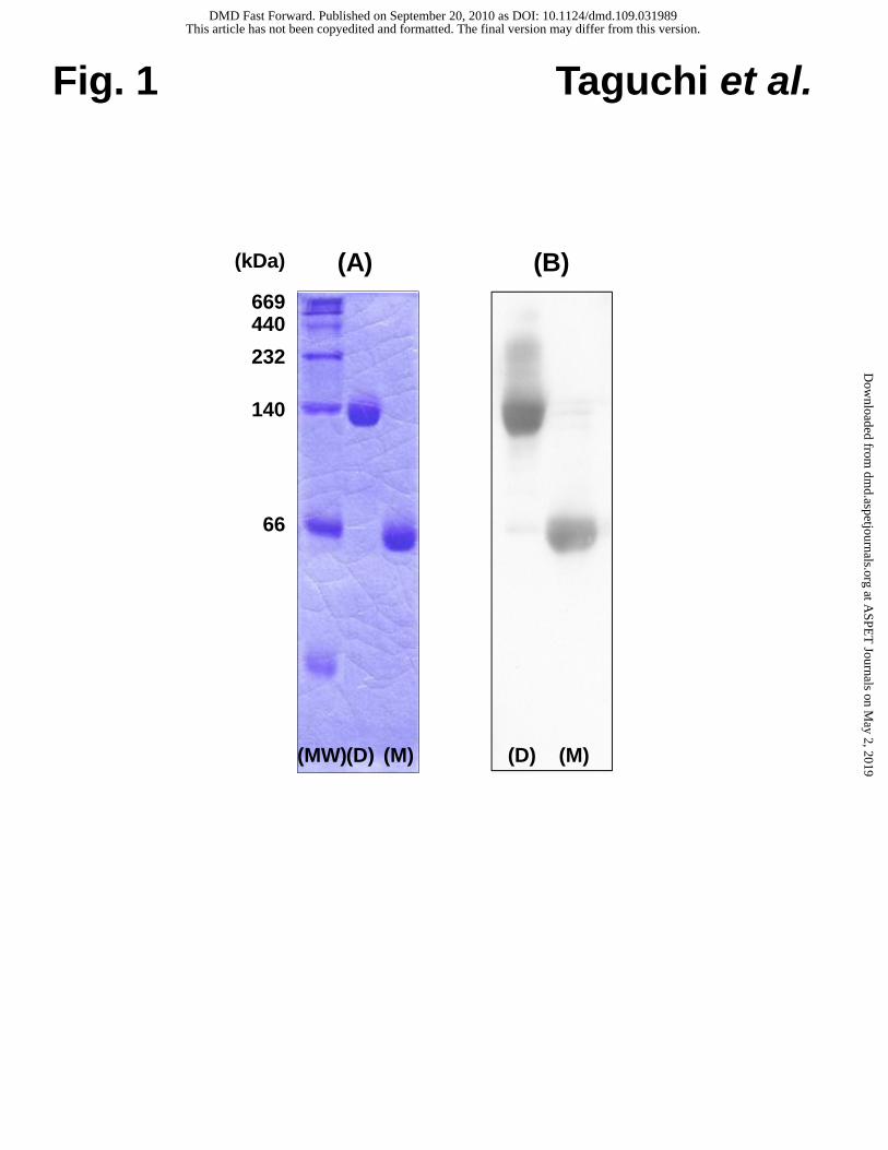
SDS-PAGE and Western Blotting of HSA Dimer
SDS-PAGE and Western Blotting were initially carried out to evaluate the efficiency
of HSA dimer synthesis. As shown in Fig. 1A, a single band was detected around 130 kDa,
and the molecular weight of the HSA dimer was approximately double that of HSA monomer.
A western blot analysis showed that the HSA dimer and the HSA monomer were both
recognized by an anti-HSA polyclonal antibody (Fig. 1B), indicating the recognition site of
the HSA dimer to the polyclonal antibody against native HSA was preserved.
Structural Characteristics of HSA Dimer
To confirm the structural characteristics of the HSA dimer, near- and far-CD
spectroscopy analyses were carried out. The near- and far-CD spectra of the HSA dimer
showed the same minima and shape as those of HSA monomer (data not shown). These
results suggest the structural characteristics of native HSA are preserved in the HSA dimer.
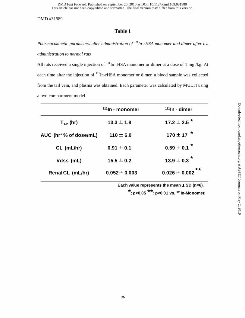
Pharmacokinetics of HSA Monomer and Dimer in Normal Rats
Figure 2 shows the time course for the plasma concentration of the 111In-HSA
monomer and dimer that had been injected into normal rats at a dose of 1 mg/kg, and Table 1
lists the pharmacokinetic parameters obtained using the two-compartment model. The
half-life (t1/2) of the HSA dimer was 1.3-times longer than that of the HSA monomer (17.2 ±
2.5 and 13.3 ± 1.8 hr, p<0.05, for the HSA dimer and monomer, respectively). This result is
consistent with our previous reported results on 125I-labeled variants (Komatsu et al., 2004a).
Accompanied by the decrease of CL and distribution volumes (Vdss) (CL; 0.59 ± 0.1 and
0.91 ± 0.1 mL/hr, p<0.05, Vdss; 13.9 ± 0.3 and 15.5 ± 0.2 mL, p<0.05, for HSA dimer and
monomer, respectively), the AUC and t1/2 were also significantly increased in the case of the
This article has not been copyedited and formatted. The final version may differ from this version.DMD Fast Forward. Published on September 20, 2010 as DOI: 10.1124/dmd.109.031989
at ASPE
T Journals on M
ay 2, 2019dm
d.aspetjournals.orgD
ownloaded from
DMD #31989
12
HSA dimer compared with the HSA monomer (AUC; 170 ± 17 and 110 ± 6.0 hr*% of
dose/mL, p<0.05, t1/2; 17.2 ± 2.5 and 13.3 ± 1.8 hr, p<0.05, for the HSA dimer and monomer,
respectively).
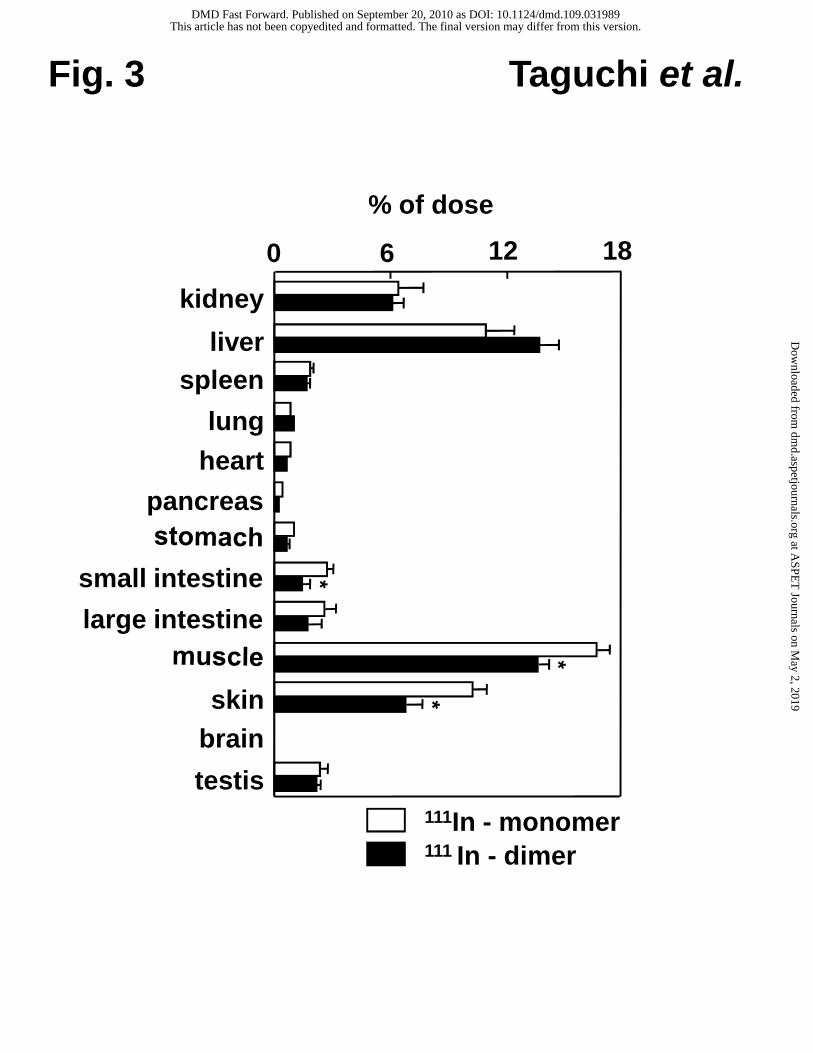
Figure 3 shows the tissue distribution of the 111In-HSA monomer and dimer (% of
injection of dose (% of ID)) at 48 hr after administration. Both the HSA monomer and dimer
were highly distributed in kidney, liver, skin and muscle. The accumulation of the HSA dimer
in skin and muscle was significantly suppressed compared with that of the HSA monomer
(Fig. 3). Furthermore, the urinary excretion of HSA labeled with 111In was also estimated. The
radioactivities of 111In at 48 hr after administration of the HSA monomer and dimer were
negligable (4.9 ± 0.2, 3.3 ± 1.7 % of ID for the HSA monomer and dimer, respectively, no
significant differences).
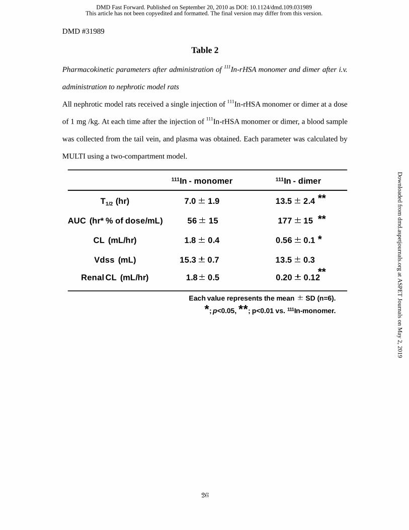
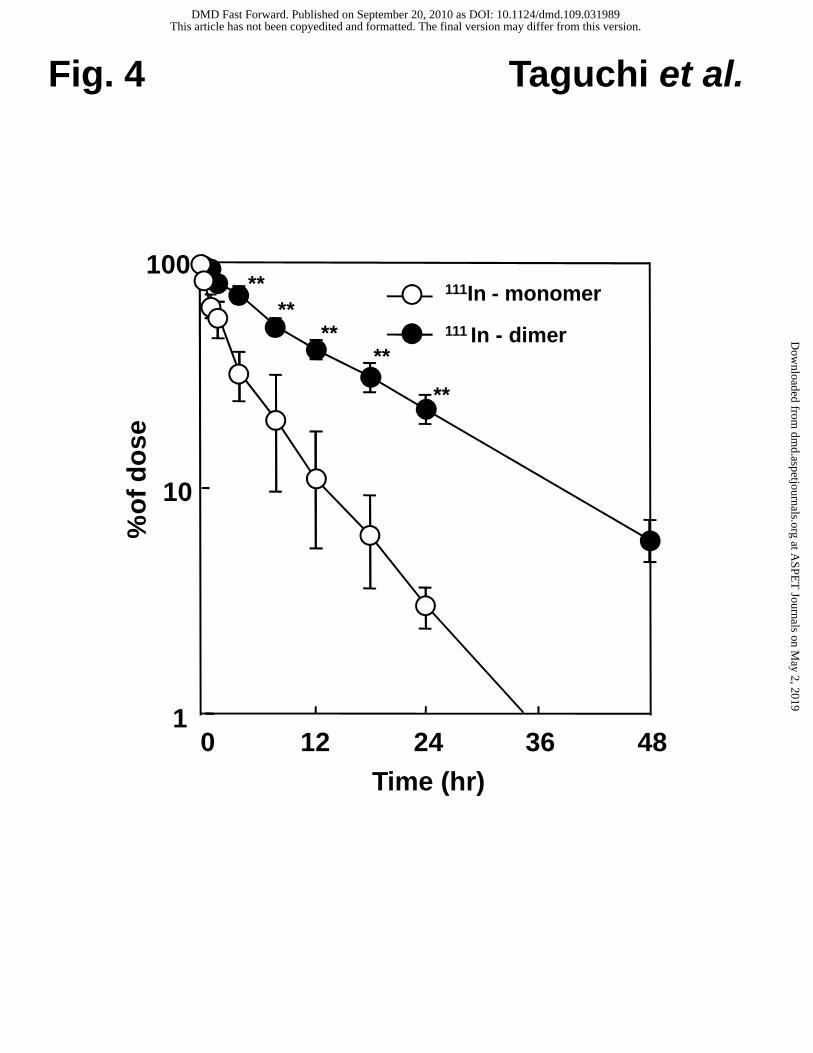
Pharmacokinetics of HSA Monomer and Dimer in Nephrotic Rats
A pharmacokinetic study of the HSA dimer was also performed in nephrotic rats
induced by adriamycin (doxorubicin) treatment. The serum albumin level in nephrotic rats
was 3.41 ± 0.2 g/dL (v.s. normal rats; 3.74 ± 0.2 g/dL, no significant differences), and urinary
protein was 213 ± 28 mg/day (v.s. normal rats; 8.0 ± 3.4 mg/day, p<0.001). These data are
consisted with previous report (Bertani T et al., 1982) and indicate that nephrosis was induced
in the adriamycin (doxorubicin) treated rats.
As shown in Fig. 4 and Table 2, the plasma concentration of HSA monomer was
rapidly cleared compared to that of the HSA dimer in nephrotic rats (13.5 ± 2.4 and 7.0 ± 1.9
hr, p<0.01, for the HSA dimer and monomer, respectively), while the plama concentration of
the HSA dimer in nephrotic rats was similar to that in normal rats. The CL for the HSA dimer
was decreased to one third of the HSA monomer (0.56 ± 0.1 and 1.8 ± 0.4 mL/hr, p<0.05, for
the HSA dimer and monomer, respectively). The AUC was also significantly increased (177 ±
This article has not been copyedited and formatted. The final version may differ from this version.DMD Fast Forward. Published on September 20, 2010 as DOI: 10.1124/dmd.109.031989
at ASPE
T Journals on M
ay 2, 2019dm
d.aspetjournals.orgD
ownloaded from
DMD #31989
13
15 and 56 ± 6.0 hr*% of dose/mL, p<0.01, for the HSA dimer and monomer, respectively).
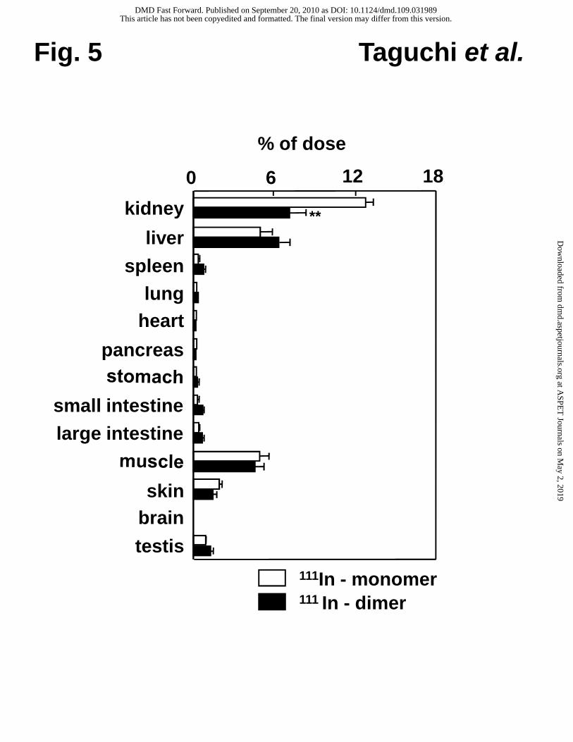
As shown in Fig. 5, the HSA monomer and dimer were both mainly distributed in the
kidney, liver, skin and muscle, the same as that in normal rats. Among these, there were no
significant differences between the accumulation of the HSA monomer and dimer in liver,
skin and muscle. Interestingly, the accumulation of the HSA dimer in the kidney was
dramatically decreased compared to that of the HSA monomer.
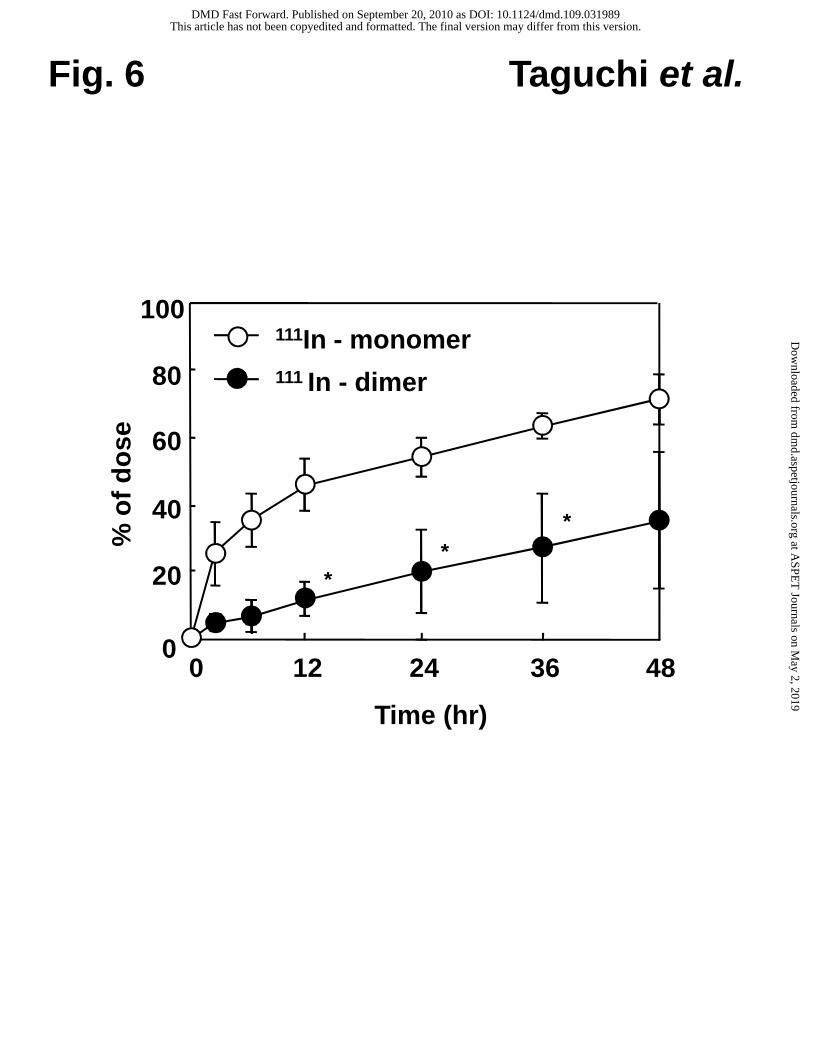
The urinary excretion of 111In in nephrotic rats were also measured (Fig. 6). The
radioactivity of 111In after administration of the HSA monomer was 75.1 ± 13.0 % of ID at 48
hr, while the radioactivity after HSA dimer administration was decreased by half compared to
that after the administration of the HSA monomer (34.6 ± 20 % of ID at 48 hr after injection,
p<0.05). In addition, the renal CL of HSA monomer was 1.81 ± 0.46 mL/hr, while that of
HSA dimer was 0.20 ± 0.12 mL/hr in nephrotic rats.
This article has not been copyedited and formatted. The final version may differ from this version.DMD Fast Forward. Published on September 20, 2010 as DOI: 10.1124/dmd.109.031989
at ASPE
T Journals on M
ay 2, 2019dm
d.aspetjournals.orgD
ownloaded from
DMD #31989
14
DISCUSSION
The major finding of this study is that the HSA dimer has the ability to maintain the
blood retention properties as well as that reported for normal conditions, even though under
the high permeability conditions, such as nephrosis, it can be expected to function as a novel
plasma expander.
In the case of normal rats, t1/2 for the HSA dimer (17.2 ± 2.5 hr) was 1.3-times higher
than that of the HSA monomer (13.3 ± 1.8 hr) accompanied by decrease the clearance from
circulation. It is generally known that approximately 40 % of native albumin exists in the
vascular space, and the remaining 60 % is present in the extravascular space. This ratio is
equilibrated between each space through the vascular endothelium. Our data suggest that the
movement of the HSA dimer from the vascular space to the cellular space through the
vascular endothelium is suppressed, the molecular weight of the dimer is double that of the
monomer (Fig. 1). In fact, in normal rats, the Vdss value for the HSA dimer (13.9 ± 0.3 mL)
was significantly decreased compared with that for the HSA monomer (15.5 ± 0.2 mL). As a
result, the accumulation of HSA dimer in skin and muscle were significantly suppressed
compared with the corresponding values for the HSA monomer (Fig. 3). This conclusion is
supported by the findings reported from Matsushita et al. (2006) who fused 2 molecules of
HSA to produce a recombinant HSA dimer using a yeast expression system. They clearly
showed that the plasma t1/2 of the recombinant HSA dimer was prolonged by 1.1-times due to
a decrease in the vascular permeability of the HSA dimer compared to that for the HSA
monomer in carrageenin-air-pouch rats. Moreover, Sejrsen et al. (1985) reported that the
predominant transcapillary transport mechanism for 131I-albumin is compatible with
transcapillary diffusion through pores with an effective equivalent pore radius of 145 Å.
Hence, the extent of extravasation of albumin may be reduced by increasing its molecular size.
It has also been shown that albumin, with an estimated radius of 35.5 Å, and water do not
This article has not been copyedited and formatted. The final version may differ from this version.DMD Fast Forward. Published on September 20, 2010 as DOI: 10.1124/dmd.109.031989
at ASPE
T Journals on M
ay 2, 2019dm
d.aspetjournals.orgD
ownloaded from
DMD #31989
15
share a common pathway in crossing the endothelial monolayer, suggesting the existence of a
large pore pathway for albumin (Dull et al., 1991). Namely, the increase in the molecular size
of HSA clearly led to a retardation in extravasation through the vascular endothelium,
resulting in a longer lifetime in the blood stream.
In the cases of certain renal injuries or obstacles, such as the nephrosis syndrome,
infused HSA is easily filtered by the renal glomerulus. In contrast, the t1/2 for the HSA dimer
in nephrotic rats (13.5 ± 2.4 hr) was comparable to that for the HSA monomer (13.3 ± 1.8 hr)
in normal rats. Thus, the t1/2 for the HSA dimer was approximately double that of the HSA
monomer in nephrotic rats (7.0 ± 1.9 hr). In general, the glomerular biological membrane has
properties that allow for high filtration rates of water and small and mid-sized molecules, but
does not allow larger proteins such as HSA to be filtered. These restrictions can be explained
by several mechanisms, including charge repulsion in the glomerular basement membrane and
barrier depending on their molecular size (Haraldsson et al., 2008). The molecular weight of
the HSA dimer is exactly 132,741, and the HSA dimer retains a negative charge as well as the
HSA monomer (pI= 4.8) (Komatsu et al., 2004a). Therefore, the increased t1/2 for the HSA
dimer in nephrotic rats is likely to be due to the increased molecular size of the molecule. In
fact, several results in this study support this hypothesis: (i) the renal distribution and urinary
excretion of the HSA dimer were decreased significantly, compared to those of the HSA
monomer (Fig. 5 and 6), (ii) the renal accumulation of the HSA dimer in nephrotic rats was
similar to that in normal rats (Fig. 3 and 5) , (iii) after administration of the HSA dimer to
nephrotic rats, the reduction in urinary excretion prolonged the t1/2 (Fig. 4 and 6). Although
the renal CL of HSA monomer was consisted with the systemic CL of HSA monomer in
nephrotic rats, the renal CL of HSA dimer was lower than the systemic CL of HSA dimer.
This suggested that the increased t1/2 of HSA dimer in nephrotic rats was not explained by
only suppression of protein leakage across the glomeruls. In the previous reports, bone was
This article has not been copyedited and formatted. The final version may differ from this version.DMD Fast Forward. Published on September 20, 2010 as DOI: 10.1124/dmd.109.031989
at ASPE
T Journals on M
ay 2, 2019dm
d.aspetjournals.orgD
ownloaded from
DMD #31989
16
one of the major distribution tissues of albumin (Yedgar et al., 1983), and albumin was a
constituent of the organic matrix in bone (Triffitt and Owen, 1973). Therefore, HSA dimer
may be distributed and utilized in bone. However, in this study, we could not examine the
clearance of HSA in bone. Further study will be needed to achieve this issue.
Very recently, Martini et al. (2008) demonstrated a new type of plasma expander,
PEG-HSA (carrying 6 copies of PEG-5000 chains per molecule; molecular weight: 97 kDa),
that is effective at reduced plasma concentrations and potentially has a better defined
pharmacokinetic profile because of its larger molecular size. Furthermore, they suggested that
the PEGylation of HSA possibly results in a more effective plasma volume expander during
hemodilution or in resuscitation fluids that are used the treatment of hemorrhagic shock with
the advantages of a longer t1/2 because of reduced glomerular filtration and movement to the
extravascular space. Since the molecular weight of the HSA dimer is approximately 130 kDa,
which is larger than the PEG-HSA alluded to above, it has promise as a new plasma volume
expander instead of native HSA monomer for patients with hypoalbuminemia, especially the
nephrosis syndrome.
As described above, since the HSA dimer shows superior blood retention properties,
it would be also expected to serve as a carrier for drug delivery in high permeability
conditions. Tsuchida et al (1997) previously reported on an HSA-based artificial oxygen
carrier “albumin-heme (HSA-FeP)”, which is a synthetic heme with a covalently bound
proximal base that is incorporated into the hydrophobic cavities of HSA. HSA-FeP reversibly
binds and releases O2 under physiological conditions, the same as hemoglobin (Komatsu et al.,
1999). An in vivo study using hemorrhagic shocked rats revealed that, the renal cortical
O2-tensions and skeletal tissue O2-tensions were increased when HSA-FeP was injected
(Tsuchida et al., 2000; Komatsu et al., 2004b). An artificial oxygen carrier is also required to
maintain high blood circulation, because it must temporarily function until a blood transfusion
This article has not been copyedited and formatted. The final version may differ from this version.DMD Fast Forward. Published on September 20, 2010 as DOI: 10.1124/dmd.109.031989
at ASPE
T Journals on M
ay 2, 2019dm
d.aspetjournals.orgD
ownloaded from
DMD #31989
17
is available or until autologous blood is recovered after a massive hemorrhage. In fact,
Komatsu et al (2004a) demonstrated that the HSA dimer enables a maximum of 16 molecules
of FeP to bind and the obtained dimeric HSA-FeP bound oxygen approximately twice higher
than blood. The HSA-FeP dimer shows promise as a new type of oxygen carrier that is
efficiently retained in circulating blood under nephrotic conditions.
Although this study provides a demonstration of the utility of the HSA dimer as a
plasma expander, our model has several limitations with respect to extrapolating it for use in a
human clinical setting. Our studies only dealt with the nephrotic syndrome, as induced by
adriamycin (doxorubicin). Since the nephrotic syndrome is a multiplex pathology induced by
several factors, such as heredity and immunity, it will be necessary to demonstrate the
pharmacokinetic properties of the HSA dimer in several nephrotic syndrome model animals.
In addition, the dose using in this study (1 mg/kg) is less than a pharmacologic dose in clinical
situation. A pharmacokinetic examination using pharmacologic doses of monomer and dimer
should be one of the subjects of future investigation. Moreover, recent evidence indicates that
the Fc receptor (FcRn) expressed in the kidney reclaimed albumin, thus maintaining the
serum concentration of albumin (Sarav et al., 2009). This finding indicates that interactions
between albumin and FcRn are important aspects of the pharmacokinetics of albumin.
However, it is well established that there is a large species difference in the interaction of
albumin and FcRn. In this study, since human albumin was administrated to rats, the
interaction between HSA and rat FcRn would likely be negligible. Thus, this interaction is not
likely to contribute to the pharmacokinetic data related to the HSA dimer as shown in this
study. However, it will be necessary to examine the interactions of the HSA dimer and human
FcRn for clinical development.
In conclusion, the cross-linked HSA dimer show potential for use as a new plasma
volume expander, since it showed superior blood retention characteristics in various clinical
This article has not been copyedited and formatted. The final version may differ from this version.DMD Fast Forward. Published on September 20, 2010 as DOI: 10.1124/dmd.109.031989
at ASPE
T Journals on M
ay 2, 2019dm
d.aspetjournals.orgD
ownloaded from
DMD #31989
18
situations. It’s use would allow us to reduce the multiple administrations required in the case
of conventional HSA preparations, and could be quite cost-effective. In addition, the HSA
dimer is also predicted to function as a versatile carrier for drug delivery systems, in particular,
an albumin based artificial oxygen carrier. Very recently, recombinant HSA has been
approved as a serum derived HSA alternative in Japan. As a result, it appears likely that a
recombinant HSA dimer could be used as a substitute of HSA preparations, even if in cases of
various pathological conditions such as the nephrosis syndrome.
This article has not been copyedited and formatted. The final version may differ from this version.DMD Fast Forward. Published on September 20, 2010 as DOI: 10.1124/dmd.109.031989
at ASPE
T Journals on M
ay 2, 2019dm
d.aspetjournals.orgD
ownloaded from
DMD #31989
19
REFERENCES
Andersson LO (1970) Hydrolysis of disulfide bonds in weakly alkaline media. II. Bovine
serum albumin dimer. Biochim Biophys Acta 200:363-369.
Bertani T, Poggi A, Pozzoni R, Delaini F, Sacchi G, Thoua Y, Mecca G, Remuzzi G and
Donati MB (1982) Adriamycin-induced nephrotic syndrome in rats: sequence of
pathologic events. Lab Invest 46:16-23.
Bradford MM (1976) A rapid and sensitive method for the quantitation of microgram
quantities of protein utilizing the principle of protein-dye binding. Anal Biochem
72:248-254.
Doumas BT, Watson WA and Biggs HG (1971) Albumin standards and the measurement of
serum albumin with bromcresol green. Clin Chim Acta 31:87-96.
Dull RO, Jo H, Sill H, Hollis TM and Tarbell JM (1991) The effect of varying albumin
concentration and hydrostatic pressure on hydraulic conductivity and albumin
permeability of cultured endothelial monolayers. Microvasc Res 41:390-407.
Haraldsson B, Nystrom J and Deen WM (2008) Properties of the glomerular barrier and
mechanisms of proteinuria. Physiol Rev 88:451-487.
Hnatowich DJ, Layne WW and Childs RL (1982) The preparation and labeling of
DTPA-coupled albumin. Int J Appl Radiat Isot 33:327-332.
Komatsu T, Hamamatsu K, Wu J and Tsuchida E (1999) Physicochemical properties and
O2-coordination structure of human serum albumin incorporating
tetrakis(o-pivalamido)phenylporphyrinatoiron(II) derivatives. Bioconjug Chem
10:82-86.
Komatsu T, Oguro Y, Teramura Y, Takeoka S, Okai J, Anraku M, Otagiri M and Tsuchida E
(2004a) Physicochemical characterization of cross-linked human serum albumin dimer
and its synthetic heme hybrid as an oxygen carrier. Biochim Biophys Acta 1675:21-31.
This article has not been copyedited and formatted. The final version may differ from this version.DMD Fast Forward. Published on September 20, 2010 as DOI: 10.1124/dmd.109.031989
at ASPE
T Journals on M
ay 2, 2019dm
d.aspetjournals.orgD
ownloaded from
DMD #31989
20
Komatsu T, Yamamoto H, Huang Y, Horinouchi H, Kobayashi K and Tsuchida E (2004b)
Exchange transfusion with synthetic oxygen-carrying plasma protein "albumin-heme"
into an acute anemia rat model after seventy-percent hemodilution. J Biomed Mater
Res A 71:644-651.
Martini J, Cabrales P, K A, Acharya SA, Intaglietta M and Tsai AG (2008) Survival time in
severe hemorrhagic shock after perioperative hemodilution is longer with
PEG-conjugated human serum albumin than with HES 130/0.4: a microvascular
perspective. Crit Care 12:R54.
Matsushita S, Chuang VT, Kanazawa M, Tanase S, Kawai K, Maruyama T, Suenaga A and
Otagiri M (2006) Recombinant human serum albumin dimer has high blood
circulation activity and low vascular permeability in comparison with native human
serum albumin. Pharm Res 23:882-891.
Robert I (1998) Human albumin administration in critically ill patients: systematic review of
randomised controlled trials. Cochrane Injuries Group Albumin Reviewers. BMJ
317:235-240.
Pulimood TB and Park GR (2000) Debate: Albumin administration should be avoided in the
critically ill. Crit Care 4:151-155.
Sarav M, Wang Y, Hack BK, Chang A, Jensen M, Bao L and Quigg RJ (2009) Renal FcRn
reclaims albumin but facilitates elimination of IgG. J Am Soc Nephrol 20:1941-1952.
Sejrsen P, Paaske WP and Henriksen O (1985) Capillary permeability of 131I-albumin in
skeletal muscle. Microvasc Res 29:265-281.
Sollenne NP, Wu HL and Means GE (1981) Disruption of the tryptophan binding site in the
human serum albumin dimer. Arch Biochem Biophys 207:264-269.
Taguchi K, Maruyama T, Iwao Y, Sakai H, Kobayashi K, Horinouchi H, Tsuchida E, Kai T
and Otagiri M (2009) Pharmacokinetics of single and repeated injection of
This article has not been copyedited and formatted. The final version may differ from this version.DMD Fast Forward. Published on September 20, 2010 as DOI: 10.1124/dmd.109.031989
at ASPE
T Journals on M
ay 2, 2019dm
d.aspetjournals.orgD
ownloaded from
DMD #31989
21
hemoglobin-vesicles in hemorrhagic shock rat model. J Control Release 136:232-239.
Triffitt JT and Owen M (1973) Studies on bone matrix glycoproteins. Incorporation of
(1-14C)glucosamine and plasma (14C)glycoprotein into rabbit cortical bone. Biochem
J 136:125-134.
Tsuchida E, Ando K, Maejima H, Kawai N, Komatsu T, Takeoka S and Nishide H (1997)
Properties of and oxygen binding by albumin-tetraphenylporphyrinatoiron(II)
derivative complexes. Bioconjug Chem 8:534-538.
Tsuchida E, Komatsu T, Hamamatsu K, Matsukawa Y, Tajima A, Yoshizu A, Izumi Y and
Kobayashi K (2000) Exchange transfusion with albumin-heme as an artificial
O2-infusion into anesthetized rats: physiological responses, O2-delivery, and
reduction of the oxidized hemin sites by red blood cells. Bioconjug Chem 11:46-50.
Vincent JL, Navickis RJ and Wilkes MM (2004) Morbidity in hospitalized patients receiving
human albumin: a meta-analysis of randomized, controlled trials. Crit Care Med
32:2029-2038.
Wilkes MM and Navickis RJ (2001) Patient survival after human albumin administration. A
meta-analysis of randomized, controlled trials. Ann Intern Med 135:149-164.
Yamaoka K, Tanigawara Y, Nakagawa T and Uno T (1981) A pharmacokinetic analysis
program (multi) for microcomputer. J Pharmacobiodyn 4:879-885.
Yedgar S, Carew TE, Pittman RC, Beltz WF and Steinberg D (1983) Tissue sites of
catabolism of albumin in rabbits. Am J Physiol 244:E101-107.
This article has not been copyedited and formatted. The final version may differ from this version.DMD Fast Forward. Published on September 20, 2010 as DOI: 10.1124/dmd.109.031989
at ASPE
T Journals on M
ay 2, 2019dm
d.aspetjournals.orgD
ownloaded from
DMD #31989
22
Footnote
This research was partially supported by Grant-in-Aid for Scientific Research (no. 20350058)
from JSPS, and Grant-in-Aid for Scientific Research for Priority Area (area 2107) from
MEXT Japan, and Health Science Research Grants (Regulatory Science) from MHLW Japan.
K.T. and Y.U. contribute equally to this work.
This article has not been copyedited and formatted. The final version may differ from this version.DMD Fast Forward. Published on September 20, 2010 as DOI: 10.1124/dmd.109.031989
at ASPE
T Journals on M
ay 2, 2019dm
d.aspetjournals.orgD
ownloaded from
DMD #31989
23
FIGURE LEGENDS
Fig. 1. SDS-PAGE (A) and Western Blot Analysis (B) of the HSA Dimer and Monomer.
MW, Molecular weight; D, HSA-dimer; M, HSA-monomer
Fig. 2. Relative Plasma Concentration of 111In-HSA Monomer and Dimer after i.v.
Administration to Normal Rats
111In-HSA monomer (open circle) and 111In-HSA dimer (closed circle) were injected at a dose
of 1 mg/kg. Each data point represents the mean ± SD (n=6). *; p<0.05, **; p<0.01 vs.
111In-HSA monomer.
Fig. 3 Tissue Distribution of Radioactivity at 48 hr after the i.v. Administration of 111In-HSA
Monomer or Dimer to Normal Rats
111In-HSA monomer (open column) or 111In-HSA dimer (closed column) were injected at a
dose of 1 mg/kg. Each column represents the mean ± SD (n=6). *; p<0.05, vs.
111In-monomer.
Fig. 4. Relative Plasma Concentration of 111In-HSA Monomer and Dimer after i.v.
Administration to Nephrotic Rats
111In-HSA monomer (open circle) or 111In-HSA dimer (closed circle) were injected at a dose
of 1 mg/kg. Each point represents the mean ± SD (n=6). **; p<0.01 vs. 111In- HSA monomer.
Fig. 5. Tissue Distribution of Radioactivity at 48 hr after i.v. Administration of 111In-HSA
Monomer or Dimer to Nephrotic Rats
111In- HSA monomer (open column) or 111In- HSA dimer (closed column) were injected at a
dose of 1 mg/kg. Each column represents the mean ± SD (n=6). **; p<0.01, vs.
This article has not been copyedited and formatted. The final version may differ from this version.DMD Fast Forward. Published on September 20, 2010 as DOI: 10.1124/dmd.109.031989
at ASPE
T Journals on M
ay 2, 2019dm
d.aspetjournals.orgD
ownloaded from
DMD #31989
24
111In-monomer.
Fig. 6. Urinary Excretion of 111In-rHSA Monomer and 111In-rHSA Dimer after i.v.
Administration to Nephrotic Rats
111In- HSA monomer (open circle) or 111In- HSA dimer (closed circle) were injected at a dose
of 1mg/kg. Each point represents the mean ± SD (n=4). *; p<0.05, vs. 111In-monomer.
This article has not been copyedited and formatted. The final version may differ from this version.DMD Fast Forward. Published on September 20, 2010 as DOI: 10.1124/dmd.109.031989
at ASPE
T Journals on M
ay 2, 2019dm
d.aspetjournals.orgD
ownloaded from
DMD #31989
25
Table 1
Pharmacokinetic parameters after administration of 111In-rHSA monomer and dimer after i.v.
administration to normal rats
All rats received a single injection of 111In-rHSA monomer or dimer at a dose of 1 mg /kg. At
each time after the injection of 111In-rHSA monomer or dimer, a blood sample was collected
from the tail vein, and plasma was obtained. Each parameter was calculated by MULTI using
a two-compartment model.
Each value represents the mean ±SD (n=6).
*; p<0.05 **; p<0.01 vs. 111In-Monomer.
T1/2 (hr) 13.3 ± 1.8 17.2 ± 2.5
AUC (hr* % of dose/mL) 110 ± 6.0 170 ± 17
CL (mL/hr) 0.91 ± 0.1 0.59 ± 0.1
Vdss (mL) 15.5 ± 0.2 13.9 ± 0.3
111In - monomer 111In - dimer
*
*
*
*
Renal CL (mL/hr) 0.052±0.003 0.026 ± 0.002 **
This article has not been copyedited and formatted. The final version may differ from this version.DMD Fast Forward. Published on September 20, 2010 as DOI: 10.1124/dmd.109.031989
at ASPE
T Journals on M
ay 2, 2019dm
d.aspetjournals.orgD
ownloaded from
DMD #31989
26
Table 2
Pharmacokinetic parameters after administration of 111In-rHSA monomer and dimer after i.v.
administration to nephrotic model rats
All nephrotic model rats received a single injection of 111In-rHSA monomer or dimer at a dose
of 1 mg /kg. At each time after the injection of 111In-rHSA monomer or dimer, a blood sample
was collected from the tail vein, and plasma was obtained. Each parameter was calculated by
MULTI using a two-compartment model.
Each value represents the mean ± SD (n=6).
*; p<0.05, **; p<0.01 vs. 111In-monomer.
T1/2 (hr) 7.0 ± 1.9 13.5 ± 2.4
AUC (hr* % of dose/mL) 56 ± 15 177 ± 15
CL (mL/hr) 1.8 ± 0.4 0.56 ± 0.1
Vdss (mL) 15.3 ± 0.7 13.5 ± 0.3
111In - monomer 111In - dimer
**
**
*
Renal CL (mL/hr) 1.8± 0.5 0.20 ± 0.12**
This article has not been copyedited and formatted. The final version may differ from this version.DMD Fast Forward. Published on September 20, 2010 as DOI: 10.1124/dmd.109.031989
at ASPE
T Journals on M
ay 2, 2019dm
d.aspetjournals.orgD
ownloaded from
669440
232
140
66
(kDa)
Taguchi et al.
(MW)(D) (M) (D) (M)
Fig. 1
(A) (B)
This article has not been copyedited and formatted. The final version may differ from this version.DMD Fast Forward. Published on September 20, 2010 as DOI: 10.1124/dmd.109.031989
at ASPE
T Journals on M
ay 2, 2019dm
d.aspetjournals.orgD
ownloaded from
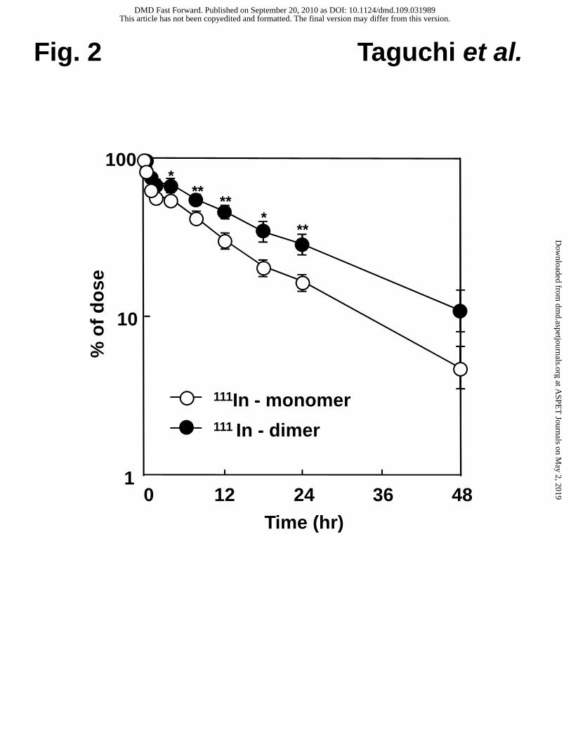
Fig. 2
0 12Time (hr)
48
100
1
111In - monomer111 In - dimer
***
***
**
% o
f d
ose
24 36
10
Taguchi et al.
This article has not been copyedited and formatted. The final version may differ from this version.DMD Fast Forward. Published on September 20, 2010 as DOI: 10.1124/dmd.109.031989
at ASPE
T Journals on M
ay 2, 2019dm
d.aspetjournals.orgD
ownloaded from
small intestine
large intestine
skinbrain
% of dose
0 6 18
kidney
liverspleen
lungheart
pancreas
testis
12*
*
*
111In - monomer111 In - dimer
Fig. 3 Taguchi et al.
This article has not been copyedited and formatted. The final version may differ from this version.DMD Fast Forward. Published on September 20, 2010 as DOI: 10.1124/dmd.109.031989
at ASPE
T Journals on M
ay 2, 2019dm
d.aspetjournals.orgD
ownloaded from
0 12
% o
f d
ose
Time (hr)24 36 48
10
100
1
Fig. 4
111In - monomer
111 In - dimer
Taguchi et al.
****
****
**
This article has not been copyedited and formatted. The final version may differ from this version.DMD Fast Forward. Published on September 20, 2010 as DOI: 10.1124/dmd.109.031989
at ASPE
T Journals on M
ay 2, 2019dm
d.aspetjournals.orgD
ownloaded from
kidney
liver
spleenlung
heart
pancreas
small intestinelarge intestine
skinbrain
testis111In - monomer111 In - dimer
Fig. 5 Taguchi et al.
**
% of dose
0 6 1812
This article has not been copyedited and formatted. The final version may differ from this version.DMD Fast Forward. Published on September 20, 2010 as DOI: 10.1124/dmd.109.031989
at ASPE
T Journals on M
ay 2, 2019dm
d.aspetjournals.orgD
ownloaded from
0 12 24 360
40
60
20
48
100
80
% o
f d
ose
Time (hr)
**
*
111In - monomer111 In - dimer
Fig. 6 Taguchi et al.
This article has not been copyedited and formatted. The final version may differ from this version.DMD Fast Forward. Published on September 20, 2010 as DOI: 10.1124/dmd.109.031989
at ASPE
T Journals on M
ay 2, 2019dm
d.aspetjournals.orgD
ownloaded from