Embed Size (px)
Citation preview


FUNCIÓN MUSCULAR PERIFÉRICA Y ENTRENAMIENTO FÍSICO EN LA ENFERMEDAD PULMONAR
OBSTRUCTIVA CRÓNICA
RELACIONES ENTRE ESTADO REDOX,INFLAMACIÓN Y RESPIRACIÓN
MITOCONDRIAL

Roberto Alejandro Rabinovich
FUNCIÓN MUSCULAR PERIFÉRICA Y ENTRENAMIENTO FÍSICO EN LA ENFERMEDAD PULMONAR
OBSTRUCTIVA CRÓNICA
RELACIONES ENTRE ESTADO REDOX,INFLAMACIÓN Y RESPIRACIÓN
MITOCONDRIAL
Tesis Doctoral
Universitat de BarcelonaDepartament de Medicina | Programa de Biopatología en Medicina
ROBERTO ALEJANDRO RABINOVICHLicenciado en Medicina y CirugíaUniversidad de Buenos Aires
Especialista en Medicina InternaUniversidad de Buenos Aires
Especialista en Cuidados IntensivosPontifi cia Universidad Católica de Buenos Aires
Alumno del programa de Biopatología en MedicinaDepartament de Medicina | Universitat de BarcelonaBienio 2000-2002
Directores de Tesis:
JOSEP ROCAProfesor TitularDepartament de Medicina, Universitat de Barcelona. IDIBAPSConsultor SeniorServei de Pneumología i Al.lèrgia RespiratòriaInstitut Clínic del TòraxHospital Clínic de Barcelona
JOSÉ CARLOS FERNANDEZ-CHECAInvestigador del CSICDepartamento de Hepatología, Universitat de BarcelonaFacultat de Medicina, IDIBAPS
© 2005 Roberto A. Rabinovich
7
A mis directores de tesis el Dr. J. Roca y J.C. Fernandez-Checa por haberme aportado su inestimable ayuda y su experiencia en forma desinteresada desde el comienzo de este largo proceso.
A los doctores J.A. Barberà y R. Rodríguez-Roisin por su contribución a la generación de un espacio en el cual he podido desarrollarme como profesional e investigador.
A todos los técnicos y personal administrativo del Laboratorio de Función Pulmonar en especial a Mirjam Hillenius, Felip Burgos y Conchi Gistau y sin los cuales no hubiese sido posible llevar a cabo este proyecto.
A todos aquellos que han hecho posible el desarrollo de la presente tesis doctoral: Carmen Hernández, Dra. Neus Carbó, Maite Figueras Polo, Dr. JM Argilés, Dr. JM Gonzalez de Suso, Dr. Juli Alonso, Dr. X Filella, Anna Capitán, Cristina Gonzalez, Eduard Vilar, Pau Vilella, Elena Gimeno, Nestor Sanchez, Dr. Mauricio Orozco-Levi, Dr. Joaquim Gea Guiral; y AM Mayer.
A todos los compañeros con quienes he compartido estos años de trabajo en el Laboratorio de Funcionalismo pulmonar, Paolo Onorati, Agustín Acuña, Loli Uribe, Jasmina Gabrijelcic, Marco Manzini y en especial a Thierry Troosters por todo lo que me ha enseñado durante el año que compartimos en el laboratorio.
A mis compañeros del laboratorio de básicas, Raquel París, Victor Peinado y en especial a Ricardo Bastos por haberme enseñado y guiado por el difícil camino de la investigación básica con una generosidad poco común y que siempre voy a agradecer; y a Esther Ardite por su ayuda y guía dentro del laboratorio, un ambiente que me era ajeno al comienzo de este trabajo.
Agradecimientos
8
A Jordi Vilaró por ser mi compañero de equipo durante todo este proceso y principal sostén del programa de rehabilitación.
Al Dr. Salvador Benito por haberme brindado su amistad y apoyo en todos estos años.A mi profesor y guía el Dr. Aquiles Roncoroni, por darme su confi anza, por su
afecto, por el honor de considerarme su discípulo, por su constante incentivo, por haberme abierto caminos desinteresadamente, por mostrarme que nunca se debe renunciar a aprender, por haber sabido sacar lo mejor de mí y por demostrarme que hay gente que nunca envejece.
A Silvia Quadrelli, por ser Silvia Quadrelli, por su entrega, por su afecto, por su consejo, por su guía, por su apoyo, por ser diferente.
A mi familia, que me acompaña a pesar de la distancia.
╯
9
Presentación
La presentación de esta tesis doctoral se realiza en forma de compendio de artículos publicados según la normativa aprobada por la Comisión de Doctorado de la Universidad de Barcelona. En la introducción se revisan aspectos esenciales de la disfunción muscular en pacientes con enfermedad pulmonar obstructiva crónica (EPOC), con el objetivo de justifi car las hipótesis de trabajo de la tesis. Se analiza la estructura-función muscular normal y las anomalías descritas en pacientes con EPOC. También se revisan los mecanismos responsables de dichas alteraciones que han sido identifi cados hasta estos momentos. El núcleo de la tesis lo constituyen cuatro artículos originales en los que el doctorando es el primer autor. Dos de ellos publicados en revistas internacionales con un alto factor de impacto en Neumología. Los restantes enviados para su publicación en revistas internacionales. Tres de dichos artículos se centran en el estudio del estado redox celular en el músculo esquelético periférico de pacientes con EPOC y en sujetos sanos. Analizan las asociaciones entre alteraciones redox musculares y la pérdida de masa muscular. Se estudia la función mitocondrial en el músculo esquelético de estos pacientes con énfasis en la inefi ciencia de la fosforilación oxidativa y su implicación fi siopatológica en la pérdida de masa muscular. En estos estudios, también se ha explorado la expresión diferencial a nivel muscular de diversos genes relacionados con la fi siopatología de la pérdida de masa muscular en la EPOC, entre ellos, genes moduladores de algunas citocinas y otros relacionados con los mecanismos de síntesis y metabolismo del glutation. El artículo restante estudia los efectos que el ejercicio agudo y el entrenamiento físico tienen sobre la expresión génica del factor de necrosis tumoral alfa (TNFα) a nivel
10
muscular y sobre las concentraciones plasmáticas de TNFα e Interleucina-6 (IL-6). Este trabajo representa un primer anáisis de las interacciones entre vías infl amatorias y alteraciones en el estado redox celular. En el anexo de la presente tesis se incluyen la editorial realizada por el Dr. Michael Reid que acompañó al primer artículo de la tesis, publicado en el American Journal of Respiratory and Critical Care Medicine, así como una carta al editor del Dr. Peter Wagner también en relación a dicho artículo. Considero que ambos materiales ayudan a contextualizar el tema de la presente tesis en el debate existente sobre la afectación sistémica de la EPOC.
╯
11
Introducción
La Enfermedad Pulmonar Obstructiva Crónica (EPOC) se caracteriza por la obstrucción crónica al fl ujo aéreo solo parcialmente reversible con la administración de broncodilatadores y que se debe a la combinación de patología de la vía aérea y a la destrucción del parénquima pulmonar1 reconociendo como factor causal fundamental el tabaquismo.
La EPOC afecta a más de 52 millones de personas en todo el mundo y causó más de 2,74 millones de muertes en el año 20002. En los países desarrollados es la cuarta causa de muerte2;3 y, según datos de la Organización Mundial de la Salud (OMS), se espera que su impacto global sobre la salud se duplique en el período comprendido entre 1990 y 2020.
La EPOC es una enfermedad crónica de progresión lenta cuyos síntomas capitales son la intolerancia al ejercicio y la disnea que lo acompaña pudiendo ésta presentarse, en etapas avanzadas de la enfermedad, incluso en reposo.
El concepto clásico de que la intolerancia al ejercicio se debe exclusivamente a la disnea ocasionada por el aumento del trabajo respiratorio ha sido cuestionado durante la última década, desde que se demostrara que un número importante de pacientes detienen el ejercicio debido a molestias en las extremidades inferiores y no debido a disnea4. Si bien los pacientes con EPOC presentan alteraciones en la mecánica pulmonar, y frecuentemente en el intercambio de gases, que pueden condicionar intolerancia al ejercicio antes de que el músculo esquelético alcance su límite de funcionalidad, se ha demostrado la existencia de una disfunción muscular periférica que contribuye de manera sustancial a reducir la tolerancia al ejercicio5;6, clásicamente
12
atribuida únicamente a factores pulmonares7. Algunos estudios incluso refi eren que la intolerancia al ejercicio en la EPOC tiene una mejor correlación con la masa/función muscular de miembros inferiores que con el grado de obstrucción bronquial8;9. Se ha descrito que pacientes con EPOC con una función pulmonar comparable, pueden presentar diferentes niveles de tolerancia al ejercicio10. El impacto de la disfunción muscular en la EPOC no solo es importante por el condicionamiento de la tolerancia al ejercicio, sino que es también un factor asociado a una mayor utilización de recursos sanitarios11. La pérdida de masa muscular es un predictor de mortalidad independiente del grado de obstrucción al fl ujo aereo12-14. Por todo esto la EPOC debe ser reconocida como una enfermedad sistémica15 y, en este sentido, la estadifi cación de esta enfermedad incorporando diferentes dominios de la misma (alteración de la capacidad ventilatoria, composición de la masa magra corporal, tolerancia al ejercicio y percepción de síntomas) permite predecir de manera más efi caz el curso evolutivo de estos pacientes que mediciones aisladas de función pulmonar en reposo, como el FEV1
16. La tolerancia al ejercicio en el paciente con EPOC, sin complicaciones cardiovasculares, debe ser analizada como un fenómeno modulado tanto por factores pulmonares como periféricos (musculares). Su medición contribuye, por tanto, a una evaluación de la severidad de la enfermedad más integral que la sola medición del funcionalismo pulmonar en reposo.
Se ha mencionado que la pérdida de peso corporal, presente en aproximadamente un 20% de los pacientes17, constituye un importante predictor de mortalidad12-14. Dicha pérdida de peso corporal es debida fundamentalmente a una disminución de la masa libre de grasa (fat free mass, FFM)18;19, aunque el fenómeno de depleción de la FFM puede observarse también en pacientes con peso corporal preservado17;20;21 debido a la concurrencia de fenómenos como la disminución de masa magra, aumento de grasa corporal y redistribución de ambas. En consecuencia, la FFM es una variable más adecuada que el BMI8 para la descripción fenotípica de los pacientes con EPOC.
La FFM se relaciona de forma estrecha con la fuerza muscular en los pacientes con EPOC22 y se correlaciona no solo con la tolerancia al ejercicio a nivel de ejercicio pico5;23
sino también con parámetros de tolerancia al ejercicio a nivel de carga submáxima5.
╯
13
Se identifi can 3 propiedades fi siológicas principales cuya preservación permite la adecuada funcionalidad del músculo esquelético: fuerza (muscle strength), resistencia (muscle endurance) y fatigabilidad (muscle fatigue).
La fuerza muscular se defi ne como la capacidad de generar una contracción muscular de intensidad adecuada ante un estímulo contráctil. Depende del número y tipo de unidades motoras reclutadas. La fuerza muscular se encuentra disminuida en aproximadamente el 20-30% de pacientes con EPOC moderado a severo23;24 y en la mayoría de pacientes con enfermedad avanzada. Es importante destacar que la fuerza normalizada por la masa muscular no es diferente entre sujetos sanos y pacientes con EPOC23. Estos hechos indican que la disminución de la fuerza se debe a la pérdida cuantitativa de fi bras musculares y no se asocia a anomalías intrínsecas funcionales en las fi bras musculares de estos pacientes25.
La fatiga muscular se defi ne como la disminución de la capacidad de las fi bras musculares para mantener una determinada fuerza de contracción durante el ejecicio prolongado y que es reversible con el reposo. Existe una cierta equivalencia entre molestias percibidas en las extremidades inferiores durante el ejercicio en los pacientes con EPOC y el fenómeno de fatiga muscular 4;26-29. Es interesante destacar que la fatiga objetivada en los músculos de las extremidades inferiores en pacientes con EPOC tras una prueba de ejercicio incremental, no se acompaña de fatiga diafragmática30.
Más aún, en aquellos pacientes que presentan fatiga muscular al fi nalizar una prueba de ejercicio incremental, la mejoría del FEV1 tras la administración de broncodilatadores no mejora la tolerancia al ejercicio 6.
La Disfunción Muscular en la EPOC
14
Por otro lado, la fatiga de miembros inferiores mejora de forma signifi cativa después de un programa de entrenamiento muscular31. Los factores que modulan la fatiga muscular son complejos, pero presentan elementos comunes con la preservación de la resistencia muscular.
La resistencia muscular se defi ne como la capacidad de sostener una contracción frente a una carga de trabajo. Es altamente dependiente de la capacidad de transporte y consumo de oxígeno del organismo y se halla claramente alterada en los pacientes con EPOC32-34. Coronell y colaboradores34 demostraron que la disminución de la resistencia muscular en la EPOC se acompaña de fatiga. Otro hallazgo importante de este estudio fue la presencia de una resistencia muscular reducida aun en el grupo de pacientes con enfermedad leve o moderada, indicando que este fenómeno tiene lugar incluso en etapas iniciales de la enfermedad. La resistencia muscular se halla íntimamente relacionada con la integridad de las funciones musculares de producción aeróbica de energía y su integración funcional con el aparato contráctil del músculo. Ello explica la mejoría de la resistencia (y de la tolerancia al ejercicio) asociada al entrenamiento muscular31.
Se han descrito una variedad de factores que pueden limitar la resistencia muscular e incrementar el fenómeno de fatiga. Podemos citar, entre otros, la acumulación de protones derivados del metabolismo glicolítico a partir de la producción de ácido láctico35, concentraciones anormalmente elevadas de fósforo inorgánico derivado de la hidrólisis de ATP36, la elevación de los niveles intracelulares de magnesio derivado de la hidrólisis del MgATP37 y la acumulación de especies reactivas de oxígeno (ROS)38. Un inadecuado fl ujo sanguíneo a los músculos en actividad puede infl uenciar el desarrollo de fatiga muscular. La preservación del fl ujo sanguíneo adecuado en la microcirculación muscular es importante no solo para asegurar un transporte adecuado de oxígeno, sino que también es clave para la eliminación de metabolitos potencialmente involucrados en el desarrollo de fatiga muscular39.
ESTRUCTURA Y FUNCIÓN DEL MÚSCULO SANO
Las células que conforman el músculo adulto son en un 90 % fi bras musculares diferenciadas. El otro 10 % corresponde a células satélite encargadas de la regeneración celular, necesaria para los procesos de crecimiento y reparación del daño muscular.
Roberto Alejandro Rabinovich • Tesis Doctoral
15
Durante un estímulo contráctil, la onda de despolarización generada a nivel de la placa neuromuscular de las fi bras musculares que componen la unidad motora se propaga a través del retículo sarcoplásmico. Este es una compleja estructura tubular interconectada longitudinal y transversalmente, de forma que permite la expansión de la onda de despolarización desde la superfi cie celular al interior de la fi bra muscular.
La unidad funcional contráctil de la fi bra muscular es la sarcómera. A grandes rasgos, esta unidad funcional se compone de proteínas contráctiles como la miosina (aproximadamente 60 % de las proteínas), la actina y la tropomiosina. Cada una de estas proteínas cumple una función clave en la contracción muscular. Los fi lamentos de actina (fi lamentos fi nos) y miosina (fi lamentos gruesos) están involucrados primariamente en el proceso de contracción muscular, en el que tiene un papel central la liberación de calcio iónico (Ca++) desde el retículo sarcoplásmico. La liberación de Ca++ genera una serie de cambios de conformación en las miofi brillas que culmina cuando los puentes de los fi lamentos de miosina son atraídos a los sitios activos de la hélice de actina. El acortamiento se produce por acción de los puentes de miosina que se mueven permitiendo el deslizamiento de los fi lamentos de actina por sobre los de miosina. En este proceso se consume energía provista por la hidrólisis de adenosina tri-fosfato (ATP). Así, durante el proceso de contracción muscular, la energía química del ATP se transforma en energía mecánica. Cuando el músculo deja de ser estimulado, el fl ujo de Ca++ cesa, éste es nuevamente transportado al interior del retículo sarcoplásmico y se produce el desacoplamiento de los puentes de actina-miosina.
Aunque la célula contiene otros compuestos de alta energía, el ATP es el más importante. Con la hidrólisis de ATP, la concentración celular de adenosina di-fosfato (ADP) y fósforo inorgánico aumentan de forma proporcional. Este cambio estimula rápidamente la utilización de los depósitos de nutrientes y la respiración muscular para proveer energía necesaria para la re-síntesis de ATP, a partir de la utilización de lípidos y carbohidratos. La fosfocreatina (PCr) es también una molécula de depósito celular con un alto valor energético, que tiene un papel crucial en la transición entre el ejercicio de baja intensidad al de alta intensidad. No obstante, los depósitos de energía en forma de ATP y PCr son rápidamente consumidos de modo que, ante situaciones que suponen un continuo uso de energía, como la contracción muscular continuada que tiene lugar durante el ejercicio, los depósitos celulares de energía (ATP y PCr) son dependientes de la capacidad de resíntesis del sistema.
La Disfunción Muscular en la EPOC
16
En el proceso de generación de energía celular la glucosa tiene un papel central como nutriente. La hidrólisis de la glucosa, presente en forma libre o formando polímeros de glucógeno en el interior de la célula muscular, es llevada a cabo por un número de reacciones enzimáticas, conocidas como glucólisis, que tienen lugar de forma secuencial en el citoplasma celular. En ausencia de oxígeno el producto fi nal de la glucólisis es el ácido láctico, mientras que en presencia de oxígeno es el ácido pirúvico. Estas dos vías de un mismo proceso se denominan glucólisis anaeróbica y aeróbica, respectivamente.
El piruvato, no así el lactato, puede ser transferido al interior de la mitocondria, y una vez convertido en acetil coA, ingresa en el ciclo de los ácidos tri-carboxílicos (ciclo de Krebs) produciéndose dióxido de carbono (CO2), nicotinamida di-nucleótido reducido (NADH) y fl avina adenina di-nucleótido reducido (FADH2). El NADH y FADH2
generados es este proceso catabólico de nutrientes celulares son moléculas ricas en energía puesto que contienen electrones transferibles, cuya liberación, genera un gran potencial energético a nivel de la cadena respiratoria ubicada en la membrana interna de la mitocondria. En este proceso, también conocido como fosforilación oxidativa, los electrones transportados por el NADH y FADH2 son transferidos a la coenzima Q por el complejo I y II de la cadena respiratoria mitocondial respectivamente. Posteriormente, se transfi eren a una serie de proteínas transportadoras de electrones que contienen hierro (Fe) denominadas citocromos. Con la aceptación de electrones, el Fe de los citocromos pasa a su forma reducida, y se forma NAD+ y FAD que son reciclados para su posterior reutilización en el metabolismo energético. El último citocromo de la cadena respiratoria (citocromo oxidasa) transfi ere sus electrones a un átomo de oxígeno. La oxidación se completa cuando un átomo de oxígeno con dos electrones libres se une a dos protones (H+) para formar una molécula de agua. Con la transferencia de un electrón a través de cada uno de los citocromos de la cadena se almacena un protón en el espacio comprendido entre las membranas interna y externa de la mitocondria generando un gradiente de protones entre el espacio inter-membrana y el interior de la mitocondria. Esta energía potencial se utiliza para la síntesis de ATP y, en último término, la re-síntesis de PCr, los dos principales depósitos energéticos celulares.
De esta forma, en condiciones anaeróbicas el producto fi nal de la hidrólisis de la glucosa es el ácido láctico, mientras que en condiciones aeróbicas, la hidrólisis de la glucosa culmina con la formación de CO2, agua y energía (ATP). La vía aeróbica
Roberto Alejandro Rabinovich • Tesis Doctoral
17
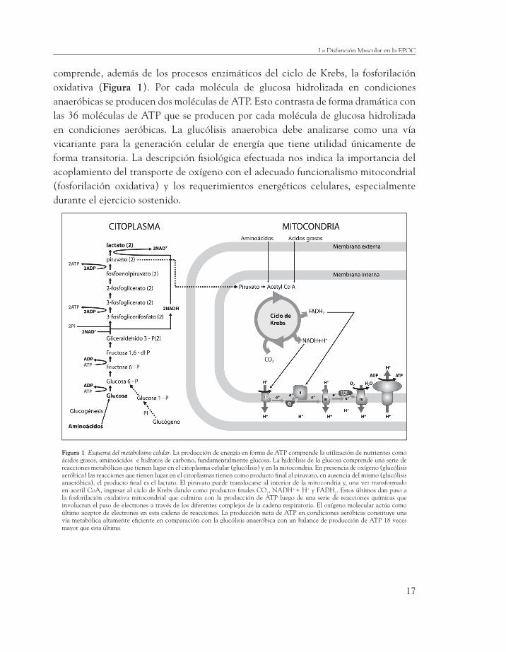
comprende, además de los procesos enzimáticos del ciclo de Krebs, la fosforilación oxidativa (Figura 1). Por cada molécula de glucosa hidrolizada en condiciones anaeróbicas se producen dos moléculas de ATP. Esto contrasta de forma dramática con las 36 moléculas de ATP que se producen por cada molécula de glucosa hidrolizada en condiciones aeróbicas. La glucólisis anaerobica debe analizarse como una vía vicariante para la generación celular de energía que tiene utilidad únicamente de forma transitoria. La descripción fi siológica efectuada nos indica la importancia del acoplamiento del transporte de oxígeno con el adecuado funcionalismo mitocondrial (fosforilación oxidativa) y los requerimientos energéticos celulares, especialmente durante el ejercicio sostenido.
Figura 1. Esquema del metabolismo celular. La producción de energía en forma de ATP comprende la utilización de nutrientes como ácidos grasos, aminoácidos e hidratos de carbono, fundamentalmente glucosa. La hidrólisis de la glucosa comprende una serie de reacciones metabólicas que tienen lugar en el citoplasma celular (glucólisis) y en la mitocondria. En presencia de oxígeno (glucólisis aeróbica) las reacciones que tienen lugar en el citoplasmas tienen como producto fi nal al piruvato, en ausencia del mismo (glucólisis anaeróbica), el producto fi nal es el lactato. El piruvato puede translocarse al interior de la mitocondria y, una vez transformado en acetil CoA, ingresar al ciclo de Krebs dando como productos fi nales CO2, NADH+ + H+ y FADH2. Estos últimos dan paso a la fosforilación oxidativa mitocondrial que culmina con la producción de ATP luego de una serie de reacciones químicas que involucran el paso de electrones a través de los diferentes complejos de la cadena respiratoria. El oxígeno molecular actúa como último aceptor de electrones en esta cadena de reacciones. La producción neta de ATP en condiciones aeróbicas constituye una vía metabólica altamente efi ciente en comparación con la glucólisis anaeróbica con un balance de producción de ATP 18 veces mayor que esta última.
La Disfunción Muscular en la EPOC

18
CAMBIOS FISIOPATOLÓGICOS EN EL MÚSCULOPERIFÉRICO DEL PACIENTE CON EPOC
Fibras musculares
El músculo esquelético humano se compone de dos tipos de fi bras: lentas (tipo I) y rápidas (tipo IIa y IIx)40, de acuerdo a sus características contráctiles. Ambos tipos de fi bras se distribuyen en proporción similar en el músculo adulto41. Su diversidad se basa fundamentalmente en la isoforma de la cadena pesada de miosina (del inglés myosine hevy chain, MHC) que contienen42. Las fi bras tipo I son de contracción lenta, reclutadas a bajas frecuencias de estimulación, desarrollan una relativa baja tensión, tienen una gran capacidad oxidativa y son más resistentes a la fatiga. Las fi bras rápidas tipo IIx son de contracción rápida, requieren una frecuencia de estimulación alta, desarrollan una gran tensión, dependen fundamentalmente de un metabolismo glicolítico y son más susceptibles a la fatiga. Las fi bras rápidas tipo IIa poseen características intermedias entre las tipo I y las tipo IIx43-45. El músculo esquelético de mamíferos contiene además la isoforma IIb42. Debido a la difi cultad para diferenciar las fi bras IIx de las IIa y IIb mediante la reacción histoquímica de ATPasa, las fi bras IIx han sido por largo tiempo confundidas con las IIb. Ha sido gracias a técnicas de inmunobloting que se ha clarifi cado la tipifi cación de las fi bras musculares. Se ha demostrado la existencia de fi bras IIx que contiene la isoforma IIx-MHC46-48, diferente de las IIa-MCH y IIb-MCH49. Así, las fi bras antiguamente identifi cadas como tipo IIb en el músculo humano han sido reclasifi cadas como fi bras tipo IIx47;48.
Existen también fi bras híbridas que co-expresan diferentes tipos de MHC: β/MHClenta+IIa-MHC (también conocidas como IIc), y IIa+IIx50-52. Estas representan un fenotipo de fi bras en transformación53 como las descritas en condiciones de inmovilización54, desuso55 o como respuesta al entrenamiento físico56.
Los pacientes con EPOC presentan un aumento de la proporción fi bras de tipo II en detrimento de las tipo I57-61;61-64. El incremento de fi bras tipo II se caracteriza fundamentalmente por un aumento del número de fi bras tipo IIx58;60;65;66. También ha sido descrita la presencia de fi bras híbridas (I/IIa y IIa/IIx) en el músculo esquelético de pacientes con EPOC. Esto sugiere que la transición entre tipos de fi bras puede ser el mecanismo que lleva a la redistribución de las mismas66.
Roberto Alejandro Rabinovich • Tesis Doctoral
19
En cuanto al tamaño de las fi bras, las que presentan mayor grado de atrofi a son las fi bras tipo IIx y las híbridas IIa/IIx65. El predominio de atrofi a de fi bras tipo IIx parecería estar indicando que el desuso no sería la única causa de atrofi a en este tipo de pacientes, puesto que esta situación se caracteriza inicialmente por atrofi a de fi bras tipo I67. Además, éste tipo de cambio en la redistribución de fi bras ha sido descrito como consecuencia a hipoxia tisular continua o intermitente68. Más aún, en estados patológicos acompañados de un desequilibrio energético, como la anorexia nerviosa, se encuentra una atrofi a predominantemente de fi bras tipo II69. De esta manera, otros mecanismos fi siopatológicos serían necesarios como coadjuvantes al mero desuso para explicar las características de la atrofi a en pacientes con EPOC. Cabe especular que los cambios en la distribución de los tipos de fi bras en la EPOC pudieran ser la expresión de fenómenos de remodelación muscular generados por los mecanismos que determinan los efectos sistémicos de la enfermedad.
Alteraciones de la capilarización y del transporte de oxigeno muscular
El transporte sistémico de oxígeno (QO2) depende de la presión parcial del gas (PaO2), la concentración y funcionalidad de la hemoglobina (Hb) y del fl ujo sanguí-neo (Q). Para un determinado valor de QO2, la oxigenación de las fi bras musculares depende de dos factores principales. En primer lugar, del equilibrio funcional entre aporte de O2 a nivel de la microcirculación y la demanda tisular de O2. El segundo factor importante para asegurar la difusión pasiva de O2 desde el capilar muscular a la mitocondria es la existencia de un área de transferencia adecuada (nº de capilares por fi bra muscular) que permita vencer el gradiente de presión entre el hematíe y la pared externa del capilar70. Una vez dentro de la fi bra muscular, el gradiente de PO2 es mínimo debido a que la mioglobina facilita la difusión de O2. Existen datos sugestivos de que ambos factores: desequilibrio de las relaciones aporte-consumo de O2 a nivel de microcirculación muscular y la capilarización anormal del músculo71;72 pueden consti-tuir factores limitantes del aporte de oxígeno a la mitocondria en estos pacientes.
Para una determinada carga submáxima, el transporte sistémico de O2 en pacientes con EPOC no suele estar disminuído73, sino que incluso puede aparecer aumentado en relación a sujetos controles64;74. Ello podría ser indicativo de un aumento de las necesidades energéticas en relación a sujetos normales o bien de un transporte/utilización
La Disfunción Muscular en la EPOC
20
muscular de O2 anómalo. En el primer supuesto, la indemnidad de los factores que determinan el transporte de O2 en el músculo (equilibrio de las relaciones QO2-VO2
(consumo de oxígeno) a nivel de la microcirculación y nº de capilares por fi bra) sería clave para evitar la hipoxia tisular durante la actividad física. Desafortunadamente el análisis funcional de las relaciones QO2-VO2 en el músculo esquelético no es aún posible, aunque existen datos indirectos sugestivos de anormalidades a este nivel75;76.
Además de los factores descritos a nivel de transporte de oxígeno muscular, cabe señalar que los pacientes con EPOC avanzado pueden (en ausencia de patología del miocardio) presentar un incremento inadecuado del débito cardíaco y el aporte sistémico de oxigeno durante el ejercicio. Ello puede explicarse por el fenómeno de atropamiento aéreo. Asimismo, algunos pacientes con EPOC pueden presentar deterioro de la PaO2 durante el ejercicio que se traduciría en una alteración del aporte sistémico de oxigeno.
Estudios de microscopía electrónica77 y óptica61 demuestran un menor número de capilares por área en el músculo esquelético de miembros inferiores de pacientes con EPOC. El número de capilares por área mitocondrial también se encontró marcadamente disminuído77. De igual forma, el número de contactos entre capilares y fi bras también está reducido de forma signifi cativa en pacientes con EPOC58;61. Por el contrario, Richardson y colaboradores64 no encontraron diferencias entre pacientes con EPOC y sujetos controles en lo que respecta a la densidad capilar (capilares por área) y al número de contactos de capilares con fi bras musculares. Cabe señalar, que los pacientes con EPOC incluidos en este estudio64 habían participado de un programa de rehabilitación de 8 a 24 meses de duración. Es bien conocido que el número de contacto capilar-fi bra muscular se incrementa con el entrenamiento en pacientes con EPOC58. Esta disminución del número de contactos entre capilares y fi bras puede contribuir a una reducción del transporte de oxígeno desde la circulación a la mitocondria fundamentalmente en situaciones de aumento de la demanda de oxígeno como ocurre durante el ejercicio.
Alteración de la bioenergética muscularDiversos estudios demuestran una disminución de la capacidad oxidativa del
músculo esquelético periférico en pacientes con EPOC. Asimismo, el potencial energético celular (ATP y PCr) se encuentra disminuida en el músculo esquelético de
Roberto Alejandro Rabinovich • Tesis Doctoral
21
estos pacientes62;78. El tiempo medio de recuperación de Pcr al fi nalizar el ejercicio, un proceso eminentemente oxidativo, se encuentra prolongado en el músculo esquelético de pacientes con EPOC74.
También se observa la aparición de un incremento precoz de la producción de ácido láctico durante el ejercicio en estos pacientes73;79 condicionando un umbral láctico temprano80. Este fenómeno no se explica por la actividad de los músculos respiratorios81 sino por la de los músculos de de las extremidades inferiores. A pesar de que los primeros (músculos respiratorios) trabajan contra una mayor resistencia y soportan una gran carga de trabajo, especialmente durante el ejercicio.
La acidosis por incremento precoz de los niveles de ácido láctico durante el ejercicio moderado genera un aumento de la demanda ventilatoria e induce la utilización de un patrón respiratorio basado en un aumento de la frecuencia respiratoria, que resulta poco favorable para el paciente por el incremento del atrapamiento aéreo. Este fenómeno, junto con la inducción de fatiga muscular por el incremento de la acidosis, constituyen factores importantes para explicar la menor tolerancia al ejercicio presentada por estos pacientes. De esta forma, la disminución del pH en la vena femoral durante el ejercicio sub-máximo se correlaciona con el consumo de oxígeno (VO2) pico73.
Se ha mencionado la existencia de un défi cit en la maquinaria oxidativa celular en los pacientes con EPOC objetivada por una disminución de la actividad de enzimas oxidativas pertenecientes al ciclo de Krebs como la citrato sintetasa y la hidroxi acil-coA deshidrogenasa82, que se correlaciona de forma signifi cativa con el VO2 pico. En este sentido, el inicio precoz de la producción de ácido láctico a niveles moderados de ejercicio puede explicarse por diversos fenómenos como la alteración del transporte muscular de O2, el reclutamiento de fi bras tipo II, de metabolismo predominantemente anaeróbico, o la disminución de la capacidad oxidativa del miocito. Aspecto, este último, en el que se profundizará en la presente tesis.
La alteración de las relaciones transporte/utilización de O2 se asocia a una disminución de la efi ciencia del músculo esquelético de estos pacientes. Así, la relación fósforo inorgánico/fosfocreatina (Pi/PCr) durante el ejercicio submáximo se encuentra incrementada en el músculo esquelético de los pacientes con EPOC en relación a sujetos normales74.
Asimismo, estos pacientes presentan mayor consumo de oxígeno en las extremidades inferiores a igual carga de submáxima64;74 que podría explicarse por el aumento del
La Disfunción Muscular en la EPOC
22
porcentaje de fi bras tipo II. En este sentido, existe evidencia convincente de que el coste energético del trabajo generado por el músculo es dependiente del tipo de fi bras siendo las fi bras tipo II las de menor efi ciencia con un mayor coste energético por unidad de trabajo83. El aumento de la actividad de la citocromo oxidasa84, último aceptor de electrones, es consistente con el incremento del consumo de oxígeno a igual carga observado en estos pacientes.
Pérdida de masa muscular
Dependiendo de la población estudiada, entre 17 y 35% de los pacientes con EPOC presentan pérdida de peso17;20;21;85;86. La asociación entre pérdida de peso y severidad de la EPOC es ampliamente reconocida. El peso corporal se correlaciona de manera positiva con la tolerancia al ejercicio en estos pacientes85;86. Más aun, la pérdida de peso se asocia a un incremento del número de hospitalizaciones secundarias a exacerbaciones87
y a una disminución de la supervivencia de estos pacientes12;88-91. La pérdida de peso corporal en esta población de pacientes tiene lugar
fundamentalmente a expensas de la masa muscular20. Además, la pérdida de masa muscular infl uye directamente en la capacidad de desarrollo de fuerza del músculo5;22;23
y en la tolerancia al ejercicio8;9;20;92, de manera independiente del grado de obstrucción bronquial expresado por el FEV1
92 y de manera más adecuada que el BMI8. Asímismo, la disminución de la masa muscular constituye un mejor predictor de calidad de vida relacionada con la salud93 y de supervivencia13;14 que el peso corporal. De esta forma, el análisis de la composición corporal en al menos dos compartimientos, el tejido graso (del inglés fat mass, FM) y el tejido libre de grasa (FFM) que representa la masa de tejido metabólicamente activo (hígado, intestinos, sistema inmune y fundamentalmente tejido muscular contráctil) más los fl uidos y sólidos extracelulares, aporta una aproximación más adecuada para la evaluación del contenido de tejido muscular.
La disminución de la FFM se presenta en un 18 a 36 % de estos pacientes17;20, pudiendo incluso presentarse en un 6 a 21 % de pacientes con peso corporal normal17;20;21. La medición de la FFM mediante bioimpedancia eléctrica, constituye un procedimiento sencillo y proporciona una variable clínica más adecuada para evaluar la pérdida de masa muscular23;94 en los pacientes con EPOC.
Roberto Alejandro Rabinovich • Tesis Doctoral
23
CAMBIOS ETIOPATOGÉNICOS DELA DISFUNCIÓN MUSCULAR EN LA EPOC
A pesar de ser la disfunción muscular el efecto sistémico de la EPOC más extensamente estudiado, los mecanismos etiopatogénicos que la condicionan no están aún del todo aclarados. Es importante resaltar que estamos ante dos fenómenos conceptualmente diferenciados aunque posiblemente relacionados entre sí: a) el funcionamiento muscular anómalo; y, b) la pérdida de masa muscular que ocurre en un subgrupo de pacientes. Diversos estudios de revisión95-97 han caracterizado las alteraciones del músculo esquelético periférico de los pacientes con EPOC y han identifi cado la naturaleza multifactorial del problema.
Alteraciones en el recambio proteico
Diversos estudios demuestran alteraciones en el recambio proteico en la EPOC98. La determinación de si la pérdida de masa muscular en los pacientes con EPOC se debe a una disminución de la síntesis proteica, a un aumento de la degradación, o a ambos factores resulta de cabal importancia por su implicación en la identifi cación de los mecanismos íntimos del proceso y por las eventuales implicaciones en el tratamiento. Se han identifi cado anomalías tanto en la síntesis proteica99 como a nivel de la regulación de la proteólisis100. El papel del sistema proteolítico ubiquitin-proteasoma101-103 en el equilibrio entre síntesis y degradación de proteínas musculares no ha sido aun explorado en la EPOC.
Los niveles de insulina, de hormona de crecimiento (GH), del factor de crecimiento simil-insulina (IGF-1) y de otras hormonas anabolizantes favorecen la síntesis proteica, fundamentalmente a nivel muscular aumentando la síntesis e inhibiendo la degradación proteica104;105. Algunos estudios encuentran niveles disminuídos de IGF-1 en la EPOC106. Estos cambios hormonales han sido relacionados con la acción de diversos factores infl amatorios, alguno de los cuales se encuentran presentes en concentraciones anormalmente elevadas en la EPOC107. La infusión de IL-1 y TNFαen animales se asocia a una disminución de los niveles circulantes de IGF-1 y a una reducción de la síntesis proteica108. A su vez, la exposición a TNFα inhibe la síntesis proteica inducida por IGF-1109.
La Disfunción Muscular en la EPOC
24
Por otra parte se han descrito niveles anormalmente bajos de testosterona en pacientes con EPOC110. La administración de diversas hormonas como la GH111;112, o la testosterona18;113-115, ha generado un incremento de la masa muscular y de la capacidad de generación de fuerza, sin cambios signifi cativos en la resistencia muscular y la tolerancia al ejercicio.
Trastornos nutricionales
La pérdida de masa muscular es el mecanismo principal de pérdida de peso observada en la EPOC20, mientras que la pérdida de masa grasa contribuye a ésta en menor medida20. Es importante diferenciar los términos “malnutrición” y “caquexia”. El primero constituye un trastorno asociado a la disminución de la ingesta calórica con una tasa metabólica disminuida y buena respuesta al soporte nutricional. En este caso existe una conservación relativa de la masa muscular. El término “caquexia” es, en realidad, el que más se ajusta al fenómeno que tiene lugar en la EPOC. La caquexia se caracteriza por una tasa metabólica elevada sin una disminución de la ingesta calórica, su origen es menos claro y la respuesta a los suplementos calóricos es pobre. En este caso la pérdida de peso se asocia a una pérdida concomitante de masa muscular. Existe una tercera posibilidad que se presenta en un porcentaje no despreciable de pacientes con EPOC. Nos referimos a la sarcopenia o pérdida de masa muscular que puede darse aún en ausencia de pérdida de peso20;21;116.
La disminución de la ingesta calórica no parece ser un factor relevante en pacientes con EPOC, excepto durante episodios de exacerbación117. El metabolismo basal se encuentra incrementado en pacientes con EPOC118, fundamentalmente en aquellos con bajo peso119. Este incremento se explicaba tradicionalmente por un aumento del consumo de oxígeno por parte de los músculos respiratorios debido al aumento del trabajo ventilatorio que caracteriza a la EPOC120. Sin embargo, recientemente se ha demostrado que los músculos no respiratorios presentan un consumo de oxígeno exagerado durante el ejercicio pudiendo contribuir a un aumento de la tasa metabólica en estos pacientes64;74. Asimismo el consumo energético necesario para la realización de las actividades de la vida diaria es signifi cativamente mayor en pacientes con EPOC que en los sujetos control121;122. Este hecho explica la menor efi ciencia mecánica, defi nida como el coste energético para realizar ejercicio a niveles submáximos64;74;123.
Roberto Alejandro Rabinovich • Tesis Doctoral
25
Un metabolismo energético elevado parcialmente compensado por una ingesta calórica inadecuada a estos requerimientos podría constituir una base para explicar la caquexia en pacientes con EPOC118.
Sedentarismo
La disnea desencadenada por el ejercicio es un elemento importante como factor explicativo del hábito sedentario que caracteriza a los pacientes con EPOC, que conlleva una disminución de la actividad contráctil del músculo.
El entrenamiento físico constituye una estrategia terapéutica que permite revertir algunas de las alteraciones musculares atribuibles al desuso muscular en pacientes con EPOC. Este apartado, de relevancia conceptual para la tesis, será desarrollado posteriormente de forma más extensa.
Corticosteroides
La miopatía esteroidea se defi ne por el conglomerado de cambios histopatológicos, bioquímicos y funcionales que ocurren a nivel muscular en individuos que han sido tratados con corticosteroides en ausencia de otras causas de miopatía. Constituye el efecto adverso asociado al uso de fármacos más frecuente a nivel muscular en la EPOC. Ha sido descrito un efecto agudo y un efecto crónico asociado al uso de éste tipo de fármacos. La miopatía esteroidea aguda es un efecto adverso raro, no descrito en pacientes con EPOC, secundario a la administración intravenosa de corticosteroides, y se acompaña de rabdomiolisis124;125. Por el contrario, la miopatía esteroidea crónica, constituye la clásica miopatía asociada al uso prolongado de corticosteroides por vía oral en la cual la rabdomiolisis esta ausente. Esta entidad se caracteriza por fenómenos de atrofi a difusa con afectación predominante de fi bras tipo IIx126. Los corticosteroides pueden afectar la producción de proteínas contráctiles y disminuir la expresión del IGF-1. Habría una relación estrecha entre dosis y duración del tratamiento con la extensión de los cambios estructurales y funcionales a nivel muscular.
Así, el uso de corticosteroides por vía oral de forma prolongada puede constituirse en una causa de miopatía específi ca en pacientes con EPOC. Sin embargo los cambios estructurales descritos en pacientes cuidadosamente seleccionados en ausencia de
La Disfunción Muscular en la EPOC
26
tratamiento corticosteroide sugiere la presencia de otros fenómenos causales en la génesis de la disfunción muscular asociada a la EPOC.
Hipoxia tisular e hipercapnia
La composición adecuada de los gases respiratorios en sangre arterial constituye una de las funciones primordiales del pulmón. La incapacidad, en forma contínua o intermitente, de mantener presiones arteriales normales de oxígeno y dióxido de carbono son fenómenos frecuentemente asociados a la EPOC.
El papel de la hipoxia tisular, aún en ausencia de hipoxemia arterial en reposo, como factor etiopatogénico de la disfunción muscular en la EPOC tiene su base en numerosas publicaciones que aportan evidencia en este sentido. La hipoxia celular, un factor que limita la producción energética celular, afecta claramente la síntesis proteica celular127. Está bien establecido que sujetos sanos en condiciones de hipoxia hipobárica, como la altura, presentan pérdida de masa muscular128;129 e incremento de la actividad de enzimas glicolíticas con disminución de la actividad de enzimas del ciclo de Krebs130. La hipoxia produce una inhibición aguda de la síntesis de proteínas mitocondriales131. Además la hipoxia crónica inhibe la síntesis proteica en células musculares causando una pérdida neta de aminoácidos y reduciendo la expresión de miosina132;133. Por su parte, niveles inadecuadamente elevados de dióxido de carbono contribuyen de manera signifi cativa a incrementar la acidosis intracelular en el músculo esquelético134. La acidosis intrace-lular conlleva efectos deletéreos en la maquinaria enzimática de la célula muscular inhibiendo la actividad de enzimas clave en el metabolismo energético135. Estudios re-alizados en pacientes con insufi ciencia respiratoria aguda demuestran niveles disminuí-dos de ATP y fosfocreatina intracelulares136. Además, la incubación de músculo aislado a niveles elevados de dióxido de carbono resulta en una disminución de los niveles de fosfocreatina y en la relación ATP/ADP137. En las exacerbaciones severas de la EPOC, la acidosis es un fenómeno que frecuentemente acompaña a la hipoxemia. La acidosis estimula la degradación proteica mediada por el sistema ubiquitina-proteasoma138. Más aún, la corrección de la acidosis tanto en modelos animales como en humanos reduce la tasa de degradación proteica139;140. En este sentido la hipoxia tisular y la hipercapnia, contínua o intermitente, puede actuar tanto como favorecedor de la pérdida de masa muscular como a nivel del metabolismo energético muscular.
Roberto Alejandro Rabinovich • Tesis Doctoral
27
Infl amación sistémica
La infl amación sistémica constituye uno de los mecanismos fi siopatológico más relevantes en la génesis de la disfunción muscular en la EPOC. Diversos estudios demuestran alteraciones a nivel de las células infl amatorias circulantes, fundamentalmente neutrófi los y linfocitos. Las propiedades quimiotácticas y de proteólisis extracelular141, así como la producción de especies reactivas de oxígeno142, se encuentra incrementada en neutrófi los aislados de pacientes con EPOC. Asimismo, la expresión de diversas moléculas de adhesión, particularmente Mac-1 en neutrófi los circulantes, se encuentra incrementada143, comprometiendo el proceso de eliminación de neutrófi los de los tejidos infl amados.
Las concentraciones plasmáticas de diversas citocinas proinfl amatorias, fundamentalmente el TNFα21;107;144-146, así como las concentraciones plasmáticas de sus receptores solubles21;144;145, se encuentran elevadas en pacientes con EPOC. El TNFα, la IL-1, la IL-6 y el IFγ son las citocinas pro-infl amatorias consideradas como γ son las citocinas pro-infl amatorias consideradas como γefectores más probables en la génesis de la pérdida de masa muscular en numerosas patologías caracterizadas por presentar sarcopenia.
La producción de TNFα por monocitos circulantes provenientes de pacientes con EPOC se encuentra incrementada147, particularmente en el subgrupo de pacientes con bajo peso107. El TNFα puede afectar a las células musculares de diversas mane-ras148. Esta citocina interrumpe el proceso de diferenciación muscular149 mediante el cual las células satélite musculares se diferencian, proliferan y se fusionan con otras células, constituyendo el mecanismo fundamental de regeneración secundaria a la injuria muscular.
En miocitos diferenciados estudiados in vitro, la exposición a TNFα produce la degradación de la cadena pesada de miosina a través del sistema ubiquitin/proteasoma148. Se trata de un efecto mediado por la activación del factor de transcripción NFkB. El TNFα puede, a su vez, inducir la expresión de otras citocinas pro-infl amatorias contribuyendo a la amplifi cación de la respuesta infl amatoria148.
Por otro lado, el TNFα puede inducir apoptosis en diversos sistemas celulares150
a través, por ejemplo, de promover la fragmentación del ADN151. La combinación de la reducción de la actividad contráctil asociada al sedentarismo y los elevados niveles de TNFα podrían inducir una pérdida signifi cativa de las células satélite.
La Disfunción Muscular en la EPOC
28
Se ha demostrado un aumento de la apoptosis en el músculo esquelético de pacientes con EPOC y bajo índice de masa corporal en relación a pacientes con peso normal y a sujetos sedentarios sin EPOC152.
Estrés oxidativo/nitrosativo
Una parte importante de los manuscritos que forman el cuerpo de esta tesis se relacionan con el papel del estrés oxidativo como mecanismo etiopatogénico en la disfunción muscular asociada a la EPOC. Es importante la comprensión de este mecanismo patogénico para favorecer un mejor entendimiento del contenido de los mismos. Este apartado, de relevancia conceptual para la tesis, será desarrollado posteriormente de forma más extensa.
╯
Roberto Alejandro Rabinovich • Tesis Doctoral
29
Los Músculos Respiratorios en la EPOC
Los cambios en la morfología de la caja torácica secundarios a la hiperinsufl ación conjuntamente con las alteraciones de la mecánica respiratoria que favorece el atrapamiento aéreo, generan una alteración en la efi cacia contráctil de la musculatura inspiratoria, fundamentalmente del diafragma (disminución de la relación longitud-tensión; disminución del área de aposición, disminución de la curvatura diafragmática, etc.). Esto impacta negativamente en la capacidad de generar fuerza y aumenta de forma notable el trabajo respiratorio, contribuyendo, en gran parte, al desencadenamiento de la sensación de disnea que aqueja a estos pacientes. De esta forma, los músculos inspiratorios se enfrentan a una sobrecarga mecánica crónica. Así, a diferencia de los músculos esqueléticos periféricos, la falta de actividad no parece ser una causa relacionada con la disfunción de la efi cacia contráctil de los músculos respiratorios153.
Existe evidencia de que el diafragma y otros músculos respiratorios expresan diversos cambios adaptativos en respuesta a la sobrecarga mecánica impuesta por la enfermedad. A diferencia de lo que ocurre en la musculatura periférica en pacientes con EPOC, el diafragma de estos pacientes presenta una mayor densidad mitocondrial que el de sujetos de igual edad y sexo sin enfermedad pulmonar154;155. La actividad de enzimas vinculadas al metabolismo aeróbico se encuentra incrementado en el diafragma de estos pacientes57. Por último el porcentaje de fi bras tipo I se encuentra incrementado en el diafragma de pacientes con EPOC mientras que el porcentaje de fi bras tipo IIa y IIx se encuentra disminuído156.
A pesar de las adaptaciones descritas, los músculos respiratorios de los pacientes con EPOC muestran una disminución de la capacidad de generar fuerza y una disminución

30
de la resistencia157 debidas al cambio en la geometría muscular impuesto por el fenómeno de hiperinsufl ación. Los fenómenos de adaptación que ocurren a nivel de los músculos respiratorios, son insufi cientes para restaurar la función muscular normal en estos pacientes.
El resultado último de las adaptaciones del músculo respiratorio y la sobrecarga crónica es un frágil equilibrio puede verse alterado de forma progresiva o aguda en situaciones tales como episodios de exacerbación o complicaciones de la enfermedad.
╯
Roberto Alejandro Rabinovich • Tesis Doctoral
31
Sedentarismo y EntrenamientoMuscular en la EPOC
La plasticidad del tejido muscular es tal, que cambios en la carga desarrollada por el músculo tienen un efecto dramático en el tamaño muscular y la capacidad metabólica de sus fi bras. El sedentarismo afecta al músculo tanto en lo que se refi ere a su trofi smo como a su capacidad oxidativa. La inactividad física causa pérdida de masa muscular, reduce la capacidad de generar fuerza, y disminuye el umbral de fatiga afectando la resistencia muscular158. Asimismo genera pérdida de masa muscular debida a una disminución de la síntesis conjuntamente con un aumento en la degradación proteica159-161. Aún por cortos períodos de tiempo, la disminución de la actividad contráctil, resulta en una pérdida de masa muscular signifi cativa162. El sistema ubiquitin-proteasoma parece tener un papel clave en la pérdida de masa muscular secundaria a la inmovilización163-165.
El hecho de que el entrenamiento físico mejore la función muscular en pacientes con EPOC refuerza el concepto de que el sedentarismo es un factor importante como condicionante de la disfunción muscular74;166;167. Asimismo, el entrenamiento físico puede incrementar de forma moderada el peso de los pacientes con EPOC a expensas de un aumento en la FFM168.
Hasta fi nes de la década de los 80´, se creía que el entrenamiento físico solo aportaba benefi cios psicológicos a los pacientes con EPOC169. Actualmente la evidencia no puede ser más clara en el sentido de que el entrenamiento físico mejora la tolerancia al ejercicio y la calidad de vida relacionada con la salud en este tipo de pacientes. Numerosos artículos científi cos lo demuestran74;96;170. Al menos un documento basado en la evidencia171 y un meta-análisis172 recientemente publicados avalan de forma clara éste hecho. Desde el punto de vista fi siológico, los pacientes con EPOC presentan
32
diferencias en los mecanismos de adaptación al entrenamiento físico en relación a individuos sanos de igual edad y sexo pero sin patología pulmonar74. Así, los mecanismos fi siológicos de adaptación al entrenamiento físico tienen lugar fundamentalmente a nivel muscular en pacientes con EPOC, en los que ésta estrategia terapéutica demostró una mejoría sustancial en la capacidad oxidativa del músculo entrenado, sin un impacto sustancial en los mecanismos centrales (fl ujo sanguíneo dirigido a los músculos, ventilación minuto, transporte de oxígeno) a nivel de ejercicio pico. Por el contrario, los individuos sanos muestran una capacidad de adaptación fundamentalmente a nivel de los factores centrales que gobiernan el transporte convectivo de oxígeno, a niveles de ejercicio pico74. Asimismo, es importante destacar que esta estrategia de tratamiento ha contribuido a arrojar luz sobre los mecanismos que condicionan la disfunción muscular en la EPOC, particularmente el fenómeno de desuso muscular provocado por el sedentarismo. Así, el entrenamiento físico en pacientes con EPOC raramente alcanza a normalizar la función muscular por completo. Más aún, algunas de las alteraciones descritas en el músculo no son explicadas por el hábito sedentario de estos pacientes. Por ejemplo la inactividad física se asocia a una disminución de la actividad de la citocromo oxidasa, actividad que se encuentra incrementada en el músculo esquelético de pacientes con EPOC84. Este mismo comportamiento de la citocromo oxidasa se encuentra en linfocitos circulantes de pacientes con EPOC, celulas que escapan a la infl uencia del sedentarismo173. Por otro lado, los cambios morfológicos en el músculo esquelético secundarios a la inmovilización se caracterizan por una atrofi a fundamentalmente de fi bras tipo I y IIa174 a diferencia de la atrofi a muscular predominantemente de fi bras tipo IIx característica de los pacientes con EPOC65.
A su vez, el entrenamiento físico induce, en el término de 24 h, la expresión de genes que favorecen el trofi smo muscular luego de la inmovilización165. La reducción del tamaño fi brilar puede revertirse, al menos parcialmente, con el entrenamiento físico58. Sin embargo estos cambios son proporcionalmente inferiores a la mejoría de la capacidad de realizar ejercicio y a la fuerza muscular168. Todo esto pone de manifi esto que, aunque el sedentarismo puede claramente relacionarse con la pérdida de masa muscular y con otros elementos que hacen a la disfunción muscular en la EPOC74;166;167, otros factores fi siopatológicos coadyuvantes son necesarios para explicar las alteraciones observadas en los pacientes. Por este motivo el entrenamiento físico en
Roberto Alejandro Rabinovich • Tesis Doctoral
33
la EPOC no solo constituye una terapia dirigida a revertir los efectos del sedentarismo sobre el músculo periférico de estos pacientes, sino que constituye una herramienta que permite profundizar en el entendimiento de los mecanismos etiopatogénicos responsables de la disfunción muscular periférica que afecta a estos pacientes. Con este espíritu, el entrenamiento muscular, se ha empleado en los diferentes trabajos originales que forman parte de esta tesis.
╯
Sedentarismo y Entrenamiento Muscular en la EPOC

35
El Estrés Oxidativo/Nitrosativo comoMecanismo Patogénico
Se entiende por estrés oxidativo/nitrosativo a la citotoxicidad causada por especies reactivas de oxígeno (ROS, reactive oxygen species) y de óxido nítrico (RNS, reactive nitrogen species)175. Las especies reactivas incluyen a moléculas como el anión superóxido (O2
-), el peróxido de hidrógeno (H2O2), el radical hidroxilo (.OH) y peroxinitrito(ONNO-). Aunque los oxidantes se generan durante procesos biológicos normales, su capacidad de modifi car diversas moléculas de forma perjudicial está bloqueada por una variedad de sistemas antioxidantes intra- y extracelulares entre los cuales se destacan: a) sistemas enzimáticos (superóxido dismutasa (SOD), catalasa y glutation peroxidasa), b) macromoléculas (ceruloplasmina, transferrina); y, c) pequeñas moléculas (glutation, metionina, vitamina C, vitamina E). La citotoxicidad se deriva del desequilibrio entre la producción de ROS/NOS y los mecanismos intracelulares de defensa antioxidantes175. Diversos estudios relacionan el desarrollo y la progresión de la EPOC con un incremento de la producción de moléculas pro-oxidantes o una disminución de los recursos antioxidantes celulares tanto en el pulmón como a nivel sistémico.
Los niveles de H2O2 en el aire exhalado se encuentran elevados en sujetos fumadores y pacientes con EPOC en comparación con ex fumadores con EPOC y sujetos no fumadores176;177. Éste fenómeno se ve incrementado durante los episodios de exacerbaciones de la enfermedad178. Asimismo, los macrófagos alveolares provenientes de sujetos fumadores presentan un incremento de la producción de anión superóxido178;179. Además, la actividad de la enzima xantino oxidasa, capaz de generar anión superóxido y peróxido de hidrógeno, se encuentra incrementada en
36
el líquido de lavado broncoalveolar y en el plasma de pacientes con EPOC y sujetos fumadores en comparación con sujetos sanos y no fumadores respectivamente180;181.
Sujetos fumadores al igual que pacientes con EPOC presentan evidencia de estrés oxidativo en la circulación sistémica, particularmente durante las exacerbaciones182. El tabaquismo incrementa los niveles plasmáticos de F2-isoprostanos, un producto de la peroxidación del ácido araquidónico183. El estrés oxidativo generado por el tabaco contribuye a las alteraciones cardiovasculares asociadas a este hábito y explican la disfunción endotelial sistémica presente en individuos fumadores184;185. El humo de tabaco contiene componentes potencialmente dañinos para el músculo esquelético a través de diversos mecanismos, además del estrés oxidativo. La nicotina puede alterar la expresión de ciertos factores de crecimiento como el TGF-β1, involucrado en el mantenimiento de la masa muscular186 y compite con la acetilcolina por el receptor en la unión neuromuscular pudiendo potencialmente afectar la contracción muscular187. De esta forma es lícito especular en que el tabaquismo resulta en si mismo un factor relevante en la génesis de la disfunción muscular que caracteriza a los pacientes con EPOC.
En orina, los niveles de F2α-III isoprostano se encuentran elevados en pacientes con EPOC en comparación con sujetos sanos; estas diferencias fueron, también, más pronunciadas durante las exacerbaciones188. A su vez, el ejercicio intenso causa oxidación del glutation plasmático189 e incrementa los niveles plasmáticos de malondialdehido (MDA), otro producto de peroxidación lipídica181 en pacientes con EPOC. Más aun, este efecto del ejercicio intenso es inhibido con la administración de fracciones inspiradas de oxígeno elevadas189 y halopurinol181, un inhibidor de la xantino oxidasa, lo que indicaría que la hipoxia tisular constituye una posible fuente de radicales libres durante el ejercicio mediada por este enzima.
A nivel muscular, los ROS ejercen un efecto bifásico en la contractilidad muscular siendo esenciales, en bajas concentraciones, para un normal desarrollo de fuerza mientras que disminuyen la capacidad contráctil del músculo a altas concentraciones190.
De esta forma, el estrés oxidativo puede tener un papel etiopatogénico en la disfunción muscular a dos niveles distintos. Además de, potencialmente interferir en la contractilidad muscular pudiendo llevar a la fatiga precoz y a la disminución de la resistencia, los ROS pueden también tener un papel importante en la facilitación de la degradación proteica en el músculo esquelético191;192, modifi cando las cadenas de
Roberto Alejandro Rabinovich • Tesis Doctoral
37
aminoácidos, formando agregados proteicos y rompiendo uniones peptídicas193. Esta modifi cación de la estructura proteica194, puede mediar mecanismos de proteólisis muscular191;192 favoreciendo la perdida de masa muscular.
Teniendo en cuenta que el daño oxidativo proteico, y la formación de proteínas car-boniladas puede ser prevenido por antioxidantes como el ácido ascórbico y el glutation, un desequilibrio entre la formación de ROS y la capacidad antioxidante celular puede jugar un papel importante en la génesis de la pérdida de masa muscular en la EPOC.
Por su parte, el NO, producido en condiciones fi siológicas, regula una innumerable cantidad de funciones celulares mediante la modifi cación post translacional de diversas proteínas. Esto ocurre fundamentalmente a nivel de residuos de cisterna (S-nitrosilación). El anión superóxido, en concentraciones fi siológicas, favorece esta reacción. Sin embargo, a concentraciones patológicas, ésta molécula interfi ere en la S-nitrosilación al interactuar directamente con las proteínas y formando RNS al unirse al NO. Así, el peroxinitrito (ONNO-) y otras especies reactivas de nitrógeno (RNS) se forman por la reacción de anión superóxido (O2
-) y óxido nítrico (NO). Los RNS, a su vez, reaccionan con residuos tirosinados de las proteínas formando 3-nitrotirosina. Este producto del estrés nitrosativo, a su vez, ha sido implicado en la etiopatogenia de diversas enfermedades cardiovasculares. El NO se forma por acción de tres sintetasas (NOS) del NO. Todas ellas se expresan en el músculo esquelético195. Dos isoformas de la NOS, la tipo I (neuronal) y la tipo III (endotelial) se expresan de manera constitutiva, mientras que la tercera isoforma, la tipo II (inducible, iNOS), se expresa en respuesta a diversos estímulos, incluyendo citocinas, oxidantes e hipoxia195. La infl amación sistémica puede inducir la expresión de iNOS en el músculo esquelético196. El músculo esquelético de pacientes con EPOC presenta, de hecho, sobreexpresión de iNOS197. La producción de NO resultante de la inducción de la iNOS puede derivar en el incremento de la producción de nitrotirosina y facilitar la degadación proteica mediada por el sistema ubiquitin/proteasoma191 e incrementar los niveles de apoptosis198. Finalmente, la inducción de la iNOS puede también ocasionar una disminución de las propiedades contráctiles del músculo199 interfi riendo, de esta forma, en la adecuada tolerancia al ejercicio.
Los oxidantes relacionados con las enfermedades humanas derivan de tres fuentes: a) generados en procesos biológicos intracelulares fi siológicos; b) generados en relación a procesos infl amatorios; y, c) de origen exógeno, como sería el caso del tabaquismo.
El Estrés Oxidativo/Nitrosativo como Mecanismo Patogénico
38
Aunque la mitocondria es la principal fuente potencialmente generadora de ROS, hay diversas fuentes de producción de ROS extra-mitocondrial como el metabolismo prostanoide, la autooxidación de catecolaminas, la actividad NAD(P)H oxidasa y la NO sintetasa. Las moléculas derivadas del metabolismo de la xantino oxidasa (XO) han sido relacionadas con el estrés oxidativo secundario a mecanismos de hipoxia/reperfusión. La mitocondria cobra cabal importancia en la generación de ROS como consecuencia de la utilización de oxígeno en la cadena de la fosforilación oxidativa con generación de ATP. Éstos se producen en segmentos específi cos de la cadena de transporte de electrones, fundamentalmente en el pool ubiquinona del complejo III,donde un electrón de ubisemiquinona es transferido directamente a una molécula de oxígeno200-203 formándose una molécula de anión superóxido y a nivel del complejo I de la cadena respiratoria mitocondrial204. Éste tiene un radio de difusión muy corto aunque puede transformarse en peróxido de hidrógeno (H2O2) por acción de la superóxido dismutasa (SOD). A su vez, el H2O2 es convertido por la catalasa en H2O + O2 siendo de esta forma neutralizado. Aunque el peróxido de hidrógeno no es estrictamente un radical libre, es un potente oxidante. Tanto el anión superóxido como el peróxido de hidrógeno pueden generar radicales hidroxilo (.OH) en presencia de metales, por ejemplo Fe++
(reacción de Fenton).Los ROS producidos en forma controlada están involucrados en un número
importante de procesos biológicos entre los que se encuentra la regulación génica a través de la activación de la ligadura al ADN de ciertos factores de transcripción dependientes del estado redox205. Numerosos estudios implican a los ROS como participantes en una variedad de señales intracelulares como activación de kinasas 206, de NFkB 207 etc. Además, la producción de ROS a nivel mitocondrial es uno de los mecanismos postulados, entre otros, como mediador de la señalización intracelular de mecanismos de adaptación a la hipoxia208;209.
Sin embargo, la generación incontrolada de ROS puede sobrepasar los mecanismos de protección antioxidante y derivar en estrés oxidativo y daño celular. Los sistemas de protección contra los oxidantes involucran a mecanismos de reparación del daño pro-ducido por estas moléculas a nivel de lípidos, proteínas o ADN y otros sistemas encar-gados de neutralizarlas, anterirmente citados. Entre estos, el sistema del glutation con-stituye uno de los mecanismos de defensa antioxidante intracelular más importantes.
El glutation no está homogéneamente distribuido en la célula, 85 % se encuentra
Roberto Alejandro Rabinovich • Tesis Doctoral
39
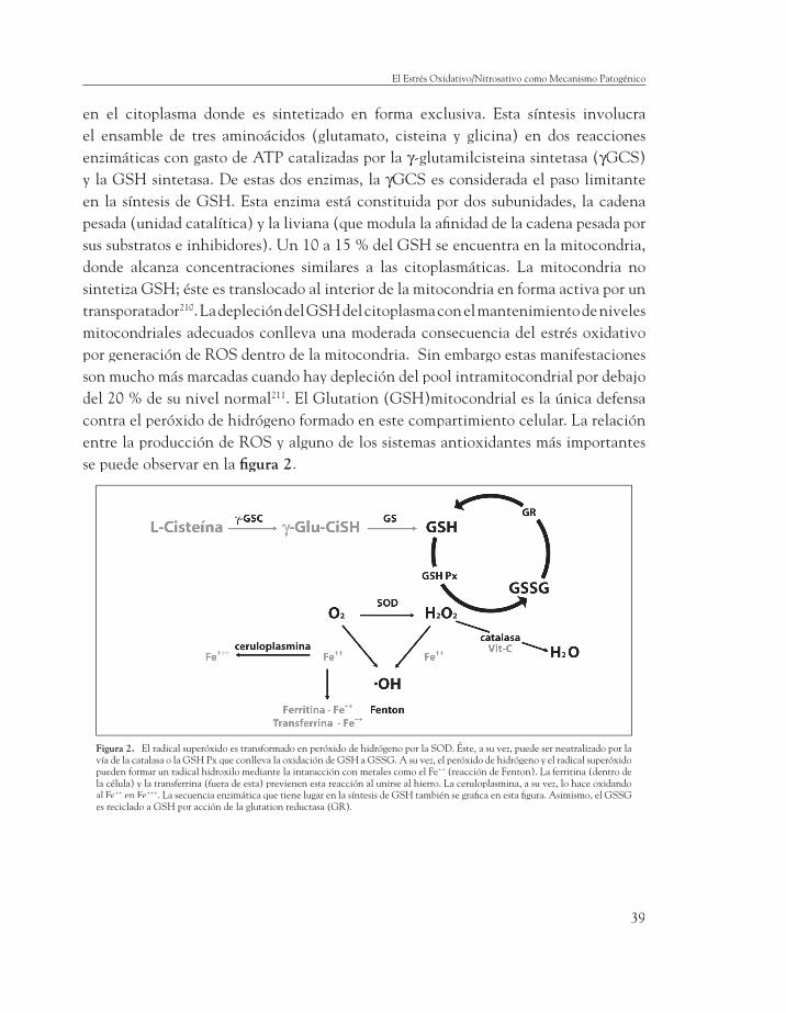
en el citoplasma donde es sintetizado en forma exclusiva. Esta síntesis involucra el ensamble de tres aminoácidos (glutamato, cisteina y glicina) en dos reacciones enzimáticas con gasto de ATP catalizadas por la γ-glutamilcisteina sintetasa (γ-glutamilcisteina sintetasa (γ γGCS) γGCS) γy la GSH sintetasa. De estas dos enzimas, la γGCS es considerada el paso limitante γGCS es considerada el paso limitante γen la síntesis de GSH. Esta enzima está constituida por dos subunidades, la cadena pesada (unidad catalítica) y la liviana (que modula la afi nidad de la cadena pesada por sus substratos e inhibidores). Un 10 a 15 % del GSH se encuentra en la mitocondria, donde alcanza concentraciones similares a las citoplasmáticas. La mitocondria no sintetiza GSH; éste es translocado al interior de la mitocondria en forma activa por un transporatador210. La depleción del GSH del citoplasma con el mantenimiento de niveles mitocondriales adecuados conlleva una moderada consecuencia del estrés oxidativo por generación de ROS dentro de la mitocondria. Sin embargo estas manifestaciones son mucho más marcadas cuando hay depleción del pool intramitocondrial por debajo del 20 % de su nivel normal211. El Glutation (GSH)mitocondrial es la única defensa contra el peróxido de hidrógeno formado en este compartimiento celular. La relación entre la producción de ROS y alguno de los sistemas antioxidantes más importantes se puede observar en la fi gura 2.
Figura 2. El radical superóxido es transformado en peróxido de hidrógeno por la SOD. Éste, a su vez, puede ser neutralizado por la vía de la catalasa o la GSH Px que conlleva la oxidación de GSH a GSSG. A su vez, el peróxido de hidrógeno y el radical superóxido pueden formar un radical hidroxilo mediante la intaracción con metales como el Fe++ (reacción de Fenton). La ferritina (dentro de la célula) y la transferrina (fuera de esta) previenen esta reacción al unirse al hierro. La ceruloplasmina, a su vez, lo hace oxidando al Fe++ en Fe+++. La secuencia enzimática que tiene lugar en la síntesis de GSH también se grafi ca en esta fi gura. Asimismo, el GSSG es reciclado a GSH por acción de la glutation reductasa (GR).
El Estrés Oxidativo/Nitrosativo como Mecanismo Patogénico

40
La medición directa de ROS in vivo presenta la difi cultad de que éstos tienen una vida media excesivamente corta175, así, son los efectos citotóxicos de los ROS, como la peroxidación lipídica, los marcadores de estrés oxidativo más extensamente estudiados.
╯
Roberto Alejandro Rabinovich • Tesis Doctoral
41
Objetivos
OBJETIVO I - EFECTOS DEL ENTRENAMIENTOSOBRE EL SISTEMA GLUTATION
Uno de los objetivos centrales de esta tesis es el análisis de las relaciones entre los efectos del entrenamiento físico y el estado redox muscular en pacientes con EPOC. La distinción entre la miopatía como efecto sistémico de la enfermedad y los fenómenos secundarios al desuso muscular que se deriva del sedentarismo continúa siendo un tema de debate. El entrenamiento físico de resistencia puede constituir una herramienta de utilidad como forma de evaluar la relación entre actividad muscular y sistema redox in-tracelular. La información generada puede tener un alto impacto en el planteamiento de nuevas estrategias de tratamiento de la disfunción muscular en la EPOC.
El entrenamiento al ejercicio constituye una estrategia efectiva en el tratamiento de los pacientes con EPOC2, con efectos positivos demostrados sobre la calidad de vida relacionada con la salud, la tolerancia al ejercicio, el curso de la enfermedad y la utilización de recursos sanitarios.
En estudios previos, se ha verifi cado que, en pacientes con EPOC de moderada-severa intensidad sin efectos sistémicos valorables, el entrenamiento restaura la bioenergética del músculo esquelético periférico a niveles próximos a los observados en los sujetos sanos74. El ejercicio físico intenso aumenta la producción de ROS en sujetos sanos212 y en pacientes con EPOC213. El estímulo repetitivo generado por el entrenamiento físico genera mecanismos de adaptación que potencian la capacidad de defensa antioxidante intracelular, siendo el sistema del glutation uno de los mecanismos
42
más relevantes de prevención del estrés oxidativo generado por el ejercicio.
Manuscrito I
Reduced Muscle Redox Capacity after Endurance Training in Patients with Chronic Obstructive Pulmonary Disease
Am J Respir Crit Care Med. 2001;164:1114-8.
Hipótesis
Los pacientes con EPOC pueden presentar mejoría de la bioenergética muscular post-entrenamiento similar a la observada en sujetos sanos, pero los fenómenos de adaptación del sistema redox al entrenamiento físico son anormales en relación a los observados en los controles sedentarios de edad similar.
Objetivo específi co
Examinar los efectos de un programa de entrenamiento de resistencia de ocho semanas de duración sobre el estado redox del cuádriceps crural en pacientes con EPOC representativos de un amplio espectro de severidad de la enfermedad y en sujetos sedentarios sanos.
OBJETIVO II - RELACIONES ENTRE ESTRÉS OXIDATIVOY PÉRDIDA DE MASA MUSCULAR
Diversos estudios indican la presencia de estrés oxidativo sistémico en la EPOC. Los radicales líbres de oxígeno pueden tener efectos deletreos sobre la contractilidad muscular y facilitar la degradación proteica. En pacientes susceptibles, la alteración del potencial redox puede constituír uno de los mecanismos fundamentales en la pérdida de masa muscular.
Roberto Alejandro Rabinovich • Tesis Doctoral
43
Manuscrito II
Training Depletes Muscle Glutathione in COPD Patientswith Low Body Mass Index
Respiration 2005 (enviado para publicación).
Hipótesis
El estrés oxidativo generado por el ejercicio altera la función del músculo esquelético y los mecanismos de regeneración muscular generando reducción de la masa muscular. Este fenómeno tiene mayor expresividad en pacientes con EPOC avanzado, que probablemente presentan una especial susceptibilidad genética.
Objetivo específi co
Explorar las diferencias de la respuesta post-entrenamiento del sistema de glutation en el vastus lateralis de pacientes con EPOC y BMI normal (BMIN), pacientes con EPOC y BMI bajo (BMIL) y sujetos sanos sedentarios.
OBJETIVO III - INTERACCIONES ENTRE INFLAMACIÓN Y EJERCICIO
La infl amación sistémica es un fenómeno relevante en la EPOC. Diversas citocinas proinfl amatorias, particularmente el TNFα, han sido implicadas en la génesis de pérdida de masa muscular asociada a diversas enfermedades crónicas y también al envejecimiento. Diferentes rutas de señalización intracelular relacionan infl amación y especies reactivas del oxígeno. Así, una alteración en el estado redox del músculo esquelético puede asociarse a una respuesta infl amatoria anómala. Por otra parte, la liberación de citocinas infl amatorias durante el ejercicio puede tener un papel relevante de señalización de la respuesta integrada del organismo al ejercicio.
Objetivos
44
Manuscrito III
Increased Tumour Necrosis Factor-α Plasma Levels during Moderate-Intensity Exercise in COPD Patients
Eur Respir J. 2003;21:789-94.
Hipótesis
El entrenamiento físico disminuye la expresión muscular y sistémica de TNFα en sujetos sanos de edad avanzada. La alteración del estado redox del músculo esquelético en los pacientes con EPOC puede asociarse a una respuesta infl amatoria anómala al entrenamiento físico
Objetivo específi co
Examinar el efecto del ejercicio moderado y del entrenamiento físico sobre los niveles plasmáticos de: TNFα, IL6, receptores hidrosolubles de TNFα y la expresión de ARN mensajero de TNFα en la porción vastus lateralis del cuádriceps crural.
OBJETIVO IV - ALTERACIONES DE LA CADENA RESPIRATORIA MITOCONDRIAL Y DISFUNCIÓN MUSCULAR
La mitocondria tiene un papel fundamental en la generación de ROS. La disminución de la capacidad oxidativa muscular por disminución de la actividad enzimática a nivel del ciclo de Krebs se ha identifi cado como una alteración característica de los pacientes con EPOC y disfunción muscular. Existe evidencia indirecta de que la limitación de la capacidad oxidativa mitocondrial puede tener un papel importante en la generación de niveles anormales de ROS durante el ejercicio con probable impacto sobre el fenómeno de pérdida de masa muscular.
Roberto Alejandro Rabinovich • Tesis Doctoral
45
Manuscrito IV
Mitochondrial Electron Transport Chain Uncoupling in COPD Patients with Muscle Wasting
Eur Respir J. 2005(Enviado para publicación).
Hipótesis
Los desequilibrios entre demanda energética celular y la capacidad del sistema de transporte y utilización de oxígeno pueden generar alteraciones a nivel de la cadena respiratoria mitocondrial que tendrían un papel relevante en la disfunción muscular de pacientes con EPOC y bajo BMI.
Objetivo específi co
Evaluar el funcionalismo de la cadena respiratoria mitocondrial y el estado redox en el cuadriceps de pacientes con EPOC BMIN, EPOC BMIL y sujetos sanos sedentarios.
╯
Objetivos


47
Trabajos Originales
MANUSCRITO I
Reduced Muscle Redox Capacity after Endurance Training in Patients with Chronic Obstructive Pulmonary DiseaseROBERTO A. RABINOVICH, ESTHER ARDITE, THIERRY TROOSTERS, NEUS CARBÓ, JULI ALONSO, JOSÉ MANUEL GONZALEZ DE SUSO, JORDI VILARÓ, JOAN ALBERT BARBERÀ, MAITE FIGUERAS POLO, JOSEP M. ARGILÉS, JOSÉ C. FERNANDEZ-CHECA, and JOSEP ROCA
Servei de Pneumologia (ICPCT) and Liver Unit (IMD) and CSIC, Hospital Clinic, Facultat de Medicina, IDIBAPS, and Departament de Bioquímica i Biologia Molecular, Facultat de Biologia, Universitat de Barcelona, Barcelona; Faculty of Physical Education and Physiotherapy, KUL, Leuven, Belgium; EUIF Blanquerna, Universitat Ramon Llull, Barcelona; Centre Diagnostic Pedralbes, Barcelona; and Centre d’Alt Rendiment, Sant Cugat del Vallès, Barcelona, Spain
(Received in original form March 13, 2001; accepted in fi nal form July 5, 2001)
Supported by Grants FIS 99/0029 and 00/0281 from the Fondo de Investigaciones Sanitarias; E-Remedy (IST-2000-25146) from the European Union (DG XIII); and, Comissionat per a Universitats i Recerca de la Generalitat de Catalunya (1999 SGR 00228).
Drs. Rabinovich and Troosters were Research Fellows supported by the European Respiratory Society, 2000.
Correspondence and requests for reprints should be addressed to Josep Roca, MD. Servei de Pneumologia, Hospital Clínic, Villarroel 170, Barcelona 08036, Spain. E-mail: [email protected]
This article has an online data supplement, which is accessible from this issue’s table of contents online at www.atsjournals.org
Am J Respir Crit Care Med Vol 164. pp 1114–1118, 2001 Internet address: www.atsjournals.or
The present study was undertaken to test whether endurance training in COPD pa-tients, along with enhancement of muscle bioenergetics, decreases muscle redox ca-pacity as a result of recurrent episodes of cell hypoxia induced by high intensity exercise sessions. Seventeen COPD patients (FEV
1
38±±4 % pred; PaO2 69±±2.7 mmHg; PaCO
2
42±±1.7 mmHg) and fi ve age-matched con-trols (C) were studied pre- and post-training. Reduced (GSH) and oxidized (GSSG) gluta-
48
thione, lipid peroxidation, and gamma-glu-tamyl cysteine synthase heavy subunit chain mRNA expression (γγGCS-HS mRNA) were GCS-HS mRNA) were measured in the vastus lateralis. Pre-train-ing redox status at rest and after moderate (40% Wpeak) constant-work rate exercise were similar between groups. After training (∆∆Wpeak, 27Wpeak, 27±±7% and 37±±18%, COPD and C, respectively) (p<0.05 each), GSSG levels in-creased only in COPD patients (from 0.7±±0.08 to 1.0±±0.15 nmol/mg protein, p<0.05) with maintenance of GSH levels, whereas GSH markedly increased in C (from 4.6±±1.03 to 8.7 ±±0.41 nmol/mg prot, p<0.01). Post-train-ing γγGCS-HS mRNA levels increased after GCS-HS mRNA levels increased after submaximal exercise in COPD patients. No evidence of lipid peroxidation was observed. We conclude that while endurance training increased muscle redox potential in healthy subjects, COPD patients showed a reduced ability to adapt to endurance training refl ect-ed in lower capacity to synthesize GSH.
Keywords: COPD; Glutathione; Muscle dysfunc-tion; Endurance training; Oxidative stress.
INTRODUCTION
Oxidative stress is a dynamic process that re-fl ects an imbalance between pro-oxidant and antioxidant factors in favor of the former (1). Reduced glutathione (GSH), the most abundant non-protein thiol in cells, plays a prominent role in the regulation of this delicate balance by quenching reactive oxygen species (ROS), aimed to maintain an appropriate cellular redox environment (1;2). GSH is synthesized exclusive-ly in the cytosol by two sequential enzymatic steps, although it is also found in mitochondria where it plays a pivotal role in maintaining vital mitochondrial functions (3).
It has been recently reported that patients with pulmonary emphysema substantiated by CT-scan show decreased muscle antioxi-
dant potential as indicated by low GSH levels in muscle at rest (4). During exercise, patients with chronic obstructive pulmonary disease (COPD) show higher peripheral blood [ROS] and higher oxidized glutathione (GSSG) levels than healthy subjects (5;6). These phenomena are partially reversed when patients exercise while breathing high O
2 concentrations (5) and
after pre-treatment with allopurinol, a xanthine oxidase inhibitor (7). It has been suggested that muscle oxidative stress generated during exer-cise might be a key mechanism of the peripheral muscle dysfunction described in COPD patients (4;8;9).
Endurance training enhances muscle O2 trans-
port/O2 utilization capability resulting in in-
creased exercise performance both in patients with COPD (10) and in healthy sedentary sub-jects (10;11). We hypothesize, however, that high intensity exercise training increases muscle oxi-dative stress in COPD patients possibly due to in-ability to cope with an increased muscle ROS pro-duction. The phenomenon could be attributed to high oxygen utilization rate in mitochondrial respiratory chain (12) or by recurrent episodes of cellular hypoxia throughout the training period. Under these conditions, hypoxia is recognized as a trigger of ROS generation from different sourc-es including the mitochondrial respiratory chain (13) or extra-mitochondrial sources such as the xanthine oxidase system (7).
The characterization of this putative phenom-enon may be of relevance in the modifi cation of training patterns, and more importantly, its analysis may shed light on the nature of skeletal muscle dysfunction in these patients (8).
The present investigation was designed to examine the effects of exercise training on limb muscle redox status in a group of COPD patients representative of a large spectrum of severity of the disease and in healthy sedentary age-matched controls. To this end, reduced glutathione, GSSG, lipid peroxidation, and gamma-glutamyl cysteine synthase heavy subunit chain mRNA expression (γGCS-HS mRNA) in muscle specimens obtained
Roberto Alejandro Rabinovich • Tesis Doctoral
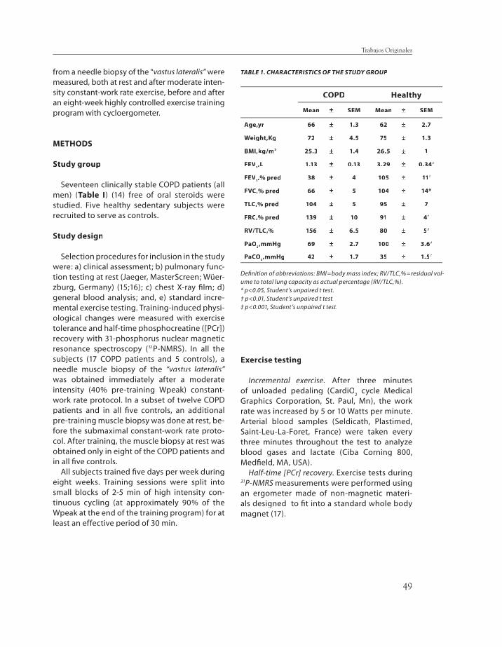
49
from a needle biopsy of the “vastus lateralis” were ” were ”measured, both at rest and after moderate inten-sity constant-work rate exercise, before and after an eight-week highly controlled exercise training program with cycloergometer.
METHODS
Study group
Seventeen clinically stable COPD patients (all men) (Table I) (14) free of oral steroids were studied. Five healthy sedentary subjects were recruited to serve as controls.
Study design
Selection procedures for inclusion in the study were: a) clinical assessment; b) pulmonary func-tion testing at rest (Jaeger, MasterScreen; Wüer-zburg, Germany) (15;16); c) chest X-ray fi lm; d) general blood analysis; and, e) standard incre-mental exercise testing. Training-induced physi-ological changes were measured with exercise tolerance and half-time phosphocreatine ([PCr]) recovery with 31-phosphorus nuclear magnetic resonance spectroscopy (31P-NMRS). In all the subjects (17 COPD patients and 5 controls), a needle muscle biopsy of the “vastus lateralis”was obtained immediately after a moderate intensity (40% pre-training Wpeak) constant-work rate protocol. In a subset of twelve COPD patients and in all fi ve controls, an additional pre-training muscle biopsy was done at rest, be-fore the submaximal constant-work rate proto-col. After training, the muscle biopsy at rest was obtained only in eight of the COPD patients and in all fi ve controls.
All subjects trained fi ve days per week during eight weeks. Training sessions were split into small blocks of 2-5 min of high intensity con-tinuous cycling (at approximately 90% of the Wpeak at the end of the training program) for at least an effective period of 30 min.
Exercise testing
Incremental exercise. After three minutes of unloaded pedaling (CardiO
2 cycle Medical
Graphics Corporation, St. Paul, Mn), the work rate was increased by 5 or 10 Watts per minute. Arterial blood samples (Seldicath, Plastimed, Saint-Leu-La-Foret, France) were taken every three minutes throughout the test to analyze blood gases and lactate (Ciba Corning 800, Medfi eld, MA, USA).
Half-time [PCr] recovery. Exercise tests during Half-time [PCr] recovery. Exercise tests during Half-time [PCr] recovery31P-NMRS measurements were performed using an ergometer made of non-magnetic materi-als designed to fi t into a standard whole body magnet (17).
Trabajos Originales
TABLE 1. CHARACTERISTICS OF THE STUDY GROUP
COPD Healthy
Mean ±± SEM Mean ±± SEM
Age,yr 66 ±± 1.3 62 ±± 2.7
Weight,Kg 72 ±± 4.5 75 ±± 1.3
BMI,kg/m2 25.3 ±± 1.4 26.5 ±± 1
FEV1,L 1.13 ±± 0.13 3.29 ±± 0.34‡
FEV1,% pred 38 ±± 4 105 ±± 11†
FVC,% pred 66 ±± 5 104 ±± 14*
TLC,% pred 104 ±± 5 95 ±± 7
FRC,% pred 139 ±± 10 91 ±± 4†
RV/TLC,% 156 ±± 6.5 80 ±± 5‡
PaO2,mmHg 69 ±± 2.7 100 ±± 3.6‡
PaCO2,mmHg 42 ±± 1.7 35 ±± 1.5†
Defi nition of abbreviations: BMI=body mass index; RV/TLC,%=residual vol-ume to total lung capacity as actual percentage (RV/TLC,%).* p<0.05, Student’s unpaired t test.† p<0.01, Student’s unpaired t test.‡ p<0.001, Student’s unpaired t test.
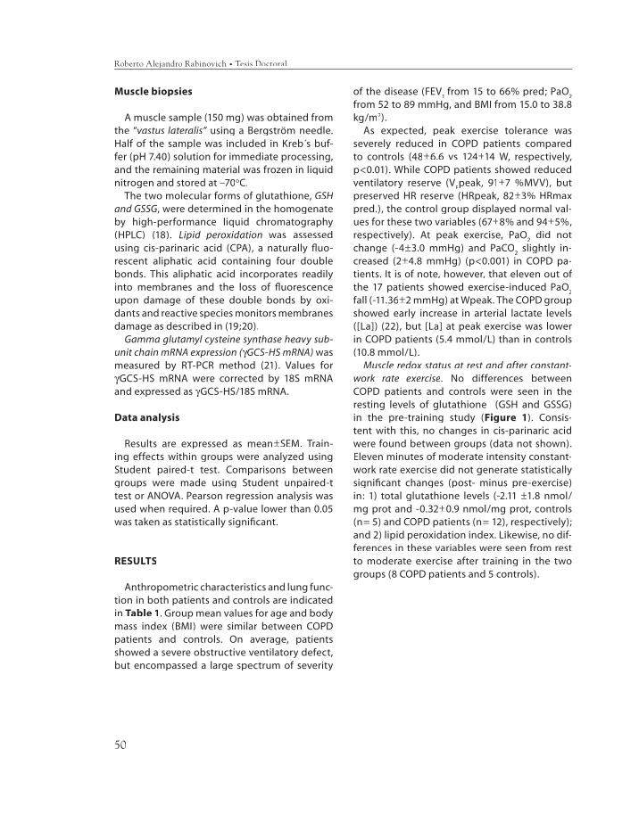
50
Muscle biopsies
A muscle sample (150 mg) was obtained from the “vastus lateralis” using a Bergström needle. vastus lateralis” using a Bergström needle. vastus lateralis”Half of the sample was included in Kreb´s buf-fer (pH 7.40) solution for immediate processing, and the remaining material was frozen in liquid nitrogen and stored at –70oC.
The two molecular forms of glutathione, GSH and GSSG, were determined in the homogenate by high-performance liquid chromatography (HPLC) (18). Lipid peroxidation was assessed using cis-parinaric acid (CPA), a naturally fl uo-rescent aliphatic acid containing four double bonds. This aliphatic acid incorporates readily into membranes and the loss of fl uorescence upon damage of these double bonds by oxi-dants and reactive species monitors membranes damage as described in (19;20).
Gamma glutamyl cysteine synthase heavy sub-unit chain mRNA expression (γunit chain mRNA expression (γunit chain mRNA expression ( GCS-HS mRNA) γGCS-HS mRNA) γ was measured by RT-PCR method (21). Values for γGCS-HS mRNA were corrected by 18S mRNA and expressed as γGCS-HS/18S mRNA.
Data analysis
Results are expressed as mean±SEM. Train-ing effects within groups were analyzed using Student paired-t test. Comparisons between groups were made using Student unpaired-t test or ANOVA. Pearson regression analysis was used when required. A p-value lower than 0.05 was taken as statistically signifi cant.
RESULTS
Anthropometric characteristics and lung func-tion in both patients and controls are indicated in Table 1. Group mean values for age and body mass index (BMI) were similar between COPD patients and controls. On average, patients showed a severe obstructive ventilatory defect, but encompassed a large spectrum of severity
of the disease (FEV1
from 15 to 66% pred; PaO2
from 52 to 89 mmHg, and BMI from 15.0 to 38.8 kg/m2).
As expected, peak exercise tolerance was severely reduced in COPD patients compared to controls (48±6.6 vs 124±14 W, respectively, p<0.01). While COPD patients showed reduced ventilatory reserve (V
Epeak, 91±7 %MVV), but
preserved HR reserve (HRpeak, 82±3% HRmax pred.), the control group displayed normal val-ues for these two variables (67±8% and 94±5%, respectively). At peak exercise, PaO
2 did not
change (-4±3.0 mmHg) and PaCO2 slightly in-
creased (2±4.8 mmHg) (p<0.001) in COPD pa-tients. It is of note, however, that eleven out of the 17 patients showed exercise-induced PaO
2
fall (-11.36±2 mmHg) at Wpeak. The COPD group showed early increase in arterial lactate levels ([La]) (22), but [La] at peak exercise was lower in COPD patients (5.4 mmol/L) than in controls (10.8 mmol/L).
Muscle redox status at rest and after constant-work rate exercise. No differences between COPD patients and controls were seen in the resting levels of glutathione (GSH and GSSG) in the pre-training study (Figure 1). Consis-tent with this, no changes in cis-parinaric acid were found between groups (data not shown). Eleven minutes of moderate intensity constant-work rate exercise did not generate statistically signifi cant changes (post- minus pre-exercise) in: 1) total glutathione levels (-2.11 ±1.8 nmol/mg prot and -0.32±0.9 nmol/mg prot, controls (n= 5) and COPD patients (n= 12), respectively); and 2) lipid peroxidation index. Likewise, no dif-ferences in these variables were seen from rest to moderate exercise after training in the two groups (8 COPD patients and 5 controls).
Roberto Alejandro Rabinovich • Tesis Doctoral
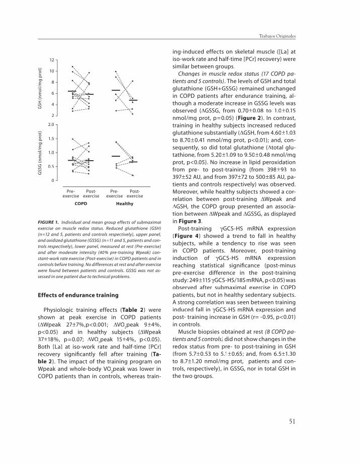
51
Effects of endurance training
Physiologic training effects (Table 2) were shown at peak exercise in COPD patients (∆Wpeak 27±7%,p<0.001; ∆VO∆VO∆
2peak 9±4%,
p<0.05) and in healthy subjects (∆Wpeak 37±18%, p=0.07; ∆VO∆VO∆
2peak 15±4%, p<0.05).
Both [La] at iso-work rate and half-time [PCr] recovery signifi cantly fell after training (Ta-ble 2). The impact of the training program on Wpeak and whole-body VO
2peak was lower in
COPD patients than in controls, whereas train-
ing-induced effects on skeletal muscle ([La] at iso-work rate and half-time [PCr] recovery) were similar between groups.
Changes in muscle redox status (17 COPD pa-tients and 5 controls). The levels of GSH and total glutathione (GSH+GSSG) remained unchanged in COPD patients after endurance training, al-though a moderate increase in GSSG levels was observed (∆GSSG, from 0.70±0.08 to 1.0±0.15 nmol/mg prot, p=0.05) (Figure 2). In contrast, training in healthy subjects increased reduced glutathione substantially (∆GSH, from 4.60±1.03 to 8.70±0.41 nmol/mg prot, p<0.01); and, con-sequently, so did total glutathione (∆total glu-tathione, from 5.20±1.09 to 9.50±0.48 nmol/mg prot, p<0.05). No increase in lipid peroxidation from pre- to post-training (from 398±93 to 397±52 AU, and from 397±72 to 500±85 AU, pa-tients and controls respectively) was observed. Moreover, while healthy subjects showed a cor-relation between post-training ∆Wpeak and ∆GSH, the COPD group presented an associa-tion between ∆Wpeak and ∆GSSG, as displayed in Figure 3.
Post-training γGCS-HS mRNA expression (Figure 4) showed a trend to fall in healthy subjects, while a tendency to rise was seen in COPD patients. Moreover, post-training induction of γGCS-HS mRNA expression reaching statistical signifi cance (post-minus pre-exercise difference in the post-training study: 249±115 γGCS-HS/18S mRNA, p<0.05) was observed after submaximal exercise in COPD patients, but not in healthy sedentary subjects. A strong correlation was seen between training induced fall in γGCS-HS mRNA expression and post- training increase in GSH (r= -0.95, p<0.01) in controls.
Muscle biopsies obtained at rest (8 COPD pa-tients and 5 controls) did not show changes in the redox status from pre- to post-training in GSH (from 5.7±0.53 to 5.1±0.65; and, from 6.5±1.30 to 8.7±1.20 nmol/mg prot, patients and con-trols, respectively), in GSSG, nor in total GSH in the two groups.
Trabajos Originales
12
10
8
6
4
2
2.0
1.5
1.0
0.5
0
GSS
G (
nm
ol/
mg
pro
t)G
SH (
nm
ol/
mg
pro
t)
Pre-exercise
Post-exercise
Pre-exercise
Post-exercise
COPD Healthy
FIGURE 1. Individual and mean group effects of submaximal exercise on muscle redox status. Reduced glutathione (GSH) (n=12 and 5, patients and controls respectively), upper panel, and oxidized glutathione (GSSG) (n=11 and 5, patients and con-trols respectively), lower panel, measured at rest (Pre-exercise) and after moderate intensity (40% pre-training Wpeak) con-stant-work rate exercise (Post-exercise) in COPD patients and in controls before training. No differences at rest and after exercise were found between patients and controls. GSSG was not as-sessed in one patient due to technical problems.
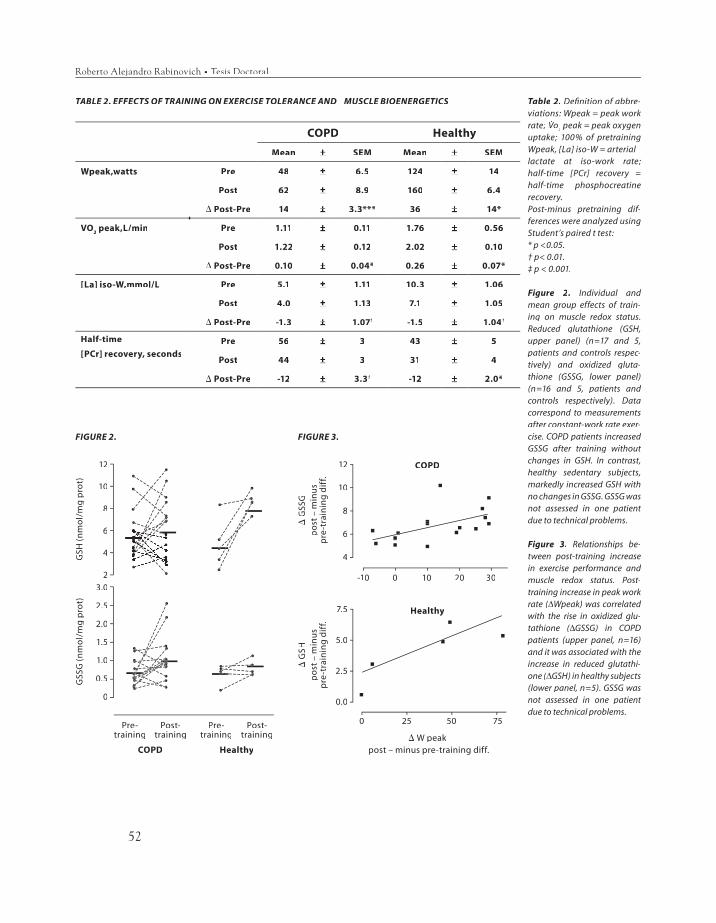
52
Roberto Alejandro Rabinovich • Tesis Doctoral
TABLE 2. EFFECTS OF TRAINING ON EXERCISE TOLERANCE AND MUSCLE BIOENERGETICS
COPD Healthy
Mean ±± SEM Mean ±± SEM
Wpeak,watts Pre 48 ±± 6.5 124 ±± 14
Post 62 ±± 8.9 160 ±± 6.4
Δ Post-Pre 14 ±± 3.3*** 36 ±± 14*
VO2 peak,L/min Pre 1.11 ±± 0.11 1.76 ±± 0.56
Post 1.22 ±± 0.12 2.02 ±± 0.10
Δ Post-Pre 0.10 ±± 0.04* 0.26 ±± 0.07*
[La] iso-W,mmol/L Pre 5.1 ±± 1.11 10.3 ±± 1.06
Post 4.0 ±± 1.13 7.1 ±± 1.05
Δ Post-Pre -1.3 ±± 1.07† -1.5 ±± 1.04†
Half-time
[PCr] recovery, secondsPre 56 ±± 3 43 ±± 5
Post 44 ±± 3 31 ±± 4
Δ Post-Pre -12 ±± 3.3† -12 ±± 2.0*
12
10
8
6
4
23.0
2.5
2.0
1.5
1.0
0.5
0
GSS
G (
nm
ol/
mg
pro
t)G
SH (
nm
ol/
mg
pro
t)
Pre-training
Post-training
Pre-training
Post-training
COPD Healthy
FIGURE 2. FIGURE 3.
12
10
8
6
4
ΔG
SSG
po
st –
min
us
pre
-tra
inin
g d
iff.
-10 0 10 20 30
0 25 50 75
ΔG
SHp
ost
– m
inu
sp
re-t
rain
ing
dif
f.
7.5
5.0
2.5
0.0
COPD
Healthy
Δ W peakpost – minus pre-training diff.
Table 2. Defi nition of abbre-viations: Wpeak = peak work rate; Vo
2 peak = peak oxygen
uptake; 100% of pretraining Wpeak, [La] iso-W = arteriallactate at iso-work rate; half-time [PCr] recovery = half-time phosphocreatine recovery.Post-minus pretraining dif-ferences were analyzed using Student’s paired t test:* p <0.05.† p< 0.01.‡ p < 0.001.
Figure 2. Individual and mean group effects of train-ing on muscle redox status. Reduced glutathione (GSH, upper panel) (n=17 and 5, patients and controls respec-tively) and oxidized gluta-thione (GSSG, lower panel) (n=16 and 5, patients and controls respectively). Data correspond to measurements after constant-work rate exer-cise. COPD patients increased GSSG after training without changes in GSH. In contrast, healthy sedentary subjects, markedly increased GSH with no changes in GSSG. GSSG was not assessed in one patient due to technical problems.
Figure 3. Relationships be-tween post-training increase in exercise performance and muscle redox status. Post-training increase in peak work rate (∆rate (∆rate ( Wpeak) was correlated with the rise in oxidized glu-tathione (∆tathione (∆tathione ( GSSG) in COPD patients (upper panel, n=16) and it was associated with the increase in reduced glutathi-one (∆one (∆one ( GSH) in healthy subjects (lower panel, n=5). GSSG was not assessed in one patient due to technical problems.
Vo
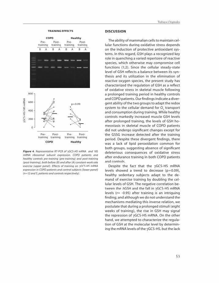
53
DISCUSSION
The ability of mammalian cells to maintain cel-lular functions during oxidative stress depends on the induction of protective antioxidant sys-tems. In this regard, GSH plays a recognized key role in quenching a varied repertoire of reactive species, which otherwise may compromise cell functions (1;2). Since the cellular steady-state level of GSH refl ects a balance between its syn-thesis and its utilization in the elimination of reactive oxygen species, the present study has characterized the regulation of GSH as a refl ect of oxidative stress in skeletal muscle following a prolonged training period in healthy controls and COPD patients. Our fi ndings indicate a diver-gent ability of the two groups to adapt the redox system to the cellular demand for O
2 transport
and consumption during training. While healthy controls markedly increased muscle GSH levels after prolonged training, the levels of GSH ho-meostasis in skeletal muscle of COPD patients did not undergo signifi cant changes except for the GSSG increase detected after the training period. Despite these divergent fi ndings, there was a lack of lipid peroxidation common for both groups, suggesting absence of signifi cant deleterious consequences of oxidative stress after endurance training in both COPD patients and controls.
Despite the fact that the γGCS-HS mRNA levels showed a trend to decrease (p=0.09), healthy sedentary subjects adapt to the de-mand of exercise training by doubling the cel-lular levels of GSH. The negative correlation be-tween the ∆GSH and the fall in γGCS-HS mRNA levels (r= -0.95) after training is an intriguing fi nding; and although we do not understand the mechanisms mediating this inverse relation, we postulate that during a prolonged stimuli (eight weeks of training), the rise in GSH may signal the repression of γGCS-HS mRNA. On the other hand, we attempted to characterize the regula-tion of GSH at the molecular level by determin-ing the mRNA levels of the γGCS-HS, but the lack
Trabajos Originales
TRAINING EFFECTS
800
600
400
200
0
γGC
S-H
S/18
S γG
CS-
HS/
18S
γm
RN
A
Pre-training
Post-training
Pre-training
Post-training
COPD Healthy
Pre-training
Post-training
COPD Healthy
B A B A
Pre-training
Post-training
B A B A
Figure 4. Representative RT-PCR of γGCS-HS mRNA and 18S mRNA ribosomal subunit expression. COPD patients and healthy controls pre-training (pre-training) and post-training (post-training), both before (B) and after (A) constant-work rate exercise (upper panel). Effects of training on γGCS-HS mRNA expression in COPD patients and control subjects (lower panel) (n=12 and 5, patients and controls respectively).
p=0.09

54
of measurement of its enzymatic activity due to the insufi cient size of muscle sample limits the interpretation of our fi ndings.
The resting muscle biopsies carried out in eight COPD patients and in all fi ve controls pre- and post-training did not display changes in the redox system. These results seems to suggest therefore that the fi ndings alluded to above are only evident after exercise.
It is well accepted that strenuous exercise in-duces higher levels of oxidative stress in COPD patients than in normal subjects (5). The phe-nomenon can be inhibited by treatment with xanthine oxidase inhibitors (allopurinol) (7) and by oxygen therapy (5). In the present study, the lack of changes in the redox system after con-stant-work rate exercise both in COPD patients and in controls (Figure 1) can be ascribed to the moderate intensity (40% pre-training Wpeak) exercise. Both intensity and duration of the protocol were chosen to perform the measure-ments after steady state exercise.
Training effects and muscle redox status
COPD patients as well as healthy sedentary subjects, signifi cantly enhanced exercise toler-ance, a phenomenon dependent on training-in-duced improvement of muscle O
2 transport and
utilization (8). It is of note that post-training val-ues of half-time [PCr] recovery in COPD patients were equivalent to the pre-training results in controls (10) (Table 2). The present study con-fi rms that in COPD patients (10), training effects on skeletal muscle in COPD patients (-∆half-time [PCr] recovery and -∆[La]iso-w) were noticeably higher than those observed at whole-body level (∆Wpeak and ∆VO∆VO∆
2peak), a fi nding that may be
of relevance in the evaluation of training out-comes.
In these patients, the reserve of the central organs to increase convective O
2 transport (ar-
terial oxygen content times blood fl ow) during heavy exercise (8;10) is limited by: 1) the se-verity of lung function impairment; and, 2) the
effects of pleural pressure swings on cardiac output (∆V∆V∆
E and ∆Q
T, respectively) (23;24). The
T, respectively) (23;24). The
T
phenomena alluded to, along with concurrent peripheral factors, such as impaired muscle oxy-gen conductance from capillary to mitochon-dria (25;26), are prone to induce cell hypoxia during moderate to high intensity exercise and consequently oxidative stress. Increased activity of cytochrome oxidase (COX) and up-regulation of this enzyme (27) can be interpreted as mito-chondrial adaptations to cell hypoxia.
Oxidative stress and muscle wasting
Engelen et al. (9) have speculated on a causal relationship between abnormally low muscle redox potential at rest and the alterations of protein metabolism observed in patients with emphysema substantiated by CT-scan (4). De-spite the fact that the present study did not identify baseline differences in GSH levels be-tween healthy subjects and COPD patients, our results are not in confl ict with those reported by these investigators (4) since we purposely stud-ied an heterogeneous group of COPD patients encompassing a large spectrum of severity of the disease.
In summary, the study demonstrates differ-ences in training-induced adaptations of mus-cle redox status between COPD patients and controls. While healthy sedentary subjects in-creased muscle GSH levels after training, COPD patients reduced their redox potential. Our re-sults highlight the importance of training-in-duced peripheral adaptations and its relevance in the assessment of training outcomes in COPD patients. Whether oxidative stress is a central factor mediating muscle mass wasting, particu-larly in susceptible subsets of COPD patients in whom a true myopathy can be observed re-mains to be elucidated.
Roberto Alejandro Rabinovich • Tesis Doctoral
55
ACKNOWLEDGEMENTS
The authors would like to thank Felip Burgos, Conxi Gistau and Jose Luis Valera and all the technical staff of the Lung Function Laboratory for their skillful support during the study. Anna Capitán, Cristina Gonzalez and Eduard Vilar from EUIF Blanquerna are acknowledged for their outstanding work supervising the training ses-sions. We thank Carme Hernandez, coordinator nurse of the Home Care Unit, for her support in the logistics of the study. Finally, the authors ac-knowledge the material support received from Erich Jaeger to conduct the study.
REFERENCES
1. Meister A, Anderson M. Glutathione. Annu Rev Biochem1983;52:711-760.
2. Fernandez Checa JC, Ookthens M. Regulation of hepatic GSH. In: N. Tavoloni and P. Berk, editors. Hepatic anion transport and bile secretion physiology and pathophysiology. New York, Raven Press ed.1993, p. 345-395
3. Fernandez Checa JC, Kaplowitz N, García-Ruiz C, Colell A. Mitochondrial glutathione: importance and transport. Semin Liver Dis 1998;18:389-401.
4. Engelen MPKJ, Schols AMWJ, Does JD, Deutz NEP, Wouters EF. Altered glutamate metabolism is associated with reduced muscle glutathione levels in patients with emphysema. Am J Respir Crit Care Med 2000;161:98-103.J Respir Crit Care Med 2000;161:98-103.J Respir Crit Care Med
5. Viña J, Servera E, Asensi M, Sastre J, Pallardó FV, Ferrero JA, Asunción JGI, Antón V, Marín J. Exercise causes blood glutathione oxidation in chronic obstructive pulmonary disease: prevention by O
2 therapy. J Appl Physiol
1996;81:2199-2202.
6. Sastre J, Asensi M, Gascó E, Pallardó FV, Ferrero JA, Furukawa T, Viña J. Exhaustive physical exercise causes oxidation of glutathione status in blood: prevention by antioxidant administration. Am J Physiol 1992;263:R992-R995.Am J Physiol 1992;263:R992-R995.Am J Physiol
7. Heunks L, Viña MJ, Van Herwaarden CL, Folgering HT, Gimeno A, Dekhuijzen PN. Xanthine oxidase is involved in exercise-induced oxidative stress in chronic obstructive pulmonary disease. Am J Physiol 1999;277:R1697-R1704.Am J Physiol 1999;277:R1697-R1704.Am J Physiol
8. American Thoracic Society and European Respiratory Society. Skeletal muscle dysfunction in chronic obstructive pulmonary disease. Am J Respir Crit Care Med 1999;159 (Suppl):S1-S40.Am J Respir Crit Care Med 1999;159 (Suppl):S1-S40.Am J Respir Crit Care Med
9. Engelen MPKJ. Muscle wasting in COPD: a metabolic and functional perspective. Maastricht: University Hospital.; 2001. Doctoral Thesis
10. Sala E, Roca J, Marrades RM, Alonso J, Gonzalez de Suso JM, Moreno A, Barbera JA, Nadal J,.Jover L, Rodriguez-Roisin R, Wagner PD. Effects of endurance training on skeletal muscle bioenergetics in chronic obstructive pulmonary disease. Am J Respir Crit Care Med 1999;159:1726-1734.Am J Respir Crit Care Med 1999;159:1726-1734.Am J Respir Crit Care Med
11. Roca J, Agustí AGN, Alonso A, Barberà JA, Rodriguez-Roisin R, Wagner PD. Effects of training on muscle O
2 transport at
VO2max. J Appl Physiol 1992;73:1067-1076.J Appl Physiol 1992;73:1067-1076.J Appl Physiol
12. Fernandez Checa JC, García-Ruiz C, Colell A, Morales A, Mari M, Miranda M, and Ardite E. Oxidative stress: role of mitochondria and protection by glutathione. Biofactors1998;8:7-11.
13. Chandel NS, Schumacker PT. Cellular oxygen sensing by mitochondria: old questions, new insight. J Appl Physiol2000;88:1880-1889.
14. A.T.S. Standards for the diagnosis and care of patients with chronic obstructive pulmonary disease. Am J Respir Crit Care Med 1995;152 (Suppl.):S78-S121.Crit Care Med 1995;152 (Suppl.):S78-S121.Crit Care Med
15. Roca J, Burgos F, Sunyer J, Saez M, Chinn S, Antó JM, Rodriguez-Roisin R, Quanjer Ph H, Nowak D, Burney P. References values for forced spirometry. Eur Respir J1998;11:1354-1362.
16. Roca J, Burgos F, Barberà JA, Sunyer J, Rodriguez-Roisin R, Castellsagué J, Sanchis J, Antó JM, Casan P, Clausen JL. Prediction equations for plethysmografi c lung volumes. Respir .Med 1998;92:454-460.Respir .Med 1998;92:454-460.Respir .Med
17. Gonzalez de Suso JM, Bernús G, Alonso A, Alay A, Capdevila A, Gili J, Prat JA, Arús C. Development and characterization of an ergometer to study the bioenergetics of the human quedriceps muscle by 31P-NMR spectroscopy inside a standard NMR scanner. Magn Reson Med 1993;29:575-581.Magn Reson Med 1993;29:575-581.Magn Reson Med
18. Fariss MW, Reed DJ. High-performance liquid chromatography of thiols and disulfi des: dinitrophenol derivatives. Methods Enzymol 1987;143:101-109.Methods Enzymol 1987;143:101-109.Methods Enzymol
19. Hedley D, Chow S. Flow cytometric measurement of lipid peroxidation in vital cells using parinaric acid. Cytometry1992;13:686-692.
20. Steenbergen RH, Drummen GP, Op den Kamp JA, Post JA. The use of cis-parinaric acid to measure lipid peroxidation in cardiomyocytes during ischemia and reperfusion. Biochim Biophys Acta 1997;1330:127-137.
21. Ardite E, Sans M, Panes J, Romero FJ, Pique JM, Fernandez Checa JC. Replenishment of glutathione levels improves mucosal function in experimental acute colitis. Lab Invest2000;80:734-744.
Trabajos Originales
56
22. Maltais F, Simard AA, Simard C, Jobin, Desgagnes P, Leblanc P. Oxidative capacity of the skeletal muscle and lactic acid kinetics during exercise in normal subjects and in patients with COPD. Am J Respir Crit Care Med 1996;153:288-293.Am J Respir Crit Care Med 1996;153:288-293.Am J Respir Crit Care Med
23. Sciurba FC, Rogers RM, Keenan RJ, Slivka, Gorcsan III J, Ferson PF, Holbert JM, Brown ML, Landreneau RJ. Improvement in pulmonary function and elastic recoil after lung-reduction surgery for diffuse emphysema. N Engl J Med 1996;334:1095-1099.Engl J Med 1996;334:1095-1099.Engl J Med
24. Montes de Oca M, Rassulo J, Celli BR. Respiratory muscle and cardiopulmonary function during exercise in very severe COPD. Am J Respir Crit Care Med 1996;154:1284-Am J Respir Crit Care Med 1996;154:1284-Am J Respir Crit Care Med1289.
25. Richardson RS, Noyszewski EA, Kendrick KF, Leigh JS, Wagner PD. Myoglobin O
2 desaturation during exercise: Evidence of
limited O2 transport. J Clin Invest 1995;96:1919-1926.J Clin Invest 1995;96:1919-1926.J Clin Invest
26. Jobin J, Maltais F, Doyon JF, Leblanc P, Simard PM, Simard AA. Chronic obstructive pulmonary disease: capillarity and fi ber characteristics of skeletal muscle. J Cardiopulm Rehabil1998;18:432-437.
27. Sauleda J, García-Palmer F, Wiesner RJ, Tarraga S, Harting I, Tomás P, Gómez C, Saus C, Palou A, Agustí AGN. Cytochrome oxidase activity and mitochondrial gene expression in skeletal muscle of patients with chronic obstructive pulmonary disease. Am J Respir Crit Care Med 1998;157:1413-Am J Respir Crit Care Med 1998;157:1413-Am J Respir Crit Care Med1417.
Roberto Alejandro Rabinovich • Tesis Doctoral
57
MANUSCRITO II
Training Depletes Muscle Glutathione in COPD Patients with Low Body Mass IndexRoberto A Rabinovich (1), Esther Ardite(1) , Ana Maria Mayer(1), Maite Figueras Polo(2), Jordi Vilaró(3), Josep M. Argilés(2), And Josep Roca(1)
Servei de Pneumologia (ICPCT), Hospital Clínic, Facultat de Medicina, IDIBAPS: Villarroel 170, 08036-Barcelona, Spain(1); Cancer Research Group, Departament de Bioquímica i Biologia Molecular, Facultat de Biologia, Universitat de Barcelona: Diagonal 645, 08028-Barcelona, Spain(2); and, EUIF Blanquerna. Universitat Ramon Llull: Padilla 326-332, 08025-Barcelona, Spain(3)
Supported by Grants FIS 00/0281 from the Fondo de Investigaciones Sanitarias; E-Remedy (IST-2000-25146) from the European Union; Comissionat per a Universitats i Recerca de la Generalitat de Catalunya (1999 SGR 00228); and Red Respira C03/011.
Roberto A Rabinovich was a Research Fellows supported by the European Respiratory Society, 2000. Ana Maria Mayer was Research Fellow supported by Fundación CAPES, Brasil, 2001-02.
Correspondence: Roberto Rabinovich, MD. Servei de Pneumologia. Hospital Clínic. Villarroel 170. Barcelona 08036. Spain. Phone 34-93-227-5540; FAX 34-93-227-5455; E-mail: [email protected]
Abstract: Physiological increase of muscle glutathione after training is not seen in COPD patients indicating abnormal peripheral muscle adaptations to exercise. We hypothesized that oxidative stress is primarily associated with low body mass index (BMI). Eleven patients with preserved BMI (BMIN: 28.2±1.2 kg.m-2), nine patients with low BMI (BMIL:19.7±0.60 kg.m-2) and fi ve age-matched controls (C:26.5±0.9 kg.m-2) were studied before and after eight weeks of high-intensity en-durance training. Reduced glutathione (GSH) and gamma-glutamyl cysteine synthase heavy subunit chain mRNA ex-pression (γGCS-HS mRNA) γGCS-HS mRNA) γ were measured in the vastus lateralis. After training, exercise capacity increased (∆VO∆VO∆ 2peak, 13±5.2%; 10±5.6%; and, 15±4.3%, BMIL, BMIN and C, respectively) (p< 0.05 each). GSH levels decreased in BMIL (from 5.2±0.7 to 3.7±0.8 nmol/mg protein, ∆GSH -1.5∆GSH -1.5∆ ±0.7 nmol/mg prot, p< 0.05); no changes were seen in BMIN (from 5.4±0.7 to 6.7±0.9 nmol/mg protein, ∆GSH∆GSH∆ 1.3±0.9 nmol/mg protein), whereas GSH markedly increased in C (from 4.6±1 to 8.7±0.4 nmol/mg prot, ∆GSH 4.1∆GSH 4.1∆ ±1 nmol/mg prot, p< 0.01). ∆GSH in BMI∆GSH in BMI∆ L was different from ∆GSH in BMI∆GSH in BMI∆ N and C (p< 0.05, each). Consistent changes were observed in γGCS-HS mRNAγGCS-HS mRNAγ expression. We conclude that GSH depletion after training in BMIL may suggest that oxidative stress plays a key role on muscle wasting in COPD.
Trabajos Originales
58
INTRODUCTION
In the last few years, systemic effects of chronic obstructive pulmonary disease (COPD) (1;2) are receiving a great deal of attention partly because of the impact of muscle wasting on poor disease prognosis (3). The underlying mechanisms of weight loss are, however, incompletely understood. We have recently reported (4) that COPD patients show an abnormal regulation of skeletal muscle glutathione (GSH). While high-intensity endurance training markedly enhanced antioxidant buffering in healthy subjects by a two-fold increase in muscle GSH, COPD patients showed a post-training increase of oxidized glutathione (GSSG) without changes in GSH. Impaired regu-lation of redox potential in the patients was closely related with the gain in exercise capacity produced by training (4). These results suggest that antioxidant buffering was not adapted to the higher rate of exercise-induced reactive oxygen species (ROS) production in COPD (4-6), thus leaving skeletal muscles more susceptible to oxidative stress.
We hypothesize that oxidative stress might be a central factor in the complex mechanisms of skeletal muscle wasting shown by COPD patients with low body mass index (BMIL)
(7). A better understanding of the underlying processes may have practical implications to refi ne evidence-based therapeutic strategies such as rehabilitation/training (8). The current investigation explores the relationships between abnormal training-induced GSH response and muscle wasting in COPD. To this end, we expanded the study group reported in (4) to compare COPD patients with preserved BMI (BMIN), BMIL and age-matched healthy sedentary controls (C).
METHODS
Study GroupTwenty clinically stable COPD patients (all men) (9) free of oral steroids were included in the
study. Five healthy sedentary subjects were recruited to serve as controls (BMI 26.5±0.9 kg.m-2). Eleven out of the twenty COPD patients (BMIN: 28.2±1.2 kg.m-2) showed preserved body mass in-dex, whereas the remaining nine patients had low BMI (BMIL: 19.7±0.60 kg.m-2). Up to seventeen patients and all fi ve controls were included in (4). All participants signed the written informed consent approved by the Committee on Investigations involving Human Subjects at the Hospital Clínic, Universitat de Barcelona.
Study designSelection procedures for inclusion in the study were: a) clinical assessment; b) pulmonary func-
tion testing at rest (Jaeger, MasterScreen; Wüerzburg, Germany) (10;11); c) chest X-ray fi lm; d) general blood analysis; and, e) standard incremental exercise testing. Training-induced physi-ological changes were measured by changes in exercise tolerance. In all the subjects (20 COPD patients and 5 controls), a needle muscle biopsy of the “vastus lateralis” was obtained immediately
Roberto Alejandro Rabinovich • Tesis Doctoral
59
after eleven minutes of moderate intensity (40% pre-training Wpeak) constant-work rate proto-col before and after 8 weeks of endurance training.
Training sessions were of 60 min duration and split into small blocks of 2 to 5 min of high inten-sity continuous cycling (60 cycles per minute) (at approximately 90% of the Wpeak at the end of the training program) for at least an effective period of 30 min. Recovery time between the high intensity periods consisted in cycling at the same speed and under 60% of pre-training Wpeak. The fi rst week was used as an adaptation period were the high intensity blocks were initially of 60% of Wpeak. After that, the work level has been raised to reach values close to the 90 % pre-training Wpeak and this intensity level was maintained until the end of the training period.
Exercise testingIncremental exercise. After three minutes of unloaded pedaling (CardiO2 cycle Medical Graph-
ics Corporation, St. Paul, Mn), the work rate was increased by 5 or 10 Watts per minute. Arterial blood samples (Seldicath, Plastimed, Saint-Leu-La-Foret, France) were taken every three minutes throughout the test to analyze blood gases and lactate (Ciba Corning 800, Medfi eld, MA, USA).
Muscle biopsiesA muscle sample (150 mg) was obtained from the “vastus lateralis” using a Bergström needle. Half of
the sample was included in Kreb s buffer (pH 7.40) solution for immediate processing, and the remain-ing material was frozen in liquid nitrogen and stored at –70oC.
The two molecular forms of glutathione, GSH and GSSG, were determined in the homogenate by high-performance liquid chromatography (HPLC) (12). Lipid peroxidation was assessed using cis-pari-naric acid (CPA), a naturally fl uorescent aliphatic acid containing four double bonds (13;14).
Gamma glutamyl cysteine synthase heavy subunit chain mRNA expression (γGCS-HS mRNA) γGCS-HS mRNA) γ was measured by RT-PCR method (15). Values for γGCS-HS mRNA were corrected by 18S mRNA γGCS-HS mRNA were corrected by 18S mRNA γand expressed as γGCS-HS/18S mRNA. γGCS-HS/18S mRNA. γ
Results are expressed as mean±SEM. Training effects within groups were analyzed using Student paired-t test. Comparisons among groups were made using ANOVA analyses. Student Neuman Keuls test was used as a post-hoc test for contrast analysis. A p-value lower than 0.05 was taken as statistically signifi cant.
RESULTS
Pre-training assessment Anthropometric characteristics and lung function in the three groups: BMIN, BMIL and C are
indicated in Table 1. On average, patients showed a severe obstructive ventilatory defect (FEV1
% pred: BMIN 40.5 ± 5.0%; BMIL 30.1±3.8%; and, C: 102±9.0%) and moderate arterial hypox-emia. Peak exercise tolerance (peak VO2/Kg) was severely reduced in COPD patients (BMIN
15.67 ± 1.14; BMIL 14.82 ± 2.2 ml.min-1.kg-1) compared to controls (C: 23.7 ± 0.85 ml.min-1.kg-1).
Trabajos Originales
60
No differences among the three groups were seen in pre-training glutathione levels (GSH: 5.2±0.7; 5.36±0.7; and 4.6±1 nmol/mg prot, BMIL, BMIN and C, respectively). Oxidized GSH (GSSG) levels and lipid peroxidation index, not reported, were also similar among groups. As described in (4), eleven minutes of moderate intensity constant-work rate exercise did not gener-ate statistically signifi cant changes (post- minus pre-exercise) in: 1) total glutathione levels; and 2) lipid peroxidation index. Likewise, no differences in these variables were seen from rest to moderate exercise after training.
Effects of physical trainingPhysiologic training effects at peak exercise (and ∆VO∆VO∆ 2peak) were observed in all three groups
(p< 0.05). Post-training ∆VO∆VO∆ 2peak were: 130±10 ml.min-1 (10%), p<0.05, in BMIN; 120±10 ml.min-
1 (13%), p<0.05, in BMIL; and, 260±70 ml.min-1 (15%), p<0.05, in healthy sedentary subjects. Changes in muscle redox status. After training, muscle GSH levels increased in healthy subjects
(from 4.6±1 to 8.70±0.4 nmol/mg, p< 0.01). In contrast, GSH decreased in BMIL (from 5.2±0.7 to 3.7±0.8 nmol/mg prot, p< 0.05); whereas no differences were seen in the BMIN (from 5.36±0.7 to 6.7±0.9 nmol/mg prot). Post-training changes in GSH (∆GSH) (Figure 1, left panel) where sig-nifi cantly different between BMIL (-1.5±0.7 nmol/mg prot) and the other two groups (1.3±0.9 and 4.1±1 nmol/mg prot, BMIN and C, respectively) (p< 0.05 each). None of the three groups showed training-induced increase in lipid peroxidation (BMIL, from 461±154 to 356±104 AU; BMIN, from 339±82 to 348±60 AU; and, C, from 358±75 to 439±72 AU).
In the BMIL group, post-training GSH fall was accompanied by a strong tendency to increase in γGCS-HS mRNA expression (γGCS-HS mRNA expression (γ Figure 1, right panel) in six out of the seven patients (p=0.06). In contrast, the post-training increase in GSH observed in healthy subjects was accompanied by a simultaneous tendency to fall in γGCS-HS mRNA expression in four out the fi ve subjects (p= γGCS-HS mRNA expression in four out the fi ve subjects (p= γ0.09). No changes in γGCS-HS mRNA expression were observed in BMIγGCS-HS mRNA expression were observed in BMIγ N (n= 8). The contrast analysis among groups (ANOVA, p= 0.05) showed statistical signifi cant differences in γGCS-HS γGCS-HS γmRNA regulation between each of the COPD groups (BMIL and BMIN) and healthy sedentary subjects.
DISCUSSION
Abnormal regulation of skeletal muscle redox system leading to higher vulnerability to oxida-tive stress has been shown in COPD patients after training (4) and during high intensity exercise (5). The impact of this phenomenon on cell function has not been established yet (16;17). The analysis of the individual results in (4) prompted the hypothesis that the abnormal adaptation of the GSH system to training might be primarily associated to the COPD phenotype character-ized by weight loss. Cell GSH seems to be an important regulator of myogenic differentiation in murine skeletal muscle C2C12 cells (18). Maintenance of optimal GSH levels might be a useful
Roberto Alejandro Rabinovich • Tesis Doctoral
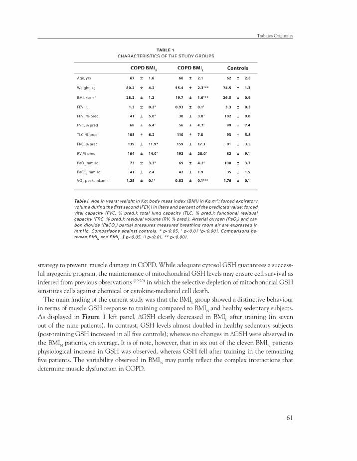
61
strategy to prevent muscle damage in COPD. While adequate cytosol GSH guarantees a success-ful myogenic program, the maintenance of mitochondrial GSH levels may ensure cell survival as inferred from previous observations (19;20) in which the selective depletion of mitochondrial GSH sensitizes cells against chemical or cytokine-mediated cell death.
The main fi nding of the current study was that the BMIL group showed a distinctive behaviour in terms of muscle GSH response to training compared to BMIN and healthy sedentary subjects. As displayed in Figure 1 left panel, ∆GSH clearly decreased in BMIL after training (in seven out of the nine patients). In contrast, GSH levels almost doubled in healthy sedentary subjects (post-training GSH increased in all fi ve controls); whereas no changes in ∆GSH were observed in the BMIN patients, on average. It is of note, however, that in six out of the eleven BMIN patients physiological increase in GSH was observed, whereas GSH fell after training in the remaining fi ve patients. The variability observed in BMIN may partly refl ect the complex interactions that determine muscle dysfunction in COPD.
Trabajos Originales
Table I. Age in years; weight in Kg; body mass index (BMI) in Kg.m-2; forced expiratory volume during the fi rst second (FEV1volume during the fi rst second (FEV1volume during the fi rst second (FEV ) in liters and percent of the predicted value; forced vital capacity (FVC, % pred.); total lung capacity (TLC, % pred.); functional residual capacity (FRC, % pred.); residual volume (RV, % pred.). Arterial oxygen (PaO2) and car-bon dioxide (PaCO2) partial pressures measured breathing room air are expressed in mmHg. Comparisons against controls. * p<0.05, † p<0.01 † p<0.01 † ‡p<0.001. Comparisons be-tween BMIN and BMIN and BMIN L. § p<0.05, 11 p<0.01, ** p<0.001.
COPD BMIN
COPD BMIL
Age, yrs 67 ±± 1.6 66 ±± 2.1 62 ±± 2.8
Weight, kg 80.2 ±± 4.2 55.4 ±± 2.3‡** 74.5 ±± 1.3
BMI, kg/m2 28.2 ±± 1.2 19.7 ±± 1.6‡** 26.5 ±± 0.9
FEV1, L 1.3 ±± 0.2‡ 0.93 ±± 0.1† 3.3 ±± 0.3
FEV1, % pred 41 ±± 5.0‡ 30 ±± 3.8† 102 ±± 9.0
FVC, % pred 68 ±± 6.4† 56 ±± 4.7† 99 ±± 7.4
TLC, % pred 105 ±± 6.2 110 ±± 7.8 93 ±± 5.8
FRC, % pred 139 ±± 11.9* 159 ±± 17.3 91 ±± 3.5
RV, % pred 164 ±± 14.0† 192 ±± 28.0† 82 ±± 9.1
PaO2,
mmHg 73 ±± 3.3‡ 69 ±± 4.2‡ 100 ±± 3.7
PaCO2,
mmHg 41 ±± 2.4 42 ±± 1.9 35 ±± 1.5
VO2, peak, mL.min-1 1.25 ±± 0.1‡ 0.82 ±± 0.1‡** 1.76 ±± 0.1
Controls
TABLE 1 CHARACTERISTICS OF THE STUDY GROUPS
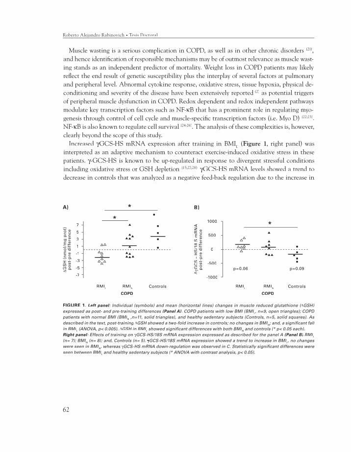
62
Muscle wasting is a serious complication in COPD, as well as in other chronic disorders (21), and hence identifi cation of responsible mechanisms may be of outmost relevance as muscle wast-ing stands as an independent predictor of mortality. Weight loss in COPD patients may likely refl ect the end result of genetic susceptibility plus the interplay of several factors at pulmonary and peripheral level. Abnormal cytokine response, oxidative stress, tissue hypoxia, physical de-conditioning and severity of the disease have been extensively reported (2) as potential triggers of peripheral muscle dysfunction in COPD. Redox dependent and redox independent pathways modulate key transcription factors such as NF-κB that has a prominent role in regulating myo-genesis through control of cell cycle and muscle-specifi c transcription factors (i.e. Myo D) (22;23). NF-κB is also known to regulate cell survival (24-26). The analysis of these complexities is, however, clearly beyond the scope of this study.
Increased γGCS-HS mRNA expression after training in BMIγGCS-HS mRNA expression after training in BMIγ L (Figure 1, right panel) was interpreted as an adaptive mechanism to counteract exercise-induced oxidative stress in these patients. γ-GCS-HS is known to be up-regulated in response to divergent stressful conditions γ-GCS-HS is known to be up-regulated in response to divergent stressful conditions γincluding oxidative stress or GSH depletion (15;27;28). γGCS-HS mRNA levels showed a trend to γGCS-HS mRNA levels showed a trend to γdecrease in controls that was analyzed as a negative feed-back regulation due to the increase in
FIGURE 1. Left panel: Individual (symbols) and mean (horizontal lines) changes in muscle reduced glutathione (: Individual (symbols) and mean (horizontal lines) changes in muscle reduced glutathione (: ∆ Individual (symbols) and mean (horizontal lines) changes in muscle reduced glutathione (∆ Individual (symbols) and mean (horizontal lines) changes in muscle reduced glutathione ( GSH) expressed as post- and pre-training differences (Panel A). COPD patients with low BMI (BMIL, n=9, open triangles); COPD patients with normal BMI (BMIN ,n=11, solid triangles), and healthy sedentary subjects (Controls, n=5, solid squares). As N ,n=11, solid triangles), and healthy sedentary subjects (Controls, n=5, solid squares). As N
described in the text, post-training ∆GSH showed a two-fold increase in controls; no changes in BMIN; and, a signifi cant fall in BMIL (ANOVA, p< 0.005). ∆GSH in BMIL showed signifi cant differences with both BMIN and controls (* p< 0.05 each).N and controls (* p< 0.05 each).N
Right panelRight panelRight : Effects of training on : Effects of training on : γGCS-HS/18S mRNA expression expressed as described for the panel A (Panel B).BMIL
(n= 7); BMIN (n= 8); and, Controls (n= 5). N (n= 8); and, Controls (n= 5). N γγGCS-HS/18S mRNA expression showed a trend to increase in BMIL, no changes were seen in BMIN, whereas N, whereas N γGCS-HS mRNA down-regulation was observed in C. Statistically signifi cant differences were seen between BMIL and healthy sedentary subjects (* ANOVA with contrast analysis, p< 0.05).
∆GS
H (
nm
ol/
mg
pro
t)p
ost
-pre
dif
fere
nce
7
5
3
1
-1
-3
-5
-7
BMIL BMIN
COPD
Controls
A)
BMIL BMIN
COPD
Controls
1000
500
0
-500
-1000
∆γG
CS
– H
S/1
8 S
mR
NA
po
st-p
re d
iffe
ren
ce
p=0.06 p=0.09
B)
Roberto Alejandro Rabinovich • Tesis Doctoral
63
GSH levels. The negative correlation between the ∆GSH and the fall in γGCS-HS mRNA levels γGCS-HS mRNA levels γ(r= -0.95) after training supports the interpretation.
In summary, the current results indicate the association between muscle wasting and abnormal response of the redox system in COPD. The impact of muscle wasting on the prognosis of the disease prompts the need for further progress in the understanding of the mechanisms involved in weight loss. In this line, a proper characterization of the underlying genomic determinants of the abnormal response of the GSH system may facilitate the identifi cation of pivotal factors modulat-ing the progress of the disease. Moreover, it may also help to guide the debate on the rationale for interventions increasing the redox potential of muscle cells in the subset of COPD patients with low BMI. Whether or not new pharmacological antioxidant agents now under development might be useful to enhance physiological adaptations to endurance training remains speculative.
ACKNOWLEDGMENTS
The authors would like to thank Felip Burgos, Conxi Gistau and Jose Luis Valera and all the technical staff of the Lung Function Laboratory for their skillful support during the study. Anna Capitán, Cristina Gonzalez and Eduard Vilar from EUIF Blanquerna are acknowledged for their outstanding work supervising the training sessions. We thank Carme Hernandez, coordinator nurse of the Home Care Unit, for her support in the logistics of the study. Finally, the authors acknowledge the material support received from Erich Jaeger to conduct the study.
(1) American Thoracic Society, European Respiratory Society. Skeletal muscle dysfunction in chronic obstructive pulmonary disease. Am J Respir Crit Care Med 1999; 159:s1-s40.
(2) Agusti AG, Noguera A, Sauleda J, Sala E, Pons J, Busquets X. Systemic effects of chronic obstructive pulmonary disease. Eur Respir J 2003; 21(2):347-360.
(3) Engelen MPKJ, Schols AMWJ, Does JD, Wouters EFM. Skeletal muscle weakness is associated with wasting of extremity fat-free mass but not with airfl ow obstruction in patients with chronic obstructive pulmonary disease. Am J Clin Nutr 2000; 71:733-738.
(4) Rabinovich RA, Ardite E, Troosters T, Carbó N, Alonso J, Gonzalez de Suso JM et al. Reduced muscle redox capacity after endurance training in COPD patients. Am J Respir Crit Care Med 2001; 164:1114-1118.
(5) Couillard A, Maltais F, Saey D, Debigare R, Michaud A, Koechlin C et al. Exercise-induced quadriceps oxidative stress and peripheral muscle dysfunction in COPD patients. Am J Respir Crit Care Med 2003; 167(12):1664-1669.
(6) Engelen MPKJ, Schols AMWJ, Does JD, Deutz NEP, Wouters EF. Altered glutamate metabolism is associated with reduced muscle glutathione levels in patients with emphysema. Am J Respir Crit Care Med 2000; 161:98-103.
(7) Chandel NS, Trzyna WX, McClintock DS, Schumacker PT. Role of oxidants in NF-kappa B activation and TNF-alpha gene transcription induced by hypoxia and endotoxin. J Immunol 2000; 165(2):1013-1021.
(8) Pauwels RA, Buist AS, Calverley PM, Jenkins CR, Hurd SS. Global strategy for the diagnosis, management, and prevention of chronic obstructive pulmonary disease. NHLBI/WHO Global Initiative for Chronic Obstructive Lung Disease
References
Trabajos Originales
64
(GOLD) Workshop summary. Am J Respir Crit Care Med 2001; 163(5):1256-1276.
(9) A.T.S. Standards for the diagnosis and care of patients with chronic obstructive pulmonary disease. Am J Respir Crit Care Med 1995; 152 (Suppl.):S78-S121.
(10) Roca J, Burgos F, Sunyer J, Saez M, Chinn S, Antó JM et al. References values for forced spirometry. Eur Respir J 1998; 11:1354-1362.
(11) Roca J, Burgos F, Barberà JA, Sunyer J, Rodriguez-Roisin R, Castellsague J et al. Prediction equations for plethysmografi c lung volumes. Respir Med 1998; 92:454-460.
(12) Fariss MW, Reed DJ. High-performance liquid chromatography of thiols and disulfi des: dinitrophenol derivatives. Methods Enzymol 1987; 143:101-109.
(13) Hedley D, Chow S. Flow cytometric measurement of lipid peroxidation in vital cells using parinaric acid. Cytometry 1992; 13:686-692.
(14) Steenbergen RH, Drummen GP, Op den Kamp JA, Post JA. The use of cis-parinaric acid to measure lipid peroxidation in cardiomyocytes during ischemia and reperfusion. Biochim Biophys Acta 1997; 1330(2):127-137.
(15) Ardite E, Sans M, Panes J, Romero FJ, Pique JM, Fernandez Checa JC. Replenishment of glutathione levels improves mucosal function in experimental acute colitis. Lab Invest 2000; 80(5):734-744.
(16) Meister A, Anderson M. Glutathione. Annu Rev Biochem 1983; 52:711-760.
(17) Fernandez Checa JC, Ookthens M. Regulation of hepatic GSH. In: Tavoloni N, Berk P, editors. Hepatic anion transport and bile secretion physiology and pathophysiology. 1993: 345-395.
(18) Ardite E, Rabinovich R, Barbera JA, Roca J, Fernandez-Checa JC. Critical Levels of Glutathione Are Needed for Differentiation in Skeletal Muscle Cells C2C12. American Journal of Respiratory and Critical Care Medicine 167[7], A129. 2003.
(19) Garcia-Ruiz C, Colell A, Mari M, Morales A, Calvo M, Enrich C et al. Defective TNF-alpha-mediated hepatocellular apoptosis and liver damage in acidic sphingomyelinase knockout mice. J Clin Invest 2003; 111(2):197-208.
(20) Ortega AL, Carretero J, Obrador E, Gambini J, Asensi M, Rodilla V et al. Tumor cytotoxicity by endothelial cells. Impairment of the mitochondrial system for glutathione uptake in mouse B16 melanoma cells that survive after in vitro interaction with the hepatic sinusoidal endothelium. J Biol Chem 2003; 278(16):13888-13897.
(21) Franssen FM, Wouters EF, Schols AM. The contribution of starvation, deconditioning and ageing to the observed alterations in peripheral skeletal muscle in chronic organ diseases. Clin Nutr 2002; 21(1):1-14.
(22) Langen RC, Schols AM, Kelders MC, Van Der Velden JL, Wouters EF, Janssen-Heininger YM. Tumor necrosis factor-alpha inhibits myogenesis through redox-dependent and -independent pathways. Am J Physiol Cell Physiol 2002; 283(3):C714-C721.
(23) Guttridge DC, Albanese C, Reuther JY, Pestell RG, Baldwin AS, Jr. NF-kappaB controls cell growth and differentiation through transcriptional regulation of cyclin D1. Mol Cell Biol 1999; 19(8):5785-5799.
(24) Wang CY, Mayo MW, Korneluk RG, Goeddel DV, Baldwin AS, Jr. NF-kappaB antiapoptosis: induction of TRAF1 and TRAF2 and c-IAP1 and c-IAP2 to suppress caspase-8 activation. Science 1998; 281(5383):1680-1683.
(25) Colell A, Garcia-Ruiz C, Roman J, Ballesta A, Fernandez-Checa JC. Ganglioside GD3 enhances apoptosis by suppressing the nuclear factor-kappa B-dependent survival pathway. FASEB J 2001; 15(6):1068-1070.
(26) Reid MB, Li YP. Tumor necrosis factor-alpha and muscle wasting: a cellular perspective. Respir Res 2001; 2(5):269-272.
(27) Meister A, Yan N, Meister. Amino acid sequence of rat kidney gamma-glutamylcysteine synthetase. J Biol Chem 1990; 265(3):1588-1593.
(28) Morales A, Miranda M, Sanchez-Reyes A, Colell A, Biete A, Fernandez Checa JC. Transcriptional regulation of the heavy subunit chain of gamma-glutamylcysteine synthetase by ionizing radiation. FEBS Lett 1998; 427(1):15-20.
Roberto Alejandro Rabinovich • Tesis Doctoral
65
MANUSCRITO III
Increased tumour necrosis factor-α plasma levels duringmoderate-intensity exercise in COPD patients
Roberto A Rabinovich (1), Maite Figueras(2) , Esther Ardite (1), Neus Carbó(2), Thierry Troosters (3), Xavier Filella(4), Joan Albert Barberà (1), José C. Fernandez- Checa (5), Josep M. Argilés (2), and, Josep Roca(1)
ABSTRACT: Post-training down-regulation of muscle tumour necrosis factor (TNFα) messenger ribonucleic acid (mRNA) expression and decrease in cellular TNFα levels have been reported in the elderly. It is hypothesized that chronic obstructive pulmonary disease (COPD) patients may not show these adaptations due to their reduced ability to increase muscle antioxidant capacity with training.Eleven COPD patients (FEV
1 36±4 % pred) and six age matched controls were
studied. Pre- and post-training plasma levels of: TNFα, soluble TNF receptors (sTNF-R, sTNF-R55 and sTNF-R75) and interleukin (IL-6) in plasma at rest and during exercise and vastus lateralis TNFα-mRNA were examined.Moderate-intensity constant-work rate exercise (eleven minutes of 40% pre-training Wpeak) increased pre-training plasma TNFα levels in COPD patients (from 17±3.2 to 23±2.7 pg/ml; p< 0.005) but not in controls (from 18.7±4.6 to 19±3.2 pg/ml). No changes were observed in sTNF-R or in IL-6 levels. After eight-weeks endurance training, moderate-intensity exercise increased plasma TNFα levels similarly to pre-training (from 16±3 to 21±4 pg/ml; p< 0.01). Pre-training muscle TNFα−mRNA expression was signifi cantly higher in COPD patients than in controls (29.3±13.9 vs 5.0±1.5 TNFα/18S, respectively; p< 0.05), but no changes were observed after exercise or training.It is concluded that moderate-intensity exercise abnormally increases plasma TNFα levels in chronic obstructive pulmonary disease patients without exercise-induced upregulation of the TNFα gene in skeletal muscle.
Eur Respir J 2003; 21: 789-794.
(1) Servei de Pneumologia (ICPCT) (1) Hospital Clínic, Facultat de Medicina, IDIBAPS; Departament de Bioquímica i Biologia Molecular, Facultat de Biologia (2), Universitat de Barcelona, Barcelona; Faculty of Physical Education and Physiotherapy, K.U.L., Leuven, Belgium (3); Servicio de Bioquímica Clínica (Unitat d’ Estudi del Càdel Càdel C ncer) (4) and Liver Unit (IMD) and CSIC (5), Hospital Clínic, IDIBAPS, Barcelona.
(2) Supported by Grants FIS 99/0029 and 00/0281 from the Fondo de Investigaciones Sanitarias; E-Remedy (IST-2000-25146) from the European Union (DG XIII); and, Comissionat per a Universitats i Recerca de la Generalitat de Catalunya (1999 SGR 00228).
(3) Roberto A. Rabinovich and Thierry Troosters were research fellows supported by the European Respiratory Society, 2000.
(4) Maite Figueras is recipient of a pre-doctoral scholarship from the Ministerio de Educación y Cultura of the Spanish Goverment.
(5) Correspondence: Josep Roca, MD. Servei de Pneumologia. Hospital Clínic. Villarroel 170. Barcelona 08036. Spain. Phone 34-93-227-5540; FAX 34-93-227-5455; E-mail: [email protected]
Running head: TNFα during exercise in COPDWord count: Abstract: 200Body text: 2649
Trabajos Originales
66
Introduction
Different lines of evidence involve cytokines, particularly tumor necrosis factor-α (TNFα), in the process of skeletal muscle wasting observed in approximately 20% of COPD patients (1-5). Muscle redox status itself and activation of transcription factors such as NFk-B by reactive oxygen species (6;7) seem to be involved in the signal-transduction pathways that modulate cellular TNFα levels. Moreover, it has been reported that exercise training in normal elderly humans down-regulates TNFα-mRNA expression and decreases muscle TNFα levels (8) likely related to enhancement of the O
2 transport/O
2utilization system.
It is well accepted that heavy exercise induces abnormally high levels of oxidative stress in COPD patients (9). We recently showed (10) that after eight weeks of endurance training at the same relative load, muscle redox potential appear to be reduced in COPD patients, but not in age-matched healthy sedentary controls.
In the present study, we hypothesize that COPD patients may present an abnormal cytokine response to exercise. They might not show training down-regulation of muscle TNFα, as reported in healthy subjects (8), because of the lower antioxidant capacity (10) after training. The study examines the effects of both endurance training and moderate intensity exercise (pre- and post-training) on: 1) cytokine plasma levels (TNFα; soluble TNF receptors: sTNF-R; sTNF-R55, sTNF-R75; and, IL-6); and, 2) mRNA expression for TNFα in limb skeletal muscle, in a population of eleven patients with COPD and six age matched healthy controls (10).
Methods
Study Group
Eleven of the seventeen COPD patients according to the ATS criteria (11) examined in (10) and six healthy sedentary age and gender-matched controls were included in the study. All participants signed the written informed consent approved by the Committee on Investigations involving Human Subjects at the Hospital Clínic, Universitat de Barcelona.
Study design
Selection procedures for inclusion in the study and details of the design have been reported in (10). Briefl y, we carried out pre- and post-training measurements to assess the effects of endurance training (incremental exercise testing; 31-phosphorus magnetic resonance spectroscopy; and, V’O
2 -on kinetics, not reported in the present study)
and to evaluate changes in cytokine plasma levels during constant-work rate exercise.
All subjects trained 5 d per week for 8 wk. Training sessions were of 60 min duration and split into small blocks of 2 to 5 min of high intensity continuous cycling (around 60 cycles per minute) (at approximately 90% of the Wpeak at the end of the training program) for at least an effective period of 30 min. Recovery time between the high intensity periods consisted in cycling at the same speed and under 60 % of pre-training Wpeak. The fi rst week was used as an adaptation period were the high intensity blocks were initially of 60 % of Wpeak. After that the work level has been raised until reach values close to the 90 % pre-training Wpeak and maintained until the end of the training period.
Constant work rate exercise
After placing an arterial catheter (Seldicath, Plastimed, Saint-Leu-La-Foret, France), subjects were installed on the cycle ergometer with the mouth piece in place which allowed on-line calculation of pulmonary O
2 uptake (V’O
2). They
cycled for eleven minutes at a constant work rate of 40% pre-training Wpeak at a speed of 60 r.p.m. The rationale for choosing a moderate intensity constant-work rate protocol was to achieve a sustainable exercise activity (12). Arterial blood sampling for measurements of cytokine plasma levels was done at rest, at 11 min of steady state exercise and at 20 min post-exercise.
Muscle Biopsies
After appropriate antisepsis and anesthetics of the skin, a muscle biopsy was obtained in a subset of nine COPD patients and the six healthy controls from the “vastus lateralis” using a Bergström needle. vastus lateralis” using a Bergström needle. vastus lateralis”Muscle samples were immediately frozen in liquid nitrogen and stored at –70oC for their later analysis.
Roberto Alejandro Rabinovich • Tesis Doctoral
67
Cytokine measurements
Plasma levels of TNFαPlasma levels of TNFαPlasma levels of TNF , soluble TNF-receptors α, soluble TNF-receptors α(sTNF-R; sTNF-R55, sTNF-R75) and IL-6 were assessed by ELISA (Medgenix, BioSource Europe, Fleurus, Belgium, for Il-6 and TNF-α; Bender MedSystems, Vienna, Austria for sTNF-R) (13); results were expressed in pg/ml. Intra-assay coeffi cients of variation (CV) at different TNFα concentrations (86.7±4.4 pg/ml, 591±22 pg/ml, and 1027±15 pg/ml; n=20) were 5.1 %, 3.7 % and 1.4 %, respectively. Inter-assay CV at different TNFα concentrations (41±4 pg/ml, 162±13 pg/ml and 664±66 pg/ml; n=10, 13 and 10 respectively) were 9.7 %, 8.0 %, and 9.9 % respectively. The minimum detectable concentration is estimated to be 3 pg/ml (Medgenix, Fleurus, Belgium).
Skeletal muscle mRNA expression for TNFα: Total RNA from vastus lateralis muscle was extracted by TriPureTM kit (Boheringer-Manheim, Germany), a commercial modifi cation of the acid guanidinium isothiocyanate/phenol/chloroform method described by Chomczynski and Sacchi (14). TNFα mRNA was measured by a two-step RT-PCR method (15) using appropiate forward (5'- AAG AGT TCC CCA GGG ACC TCT -3') and reverse (5'- CCT GGG AGT AGA TGA GGT ACA- 3') primers (Gene specifi c relative RT-PCR kit 5345). The predicted size of the amplifi ed product using these primers is 233 bp. Briefl y, for each sample, 5 µg of RNA were reverse-transcribed (RT) into cDNA in the presence of appropriate anti-sense primers. The PCR reaction was carried out as follows: 1x (94ºC, 1:00 min); 33x (94ºC, 1:00 min; 61ºC, 1:00 min; 72ºC, 2:00 min); 1x (72ºC, 4 min) in the presence of 1.5 mM MgCl
2 . All the RT-
PCR were standardized at 33 cycles to ensure that the samples were assayed within the exponential phase of the PCR reaction. The RT-PCR products were electrophoresed in 2% agarose gels in TBE buffer. The gel was stained with ethidium bromide and quantitated by scanning densitometry. Values for TNFα-mRNA were corrected by 18S RNA expression and expressed as % TNFα/18S ratio. The set of primers used amplify a segment of the 18S rRNA with a predicted size of 495 bp. The abundance of rRNA is a major limitation to its utility as a control, since an endogenous control must be in the same linear ranges as the RNA under study when amplifi ed under the same conditions.
Ambion’s Competimer™ technology (patent pending) allow to attenuate the amplifi cation of 18S-rRNA by using a modifi ed primer (competimer) that cannot be extended. In this way the expression of the internal control can be modulated to the level of even rare messages, without affecting the performance of other targets in a multiplex PCR reaction. The proportion of primer/competimer used was 3:17.
Data analysis
Results are expressed as mean±SEM. Changes within groups were analyzed using Student paired-t test. Comparisons between groups were done using Student unpaired-t test. When normality test fail, a logarithmic transformation was applied to the data. A p-value < 0.05 was taken as statistically signifi cant.
Results
The COPD group (age 65±1.4 yrs, range 59 to 72 yrs; and, BMI 25±1.5 kg/m2, range 15 to 36 kg/m2) (Table I) encompassed a large spectrum of severity of the disease. These patients showed a severe obstructive ventilatory defect (FEV
1 40±4.4
% pred, range 15 to 57% pred; PaO2 69±3.3 mmHg,
range 54 to 89 mmHg) with limited exercise tolerance (V’O
2 peak 17±1.6 ml/Kg/min, range 9 to
27 mL/Kg/min). Healthy sedentary controls were matched by gender (all men), age (age 63±1.9 yrs) and body mass index (BMI, 26.9±0.9 kg/m2) and showed normal pulmonary function test values (FEV
1 112±11 % pred) and exercise tolerance
(V’O2peak 24±0.8 ml/Kg/min). As reported in
detail in (10), a signifi cant training effect was observed in the two groups. In COPD patients, exercise tolerance increased (∆Wpeak 41±12% and ∆V’O
2peak 10.5±3%, p< 0.01 each) and arterial
[La] at iso-work rate fell -20±6 % after training (p< 0.01). Likewise, healthy sedentary controls enhanced exercise tolerance (∆Wpeak 32±13%, p= 0.05; and, ∆V’Opeak 14±3% p< 0.005) and arterial [La] at iso-work rate fell -12.5 ± 3.9 % after training (p< 0.05).
Cytokine plasma levels during exercise and after training
No differences in TNFα plasma levels at rest were observed between patients and controls
Trabajos Originales
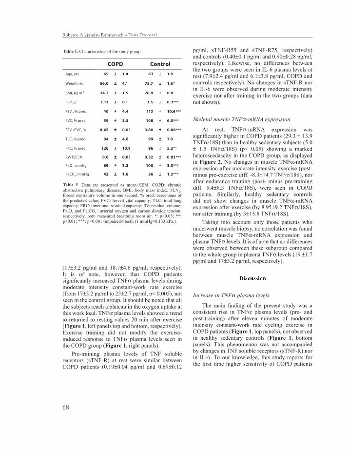
68
(17±3.2 pg/ml and 18.7±4.6 pg/ml; respectively). It is of note, however, that COPD patients signifi cantly increased TNFα plasma levels during moderate intensity constant-work rate exercise (from 17±3.2 pg/ml to 23±2.7 pg/ml, p< 0.005), not seen in the control group. It should be noted that all the subjects reach a plateau in the oxygen uptake at this work load. TNFα plasma levels showed a trend to returned to resting values 20 min after exercise (Figure 1, left panels top and bottom, respectively). Exercise training did not modify the exercise-induced response to TNFα plasma levels seen in the COPD group (Figure 1, right panels).
Pre-training plasma levels of TNF soluble receptors (sTNF-R) at rest were similar between COPD patients (0.19±0.04 pg/ml and 0.69±0.12
pg/ml, sTNF-R55 and sTNF-R75, respectively) and controls (0.40±0.1 pg/ml and 0.90±0.28 pg/ml, respectively). Likewise, no differences between the two groups were seen in IL-6 plasma levels at rest (7.9±2.4 pg/ml and 6.1±3.8 pg/ml, COPD and controls respectively). No changes in sTNF-R nor in IL-6 were observed during moderate intensity exercise nor after training in the two groups (data not shown).
Skeletal muscle TNFαSkeletal muscle TNFαSkeletal muscle TNF -mRNA expression
At rest, TNFα-mRNA expression was signifi cantly higher in COPD patients (29.3 ± 13.9 TNFα/18S) than in healthy sedentary subjects (5.0 ± 1.5 TNFα/18S) (p< 0.05) showing a marked heteroscedascity in the COPD group, as displayed in Figure 2. No changes in muscle TNFα-mRNA expression after moderate intensity exercise (post- minus pre-exercise diff. -8.3±14.7 TNFα/18S), nor after endurance training (post- minus pre-training diff. 5.4±8.3 TNFα/18S), were seen in COPD patients. Similarly, healthy sedentary controls did not show changes in muscle TNFα-mRNA expression after exercise (by 8.95±9.2 TNFα/18S), nor after training (by 3±15.8 TNFα/18S).
Taking into account only those patients who underwent muscle biopsy, no correlation was found between muscle TNFα-mRNA expression and plasma TNFα levels. It is of note that no differences were observed between these subgroup compared to the whole group in plasma TNFα levels (19 ±1.7 pg/ml and 17±3.2 pg/ml, respectively).
Discussion
Increase in TNFαIncrease in TNFαIncrease in TNF plasma levels
The main fi nding of the present study was a consistent rise in TNFα plasma levels (pre- and post-training) after eleven minutes of moderate intensity constant-work rate cycling exercise in COPD patients (Figure 1, top panels), not observed in healthy sedentary controls (Figure 1, bottom panels). This phenomenon was not accompanied by changes in TNF soluble receptors (sTNF-R) nor in IL-6. To our knowledge, this study reports for the fi rst time higher sensitivity of COPD patients
Table 1. Characteristics of the study group
COPD Control
Age, yrs 65 ±± 1.4 63 ±± 1.9
Weight, kg 66.0 ±± 4.1 75.7 ±± 1.6*
BMI, kg. m-2 24.7 ±± 1.5 26.9 ±± 0.9
FEV1, L 1.13 ±± 0.1 3.5 ±± 0.3***
FEV1, % pred 40 ±± 4.4 112 ±± 10.6***
FVC, % pred 59 ±± 3.5 108 ±± 6.5***
FEV1/FVC, % 0.45 ±± 0.03 0.80 ±± 0.06***
TLC, % pred 94 ±± 4.6 99 ±± 7.0
FRC, % pred 126 ±± 10.9 96 ±± 5.3**
RV/TLC, % 0.6 ±± 0.03 0.32 ±± 0.03***
PaO2, mmHg 69 ±± 3.3 100 ±± 3.3***
PaCO2, mmHg 42 ±± 1.6 36 ±± 1.3***
Table I. Data are presented as mean+SEM. COPD: chronic obstructive pulmonary disease; BMI: body mass index; FEV
1:
forced expiratory volume in one second; % pred: percentage of the predicted value; FVC: forced vital capacity; TLC: total lung capacity; FRC: functional residual capacity; RV: residual volume; Pa,O
2 and Pa,CO
2: arterial oxygen and carbon dioxide tension,
respectively, both measured breathing room air. *: p<0.05; **: p<0.01; ***: p<0.001 (unpaired-t test); (1 mmHg=0.133 kPa.).
Roberto Alejandro Rabinovich • Tesis Doctoral
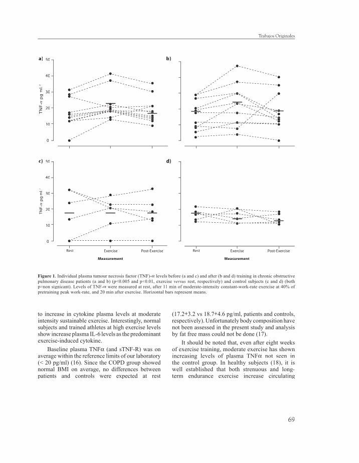
69
to increase in cytokine plasma levels at moderate intensity sustainable exercise. Interestingly, normal subjects and trained athletes at high exercise levels show increase plasma IL-6 levels as the predominant exercise-induced cytokine.
Baseline plasma TNFα (and sTNF-R) was on average within the reference limits of our laboratory (< 20 pg/ml) (16). Since the COPD group showed normal BMI on average, no differences between patients and controls were expected at rest
(17.2±3.2 vs 18.7±4.6 pg/ml, patients and controls, respectively). Unfortunately body composition have not been assessed in the present study and analysis by fat free mass could not be done (17).
It should be noted that, even after eight weeks of exercise training, moderate exercise has shown increasing levels of plasma TNFα not seen in the control group. In healthy subjects (18), it is well established that both strenuous and long-term endurance exercise increase circulating
Figure 1. Individual plasma tumour necrosis factor (TNF)-α levels before (a and c) and after (b and d) training in chronic obstructive pulmonary disease patients (a and b) (p<0.005 and p<0.01, exercise versus rest, respectively) and control subjects (c and d) (both p=non signicant). Levels of TNF-α were measured at rest, after 11 min of moderate-intensity constant-work-rate exercise at 40% of pretraining peak work-rate, and 20 min after exercise. Horizontal bars represent means.
50
40
30
20
10
0
TN
F-α
pg
. mL-1
Rest Exercise Post-Exercise
50
40
30
20
10
0
TNF-
αp
g. m
L-1m
L-1m
L
Measurement
Rest Exercise Post-Exercise
Measurement
d)c)
Trabajos Originales
a) b)
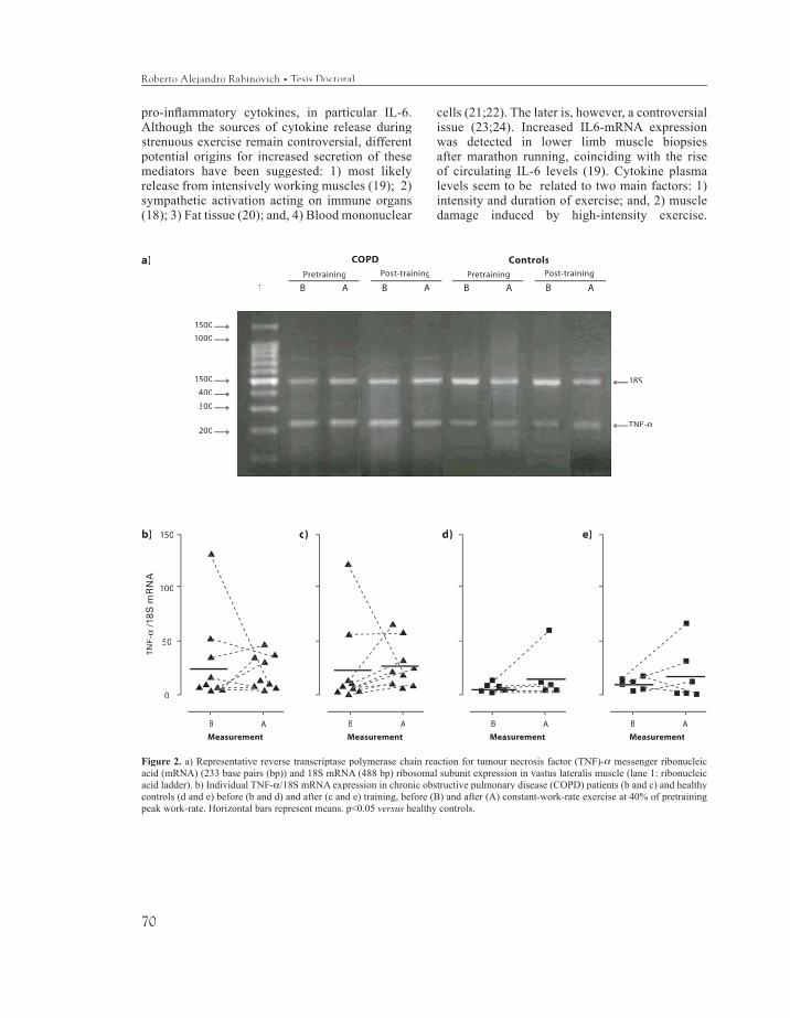
70
pro-infl ammatory cytokines, in particular IL-6. Although the sources of cytokine release during strenuous exercise remain controversial, different potential origins for increased secretion of these mediators have been suggested: 1) most likely release from intensively working muscles (19); 2) sympathetic activation acting on immune organs (18); 3) Fat tissue (20); and, 4) Blood mononuclear
cells (21;22). The later is, however, a controversial issue (23;24). Increased IL6-mRNA expression was detected in lower limb muscle biopsies after marathon running, coinciding with the rise of circulating IL-6 levels (19). Cytokine plasma levels seem to be related to two main factors: 1) intensity and duration of exercise; and, 2) muscle damage induced by high-intensity exercise.
Figure 2. a) Representative reverse transcriptase polymerase chain reaction for tumour necrosis factor (TNF)-α messenger ribonucleic acid (mRNA) (233 base pairs (bp)) and 18S mRNA (488 bp) ribosomal subunit expression in vastus lateralis muscle (lane 1: ribonucleic acid ladder). b) Individual TNF-α/18S mRNA expression in chronic obstructive pulmonary disease (COPD) patients (b and c) and healthy controls (d and e) before (b and d) and after (c and e) training, before (B) and after (A) constant-work-rate exercise at 40% of pretraining peak work-rate. Horizontal bars represent means. p<0.05 versus healthy controls.
Roberto Alejandro Rabinovich • Tesis Doctoral
Pretraining Post-training
COPD
B A B A1
Pretraining Post-training
Controls
B A B A
1500
1000
1500
400
300
200
18S
TNF-α
150
100
50
0
TNF-
α /1
8S
mR
NA
B A
Measurement
b)
B A
Measurement
c)
B A
Measurement
d)
B A
Measurement
e)
a)
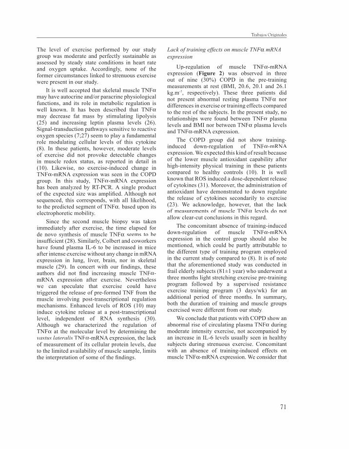
71
The level of exercise performed by our study group was moderate and perfectly sustainable as assessed by steady state conditions in heart rate and oxygen uptake. Accordingly, none of the former circumstances linked to strenuous exercise were present in our study.
It is well accepted that skeletal muscle TNFα may have autocrine and/or paracrine physiological functions, and its role in metabolic regulation is well known. It has been described that TNFα may decrease fat mass by stimulating lipolysis (25) and increasing leptin plasma levels (26). Signal-transduction pathways sensitive to reactive oxygen species (7;27) seem to play a fundamental role modulating cellular levels of this cytokine (8). In these patients, however, moderate levels of exercise did not provoke detectable changes in muscle redox status, as reported in detail in (10). Likewise, no exercise-induced change in TNFα-mRNA expression was seen in the COPD group. In this study, TNFα-mRNA expression has been analyzed by RT-PCR. A single product of the expected size was amplifi ed. Although not sequenced, this corresponds, with all likelihood, to the predicted segment of TNFα, based upon its electrophoretic mobility.
Since the second muscle biopsy was taken immediately after exercise, the time elapsed for de novo synthesis of muscle TNFα seems to be insuffi cient (28). Similarly, Colbert and coworkers have found plasma IL-6 to be increased in mice after intense exercise without any change in mRNA expression in lung, liver, brain, nor in skeletal muscle (29). In concert with our fi ndings, these authors did not fi nd increasing muscle TNFα-mRNA expression after exercise. Nevertheless we can speculate that exercise could have triggered the release of pre-formed TNF from the muscle involving post-transcriptional regulation mechanisms. Enhanced levels of ROS (10) may induce cytokine release at a post-transcriptional level, independent of RNA synthesis (30). Although we characterized the regulation of TNFα at the molecular level by determining the vastus lateralis TNFα-mRNA expression, the lack of measurement of its cellular protein levels, due to the limited availability of muscle sample, limits the interpretation of some of the fi ndings.
Lack of training effects on muscle TNFαLack of training effects on muscle TNFαLack of training effects on muscle TNF mRNA expression
Up-regulation of muscle TNFα-mRNA expression (Figure 2) was observed in three out of nine (30%) COPD in the pre-training measurements at rest (BMI, 20.6, 20.1 and 26.1 kg.m-2, respectively). These three patients did not present abnormal resting plasma TNFα nor differences in exercise or training effects compared to the rest of the subjects. In the present study, no relationships were found between TNFα plasma levels and BMI nor between TNFα plasma levels and TNFα-mRNA expression.
The COPD group did not show training-induced down-regulation of TNFα-mRNA expression. We expected this kind of result because of the lower muscle antioxidant capability after high-intensity physical training in these patients compared to healthy controls (10). It is well known that ROS induced a dose-dependent release of cytokines (31). Moreover, the administration of antioxidant have demonstrated to down regulate the release of cytokines secondarily to exercise (23). We acknowledge, however, that the lack of measurements of muscle TNFα levels do not allow clear-cut conclusions in this regard.
The concomitant absence of training-induced down-regulation of muscle TNFα-mRNA expression in the control group should also be mentioned, which could be partly attributable to the different type of training program employed in the current study compared to (8). It is of note that the aforementioned study was conducted in frail elderly subjects (81±1 year) who underwent a three months light stretching exercise pre-training program followed by a supervised resistance exercise training program (3 days/wk) for an additional period of three months. In summary, both the duration of training and muscle groups exercised were different from our study.
We conclude that patients with COPD show an abnormal rise of circulating plasma TNFα during moderate intensity exercise, not accompanied by an increase in IL-6 levels usually seen in healthy subjects during strenuous exercise. Concomitant with an absence of training-induced effects on muscle TNFα-mRNA expression. We consider that
Trabajos Originales
72
our results open striking questions, particularly those related with the abnormal TNFα response during moderate exercise and its relationships with systemic effects of the disease. Further research is needed to clarify the origin and physiological meaning of these fi ndings.
Acknowledgements. The authors are grateful to Felip Burgos, Conxi Gistau and Jose Luis Valera and all the technical staff of the Lung Function Laboratory for their skillful support during the study. Jordi Vilaró, Anna Capitán, Cristina Gonzalez and Eduard Vilar from EUIF Blanquerna are acknowledged for their outstanding work conducting the training sessions. We acknowledge Carmen Hernandez, coordinator nurse of the Home Care Unit, for the help in the patient recruitment. We thank especially to Dr Ricardo Bastos for his valuable help in the revision of the manuscript. Dr. Peter Wagner is acknowledged for carefully and critical reading the manuscript. Finally, the authors are grateful for the support received from Erich Jaeger to conduct the study.
References
1. Takabatake M, Nakamura H, Abe S, Hino T, Saito H, Yuki H, Kato S, Tomoike H. Circulating leptin in patients with chronic obstructive pulmonary disease. Am J Respir Crit Care Med 1999; 159:1215-1219.Am J Respir Crit Care Med 1999; 159:1215-1219.Am J Respir Crit Care Med
2. Di Francia M, Barbier D, Mege J, Orehek J. Tumor necrosis factor-alpha and weight loss in chronic obstructive pulmonary disease. Am J Respir Crit Care Med 1994; 150:1453-1455.Care Med 1994; 150:1453-1455.Care Med
3. de Godoy I, Donahoe M, Calhoun WJ, Mancino J, Rogers RM. Elevated TNF-α production by peripheral blood monocytes of weight-losing COPD patients. Am J Respir Crit Care Med 1996; 153:633-637.Am J Respir Crit Care Med 1996; 153:633-637.Am J Respir Crit Care Med
4. Creutzberg EC, Schols AMWJ, Weling-Scheepers CAPM, Buurman WAW, Wouters EF. Characterization of nonresponse to high caloric oral nutritional therapy in depleted patients with chronic obstructive pulmonary disease. Am J Respir Crit Care Med 2000; Am J Respir Crit Care Med 2000; Am J Respir Crit Care Med161:745-752.
5. Takabatake M, Nakamura H, Abe H, Inoue S, Hino T, Saito H, Yuki H, Kato S, Tomoike H. The Relationship between Chronic Hypoxemia and Activation of the Tumor Necrosis Factor-a System in Patients with Chronic Obstructive Pulmonary Disease. Am J Respir Crit Care Med 2000; 161:1179-Am J Respir Crit Care Med 2000; 161:1179-Am J Respir Crit Care Med1184.
6. Li YP, Schwartz RJ, Waddell IA, Holloway BR, Reid MB. Skeletal muscle myocites undergo ptotein loss and reactive oxygen-mediated NF-kB activation in response to TNF-α. FASEB J 1998; 12:871-880.FASEB J 1998; 12:871-880.FASEB J
7. Chandel NS, Trzyna WX, McClintock DS, Schumacker PT. Role of oxidants in NF-kappa B activation and TNF-alpha gene transcription induced by hypoxia and endotoxin. J Immunol 2000; J Immunol 2000; J Immunol165:1013-1021.
8. Greiwe JS, Cheng B, Rubin DC, Yarasheski KE, Semenkovich CF. Resistance exercise decreases skeletal muscle tumor necrosis factor α in frail elderly humans. FASEB J 2001; 15:475-482.FASEB J 2001; 15:475-482.FASEB J
9. Viña J, Servera E, Asensi M, Sastre J, Pallardó FV, Ferrero JA, Asunción JGdl, Antón V, Marín J. Exercise causes blood glutathione oxidation in chronic obstructive pulmonary disease: prevention of O
2 therapy. J Appl Physiol 1996; 81:2199-2202.J Appl Physiol 1996; 81:2199-2202.J Appl Physiol
10. Rabinovich RA, Ardite E, Troosters T, Carbó N, Alonso J, Gonzalez de Suso JM, Vilaró J, Barberà JA, Figueras M, Argiles JM, Fernandez Checa JC, Roca J. Reduced muscle redox capacity after endurance training in COPD patients. Am J Respir Crit Care Med 2001; 164:1114-1118.Med 2001; 164:1114-1118.Med
11. A.T.S. Standards for the diagnosis and care of patients with chronic obstructive pulmonary disease. Am J Respir Crit Care Med 1995; 152 (Suppl.):S78-S121.Respir Crit Care Med 1995; 152 (Suppl.):S78-S121.Respir Crit Care Med
12. Sala E, Roca J, Marrades RM, Alonso J, Gonzalez de Suso JM, Moreno A, Barbera J, Nadal J, Jover Ll, Rodriguez-Roisin R, Wagner PD. Effects of endurance training on skeletal muscle bioenergetics in chronic obstructive pulmonary disease. Am J Respir Crit Care Med 1999; 159:1726-1734.Respir Crit Care Med 1999; 159:1726-1734.Respir Crit Care Med
13. Beutler B, Cerami A. Cachectin: more than a tumor necrosis factor. N Engl J Med 1987; 316:379-385.N Engl J Med 1987; 316:379-385.N Engl J Med
14. Chomczynski P, Sacchi N. Single-step method of RNA isolation by acid guanidinium thiocyanate-phenol-chloroform extraction. Anal Biochem 1987; 162:156-159.
15. Ardite E, Sans M, Panes J, Romero FJ, Pique JM, Fernandez Checa JC. Replenishment of glutathione levels improves mucosal function in experimental acute colitis. Lab Invest 2000; 80:734-744.Lab Invest 2000; 80:734-744.Lab Invest
Roberto Alejandro Rabinovich • Tesis Doctoral
73
16. Kreuzer KA, Rockstroh JK, Sauerbruch T, Spengler U. A comparative study of different enzyme immunosorbent assays for human tumor necrosis factor-alpha. J Immunol Methods 1996; 195:49-54.
17. Eid AA, Ionescu AA, Nixon LS, Lewis-Jenkins V, Matthews SB, Griffi ths TL, Shale DJ. Infl ammatory response and body composition in chronic obstructive pulmonary disease. Am J Respir Crit Care Med 2001; Am J Respir Crit Care Med 2001; Am J Respir Crit Care Med164:1414-1418.
18. Drenth JP, Van Uum SH, Van Deuren M, Pesman GJ, Van d, V, Van der Meer JW. Endurance run increases circulating IL-6 and IL-1ra but downregulates ex vivo TNF-alpha and IL-1 beta production. J Appl Physiol1995; 79:1497-1503.
19. Ostrowski K, Schjerling P, Pedersen BK. Physical activity and plasma interleukin-6 in humans--effect of intensity of exercise. Eur J Appl Physiol 2000; Eur J Appl Physiol 2000; Eur J Appl Physiol83:512-515.
20. Nara M, Kanda T, Tsukui S, Inukai T, Shimomura Y, Inoue S, Kobayashi I. Running exercise increases tumor necrosis factor-alpha secreting from mesenteric fat in insulin-resistant rats. Life Sci 1999; 65:237-244.Life Sci 1999; 65:237-244.Life Sci
21. Rhind SG, Castellani JW, Brenner IK, Shephard RJ, Zamecnik J, Montain SJ, Young AJ, Shek PN. Intracellular monocyte and serum cytokine expression is modulated by exhausting exercise and cold exposure. Am J Physiol Regul Integr Comp Physiol 2001; 281:R66-R75.Physiol 2001; 281:R66-R75.Physiol
22. Rivier A, Pene J, Chanez P, Anselme F, Caillaud C, Prefaut C, Godard P, Bousquet J. Release of cytokines by blood monocytes during strenuous exercise. Int J Sports Med 1994; 15:192-198.Sports Med 1994; 15:192-198.Sports Med
23. Vassilakopoulos T, Katsaounou P, Karatza MH, Kollintza A, Zakynthinos S, Roussos C. Strenuous resistive breathing induces plasma cytokines: role of antioxidants and monocytes. Am J Respir Crit Care Med 2002; 166:1572-1578.Med 2002; 166:1572-1578.Med
24. Moldoveanu AI, Shephard RJ, Shek PN. Exercise elevates plasma levels but not gene expression of IL-1beta, IL-6, and TNF-alpha in blood mononuclear cells. J Appl Physiol 2000; 89:1499-1504.J Appl Physiol 2000; 89:1499-1504.J Appl Physiol
25. Hauner H, Petruschke T, Russ M, Rohrig K, Eckel J. Effects of tumour necrosis factor alpha (TNF alpha) on glucose transport and lipid metabolism of newly-differentiated human fat cells in cell culture. Diabetologia 1995; 38:764-771.
26. Grunfeld C, Zhao C, Fuller J, Pollack A, Moser A, Friedman J, Feingold KR. Endotoxin and cytokines induce expression of leptin, the ob gene product, in hamsters. J Clin Invest 1996; 97:2152-2157.J Clin Invest 1996; 97:2152-2157.J Clin Invest
27. Vassilakopoulos T, Zakynthinos S, Roussos C. Strenuous resistive breathing induces proinfl ammatory cytokines and stimulates the HPA axis in humans. Am J Physiol 1999; 277:R1013-R1019.J Physiol 1999; 277:R1013-R1019.J Physiol
28. Albarenque SM, Suzuki K, Nakayama H, Doi K. Kinetics of cytokines mRnas expression in the dorsal skin of WBN/ILA-Ht rats following topical application of T-2 toxin. Exp Toxicol Pathol 2001; Exp Toxicol Pathol 2001; Exp Toxicol Pathol53:271-274.
29. Colbert LH, Davis JM, Essig DA, Ghaffar A, Mayer EP. Tissue expression and plasma concentrations of TNFalpha, IL-1beta, and IL-6 following treadmill exercise in mice. Int J Sports Med 2001; 22:261-267.Int J Sports Med 2001; 22:261-267.Int J Sports Med
30. Yoshida Y, Maruyama M, Fujita T, Arai N, Hayashi R, Araya J, Matsui S, Yamashita N, Sugiyama E, Kobayashi M. Reactive oxygen intermediates stimulate interleukin-6 production in human bronchial epithelial cells. Am J Physiol 1999; 276:Am J Physiol 1999; 276:Am J PhysiolL900-L908.
31. Haddad JJ, Safi eh-Garabedian B, Saade NE, Kanaan SA, Land SC. Chemioxyexcitation (delta pO2/ROS)-dependent release of IL-1 beta, IL-6 and TNF-alpha: evidence of cytokines as oxygen-sensitive mediators in the alveolar epithelium. Cytokine 2001; 13:138-147.
Trabajos Originales

75
MANUSCRITO IV
Mitochondrial electron transport chain uncoupling in COPD patients with muscle wasting
Roberto A Rabinovich(1), Ricardo Bastos(1), Esther Ardite(1), Mauricio Orozco-Levi(2), Joaquim Gea(2), Jordi Vilaró(3), Joan A Barberà(1), Robert Rodriguez-Roisin(1), José C. Fernandez-Checa(4), Josep Roca(1)
Servei de Pneumologia (ICT)(1), Hospital Clínic, IDIBAPS, Universitat de Barcelona, Barcelona; Muscle and Respiratory System Research Unit, IMIM, CEXS, Universitat Pompeu Fabra; and Respiratory Medicine Department, Hospital del Mar, Barcelona(2); EUIF Blanquerna. Universitat Ramon Llull, Barcelona (3);and Liver Unit (IMD)-CSIC (4), IDIBAPS, Universitat de Barcelona, Barcelona, Spain.
Supported by Grants La Marató de TV3 - 072(2004); SEPAR (N-2003-500621-D); Comissionat per a Universitats i Recerca de la Generalitat de Catalunya (SGR-00386) and Red Respira - ISCIII - RTIC-03/11
Correspondence: Josep Roca, MD. Servei de Pneumologia. Hospital Clínic. Villarroel 170. Barcelona 08036 Spain. Phone 34-93-227-5540; FAX 34-93-227-5455; E-mail:[email protected]
Abstract: We hypothesized that mitochondrial respiratory chain uncoupling may contribute to limb muscle dysfunc-tion in chronic obstructive pulmonary disease (COPD) patients with low body mass index (BMIL). Seven BMIL (FEV133±6% pred, BMI 19±1 Kg.m-2 ), eight COPD patients with normal BMI (BMIN) (FEV1 47±6% pred, BMI 29±2 Kg.m-2), and seven healthy sedentary individuals (FEV1 95±5% pred, BMI 28±2 Kg.m-2) were studied before and after 3-week endurance training. Two open biopsies of the vastus lateralis were carried out to assess muscle structure, in vitro mito-chondrial respiratory function, isolated mitochondria and whole muscle glutathione levels.
Pre-training respiratory chain function (ACR, acceptor control ratio) was lower in BMIL (2±1) than in both BMIN(5±1) and controls (8±1) (ANOVA p<0.01). Arterial oxygenation at rest (r=0.70, p=0.0016) and at peak exercise (r=0.63, p=0.009) correlated with ACR which, in turn, showed a negative association with the increase in arterial blood lactate levels at 45 W (r=-0.60, p=0.01). No training-induced changes in ACR were observed.
We conclude that COPD patients with reduced muscle mass show electron transport chain dysfunction which seems to contribute to low muscle endurance in this subset of patients.
Trabajos Originales
76
INTRODUCTION
Chronic obstructive pulmonary disease (COPD)1 may generate major extra-pulmonary effects, being skeletal muscle dysfunction a prominent contributor to exercise limitation2;3. Limb muscle abnormalities have a multi-factorial nature, including physical inactivity, systemic infl ammation/oxidative stress and cell hypoxia4-6.
It is acknowledged that COPD severity1It is acknowledged that COPD severity1It is acknowledged that COPD severity is associated with an augmented prevalence of sys-temic effects leading to loss of fat free mass (FFM). The phenomenon has a deleterious impact on health status, increased use of health care resources and poor disease prognosis, which is partly independent of the degree of FEV1 impairment7;8 impairment7;8 impairment . A better knowledge of the interplay among mechanisms determining limb muscle dysfunction and loss of fat free mass in COPD may enhance our understanding of the functional heterogeneities of the disease which, in turn, may lead to a better management of the patients.
In previous studies9;10 we reported that physical training improves muscle bioenergetics in COPD patients, irrespective of the staging severity, but they show an abnormal adaptation to muscle oxidative stress generated by repeated high-intensity training sessions. Since altered adap-tations of muscle redox status after 8-week endurance training are particularly evident in patients with low body mass index (BMIL)
9;10, we hypothesized an association between loss of FFM and abnormal mitochondrial respiratory chain function in COPD. It is known that the mitochondria are important generators of reactive oxygen species playing a central role to regulate muscle cell regeneration through several oxygen-sensitive pathways some of them mediated by NF-κB11;12
In the current investigation, we explored this hypothesis in eight COPD patients with normal BMI (BMIN), seven BMIL (< 21 kg.m-2), and seven healthy sedentary individuals. All were exam-ined at rest, before training, and after 3-weeks of high intensity endurance exercise.
Along with the analysis of the respiratory chain function at baseline, which was the primary objective of the study, we also aimed to explore the time course of training-induced changes in muscle glutathione in the scope of those previously described after an 8-week training period9;10. At both time points, baseline and after 3-weeks training, we examined in vitro muscle mitochon-drial respiratory chain function, and glutathione levels in muscle homogenate and in isolated mitochondria13. It is well accepted that cellular training-induced changes can be already evident at 3-weeks14-16.
METHODS
Study GroupFifteen clinically stable COPD patients (all men) (Table 1)17 were included in the study. All
patients were on bronchodilators and inhaled corticosteroids. They were clinically stable at the time of the study, without episodes of exacerbation or oral steroid treatment in the previous four
Roberto Alejandro Rabinovich • Tesis Doctoral
77
months. None of them presented signifi cant co-morbidities. Seven age-matched healthy seden-tary subjects were included as controls. Eight COPD patients were characterized as BMIN (BMI ≥ 21 kg.m-2) and the remaining seven COPD patients were BMIL (BMI <21 kg.m-2)18-20. Selection procedures for inclusion in the study were: a) Clinical assessment; b) Pulmonary function test-ing (Jaeger, Master Screen; Wüerzburg, Germany)21;22; c) Chest X-ray fi lm; and, d) General blood analysis. Measurements to characterize the subjects included: i) Body composition analysis by Bio Impedance (Quantum X, RJL Systems instruments, Clinton Twp., MI - USA); ii) Incremental cycling exercise protocol (CardiO2 cycle Medical Graphics Corporation, St. Paul, Mn. US)23; iii) Left quadriceps static (isometric) and dynamic (isokinetic) strength and endurance (Cybex 6000. Lumex Inc., Ronkonkoma, NY, USA)24; and, iv) Muscle morphometry using immunohistochemi-cal procedures25;26. All participants were informed of any risks and discomfort associated with the experiment, and written informed consent was obtained in accordance with the Committee on Investigations Involving Human Subjects at the Hospital Clínic, Universitat de Barcelona, which approved the study.
Study designMeasurements were performed before and after three weeks of exercise training consisting of
exercise sessions fi ve days per week for three weeks. Open muscle biopsies of the “vastus lateralis”(~0.8 g) were obtained before (at rest) and after the training period. Muscle specimens were processed as follows: a) approximately ~0.7 g of the muscle sample was included in Kreb s buffer (pH 7.40) solution for immediate processing assays (homogenate and mitochondrial glutathione concentrations and mitochondrial respiration assays); and, b) ~0.1 g was included in RNA stabi-lization reagent (RNAlater®, Ambion, Inc., 2130, Woodward street, Austin TX-USA) and stored at -20oC for RNA extraction. Two COPD patients (1 BMIL and 1 BMIN) and two healthy controls studied at baseline refused to underwent the post-training biopsy (post-training BMIL n= 6, BMIN
n= 7, and controls, n= 5).
Lung function at rest, body composition and exercise testingLung function. Forced spirometry, lung volumes, and carbon monoxide transfer capacity (TLCO)
were measured (Jaeger, MasterScreen; Wüerzburg, Germany) and the results were expressed as a percentage of the reference values obtained in our own laboratory21;22percentage of the reference values obtained in our own laboratory21;22percentage of the reference values obtained in our own laboratory . Arterial oxygen tension (PaO2), carbon dioxide tension (PaCO2), pH and blood lactate were analyzed on a blood gas analyzer (Ciba Corning 800, Medfi eld, MA, USA).
Body composition. Body composition was estimated using single frequency (50 kHz) bio-electri-cal impedance analysis (Quantum X, RJL Systems instruments, Clinton Twp., MI - USA) while subjects were in supine position. Fat free mass (FFM) was calculated from gender-specifi c regres-sion equations27. Fat free mass index was obtained by dividing FFM in Kg by height in m2.
Trabajos Originales
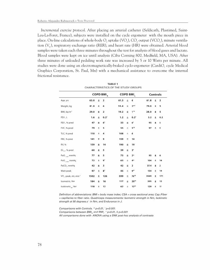
78
Incremental exercise protocol. After placing an arterial catheter (Seldicath, Plastimed, Saint-Leu-La-Foret, France), subjects were installed on the cycle ergometer with the mouth piece in place. On-line calculations of whole-body O2 uptake (VO2), CO2 output (VCO2), minute ventila-tion (VE), respiratory exchange ratio (RER), and heart rate (HR) were obtained. Arterial blood samples were taken each three minutes throughout the test for analysis of blood gases and lactate. Blood samples were kept on ice until analysis (Ciba Corning 800, Medfi eld, MA, USA). After three minutes of unloaded pedaling work rate was increased by 5 or 10 Watts per minute. All studies were done using an electromagnetically-braked cyclo-ergometer (CardiO2 cycle Medical Graphics Corporation, St. Paul, Mn) with a mechanical assistance to overcome the internal frictional resistance.
Roberto Alejandro Rabinovich • Tesis Doctoral
Defi nition of abbreviations: BMI = body mass index; CSA = cross sectional area; Cap./Fiber = capillaries to fi ber ratio. Quadriceps measurements: Isometric strength in Nm, Isokinetic strength at 90 degrees.s-1 in Nm, and Endurance in J.
Comparisons with Controls. * p<0.01, † p<0.001. Comparisons between BMINComparisons between BMINComparisons between BMI and BMIN and BMIN L and BMIL and BMI . ‡ p<0.01, ‡ p<0.01, ‡ § p<0.001All comparisons done with ANOVA using a SNK post-hoc analysis of contrasts
COPD BMIN
COPD BMIL
Age, yrs 63.0 ±± 2 61.3 ±± 6 61.8 ±± 2
Weight, kg 81.0 ±± 6 55.4 ±± 2*‡ 79.0 ±± 5
BMI, kg/m2 29.0 ±± 2 19.2 ±± 1†§ 28.0 ±± 5
FEV1, L 1.6 ±± 0.2† 1.2 ±± 0.2† 3.2 ±± 0.2
FEV1, % pred 47 ±± 6† 33 ±± 6† 95 ±± 5
FVC, % pred 79 ±± 5 55 ±± 5†§ 97 ±± 5
TLC, % pred 110 ±± 4 108 ±± 6
FRC, % pred 141 ±± 9 159 ±± 14
RV, % 159 ±± 14 190 ±± 19
DLCO
, % pred 60 ±± 5 39 ±± 3‡
PaO2, rest
mmHg 77 ±± 5 73 ±± 2* 95 ±± 6
PaO2, exer
mmHg 72 ±± 9* 63 ±± 4* 104 ±± 14
PaCO2, mmHg 42 ±± 3 42 ±± 2 37.4 ±± 2
Watt peak 97 ±± 8† 45 ±± 9‡§ 154 ±± 14
VO2, peak, mL.min-1 1502 ±± 126 599 ±± 76‡§ 2049 ±± 171
Isometric, Nm 184 ±± 16 117 ±± 20†‡ 205 ±± 12
Isokinetic90
, Nm 118 ±± 12 62 ±± 12†‡ 129 ±± 11
Controls
TABLE 1 CHARACTERISTICS OF THE STUDY GROUPS
79
Muscle StrengthMeasurements of maximal voluntary contractions of the left quadriceps were made during
static (isometric) and dynamic (isokinetic) contractions against an isokinetic system (Cybex 6000. Lumex Inc., Ronkonkoma, NY, USA). After a previous muscle warm up peak extension torque was evaluated at 60 degrees of knee fl exion, the best of fi ve repetitions was taken for the analysis. Isokinetic strength test consisting on a fi ve repetitions at the following different angular speeds (60, 90, 120, 180 and 240 degrees s-1). The best peak torque and peak power were taken for the analysis. Also an endurance test was carried out in all patients. This test consisted in a set of 30 knee extensions at a speed of 90 deg s-1: The total work done during the repetitions was used to analyse the quadriceps endurance. To ensure the maximal muscle strength in each test, rest periods were introduced between them. Reference values for quadriceps force were used24.
Muscle structureThe muscle was embedded in paraffi n and ten-micrometer thick sections were cut, varying
the inclination of the holder by 5° increments until the minimum fi ber cross-sectional area wasobtained,which was defi ned as truly transverse25;28. Consecutive cross-sections were processed by immunohistochemical techniques using monoclonal antibodies directed against myosin heavy chain (MyHC) isoforms type I and type II (MHCs and MHCf clones; Biogenesis, New Fields, Poole, UK). The fi ber cross-sectional area (CSA), mean least diameter, and proportions of type I and IIfi bers were assessed using a light microscope (OLYMPUS, SeriesBX50F3; Olympus Optical Co., Hamburg, Germany) coupled with an image-digitizing camera (Pixera Studio, Version 1.2; Pixera Corporation, Los Gatos, CA) and a morphometry program (NIH IMAGE, Version 1.60). At least 100 fi bers were measured from each biopsy25At least 100 fi bers were measured from each biopsy25At least 100 fi bers were measured from each biopsy . Fiber diameters between 40 and 80 µm were considered normal26;29.
Mitochondrial respirationMitochondrial were obtained by percoll centrifugation gradient30. The rate of oxygen consump-
tion was measured using Clark-type oxygen electrode (Hansatech Instruments Limited, Norfolk-UK) within one hour after completion of mitochondrial isolation31. The respiratory function was analyzed in medium containing 225 mM sucrose, 5 mM MgCl2, 10 mM KH2PO4, 20 Mm KCl, 10 mM Tris, and, 5mM HEPES at 25oC continuously stirred, using an electromagnetic stirrer and bar fl ea. The mitochondrial suspension was added to the reaction medium and ADP-limited respiration (state 4) was initiated by the addition of (13.5 mM) succinic acid in absence of ADP. Release of state 4 was obtained by the addition of (0.63 mM) ADP, a phosphate acceptor (state 3). Oxidative phosphorylation effi ciency was assessed by acceptor control ratio (ACR) by dividing the slope of state 3 to the slope of state 432.
Trabajos Originales
80
Assessment of muscle redox statusFresh muscle samples were homogenized in a buffer consisting in 20 mM Tris, 0.25 M sucrose,
40 mM KCl, 2 mM EGTA and 1mg/ml bovine serum albumin (BSA) and 500 μl were separated by glutathione concentration assessment. The rest of the sample served to mitochondrial isola-tion by percoll centrifugation gradient30. In both fractions (homogenate and mitochondria), the two molecular forms of glutathione, reduced (GSH) and oxidized gluthatione (GSSG) were ob-tained by high-performance liquid chromatography (HPLC) as described previously in detail33. An HPLC equipped with a 3-aminopropyl column and an UV spectrophotometric detector (365 nm) was used. GSH and GSSG standards were used to quantify glutathione concentrations.
Training program Patients exercised on a cycle ergometer (Jaeger ER 550; Wüerzburg, Germany) fi ve days per
week during three weeks. During the cycling sessions, HR was continuously monitored (Sport-Tester PE 3000 System, Polar Electro, Kemple, Finland). Patients trained during one-hour sessions for at least an effective period of 30 min. Training sessions were split into small blocks of 2-5 min of continuous cycling which allowed achievement of high intensity exercise. Initial work rates were programmed between the 60 and 75% of the Wpeak until reaching 95% of the Wpeak at the end of the period.
Data analysisResults are expressed as mean±SEM. Multiple comparisons were performed using ANOVA or
a Kruskal-Wallis test depending upon the distribution of variables. Training effects were analyzed using Student paired-t test. Pearson regression analysis was used to assess univariate correlations. Stepwise multiple regression analyses were used to explore associations between ACR and dif-ferent baseline variables. Also, we carried out multiple regression analyses of training-induced glutathione changes as dependent variable and different covariates, namely, FEV1, peak oxygen uptake, FFM index (FFMi), muscle strength and endurance, and training-induced changes at peak exercise. A p value lower than 0.05 was taken as statistically signifi cant.
RESULTS
Anthropometric and functional characteristics of COPD patients (BMIN
n= 8, and BMIL
n= 7) and controls (n= 7) are indicated in Table 1. As expected, FFMi was signifi cantly lower in BMI
L
(17±1 kg.m-2) than in both BMIN (21±1 kg.m-2) and controls (22±1 kg.m-2) (p<0.001). None of the
patients with normal BMI showed abnormally low FFMi. Patients with normal BMI showed high-er FEV
1er FEV
1er FEV % predicted than those with reduced muscle mass. The distribution of patients according to the GOLD1 classifi cation was as follows: 4 in stage II (3 BMI
N and 1 BMI
L); 6 in stage III (4
BMIN and 2 BMI
L), and 5 in stage IV (1 BMI
N and 4 BMI
L). The two groups of patients showed
Roberto Alejandro Rabinovich • Tesis Doctoral
81
a similar degree of mild arterial hypoxemia, but exercise-induced hypoxemia was slightly more pronounced in BMI
L. Likewise, BMI
L had lower exercise tolerance (VO
2 peak 11±1 ml.kg-1.min-1)
than both BMIN (19±2 ml.kg-1.min-1) and controls (26±2 ml.kg-1.min-1) (ANOVA, p<0.0001).
Quadriceps muscle strength and endurance were also lower in BMIL compared to the other two groups (Table 1). Muscle-fi ber CSA showed a trend to be higher (24%) in controls than in COPD pa-tients, with identical results between BMIN and BMIL. But no statistically signifi cant differences among groups were detected. It is of note that muscle endurance normalized by CSA (endurance/CSA ratio) was signifi cantly lower in BMIL (73±17 J/µm2) than in both BMIN (94±6 J/µm2) and C (90±9 J/µm2), (ANOVA, p<0.001). The ratio muscle strength/CSA was similar among groups (data not shown).Figure 1 indicates that BMIL presented earlier arterial blood lactate ([La]) release than both BMIN and controls. As expected, exercise-induced increases in [La] showed a strong association with VO2peak (r= 0.84, p<0.0001) and Watts peak (r= 0.85, p<0.0001). After 3 weeks, a clear training-induced effect on ∆[La] was showed in COPD (BMIN and BMIL) patients. In BMIL, [La] fell from 3.5±0.4 nmol.L-1 to 2.1±0.2 nmol.L-1 at iso-VO2 (100% of pre-training peak VO2, p=0.05) which corresponded to a workload of 45 W. Likewise, [La] decreased from 7.9±0.8 nmol.L-1 to 5.2±0.6 nmol.L-1 after training in BMIN (p<0.05). No training-induced changes in [La] were observed in controls (from 8.7±1.4 to 8.1±0.8 nmol.L-1). Early [La] release persisted in BMIL after training.
Mitochondrial respirationIndividual fi gures for ACR at baseline are illustrated in Figure 2. On average, ACR at baseline
was signifi cantly lower in BMIL (2.2±1) than in both BMIN (5.3±1) and in healthy sedentary subjects (8.2±1) (ANOVA p<0.01). Abnormal ACR (< 3) was seen in fi ve out of seven BMIL and in only two out of eight BMIN, whereas it was absent in the controls. Arterial PO2 correlated sig-nifi cantly with ACR, both at rest (r=0.70, p=0.0016) and at peak exercise (r=0.63, p=0.009). Ex-ercise-induced increase in [La] from rest to 45 watts was negatively correlated with mitochondrial respiratory function (r=-0.60, p=0.01). Moreover, a positive correlation was shown between ACR and the endurance/CSA ratio both before (r=0.40, p=0.07) and after (r=0.50, p=0.04) training.
After training, no signifi cant changes in ACR (∆ACR) were observed in the three subsets (BMIL, 2±1; BMIN, 1±2; and, controls -1±2).
Muscle glutathione levels No differences among groups were observed in muscle homogenate concentrations of GSH
(BMIL 7±1; BMIN, 9±2; and, controls 5±1 nmol/mg), GSSG and total glutathione at baseline. After training, a pattern of GSH consumption was observed in BMIN and controls. In these two subsets, we observed a marked individual variability. In contrast, none of the BMIL patients showed training-induced GSH changes (Figure 3). The multiple regression analyses indicat-ed that only FFMi was signifi cantly associated with training-induced glutathione adaptations:
Trabajos Originales
82
∆GSH (r=-0.46, p=0.056), ∆GSSG (r=-0.55, p=0.018) and ∆Total glutathione (r=-0.47, p=0.046). No differences in mitochondrial glutathione levels were observed among groups at baseline and after training.
DISCUSSION
The current study provides original evidence of mitochondrial respiratory chain uncoupling in COPD patients with reduced muscle mass (Figure 2), not seen in BMIN, nor in healthy seden-tary subjects. The BMIL group presented low muscle endurance after correction by muscle mass (muscle endurance/CSA ratio) which showed a moderate but signifi cant correlation with ACR before and after training.
Potential mechanisms Although speculative, it can be hypothesized that electron transport chain dysfunction in
BMIL may indicate an abnormal mitochondrial adaptation to long-term repeated episodes of cell hypoxia. The association between ACR and arterial hypoxemia seems to support this contention. We acknowledge, however, that causality can not be established from the current study.
Relationships between poor arterial oxygenation and systemic manifestations of COPD, namely low FFMi and high TNFα plasma levels, have been reported by other authors34,35. Moreover, abnormal mitochondrial respiratory chain function has been identifi ed as a contributing mechanism to limb muscle dysfunction in patients with tissue hypoxia due to chronic peripheral arterial obstruction36;37.
In addition to potential perturbations of the main determinants of systemic oxygen fl ow, COPD patients are prone to present low muscle O2 conductance38 from capillaries to mitochondria and muscle O2 supply-O2 demand heterogeneities. These two factors may generate cell hypoxia during exercise, even in absence of overt arterial hypoxemia. In spite of the fact that low O2 conductance and inequality of O2 supply-O2 demand can not be directly assessed, there is evidence supporting a role for muscle hypoxia during sub-maximal work rate.
A fi rst indirect set of data comes from the abnormally high muscle O2 fl ow at a given sub-maximal work rate seen in COPD patients39;40. It most likely indicates augmented cellular oxy-gen demand due to mechanical ineffi ciency, not explainable by abnormal ergonomics during exercise39;40. It has been recently suggested that nitroso-redox imbalance in the cardiovascular system may play a central role in different chronic disorders41-43 (congestive heart failure, COPD, diabetes) triggering tissue hypoxia due to mismatching between O2 fl ow-O2 demand. Further research, however, is still needed to elucidate the mechanisms by which red blood cells act as oxygen-responsive transducers of nitric oxide bioactivity, as well as to identify the clinical impact of this pathway.
Moreover, it is well known that in healthy sedentary young subjects44;45 mitochondrial oxidative capacity limits maximum oxygen uptake, at difference with athletes in whom VO2max is clearly
Roberto Alejandro Rabinovich • Tesis Doctoral
83
limited by O2 transport46 transport46 transport . Likewise, it seems reasonable to hypothesize that muscle energy demand during heavy exercise in sedentary old COPD patients may exceed mitochondrial oxidative ca-pacity leading to abnormal exercise-induced oxidative stress seen in these patients9,47-50. Because mitochondria are the major consumers of molecular oxygen, they stand as one of the most impor-tant generators of reactive oxygen species therefore constituting potential targets for therapeutic interventions in disease states in which oxidative stress originates from these organelles.
Maltais et al.51,52 reported altered muscle oxidative capacity at the level of Krebs cycle in COPD patients, irrespective of their muscle mass status. These abnormalities partly reversed after endurance training51. The activity of oxidative enzymes of the Krebs cycle is usually associated with mitochondrial volume and the changes in these two characteristics may refl ect muscle plasticity to physical training. Despite our training program only lasted three weeks, we showed clear effects in the patients, as indicated by a signifi cant post-training fall in arterial [La] at iso-VO2. This observation is consistent with previous studies14;15 showing short-term training effects at cellular level.
Muscle glutathione levels At difference with other disorders15, the current research showed that GSH information obtained
in muscle homogenate was not improved by measurements performed in isolated mitochondria. This is a relevant methodological fi nding because it simplifi es further studies in the fi eld.
The current study does not reproduce the pattern of muscle GSH adaptations to training seen in a previous report9 wherein healthy sedentary subjects signifi cantly increased muscle GSH levels after 8-weeks high intensity endurance training. Muscle redox adaptations were modest in BMIN
and absent in BMIL9;10. Precisely, the latter subset of patients showed a training-induced decrease in
muscle GSH10 after 8-weeks. In the current study, we confi rm that BMIL after a 3-weeks endurance program show different
training-induced GSH changes than both BMIN and controls (Figure 3). It can be speculated that, at this time point (3-weeks training), glutathione consumption seen in BMIN and in controls is due to exercise-induced ROS production. Mitochondrial respiratory chain abnormalities in BMIL
might be transiently associated with lower rate of ROS production, but the scenario observed in BMIL after 3-weeks of training is not sustainable according to the previous results obtained after an 8-weeks training program9;10. With the current results, the analysis of the time course of GSH adaptations becomes an issue to be explored with a specifi c study design including muscle redox measurements carried out before training, at mid-term and after completion of an 8-weeks training program in the same set of subjects.
Methodological aspects The rather invasive approach of the study including two open muscle biopsies accounts for the
small sample size which may constitute a limitation for some of the areas explored. In particular, the lack of signifi cant post-training changes in ACR should be taken cautiously. A type II error
Trabajos Originales
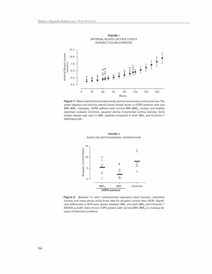
84
15
10
5
0
Acc
epto
r C
on
tro
l Rat
io
BMIN BMIL
COPD patients
Controls
FIGURE 2BASELINE MITOCHINDRIAL RESPIRATION
12.5
10.0
7.0
5.0
2.5
0.0
Art
eria
l Blo
od
Lac
tate
(mm
ol/
L)
0 20 40 60 80 100 120 140
Watts
FIGURE 1ARTERIAL BLOOD LACTATE LEVELS
DURING CYCLING EXERCICE
Figure 1. Mean arterial blood lactate levels during incremental cycling exercise. The graph displays pre-training arterial blood lactate levels in COPD patients with low BMI (BMILBMI (BMILBMI (BMI , triangles), COPD patients with normal BMI (BMIN, triangles), COPD patients with normal BMI (BMIN, triangles), COPD patients with normal BMI (BMI , circles), and healthy N, circles), and healthy N
sedentary subjects (Controls, squares) during incremental cycling exercise. Early lactate release was seen in BMILlactate release was seen in BMILlactate release was seen in BMI patients compared to both BMIN patients compared to both BMIN patients compared to both BMI and Controls (* N and Controls (* N
ANOVAp<0.05).
Figure 2. Baseline “in vitro” mitochondrial respiratory chain function. Individual (circles) and mean group (solid lines) data for acceptor control ratio (ACR). Signifi -cant differences in ACR were shown between BMILcant differences in ACR were shown between BMILcant differences in ACR were shown between BMI and both BMIN and both BMIN and both BMI and Controls (* N and Controls (* N
ANOVA p<0.05). Data of one COPD patient with normal BMI (BMINANOVA p<0.05). Data of one COPD patient with normal BMI (BMINANOVA p<0.05). Data of one COPD patient with normal BMI (BMI ) is missing be-cause of technical problems.
Roberto Alejandro Rabinovich • Tesis Doctoral
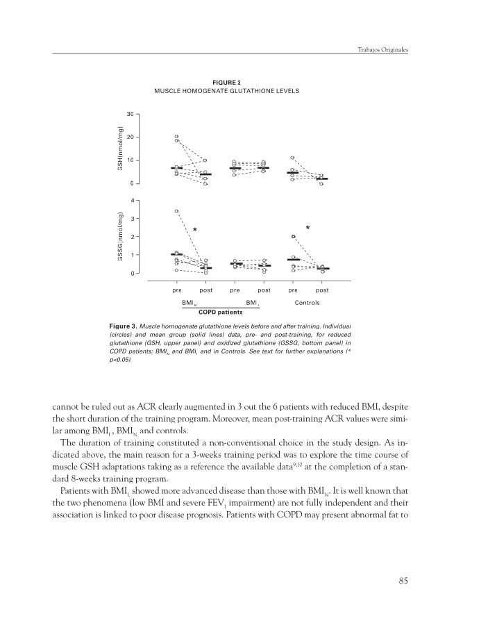
85
cannot be ruled out as ACR clearly augmented in 3 out the 6 patients with reduced BMI, despite the short duration of the training program. Moreover, mean post-training ACR values were simi-lar among BMIL, BMIN and controls.
The duration of training constituted a non-conventional choice in the study design. As in-dicated above, the main reason for a 3-weeks training period was to explore the time course of muscle GSH adaptations taking as a reference the available data9;10 at the completion of a stan-dard 8-weeks training program.
Patients with BMIL showed more advanced disease than those with BMIN. It is well known that the two phenomena (low BMI and severe FEV1 impairment) are not fully independent and their association is linked to poor disease prognosis. Patients with COPD may present abnormal fat to
30
20
10
0
GS
H(n
mo
l/m
g)
FIGURE 3MUSCLE HOMOGENATE GLUTATHIONE LEVELS
4
3
2
1
0
GS
SG
(nm
ol/
mg
)
pre post pre post pre post
ControlsBMIN BMIL
COPD patients
Figure 3. Muscle homogenate glutathione levels before and after training. Individual (circles) and mean group (solid lines) data, pre- and post-training, for reduced glutathione (GSH, upper panel) and oxidized glutathione (GSSG, bottom panel) in COPD patients: BMINCOPD patients: BMINCOPD patients: BMI and BMIN and BMIN L and BMIL and BMI and in Controls. See text for further explanations (* p<0.05).
Trabajos Originales
86
lean body mass ratio such that BMI may not properly indicate patient’s muscle mass. This fact does not seem to alter the interpretation of the current results since none of the COPD patients with normal BMI presented abnormally low FFMi. The relative contribution of muscle wasting on the study fi ndings was estimated by multiple regression analysis taking FFMi as covariate.
One intriguing fi nding in the study was that functional abnormalities (ACR) were not accompa-nied by substantial alterations in muscle structure. We understand that the analysis of muscle struc-ture-function relationships requires further attention due to several confounding factors, namely physical de-conditioning and geographical-ethnic differences that may partly explain the heteroge-neity of the results reported in the literature.
In summary, the current investigation clearly identifi es mitochondrial respiratory chain un-coupling in COPD patients with low muscle mass. Whether the phenomenon occurs only in a subset of susceptible advanced COPD patients or it is a general feature of end-stage disease needs to be elucidated. Further research is also required to explore the underlying post-transcriptional mechanisms that are likely shared by other chronic conditions.
ACKNOWLEDGMENTS
The authors would like to thank Felip Burgos, Conxi Gistau and Jose Luis Valera and all the technical staff of the Lung Function Laboratory for their skillful support during the study. Elena Gimeno, Nestor Sanchez, Carlos Hernando and Isaac Diaz from EUIF Blanquerna are acknowl-edged for their outstanding work supervising the training sessions.
1. 2004. Global Initiative for Chronic Obstructive Lung Disease - Updated 2004.www.goldcopd.com.
2. Gosselink, R., T. Troosters, and M. Decramer. 1996. Peripheral muscle weakness contributes to exercise limitation in COPD. Am J Respir Crit Care Med 153:976-980.
3. Saey, D., R. Debigare, P. Leblanc, M. J. Mador, C. H. Cote, J. Jobin, and F. Maltais. 2003. Contractile leg fatigue after cycle exercise: a factor limiting exercise in patients with chronic obstructive pulmonary disease. Am.J.Respir.Crit Care Med. 168:425-430.
4. ATS/ERS statement. 1999. “Skeletal Muscle Dysfunction in Chronic Obstructive Pulmonary Disease”. Am J Respir Crit Care Med 159:S1-S40.
5. Agusti, A. G., A. Noguera, J. Sauleda, E. Sala, J. Pons, and X. Busquets. 2003. Systemic effects of chronic obstructive pulmonary disease. Eur.Respir.J. 21:347-360.
6. Wouters, E. F., E. C. Creutzberg, and A. M. Schols. 2002. Systemic effects in COPD. Chest 121:127S-130S.
7. Jones, P. W. 1995. Issues concerning health-related quality of life in COPD. Chest 107 Suppl.:187S-193S.
8. Connors, A. F., N. V. Dawson, C. Thomas, F. H. Harrel Jr, N. Desbiens, W. J. Fulkerson, P. Kussin, P. Bellamy, L. Goldman, and W. A. Knaus. 1996. Outcomes following acute exacerbation of severe chronic obstructive lung disease. Am J Respir Crit Care Med 154:959-967.
9. Rabinovich, R. A., E. Ardite, T. Troosters, N. Carbó, J. Alonso, J. M. Gonzalez de Suso, J. Vilaró, J. A. Barberà, M. Figueras, J. M. Argiles, J. C. Fernandez Checa, and J. Roca. 2001. Reduced muscle redox capacity after endurance training in COPD patients. Am J Respir Crit Care Med164:1114-1118.
References
Roberto Alejandro Rabinovich • Tesis Doctoral
87
10. Rabinovich, R. A. and J. Roca. 2002. Is COPD also a disease of skeletal muscle?, author reply. Am.J Respir Crit Care Med.165:1337-1338.
11. Chandel, N. S., W. X. Trzyna, D. S. McClintock, and P. T. Schumacker. 2000. Role of oxidants in NF-kappa B activation and TNF-alpha gene transcription induced by hypoxia and endotoxin. J Immunol 165:1013-1021.
12. Ardite, E., J. A. Barbera, J. Roca, and J. C. Fernandez-Checa. 2004. Glutathione depletion impairs myogenic differentiation of murine skeletal muscle C2C12 cells through sustained NF-kappaB activation. Am.J Pathol. 165:719-728.
13. Fernandez-Checa, J. C., A. Colell, and C. Garcia-Ruiz. 2002. S-Adenosyl-L-methionine and mitochondrial reduced glutathione depletion in alcoholic liver disease. Alcohol27:179-183.
14. Chen, Y. W., M. J. Hubal, E. P. Hoffman, P. D. Thompson, and P. M. Clarkson. 2003. Molecular responses of human muscle to eccentric exercise. J Appl.Physiol 95:2485-2494.
15. Spina, R. J., M. M. Chi, M. G. Hopkins, P. M. Nemeth, O. H. Lowry, and J. O. Holloszy. 1996. Mitochondrial enzymes increase in muscle in response to 7-10 days of cycle exercise. J Appl.Physiol 80:2250-2254.
16. Bickel, C. S., J. Slade, E. Mahoney, F. Haddad, G. A. Dudley, and G. R. Adams. 2005. Time course of molecular responses of human skeletal muscle to acute bouts of resistance exercise. J Appl.Physiol 98:482-488.
17. Celli, B. R. and W. MacNee. 2004. Standards for the diagnosis and treatment of patients with COPD: a summary of the ATS/ERS position paper. Eur Respir J 23:932-946.
18. Baarends, E. M., A. M. Schols, W. D. Van Marken Lichtenbelt, and E. F. Wouters. 1997. Analysis of body water compartments in relation to tissue depletion in clinically stable patients with chronic obstructive pulmonary disease. Am J Clin Nutri 65:88-94.
19. Heijdra, Y. F., V. Pinto-Plata, R. Frants, J. Rassulo, L. Kenney, and B. R. Celli. 2003. Muscle strength and exercise kinetics in COPD patients with a normal fat-free mass index are comparable to control subjects. Chest 124:75-82.
20. Schols, A. M. and E. F. Wouters. 2000. Nutritional abnormalities and supplementation in chronic obstructive pulmonary disease. Clin.Chest Med. 21:753-762.
21. Roca, J., F. Burgos, J. Sunyer, M. Saez, S. Chinn, J. M. Antó, R. Rodriguez-Roisin, Ph. H. Quanjer, D. Nowak, and P. Burney. 1998. References values for forced spirometry. Eur Respir J 11:1354-1362.
22. Roca, J., F. Burgos, J. A. Barberà, J. Sunyer, R. Rodriguez-Roisin, J. Castellsague, J. Sanchis, J. M. Antó, P. Casan, and J. L. Clausen. 1998. Prediction equations for plethysmografi c lung volumes. Respir.Med. 92:454-460.
23. Roca, J. and Whipp, B. J. eds., 1997. Clinical Exercise Testing. Eur Respir Monograph. European Respiratory society Journals Ltd, Sheffi eld, UK, p 164.
24. Decramer, M., L. M. Lacquet, R. Fagard, and P. Rogiers. 1994. Corticosteroids contribute to muscle weakness in chronic airfl ow obstruction. Am J Respir Crit Care Med 150:11-16.
25. Dubowitz, V. and M. Brooke 1973. Muscle biopsy: a modern approach. In V. Dubowitz and M. Brooke, editors Major Problems in Neurology Saunders, London. p 74-85.
26. Hards, J. M., W. D. Reid, R. L. Pardy, and P. D. Pare. 1990. Respiratory muscle fi ber morphometry. Correlation with pulmonary function and nutrition. Chest 97:1037-1044.
27. Kotler, D. P., S. Burastero, J. Wang, and R. N. Pierson, Jr. 1996. Prediction of body cell mass, fat-free mass, and total body water with bioelectrical impedance analysis: effects of race, sex, and disease. Am J Clin.Nutr. 64:489S-497S.
28. Poole, D. C., R. L. Lieber, and O. Mathieu-Costello. 1994. Myosin and actin fi lament lengths in diaphragms from emphysematous hamsters. J.Appl.Physiol 76:1220-1225.
29. Brooke, M. H. and W. K. Engel. 1969. The histographic analysis of human muscle biopsies with regard to fi ber types. 4. Children’s biopsies. Neurology 19:591-605.
30. Dunkley, P. R., J. W. Heath, S. M. Harrison, P. E. Jarvie, P. J. Glenfi eld, and J. A. Rostas. 1988. A rapid Percoll gradient procedure for isolation of synaptosomes directly from an S1 fraction: homogeneity and morphology of subcellular fractions. Brain Res. 441:59-71.
31. Colell, A., C. Garcia-Ruiz, J. M. Lluis, O. Coll, M. Mari, and J. C. Fernandez-Checa. 2003. Cholesterol impairs the adenine nucleotide translocator-mediated mitochondrial permeability transition through altered membrane fl uidity. J Biol.Chem. 278:33928-33935.
32. Ferreira, J., L. Gil, A. Stutzin, and F. Orrego. 1985. Effects of guanethidine on electron transport and proton movements in rat heart, brain and liver mitochondria. Biochem.Pharmacol.34:2507-2512.
33. Fariss, M. W. and D. J. Reed. 1987. High-performance liquid chromatography of thiols and disulfi des: dinitrophenol derivatives. Methods Enzymol 143:101-109.
34. Takabatake, M., H. Nakamura, S. Abe, T. Hino, H. Saito, H. Yuki, S. Kato, and H. Tomoike. 1999. Circulating leptin in
Trabajos Originales
88
patients with chronic obstructive pulmonary disease. Am J Respir Crit Care Med 159:1215-1219.
35. Takabatake, M., H. Nakamura, H. Abe, S. Inoue, T. Hino, H. Saito, H. Yuki, S. Kato, and H. Tomoike. 2000. The Relationship between Chronic Hypoxemia and Activation of the Tumor Necrosis Factor-a System in Patients with Chronic Obstructive Pulmonary Disease. Am J RespirCrit CareMed 161:1179-1184.
36. Brass, E. P., H. Wang, and W. R. Hiatt. 2000. Multiple skeletal muscle mitochondrial DNA deletions in patients with unilateral peripheral arterial disease. Vasc.Med 5:225-230.Vasc.Med 5:225-230.Vasc.Med
37. Pipinos, I. I., V. G. Sharov, A. D. Shepard, P. V. Anagnostopoulos, A. Katsamouris, A. Todor, K. A. Filis, and H. N. Sabbah. 2003. Abnormal mitochondrial respiration in skeletal muscle in patients with peripheral arterial disease. J Vasc.SurgVasc.SurgVasc.Sur . 38:827-832.
38. Simon, M., P. Leblanc, J. Jobin, M. Desmeules, M. J. Sullivan, and F. Maltais. 2001. Limitation of lower limb VO(2) during cycling exercise in COPD patients. J Appl.Physiol 90:1013-1019.
39. Sala, E., J. Roca, R. M. Marrades, J. Alonso, J. M. Gonzalez de Suso, A. Moreno, J. Barbera, J. Nadal, Ll. Jover, R. Rodriguez-Roisin, and P. D. Wagner. 1999. Effects of endurance training on skeletal muscle bioenergetics in chronic obstructive pulmonary disease. Am J Respir Crit Care Med 159:1726-1734.
40. Richardson, R. S., B. T. Leek, T. P. Gavin, L. J. Haseler, S. R. Mudaliar, R. Henry, O. Mathieu-Costello, and P. D. Wagner. 2004. Reduced mechanical effi ciency in chronic obstructive pulmonary disease but normal peak VO2 with small muscle mass exercise. Am.J.Respir.Crit Care Med. 169:89-96.
41. Hare, J. M. 2004. Nitroso-redox balance in the cardiovascular system. N.Engl.J Med 351:2112-2114.
42. Hare, J. M. 2003. Nitric oxide and excitation-contraction coupling. J Mol.Cell Cardiol. 35:719-729.
43. Singel, D. J. and J. S. Stamler. 2004. Blood traffi c control. Nature 430:297.
44. Sala, E., E. A. Noyszewski, J. M. Campistol, R. Marrades, S. Dreha, J. V. Torregrosa, J. S. Beers, P. D. Wagner, and J. Roca. 2001. Impaired muscle oxygen transfer in patients with chronic renal failure. Am J Physiol 280:R1240-R1248.
45. Haseler, L. J., A. P. Lin, and R. S. Richardson. 2004. Skeletal muscle oxidative metabolism in sedentary humans: 31P-MRS assessment of O2 supply and demand limitations. J Appl.Physiol 97:1077-1081.
46. Richardson, R. S., E. A. Noyszewski, K. F. Kendrick, J. S. Leigh, and P. D. Wagner. 1995. Myoglobin O2 desaturation during exercise: Evidence of limited O2 transport. J Clin Invest 96:1916-1926.
47. Miwa, S. and M. D. Brand. 2003. Mitochondrial matrix reactive oxygen species production is very sensitive to mild uncoupling. Biochem.Soc.Trans. 31:1300-1301.
48. Barreiro, E., J. Gea, J. M. Corominas, and S. N. Hussain. 2003. Nitric oxide synthases and protein oxidation in the quadriceps femoris of patients with chronic obstructive pulmonary disease. Am.J Respir Cell Mol.Biol. 29:771-778.
49. Koechlin, C., A. Couillard, D. Simar, J. P. Cristol, H. Bellet, M. Hayot, and C. Prefaut. 2004. Does oxidative stress alter quadriceps endurance in chronic obstructive pulmonary disease? Am.J Respir Crit Care Med. 169:1022-1027.
50. Agusti, A., M. Morla, J. Sauleda, C. Saus, and X. Busquets. 2004. NF-kappaB activation and iNOS upregulation in skeletal muscle of patients with COPD and low body weight. Thorax 59:483-487.
51. Maltais, F., P. Leblanc, C. Simard, J. Jobin, C. Bérubé, J. Bruneau, L. Carrier, and R. Belleau. 1996. Skeletal muscle adaptation to endurance training in patients with Chronic Obstructive Pulmonary Disease. Am J Respir Crit Care Med154:442-447.
52. Allaire, J., F. Maltais, J. F. Doyon, M. Noel, P. Leblanc, G. Carrier, C. Simard, and J. Jobin. 2004. Peripheral muscle endurance and the oxidative profi le of the quadriceps in patients with COPD. Thorax 59:673-678.
Roberto Alejandro Rabinovich • Tesis Doctoral
89
MANUSCRITO I
Reduced Muscle Redox Capacity after Endurance Training in Patients with Chronic Obstructive Pulmonary Disease
Después de ocho semanas de entrenamiento físico, los sujetos sanos participantes en este estudio como grupo control doblaron la concentración de GSH en el homogenado del vastus lateralis. Por el contrario, los pacientes con EPOC no aumentaron los niveles de GSH e incrementaron los de GSSG indicando que el entrenamiento físico disminuyó la capacidad antioxidante muscular.
Cabe señalar que los pacientes con EPOC incrementaron la expresión del γGCS-γGCS-γARNm en el vastus lateralis post-entrenamiento mientras que se observó el efecto opuesto en los sujetos sanos. A pesar de estas diferencias en los mecanismos de adaptación del sistema redox, tanto los pacientes con EPOC como los sujetos control mostraron un efecto benefi cioso del entrenamiento físico en lo que respecta a tolerancia al ejercicio (medida como incremento de la carga pico y consumo de oxígeno pico), y en la capacidad oxidativa muscular (medida con espectroscopia del 31P).
Resumen de resultados
90
MANUSCRITO II
Training Depletes Muscle Glutathione in COPD Patients with Low Body Mass Index
El análisis de los cambios del estado redox muscular post-entrenamiento separando los pacientes en dos grupos: índice de masa corporal bajo (BMIL) y BMI normal (BMIN), mostró que los primeros presentaban una depleción de los niveles musculares de GSH mientras que en los segundos el GSH presentaba una tendencia al incremento sin alcanzar la signifi cación estadística. Cabe señalar que el comportamiento del GSH muscular en los pacientes con índice de masa corporal normal se diferencia claramente del observado en los sujetos sedentarios sanos en los que los niveles de GSH post-enternamiento prácticamente se doblaron. Estos resultados demuestran que la alteración en la capacidad de adaptación del sistema del glutation es más exagerada en el grupo de pacientes BMIL. Nuevamente, ambos grupos de pacientes muestran un incremento de los niveles de γGCS-ARNm con el entrenamiento.γGCS-ARNm con el entrenamiento.γ
MANUSCRITO III
Increased Tumour Necrosis Factor-α Plasma Levels during Moderate-Intensity Exercise in COPD Patients
La expresión de ARNm de TNFα (ARNm-TNFα) se encuentra incrementada en el músculo esquelético periférico de pacientes con EPOC en comparación con sujetos sanos de igual edad y sexo. Si bien no se observaron diferencias en las concentraciones plasmáticas de TNFα entre pacientes y sujetos sanos, el ejercicio de moderada intensidad (40 % de la capacidad máxima de ejercicio sostenida durante 11 minutos) generó un incremento de los niveles plasmáticos de TNFα en los pacientes con EPOC
Roberto Alejandro Rabinovich • Tesis Doctoral
91
no observado en los sujetos control. El entrenamiento físico no produjo modifi caciones en los niveles de expresión muscular de ARNm-TNFα ni en las concentraciones plasmáticas de TNFα.
No se observaron diferencias en las concentraciones plasmáticas de interleucina-6 (IL-6), otra de las citocinas potencialmente relacionadas con la pérdida de masa muscular, ni en las concentraciones plasmáticas de los receptores hidrosolubles para el TNFα, entre pacientes con EPOC y sujetos controles. Tampoco se observaron cambios en las concentraciones plasmáticas de IL-6 o de los receptores para el TNFα en relación al ejercicio o al entrenamiento.
MANUSCRITO IV
Mitochondrial Electron Transport Chain Uncoupling in COPD Patients with Muscle Wasting
La funcionalidad de la cadena respiratoria mitocondrial (acceptor control ratio, ACR) presentó una alteración signifi cativa en los pacientes BMIL en comparación con los BMIN y los sujetos sanos sedentarios. El ACR se correlacionó de forma altamente signifi cativa con el nivel de PaO2 tanto a nivel basal como al fi nalizar el ejercicio incremental con cicloergómetro. También se correlacionó de forma inversa con el incremento precoz de ácido láctico durante el ejercicio.
Se encontraron diferencias en la adaptación del estado redox muscular a las tres semanas de entrenamiento entre el grupo BMIL y los pacientes BMIN y el grupo Control. Mientras que los pacientes BMIL presentaron una respuesta uniforme sin cambios en el estado redox muscular en relación al entrenamiento, tanto los pacientes BMIN como el grupo control presentaron un patrón de comportamiento similar con una gran variabilidad inter-individual. En estos dos grupos (BMIN y controles) se observó una disminución de los niveles de GSSG concomitantemente con una tendencia a disminuir la concentración de GSH y glutation total (GSH+GSSG). Se
Resumen de resultados
92
utilizaron análisis de regresión lineal múltiple para explorar las asociaciones entre los cambios producidos por el entrenamiento en las concentraciones de GSH, GSSG y glutation total como variables dependientes y diversas variables independientes relacionadas con la función pulmonar, tolerancia al ejercicio y composición corporal. Todas las variables independientes incluidas en los modelos fueron excluidas excepto el índice de FFM (FFMi) que mostró una asociación consistente con los cambios en las concentraciones de GSH, GSSG y glutation total inducidas por el entrenamiento.
╯
Roberto Alejandro Rabinovich • Tesis Doctoral
93
EFECTOS DEL ENTRENAMIENTO SOBRE EL SISTEMA GLUTATION
En el primer trabajo de esta tesis se demuestra que la regulación del sistema del glutation se encuentra alterada en el músculo esquelético periférico de pacientes con EPOC. El incremento de la capacidad antioxidante evidenciada en el grupo control debe ser interpretado como un mecanismo de adaptación al estrés oxidativo provocado por las sucesivas sesiones de ejercicio que componen el programa de entrenamiento. Por el contrario, el entrenamiento físico disminuyó la capacidad antioxidante en el músculo de los pacientes con EPOC. Asímismo, el incremento de la expresión del γGCS-ARNm, enzima que constituye el paso limitante en la síntesis de GSH y es γGCS-ARNm, enzima que constituye el paso limitante en la síntesis de GSH y es γregulada alostéricamente por éste, no se traduce en un incremento de la concentración de GSH.
Aunque en la síntesis de novo de GSH intervienen diversos mecanismos, de hecho existen dos factores que infl uyen de forma predominante: a) la cantidad y actividad de la γGCS presente en la célula, regulada por feed-back negativo que ejerce el γGCS presente en la célula, regulada por feed-back negativo que ejerce el γpropio GSH sobre la misma; y, b) la biodisponibilidad de substratos, particularmente L-cisteína.
El incremento de la expresión del γGCS-ARNm es indicativa de que la transcripción γGCS-ARNm es indicativa de que la transcripción γdel gen tiene lugar en este caso. Sin embargo los datos disponibles en el presente estudio no permiten dilucidar el tipo de alteración post-transcripcional que impide que esta señal se traduzca en un incremento de la concentración de GSH. Basados en datos de estudios anteriores, podemos especular que existe alguna alteración a nivel de
Discusión
94
la disponibilidad de sustratos necesarios para la adecuada síntesis de GSH214;215.A pesar de que la expresión de γGCS-ARNm disminuye con el entrenamiento γGCS-ARNm disminuye con el entrenamiento γ
en los sujetos control, estos incrementan los niveles de GSH en el homogenado del vastus lateralis. La alta correlación entre los cambios de γGCS-ARNm y GSH γGCS-ARNm y GSH γprovocados por el entrenamiento indicaría que el incremento en los niveles de GSH podría reprimir la transcripción de la γGCS en este grupo.γGCS en este grupo.γ
En la introducción se ha mencionado la existencia de un incremento del estrés oxidativo sistémico en pacientes con EPOC, especialmente durante las exacerbaciones. El papel potencial de este fenómeno en la etiopatogenia de la disfunción muscular en la EPOC, adquiere especial relevancia en situaciones en las que la regulación de los mecanismos antioxidantes como el sistema del glutation se encuentra alterada como se demuestra en este primer trabajo de la tesis. En este contexto, la reacción de moléculas pro-oxidantes con lípidos y proteínas194 puede interferir en el funcionamiento adecuado del músculo (adecuada contractilidad) y alterar los fenómenos regenerativos musculares dando lugar a la pérdida de masa muscular observada en algunos de estos pacientes. El hecho de que la n-acetil cisterna (NAC), un precursor de la síntesis de glutation, mejore la resistencia muscular en pacientes con EPOC previniendo el estrés oxidativo inducido por el ejercicio216 es indicativo del papel de los ROS en la disfunción muscular esquelética de los pacientes con EPOC. Asimismo, el papel benefi cioso de la NAC daría soporte a la hipótesis de que el défi cit de sustratos constituiría un factor relevante en la disfunción muscular de la EPOC.
Por otro lado, las modifi cación de proteínas estructurales celulares194 generada por los ROS, puede mediar mecanismos de proteólisis muscular191;192 favoreciendo la perdida de masa muscular.
RELACIONES ENTRE ESTRÉS OXIDATIVO Y PÉRDIDA DE MASA MUSCULAR
En el segundo trabajo de la tesis se incrementó el número de pacientes con EPOC del primer estudio a expensas de enfermos con índice de masa corporal bajo (BMIL). El objetivo era efectuar un análisis diferenciado de las adaptaciones redox al entrenamiento muscular entre BMIL y BMIN. Este estudio demuestra que la alteración en la capacidad de adaptación del sistema del glutation es más exagerada en el grupo
Roberto Alejandro Rabinovich • Tesis Doctoral
95
de pacientes con BMIL. La pérdida de masa muscular en estos pacientes probablemente refl eja la activación de mecanismos fi siopatológicos ya mencionados en la introducción. El mantenimiento de los niveles de GSH constituiría una importante estrategia celular para prevenir el daño muscular producido por el incremento de radicales libres potencialmente citotóxicos. La alteración en los mecanismos de adaptación de sistemas antioxidantes pone en evidencia la desventaja del músculo de estos pacientes, particularmente del subgrupo BMIL. Éste hallazgo abre nuevos interrogantes en lo que respecta al potencial efecto deletéreo del entrenamiento muscular en los pacientes con pérdida de masa muscular.
El ejercicio localizado en miembros inferiores incrementa los niveles plasmáticos de TBARs (thiobarbituric reactive substances) en pacientes con EPOC en comparación con sujetos sanos217. Esta expresión de estrés oxidativo a nivel plasmático parece ser el refl ejo del producido por el ejercicio a nivel muscular218.
Con el objetivo de profundizar en el conocimiento de la secuencia temporal de la adaptación del estado redox en respuesta al entrenamiento muscular, en el cuarto trabajo de esta tesis se llevó a cabo el análisis de las concentraciones de GSH y GSSG en el homogenado muscular y en el compartimiento mitocondrial a las tres semanas de entrenamiento. Los resultados indican que las mediciones efectuadas en el compartimiento mitocondrial no aportan información de interés en relación a la obtenida en el homogenado muscular.
El patrón de comportamiento del GSH en el cuarto manuscrito debe ser analizado en relación a los cambios en el estado redox producidos por un programa de ocho semanas de entrenamiento descritos en el primer y segundo estudios. La disminución de los niveles de GSH, GSSG y glutation total en los pacientes BMIN y los sujetos control podrían ser interpretados como un incremento en el consumo de este tiol intracelular por parte de moléculas oxidantes producidas durante las sesiones de entrenamiento. En los controles, la extensión del entrenamiento a 8 semanas generaría los fenómenos adaptativos descritos anteriormente con el consiguiente aumento de GSH, que apenas se observaría en los pacientes con BMIN.
En cambio, la estabilidad de la concentración muscular de GSH observada en los pacientes con BMIL a las tres semanas de entrenamiento no se mantiene en el tiempo, sino que daría lugar a un progresivo deterioro del potencial redox muscular perfectamente observable a las 8 semanas. La posible relación entre este patrón de
Discusión
96
comportamiento y una anormalidad en el acoplamiento de la cadena respiratoria mitocondrial será discutido más adelante.
El aumento de GSSG a las 8 semanas de entrenamiento observado en los pacientes con EPOC en relación a los controles sugiere que el incremento de la producción de moléculas pro oxidantes no fue adecuadamente neutralizado por los sistemas de defensa antioxidante dejando al músculo entrenado más susceptible al estrés oxidativo. Estos resultados, junto con los observados en el segundo protocolo, plantean la necesidad de profundizar en el conocimiento de los efectos del entrenamiento muscular en los pacientes con EPOC y pérdida de masa muscular. Ante la evidencia de los efectos benefi ciosos del entrenamiento al ejercicio en pacientes con EPOC, no parece verosímil plantearse desterrar esta herramienta terapéutica. Por el contrario, en estos momentos el principal reto radica en el hallazgo de formulas de entrenamiento físico que permitan asegurar la sostenibilidad de los efectos del entrenamiento en el tiempo con un coste asumible por el sistema sanitario. Se trata, por tanto, de desarrollar estrategias de desplegamiento extensivo de los programas de entrenamiento en fases relativamente tempranas de la enfermedad. En lo que respecta al potencial efecto deletéreo del entrenamiento sobre el sistema redox muscular, especialmente en los pacientes con BMIL, está en el terreno de investigaciones futuras el conocer el impacto clínico de las alteraciones en la capacidad antioxidante inducida por el entrenamiento, cuales son las vías metabólicas afectadas y como podemos desarrollar estrategia de prevención del problema. En este sentido, la eventual administración de suplementos nutricionales o farmacológicos tendientes a limitar el estrés oxidativo generado por el ejercicio debe ser objeto de futuros estudios.
INTERACCIONES ENTRE INFLAMACIÓN Y ESTRÉS OXIDATIVO.
En el tercer trabajo se demuestra que la expresión de ARNm de TNFα (ARNm-TNFα) se encuentra incrementada en el cuádriceps de pacientes con EPOC en comparación con sujetos sanos de igual edad y sexo. Si bien no se observaron diferencias en las concentraciones plasmáticas de TNFα entre pacientes y sujetos sanos, el ejercicio de moderada intensidad (40 % de la capacidad máxima de ejercicio sostenida durante 11 minutos) generó un incremento de los niveles plasmáticos de TNFα en los pacientes con EPOC no observado en los sujetos control. El entrenamiento físico
Roberto Alejandro Rabinovich • Tesis Doctoral
97
no produjo modifi caciones en los niveles de expresión muscular de ARNm-TNFαni en las concentraciones plasmáticas de TNFα. En este trabajo no se observaron diferencias entre pacientes con EPOC y controles, en las concentraciones plasmáticas de interleucina-6 (IL-6), otra de las citocinas potencialmente relacionadas con la pérdida de masa muscular, ni en los niveles plasmáticos de los receptores hidrosolubles para el TNFα. Tampoco se observaron cambios en las concentraciones plasmáticas de IL-6 o de los receptores para el TNFα en relación al ejercicio o al entrenamiento.
El factor de necrosis tumoral alfa (TNFα), originalmente denominada “caquectina” por su reconocida acción catabólica219, ha sido extensamente asociada a la pérdida de masa muscular que ocurre en diversas patologías como el cáncer, infecciones crónicas, artritis reumatoidea, y otras enfermedades crónicas220.
Takabatake y colaboradores demostraron que en pacientes con EPOC los niveles séricos de TNFα se encuentran elevados en comparación con sujeto sanos221. El índice de masa corporal de estos pacientes fue signifi cativamente menor que el del grupo control y por debajo de los valores de la normalidad. Asimismo, pacientes con EPOC y peso corporal por debajo del normal presentan niveles séricos de TNFα elevado en comparación con pacientes con EPOC y peso corporal normal107. Esta elevación de los niveles séricos de TNFα podría ser refl ejo una producción exagerada de esta citocina por parte de monocitos circulantes147. Recientemente Greiwe y colaboradores han demostrado que el TNFα contribuye a la pérdida de masa muscular asociada con la edad222. Más aun, estos mismos autores demuestran que el entrenamiento físico disminuye la expresión de TNFα en el músculo esquelético de sujetos añosos. A diferencia de lo observado por Greiwe y colaboradores, el entrenamiento al ejercicio al que fueron sometidos los sujetos del estudio presentado en esta tesis doctoral, no disminuyó la expresión de ARNm-TNFα ni en los pacientes con EPOC ni en los sujetos sanos. Asimismo, el entrenamiento tampoco modifi có el incremento anormal de los niveles plasmáticos de TNFα producidos por el ejercicio moderado en los pacientes con EPOC.
En el tercer estudio de la tesis, el índice de masa corporal de los pacientes se halla dentro de los valores normales. Por ello, no esperábamos encontrar diferencias signifi cativas en los niveles plasmáticos basales de TNFα en relación a los controles21. Desafortunadamente, no se realizaron mediciones de composición corporal (FFM) de los pacientes.
Discusión
98
En sujetos sanos está bien establecido que tanto el ejercicio intenso como el ejercicio prolongado incrementan los niveles de citocinas pro-infl amatorias, particularmente IL-6223. La expresión de ARNm de IL-6 aumenta en el músculo esquelético de las piernas en corredores de maratón, coincidiendo con el aumento de los niveles plasmáticos de IL-6224. El nivel de ejercicio realizado por los sujetos del presente estudio ha sido de intensidad moderada y de corta duración, lo que explica los resultados obtenidos en estas variables.
La ausencia de efecto del entrenamiento en los niveles de ARNm-TNFα en los sujetos sanos de nuestro estudio, a diferencia de Greiwe y colaboradores222, puede ser atribuida a las diferencias en el entrenamiento. Estos autores222 efectuaron un programa de estiramientos musculares de 3 meses de duración, seguido de 3 meses de entrenamiento muscular con 3 sesiones semanales. En defi nitiva, se trata de un programa de mayor duración y volumen que el empleado en la presente tesis doctoral.
En este tercer estudio, se identifi ca un punto que plantea interrogantes. Es en relación al origen del TNFα durante el ejercicio moderado en los pacientes con EPOC. Se han efectuado diferentes hipótesis al respecto: 1) músculos en actividad224, 2) estímulo simpático sobre diversos órganos223, 3) tejido adiposo225; y, 4) células mononucleares circulantes226. Puesto que la segunda biopsia muscular fue obtenida inmediatamente después de fi nalizar el ejercicio, el tiempo transcurrido parece ser insufi ciente para que la síntesis de novo de TNFα tenga lugar227. Sin embargo, es razonable especular que el ejercicio pudo haber provocado la liberación de TNFα “preformado” a nivel muscular a través de mecanismos de regulación post-transcripcional. El aumento de los niveles de moléculas pro-oxidantes puede inducir la liberación de citocinas a nivel post-transcripcional, independientemente de la síntesis de ARNm228. Dado que el músculo esquelético de pacientes con EPOC presenta un incremento en los niveles de oxidación proteica, refl ejando una situación de estrés oxidativo194, podríamos especular que la situación antes descrita puede verse favorecida en este grupo de pacientes. La alteración del potencial redox en la EPOC podría aumentar la susceptibilidad de estos pacientes para incrementar la liberación de TNFα muscular. Desafortunadamente en el presente estudio no se analizaron los niveles proteicos de TNFα a nivel muscular mediante western bloot con lo cual está hipótesis queda en el terreno de lo especulativo. También se desconoce el impacto que pudiera tener el incremento de esta citocina durante el ejercicio.
Roberto Alejandro Rabinovich • Tesis Doctoral
99
A pesar de las limitaciones que pudiera tener el presente estudio, quedan claramente defi nidos dos aspectos fundamentales en lo que respecta al TNFα. Por un lado, la mayor expresión muscular de ARNm-TNFα en los pacientes con EPOC en comparación con los sujetos sanos. Por otro lado, el incremento anómalo de las concentraciones plasmáticas de TNFα con el ejercicio moderado observado en estos mismos pacientes. Se ha de destacar también la ausencia de cambios con el entrenamiento físico en la expresión de ARNm-TNFα en el músculo esquelético y en el incremento de TNFα plasmático producido por el ejercicio moderado. Otro aspecto importante de este tercer trabajo es que el hallazgo de un aumento en la expresión de ARNm-TNFα en el vastus lateralis de los pacientes con EPOC, abre una nueva posibilidad al origen in situ del TNFα. En este sentido, investigaciones recientes indican que el TNFα tiene funciones de mediador endógeno de los mecanismos de adaptación muscular mediante efectos autocrino-paracrinos229. Li y colaboradores demuestran que el TNFα se encuentra elevado en cultivos de células musculares durante las 24 h iniciales de la diferenciación230.
En la introducción se mencionan, entre otros factores, los fenómenos infl amatorios sistémicos presentes en los pacientes con EPOC en la génesis de la disfunción muscular. Se especula que el incremento de los niveles plasmáticos de moléculas pro-infl amatorias puedan afectar al músculo. De todas las citocinas relacionadas con el músculo es el TNFα la más extensamente estudiada. Se trata de un polipéptido capaz de promover la respuesta inmune y antitumoral231. Es liberada fundamentalmente por macrófagos circulantes aunque otras células, incluyendo el músculo esquelético, son capaces de sintetizar TNFα 232;233.
La respuesta de las células musculares al TNFα está mediada por dos receptores de membrana, el TNFR1 (55kDa) y el TNFR2 (75kDa)234. De estos, el TNFR1 sería el receptor involucrado en la pérdida de masa muscular mediada por TNFα 235;236. A través la interacción con sus receptores específi cos, el TNFα desencadena una compleja cas-cada de eventos de señalización intracelular237. Estas vías de señalización incluyen a los ROS y a los RNS como mediadores231. Estas moléculas pro-oxidantes son claves para la señalización de los eventos intracelulares desencadenados por el TNFα a nivel del mús-culo esquelético148;238;239. Existen al menos tres mecanismos dependientes de la acción de oxidantes mediante el cual el TNFα puede relacionarse con la disfunción muscular: 1) alteración en la capacidad contráctil muscular, 2) la pérdida de proteínas musculares promovida por el TNFα; y, 3) alteración en el proceso de diferenciación muscular.
Discusión
100
Diversos estudios recientes indican que la función muscular puede estar comprometida por la elevación crónica de TNFα sin cambios musculares ultraestructurales240. Así, numerosos artículos demuestran que el TNFα inhibe la contractilidad muscular240-244. La administración sistémica de TNFα disminuye la fuerza muscular en animales anestesiados241. Este efecto del TNFα sobre la capacidad contráctil muscular ha sido demostrado también en tejido muscular in vitro242. El efecto del TNFα sobre la contractilidad muscular se atenúa con la administración de antioxidantes240 sugiriendo que esta disminución de la fuerza provocada por el TNFαestaría mediada por un incremento en el nivel de oxidantes intracelulares. El artículo de Greiwe y colaboradores222 refuerza el hecho de que la expresión de TNFα está regulada por la contractilidad muscular.
Por su demostrada acción catabólica y por el hecho de que los niveles plasmáticos de esta citocina se encuentran incrementados en pacientes con EPOC y bajo peso corporal, el TNFα es, entre las citocinas pro-infl amatorias, el primer candidato a estar involucrado en la etiopatogenia de la pérdida de masa muscular en la EPOC. Los efectos catabólicos indirectos del TNFα han sido largamente reconocidos. De esta forma, el TNFα puede alterar los niveles plasmáticos de diversas hormonas que regulan el crecimiento muscular como la insulina, glucagon, hormonas tiroideas, glucocorticoides y catecolaminas245-247. También existe evidencia de que el TNFαpuede afectar al músculo de manera directa. Esta citocina afectaría el trofi smo muscular tanto por efecto sobre células musculares diferenciadas como por interferencia en los procesos de diferenciación muscular. Solo o en combinación con otras citocinas, el TNF puede inducir pérdida de proteínas contráctiles como la cadena pesada de miosina en células musculares diferenciadas148;248;249.
Numerosos estudios demuestran que este efecto estaría mediado por la activación del factor de transcripción nuclear kB (NFkB)148;149;238;248;250. A diferencia de la mayoría de los factores de transcripción, el NFkB reside fundamentalmente en el citoplasma de la célula en forma inactiva por su unión a la proteína inhibitoria de NFkB (IkB). El IkB enmascara la secuencia de localización nuclear del NFkB. La translocación del NFkB al nucleo ocurre cuando el IkB es fosforilado en sus residuos de serina causando su poliubiquitinación y proteólisis subsiguiente por el proteasoma 26S251.
Esta vía de señalización intracelular parece ser redox sensible. Sen y colaboradores238
demuestran que la depleción intracelular de GSH potencia la activación de NFkB
Roberto Alejandro Rabinovich • Tesis Doctoral
101
inducida por el TNFα. Más aun, recientemente se ha demostrado el papel clave del GSH en el proceso normal de diferenciación muscular252. Nuevamente, la disminución de la capacidad de síntesis de GSH observada en estos pacientes, particularmente en el grupo con bajo BMI, cobra especial interés en este contexto. Li y colaboradores demuestran que la activación del NFkB en presencia de TNFα es mediada por un incremento de ROS148 que activarían a la protein-kinasa C (PKC) que fosforilaria el IkB permitiendo su degradación por el proteasoma 26S239. Además, estos239 y otros autores253
demuestran que estos ROS se originan en la cadena respiratoria mitocondrial.El NFkB indudablemente estimula la pérdida proteica a través de su efecto en
la regulación de la expresión génica. De estos genes, los involucrados con mayor probabilidad en este proceso son los ligados a la vía ubiquitin-proteasoma. La mayoría de las proteínas intracelulares degradadas por éste sistema son previamente marcadas para ser destruídas mediante la unión covalente a una pequeña proteína llamada ubiquitina. La ubiquitina es inicialmente activada mediante la conversión del extremo carboxilico de la misma a un thiol ester mediante una enzima conocida como E1. A su vez la E1 une la ubiquirina a una proteína transportadora (proteína E2). Finalmente la ubiquitina es unida a la proteína diana por ligasas ubiquitina-proteína (proteína E3). La modifi cación sufrida por la proteína marca a esta para ser degradada por el complejo proteolítico proteasoma 26S. Existe una gran variedad de transpotadores de ubiquitina (E2) dentro de la célula. La selectividad de este mecanismo reside fundamentalmente en las proteínas E2-E3 involucradas en el proceso de marcado. Los diferentes pares E2-E3 tienen una alta selectividad para determinadas proteínas específi cas.
Diversos estudios indican que el TNFα incrementa la expresión de ARNm-ubiquitina y niveles proteicos de ubiquitina en el músculo esquelético254;255. Li y colaboradores demuestran que el incremento de la ubiquitinación producida por el TNFα en el músculo esquelético es mediada por un incremento en la expresión de la proteína E2 UbcH2. Esta acción del TNFα es, a su vez, mediada por NFkB256. Recientemente Agustí y colaboradores demuestran que la actividad del NFkB se encuentra incrementada en pacientes con EPOC y bajo índice de masa corporal197.
Además de su efecto catabólico sobre el músculo, el TNFα es un potente inhibidor del proceso de miogénesis149;248. Este proceso estaría mediado por una disminución de la expresión del factor de la determinación de la biogénesis (MyoD) inducida por NFkB. La diferenciación muscular representa el paradigma de programa de múltiples pasos
Discusión
102
regulado a nivel de la transcripión génica. La diferenciación de las células satélite en fi bras musculares adultas comprende la fusión de las precursoras musculares en miotubos multinucleados, un proceso regulado por la activación en cascada de un programa genético muscular257. Los factores reguladores de la miogénesis (del inglés myogenic regulatory factors, MRFs) son parte de una superfamilia de factores de transcripión b helix-loop-helix (bHLH) involucrados en la diferenciación de la célula satélite. Se encuentran dentro de esta familia el MyoD258, Myf5259, Myogenin260 y el MRF4261. Los MRFs primarios, MyoD y Myf5, están involucrados en la “determinación” de la célula satélite mientras que los secundarios, myogenin y MRF4, son requeridos como factores de “diferenciación”. Este programa muscular requiere la expresión cronológica ordenada de estos factores.
En condiciones fi siológicas el NFkB es un importante regulador del proceso de diferenciación muscular. Numerosos autores han observado que los mioblastos en proliferación muestran una actividad alta del NFkB que disminuye durante el proceso de differenciación149;262;263. El NFkB es un potente inhibidor de la diferenciación manteniendo los mioblastos en un estado de indiferenciación. Recientemente ha sido demostrado que uno de los mecanismos mediante los cuales el NFkB bloquea la diferenciación es a través de la regulación de la ciclina D1, un potente inhibidor de la miogénesis262. A su vez, el NFkB regula la expresión de MyoD, posiblemente a través de un mecanismo post transcripcional248;264.
Además del estímulo que el TNFα ejerce en la activación del NFkB, este factor de transcripción regula, entre muchos otros genes, la expresión de la propia citocina265
indicando la existencia de un círculo de feedback entre este factor de transcripción y el TNFα. Es interesante el hecho de que los ROS, además de mediar la activación del NFkB inducida por el TNFα también son los responsables de mediar el incremento de la expresión de NFkB en respuesta a estímulos como la hipoxia o la exposición a lipopolisacáridos (endotoxinas) que inducen un aumento en la expresión de TNFαen cultivos celulares253. Estos mecanismos intracelulares de señalización pueden verse claramente en la Figura 3.
Los pacientes con EPOC se encuentran potencialmente expuestos a situaciones de hipoxia tisular. Un porcentaje no despreciable de pacientes presentan hipoxemia en reposo o en situaciones especiales como durante el sueño o el ejercicio. Más aún, la alteración de la capilarización del músculo descrita por algunos autores, permite
Roberto Alejandro Rabinovich • Tesis Doctoral
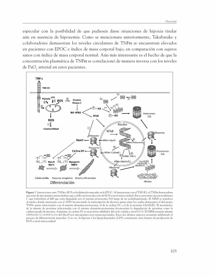
103
especular con la posibilidad de que pudiesen darse situaciones de hipoxia tisular aún en ausencia de hipoxemia. Como se mencionara anteriormente, Takabatake y colaboradores demuestran los niveles circulantes de TNFα se encuentran elevados en pacientes con EPOC e índice de masa corporal bajo, en comparación con sujetos sanos con índice de masa corporal normal. Aún más interesante es el hecho de que la concentración plasmática de TNFα se correlacionó de manera inversa con los niveles de PaO2 arterial en estos pacientes.
Figura 3. Interacciones entre TNFα y ROS en la disfunción muscular en la EPOC. Al interactuar con el TNF-R1, el TNFα desencadena una serie de mecanismos intracelulares que conllevan la producción de ROS a nivel mitocondrial. Estos activarían una proteinkinasa C que fosforilaría al IkB que sería degradado por el sistema proteasoma S26 luego de ser poliubiquitinado. El NFkB se transloca al núcleo donde interactúa con el ADN favoreciendo la transcripción de diversos genes entre los cuales destacamos el del propio TNFα, genes relacionados con el sistema ubiquitin-proteasoma, el de la ciclina D1 y el de la proteína UbcH2/E2. El incremento de la síntesis de proteínas relacionadas con el sistema ubiquitin-proteasoma favorecerían la degradación de proteínas como la cadena pesada de miosina. Asimismo, la ciclina D1 es un potente inhibidor del ciclo celular a nivel G1-S. El NFkB actuaría además inhibiendo la estabilización del MyoD por mecanismos post-transcripcionales. Estos dos últimos aspectos actuarían inhibiendo el proceso de diferenciación muscular. A su vez, la hipoxia y los lipopolisacáridos (LPS) constituyen otras fuentes de producción de ROS a nivel mitocondrial.
Discusión
104
Con todo, la relación entre moléculas oxidantes y TNFα puede adquirir una especial relevancia en situaciones en donde la capacidad antioxidante muscular se encuentra alterada, como se demuestra en el primer trabajo de esta tesis. En este contexto debe analizarse la ausencia de cambios con el entrenamiento físico en la expresión de ARNm-TNFα a nivel muscular y en el incremento de los niveles plasmáticos de TNFα producidos por el ejercicio moderado en el grupo de pacientes EPOC incluido en el tercer trabajo presentado en esta tesis doctoral.
RELACIÓN ENTRE DESACOPLAMIENTO DE LA CADENA RESPIRATORIA Y DISFUNCIÓN MUSCULAR
En el cuarto trabajo de la tesis, el análisis de composición corporal mediante bioimpedancia reveló una disminución signifi cativa de la FFM en el grupo de pacientes con bajo BMI en comparación con los sujetos control y los EPOC BMIN. Asimismo, estos sujetos presentaron una marcada intolerancia al ejercicio (ejercicio incremental en cicloergómetro), un incremento temprano de los niveles plasmáticos de lactato durante el mismo protocolo de ejercicio y una disminución de la fuerza y la resistencia muscular estadísticamente signifi cativa con respecto a los otros dos grupos. Cabe mencionar que la fuerza muscular corregida por la superfi cie muscular medida como cross-sectional área (CSA) a partir del análisis histológico de las muestras de músculo, no presentó diferencias signifi cativas entre los grupos. Sin embargo, la resistencia muscular normalizada por la CSA mantuvo las diferencias signifi cativas entre los BMIL
y los otros dos grupos estudiados. Esto demuestra que la capacidad de desarrollar fuerza muscular depende de manera importante de la masa muscular. Sin embargo, este no parece ser el caso de la resistencia, o capacidad de sostener el esfuerzo, que depende fundamentalmente de la capacidad de transporte y utilización muscular del oxígeno.
Este estudio profundiza en las posibles explicaciones a estos fenómenos. De esta forma, el hallazgo más signifi cativo de este estudio fue, sin duda, la existencia de una alteración en el acoplamiento de la cadena respiratoria mitocondrial en los BMIL en comparación con los BMIN y sujetos C. Esto se sustenta en el hallazgo de una marcada disminución del ACR (acceptor control ratio), un parámetro de efi ciencia de la fosforilación oxidativa mitocondrial. Es interesante el hecho de que el ACR se correlacionó de manera inversa y altamente signifi cativa con el nivel de PaO2, tanto a nivel basal como al fi nalizar
Roberto Alejandro Rabinovich • Tesis Doctoral
105
el ejercicio incremental en cicloergómetro. Asimismo, éste índice de acoplamiento de la cadena respiratoria también se correlacionó inversa y signifi cativamente con el incremento precoz de los niveles de ácido láctico durante el ejercicio.
En defi nitiva, este estudio demuestra una marcada diferencia en la tolerancia al ejercicio conjuntamente con un incremento de la producción de lactato a iso-carga. La asociación entre ACR y el incremento en la producción de ácido láctico permite especular que la inefi ciencia en la cadena respiratoria mitocondrial comprometa la adecuada producción de ATP del músculo favoreciendo una activación precoz del metabolismo láctico. Hasta el momento, la inefi ciencia muscular representada por el incremento temprano del ácido láctico ha sido atribuida a factores como la disminución de la actividad de enzimas del metabolismo oxidativo muscular ampliamente demostrada en la literatura74;82. Además, el incremento en el consumo de oxígeno a igual carga de trabajo demostrado por, al menos, dos trabajos anteriormente publicados64;74 ha sido atribuido al incremento de la proporción de fi bras tipo II existente en el músculo periférico de los pacientes con EPOC. Esta es la primera evidencia de la existencia de alteraciones en la cadena respiratoria mitocondrial en el músculo esquelético de este tipo de pacientes. Otro factor destacable es la asociación entre el ACR y la resistencia muscular normalizada por la CSA tanto a nivel basal como después del entrenamiento. A diferencia de la alteración en la fuerza muscular observada en los pacientes con bajo BMI, que puede ser atribuida a la disminución de la CSA, la persistencia de las diferencias en la resistencia corregida por la CSA y su asociación con el ACR establecen una clara relación entre la disfunción mitocondrial y la disminución de la resistencia muscular.
Las tres semanas de entrenamiento físico tuvieron un efecto positivo en la función muscular demostrado por una disminución del incremento de los niveles plasmáticos de ácido láctico durante el ejercicio incremental en cicloergómetro en los pacientes BMIL en comparación con los BMIN y el grupo C. Sin embargo, no existieron cambios en el ACR en relación con el entrenamiento en ninguno de los tres grupos. Cualquier conclusión al respecto debe ser sumamente prudente por la corta duración del programa de entrenamiento y el limitado número de pacientes estudiados.
En cuanto a los posibles mecanismos íntimos que generan esta alteración de la cadena respiratoria mitocondrial, la asociación entre los niveles de PaO2 y ACR permite especular en una posible relación causal hipoxia celular-alteración en la
Discusión
106
fosforilación oxidativa. Además, es bien conocido que una elevada producción de peroxinitritos, se relaciona con el desacople de la fosforilación oxidativa266. En este sentido, los niveles de 3-nitrotirosina se encuentran incrementados en el músculo esquelético de pacientes con EPOC194. Más aun, la expresión de la sintetasa inducible de óxido nítrico (iNOS) se encuentra elevada en el músculo esquelético de pacientes con EPOC y bajo BMI197. El estrés oxidativo/nitrosativo puede causar peroxidación de lípidos de las membranas celulares. Esto puede incrementar la permeabilidad de las mismas. En el caso de la membrana interna mitocondrial puede llevar a una fuga de iones, resultando en un daño de la función mitocondrial.
En este sentido, ha sido mencionado en la literatura que el desacoplamiento de la cadena respiratoria mitocondrial puede constituir un mecanismo de defensa antioxidante al asociarse a una disminución de la producción de especies reactivas de oxígeno204. De esta forma, el desacoplamiento de la cadena respiratoria reportado en el cuarto estudio de esta tesis doctoral, podría estar ocasionado por un incremento de peroxinitritos en el músculo esquelético que ha sido descrito en pacientes con EPOC y BMI bajo194. Si esto es así, las diferencias, anteriormente discutidas, entre BMIN, BMIL y controles en el comportamiento de las concentraciones de glutation en relación al entrenamiento, podrían relacionarse a una disminución en la producción de ROS asociada al ejercicio en los pacientes con bajo índice de masa corporal ocasionado por este desacoplamiento de la cadena respiratoria mitocondrial204. Es importante destacar, de acuerdo a los resultados del primer y segundo trabajo, que este potencial mecanismo de defensa antioxidante parecería ser superado por la persistencia del estímulo prolongado, tal como sugieren los resultados del entrenamiento de 8 semanas de duración.
╯
Roberto Alejandro Rabinovich • Tesis Doctoral
107
La intolerancia al ejercicio que sufren los pacientes con EPOC se debe no solo a factores pulmonares sino a la disfunción de músculos periféricos presente en este grupo de pacientes que se engloba dentro de los denominados efectos sistémicos de la enfermedad. La disfunción muscular en pacientes con EPOC responde a dos fenómenos diferenciados pero íntimamente relacionados como son la alteración en la función muscular normal y la pérdida de masa muscular que ocurre en un subgrupo de pacientes. La rehabilitación basada en el entrenamiento muscular es una herramienta terapéutica orientada a restaurar la función muscular normal. Ésta consigue incrementar la tolerancia al ejercicio conjuntamente con la mejoría de la función muscular caracterizada por un incremento en la capacidad de desarrollar fuerza, una disminución de la fatigabilidad del músculo y un incremento en la resistencia. Esto impacta positiva y signifi cativamente en la calidad de vida de estos pacientes. Sin embargo, el entrenamiento físico en pacientes con EPOC raramente alcanza a normalizar la función muscular por completo. Por ello, el entrenamiento constituye una herramienta que permite profundizar en el entendimiento de los mecanismos etiopatogénicos responsables de la disfunción muscular periférica que afecta a estos pacientes. La disfunción muscular responde a causas multifactoriales. Entre ellas, factores infl amatorios como determinadas citocinas, fundamentalmente el TNFα y el estrés oxidativo han cobrado relevancia en los últimos años. Esta tesis profundiza en el análisis de las relaciones entre el efecto del entrenamiento físico en pacientes con EPOC y el estado redox muscular. También examina la relación entre estrés oxidativo y la pérdida de masa muscular que tiene lugar en un subgrupo de pacientes con EPOC.
Resumen
108
Finalmente, la tesis explora las interacciones entre infl amación y estrés oxidativo. Asimismo, se describen por primera vez alteraciones en la efi ciencia de la fosforilación oxidativa en pacientes con índice de masa corporal disminuido y su relación con los cambios fi siopatológicos responsables de la disfunción muscular.
Del cuidadoso análisis de los resultados obtenidos a lo largo de estos años, plasmados en los cuatro trabajos presentados en la tesis podemos elaborar una serie de conclusiones que se detallan en el apartado siguiente.
╯
Roberto Alejandro Rabinovich • Tesis Doctoral
109
1) Los mecanismos de adaptación al estrés oxidativo asociados al ejercicio se encuentran alterados en el músculo esquelético de pacientes con EPOC. A diferencia del grupo control, después de un programa de entrenamiento de ocho semanas de duración, los pacientes con EPOC presentan una alteración en la capacidad de incrementar las concentraciones de GSH concomitantemente con un incremento en el GSSG. Dicha alteración es más evidente en los pacientes con bajo índice de masa corporal.
2) El incremento en la expresión de ARNm-γGCS en el músculo de estos pacientes γGCS en el músculo de estos pacientes γpodría indicar que la alteración del estado redox muscular en los pacientes con EPOC no parece responder a una mala regulación transcripcional sino a alteraciones de carácter post-transcripcional.
3) Los pacientes con EPOC presentan un incremento en la expresión de ARNm-TNFα en el músculo periférico. Existe además un moderado incremento de las concentraciones plasmáticas de TNFα en relación al ejercicio de moderada intensidad, no observado en sujetos sanos.
Conclusiones
110
4) Los pacientes con EPOC e índice de masa corporal bajo presentan un desacoplamiento de la cadena respiratoria mitocondrial, que se relaciona con los niveles de hipoxemia. Ello permite especular sobre posibles relaciones causales entre hipoxia tisular, estrés oxidativo y el fenómeno de pérdida de masa muscular observado en un subgrupo de pacientes con EPOC.
5) Asimismo, una alteración en el adecuado acoplamiento de la cadena respiratoria mitocondrial puede explicar, en parte, el incremento temprano de los niveles de ácido láctico sanguíneo que ocurre en los pacientes con EPOC y BMIL durante el ejercicio.
╯
Roberto Alejandro Rabinovich • Tesis Doctoral
111
Las conclusiones de la tesis generan nuevos interrogantes que constituyen retos para investigaciones futuras. De este modo, es importante conocer cual es la extensión de las alteraciones en la capacidad antioxidante inducida por el entrenamiento, cuales son las vías afectadas y en que grado esta defi ciencia está ligada a un régimen específi co de programa entrenamiento para poder establecer estrategias futuras dirigidas a modelar los mismos a las necesidades particulares de cada paciente. En este sentido, la eventual administración de suplementos nutricionales o farmacológicos tendientes a limitar el estrés oxidativo generado por el ejercicio debe ser objeto de futuros estudios.
╯
Perspectivas de futuro

113
Bibliografía
1. Celli, B. R. and W. MacNee. 2004. Standards for the diagnosis and treatment of patients with COPD: a summary of the ATS/ERS position paper. Eur Respir J. 23:932-946.
2. Pauwels, R. A., A. S. Buist, P. M. Calverley, C. R. Jenkins, and S. S. Hurd. 2001. Global strategy for the diagnosis, management, and prevention of chronic obstructive pulmonary disease. NHLBI/WHO Global Initiative for Chronic Obstructive Lung Disease (GOLD) Workshop summary. Am J Respir Crit Care Med. 163:1256-1276.
3. Plan de Salud. Madrid: Secretaría General Técnica. Centro de Publicaciones. Ministerio de Sanidad y Consumo, 1995; Nipo 351-95-02-7.
4. Killian, K. J., P. Leblanc, D. H. Martin, E. Summers, N. L. Jones, and E. J. M. Campbell. 1992. Exercise capacity and ventilatory, circulatory, and symptom limitation in patients with chronic airfl ow limitation. Am Rev Respir Dis.146:935-940.
5. Gosselink, R., T. Troosters, and M. Decramer. 1996. Peripheral muscle weakness contributes to exercise limitation in COPD. Am J Respir Crit Care Med. 153:976-980.
6. Saey, D., R. Debigare, P. Leblanc, M. J. Mador, C. H. Cote, J. Jobin, and F. Maltais. 2003. Contractile leg fatigue after cycle exercise: a factor limiting exercise in patients with chronic obstructive pulmonary disease. Am J Respir Crit Care Med.168:425-430.
7. Gallagher, C. 1990. Exercise and chronic obstructive pulmonary disease. Med Clinics N Am. 74:619-641.
8. Baarends, E. M., A. M. Schols, R. Mostert, and E. F. Wouters. 1997. Peak exercise response in relation to tissue depletion in patients with chronic obstructive pulmonary disease. Eur Respir J. 10:2807-2813.
9. Kobayashi, A., T. Yoneda, M. Yoshikawa, M. Ikuno, H. Takenaka, A. Fukuoka, N. Narita, and K. Nezu. 2000. The relation of fat-free mass to maximum exercise performance in patients with chronic obstructive pulmonary disease. Lung. 178:119-127.
10. Jones, N. L. and K. J. Killian 1991. Limitation of exercise in chronic airwa obstruction. In N. S. Cherniack, editor Chronic obstructive Pulmonary diseas Saunders, Philadelphia. p 196-206.
11. Decramer, M., L. E. Gosselink, T. Troosters, and M. Verschueren. 1997. Muscle weakness is related to utilization of health care resources in COPD patients. Eur Respir J.10:417-423.
12. Schols, A. M. W. J. 1998. Weight loss is a reversible factor in the prognosis of chronic obstructive pulmonary disease. Am J Respir Crit Care Med. 157:1791-1797.
13. Marquis, K., R. Debigare, Y. Lacasse, P. Leblanc, J. Jobin, G. Carrier, and F. Maltais. 2002. Midthigh muscle cross-sectional area is a better predictor of mortality than body mass index in patients with chronic obstructive pulmonary disease. Am J Respir Crit Care Med. 166:809-813.
14. Mador, M. J. 2002. Muscle mass, not body weight, predicts outcome in patients with chronic obstructive pulmonary disease. Am J Respir Crit Care Med. 166:787-789.
15. Andreassen, H. and J. Vestbo. 2003. Chronic obstructive pulmonary disease as a systemic disease: an epidemiological perspective. Eur Respir J Suppl. 46:2s-4s.
16. Celli, B. R., C. G. Cote, J. M. Marin, C. Casanova, d. O. Montes, R. A. Mendez, P. Pinto, V, and H. J. Cabral. 2004. The body-mass index, airfl ow obstruction, dyspnea, and exercise capacity index in chronic obstructive pulmonary disease. N Engl J Med. 350:1005-1012.
17. Engelen, M. P. K. J., A. M. W. J. Schols, W. C. Baken, G. J. Wesseling, and E. F. M. Wouters. 1994. Nutritional depletion in relation to respiratory and peripheral skeletal muscle function in out-patients with COPD. Eur Respir J.7:1793-1797.
18. Schols, A. M., P. B. Soeters, R. Mostert, R. J. Pluymers, and E. F. Wouters. 1995. Physiologic effects of nutritional support and anabolic steroids in patients with chronic obstructive pulmonary disease. A placebo-controlled randomized trial. Am J Respir Crit Care Med. 152:1268-1274.
114
19. Laaban, J.-P., B. Kouchakji, M.-F. Dore, E. Orvoen-Frija, P. David, and J. Rochemaure. 1993. Nutritional status of patients with chronic obstructive pulmonary disease and acute respiratory failure. Chest. 103:1362-1368.
20. Schols, A. M. W. J., P. B. Soeters, A. M. C. Dingemans, R. Mostert, P. J. Frantzen, and E. F. M. Wouters. 1993. Prevalence and characteristics of nutritional depletion in patients with stable COPD eligible for pulmonary rehabilitation. Am Rev Respir Dis.147:1151-1156.
21. Eid, A. A., A. A. Ionescu, L. S. Nixon, V. Lewis-Jenkins, S. B. Matthews, T. L. Griffi ths, and D. J. Shale. 2001. Infl ammatory response and body composition in chronic obstructive pulmonary disease. Am J Respir Crit Care Med.164:1414-1418.
22. Engelen, M. P. K. J., A. M. W. J. Schols, J. D. Does, and E. F. M. Wouters. 2000. Skeletal muscle weakness is associated with wasting of extremity fat-free mass but not with airfl ow obstruction in patients with chronic obstructive pulmonary disease. Am J Clin Nutr. 71:733-738.
23. Bernard, S., P. Leblanc, F. Whittom, G. Carrier, J. Jobin, R. Belleau, and F. Maltais. 1998. Peripheral Muscle Weakness in Patients with Chronic Obstructive Pulmonary Disease. Am J Respir Crit Care Med. 158:629-634.
24. Hamilton, A. L., K. J. Killian, E. Summers, and N. L. Jones. 1995. Muscle strenght, symptom intensity, and exercise capacity in patients with cardiorespiratory disorders. Am J Respir Crit Care Med. 152:2021-2031.
25. Debigare, R., C. H. Cote, F. S. Hould, P. Leblanc, and F. Maltais. 2003. In vitro and in vivo contractile properties of the vastus lateralis muscle in males with COPD. Eur Respir J.21:273-278.
26. Killian, K. J., E. Summers, N. L. Jones, and E. J. M. Campbell. 1992. Dyspnea and leg efforts during incremental cycle ergometry. Am Rev Respir Dis. 145:1339-1345.
27. Mador, M. J., T. J. Kufel, and L. Pineda. 2000. Quadriceps fatigue after cycle exercise in patients with chronic obstructive pulmonary disease. Am J Respir Crit Care Med.161:447-453.
28. Mador, M. J., E. Bozkanat, and T. J. Kufel. 2003. Quadriceps fatigue after cycle exercise in patients with COPD compared with healthy control subjects. Chest. 123:1104-1111.
29. Mador, M. J., O. Deniz, A. Aggarwal, and T. J. Kufel. 2003. Quadriceps fatigability after single muscle exercise in patients with chronic obstructive pulmonary disease. Am J Respir Crit Care Med. 168:102-108.
30. Jeffery, M. M., T. J. Kufel, and L. A. Pineda. 2000. Quadriceps and diaphragmatic function after exhaustive cycle exercise in the healthy elderly. Am J Respir Crit Care Med. 162:1760-1766.
31. Mador, M. J., T. J. Kufel, L. A. Pineda, A. Steinwald, A. Aggarwal, A. M. Upadhyay, and M. A. Khan. 2001. Effect of pulmonary rehabilitation on quadriceps fatiguability during exercise. Am J Respir Crit Care Med. 163:930-935.
32. Serres, I., V. Gautier, A. Varray, and C. Prefaut. 1998. Impaired skeletal muscle endurance related to physical inactivity and altered lung function in COPD patients. Chest. 113:900-905.
33. Newell, S. Z., D. K. McKenzie, and S. C. Gandevia. 1989. Inspiratory and skeletal muscle strength and endurance and diaphragmatic activation in patients with chronic airfl ow limitation. Thorax. 44:903-912.
34. Coronell, C., M. Orozco-Levi, R. Mendez, A. Ramirez-Sarmiento, J. B. Galdiz, and J. Gea. 2004. Relevance of assessing quadriceps endurance in patients with COPD. Eur Respir J. 24:129-136.
35. Metzger, J. M. and R. L. Moss. 1987. Greater hydrogen ion-induced depression of tension and velocity in skinned single fi bres of rat fast than slow muscles. J Physiol .393:727-742.
36. Cooke, R., K. Franks, G. B. Luciani, and E. Pate. 1988.The inhibition of rabbit skeletal muscle contraction by hydrogen ions and phosphate. J Physiol. 395:77-97.
37. Lamb, G. D. and D. G. Stephenson. 1991. Effect of Mg2+ on the control of Ca2+ release in skeletal muscle fi bres of the toad. JPhysiol. 434:507-528.
38. Reid, M. B. 2001. Invited Review: redox modulation of skeletal muscle contraction: what we know and what we don’t. J Appl Physiol. 90:724-731.
39. Kosmas, E. N., S. A. Hussain, J. M. Montserrat, and R. D. Levy. 1996. Relationship of peak exercise capacity with indexes of peripheral muscle vasodilation. Med Sci Sports Exerc. 28:1254-1259.
40. Schiaffi no, S. and G. Salviati. 1997. Molecular diversity of myofi brillar proteins: isoforms analysis at the protein and mRNA level. Methods Cell Biol. 52:349-369.
41. Gollnick, P. D., R. B. Armstrong, C. W. Saubert, K. Piehl, and B. Saltin. 1972. Enzyme activity and fi ber composition in skeletal muscle of untrained and trained men. J Appl Physiol. 33:312-319.
42. Schiaffi no, S. and C. Reggiani. 1996. Molecular diversity of myofi brillar proteins: gene regulation and functional signifi cance. Physiol Rev. 76:371-423.
43. Armstrong, R. B. 1988. Muscle fi ber recruitment paterns and their metabolic correlates. In H. S. Horton and R. L. Terjung, editors Exercise, nutrition, and energy metabolism Macmillan Publishing Company, New York. p 9-26.
Roberto Alejandro Rabinovich • Tesis Doctoral
115
44. Harridge, S. D., R. Bottinelli, M. Canepari, M. A. Pellegrino, C. Reggiani, M. Esbjornsson, and B. Saltin. 1996. Whole-muscle and single-fi bre contractile properties and myosin heavy chain isoforms in humans. Pfl ugers Arch. 432:913-920.
45. McComas, A. J. 1996. Skeletal muscle: forma and function. Champaign, IL:Human Kinetics.
46. Pereira Sant’Ana, J. A., S. Ennion, A. J. Sargeant, A. F. Moorman, and G. Goldspink. 1997. Comparison of the molecular, antigenic and ATPase determinants of fast myosin heavy chains in rat and human: a single-fi bre study. Pfl ugers Arch. 435:151-163.
47. Ennion, S., P. J. Sant’ana, A. J. Sargeant, A. Young, and G. Goldspink. 1995. Characterization of human skeletal muscle fi bres according to the myosin heavy chains they express. J Muscle Res Cell Motil. 16:35-43.
48. Smerdu, V., I. Karsch-Mizrachi, M. Campione, L. Leinwand, and S. Schiaffi no. 1994. Type IIx myosin heavy chain transcripts are expressed in type IIb fi bers of human skeletal muscle. Am J Physiol. 267:C1723-C1728.
49. Schiaffi no, S., L. Gorza, S. Sartore, L. Saggin, S. Ausoni, M. Vianello, K. Gundersen, and T. Lomo. 1989. Three myosin heavy chain isoforms in type 2 skeletal muscle fi bres. J Muscle Res Cell Motil. 10:197-205.
50. Biral, D., R. Betto, D. Danieli-Betto, and G. Salviati. 1988. Myosin heavy chain composition of single fi bres from normal human muscle. Biochem J. 250:307-308.
51. Larsson, L. and R. L. Moss. 1993. Maximum velocity of shortening in relation to myosin isoform composition in single fi bres from human skeletal muscles. J Physiol. 472:595-614.
52. Klitgaard, H., M. Zhou, S. Schiaffi no, R. Betto, G. Salviati, and B. Saltin. 1990. Ageing alters the myosin heavy chain composition of single fi bres from human skeletal muscle. Acta Physiol Scand. 140:55-62.
53. Andersen, J. L. and S. Schiaffi no. 1997. Mismatch between myosin heavy chain mRNA and protein distribution in human skeletal muscle fi bers. Am J Physiol. 272:C1881-C1889.
54. Haggmark, T., E. Eriksson, and E. Jansson. 1986. Muscle fi ber type changes in human skeletal muscle after injuries and immobilization. Orthopedics. 9:181-185.
55. Larsson, L. and T. Ansved. 1985. Effects of long-term physical training and detraining on enzyme histochemical and functional skeletal muscle characteristic in man. Muscle Nerve. 8:714-722.
56. Howald, H., H. Hoppeler, H. Claassen, O. Mathieu, and R. Straub. 1985. Infl uences of endurance training on the ultrastructural composition of the different muscle fi ber types in humans. Pfl ugers Arch. 403:369-376.
57. Hughes, R. L., H. Katz, V. Sahgal, J. A. Campbell, R. Hartz, and T. W. Shields. 1983. Fiber size and energy metabolites in fi ve separate muscles from patients with chronic obstructive lung diseases. Respiration. 44:321-328.
58. Whittom, F., J. Jobin, P. M. Simard, P. Leblanc, C. Simard, S. Bernard, R. Belleau, and F. Maltais. 1998. Histochemical and morphological characteristics of the vastus lateralis muscle in patients with chronic obstructive pulmonary disease. Med Sci Sports Exerc. 30:1467-1474.
59. Maltais, F., M. J. Sullivan, P. Leblanc, B. D. Duscha, F. H. Schachat, C. Simard, J. M. Blank, and J. Jobin. 1999. Altered expression of myosin heavy chain in the vastus lateralis muscle in patients with COPD. Eur Respir J.13:850-854.
60. Satta, A., G. B. Migliori, A. Spanevello, M. Neri, R. Bottinelli, M. Canepari, M. A. Pellegrino, and C. Reggiani. 1997. Fibre types in skeletal muscles of chronic obstructive pulmonary disease patients related to respiratory function and exercise tolerance. Eur Respir J. 10:2853-2860.
61. Jobin, J., F. Maltais, J. F. Doyon, P. Leblanc, P. M. Simard, and A. A. Simard. 1998. Chronic obstructive pulmonary disease: capillarity and fi ber characteristics of skeletal muscle. J Cardiopulm Rehabil. 18:432-437.
62. Jakobsson, P., L. Jorfeldt, and A. Brundin. 1990. Skeletal muscle metabolits and fi bre types in patients with advanced chronic obstructive pulmonary disease (COPD), with and without chronic respiratory failure. Eur Respir J. 3:192-196.
63. Hildebrand, I. L., C. Sylven, M. Esbjornsson, K. Hellstrom, and E. Jansson. 1991. Does chronic hypoxaemia induce transformations of fi bre types? Acta Physiol Scand. 141:435-439.
64. Richardson, R. S., B. T. Leek, T. P. Gavin, L. J. Haseler, S. R. Mudaliar, R. Henry, O. Mathieu-Costello, and P. D. Wagner. 2004. Reduced mechanical effi ciency in chronic obstructive pulmonary disease but normal peak VO2 with small muscle mass exercise. Am J Respir Crit Care Med. 169:89-96.
65. Gosker, H. R., M. P. Engelen, H. van Mameren, P. J. van Dijk, G. J. Van der Vusse, E. F. Wouters, and A. M. Schols. 2002. Muscle fi ber type IIX atrophy is involved in the loss of fat-free mass in chronic obstructive pulmonary disease. Am J Clin Nutr. 76:113-119.
66. Gosker, H. R., H. van Mameren, P. J. van Dijk, M. P. Engelen, G. J. Van der Vusse, E. F. Wouters, and A. M. Schols. 2002. Skeletal muscle fi bre-type shifting and metabolic profi le in patients with chronic obstructive pulmonary disease. Eur Respir J. 19:617-625.
67. Booth, F. W. and P. D. Gollnick. 1983. Effects of disuse on the structure and function of skeletal muscle. Med Sci Sports Exerc. 15:415-420.
Bibliografía
116
68. Itoh, K., T. Moritani, K. Ishida, C. Hirofuji, S. Taguchi, and M. Itoh. 1990. Hypoxia-induced fi bre type transformation in rat hindlimb muscles. Histochemical and electro-mechanical changes. Eur J Appl Physiol Occup Physiol. 60:331-336.
69. Lindboe, C. F., F. Askevold, and M. Slettebo. 1982. Changes in skeletal muscles of young women with anorexia nervosa. An enzyme histochemical study. Acta Neuropathol.(Berl)56:299-302.
70. Richardson, R. S., E. A. Noyszewski, K. F. Kendrick, J. S. Leigh, and P. D. Wagner. 1995. Myoglobin O2 desaturation during exercise: Evidence of limited O2 transport. J Clin Invest. 96:1916-1926.
71. Groebe, K. and G. Thews. 1990. Calculated intra- and extracellular PO2 gradients in heavily working red muscle. Am J Physiol. 259:H84-H92.
72. Honig, C. R., T. E. Gayeski, W. Federspiel, A. Clark, Jr., and P. Clark. 1984. Muscle O2 gradients from hemoglobin to cytochrome: new concepts, new complexities. Adv Exp Med Biol. 169:23-38.
73. Maltais, F., J. Jobin, M. J. Sullivan, S. Bernard, F. Whittom, K. J. Killian, M. Desmeules, P. Belanger, and P. Le Blanc. 1998. Lower limb metabolic and hemodynamic responses during exercise in normal subjects and in COPD. J Appl Physiol. 84:1573-1580.
74. Sala, E., J. Roca, R. M. Marrades, J. Alonso, J. M. Gonzalez de Suso, A. Moreno, J. Barbera, J. Nadal, Ll. Jover, R. Rodriguez-Roisin, and P. D. Wagner. 1999. Effects of endurance training on skeletal muscle bioenergetics in chronic obstructive pulmonary disease. Am J Respir Crit Care Med. 159:1726-1734.
75. Hare, J. M. 2004. Nitroso-redox balance in the cardiovascular system. N Engl J Med. 351:2112-2114.
76. Hare, J. M. 2003. Nitric oxide and excitation-contraction coupling. J Mol Cell Cardiol. 35:719-729.
77. Simard, C., Maltais, F., Leblanc, P., Simard, P, and Jobin, J., 1996. Mitochondrial and Capillarity Changes in Vastus Lateralis Muscle of COPD Patients: Electron Microscopy Study. Med.Sci.Sports Exerc. 28[5], S95.
78. Fiaccadori, E., S. Del Canale, P. Vitali, E. Coffrini, N. Ronda, and A. Guariglia. 1987. Skeletal muscle energetics,acid-base equilibrium and lactate metabolism in patients with severe hypercapnia and hypoxemia. Chest. 92:883-887.
79. Casaburi, R., A. Patessio, F. Ioli, S. Zanaboni, C. F. Donner, and K. Wasserman. 1991. Reductions in exercise lactic acidosis and ventilation as a result of exercise training in patients with obstructive lung disease. Am Rev Respir Dis. 143:9-18.
80. Patessio, A., R. Casaburi, M. Carone, L. Appendini, C. F. Donner, and K. Wasserman. 1993. Comparison of gas ex-change, lactate, and lactic acidosis thresholds in patients
with chronic obstructive pulmonary disease. Am.Rev.Respir.Dis. 148:622-626.
81. Engelen, M. P., R. Casaburi, R. Rucker, and E. Carithers. 1995. Contribution of the respiratory muscles to the lactic acidosis of heavy exercise in COPD. Chest. 108:1246-1251.
82. Maltais, F., P. Leblanc, F. Whittom, C. Simard, K. Marquis, M. Belanger, M. J. Breton, and J. Jobin. 2000. Oxidative enzyme activities of the vastus lateralis muscle and the functional status in patients with COPD. Thorax. 55:848-853.
83. Hunter, G. R., B. R. Newcomer, D. E. Larson-Meyer, M. M. Bamman, and R. L. Weinsier. 2001. Muscle metabolic economy is inversely related to exercise intensity and type II myofi ber distribution. Muscle Nerve. 24:654-661.
84. Sauleda, J., F. García-Palmer, R. J. Wiesner, S. Tarraga, I. Harting, P. Tomás, C. Gómez, C. Saus, A. Palou, and A. G. N. Agustí. 1998. Cytochrome oxidase activity and mitochondrial gene expression in skeletal muscle of patients with chronic obstructive pulmonary disease. Am J Respir Crit Care Med. 157:1413-1417.
85. Braun, S. R., N. L. Keim, R. M. Dixon, P. Clagnaz, A. Anderegg, and E. S. Shrago. 1984. The prevalence and determinants of nutritional changes in chronic obstructive pulmonary disease. Chest. 86:558-563.
86. Gray-Donald, K., L. Gibbons, S. H. Shapiro, and J. G. Martin. 1989. Effect of nutritional status on exercise performance in patients with chronic obstructive pulmonary disease. Am Rev Respir Dis. 140:1544-1548.
87. Kessler, R., M. Faller, G. Fourgaut, B. Mennecier, and E. Weintzenblum. 1999. Predictive factors of hospitalization for acute exacerbation in a series of 64 patients with chronic obstruc-tive pulmonary disease. Am J Respir Crit Care Med. 159:158-164.
88. Wilson, D. O., R. M. Rogers, E. C. Wright, and N. R. Anthonisen. 1989. Body weight in chronic obstructive pulmonary disease. The National Institutes of Health Intermittent Positive-Pressure Breathing Trial. Am Rev Respir Dis. 139:1435-1438.
89. Connors, A. F., N. V. Dawson, C. Thomas, F. H. Harrel Jr, N. Desbiens, W. J. Fulkerson, P. Kussin, P. Bellamy, L. Goldman, and W. A. Knaus. 1996. Outcomes following acute exacerbation of severe chronic obstructive lung disease. Am J Respir Crit Care Med. 154:959-967.
90. Landbo, CH., E. Prescott, P. Lange, J. Vestbo, and T. P. Almdal. 1999. Prognostic Value of Nutritional Status in Chronic Obstructive Pulmonary Disease. Am J Respir Crit Care Med. 160:1856-1861.
91. Gray-Donald, K., L. Gibbons, S. H. Shapiro, P. T. Macklem, and J. G. Martin. 1996. Nutritional status and mortality in chronic obstructive pulmonary disease. Am J Respir Crit Care Med. 153:961-966.
Roberto Alejandro Rabinovich • Tesis Doctoral
117
92. Schols, A. M., R. Mostert, P. B. Soeters, and E. F. Wouters. 1991. Body composition and exercise performance in patients with chronic obstructive pulmonary disease. Thorax. 46:695-699.
93. Mostert, R., A. Goris, C. Weling-Scheepers, E. F. Wouters, and A. M. Schols. 2000. Tissue depletion and health related quality of life in patients with chronic obstructive pulmonary disease. Respir Med. 94:859-867.
94. Debigare, R., C. H. Cote, and F. Maltais. 2001. Peripheral Muscle Wasting in Chronic Obstructive Pulmonary Disease. Clinical relevance and mechanisms. Am J Respir Crit Care Med. 164:1712-1717.
95. ATS/ERS statement. 1999. “Skeletal Muscle Dysfunction in Chronic Obstructive Pulmonary Disease”. Am J Respir Crit Care Med. 159:S1-S40.
96. Agusti, A. G., A. Noguera, J. Sauleda, E. Sala, J. Pons, and X. Busquets. 2003. Systemic effects of chronic obstructive pulmonary disease. Eur Respir J. 21:347-360.
97. Wouters, E. F., E. C. Creutzberg, and A. M. Schols. 2002. Systemic effects in COPD. Chest. 121:127S-130S.
98. Engelen, M. P. K. J., N. E. P. Deutz, E. F. M. Wouters, and A. M. W. J. Schols. 2000. Enhanced levels of whole-body protein turnover in patients with chronic obstructive pulmonary disease. Am J Respir Crit Care Med. 162:1488-1492.
99. Morrison, W. L., J. Gibson, C. Scrimegeour, and M. J. Rennie. 1988. Muscle wasting in emphysema. Clin Sci. 75:415-420.
100. Aguilaniu, B., S. Goldstein-Shapses, A. Pajon, P. Levy, F. Sarrot, X. Leverve, E. Page, and J. Askanazi. 1992. Muscle protein degradation in severely malnourished patients with chronic obstructive pulmonary disease subject to short-term total parenteral nutrition. JPEN J Parenter Enteral Nutr.16:248-254.
101. Jagoe, R. T. and A. L. Goldberg. 2001. What do we really know about the ubiquitin-proteasome pathway in muscle atrophy? Curr Opin Clin Nutr Metab Care. 4:183-190.
102. Bossola, M., M. Muscaritoli, P. Costelli, R. Bellantone, F. Pacelli, S. Busquets, J. Argiles, F. J. Lopez-Soriano, I. M. Civello, F. M. Baccino, F. F. Rossi, and G. B. Doglietto. 2001. Increased muscle ubiquitin mRNA levels in gastric cancer patients. Am J Physiol Regul Integr Comp Physiol. 280:R1518-R1523.
103. Williams, A., X. Sun, J. E. Fischer, and P. O. Hasselgren. 1999. The expression of genes in the ubiquitin-proteasome proteolytic pathway is increased in skeletal muscle from patients with cancer. Surgery. 126:744-749.
104. Fryburg, D. A. 1994. Insulin-like growth factor I exerts growth hormone- and insulin-like actions on human muscle protein metabolism. Am J Physiol. 267:E331-E336.
105. Fryburg, D. A. and E. J. Barrett. 1993. Growth hormone acutely stimulates skeletal muscle but not whole-body protein synthesis in humans. Metabolism. 42:1223-1227.
106. Creutzberg, E. C. and R. Casaburi. 2003. Endocrinological disturbances in chronic obstructive pulmonary disease. Eur Respir J Suppl. 46:76s-80s.
107. Di Francia, M., D. Barbier, J. Mege, and J. Orehek. 1994. Tumor necrosis factor-alpha and weight loss in chronic obstructive pulmonary disease. Am J Respir Crit Care Med.150:1453-1455.
108. Jurasinski, C. V., L. Kilpatrick, and T. C. Vary. 1995. Amrinone prevents muscle protein wasting during chronic sepsis. Am J Physiol. 268:E491-E500.
109. Frost, R. A., C. H. Lang, and M. C. Gelato. 1997. Transient exposure of human myoblasts to tumor necrosis factor-alpha inhibits serum and insulin-like growth factor-I stimulated protein synthesis. Endocrinology. 138:4153-4159.
110. Kamischke, A., D. E. Kemper, M. A. Castel, M. Luthke, C. Rolf, H. M. Behre, H. Magnussen, and E. Nieschlag. 1998. Testosterone levels in men with chronic obstructive pulmonary disease with or without glucocorticoid therapy. Eur Respir J. 11:41-45.
111. Pape, G. S., M. Friedman, L. E. Underwood, and D. R. Clemmons. 1991. The effect of growth hormone on weight gain and pulmonary function in patients with chronic obstructive lung disease. Chest. 99:1495-1500.
112. Burdet, L., B. de Muralt, Y. Schutz, C. Pichard, and J. W. Fitting. 1997. Administration of growth hormone to underweight patients with chronic obstructive pulmonary disease. A prospective, randomized, controlled study. Am J Respir Crit Care Med. 156:1800-1806.
113. Ferreira, I. M., I. T. Verreschi, L. E. Nery, R. S. Goldstein, N. Zamel, D. Brooks, and J. R. Jardim. 1998. The infl uence of 6 months of oral anabolic steroids on body mass and respiratory muscles in undernourished COPD patients. Chest. 114:19-28.
114. Creutzberg, E. C., E. F. Wouters, R. Mostert, R. J. Pluymers, and A. M. Schols. 2003. A role for anabolic steroids in the rehabilitation of patients with COPD? A double-blind, placebo-controlled, randomized trial. Chest. 124:1733-1742.
115. Yeh, S. S., B. DeGuzman, and T. Kramer. 2002. Reversal of COPD-associated weight loss using the anabolic agent oxandrolone. Chest. 122:421-428.
116. Engelen, M. P., E. F. Wouters, N. E. Deutz, J. D. Does, and A. M. Schols. 2001. Effects of exercise on amino acid metabolism in patients with chronic obstructive pulmonary disease. Am J Respir Crit Care Med. 163:859-864.
117. Schols, A. M. and E. F. Wouters. 2000. Nutritional abnormalities and supplementation in chronic obstructive pulmonary disease. Clin.Chest Med. 21:753-762.
Bibliografía
118
118. Schols, A. M. W. J., P. B. Soeters, R. Mostert, W. H. M. Saris, and E. F. M. Wouters. 1991. Energy balance in chronic obstructive pulmonary disease. Am Rev Respir Dis.143:1248-1252.
119. Wilson, D. O., M. Donahoe, R. M. Rogers, and B. E. Pennock. 1990. Metabolic rate and weight loss in chronic obstructive lung disease. JPEN J Parenter Enteral Nutr.14:7-11.
120. Baarends, E. M., A. M. W. J. Schols, D. J. Slebos, R. Mostert, P. P. Janssen, and E. F. M. Wouters. 1995. Metabolic and ventilatory response pattern to arm elevation in patients with COPD and helathy age-matched subjects. Eur Respir J.8:1345-1351.
121. Baarends, E. M., A. M. W. J. Schols, D. L. E. Pannemans, K. R. Westerterp, and E. F. M. Wouters. 1997. Total free living energy expenditure in patients with severe COPD. Eur Respir J. 155:549-554.
122. Hugli, O., Y. Schutz, and J. W. Fitting. 1996. The daily energy expenditure in stable chronic obstructive pulmonary disease. Am J Respir Crit Care Med. 153:294-300.
123. Baarends, E. M., A. M. Schols, M. A. Akkermans, and E. F. Wouters. 1997. Decreased mechanical effi ciency in clinically stable patients with COPD. Thorax. 52:981-986.
124. Williams, T. J., R. E. O’Hehir, D. Czarny, M. Horne, and G. Bowes. 1988. Acute myopathy in severe acute asthma treated with intravenously administered corticosteroids. Am Rev Respir Dis. 137:460-463.
125. Shee, C. D. 1990. Risk factors for hydrocortisone myopathy in acute severe asthma. Respir Med. 84:229-233.
126. Decramer, M., V. De Bock, and R. Dom. 1996. Functional and histologic picture of steroid-induced myopathy in chronic obstructive pulmonary disease. Am J Respir Crit Care Med. 153:1958-1964.
127. Preedy, V. R., D. M. Smith, and P. H. Sugden. 1985. The effects of 6 hours of hypoxia on protein synthesis in rat tissues in vivo and in vitro. Biochem.J 228:179-185.
128. Green, H. J., J. R. Sutton, A. Cymerman, P. M. Young, and C. S. Houston. 1989. Operation Everest II: adaptations in human skeletal muscle. J Appl Physiol. 66:2454-2461.
129. Bigard, A. X., A. Brunet, C. Y. Guezennec, and H. Monod. 1991. Skeletal muscle changes after endurance training at high altitude. J Appl Physiol. 71:2114-2121.
130. Howald, H., D. Pette, J. A. Simoneau, A. Uber, H. Hoppeler, and P. Cerretelli. 1990. Effects of chronic hypoxia on muscle enzyme activities. Int J Sports Med. 11:S10-S14.
131. Kwast, K. E. and S. C. Hand. 1996. Acute depression of mitochondrial protein synthesis during anoxia: contributions of oxygen sensing, matrix acidifi cation, and redox state. J Biol Chem. 271:7313-7319.
132. Bigard, A. X., H. Sanchez, O. Birot, and B. Serrurier. 2000. Myosin heavy chain composition of skeletal muscles in young rats growing under hypobaric hypoxia conditions. J Appl Physiol. 88:479-486.
133. Rennie, M. J., R. H. Edwards, P. W. Emery, D. Halliday, K. Lundholm, and D. J. Millward. 1983. Depressed protein synthesis is the dominant characteristic of muscle wasting and cachexia. Clin Physiol. 3:387-398.
134. Cechetto, D. and G. W. Mainwood. 1978. Carbon dioxide and acid base balance in the isolated rat diaphragm. Pfl ugers Arch. 376:251-258.
135. Trivedi, B. and W. H. Danforth. 1966. Effect of pH on the kinetics of frog muscle phosphofructokinase. J Biol Chem.241:4110-4112.
136. Gertz, I., G. Hedenstierna, G. Hellers, and J. Wahren. 1977. Muscle metabolism in patients with chronic obstructive lung disease and acute respiratory failure. Clin Sci. 52:395-403.
137. Sahlin, K., L. Edstrom, and H. Sjoholm. 1983. Fatigue and phosphocreatine depletion during carbon dioxide-induced acidosis in rat muscle. Am J Physiol. 245:C15-C20.
138. Mitch, W. E. 1998. Robert H Herman Memorial Award in Clinical Nutrition Lecture, 1997. Mechanisms causing loss of lean body mass in kidney disease. Am J Clin Nutr.67:359-366.
139. Bailey, J. L., X. Wang, B. K. England, S. R. Price, X. Ding, and W. E. Mitch. 1996. The acidosis of chronic renal failure activates muscle proteolysis in rats by augmenting transcription of genes encoding proteins of the ATP-dependent ubiquitin-proteasome pathway. J Clin Invest.97:1447-1453.
140. Reaich, D., S. M. Channon, C. M. Scrimgeour, S. E. Daley, R. Wilkinson, and T. H. Goodship. 1993. Correction of acidosis in humans with CRF decreases protein degradation and amino acid oxidation. Am J Physiol. 265:E230-E235.
141. Burnett, D., A. Chamba, S. L. Hill, and R. A. Stockley. 1987. Neutrophils from subjects with chronic obstructive lung disease show enhanced chemotaxis and extracellular proteolysis. Lancet. 2:1043-1046.
142. Noguera, A., S. Batle, C. Miralles, J. Iglesias, X. Busquets, W. MacNee, and A. G. Agusti. 2001. Enhanced neutrophil response in chronic obstructive pulmonary disease. Thorax.56:432-437.
143. Noguera, A., X. Busquets, J. Sauleda, J. M. Villaverde, W. MacNee, and A. G. Agusti. 1998. Expression of adhesion molecules and G proteins in circulating neutrophils in chronic obstructive pulmonary disease. Am J Respir Crit Care Med. 158:1664-1668.
144. Schols, A. M. W. J., W. A. Buurman, and D. M. W. E. Staal van den Brekel AJ. 1996. Evidence for a relation between
Roberto Alejandro Rabinovich • Tesis Doctoral
119
metabolic derangements and increased levels of infl ammatory mediators in a subgroup of patients with chronic obstructive pulmonary disease. Thorax. 51:819-824.
145. Yasuda, N., K. Gotoh, S. Minatoguchi, K. Asano, K. Nishigaki, M. Nomura, A. Ohno, M. Watanabe, H. Sano, H. Kumada, T. Sawa, and H. Fujiwara. 1998. An increase of soluble Fas, an inhibitor of apoptosis, associated with progression of COPD. Respir Med. 92:993-999.
146. Malo, O., J. Sauleda, X. Busquets, C. Miralles, A. G. Agusti, and A. Noguera. 2002. [Systemic infl ammation during exacerbations of chronic obstructive pulmonary disease]. Arch Bronconeumol. 38:172-176.
147. de Godoy, I., M. Donahoe, W. J. Calhoun, J. Mancino, and R. M. Rogers. 1996. Elevated TNF-α production by peripheral blood monocytes of weight-losing COPD patients. Am J Respir Crit Care Med. 153:633-637.
148. Li, Y. P., R. J. Schwartz, I. A. Waddell, B. R. Holloway, and M. B. Reid. 1998. Skeletal muscle myocites undergo ptotein loss and reactive oxygen-mediated NF-kB activation in response to TNF-α. FASEB J. 12:871-880.
149. Langen, R. C., A. M. Schols, M. C. Kelders, E. F. Wouters, and Y. M. Janssen-Heininger. 2001. Infl ammatory cytokines inhibit myogenic differentiation through activation of nuclear factor-kappaB. FASEB J. 15:1169-1180.
150. Petrache, I., L. E. Otterbein, J. Alam, G. W. Wiegand, and A. M. Choi. 2000. Heme oxygenase-1 inhibits TNF-alpha-induced apoptosis in cultured fi broblasts. Am J Physiol Lung Cell Mol Physiol. 278:L312-L319.
151. Carbo, N., S. Busquets, M. van Royen, B. Alvarez, F. J. Lopez-So-riano, and J. M. Argiles. 2002. TNF-alpha is involved in activating DNA fragmentation in skeletal muscle. BrJCancer. 86:1012-1016.
152. Agusti, A. G., J. Sauleda, C. Miralles, C. Gomez, B. Togores, E. Sala, S. Batle, and X. Busquets. 2002. Skeletal muscle apoptosis and weight loss in chronic obstructive pulmonary disease. Am J Respir Crit Care Med. 166:485-489.
153. Orozco-Levi, M. 2003. Structure and function of the respiratory muscles in patients with COPD: impairment or adaptation? Eur Respir J Suppl. 46:41s-51s.
154. Orozco-Levi, M., J. Gea, J. L. Lloreta, M. Felez, J. Minguella, S. Serrano, and J. M. Broquetas. 1999. Subcellular adaptation of the human diaphragm in chronic obstructive pulmonary disease. Eur Respir J. 13:371-378.
155. Lloreta, J., M. Orozco, J. Gea, J. M. Corominas, and S. Serrano. 1996. Selective diaphragmatic mitochondrial abnormalities in a patient with marked air fl ow obstruction. Ultrastruct Pathol. 20:67-71.
156. Levine, S., L. Kaiser, J. Leferovich, and B. Tikunov. 1997. Cellular adaptations in the diaphragm in chronic obstructive pulmonary disease. N Engl J Med. 337:1799-1806.
157. Levine, S., C. Gregory, T. Nguyen, J. Shrager, L. Kaiser, N. Rubinstein, and G. Dudley. 2002. Bioenergetic adaptation of individual human diaphragmatic myofi bers to severe COPD. J Appl Physiol .92:1205-1213.
158. Roca, J., Weisman, I. M., Palange, P., and Whipp, B., Roca, J. and Whipp, B. 1997. Clinical Exercise Testing. European Respiratory Monograph [Chapter 5], 88-114.
159. Jaspers, S. R. and M. E. Tischler. 1984. Atrophy and growth failure of rat hindlimb muscles in tail-cast suspension. J Appl Physiol. 57:1472-1479.
160. Ferrando, A. A., H. W. Lane, C. A. Stuart, J. Davis-Street, and R. R. Wolfe. 1996. Prolonged bed rest decreases skeletal muscle and whole body protein synthesis. Am J Physiol. 270:E627-E633.
161. Gibson, J. N., D. Halliday, W. L. Morrison, P. J. Stoward, G. A. Hornsby, P. W. Watt, G. Murdoch, and A. J. Rennie. 1987. Decrease in human quadriceps muscle protein turnover consequent upon leg immobilization. Clin Sci (Lond). 72:503-509.
162. Adams, G. R., B. M. Hather, and G. A. Dudley. 1994. Effect of short-term unweighting on human skeletal muscle strength and size. Aviat Space Environ Med. 65:1116-1121.
163. Taillandier, D., E. Aurousseau, D. Meynial-Denis, D. Bechet, M. Ferrara, P. Cottin, A. Ducastaing, X. Bigard, C. Y. Guezennec, H. P. Schmid, and . 1996. Coordinate activation of lysosomal, Ca 2+-activated and ATP-ubiquitin-dependent proteinases in the unweighted rat soleus muscle. Biochem J.316 ( Pt 1):65-72.
164. Bodine, S. C., E. Latres, S. Baumhueter, V. K. Lai, L. Nunez, B. A. Clarke, W. T. Poueymirou, F. J. Panaro, E. Na, K. Dharmarajan, Z. Q. Pan, D. M. Valenzuela, T. M. DeChiara, T. N. Stitt, G. D. Yancopoulos, and D. J. Glass. 2001. Identifi cation of ubiquitin ligases required for skeletal muscle atrophy. Science. 294:1704-1708.
165. Jones, S. W., R. J. Hill, P. A. Krasney, B. O’Conner, N. Peirce, and P. L. Greenhaff. 2004. Disuse atrophy and exercise rehabilitation in humans profoundly affects the expression of genes associated with the regulation of skeletal muscle mass. FASEB J. 18:1025-1027.
166. Maltais, F., P. Leblanc, C. Simard, J. Jobin, C. Bérubé, J. Bruneau, L. Carrier, and R. Belleau. 1996. Skeletal muscle adaptation to endurance training in patients with Chronic Obstructive Pulmonary Disease. Am J Respir Crit Care Med.154:442-447.
167. Maltais, F., P. Leblanc, J. Jobin, C. Berube, J. Bruneau, L. Carrier, M. J. Breton, G. Falardeau, and R. Belleau. 1997. Intensity of training and physiologic adaptation in patients with chronic obstructive pulmonary disease. Am J Respir Crit Care Med. 155:555-561.
Bibliografía
120
168. Franssen, F. M., R. Broekhuizen, P. P. Janssen, E. F. Wouters, and A. M. Schols. 2004. Effects of Whole-Body Exercise Training on Body Composition and Functional Capacity in Normal-Weight Patients With COPD. Chest.125:2021-2028.
169. Belman, M. J. 1986. Exercise in chronic obstructive pulmonary disease. Clin Chest Med. 7:585-597.
170. Barbera, J. A., G. Peces-Barba, A. G. Agusti, J. L. Izquierdo, E. Monso, T. Montemayor, and J. L. Viejo. 2001. [Clinical guidelines for the diagnosis and treatment of chronic obstructive pulmonary disease]. Arch Bronconeumol. 37:297-316.
171. Ries, A. L., B. W. Carlin, V. Carrieri-Kohlman, and et al. 1997. Pulmonary rehabilitation: joint ACCP/AACVPR evidence-based guidelines. ACCP/AACVPR Pulmonary Rehabilitation Guidelines Panel. American College of Chest Physicians. American Association of Cardiovascular and Pulmonary Rehabilitation. Chest. 112:1363-1396.
172. Lacasse, Y., E. Wong, G. H. Guyatt, D. King, D. J. Cook, and R. S. Goldstein. 1996. Meta-analysis of respiratory rehabilitation in chronic obstructive pulmonary disease. Lancet. 348:1115-1119.
173. Sauleda, J., F. J. Garcia-Palmer, G. Gonzalez, A. Palou, and A. G. Agusti. 2000. The activity of cytochrome oxidase is increased in circulating lymphocytes of patients with chronic obstructive pulmonary disease, asthma, and chronic arthritis. Am J Respir Crit Care Med. 161:32-35.
174. Hather, B. M., G. R. Adams, P. A. Tesch, and G. A. Dudley. 1992. Skeletal muscle responses to lower limb suspension in humans. J Appl Physiol. 72:1493-1498.
175. Repine, J. E., A. Bast, and I. Lankhorst. 1997. Oxidative stress in chronic obstructive pulmonary disease. Oxidative Stress Study Group. Am J Respir Crit Care Med. 156:341-357.
176. Nowak, D., A. Antczak, M. Krol, T. Pietras, B. Shariati, P. Bialasiewicz, K. Jeczkowski, and P. Kula. 1996. Increased content of hydrogen peroxide in the expired breath of cigarette smokers. Eur Respir J. 9:652-657.
177. MacNee, W. and I. Rahman. 1999. Oxidants and antioxidants as therapeutic targets in chronic obstructive pulmonary disease. Am J Respir Crit Care Med. 160:S58-S65.
178. Dekhuijzen, P. N., K. K. Aben, I. Dekker, L. P. Aarts, P. L. Wielders, C. L. van Herwaarden, and A. Bast. 1996. Increased exhalation of hydrogen peroxide in patients with stable and unstable chronic obstructive pulmonary disease. Am J Respir Crit Care Med. 154:813-816.
179. Thompson, A. B., T. Bohling, A. Heires, J. Linder, and S. I. Rennard. 1991. Lower respiratory tract iron burden is increased in association with cigarette smoking. J Lab Clin Med. 117:493-499.
180. Pinamonti, S., M. Muzzoli, M. C. Chicca, A. Papi, F. Ravenna, L. M. Fabbri, and A. Ciaccia. 1996. Xanthine oxidase activity in bronchoalveolar lavage fl uid from patients with chronic obstructive pulmonary disease. Free Radic Biol Med. 21:147-155.
181. Heunks, L. M., J. Viña, C. L. van Herwaarden, H. T. Folgering, A. Gimeno, and P. N. Dekhuijzen. 1999. Xanthine oxidase is involved in exercise-induced oxidative stress in chronic obstructive pulmonary disease. Am J Physiol. 277:R1697-R1704.
182. Rahman, I., D. Morrison, K. Donaldson, and W. Mac Nee. 1996. Systemic oxidative stress in asthma, COPD, and smokers. Am J Respir Crit Care Med. 154:1055-1060.
183. Morrow, J. D., B. Frei, W. A. Longmire, M. Gaziano, S. M. Lynch, Y. Shyr, W. E. Strauss, J. A. Oates, and L. J. I. Roberts. 1995. Increase in circulating products of lipid peroxidation (F2-isoprostanes) in smokers. N Engl J Med.332:1198-1203.
184. Celermajer, D. S. 1996. Passive smoking and impaired endothelium-dependent arterial dilatation in healthy young adults. New Engl J Med. 334:150-154.
185. Raitakari, O. T., M. R. Adams, R. J. McCredie, K. A. Griffi ths, and D. S. Celermajer. 1999. Arterial endothelial dysfunction related to passive smoking is potentially reversible in healthy young adults. Ann Intern Med. 130:578-581.
186. Cucina, A., P. Sapienza, V. Corvino, V. Borrelli, V. Mariani, B. Randone, D. L. Santoro, and A. Cavallaro. 2000. Nicotine-induced smooth muscle cell proliferation is mediated through bFGF and TGF-beta 1. Surgery. 127:316-322.
187. Broal, P. 1992. Main features of structure and function. In P. Broal, editor The central nervous system Oxford University Press, New York. p 5-50.
188. Pratico, D., S. Basili, M. Vieri, C. Cordova, F. Violi, and G. A. Fitzgerald. 1998. Chronic obstructive pulmonary disease is associated with an increase in urinary levels of isoprostane F2alpha-III, an index of oxidant stress. Am J Respir Crit Care Med. 158:1709-1714.
189. Viña, J., E. Servera, M. Asensi, J. Sastre, F. V. Pallardó, J. A. Ferrero, J. G. d. l. Asunción, V. Antón, and J. Marín. 1996. Exercise causes blood glutathione oxidation in chronic obstructive pulmonary disease: prevention of O2 therapy. J Appl Physiol. 81:2199-2202.
190. Reid, M. B. 2001. Plasticity in skeletal, cardiac, and smooth muscle. Invited review: Redox modulation of skeletal muscle contraction: what we know and what we don´t. J Appl Physiol. 90:724-731.
191. Mitch, W. E. and A. L. Goldberg. 1996. Mechanisms of muscle wasting. The role of the ubiquitin-proteasome pathway. N Engl J Med. 335:1897-1905.
Roberto Alejandro Rabinovich • Tesis Doctoral
121
192. Buck, M. and M. Chojkier. 1996. Muscle wasting and dedifferentiation induced by oxidative stress in a murine model of cachexia is prevented by inhibitors of nitric oxide synthesis and antioxidants. EMBO J. 15:1753-1765.
193. Stadtman, E. R. 1990. Metal ion-catalyzed oxidation of proteins: biochemical mechanism and biological consequences. Free Radic Biol Med. 9:315-325.
194. Barreiro, E., J. Gea, J. M. Corominas, and S. N. Hussain. 2003. Nitric oxide synthases and protein oxidation in the quadriceps femoris of patients with chronic obstructive pulmonary disease. Am J Respir Cell Mol Biol. 29:771-778.
195. Kobzik, L., M. B. Reid, D. S. Bredt, and J. S. Stamler. 1994. Nitric oxide in skeletal muscle. Nature. 372:546-548.
196. Williams, G., T. Brown, L. Becker, M. Prager, and B. P. Giroir. 1994. Cytokine-induced expression of nitric oxide synthase in C2C12 skeletal muscle myocytes. Am J Physiol267:R1020-R1025.
197. Agusti, A., M. Morla, J. Sauleda, C. Saus, and X. Busquets. 2004. NF-kappaB activation and iNOS upregulation in skeletal muscle of patients with COPD and low body weight. Thorax. 59:483-487.
198. Brune, B., A. von Knethen, and K. B. Sandau. 1998. Nitric oxide and its role in apoptosis. Eur J Pharmacol. 351:261-272.
199. Lanone, S., A. Mebazaa, C. Heymes, D. Henin, J. J. Poderoso, Y. Panis, C. Zedda, T. Billiar, D. Payen, M. Aubier, and J. Boczkowski. 2000. Muscular contractile failure in septic patients: role of the inducible nitric oxide synthase pathway. Am J Respir Crit Care Med. 162:2308-2315.
200. Boveris, A., N. Oshino, and B. Chance. 1972. The cellular production of hydrogen peroxide. Biochem J. 128:617-630.
201. Turrens, J. F., A. Alexandre, and A. L. Lehninger. 1985. Ubisemiquinone is the electron donor for superoxide formation by complex III of heart mitochondria. Arch Biochem Biophys. 237:408-414.
202. Fernandez Checa, J. C., N. Kaplowitz, C. García Ruiz, M. Miranda, M. Mari, E. Ardite, and A. Morales. 1997. GSH transport in mitochondria: defense against TNF-induced oxidative stress and alcohol-induced defect. Am J Physiol.273:G7-G17.
203. Chance B., Sies H and Boveris A. 1979. Hydroperoxide metabolism in mammalian organs. Physiol Rev 59:527-605.
204. Miwa, S. and M. D. Brand. 2003. Mitochondrial matrix reactive oxygen species production is very sensitive to mild uncoupling. Biochem Soc Trans. 31:1300-1301.
205. Abate C., Patel FJ, RAuscher III FJ, and Curran T. 1990. Redox regulation of Fos and Jun DNA binding actibity in vitro. Science 249:1157-1161.
206. Guyton, K. Z., Y. Liu, M. Gorospe, Q. Xu, and N. J. Holbrook. 1996. Activation of mitogen-activated protein kinase by H2O2. Role in cell survival following oxidant injury. J Biol Chem. 271:4138-4142.
207. Flohe, L., R. Brigelius-Flohe, C. Saliou, M. G. Traber, and L. Packer. 1997. Redox regulation of NF-kappa B activation. Free Radic Biol Med. 22:1115-1126.
208. Chandel, N. S. and P. T. Schumacker. 2000. Cellular oxygen sensing by mitochondria: old questions, new insight. J Appl Physiol. 88:1880-1889.
209. Duranteau, J., N. S. Chandel, and P. T. Schumacker. 1998. Intracellular signaling by Reactive Oxygen Species during Hypoxia. J Biol Chem. 273:11619-11624.
210. Martenson J., Lai CK and Meister A. 1990. High-affi nity transport of glutathione is part of a multicomponent system essential for mitochondrial function. Proc Natl Acad Sci USA 87:7185-7189.
211. Garcia-Ruiz, C., A. Colell, A. Morales, N. Kaplowitz, and J. C. Fernandez-Checa. 1995. Role of oxidative stress generated from the mitochondrial electron transport chain and mitochondrial glutathione status in loss of mitochondrial function and activation of transcription factor nuclear factor-kappa B: studies with isolated mitochondria and rat hepatocytes. Mol Pharmacol. 48:825-834.
212. Jammes, Y., J. G. Steinberg, F. Bregeon, and S. Delliaux. 2004. The oxidative stress in response to routine incremen-tal cycling exercise in healthy sedentary subjects. Respir Physiol Neurobiol. 144:81-90.
213. Sastre, J., M. Asensi, E. Gascó, F. V. Pallardó, J. A. Ferrero, T. Furukawa, and J. Viña. 1992. Exhaustive physical exercise causes oxidation of glutathione status in blood: prevention by antioxidant administration. Am J Physiol. 263:R992-R995.
214. Engelen, M. P. K. J., A. M. W. J. Schols, J. D. Does, N. E. P. Deutz, and E. F. Wouters. 2000. Altered glutamate metabolism is associated with reduced muscle glutathione levels in pa-tients with emphysema. Am J Respir Crit Care Med. 161:98-103.
215. Engelen, M. P., A. M. Schols, J. D. Does, H. R. Gosker, N. E. Deutz, and E. F. Wouters. 2000. Exercise-induced lactate increase in relation to muscle substrates in patients with chronic obstructive pulmonary disease. Am J Respir Crit Care Med. 162:1697-1704.
216. Koechlin, C., A. Couillard, D. Simar, J. P. Cristol, H. Bellet, M. Hayot, and C. Prefaut. 2004. Does oxidative stress alter quadriceps endurance in chronic obstructive pulmonary disease? Am J Respir Crit Care Med. 169:1022-1027.
217. Couillard, A., C. Koechlin, J. P. Cristol, A. Varray, and C. Prefaut. 2002. Evidence of local exercise-induced systemic oxidative stress in chronic obstructive pulmonary disease patients. Eur Respir J. 20:1123-1129.
Bibliografía
122
218. Couillard, A., F. Maltais, D. Saey, R. Debigare, A. Michaud, C. Koechlin, P. Leblanc, and C. Prefaut. 2003. Exercise-induced quadriceps oxidative stress and peripheral muscle dysfunction in COPD patients. Am J Respir Crit Care Med. 167:1664-1669.
219. Beutler, B. and A. Cerami. 1987. Cachectin: more than a tumor necrosis factor. N Engl J Med. 316:379-385.
220. Espat, N. J., E. M. Copeland, and L. L. Moldawer. 1994. Tumor necrosis factor and cachexia: a current perspective. Surg Oncol. 3:255-262.
221. Takabatake, M., H. Nakamura, S. Abe, T. Hino, H. Saito, H. Yuki, S. Kato, and H. Tomoike. 1999. Circulating leptin in patients with chronic obstructive pulmonary disease. Am J Respir Crit Care Med. 159:1215-1219.
222. Greiwe, J. S., B. Cheng, D. C. Rubin, K. E. Yarasheski, and C. F. Semenkovich. 2001. Resistance exercise decreases skeletal muscle tumor necrosis factor α in frail elderly humans. FASEB J 15:475-482.
223. Drenth, J. P., S. H. Van Uum, M. Van Deuren, G. J. Pesman, d. Van, V, and J. W. Van der Meer. 1995. Endurance run increases circulating IL-6 and IL-1ra but downregulates ex vivo TNF-alpha and IL-1 beta production. J Appl Physiol.79:1497-1503.
224. Ostrowski, K., P. Schjerling, and B. K. Pedersen. 2000. Physical activity and plasma interleukin-6 in humans--effect of intensity of exercise. Eur J Appl Physiol. 83:512-515.
225. Nara, M., T. Kanda, S. Tsukui, T. Inukai, Y. Shimomura, S. Inoue, and I. Kobayashi. 1999. Running exercise increases tumor necrosis factor-alpha secreting from mesenteric fat in insulin-resistant rats. Life Sci. 65:237-244.
226. Rhind, S. G., J. W. Castellani, I. K. Brenner, R. J. Shephard, J. Zamecnik, S. J. Montain, A. J. Young, and P. N. Shek. 2001. Intracellular monocyte and serum cytokine expression is modulated by exhausting exercise and cold exposure. Am J Physiol Regul Integr Comp Physiol. 281:R66-R75.
227. Albarenque, S. M., K. Suzuki, H. Nakayama, and K. Doi. 2001. Kinetics of cytokines mRnas expression in the dorsal skin of WBN/ILA-Ht rats following topical application of T-2 toxin. Exp Toxicol Pathol. 53:271-274.
228. Yoshida, Y., M. Maruyama, T. Fujita, N. Arai, R. Hayashi, J. Araya, S. Matsui, N. Yamashita, E. Sugiyama, and M. Kobayashi. 1999. Reactive oxygen intermediates stimulate interleukin-6 production in human bronchial epithelial cells. Am J Physiol. 276:L900-L908.
229. Li, Y. P. and M. B. Reid. 2001. Effect of tumor necrosis factor-alpha on skeletal muscle metabolism. Curr Opin Rheumatol.13:483-487.
230. Li, Y. P. and R. J. Schwartz. 2001. TNF-alpha regulates early differentiation of C2C12 myoblasts in an autocrine fashion. FASEB J 15:1413-1415.
231. Schutze, S., T. Machleidt, and M. Kronke. 1992. Mechanisms of tumor necrosis factor action. Semin Oncol. 19:16-24.
232. Shindoh, C., W. Hida, Y. Ohkawara, K. Yamauchi, I. Ohno, T. Takishima, and K. Shirato. 1995. TNF-alpha mRNA expression in diaphragm muscle after endotoxin administration. Am J Respir Crit Care Med. 152:1690-1696.
233. Saghizadeh, M., J. M. Ong, W. T. Garvey, R. R. Henry, and P. A. Kern. 1996. The expression of TNF alpha by human muscle. Relationship to insulin resistance. J Clin Invest.97:1111-1116.
234. Tartaglia, L. A. and D. V. Goeddel. 1992. Two TNF receptors. Immunol.Today 13:151-153.
235. Llovera, M., C. Garcia-Martinez, J. Lopez-Soriano, N. Carbo, N. Agell, F. J. Lopez-Soriano, and J. M. Argiles. 1998. Role of TNF receptor 1 in protein turnover during cancer cachexia using gene knockout mice. Mol Cell Endocrinol.142:183-189.
236. Llovera, M., C. Garcia-Martinez, J. Lopez-Soriano, N. Agell, F. J. Lopez-Soriano, I. Garcia, and J. M. Argiles. 1998. Protein turnover in skeletal muscle of tumour-bearing transgenic mice overexpressing the soluble TNF receptor-1. Cancer Lett. 130:19-27.
237. Liu, Z. G., H. Hsu, D. V. Goeddel, and M. Karin. 1996. Dissection of TNF receptor 1 effector functions: JNK activation is not linked to apoptosis while NF-kappaB activation prevents cell death. Cell. 87:565-576.
238. Sen, C. K., S. Khanna, A. Z. Reznick, S. Roy, and L. Packer. 1997. Glutathione regulation of tumor necrosis factor-alpha-induced NF-kappa B activation in skeletal muscle-derived L6 cells. Biochem Biophys Res Commun. 237:645-649.
239. Li, Y. P., C. M. Atkins, J. D. Sweatt, and M. B. Reid. 1999. Mitochondria mediate tumor necrosis factor-alpha/NF-kappaB signaling in skeletal muscle myotubes. Antioxid Redox Signal. 1:97-104.
240. Li, X., M. R. Moody, D. Engel, S. Walker, F. J. Clubb, Jr., N. Sivasubramanian, D. L. Mann, and M. B. Reid. 2000. Cardiac-specifi c overexpression of tumor necrosis factor-alpha causes oxidative stress and contractile dysfunction in mouse diaphragm. Circulation. 102:1690-1696.
241. Wilcox, P. G., Y. Wakai, K. R. Walley, D. J. Cooper, and J. Road. 1994. Tumor necrosis factor alpha decreases in vivo diaphragm contractility in dogs. Am J Respir Crit Care Med.150:1368-1373.
242. Wilcox, P., C. Milliken, and B. Bressler. 1996. High-dose tumor necrosis factor alpha produces an impairment of hamster diaphragm contractility. Attenuation with a prostaglandin inhibitor. Am J Respir Crit Care Med.153:1611-1615.
Roberto Alejandro Rabinovich • Tesis Doctoral
123
243. Alloatti, G., C. Penna, F. Mariano, and G. Camussi. 2000. Role of NO and PAF in the impairment of skeletal muscle contractility induced by TNF-alpha. Am J Physiol Regul Integr Comp Physiol. 279:R2156-R2163.
244. Diaz, P. T., M. W. Julian, M. D. Wewers, and T. L. Clanton. 1993. Tumor necrosis factor and endotoxin do not directly affect in vitro diaphragm function. Am Rev Respir Dis148:281-287.
245. Tessitore, L., P. Costelli, and F. M. Baccino. 1993. Humoral mediation for cachexia in tumour-bearing rats. Br J Cancer.67:15-23.
246. Lawson, D. H., A. Richmond, D. W. Nixon, and D. Rudman. 1982. Metabolic approaches to cancer cachexia. Annu Rev Nutr. 2:277-301.
247. Mealy, K., J. J. van Lanschot, B. G. Robinson, J. Rounds, and D. W. Wilmore. 1990. Are the catabolic effects of tumor necrosis factor mediated by glucocorticoids? Arch Surg. 125:42-47.
248. Guttridge, D. C., M. W. Mayo, L. V. Madrid, C. Y. Wang, and A. S. Baldwin, Jr. 2000. NF-kappaB-induced loss of MyoD messenger RNA: possible role in muscle decay and cachexia. Science. 289:2363-2366.
249. Ladner, K. J., M. A. Caligiuri, and D. C. Guttridge. 2003. Tumor necrosis factor-regulated biphasic activation of NF-kappa B is required for cytokine-induced loss of skeletal muscle gene products. J Biol Chem. 278:2294-2303.
250. Li, Y. P. and M. B. Reid. 2000. NF-kappaB mediates the protein loss induced by TNF-alpha in differentiated skeletal muscle myotubes. Am.J Physiol Regul Integr Comp Physiol.279:R1165-R1170.
251. Karin, M. and Y. Ben Neriah. 2000. Phosphorylation meets ubiquitination: the control of NF-[kappa]B activity. Annu Rev Immunol. 18:621-663.
252. Ardite, E., J. A. Barbera, J. Roca, and J. C. Fernandez-Checa. 2004. Glutathione depletion impairs myogenic differentiation of murine skeletal muscle C2C12 cells through sustained NF-kappaB activation. Am J Pathol. 165:719-728.
253. Chandel, N. S., W. X. Trzyna, D. S. McClintock, and P. T. Schumacker. 2000. Role of oxidants in NF-kappa B activation and TNF-alpha gene transcription induced by hypoxia and endotoxin. J Immunol. 165:1013-1021.
254. Garcia-Martinez, C., N. Agell, M. Llovera, F. J. Lopez-Soriano, and J. M. Argiles. 1993. Tumour necrosis factor-alpha increases the ubiquitinization of rat skeletal muscle proteins. FEBS Lett. 323:211-214.
255. Garcia-Martinez, C., M. Llovera, N. Agell, F. J. Lopez-Soriano, and J. M. Argiles. 1995. Ubiquitin gene expression in skeletal muscle is increased during sepsis: involvement of TNF-alpha but not IL-1. Biochem Biophys Res Commun.217:839-844.
256. Li, Y. P., S. H. Lecker, Y. Chen, I. D. Waddell, A. L. Goldberg, and M. B. Reid. 2003. TNF-alpha increases ubiquitin-conjugating activity in skeletal muscle by up-regulating UbcH2/E220k. FASEB J. 17:1048-1057.
257. Sabourin, L. A. and M. A. Rudnicki. 2000. The molecular regulation of myogenesis. Clin Genet. 57:16-25.
258. Davis, R. L., H. Weintraub, and A. B. Lassar. 1987. Expression of a single transfected cDNA converts fi broblasts to myoblasts. Cell. 51:987-1000.
259. Braun, T., G. Buschhausen-Denker, E. Bober, E. Tannich, and H. H. Arnold. 1989. A novel human muscle factor related to but distinct from MyoD1 induces myogenic conversion in 10T1/2 fi broblasts. EMBO J. 8:701-709.
260. Edmondson, D. G. and E. N. Olson. 1989. A gene with homology to the myc similarity region of MyoD1 is expressed during myogenesis and is suffi cient to activate the muscle differentiation program. Genes Dev. 3:628-640.
261. Braun, T., E. Bober, B. Winter, N. Rosenthal, and H. H. Arnold. 1990. Myf-6, a new member of the human gene family of myogenic determination factors: evidence for a gene cluster on chromosome 12. EMBO J. 9:821-831.
262. Guttridge, D. C., C. Albanese, J. Y. Reuther, R. G. Pestell, and A. S. Baldwin, Jr. 1999. NF-kappaB controls cell growth and differentiation through transcriptional regulation of cyclin D1. Mol Cell Biol. 19:5785-5799.
263. Conejo, R., A. M. Valverde, M. Benito, and M. Lorenzo. 2001. Insulin produces myogenesis in C2C12 myoblasts by induction of NF-kappaB and downregulation of AP-1 activities. J Cell Physiol. 186:82-94.
264. Langen, R. C., J. L. Van Der Velden, A. M. Schols, M. C. Kelders, E. F. Wouters, and Y. M. Janssen-Heininger. 2004. Tumor necrosis factor-alpha inhibits myogenic differentiation through MyoD protein destabilization. FASEB J. 18:227-237.
265. Argiles, J. M. and F. J. Lopez-Soriano. 1999. The role of cytokines in cancer cachexia. Med Res Rev. 19:223-248.
266. Vatassery, G. T., K. S. SantaCruz, E. G. DeMaster, H. T. Quach, and W. E. Smith. 2004. Oxidative stress and inhibition of oxidative phosphorylation induced by peroxynitrite and nitrite in rat brain subcellular fractions. Neurochem Int. 45:963-970.
Bibliografía

125
Acrónimos
ACR = acceptor control ratio
ADN = ácido desoxirribonucléico
ADNc = ácido desoxirribonucléico complementario
ADP = adenosin difosfato
ARNm = ácido ribonucléico mensajero
ATP = adenosin trifosfato
BMI = índice de masa corporal
BMIL = índice de masa corporal bajo
BMIN = índice de masa corporal normal
Ca++ = cálcio iónico
CSA = cross sectional area
CO2 = dióxido de carbono
EPOC = enfermedad pulmonar obstructiva crónica
FAD = fl avin-adenin-dinucleótido
Fe = hierro
FEV1 = volumen espirado en el primer segundo
FFM = masa libre de grasa
FM = masa grasa
GH = hormona de crecimiento
γGCS = gamma-glutamilcisteina sintetasaγGCS = gamma-glutamilcisteina sintetasaγγGCS-ARNm = ARN mensajero de la gamma-γGCS-ARNm = ARN mensajero de la gamma-γglutamilcisteina sintetasa
GPX = glutation peroxidasa
GR = glutation reductasa
GSH = glutation reducido
GSSG = glutation oxidado
H+ = protón
H2O2 = peróxido de hidrógeno
HGF = factor de crecimiento hepatocitario
ID-1 = proteína inhibidora de la diferenciación-1
ID-3 = proteína inhibidora de la diferenciación-3
IFγ = interferón gammaγ = interferón gammaγIGF-1 = factor de crecimiento simil insulina
IL-1 = interleucina-1
IL-6 = interleucina-6
IL-8 = interleucina-8
iNOS = sintetasa inducible del óxido nítrico
MDA = malondialdehido
MgATP = ATPasa dependiente de magnesio
MHC = cadena pesada de miosina
MRF4 = factor regulador de la miogénesis-4
MyoD = factor de determinación miogénico
Myf5 = factor miogénico-5
NAD = nicotinamida-adenín-dinucleótido
NADH = nicotinamida-adenín-dinucleótido reducido
NAC = n-acetil-cisteína
NFkB = factor de transcripción nuclear kappa B.
NOS = sintetasa del óxido nítrico
O2 = oxígeno molecular
O2- = anión superóxido
. OH = radical hidroxilo
OMS = organización mundial de la salud
ONNO- = peróxinitrito31P = fósforo-31
PaO2 = presión parcial de oxígeno arterial
PCr = fosfocreatina
Pi = fósforo inorgánico
PKC = protein-kinasa C
PO2 = presión parcial de oxígeno
QO2 = fl ujo de oxígeno
ROS = especies reactivas de oxígeno
RNS = especies reactivas de nitrógeno
SOD = superóxido dismutasa
TBARs = sustancias reactivas tiobarbitúricas
TNFα = factor de necrosis tumoral alpha
TNFR1 = receptor hidrosoluble del TNF-1
TNFR2 = receptor hidrosoluble del TNF-2
UCP-3 = proteína desacopladora-3
VEGF = factor de crecimiento del endotelio vascular
VO2 = consumo de oxígeno
XO = xantino oxidasa

127
Anexo
Artists have much to teach us about respiratory pathophysiology. In his classic depiction of advanced emphysema (1), Dr. Frank Netter provides distinct cues that breathing is labored. The gentleman’s lips are pursed and he sits with his shoulder girdle braced. However, the most striking physical sign is an asthenic phenotype, with obvious weight loss and marked atrophy of the trunk and limb muscles. This image clearly illustrates the muscle loss that commonly occurs in chronic obstructive pulmonary disease (COPD) and results in muscle dysfunction (2).
The mechanisms responsible for muscle dysfunction in COPD include the chronic effects of infl ammatory mediators (2). In particular, skeletal muscle is exposed to increased concentrations of reactive oxygen species (ROS) that can inhibit force production, contribute to premature muscle fatigue, and promote muscle atrophy. Such responses require that ROS concentrations exceed the buffering capacity of antioxidants in skeletal muscle. The most important antioxidant is glutathione (GSH), a low-molecular-weight molecule present in muscle at millimolar concentrations. GSH inactivates ROS and other biologic oxidants, forming glutathione
disulfi de (GSSG) as the oxidation by-product. GSSG is then recycled enzymatically to GSH. This cycle is dynamic; GSH is continually being oxidized to GSSG and resynthesized within mammalian cells. The GSH/GSSG cycle plays a fundamental role in cellular homeostasis. Indeed, the balance between GSH and GSSG can be used to assess overall redox environment of the cell (3).
In the current issue (pp. 1114–1118), Rabinovich and associates (4) provide new evidence that GSH regulation is abnormal in muscles of patients with COPD. These investigators tested the postulate that GSH buffering capacity could be enhanced in the lower limb muscles by exercise training. The 8-wk protocol of high-intensity training had the expected effect in control subjects. Compared with the untrained state, the trained vastus lateralis muscle maintained GSH at signifi cantly higher levels after a bout of exhaustive exercise. Thus, training appeared to increase the effi cacy of antioxidant buffering in control subjects as expected (5). No such benefi t was seen in individuals with COPD. GSH concentrations measured after exhaustive exercise were unaltered by training. Worse, training increased the GSSG levels measured after exhaustive exercise.
EditorialsCOPD as a Muscle Disease
EDITORIAL
128
The latter fi nding is paradoxical. Accumulation of the oxidized GSSG molecule suggests that training can be disadvantageous in patients with COPD, making the muscles more susceptible to exercise-induced oxidative stress. How might this be? Where did the system go wrong? The data of Rabinovich and associates provide us with several clues: First, before exercise, GSH and GSSG concentrations in the muscles of trained patients with COPD were not different from the values obtained in trained control subjects. This argues that training did not induce GSH depletion. Nor does it appear that oxidative stress in the lungs or other remote organs starved the muscle of circulating substrate for GSH synthesis. Second, the loss of GSH/GSSG regulation was closely correlated with the gain in exercise capacity produced by training. The strenuous contraction required for skeletal muscle training causes the muscles to generate oxidants at an accelerated rate (6). In patients with COPD, it appears that the increased oxidant production was not adequately buffered. The data suggest that antioxidant buffering did not fully adapt to the higher rate of production, leaving the trained muscle more susceptible to oxidative stress.
Which regulatory element failed to respond? The most obvious candidates are the enzymes that regulate GSH/GSSG cycling. Rabinovich and associates recognized this possibility and measured messenger ribonucleic acid (mRNA) concentrations for a key enzyme in GSH synthesis, γ-glutamylcysteine synthetase. Results of this γ-glutamylcysteine synthetase. Results of this γanalysis were not conclusive, however, and inadequate adaptation of the GSH/GSSG cycle cannot be ruled out. It is equally plausible that training compromised other antioxidant pathways that work in conjunction with GSH. For example, catalase, the superoxide dismutases, α-lipoic acid, and vitamins E and C all contribute to antioxidant buffering. Losses in one or more of these pathways would increase the oxidative burden on GSH metabolism, predisposing the muscles to GSSG accumulation during exercise.
So what? Why might we care that GSSG concentrations rise during exercise? The obvious answer lies at the level of muscle function. The rise in GSSG indicates that antioxidant buffering is less effective and that activity of unbuffered oxidants is increased in the working muscle. Oxidants act directly on muscle myofi laments to decrease force production and predispose muscles of the respiratory pump and limbs to premature fatigue (7). Over the long haul, chronic increases in oxidant levels can also infl uence skeletal muscle gene expression (8). Deleterious effects that may result include accelerated muscle atrophy (9). If training predisposes the muscles of patients with COPD to such effects, it could be wildly counterproductive. Thus, the data of Rabinovich and associates call into question the basic strategy of exercise training for patient rehabilitation.
What is to be done? It is clear that additional data are needed to fl esh out this limited but very stimulating report. Assuming the data are reproducible—and there is no reason to suppose otherwise—it becomes important to defi ne the problem in greater detail. We need to know the extent to which training induces antioxidant insuffi ciency, the antioxidant pathways that are affected, and the degree to which antioxidant defi ciency is linked to a specifi c training regimen. This information may make it possible to avoid antioxidant depletion by tailoring the training protocols appropriately. Alternatively, it may be possible to provide nutritional or pharmacologic supplements that limit oxidative stress in working muscle. The latter intervention has been considered in COPD for pulmonary pharmacotherapy (10) and may also prove benefi cial for muscles.
Like Dr. Netter’s painting, the data of Rabinovich and associates remind us that lung disease has remote effects on skeletal muscle. It has long been recognized that redox homeostasis is jeopardized by COPD. Rabinovich and associates have provided new evidence to reinforce this principle and to pointedly illustrate its unexpected impact on the muscles of patients.
MICHAEL B. REID, PH.D.Baylor College of MedicineHouston, Texas
Roberto Alejandro Rabinovich • Tesis Doctoral
129
1. Netter FH. Respiratory system. In: Divertie MB, editor. 2nd ed., vol. 7, The CIBA Collection of Medical Illustrations. Summit, NJ: CIBA Pharmaceutical Co., 1980. p. 148.
2. American Thoracic Society and European Respiratory Society. Skeletal muscle dysfunction in chronic obstructive pulmonary disease. Am J Respir Crit Care Med 1999;159:S1–S40.Am J Respir Crit Care Med 1999;159:S1–S40.Am J Respir Crit Care Med
3. Schafer FQ, Buettner GR. Redox environment of the cell as viewed through the redox state of the glutathione disulfi de/glutathione couple. Free Rad Biol Med 2001;30:1191–1212.
4. Rabinovich RA, Ardite E, Troosters T, Carbo N, Alonso J, Gonzalez de Suso JM, Vilaro J, Barbera JA, Polo MF, Argiles JM, Fernandez-Checa JC, Roca J. Reduced muscle redox capacity after endurance training in COPD patients. Am J Respir Crit Care Med 2001;164:1114–1118.
5. Powers SK, Ji LL, Leeuwenburgh C. Exercise training-induced alterations in skeletal muscle antioxidant capacity: a brief review. Med Sci Sports Exerc 1999;31:987–997.
6. Clanton TL, Zuo L, Klawitter P. Oxidants and skeletal muscle function: physiologic and pathophysiologic implications. Proc Soc Exp Biol Med 1999;222:253–262.
7. Reid MB. Redox modulation of skeletal muscle contraction: what we know and what we don’t. J Appl Physiol 2001;90:724–731.
8. Jackson MJ. Free radicals in skin and muscle: damaging agents or signals for adaptation? Proc Nutr Soc 1999;58:673–676.
9. Kondo H. Oxidative stress in muscular atrophy. In: Sen CK, Packer L, Hanninen O, editors. Handbook of oxidants and antioxidants in exercise. New York: Elsevier; 2000. p. 631–653.
10. Buhl R, Meyer A, Vogelmeier C. Oxidant–protease interaction in the lung: prospects for antioxidant therapy. Chest 1996;110:267S–272S.
References
Anexo

131
IS COPD ALSO A DISEASE OF SKELETAL MUSCLE?
To the Editor:
The article by Rabinovich and colleagues (1) and editorial by Reid (2) are very stimulating. Rabinovich’s abstract ends “. . . patients with COPD showed a reduced ability to adapt to endurance training refl ected in lower capacity to synthesize GSH” and Reid’s title is “COPD as a muscle disease.” They infer an intrinsic muscle defect in COPD that refl ects a pathological state rather than different levels of activity and/or muscle environment. In the latter, muscle would function normally if activity and/or environment were restored; in the former, this would not happen. Which of these two possibilities proves correct may have therapeutic consequences.
Consider the GSH response as the product of the stimulus (amount of training) and the gain (GSH change per unit of training). Is the diminished response in COPD due to lower gain (i.e., muscle pathology) or just lesser stimulus? The stimulus was certainly less in COPD: peak watts increased by 14 from 48 to 62 in COPD, but by 36 from 124 to 160 in controls. Therefore one cannot refute the hypotheses that in COPD the lower GSH response was due to lower stimulus and ability to increase GSH with training is not impaired. Note that the authors could be correct. It may be that the relative training stimulus is more important than the absolute stimulus; however, we do not know that.
Rabinovich’s data for GSH levels (in subjects with normal BMI) before and after training appear in Figure 1 as a function of exercise capacity. Both before (top) and after (bottom) training, most patients (triangles) had higher GSH levels in relation to peak power than the normal subjects (circles). Pretraining GSH levels were higher even in absolute terms in the patients. The capacity to form GSH is thus enhanced, not compromised, in COPD. In the lower panel, note the mean pre- and post-training data (solid symbols). The slopes are similar. Thus, much of the reater increase in GSH in the controls could be due to their greater increase in peak work rate.
Both the article and editorial should conclude only that in COPD, the GSH/GSSG system fails to respond as in normal subjects trained at much higher intensities. Whether this is due to reduced ability to respond, or due to lower training stimulus remains to be established. This does not lessen the value of either the article or the editorial. That the poor response in COPD may be due to a low stimulus and not pathology, the opposite of what is implied, is suggested by Figure 1, showing little difference in GSH response per watt gained with training. Rather, the data suggest that in COPD with normal BMI, the problem is predominantly low muscle use and limited increase in peak power with training due to the limitations imposed by the lung disease itself.
PETER D. WAGNER
University of California, San DiegoLa Jolla, California
CorrespondenceCorrespondence
CARTA AL EDITOR
Anexo
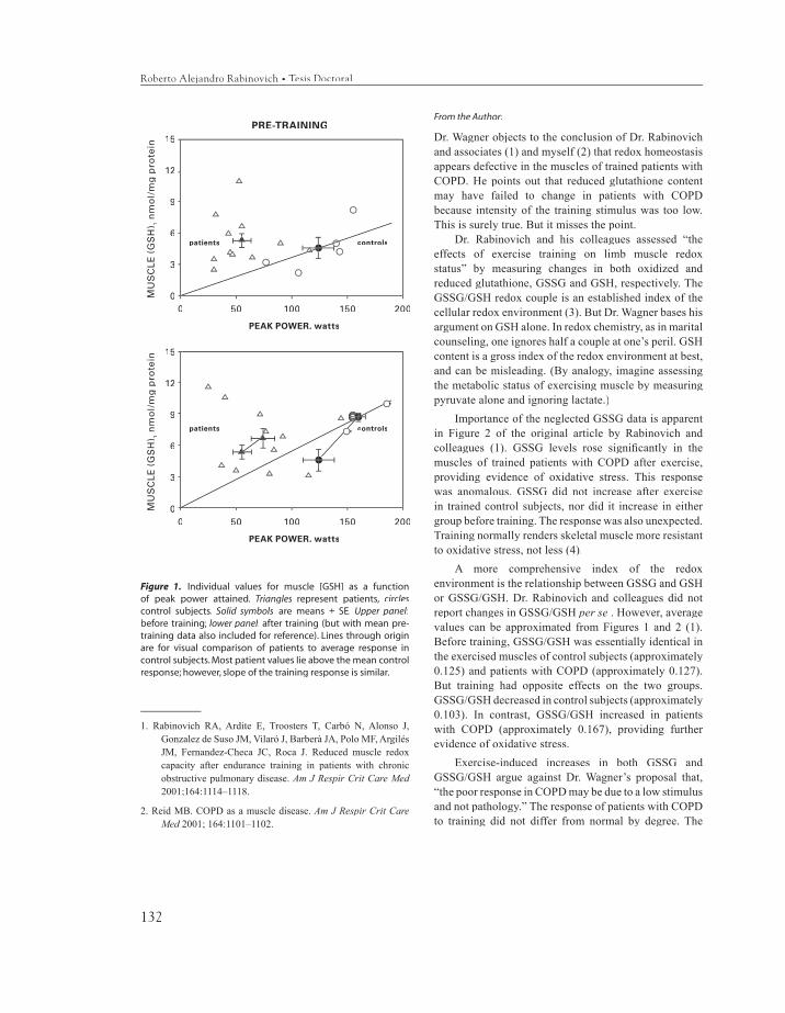
132
Figure 1. Individual values for muscle [GSH] as a function of peak power attained. Triangles represent patients, circles control subjects. Solid symbols are means ± SE. Upper panel:before training; lower panel: after training (but with mean pre-training data also included for reference). Lines through origin are for visual comparison of patients to average response in control subjects. Most patient values lie above the mean control response; however, slope of the training response is similar.
1. Rabinovich RA, Ardite E, Troosters T, Carbó N, Alonso J, Gonzalez de Suso JM, Vilaró J, Barberà JA, Polo MF, Argilés JM, Fernandez-Checa JC, Roca J. Reduced muscle redox capacity after endurance training in patients with chronic obstructive pulmonary disease. Am J Respir Crit Care Med 2001;164:1114–1118.
2. Reid MB. COPD as a muscle disease. Am J Respir Crit Care Med 2001; 164:1101–1102.
From the Author:
Dr. Wagner objects to the conclusion of Dr. Rabinovich and associates (1) and myself (2) that redox homeostasis appears defective in the muscles of trained patients with COPD. He points out that reduced glutathione content may have failed to change in patients with COPD because intensity of the training stimulus was too low. This is surely true. But it misses the point.
Dr. Rabinovich and his colleagues assessed “the effects of exercise training on limb muscle redox status” by measuring changes in both oxidized and reduced glutathione, GSSG and GSH, respectively. The GSSG/GSH redox couple is an established index of the cellular redox environment (3). But Dr. Wagner bases his argument on GSH alone. In redox chemistry, as in marital counseling, one ignores half a couple at one’s peril. GSH content is a gross index of the redox environment at best, and can be misleading. (By analogy, imagine assessing the metabolic status of exercising muscle by measuring pyruvate alone and ignoring lactate.)
Importance of the neglected GSSG data is apparent in Figure 2 of the original article by Rabinovich and colleagues (1). GSSG levels rose signifi cantly in the muscles of trained patients with COPD after exercise, providing evidence of oxidative stress. This response was anomalous. GSSG did not increase after exercise in trained control subjects, nor did it increase in either group before training. The response was also unexpected. Training normally renders skeletal muscle more resistant to oxidative stress, not less (4).
A more comprehensive index of the redox environment is the relationship between GSSG and GSH or GSSG/GSH. Dr. Rabinovich and colleagues did not report changes in GSSG/GSH per se . However, average values can be approximated from Figures 1 and 2 (1). Before training, GSSG/GSH was essentially identical in the exercised muscles of control subjects (approximately 0.125) and patients with COPD (approximately 0.127). But training had opposite effects on the two groups. GSSG/GSH decreased in control subjects (approximately 0.103). In contrast, GSSG/GSH increased in patients with COPD (approximately 0.167), providing further evidence of oxidative stress.
Exercise-induced increases in both GSSG and GSSG/GSH argue against Dr. Wagner’s proposal that, “the poor response in COPD may be due to a low stimulus and not pathology.” The response of patients with COPD to training did not differ from normal by degree. The
PRE-TRAINING
Roberto Alejandro Rabinovich • Tesis Doctoral
patients controls
15
12
9
6
3
0
PEAK POWER, watts
0 50 100 150 200
MU
SC
LE
(G
SH
) 1 n
mo
l/m
g p
rote
inM
US
CL
E (
GS
H) 1
nm
ol/
mg
pro
tein
15
12
9
6
3
0
patients controls
PEAK POWER, watts
0 50 100 150 200
133
difference was qualitative, explicitly abnormal. If one considers the data en toto, pathology seems the most logical explanation.
MICHAEL B. REID
Baylor College of MedicineHouston, Texas
1. Rabinovich RA, Ardite E, Troosters T, Carbó N, Alonso J, Gonzalez de Suso JM, Vilaró J, Barberà JA, Polo MF, Argilés JM, Fernandez-Checa JC, Roca J. Reduced muscle redox capacity after endurance training in patients with chronic obstructive pulmonary disease. Am J Respir Crit Care Med 2001;164:1114–1118.
2. Reid MB. COPD as a muscle disease. Am J Respir Crit Care Med 2001; 164:1101–1102.
3. Schafer FQ, Buettner GR. Redox environment of the cell as viewed through the redox state of the glutathione disulfi de/glutathione couple. Free Rad Biol Med 2001;30:1191–1212.
4. Powers SK, Ji LL, Leeuwenburgh C. Exercise training-induced alterations in skeletal muscle antioxidant capacity: a brief review. Med Sci Sports Exerc 1999;31:987–997.
From the Authors:
We thank Dr. Wagner for his thorough considerations on our work (1). The letter clearly addresses two relevant points:
(1) Magnitude of the stimulus . The arguments regarding effects of absolute versus relative intensity of the training stimulus on skeletal muscle are well taken, but conclusions should not be drawn from pre-training GSH data. Relative (not absolute) intensity of the training stimulus determines muscle adaptation. Although COPD patients (FEV
1 38 ±4 % pred) were trained at a lower
external power than healthy subjects, they showed similar post-training reduction of half-time phosphocreatine ([PCr]) recovery (31P-NMRS) (-12.5±3.3 seconds versus -12.0±2.0 seconds, COPD and controls, respectively). Moreover, a subanalysis in the six COPD patients with low body mass index (FEV
1 27±5 % pred) displayed an
even higher post-training reduction of half-time [PCr] recovery (by -22±3 seconds) (p=0.02). These results indicate that in patients with severe pulmonary disease, the training stimulus produced substantial skeletal muscle adaptations.
Training generated different redox responses in COPD patients and in control subjects. Pre-training muscle GSH levels were not different between COPD patients and healthy sedentary subjects (5.36±0.72 and 4.6±1.03 nmol/
mg protein, respectively), yet COPD patients showed a signifi cant relationship between ∆GSSG (oxidized glutathione) and ∆Wpeak after training (r=0.55, p=0.02). In these patients, no changes in GSH (reduced glutathione) were shown, despite upregulation of gamma-glutamyl cystein synthase (one of the key enzymes in the synthesis of GSH). Briefl y, while training decreased muscle redox potential in these patients, the GSH/GSSG ratio increased after training in controls.
(2) Heterogeneity of COPD patients . The distinction among intrinsic muscle disease, muscle deconditioning, and dysfunction related to abnormal muscle environment was clearly beyond the scope of our study. We must acknowledge, however, that the subset of six low BMI patients showed a post-training fall in total glutathione (-0.90±0.66 nmol/mg protein, p=0.04) not seen in normal BMI patients (2.0±1.11 nmol/mg protein) or in healthy subjects (4.27±1 nmol/mg protein p=0.01 each). Our group is actively working on the notion that although COPD patients with low fat free mass (FFM) may constitute a subset of COPD patients with intrinsic muscle disease, COPD patients with normal FFM may predominantly show the effects of muscle deconditioning. This hypothesis raises a potential debate on the rationale for different types of interventions to enhance physiological adaptations to endurance training in COPD patients that will remain speculative until further progress in this area is achieved.
ROBERTO A. RABINOVICH
JOSEP ROCA
Hospital Clinic, IDIBAPS, University of BarcelonaBarcelona, Spain
1. Rabinovich RA, Ardite E, Troosters T, Carbó N, Alonso J, Gonzalez de Suso JM, Vilaró J, Barberà JA, Polo MF, Argilés JM, Fernandez-Checa JC, Roca J. Reduced muscle redox capacity after endurance training in patients with chronic obstructive pulmonary disease. Am J Respir Crit Care Med 2001;164:1114–1118.
Anexo

135
Índice
Agradecimientos 7
Presentación 9
Introducción 11
La Disfunción Muscular en la EPOC 13 ESTRUCTURA Y FUNCIÓN DEL MÚSCULO SANO 14
CAMBIOS FISIOPATOLÓGICOS EN EL MÚSCULOPERIFÉRICO DEL PACIENTE CON EPOC 18
Fibras musculares 18 Alteraciones de la capilarización y del transporte de oxígeno muscular 19 Alteración de la bioenergética muscular 20 Pérdida de masa muscular 22 CAMBIOS ETIOPATOGÉNICOS DE LA DISFUNCIÓN MUSCULAR EN LA EPOC 23 Alteraciones en el recambio proteico 23 Trastornos nutricionales 24 Sedentarismo 25 Corticosteroides 25
Hipoxia tisular e hipercapnia 26
136
Infl amación sistémica 27 Estrés oxidativo/nitrosativo 28
Los Músculos Respiratorios en la EPOC 29
Sedentarismo y Entrenamiento Muscular en la EPOC 31
El Estrés Oxidativo/Nitrosativo como Mecanismo Patogénico 35
Objetivos 41OBJETIVO I - EFECTOS DEL ENTRENAMIENTO SOBRE
EL SISTEMA GLUTATION 41 Manuscrito I 42 Hipótesis 42 Objetivo específi co 42
OBJETIVO II - RELACIONES ENTRE ESTRÉS OXIDATIVO Y PERDIDA DE MASA MUSCULAR 42 Manuscrito II 43 Hipótesis 43 Objetivo específi co 43
OBJETIVO III - INTERACCIONES ENTRE INFLAMACIÓN Y EJERCICIO 43 Manuscrito III 44 Hipótesis 44 Objetivo específi co 44
OBJETIVO IV - ALTERACIONES DE LA CADENA RESPIRATORIA MITOCONDRIAL Y DISFUNCIÓN MUSCULAR 44 Manuscrito IV 45 Hipótesis 45 Objetivo específi co 45
Trabajos Originales 47
Roberto Alejandro Rabinovich • Tesis Doctoral
137
MANUSCRITO I 47MANUSCRITO II 57MANUSCRITO III 65MANUSCRITO IV 75
Resumen de Resultados 89MANUSCRITO I 89MANUSCRITO II 90MANUSCRITO III 90MANUSCRITO IV 91
Discusión 93EFECTOS DEL ENTRENAMIENTO SOBRE EL SISTEMA GLUTATION 93RELACIONES ENTRE ESTRÉS OXIDATIVO Y PÉRDIDA DE MASA
MUSCULAR 94INTERACCIONES ENTRE INFLAMACIÓN Y ESTRÉS OXIDATIVO 96RELACIÓN ENTRE DESACOPLAMIENTO DE LA CADENA
RESPIRATORIA Y DISFUNCIÓN MUSCULAR 104
Resumen 107
Conclusiones 109
Perspectivas de Futuro 111
Bibliografía 113
Acrónimos 125
Anexo 127EDITORIAL 127CARTA AL EDITOR 131
Índice