Embed Size (px)
Citation preview
ÖZET
Amaç: Bu çalışmada, parotis bezinde yerleşmiş benign ve malign lezyonların tanı yöntemleri ve tedavi metodla-rı, radyolojik görüntüleme ve ince iğne aspirasyon biopsi-sinin (İİAB) lezyonun histopatolojik tanısını desteklemede ve yapılacak cerrahinin şeklini belirlemedeki rolü ve fonk-siyonel sonuçlar değerlendirildi.
Gereç ve Yöntem: Ekim 2005 ile Aralık 2010 tarihleri arasında kliniğimizde yapılan 79 parotidektomi retrospek-tif olarak tarandı. Yaş, cinsiyet, klinik bulgular, İİAB si-tolojisi, radyolojik değerlendirme, cerrahi metodlar, his-topatolojik değerlendirme ve komplikasyonlar kaydedildi.
Bulgular: Yetmiş dokuz hastanın lezyon dağılımı; 59 be-nign (%75), 19 malign (%24) ve 1 enflamatuvardı (%1). En yaygın benign tümör pleomorfik adenom (%69), en yaygın malign tümör asinik hücreli karsinomdu (%15.8). Malign tümörlü hastaların ortalama yaşı benign tümörlü hastaların yaşına göre anlamlı olarak yüksekti. İİAB’nin doğruluk oranı malign tümörler için %90.63, benign tü-mörler için %76.56 idi. Yüzeyel lobta yerleşik 37 pleo-morfik adenom ve 13 Warthin tümörüne superfisiyal paro-tidektomi yapılırken, derin lobta yerleşmiş 12 malign ve 5 benign tümör total parotidektomi ile çıkarılmıştır. Seri-mizde en yaygın komplikasyon fasiyal paraliziydi (%5).
Sonuç: İİAB sitolojisinin malign benign ayırımındaki be-lirgin ayırıcılığına rağmen, parotis bezi lezyonlarının tanı-sı radyolojik ve klinik verilerin İİAB verilerini tamamlaması ile konur. Ultrasonografi ucuz maliyeti ve radyasyon riskinin olmaması ile yüzeyel lob lezyonlarında rutin olarak kullanı-lır. Bilgisayarlı tomografi ve manyetik rezonans görüntüleme malign ve bazı seçici benign lezyonlarda tercih edilir. Cerra-hi tercihimiz süperfisiyal lob benign tümörlerde superfisiyal parotidektomi iken derin lob benign tümörlerde ve ileri evre malign tümörlerde total parotidektomidir. Anahtar sözcükler: İnce iğne aspirasyon biopsisi; parotidek-tomi; parotis tümörü.
İstanbul Tıp Derg - Istanbul Med J 2012;13(3):125-132doi: 10.5505/1304.8503.2012.27146
ORIGINAL ARTICLE - KLİNİK ÇALIŞMA
The Clinical Management of the Parotid Masses:A Five Year StudyParotis Kitlelerine Klinik Yaklaşım: 5 Yıllık DeneyimSuat BİLİCİ, Meltem Esen AKPINAR, Özgür YİĞİT, Zehra DÖNMEZ
SUMMARY Objectives: Our objective was to analyze the diagnostic mea-sures and management methods for benign and malignant le-sions localized in the parotid gland and to assess the role of radiological imaging, fine-needle aspiration biopsy (FNAB), the type and extent of surgery in respect to histopathology of the lesion and subsequent functional results.Methods: The data related to 79 parotidectomies performed in our clinic between October 2005 and December 2010 were retrospectively reviewed. Age, gender, clinical findings, FNAB cytology, radiological evaluation, surgical methods, histo-pathological evaluation, and complications were recorded.Results: The distribution of the lesions found on the 79 pa-tients was as follows: 59 benign (75%), 19 malignant (24%), and one inflammatory (1%). The most common benign tumor was pleomorphic adenoma (69%) and the most common ma-lignant tumor was acinic cell carcinoma (15.8%). The mean age of patients with malignant tumor was significantly higher than the mean age of patients with benign tumors. The ac-curacy rate of FNAB was 90.63% for malignant tumors and 76.56% for benign tumors. The 37 pleomorphic adenomas localized in the superficial lobe and the 13 Warthin’s tumors were managed with superficial parotidectomy, whereas the 12 malignant and 5 benign tumors localized in the deep lobe were removed with total parotidectomy. The most common complication in our series was facial paralysis (5%).Conclusion: Despite the significant contribution of FNAB cytology, particularly in malignant-benign differentiation, the diagnosis of parotid gland lesions should include either clini-cal or radiological data as complementary to FNAB cytology data. Ultrasonography, owing to the low cost and absence of radiation exposure, may be used routinely for locating super-ficial lobe masses. CT and MRI may be reserved for malignant and selected benign cases. Our surgical preference was su-perficial parotidectomy in superficial benign parotid masses and total parotidectomy in deep-lobe benign tumors and ad-vanced-stage malignant tumors.Key words: Fine-needle aspiration biopsy; parotidectomy; pa-rotid tumor.
Submitted (Geliş tarihi): 19.03.2012 Accepted (Kabul tarihi): 22.06.2012
İstanbul Eğitim ve Araştırma Hastanesi, Kulak Burun Boğaz Kliniği, İstanbul
Correspondence (İletişim): Dr. Suat Bilici. e-mail (e-posta): [email protected]
125
İstanbul Tıp Derg
INTRODUCTIONThe management of parotid masses including di-
agnostic and therapuetic aspects warrants systemati-cal approach.
In diagnostic work-up apart form the history, the radiological imaging techniques including ul-trasonography (US), computed tomography (CT), and magnetic resonance imaging (MRI) are fre-quently encountered.[1] Fine-needle aspiration biopsy (FNAB) is of value in benign and malignant dif-ferentiation. The accuracy rate of FNAB in benign and malignant lesion differentiation was reported as 81-98%.[2,3] In another study, the validity of FNAB was found nearly 80%.[3,4] The major complications associated with fine-needle aspiration biopsy were not reported previously. FNAB is an easy, safe, and minimally invasive procedure.[4-6]
In parotid masses, the combined data derived from clinical, radiological and pathological assess-ment provides the final diagnosis.[7]
Seventy to eighty percent of all salivary gland tu-mors are localized in parotid gland. Parotid masses are predominantly benign tumors (80%). The most commonly encountered benign tumors are pleomor-phic adenomas, followed by Warthin’s tumor. The most common malignant tumor of the parotid gland is mucoepidermoid carcinoma.[8]
Benign lesions localized in the superficial lobe are managed with superficial parotidectomy, whereas benign lesions localized in the deep lobe necessitate total parotidectomy.[9,10] In the early stages, superfi-cial malignant lesions also only require superficial parotidectomy; however, total parotidectomy is per-formed for deep lobe lesions. In advanced stages and for high grade lesions, neck dissection and/or radio-therapy are combined with total parotidectomy.[8]
In this study, 79 patients diagnosed with parotid tumors were retrospectively analyzed. The clinical findings, fine-needle aspiration biopsy results, type of the surgery, postoperative histopathology, and any resulting complications. The diagnostic measures were reviewed to assess the role of radiological im-aging and FNAB. The management including the type and extent of surgical treatment was discussed
in respect to histopathology of the lesion and subse-quent functional results.
MATERIALS AND METHODS
This study retrospectively analyzed 79 patients who received operations for parotid masses and met-astatic lesions at the Istanbul Training and Research Hospital between 2005 and 2010. The demographic, clinical, surgical, histopathological and postoperative follow-up data. Postoperative complications, ma-lignant tumor stages for malignant lesions and data were collected from their records. The demographic data included age, gender. Clinical data consisted of symptoms, signs, staging for malignant lesions, signs revealed through clinical examination and radiologi-
126
Table 1. Classification of cytology
Positive value Exclusively benign lesion
Negative value Malignant, inflammatory, or suspicious
Positive value Exclusively malignant lesion
Negative value Malignant, inflammatory, or suspicious
Table 2. Histopathology of the parotid tumors (October 2005-December 2010)
Histology n %
Benign tumors 59 75
Pleomorphic adenoma 41 69
Warthin’s tumor 14 24
Myoepithelioma 2
Oncocytoma 1
Lymphoepitalial cyst 1
Malignant tumors 19 24
Acinic cell carcinoma 3
Mucoepidermoid carcinoma 2
Adenoid cystic carcinoma 2
Epidermoid carcinoma 2
Malignant fibrous histiocytoma 1
Spindle cell carcinoma 1
Myoepitelial carcinoma 1
Auricula basal cell carcinoma 1
Retromolar trigone
epidermoid carcinoma 1
Salivary duct carcinoma 1
Folliculer lymphoma 1
Buccal region epidermoid carcinoma 1
Undiferantiated pleomorphic sarcoma 1
Inflammatory parotid disease 1 1
cal imaging. Surgical data included type of surgery, perioperative findings, histopathological data con-sisted of FNAB cytology and final specimen pathol-ogy reports. FNAB cytology was categorized (Table 1) to calculate specificity, sensitivity, and accuracy values were also calculated.
The statistical analysis was performed using SAS package (9.2 version, cary, NC, USA). A student t- test was used to compare categorical data on benign and malignant lesions. The predictive value of fine- needle aspiration biopsy in the diagnosis of benign and malignant tumors was calculated using a chi-square test.
RESULTSSeventy-nine patients had a parotidectomy at
the Istanbul Training and Research Hospital Second Otolaryngology Clinic between 2005 and 2010. Post-operative histological diagnoses are summarized in Table 2.
The male and female distribution was 48 (61%) and 31 (39%), respectively. The distribution of pa-tients with benign lesions is summarized in Table 3. The average age for patients with benign and malig-nant lesions was 47.9 and 64 years old, respectively. The difference between the two groups was statisti-
cally significant (p=0.0002) (Table 4).
Pleomorphic adenoma (69%) was the most com-monly encountered benign lesion, with the second most common lesion being Warthin’s tumor (23%). The Warthin’s tumor was bilateral in one patient out of 14. In our series, the most common malignant tumors were acinic cell carcinoma (15.8%), muco-epidermoid carcinoma (10.5%), adenoid cystic carci-noma (10.5%), and epidermoid carcinoma (10.5%), respectively.
Sixty-one superficial and 15 deep- lobe localized tumors were encountered. The distribution is sum-marized in Table 3.
Radiological Evaluation
During the diagnostic work-up, every patient had an ultrasonographical examination. The distribution of selected patients with CT and MRI exams during the preoperative diagnostic work-up is summarized in Table 5.
127
The Clinical Management of the Parotid Masses: A Five Year Study
Table 3. Comparison of benign and malignant parotid tumors
Benign Malignant
No. of patients 60 18
Male / Female 35/25 12/6
Age (year) 47.9 (18-82) 64.6 (28-86)
Right/Left/Bilateral 32/27/1 10/8
Superficial/Deep/Safe 55/5 6/9/3
Table 4. Comparison of age and gender in the benign and malignant parotid tumors (p<0.05)
n Avarage Standard deviation p
Age All groups 78 51.818 16.602
Benign 60 47.898 14.874 0.00022
Malignant 18 64.667 15.789
Gender (Male/Female) All groups 47/31 0.597/0.403 –
Benign 35/25 0.576/0.424 – 0.4858
Malignant 12/6 0.667/0.333 –
Table 5. Radiological imaging (BT and MRI)
Patient groups BT MRI BT+MRI
Malignant
Malignant fibrous histiocytoma +
Retromolar trigone
epidermoid carcinoma +
Spindle cell carcinoma +
İndiferantiated anaplastic carcinoma +
Pleomorphic undiferantiated sarcoma +
Myoepitelial carcinoma +
Renal clear cell carcinoma metastasis +
Benign
Giant pleomorphic adenoma +
Pleomorphic adenoma recurrence +

Table 7. Specificity, sensitivity and accuracy of the FNAB for malignant parotid tumors
Cytology Histology Total
Positive Negative
Positive 42 5 47
Negative 10 7 17
Total 52 12 64
Specificity= 51/52= 0.9808; Sensitivity= 7/12= 0.5833;Accuracy= 58/64= 0.9063.
Fine-Needle Aspiration Biopsy (FNAB) Cytol-ogy:
FNAB was performed in 64 of 79 patients. Five of the remaining 15 patient had neighboring metastatic lesions with incisional biopsy results. Ten patient had their biopsy performed in other centres and had no records at our institution. FNAB was consistent with the final diagnosis in 33 of 41 patients diagnosed with pleomorphic adenoma. The records of five patients were missing. The remaining patients were reported as having chronic inflammation (1 case), intraparot-id neoplasia (1 case), and spindle cell neoplasia (1 case). The FNAB was consistent with the final di-agnosis of Warthin’s tumors in seven of 14 patients. In the remaining five patients, FNAB was revealed acute inflammation (1 case), suspicious cytology (1 case), oncocytic epithelial cells (1 case) and cyst con-sistent findings (1 case). In three cases with a final diagnosis of acinic cell carcinoma, FNAB revealed a benign oncocytic tumor, suspicious cytology, and was inconclusive in discrimination between muco-epidermoid carcinoma and benign epithelial tumors.
In this study, the specificity, sensitivity, and accu-racy of FNAB for malignant tumors was calculated
as 0.98, 0.58 and 0.91, respectively. FNAB specific-ity, sensitivity and accuracy for benign tumors were calculated as 0.58, 0.81, and 0.77, respectively (Ta-bles 6,7,8).
Surgical Procedure and Evaluation of Results Thirthy-seven pleomorphic adenoma (59.6%)
and 13 Warthin’s tumors (20%) were diagnosed in 62 patients undergoing superficial parotidectomy. Our surgical approach was a classical superficial pa-rotidectomy, involving a preauriculocervical incision (Blair), and the detection of the main trunk of the facial nerve with through an anterograde approach. Facial nerve monitorization was not routine, except revision surgeries. One of the 62 patients with the basal cell carcinoma of the auricula had a superficial parotidectomy together with a skin excision, and an-other patient with the final diagnosis of Warthin’s tu-mor had a superficial parotidectomy together with a skin excision and functional neck dissection. A total parotidectomy was performed in one recurrent case, FNAB reported pleomorphic adenoma with a final histopathological diagnosis of epidermoid carci-noma. Three patients with maxillar skin epidermoid
İstanbul Tıp Derg
128
Table 6. The patient distribution according to FNAB results
Cytology Histology Total
Benign Malignant Inflammation Not available
Benign 42 5 0 0 47
Malignant 1 7 0 0 8
Inflammation 4 0 1 0 5
Suspicious 4 0 0 0 4
Not available 8 6 0 1 15
Total 59 18 1 1 79
Table 8. Specificity, sensitivity and accuracy of the FNAB for benign parotid tumors
Cytology Histology Total
Positive Negative
Positive 7 1 8
Negative 5 51 56
Total 12 52 64
Specificity= 7/12= 0.5833; Sensitivity= 42/52= 0.8077;Accuracy= 49 /64= 0.7656
The Clinical Management of the Parotid Masses: A Five Year Study
carcinoma, basal cell auricular carcinoma, and ret-romolar trigone carcinoma had a superficial paroti-dectomy combined with an excision of the original lesion, but a histopathological parotid gland infiltra-tion was not detected.
Seventeen patients with 12 malignant and five benign lesions had a total parotidectomy. Seven pa-tient had a neck dissection (two radical, one modified radical, and four functional) combined with a total parotidectomy. Deep -lobe localized pleomorphic adenomas (4 cases) and Warthin’s tumor (1 case) were managed with a total parotidectomy.
Two patients with a histopathological diagnosis of follicular lymphoma and malign fibrous histiocy-toma had chemotherapy. One patient with malignant fibrous histiocytoma had a recurrence, and one pa-tient with a tumor in retromolar trigone had a recur-rence despite postoperative radiotheraphy and che-motherapy.
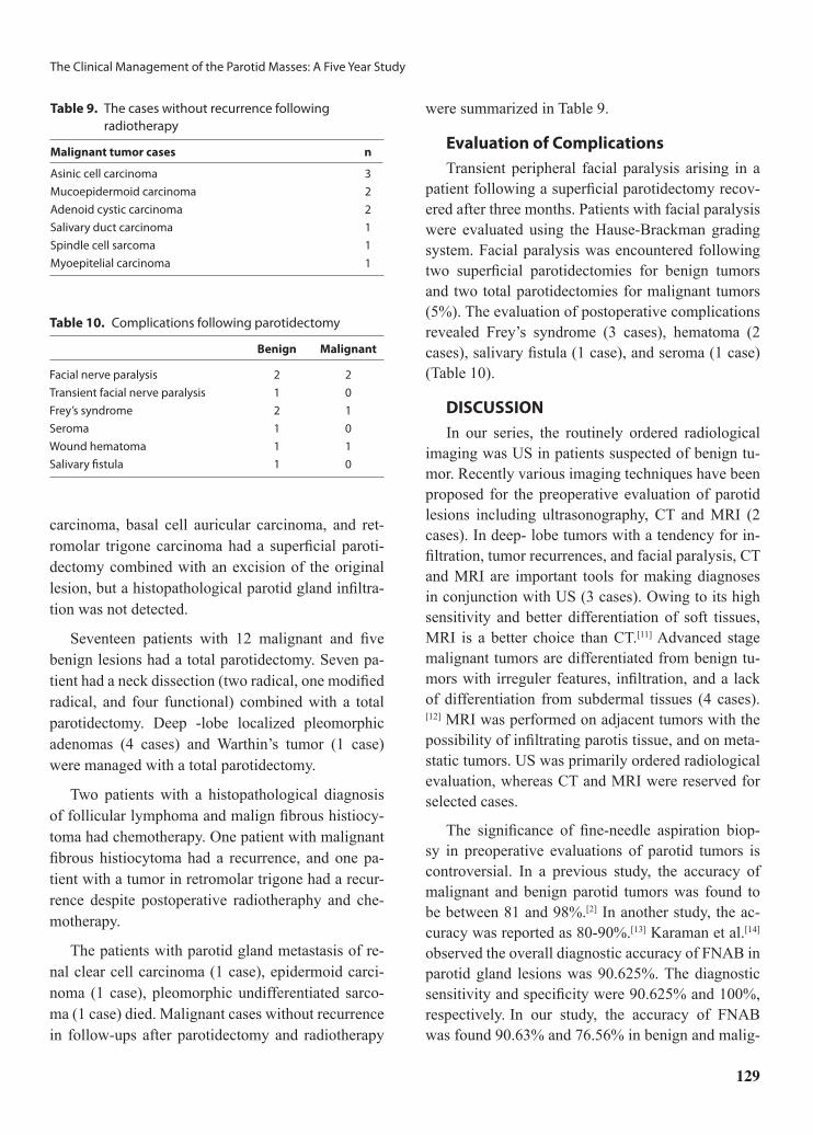
The patients with parotid gland metastasis of re-nal clear cell carcinoma (1 case), epidermoid carci-noma (1 case), pleomorphic undifferentiated sarco-ma (1 case) died. Malignant cases without recurrence in follow-ups after parotidectomy and radiotherapy
were summarized in Table 9.
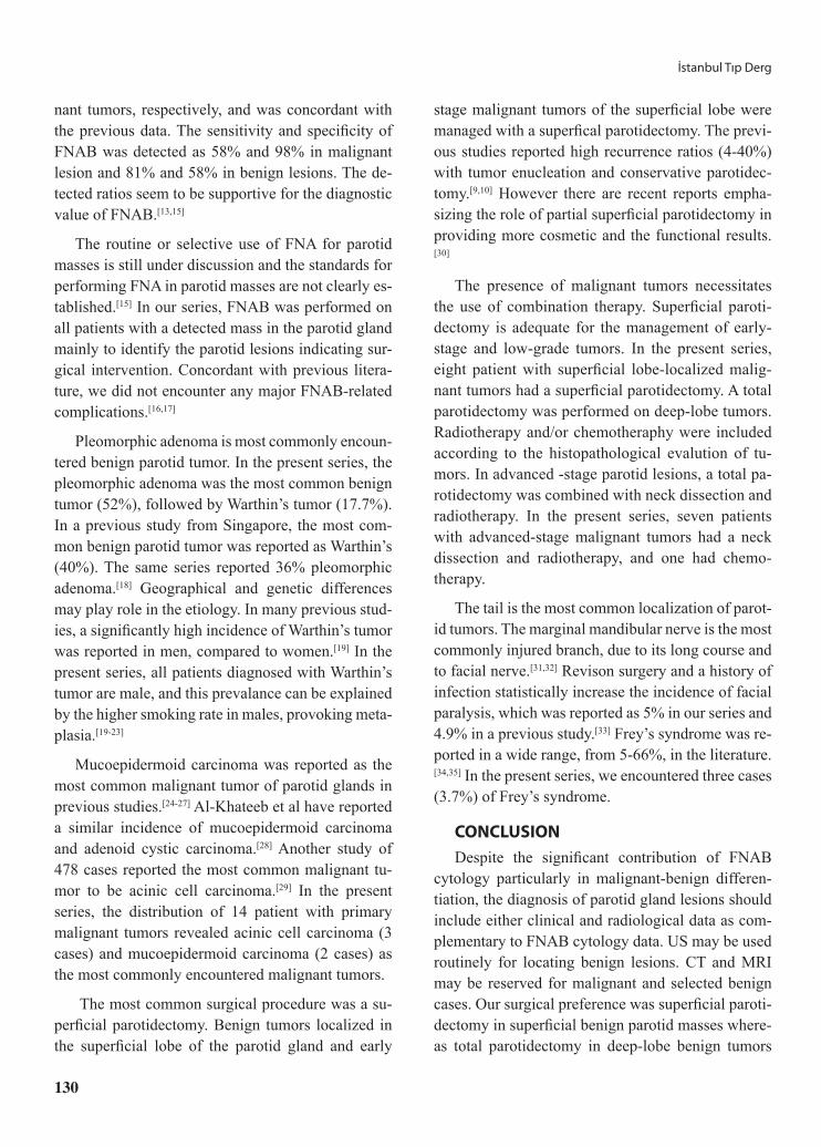
Evaluation of ComplicationsTransient peripheral facial paralysis arising in a
patient following a superficial parotidectomy recov-ered after three months. Patients with facial paralysis were evaluated using the Hause-Brackman grading system. Facial paralysis was encountered following two superficial parotidectomies for benign tumors and two total parotidectomies for malignant tumors (5%). The evaluation of postoperative complications revealed Frey’s syndrome (3 cases), hematoma (2 cases), salivary fistula (1 case), and seroma (1 case) (Table 10).
DISCUSSIONIn our series, the routinely ordered radiological
imaging was US in patients suspected of benign tu-mor. Recently various imaging techniques have been proposed for the preoperative evaluation of parotid lesions including ultrasonography, CT and MRI (2 cases). In deep- lobe tumors with a tendency for in-filtration, tumor recurrences, and facial paralysis, CT and MRI are important tools for making diagnoses in conjunction with US (3 cases). Owing to its high sensitivity and better differentiation of soft tissues, MRI is a better choice than CT.[11] Advanced stage malignant tumors are differentiated from benign tu-mors with irreguler features, infiltration, and a lack of differentiation from subdermal tissues (4 cases).[12] MRI was performed on adjacent tumors with the possibility of infiltrating parotis tissue, and on meta-static tumors. US was primarily ordered radiological evaluation, whereas CT and MRI were reserved for selected cases.
The significance of fine-needle aspiration biop-sy in preoperative evaluations of parotid tumors is controversial. In a previous study, the accuracy of malignant and benign parotid tumors was found to be between 81 and 98%.[2] In another study, the ac-curacy was reported as 80-90%.[13] Karaman et al.[14] observed the overall diagnostic accuracy of FNAB in parotid gland lesions was 90.625%. The diagnostic sensitivity and specificity were 90.625% and 100%, respectively. In our study, the accuracy of FNAB was found 90.63% and 76.56% in benign and malig-
129
Table 9. The cases without recurrence following radiotherapy
Malignant tumor cases n
Asinic cell carcinoma 3
Mucoepidermoid carcinoma 2
Adenoid cystic carcinoma 2
Salivary duct carcinoma 1
Spindle cell sarcoma 1
Myoepitelial carcinoma 1
Table 10. Complications following parotidectomy
Benign Malignant
Facial nerve paralysis 2 2
Transient facial nerve paralysis 1 0
Frey’s syndrome 2 1
Seroma 1 0
Wound hematoma 1 1
Salivary fistula 1 0
nant tumors, respectively, and was concordant with the previous data. The sensitivity and specificity of FNAB was detected as 58% and 98% in malignant lesion and 81% and 58% in benign lesions. The de-tected ratios seem to be supportive for the diagnostic value of FNAB.[13,15]
The routine or selective use of FNA for parotid masses is still under discussion and the standards for performing FNA in parotid masses are not clearly es-tablished.[15] In our series, FNAB was performed on all patients with a detected mass in the parotid gland mainly to identify the parotid lesions indicating sur-gical intervention. Concordant with previous litera-ture, we did not encounter any major FNAB-related complications.[16,17]
Pleomorphic adenoma is most commonly encoun-tered benign parotid tumor. In the present series, the pleomorphic adenoma was the most common benign tumor (52%), followed by Warthin’s tumor (17.7%). In a previous study from Singapore, the most com-mon benign parotid tumor was reported as Warthin’s (40%). The same series reported 36% pleomorphic adenoma.[18] Geographical and genetic differences may play role in the etiology. In many previous stud-ies, a significantly high incidence of Warthin’s tumor was reported in men, compared to women.[19] In the present series, all patients diagnosed with Warthin’s tumor are male, and this prevalance can be explained by the higher smoking rate in males, provoking meta-plasia.[19-23]
Mucoepidermoid carcinoma was reported as the most common malignant tumor of parotid glands in previous studies.[24-27] Al-Khateeb et al have reported a similar incidence of mucoepidermoid carcinoma and adenoid cystic carcinoma.[28] Another study of 478 cases reported the most common malignant tu-mor to be acinic cell carcinoma.[29] In the present series, the distribution of 14 patient with primary malignant tumors revealed acinic cell carcinoma (3 cases) and mucoepidermoid carcinoma (2 cases) as the most commonly encountered malignant tumors.
The most common surgical procedure was a su-perficial parotidectomy. Benign tumors localized in the superficial lobe of the parotid gland and early
stage malignant tumors of the superficial lobe were managed with a superfical parotidectomy. The previ-ous studies reported high recurrence ratios (4-40%) with tumor enucleation and conservative parotidec-tomy.[9,10] However there are recent reports empha-sizing the role of partial superficial parotidectomy in providing more cosmetic and the functional results.[30]
The presence of malignant tumors necessitates the use of combination therapy. Superficial paroti-dectomy is adequate for the management of early-stage and low-grade tumors. In the present series, eight patient with superficial lobe-localized malig-nant tumors had a superficial parotidectomy. A total parotidectomy was performed on deep-lobe tumors. Radiotherapy and/or chemotheraphy were included according to the histopathological evalution of tu-mors. In advanced -stage parotid lesions, a total pa-rotidectomy was combined with neck dissection and radiotherapy. In the present series, seven patients with advanced-stage malignant tumors had a neck dissection and radiotherapy, and one had chemo-therapy.
The tail is the most common localization of parot-id tumors. The marginal mandibular nerve is the most commonly injured branch, due to its long course and to facial nerve.[31,32] Revison surgery and a history of infection statistically increase the incidence of facial paralysis, which was reported as 5% in our series and 4.9% in a previous study.[33] Frey’s syndrome was re-ported in a wide range, from 5-66%, in the literature.[34,35] In the present series, we encountered three cases (3.7%) of Frey’s syndrome.
CONCLUSIONDespite the significant contribution of FNAB
cytology particularly in malignant-benign differen-tiation, the diagnosis of parotid gland lesions should include either clinical and radiological data as com-plementary to FNAB cytology data. US may be used routinely for locating benign lesions. CT and MRI may be reserved for malignant and selected benign cases. Our surgical preference was superficial paroti-dectomy in superficial benign parotid masses where-as total parotidectomy in deep-lobe benign tumors
İstanbul Tıp Derg
130
The Clinical Management of the Parotid Masses: A Five Year Study
and advanced-stage malignant tumors.
REFERENCES1. Urquhart A, Hutchins LG, Berg RL. Preoperative
computed tomography scans for parotid tumor evalu-ation. Laryngoscope 2001;111:1984-8.
2. Seethala RR, LiVolsi VA, Baloch ZW. Relative accu-racy of fine-needle aspiration and frozen section in the diagnosis of lesions of the parotid gland. Head Neck 2005;27:217-23.
3. Howlett DC. Diagnosing a parotid lump: fine nee-dle aspiration cytology or core biopsy? Br J Radiol 2006;79:295-7.
4. Zbären P, Nuyens M, Loosli H, et al. Diagnostic ac-curacy of fine-needle aspiration cytology and fro-zen section in primary parotid carcinoma. Cancer 2004;100:1876-83.
5. Herrera Hernández AA, Díaz Pérez JA, García CA, et al. Evaluation of fine needle aspiration cytology in the diagnosis of cancer of the parotid gland. [Article in Spanish] Acta Otorrinolaringol Esp 2008;59:212-6. [Abstract]
6. Salgarelli AC, Capparè P, Bellini P, et al. Usefulness of fine-needle aspiration in parotid diagnostics. Oral Maxillofac Surg 2009;13:185-90.
7. Bussu F, Parrilla C, Rizzo D, et al. Clinical approach and treatment of benign and malignant parotid mass-es, personal experience. Acta Otorhinolaryngol Ital 2011;31:135-43.
8. Lin CC, Tsai MH, Huang CC, et al. Parotid tumors: a 10-year experience. Am J Otolaryngol 2008;29:94-100.
9. Bradley PJ. Pleomorphic salivary adenoma of the pa-rotid gland: which operation to perform? Curr Opin Otolaryngol Head Neck Surg 2004;12:69-70.
10. Zbären P, Tschumi I, Nuyens M, et al. Recurrent pleo-morphic adenoma of the parotid gland. Am J Surg 2005;189:203-7.
11. Lee YY, Wong KT, King AD, et al. Imaging of sali-vary gland tumours. Eur J Radiol 2008;66:419-36.
12. Harish K. Management of primary malignant epithe-lial parotid tumors. Surg Oncol 2004;13:7-16.
13. Cohen EG, Patel SG, Lin O, et al. Fine-needle aspi-ration biopsy of salivary gland lesions in a selected patient population. Arch Otolaryngol Head Neck Surg 2004;130:773-8.
14. Karaman M, Tuncel A, Tel A, Erdem Habeşoğlu T. Correlation between fine needle aspiration biopsy and histologic findings in parotid masses. KBB ve BBC Dergisi 2010;18:1-5.
15. Zbären P, Schär C, Hotz MA, et al. Value of fine-nee-dle aspiration cytology of parotid gland masses. La-
ryngoscope 2001;111:1989-92.16. Awan MS, Ahmad Z. Diagnostic value of fine needle
aspiration cytology in parotid tumors. J Pak Med As-soc 2004;54:617-9.
17. Aversa S, Ondolo C, Bollito E, et al. Preoperative cy-tology in the management of parotid neoplasms. Am J Otolaryngol 2006;27:96-100.
18. Lim LH, Chao SS, Goh CH, et al. Parotid gland sur-gery: 4-year review of 118 cases in an Asian popula-tion. Head Neck 2003;25:543-8.
19. Yoo GH, Eisele DW, Askin FB, et al. Warthin’s tumor: a 40-year experience at The Johns Hopkins Hospital. Laryngoscope 1994;104:799-803.
20. Chung YF, Khoo ML, Heng MK, et al. Epidemiology of Warthin’s tumour of the parotid gland in an Asian population. Br J Surg 1999;86:661-4.
21. Eveson JW, Cawson RA. Warthin’s tumor (cystadeno-lymphoma) of salivary glands. A clinicopathologic investigation of 278 cases. Oral Surg Oral Med Oral Pathol 1986;61:256-62.
22. Seifert G, Donath K. Multiple tumours of the salivary glands--terminology and nomenclature. Eur J Cancer B Oral Oncol 1996;32:3-7.
23. Batsakis JG, el-Naggar AK. Warthin’s tumor. Ann Otol Rhinol Laryngol 1990;99:588-91.
24. Li LJ, Li Y, Wen YM, et al. Clinical analysis of sali-vary gland tumor cases in West China in past 50 years. Oral Oncol 2008;44:187-92.
25. Subhashraj K. Salivary gland tumors: a single institu-tion experience in India. Br J Oral Maxillofac Surg 2008;46:635-8.
26. Vargas PA, Gerhard R, Araújo Filho VJ, et al. Salivary gland tumors in a Brazilian population: a retrospective study of 124 cases. Rev Hosp Clin Fac Med Sao Paulo 2002;57:271-6.
27. Pinkston JA, Cole P. Incidence rates of salivary gland tumors: results from a population-based study. Otolar-yngol Head Neck Surg 1999;120:834-40.
28. Al-Khateeb TH, Ababneh KT. Salivary tumors in north Jordanians: a descriptive study. Oral Surg Oral Med Oral Pathol Oral Radiol Endod 2007;103:53-9.
29. van der Wal JE, Leverstein H, Snow GB, et al. Parotid gland tumors: histologic reevaluation and reclassifica-tion of 478 cases. Head Neck 1998;20:204-7.
30. Roh JL, Kim HS, Park CI. Randomized clinical trial comparing partial parotidectomy versus superficial or total parotidectomy. Br J Surg 2007;94:1081-7.
31. Al Salamah SM, Khalid K, Khan IA, et al. Outcome of surgery for parotid tumours: 5-year experience of a general surgical unit in a teaching hospital. ANZ J Surg 2005;75:948-52.
32. Astor FC, Ackerman EB, Hanft KL, et al. Surgical
131
treatment of parotid tumors in the general community hospital. South Med J 2002;95:1404-7.
33. Granell J, Sánchez-Jara JL, Gavilanes J, et al. Man-agement of the surgical pathology of the parotid gland: A review of 54 cases. [Article in Spanish] Acta Otorrinolaringol Esp 2010;61:189-95. [Abstract]
34. Guntinas-Lichius O, Klussmann JP, Wittekindt C, et
al. Parotidectomy for benign parotid disease at a uni-versity teaching hospital: outcome of 963 operations. Laryngoscope 2006;116:534-40.
35. Marshall AH, Quraishi SM, Bradley PJ. Patients’ per-spectives on the short- and long-term outcomes fol-lowing surgery for benign parotid neoplasms. J Lar-yngol Otol 2003;117:624-9.
İstanbul Tıp Derg
132