Embed Size (px)
Citation preview

Treatment methods of long bone fractures in equines
– comparing Swedish and foreign experience
Behandlingsmetoder för frakturer på långa rörformiga ben hos häst
- en jämförelse mellan svenska och utländska erfarenheter
Sergey Gazeev
Degree project/Independent project • 30 credits
Swedish University of Agricultural Sciences, SLU
Faculty of Veterinary Medicine and Animal Science
Veterinary Medicine Programme
Uppsala 2021

Treatment methods of long bone fractures in equines
– comparing Swedish and foreign experience
Behandlingsmetoder för frakturer på långa rörformiga ben hos häst - en jämförelse mellan svenska och utländska erfarenheter
Sergey Gazeev
Supervisor: Ove Wattle, Swedish University of Agricultural Sciences, Department of
Clinical Sciences
Assistant supervisor: Karl Ljungvall, Mälaren Hästklinik
Examiner: Lena Ström, Swedish University of Agricultural Sciences, Department of
Clinical Sciences
Credits: 30 credits
Level: A2E
Course title: Independent project in Veterinary Medicine
Course code: EX0869
Programme/education: Veterinary Medicine Programme
Course coordinating dept: Department of Clinical Sciences
Place of publication: Uppsala
Year of publication: 2021
Cover picture: Lena Lopes
Keywords: equine, long bone, fracture, osteosynthesis
Swedish University of Agricultural Sciences
Faculty of Veterinary Medicine and Animal Science
Department of Clinical Sciences
Approved students’ theses at SLU are published electronically. As a student, you
have the copyright to your own work and need to approve the electronic publishing.
If you check the box for YES, the full text (pdf file) and metadata will be visible
and searchable online. If you check the box for NO, only the metadata and the ab-
stract will be visible and searchable online. Nevertheless, when the document is
uploaded it will still be archived as a digital file. If you are more than one author you all need to agree on a decision. Read about SLU’s
publishing agreement here: https://www.slu.se/en/subweb/library/publish-and-ana-
lyse/register-and-publish/agreement-for-publishing/.
☒ YES, I/we hereby give permission to publish the present thesis in accordance
with the SLU agreement regarding the transfer of the right to publish a work.
Publishing and archiving
Abstract
The treatment of long bone fractures in horses remains to be a challenge for equine veterinarians,
since it is necessary to succeed treating an animal sometimes weighing over 500kg that will have to
endure fracture repair procedures and rehabilitation for at least 3 months and manage the resulting
complications. The objective of this study is to investigate and shed light over some of the treatment
techniques for long bone fractures in equines, and to sample new data from a questionnaire, an-
swered by experienced equine surgeons in Sweden, summarising the obtained results with the al-
ready existing research. In a literature review, this work starts by describing bone anatomy and
physiology, gradually narrowing down to long bones. Then the different types of bone fractures are
discussed, both in regards to their aetiology, impact and classification. This is followed by the mech-
anisms of bone healing through which bone fractures are healed. Further, this work investigates the
most common and current treatment techniques of long bone fractures in equines. Various treatment
techniques are reviewed, such as the internal and the external fixation, as well as the advantages and
the disadvantages of every respective technique. Furthermore, the factors that affect long bone frac-
ture repair are described, as well as the biomechanics of the strain forces that need to be counteracted
for successful fracture treatment. Finally, the literature study describes the various complications
that may follow bone fracture repair post-operatively or during the rehabilitation phase. In addition,
the study encompasses a questionnaire that was sent to 20 Swedish senior equine surgeons, asking
questions about long bone fracture treatment. Some of them replied to the questionnaire, elucidating
on how the different types of long bone fractures are dealt with in Sweden, thus helping the study to
exhibit the current practices of equine orthopaedic surgery, as they are adapted to the Swedish real-
ities, including the animal welfare norms. The results of the survey showed that veterinarians who
heavily rely on surgical treatment do tend to have a lesser need to resort to euthanasia, than other
veterinarians, who do not apply osteosynthesis as their primary treatment. Moreover, there seems to
be a general correlation between the orthopaedic practices applied by equine surgeons in Sweden
and the ones described in foreign literature, with the distinction that the Swedish veterinary policy
applies conservative treatment as standard, unless the fracture is displaced.
Keywords: equine, long bone, fracture, osteosynthesis
Sammanfattning
Behandling av frakturer på långa rörformiga ben hos häst är en utmaning för hästveterinärer. Det är
svårt att behandla ett i grunden flocklevande flyktdjur, som kan väga över 500 kg, för skador som
måste läka i minst 3 månader. Därtill kan man behöva hantera resulterande komplikationer. Syftet
med denna studie var att undersöka och belysa några av de behandlingstekniker som används för
frakturer på långa rörformiga ben hos häst. Dessutom, via en enkät besvarad av erfarna hästkirurger
som arbetar i Sverige, få en uppfattning om hur dylika frakturer vanligen behandlas i detta land. I
litteraturöversikten beskrivs benanatomi och fysiologi med fokus på hästens långa rörformiga ben.
Olika typerna av benfrakturer tas upp avseende sin etiologi, inverkan och klassificering följt av frak-
turläkningens grunder. Behandlingstekniker såsom intern och extern fixeringen, samt fördelar och
nackdelar med varje teknik gås igenom översiktligt. Vidare tas faktorer som påverkar reparation av
frakturer på långa rörformiga ben upp inklusive de biomekaniska krafter som behöver motverkas för
en framgångsrik frakturbehandling. Även olika komplikationer som kan uppstå perioperativt och
under rehabiliteringsfasen berörs. Det frågeformulär som skickades till 20 erfarna hästkirurger i
Sverige med frågor om behandling av frakturer på långa rörformiga ben hos häst besvarades av 9
personer som alla hade över 20 års erfarenhet av hästkirurgi. De redogjorde för hur de hanterat olika
typer av frakturer i hästens långa rörben. Även djurskyddsaspekter förknippade med fraktur hos
häst berördes. Resultaten visade att veterinärer som arbetar på klinik och lutar sig mot kirurgisk
behandling i sin praktik troligen har ett mindre behov av att avliva patienter än veterinärer som inte
har möjlighet använda osteosyntes i sin behandling. Det fanns en överensstämmelse mellan de or-
topediska metoder som tillämpas av hästkirurger i Sverige och de som beskrivs i utländsk litteratur.
Dock med skillnaden att svenska hästkiruger oftast tillämpar konservativ behandling, ofta i häng-
matta, som standard då frakturen inte är öppen, dislocerad och/eller instabil
Nyckelord: häst, rörben, fraktur, osteosyntes
Table of contents
List of tables ...................................................................................................................... 8
List of figures ..................................................................................................................... 9
Abbreviations .................................................................................................................. 10
1. Introduction ............................................................................................................. 11
2. Literature Review .................................................................................................... 12
2.1. Structure and function of bones .................................................................. 12 2.2. Fractures of equine bones ........................................................................... 13 2.3. Fracture healing ........................................................................................... 16 2.4. Surgical techniques used for stabilising fractures in horses ........................ 17
2.5. Equine long bone fracture repair ................................................................. 22 2.6. Long bones with their respective types of fractures and repair...................26
3. Materials and Methods ........................................................................................... 29
4. Results .................................................................................................................... 30
4.1. A case illustrating when sling was used to treat an MTIII fracture.....................32
5. Discussion ............................................................................................................... 34
6. Conclusion ............................................................................................................... 38
7. References ............................................................................................................... 39
8. Acknowledgements ................................................................................................ 44
9. Popular science summary ...................................................................................... 45
10. Appendix – Questionnaire ...................................................................................... 47

8
List of tables
Table 1. Contributing and determining causes of equine fractures ....................... 14
Table 2. Example of treatments according to fracture configurations ................... 22
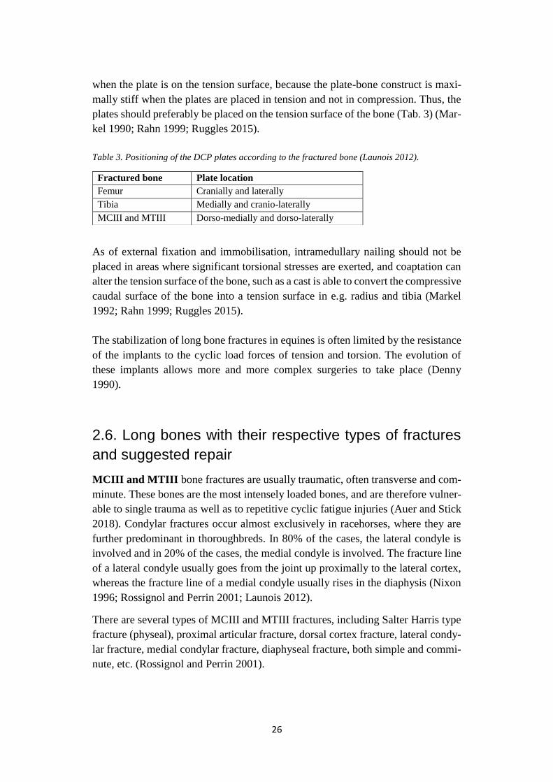
Table 3. Positioning of the DCP plates according to the fractured bone ............... 26
9
List of figures
Figure 1. Sagittal section of an animal long bone .................................................. 13
Figure 2. Cross section of an animal long bone ..................................................... 14
Figure 3. Classification of Salter Harris fractures ................................................. 15
Figure 4. Internal plate fixation of an almost healed fracture ................................ 17
Figure 5. Different bone screws ............................................................................. 18
Figure 6. Cross-section of plate holes .................................................................... 19
Figure 7. LC-DCP plate ......................................................................................... 19
Figure 8. LCP plate ................................................................................................ 20
Figure 9. A Intramedullary pins in a recently fractured bone ................................ 20
Figure 9. B Successful periosteal and intramedullary callus formation ................ 20
Figure 10. Interlocking nails .................................................................................. 21
Figure 11. External fixators ................................................................................... 21
Figure 12. Sling ...................................................................................................... 22
Figure 13. Biomechanical forces applied to long bones ........................................ 25
Figure 14. A radiographic x-ray image of the fractured MTIII ............................. 33
10
Abbreviations
DCP Dynamic Compression Plates
LCP Locking Compression Plates
LC-DCP Dynamic Compression Plates with Limited Bone Contact
MCIII
MIPO
Third Metacarpal Bone
Minimally Invasive Plate Osteosynthesis
MTIII Third Metatarsal Bone
11
1. Introduction
Long tubular bone fractures are among the most complicated types of fractures in
horses and generally they have a poor prognosis due to the body mass of the horse
and the necessity of weight bearing on all the four limbs. There are two basic diffi-
culties to it. Firstly, the veterinary-medical challenge of fixating a long bone frac-
ture of an over 300 kg highly dynamic animal and, secondly, the ethical aspects of
leaving a lame horse for a long rehabilitation period with all the related pain and
suffering. The problem reaches its climax when an otherwise healthy young horse
has to be put down because of such a fracture. Surgical treatment of long bone frac-
tures in horses encompasses several stages, such as box rest, external fixation for
the immobilisation of the fractured limb, internal bone fixation by osteosynthetic
surgery, patient management, anaesthesia, recovery from anaesthesia and the post-
operative rehabilitation. The aim of fracture repair is to return the bone to its origi-
nal structure and function by, preferably, minimally invasive osteosynthesis, care-
fully dealing with the surrounding vascularised soft tissues. It is, therefore, im-
portant to opt for an implant system that is able to neutralize the biomechanical
forces at the fracture site and allow for an accelerated consolidation of the fractured
bone fragments (Perren 2002).Veterinary orthopaedic surgery has kept on deve-
loping, but the technical advances in this domain are mostly derived from extrapo-
lating human surgery data. However, even at the beginning of the 21st century, long
bone fracture repair in horses remains a challenge for veterinarians.
The objective of this paper was to delve into the available veterinary methods of
treating equine long bone fractures, to sample new data from a questionnaire, an-
swered by veterinarians that have practiced equine surgery at animal hospitals in
Sweden, and intertwine the obtained results with the already existing research in
the frontiers of contemporary veterinary medicine.
12
2. Literature Review
2.1. The structure and function of bones
The skeleton serves as the structural protection and carrier of internal organs (Mail-
let 1979), as a fastening for muscles, as well as for haematopoietic purposes (André
et al. 2008), and as the body’s mineral station depo for calciphosphoric metabolism
(Maillet and Chiarasini 1979; Kierszenbaum 2002). The main component of the
skeleton is the bone, which is characterised by its high density, its ability to undergo
constant turnover and self-repair. The equine skeleton is held together by a system
of ligaments and tendons, where ligaments link the bones together and tendons
transmit forces between bones and muscles. In the animal kingdom, equine bones
are among the densest bones (Junqueira and Carneiro 2005; Markel 2015a).
The equine skeleton reaches its maximal size by the age of five years; however, that
highly depends upon the breed and condition of the horse. There are three types of
bones in the skeleton:
- Short (irregular) bones, such as the small bones of carpus and tarsus, which are of
cortico-spongy type and their cortex is thinner than that of the long bones.
- Flat bones, such as the scapula and the mandible, which are made up of two thin
layers of compact bone enclosing a spongy bone.
- Long tubular bones, such as the humerus, radius, ulna, third metacarpal and third
metatarsal bones, femur and tibia.
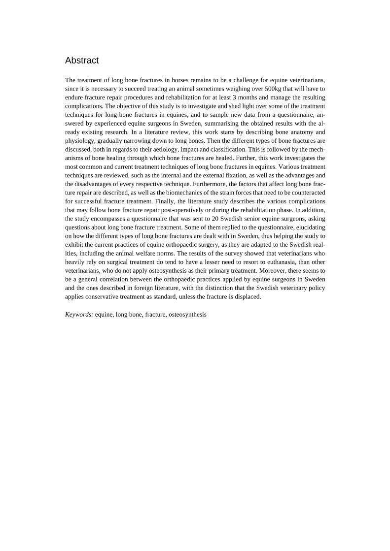
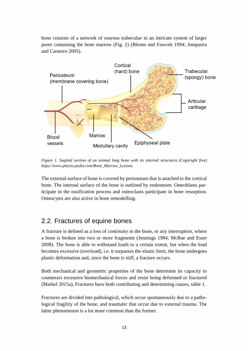
In this work, we shall be focusing on long bones. Long tubular bones consist of a
diaphysis (hollow cylinder of compact bone with medullar yellow bone marrow
inside), a metaphysis (binding the epiphysis and the diaphysis together), and an
epiphysis (spongy cancellous bone with red bone marrow inside) (Fig. 1). The
growth plate (physis), which lies within the metaphysis, regresses with age and it
becomes absent in fully-grown animals, thereby the epiphysis and metaphysis are
united with the diaphysis in adults (Junqueira and Carneiro 2005; Markel 2015a).
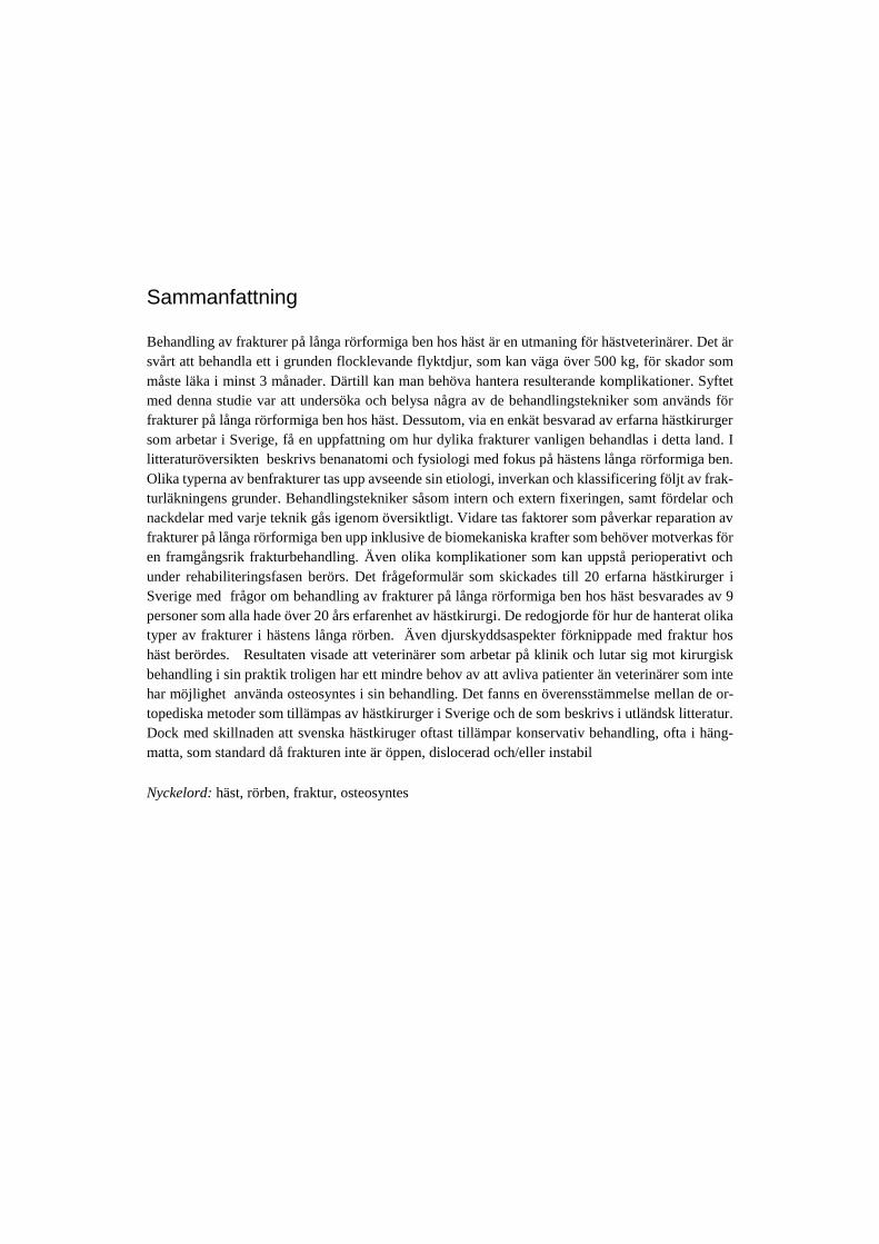
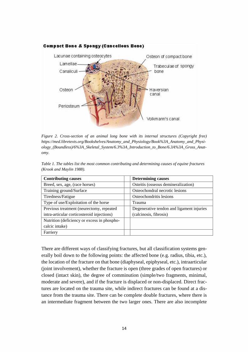
After enamel and dentin, bone is the hardest structure in the body. Its strength de-
pends on both the compact (cortical) bone and on the trabeculae of the cancellous
bone. The degree of porosity of the compact bone lies between 5% and 30% and
that of the trabecular bone lies between 30% and 90%. The compact bone contains
such tiny pores that they are only visible under the microscope, while the spongy
13
bone consists of a network of osseous trabeculae in an intricate system of larger
pores containing the bone marrow (Fig. 2) (Bloom and Fawcett 1994; Junqueira
and Carneiro 2005).
Figure 1. Sagittal section of an animal long bone with its internal structures (Copyright free)
https://www.physio-pedia.com/Bone_Marrow_Lesions.
The external surface of bone is covered by periosteum that is attached to the cortical
bone. The internal surface of the bone is outlined by endosteum. Osteoblasts par-
ticipate in the ossification process and osteoclasts participate in bone resorption.
Osteocytes are also active in bone remodelling.
2.2. Fractures of equine bones
A fracture is defined as a loss of continuity in the bone, or any interruption, where
a bone is broken into two or more fragments (Jennings 1984; McRae and Esser
2008). The bone is able to withstand loads to a certain extent, but when the load
becomes excessive (overload), i.e. it surpasses the elastic limit, the bone undergoes
plastic deformation and, since the bone is stiff, a fracture occurs.
Both mechanical and geometric properties of the bone determine its capacity to
counteract excessive biomechanical forces and resist being deformed or fractured
(Markel 2015a). Fractures have both contributing and determining causes, table 1.
Fractures are divided into pathological, which occur spontaneously due to a patho-
logical fragility of the bone, and traumatic that occur due to external trauma. The
latter phenomenon is a lot more common than the former.
14
Figure 2. Cross-section of an animal long bone with its internal structures (Copyright free)
https://med.libretexts.org/Bookshelves/Anatomy_and_Physiology/Book%3A_Anatomy_and_Physi-
ology_(Boundless)/6%3A_Skeletal_System/6.3%3A_Introduction_to_Bone/6.3A%3A_Gross_Anat-
omy.
Table 1. The tables list the most common contributing and determining causes of equine fractures
(Krook and Maylin 1988).
Contributing causes Determining causes
Breed, sex, age, (race horses) Osteitis (osseous demineralization)
Training ground/Surface Osteochondral necrotic lesions
Tiredness/Fatigue Osteochondritis lesions
Type of use/Exploitation of the horse Trauma
Previous treatment (neurectomy, repeated
intra-articular corticosteroid injections)
Degenerative tendon and ligament injuries
(calcinosis, fibrosis)
Nutrition (deficiency or excess in phospho-
calcic intake)
Farriery
There are different ways of classifying fractures, but all classification systems gen-
erally boil down to the following points: the affected bone (e.g. radius, tibia, etc.),
the location of the fracture on that bone (diaphyseal, epiphyseal, etc.), intraarticular
(joint involvement), whether the fracture is open (three grades of open fractures) or
closed (intact skin), the degree of comminution (simple/two fragments, minimal,
moderate and severe), and if the fracture is displaced or non-displaced. Direct frac-
tures are located on the trauma site, while indirect fractures can be found at a dis-
tance from the trauma site. There can be complete double fractures, where there is
an intermediate fragment between the two larger ones. There are also incomplete
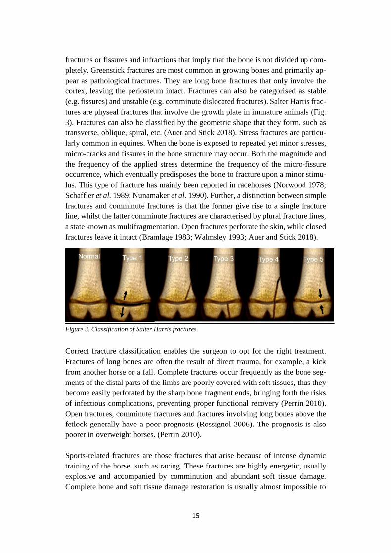
15
fractures or fissures and infractions that imply that the bone is not divided up com-
pletely. Greenstick fractures are most common in growing bones and primarily ap-
pear as pathological fractures. They are long bone fractures that only involve the
cortex, leaving the periosteum intact. Fractures can also be categorised as stable
(e.g. fissures) and unstable (e.g. comminute dislocated fractures). Salter Harris frac-
tures are physeal fractures that involve the growth plate in immature animals (Fig.
3). Fractures can also be classified by the geometric shape that they form, such as
transverse, oblique, spiral, etc. (Auer and Stick 2018). Stress fractures are particu-
larly common in equines. When the bone is exposed to repeated yet minor stresses,
micro-cracks and fissures in the bone structure may occur. Both the magnitude and
the frequency of the applied stress determine the frequency of the micro-fissure
occurrence, which eventually predisposes the bone to fracture upon a minor stimu-
lus. This type of fracture has mainly been reported in racehorses (Norwood 1978;
Schaffler et al. 1989; Nunamaker et al. 1990). Further, a distinction between simple
fractures and comminute fractures is that the former give rise to a single fracture
line, whilst the latter comminute fractures are characterised by plural fracture lines,
a state known as multifragmentation. Open fractures perforate the skin, while closed
fractures leave it intact (Bramlage 1983; Walmsley 1993; Auer and Stick 2018).
Figure 3. Classification of Salter Harris fractures.
Correct fracture classification enables the surgeon to opt for the right treatment.
Fractures of long bones are often the result of direct trauma, for example, a kick
from another horse or a fall. Complete fractures occur frequently as the bone seg-
ments of the distal parts of the limbs are poorly covered with soft tissues, thus they
become easily perforated by the sharp bone fragment ends, bringing forth the risks
of infectious complications, preventing proper functional recovery (Perrin 2010).
Open fractures, comminute fractures and fractures involving long bones above the
fetlock generally have a poor prognosis (Rossignol 2006). The prognosis is also
poorer in overweight horses. (Perrin 2010).
Sports-related fractures are those fractures that arise because of intense dynamic
training of the horse, such as racing. These fractures are highly energetic, usually
explosive and accompanied by comminution and abundant soft tissue damage.
Complete bone and soft tissue damage restoration is usually almost impossible to
16
attain after such an injury, thus fracture reduction becomes more complicated. Mus-
cle contractions and the inability of the horse to avoid bearing weight on its frac-
tured limb complicate reduction even further and may cause eburnation. Another
issue is that equine tissues are sensitive and tend to react by excessive oedema and
soft tissue swelling (Denny 1990; Nixon 1996).
2.3. Fracture healing
Fracture healing aims to render the damaged bone back to its original state. De-
pending on the extent of the trauma, one of the two bone-healing processes can take
place, namely healing by primary or by secondary intention (Jennings 1984).
Healing by primary intention depends on special conditions that must be fulfilled
at the site of the fracture. These conditions include containment of the bone frag-
ments with a proper anatomical reduction without loss of substance, exerted pres-
sure upon the fracture line, a non-contaminated wound, intact peripheral soft tissues
and vascularization via Haversian canaliculi (Jennings 1984). The phase of resorp-
tion and reconstruction between the two fragments follows the phase of acute in-
flammation, same as in the secondary intention healing (Mansmaan et al. 1982).
Revascularization takes place through Haversian canaliculi and osteoclasts resorb
the opposite fragment, whereupon osteoblasts form new osteons. Bone continuity
is thus regained within ten weeks without callus formation (Mathon 1999).
Healing by secondary intention, or indirect healing, occurs in fractures with evident
interfragmentary movements and where the fracture space is greater than 1 mm
(Reikerås 1990; McKinley 2003). This type of healing can be subdivided into four
stages or three overlapping phases: inflammatory phase (first 2-3 weeks of injury,
characterised by vasodilatation and chemotaxis), reparative phase (formation of soft
and hard callus, also known as consolidation phase) and remodelling phase (ne-
crotic regions are replaced by osteonal remodelling) (McKibbin 1978; Nixon 1996).
These phases involve a sequence of events, where different tissues substitute each
other during fracture healing, which, eventually, renders the bone stiff and strong;
the rule of thumb being that tissues cannot be formed that could not exist under the
given biomechanical conditions. The sequence of events includes hematoma for-
mation, inflammatory phase, formation of soft then hard callus, and remodelling.
The aim of these phases is to stabilise the bone fragments, starting by means of
external callus formation, which characterizes this type of healing (by secondary
intention) (Perren and Rahn 1980; Nixon 1996).
17
Bone consolidation takes place from 3 weeks to 3 months post-trauma, starting with
the differentiation of the periosteum and of the endosteum stem cells into osteo-
blasts, which synthesise the bone matrix. Intramembranous ossification progresses
centripetally towards the interfragmentary gap, this occurring rapidly in the perios-
teum and slowly in the endosteum. Endochondral ossification goes on until com-
plete bone union of the fragment ends is reached, thus forming the hard callus,
whereas the soft callus is destroyed with its constituent chondrocytes, becoming
hypertrophic and calcified. Therefore, it can be said that the synergy of endochon-
dral and intramembranous ossifications forms the hard callus (immature bone) (Ein-
horn 1998). The consolidation phase stabilizes the fracture site by further increasing
its stiffness, due to both a larger diameter of the callus improved bone and succes-
sive tissue differentiation. The interfragmentary movements decrease, enabling la-
mellar bone formation in the fracture site. Sufficient strength and stiffness of the
newly formed bone is now gained, allowing its further functionality resumption,
even though the bone is still not identical to its original state (Augat et al. 2004).
The remodelling phase follows the hard callus formation. Immature bone is gra-
dually replaced by mechanically competent lamellar bone (compact bone/cortical
bone). The remodelling phase can extend from several months to 6 or even 9 years
after the initial trauma. Its intensity and duration are strongly linked to the con-
straints applied at the level of the fracture site (Frost 1994; Ruff et al. 2006; Wolff
2011).
2.4. Surgical techniques used for stabilizing fractures in
horses
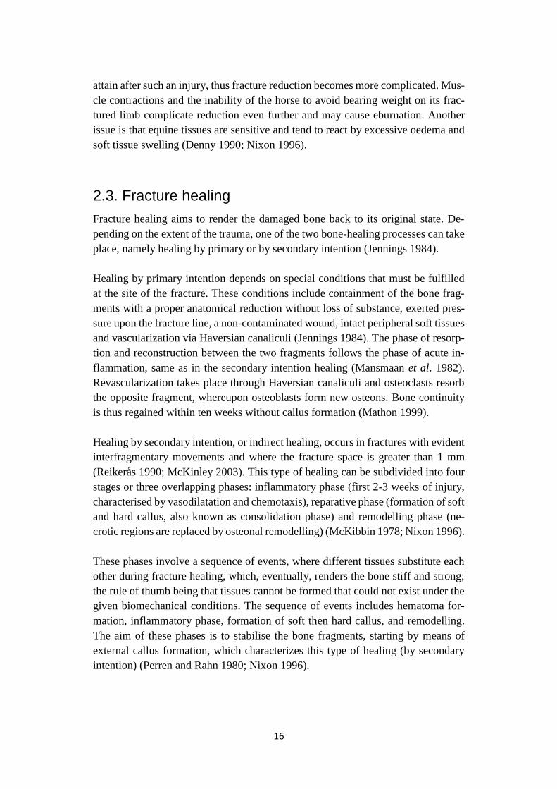
Internal fixation consists of bone reconstruction by placing internal compression
implants that will aid bone healing by restoring bone continuity and holding it to-
gether, e.g. metal devices such as plates are applied in and on the bone to align and
stabilize the bone fragments (Fig. 4). Compression devices, consisting of metal
plates and screws, align the bone fragments, thus enabling primary healing to occur.
Transfixation devices bind the bone fragments together through horizontally in-
serted pins, where external casts support them.
Figure 4. Internal plate fixation of an almost healed fracture. Courtesy: Prof Nabil Ebraheim,
University of Toledo, Ohio, USA.
18
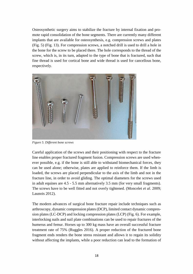
Osteosynthetic surgery aims to stabilize the fracture by internal fixation and pro-
mote rapid consolidation of the bone segments. There are currently many different
implants that are available for osteosynthesis, e.g. compression screws and plates
(Fig. 5) (Fig. 13). For compression screws, a notched drill is used to drill a hole in
the bone for the screw to be placed there. The hole corresponds to the thread of the
screw, which is, in its turn, adapted to the type of bone that is fractured, such that
fine thread is used for cortical bone and wide thread is used for cancellous bone,
respectively.
Figure 5. Different bone screws
Careful application of the screws and their positioning with respect to the fracture
line enables proper fractured fragment fusion. Compression screws are used when-
ever possible, e.g. if the bone is still able to withstand biomechanical forces, they
can be used alone; otherwise, plates are applied to reinforce them. If the limb is
loaded, the screws are placed perpendicular to the axis of the limb and not in the
fracture line, in order to avoid gliding. The optimal diameters for the screws used
in adult equines are 4.5 - 5.5 mm alternatively 3.5 mm (for very small fragments).
The screws have to be well fitted and not overly tightened. (Moncelet et al. 2009;
Launois 2012).
The modern advances of surgical bone fracture repair include techniques such as
arthroscopy, dynamic compression plates (DCP), limited contact dynamic compres-
sion plates (LC-DCP) and locking compression plates (LCP) (Fig. 6). For example,
interlocking nails and nail plate combinations can be used to repair fractures of the
humerus and femur. Horses up to 300 kg mass have an overall successful fracture
treatment rate of 75% (Ruggles 2016). A proper reduction of the fractured bone
fragment ends renders the bone stress resistant and allows it to regain its solidity
without affecting the implants, while a poor reduction can lead to the formation of
19
bone calluses. Wound infections that may follow in-
ternal fixation can alone compromise the success of
the surgery (Markel 2015a; 2015b; Ruggles 2016).
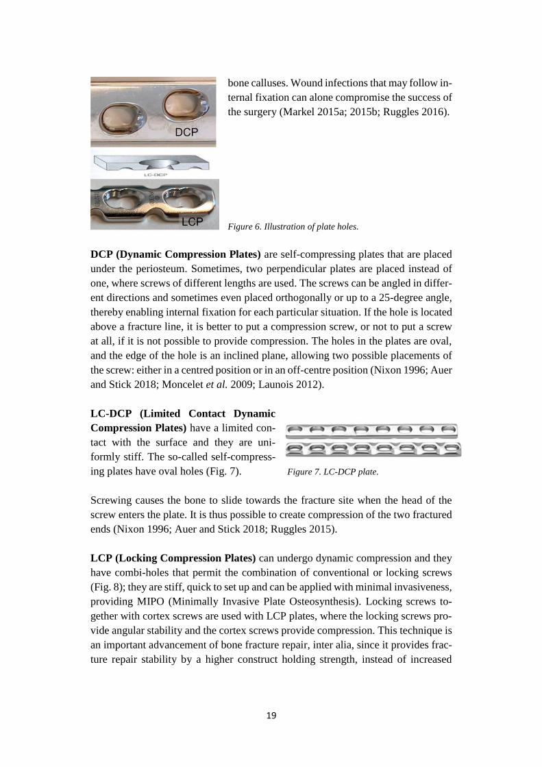
Figure 6. Illustration of plate holes.
DCP (Dynamic Compression Plates) are self-compressing plates that are placed
under the periosteum. Sometimes, two perpendicular plates are placed instead of
one, where screws of different lengths are used. The screws can be angled in differ-
ent directions and sometimes even placed orthogonally or up to a 25-degree angle,
thereby enabling internal fixation for each particular situation. If the hole is located
above a fracture line, it is better to put a compression screw, or not to put a screw
at all, if it is not possible to provide compression. The holes in the plates are oval,
and the edge of the hole is an inclined plane, allowing two possible placements of
the screw: either in a centred position or in an off-centre position (Nixon 1996; Auer
and Stick 2018; Moncelet et al. 2009; Launois 2012).
LC-DCP (Limited Contact Dynamic
Compression Plates) have a limited con-
tact with the surface and they are uni-
formly stiff. The so-called self-compress-
ing plates have oval holes (Fig. 7). Figure 7. LC-DCP plate.
Screwing causes the bone to slide towards the fracture site when the head of the
screw enters the plate. It is thus possible to create compression of the two fractured
ends (Nixon 1996; Auer and Stick 2018; Ruggles 2015).
LCP (Locking Compression Plates) can undergo dynamic compression and they
have combi-holes that permit the combination of conventional or locking screws
(Fig. 8); they are stiff, quick to set up and can be applied with minimal invasiveness,
providing MIPO (Minimally Invasive Plate Osteosynthesis). Locking screws to-
gether with cortex screws are used with LCP plates, where the locking screws pro-
vide angular stability and the cortex screws provide compression. This technique is
an important advancement of bone fracture repair, inter alia, since it provides frac-
ture repair stability by a higher construct holding strength, instead of increased
20
bone-plate friction. A disadvantage of this method is the requirement of the specific
surgical skill of angling screws across the medullar cavity (Ruggles 2016).
Figure 8. LCP plate.
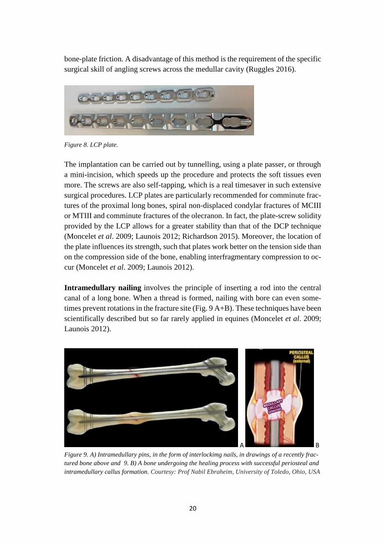
The implantation can be carried out by tunnelling, using a plate passer, or through
a mini-incision, which speeds up the procedure and protects the soft tissues even
more. The screws are also self-tapping, which is a real timesaver in such extensive
surgical procedures. LCP plates are particularly recommended for comminute frac-
tures of the proximal long bones, spiral non-displaced condylar fractures of MCIII
or MTIII and comminute fractures of the olecranon. In fact, the plate-screw solidity
provided by the LCP allows for a greater stability than that of the DCP technique
(Moncelet et al. 2009; Launois 2012; Richardson 2015). Moreover, the location of
the plate influences its strength, such that plates work better on the tension side than
on the compression side of the bone, enabling interfragmentary compression to oc-
cur (Moncelet et al. 2009; Launois 2012).
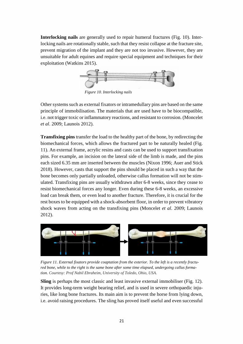
Intramedullary nailing involves the principle of inserting a rod into the central
canal of a long bone. When a thread is formed, nailing with bore can even some-
times prevent rotations in the fracture site (Fig. 9 A+B). These techniques have been
scientifically described but so far rarely applied in equines (Moncelet et al. 2009;
Launois 2012).
A B
Figure 9. A) Intramedullary pins, in the form of interlockimg nails, in drawings of a recently frac-
tured bone above and 9. B) A bone undergoing the healing process with successful periosteal and
intramedullary callus formation. Courtesy: Prof Nabil Ebraheim, University of Toledo, Ohio, USA
21
Interlocking nails are generally used to repair humeral fractures (Fig. 10). Inter-
locking nails are rotationally stable, such that they resist collapse at the fracture site,
prevent migration of the implant and they are not too invasive. However, they are
unsuitable for adult equines and require special equipment and techniques for their
exploitation (Watkins 2015).
Figure 10. Interlocking nails
Other systems such as external fixators or intramedullary pins are based on the same
principle of immobilisation. The materials that are used have to be biocompatible,
i.e. not trigger toxic or inflammatory reactions, and resistant to corrosion. (Moncelet
et al. 2009; Launois 2012).
Transfixing pins transfer the load to the healthy part of the bone, by redirecting the
biomechanical forces, which allows the fractured part to be naturally healed (Fig.
11). An external frame, acrylic resins and casts can be used to support transfixation
pins. For example, an incision on the lateral side of the limb is made, and the pins
each sized 6.35 mm are inserted between the muscles (Nixon 1996; Auer and Stick
2018). However, casts that support the pins should be placed in such a way that the
bone becomes only partially unloaded, otherwise callus formation will not be stim-
ulated. Transfixing pins are usually withdrawn after 6-8 weeks, since they cease to
resist biomechanical forces any longer. Even during these 6-8 weeks, an excessive
load can break them, or even lead to another fracture. Therefore, it is crucial for the
rest boxes to be equipped with a shock-absorbent floor, in order to prevent vibratory
shock waves from acting on the transfixing pins (Moncelet et al. 2009; Launois
2012).
Figure 11. External fixators provide coaptation from the exterior. To the left is a recently fractu-
red bone, while to the right is the same bone after some time elapsed, undergoing callus forma-
tion. Courtesy: Prof Nabil Ebraheim, University of Toledo, Ohio, USA.
Sling is perhaps the most classic and least invasive external immobiliser (Fig. 12).
It provides long-term weight bearing relief, and is used in severe orthopaedic inju-
ries, like long bone fractures. Its main aim is to prevent the horse from lying down,
i.e. avoid raising procedures. The sling has proved itself useful and even successful
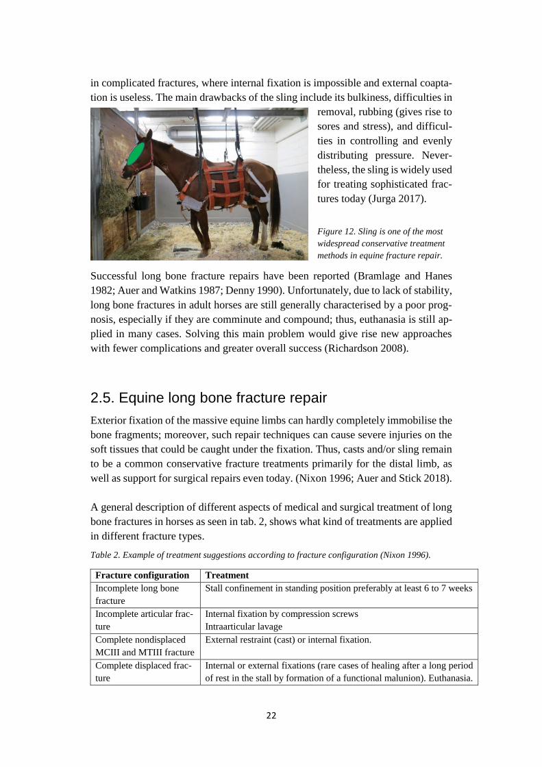
22
in complicated fractures, where internal fixation is impossible and external coapta-
tion is useless. The main drawbacks of the sling include its bulkiness, difficulties in
removal, rubbing (gives rise to
sores and stress), and difficul-
ties in controlling and evenly
distributing pressure. Never-
theless, the sling is widely used
for treating sophisticated frac-
tures today (Jurga 2017).
Figure 12. Sling is one of the most
widespread conservative treatment
methods in equine fracture repair.
Successful long bone fracture repairs have been reported (Bramlage and Hanes
1982; Auer and Watkins 1987; Denny 1990). Unfortunately, due to lack of stability,
long bone fractures in adult horses are still generally characterised by a poor prog-
nosis, especially if they are comminute and compound; thus, euthanasia is still ap-
plied in many cases. Solving this main problem would give rise new approaches
with fewer complications and greater overall success (Richardson 2008).
2.5. Equine long bone fracture repair
Exterior fixation of the massive equine limbs can hardly completely immobilise the
bone fragments; moreover, such repair techniques can cause severe injuries on the
soft tissues that could be caught under the fixation. Thus, casts and/or sling remain
to be a common conservative fracture treatments primarily for the distal limb, as
well as support for surgical repairs even today. (Nixon 1996; Auer and Stick 2018).
A general description of different aspects of medical and surgical treatment of long
bone fractures in horses as seen in tab. 2, shows what kind of treatments are applied
in different fracture types.
Table 2. Example of treatment suggestions according to fracture configuration (Nixon 1996).
Fracture configuration Treatment
Incomplete long bone
fracture
Stall confinement in standing position preferably at least 6 to 7 weeks
Incomplete articular frac-
ture
Internal fixation by compression screws
Intraarticular lavage
Complete nondisplaced
MCIII and MTIII fracture
External restraint (cast) or internal fixation.
Complete displaced frac-
ture
Internal or external fixations (rare cases of healing after a long period
of rest in the stall by formation of a functional malunion). Euthanasia.
23
Fracture repair depends on factors that can be split up into two categories: con-
trolled and uncontrolled. Controlled factors are listed below:
- biomechanical forces
- anatomical fracture reduction including thorough debridement of the fracture bed
- radiographic control
- surgical skill
- immobilisation,
- equipment
- soft tissue management
- sterile environment (preventing infection)
Sufficient preoperative preparations include appropriate choice of the implants. Ex-
amples of uncontrolled factors:
- patient’s size
- handling, age and condition
- fracture’s site and classification (open or closed, severe or mild, etc.)
- intact blood supply and prognosis. (Denny 1990; Ruggles 2015; American
College of Veterinary Surgeons 2020).
The horse mass is a factor to consider lest both the fractured limb and even the
contralateral limb bear the entire load, increasing the risk of complications in the
fractured bone and supporting limb laminitis. The greater the weight the higher the
risk of developing laminitis (Perrin 2010).
Surgical treatment of fractures usually requires general anaesthesia, which is has a
certain risk especially in the recovery phase, when the horse sometimes does prem-
ature attempts to stand up before the effect of anaesthesia has ceased. During such
attempts, a heavy or stressed horse is more likely to turn even a correctly executed
surgical operation into a disaster by placing an excessive load on the newly inserted
implants. Therefore, an important task of surgical implants is to render horses am-
bulatory and fully weight bearing in the immediate post-operative period after os-
teosynthetic surgery (Nixon 1996; Auer and Stick 2018). The recovery prognosis is
naturally ameliorated for closed and non-displaced fractures, especially in calm
horses that are adapted to human environment and handling. Conversely, open dis-
placed comminute fractures in heavy horses (>250kg) that are difficult to handle
have a poor recovery prognosis. Challenges of surgical fracture repair in large ani-
mals include large plates that complicate skin closure, implant failure and post-op-
erative lameness (Nixon 1996; Auer and Stick 2018).
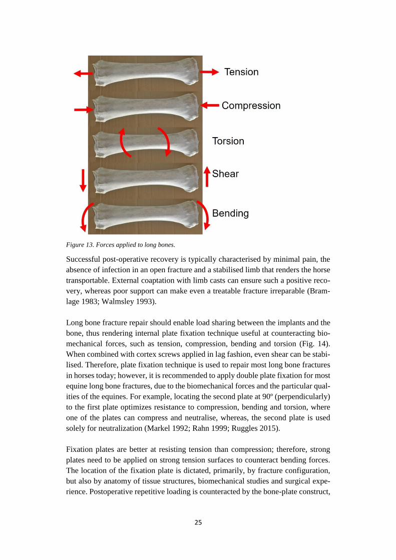
Orthopaedic surgery requires knowledge of the biomechanical forces that are
exerted on the limb, such as tension, compression, shear, bending, avulsion and
torsion, since the bone experiences these forces (Fig. 13). MCIII and MTIII are
24
mostly exposed to compression, which occurs during weight bearing but when suf-
fering transverse fracture, they have been subjected to multiple forces such as shear,
bending and torsion forces at the same time. Excessive tension typically causes a
transverse fracture, excessive compression produces a short oblique fracture and
excessive torsion gives rise to long, oblique, spiral fractures (Schneider 2015).
When having to repair a fracture, it is essential that the surgeon be aware of the
specific constraints of each bone surface. For example, the upper limbs (radius) of
a walking horse is subject to craniolateral-caudomedial bending, the cranial surface
experiences tension, the caudal surface is subject to compression, the lateral surface
is under tension, the medial surface is under compression, and the distal radius ex-
periences torsion. A fixation cast can counteract these natural strain forces. For
example, by placing the cast on the cranial surface enables it to experience com-
pression and the caudal surface experiences, respectively, tension. Casts are usually
not applied to radial fractures repaired by double plate fixation (Schneider 2015).
The same can be considered when investigating strains in tibia of a walking horse.
Tension in the tibia occurs on its cranial surface, whereas caudal tibia is subject to
compression. These strain forces reach their maximal magnitude in the proximal
metaphysis and mid-diaphysis. Distal tibia is under torsion, which even exhibits
itself in lateral and medial tibia. The cast placed in the proximal and midshaft region
of the tibia did not alter the magnitude and the direction of the strains. However,
the cast did manage to reverse the direction of torsion distally. The cast only af-
fected torsion, and, therefore, cannot be used to protect the bone from axial com-
pression (weight-bearing loads) and tension (Schneider 2015).
25
Figure 13. Forces applied to long bones.
Successful post-operative recovery is typically characterised by minimal pain, the
absence of infection in an open fracture and a stabilised limb that renders the horse
transportable. External coaptation with limb casts can ensure such a positive reco-
very, whereas poor support can make even a treatable fracture irreparable (Bram-
lage 1983; Walmsley 1993).
Long bone fracture repair should enable load sharing between the implants and the
bone, thus rendering internal plate fixation technique useful at counteracting bio-
mechanical forces, such as tension, compression, bending and torsion (Fig. 14).
When combined with cortex screws applied in lag fashion, even shear can be stabi-
lised. Therefore, plate fixation technique is used to repair most long bone fractures
in horses today; however, it is recommended to apply double plate fixation for most
equine long bone fractures, due to the biomechanical forces and the particular qual-
ities of the equines. For example, locating the second plate at 90º (perpendicularly)
to the first plate optimizes resistance to compression, bending and torsion, where
one of the plates can compress and neutralise, whereas, the second plate is used
solely for neutralization (Markel 1992; Rahn 1999; Ruggles 2015).
Fixation plates are better at resisting tension than compression; therefore, strong
plates need to be applied on strong tension surfaces to counteract bending forces.
The location of the fixation plate is dictated, primarily, by fracture configuration,
but also by anatomy of tissue structures, biomechanical studies and surgical expe-
rience. Postoperative repetitive loading is counteracted by the bone-plate construct,
26
when the plate is on the tension surface, because the plate-bone construct is maxi-
mally stiff when the plates are placed in tension and not in compression. Thus, the
plates should preferably be placed on the tension surface of the bone (Tab. 3) (Mar-
kel 1990; Rahn 1999; Ruggles 2015).
Table 3. Positioning of the DCP plates according to the fractured bone (Launois 2012).
As of external fixation and immobilisation, intramedullary nailing should not be
placed in areas where significant torsional stresses are exerted, and coaptation can
alter the tension surface of the bone, such as a cast is able to convert the compressive
caudal surface of the bone into a tension surface in e.g. radius and tibia (Markel
1992; Rahn 1999; Ruggles 2015).
The stabilization of long bone fractures in equines is often limited by the resistance
of the implants to the cyclic load forces of tension and torsion. The evolution of
these implants allows more and more complex surgeries to take place (Denny
1990).
2.6. Long bones with their respective types of fractures
and suggested repair
MCIII and MTIII bone fractures are usually traumatic, often transverse and com-
minute. These bones are the most intensely loaded bones, and are therefore vulner-
able to single trauma as well as to repetitive cyclic fatigue injuries (Auer and Stick
2018). Condylar fractures occur almost exclusively in racehorses, where they are
further predominant in thoroughbreds. In 80% of the cases, the lateral condyle is
involved and in 20% of the cases, the medial condyle is involved. The fracture line
of a lateral condyle usually goes from the joint up proximally to the lateral cortex,
whereas the fracture line of a medial condyle usually rises in the diaphysis (Nixon
1996; Rossignol and Perrin 2001; Launois 2012).
There are several types of MCIII and MTIII fractures, including Salter Harris type
fracture (physeal), proximal articular fracture, dorsal cortex fracture, lateral condy-
lar fracture, medial condylar fracture, diaphyseal fracture, both simple and commi-
nute, etc. (Rossignol and Perrin 2001).
Fractured bone Plate location
Femur Cranially and laterally
Tibia Medially and cranio-laterally
MCIII and MTIII Dorso-medially and dorso-laterally
27
Internal fixation surgery with two DCP plates can be applied for treating simple
traumatic diaphyseal fractures (Nixon 1996; Rossignol and Perrin 2001; Launois
2012). This can be combined with external coaptation depending on the fracture
configuration, if seen as necessary by the surgeon (Nixon 1996; Auer and Stick
2018). A combination of LCP plates, compression screws, and a transfixing cast
with transfixing wires in the distal radius can be applied for repairing comminute
fractures. Compression screws can be successfully used for lateral condylar frac-
tures, whereas LCP plate and compression screws are recommended to be com-
bined for treating medial condylar fractures, since there is a risk that the fracture
progresses proximally, especially in the post-operative phase (Nixon 1996; Ros-
signol and Perrin 2001; Launois 2012). However, other authors suggest repairing
comminute fractures merely by the transfixation pin casting technique (Nixon 1996;
Auer and Stick 2018).
Radial fractures can be distinguished as strain fractures, diaphyseal fractures, met-
aphyseal fractures and Salter Harris (physeal fractures) (Rossignol and Perrin
2001).
Conservative treatment such as stall confinement, sling and external coaptation is
indicated for non-displaced fractures. Transfixation pinning can sometimes be used,
in foals or small ponies, keeping in mind that fracture configuration and extent of
soft tissue damage determine the therapeutic choice (Nixon 1996). Post-operative
rehabilitation includes a Robert Jones bandage that is remounted every 3 weeks.
Humeral fractures can be divided into 6 types. Type I is typical to foals and is a
Salter Harris fracture of the glenoidal head of the humerus, type II are tuberculum
majus fractures, type III are deltoid tuberosity fractures, typeIVare mid-diaphyseal
fractures, type V are distal metaphyseal fractures, and type VI are condylar and
epicondylar fractures (Rossignol and Perrin 2001).
Type I non-displaced fractures can be treated conservatively, whereas patients with
displaced fractures of this type are usually euthanised. Type II fractures are treated
differently according to the size of the minor fragment displacement, which if it is
less than 2 cm it is treated conservatively by immobilization in the box for 2 months
with Robert-Jones with one month sling; a palmar splint may be added to prevent
contracture. If the minor fragment has a displacement of greater than 2 cm, then
resection is applied. In cases of a large fragment, either conservative treatment or
osteosynthesis by compression screw and plate is used. Type III fractures with mi-
nor fragments are repaired by resection, while if the fragments are large, osteosyn-
thesis is usually used. Adults with unstable type IV fractures are usually euthanised;
otherwise, conservative treatment that encompasses immobilization with stall con-
finement for 3 months together with a month sling, as applied in stress fractures,
may be attempted. Type V fractures are attempted osteosynthetically by double
plate fixation with dorsal and lateral plates. Type VI fractures are also attempted
28
surgically by compression screws on the medial epicondyle (Rossignol and Perrin
2001).
Femoral fractures include physeal fractures that can both be proximal or distal
Salter Harris type I or type II, both displaced and non-displaced. (Rossignol and
Perrin 2001; Simon 2009; Launois 2012).
Distal physeal fractures with minimal displacement are treated conservatively with
stall confinement. If the displacement is more substantial, osteosynthesis by internal
fixator with cross pins can repair such fractures in foals or small ponies. Proximal
physeal fractures can be repaired osteosynthetically by reduction and internal fixa-
tion with 6.5mm screws. Diaphyseal fractures have a generally pessimistic progno-
sis, such that if the patient weighs more than 150 kg, euthanasia remains to be the
only choice. However, osteosynthesis by double plate fixation with one cranial and
one lateral plate together with 5.5mm screws can repair such fractures in lighter
patients weighing less than 150 kg (Rossignol and Perrin 2001; Launois 2012).
Tibial fractures. Incomplete stress fractures, complete simple non-displaced closed
diaphyseal fractures, as well as tibial tuberosity fractures in adults can be treated
conservatively by sling. Displaced tibial tuberosity fractures can be repaired by in-
ternal fixation with tension plates and/or compression screws. Simple closed dis-
placed diaphyseal as well as open diaphyseal fractures are only attempted if the
patient weighs less than 150 kg; otherwise, only euthanasia remains available. In
the case when the animal is under 150 kg, double plate internal fixation is carried
out, the plates placed cranio-laterally and cranio-medially, respectively. Unfortu-
nately, all patients with comminute diaphyseal fractures still do not have any suc-
cessful treatment available and are therefore euthanised (Launois 2012).
29
3. Materials and Methods
A questionnaire was sent out to 21 veterinarians, with long time experience of eq-
uine surgery, to investigate their attitudes, knowledge and experience in equine long
bone fracture repair. The respondents reside in different equine clinics in Sweden.
Some of them are already retired (for less than a 10 year period), but others are still
professionally active and they could all answer anonymously. The survey contained
the questions below. For full questionnaire, see Appendix.
1. How long have you worked as a veterinarian in general and with equine
surgery in specific?
2. Are you currently active or already retired?
3. Do you perform equine orthopaedic surgery and if yes then how frequently?
4. Do you encounter fractures in your clinical practice and if yes then how
often?
5. Have you dealt with long bone fractures in you clinical career and if yes
then which ones?
6. How frequently have you dealt with each long bone fracture, in specific?
7. How did you treat the cases that you encountered, in specific?
8. What is the success rate? Please, distinguish between the different fractures.
9. What is the most reliable treatment method according to your clinical expe-
rience?
10. In which cases of equine long bone fractures is euthanasia the only alterna-
tive?
11. What is the general prognosis of equine long bone fracture repair?
12. What type of fractures would you say are the most complicated to treat and
why?
The questionnaire contained multiple choice questions, yes/no questions, and
slightly longer text questions. If the respondent had no experience in equine ortho-
paedic surgery, or never performed long bone fracture repair, the surgeon was
guided away from answering questions about the following more specific areas of
expertise. The survey encompassed only adult equines, leaving the foals outside the
spectrum of our current study.
30
4. Results
Nine respondents completed the entire survey, seven currently active and two re-
tired. All respondents had more than 20 years’ experience as veterinary surgeons
and clinical experience of working with orthopaedic surgery. All of the respondents
had also dealt with long bone fractures during their career. Five had performed or-
thopaedic surgery a few times a month and encountered fractures at least once a
month.
Regarding question 6, type of long bone fractures together with the frequency of
occurrence, the most frequently presented fractures were those of MCIII, MTIII,
radius and tibia. Cannon bone fractures were seen more frequently than fractures of
other long bones.
The different types of long bone fractures were treated both surgically and conser-
vatively, depending on the fracture configuration and the surgeon’s clinical prac-
tice.
- Scapular fractures were usually treated by sling; however, some specific
cases were treated by certain surgeons osteosynthetically by glenoidal fixa-
tion with surgical resection of the fragments. The patients with severe frac-
tures were euthanised.
- Humeral fractures were also repaired either by sling or by osteosynthesis
and severe cases were euthanised as well.
- Radial fractures were treated surgically or conservatively, depending on
both fracture configuration and surgeon’s preference.
- MCIII and MTIII fractures were treated osteosynthetically in the over-
whelming majority of cases. However, in case of a closed and stable fracture
conservative treatment, i.e. sling was always considered as an option for
treatment.
Most of the respondents considered femoral and tibial fractures as among the most
complicated equine long bone fractures.
- Femoral fractures were attempted conservatively, e.g. sling, or euthanasia.
Only intraarticular fractures were repaired by surgical resection.
31
- Tibial fractures were attempted surgically (osteosynthesis) or conserva-
tively (sling), where sling showed itself more successful. Apart from frac-
tured crista tibia cases, almost every surgical treatment attempt ended up
in euthanasia for dislocated tibial fractures; sling left at least some suc-
cessfully repaired tibial fractures.
The respondents differed in opinion as to what treatment technique should be con-
sidered the best for equine long bone fracture repair. Most preference was given to,
and divided between osteosynthesis and sling. Certain surgeons preferred applying
a combination of methods. Immobilisation techniques, such as plaster casts or ex-
ternal fixation pins, were rarely used on their own, as opposed to the sling, which
was often used on its own. Some surgeons even mentioned that immobilisation
methods, as well as stall confinement, should only be used in combination with
other treatment techniques. Most surgeons tend to agree that conservative treatment
(sling) was useful for the cases where surgical treatment (osteosynthesis) either can-
not be applied or when there was a reasonable chance for fracture healing without
external fixation, i.e. non-displaced reasonable stable fractures.
Most of the respondents were in agreement as to what factors inevitably lead to
euthanasia, naming open fractures, comminute fractures, infected fractures, exten-
sive soft tissue damage, great mass of the patient (> 300 kg), old patient and low
condition of the patient.
The general prognosis for long bone fractures was seen as unfavourable by the re-
spondents; however, it differed depending on the fractured bone and the configura-
tion of the fracture. Displaced humeral/scapular, femoral and tibial fractures were
generally considered to have an unfavourable prognosis. Radial fractures were be-
lieved to have a guarded prognosis, whereas MCIII and MTIII fractures were con-
sidered to have a favourable prognosis, unless they were open and displaced.
Most surgeons agreed on that humeral/scapular, femoral and tibial fractures were
the most difficult to treat and that they still give the highest euthanasia rate.
Humeral and femoral fractures were considered difficult to treat because they are
located deeply in soft tissue and surrounded by thick musculature, thus the access
to them is limited. Moreover, humeral and femoral fractures were both considered
complicated to treat due the abundant soft tissue damage that usually accompanies
these fractures, as well as the impossibility to fixate bone ends.
Tibial fractures were considered difficult to treat, since only a thin layer of soft
tissue and skin covers them, thereby the fractures easily become open and infected.
32
In addition, tibial fractures are usually severely overriding, comminute and unsta-
ble.
Both humeral and tibial fractures are often also severe, due to the great mass of the
horse that they both have to bear and that hinders fracture healing.
When thinking of the type of fracture, open fractures and splitter fractures were
considered as the most complicated to treat. Blood supply was mentioned as an
important factor too and when it was lost, then fracture repair was no longer possi-
ble. Comminute and contaminated fractures appeared to be the most difficult to
treat as well.
Ulnar and radial fractures were generally treatable and MCIII and MTIII (cannon
bone) fractures were the most convenient to treat.
4.1 A case illustrating when sling was used to treat an
MTIII fracture
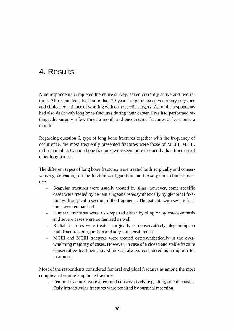
A case with a 12-year old Anglo-Arabian thoroughbred mare weighing 482 kg with
a fractured MTIII on the right hind limb is brought up in this paper. The reason for
bringing up this case is to demonstrate a real clinical example that took place at our
university equine clinic, where a successful treatment of a long bone fracture was
witnessed by the author of this study. In October this year, during a training session,
the horse exhibited lameness on the left forelimb. This lameness disappeared after
4 days but it was followed by lameness on the right hind limb instead. The rider
experienced a notable difference on the diverse running laps, such that left laps were
performed well, while right laps were carried out with difficulty. At the equine
clinic, the horse was examined both radiographically and ultrasonographically,
where the patient was diagnosed with a non-displaced fissure/fracture of the MTIII
right hind limb. The horse was treated by sling for approximately 2 months, where
the treatment went successfully and the horse was back home. Since the fracture
was not 100% complete, sling was applied and it became a successful treatment in
this case. (Fig. 14)
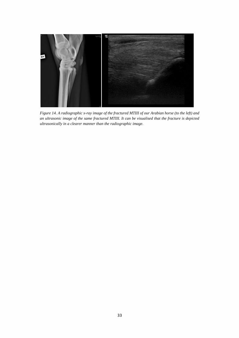
33
Figure 14. A radiographic x-ray image of the fractured MTIII of our Arabian horse (to the left) and
an ultrasonic image of the same fractured MTIII. It can be visualised that the fracture is depicted
ultrasonically in a clearer manner than the radiographic image.
34
5. Discussion
Swedish equine surgeons are in concord with what is described in the literature in
treating humeral fractures either by sling or osteosynthetic fragment resection, de-
pending on the configuration of the fracture (Rossignol and Perrin 2001). Mez et
al. (2007) mentions a 90% successful outcome of surgically treated humeral frac-
tures of the greater tubercle and a 40% success rate after conservative treatment by
stall confinement. The study only considered 15 fractures of the greater tubercle,
thus the trend that surgical treatment can arrange for a higher success rate, provided
it is done properly and the fracture can be accessed through all the muscular layers,
still cannot be backed up with satisfactory scientific evidence. On the other hand,
Zamos and Park (1992) suggest the opposite, namely that in the 22 cases, conserva-
tive treatment had a higher success outcome (at least 70%) compared to the surgi-
cally treated humeral fractures (30%). A similar conclusion is drawn from another
subsequent study with 54 cases, carried out a year later (Carter et al. 1993). It
showed that conservative treatment had a 53% success rate, compared to the 23%
outcome success after the surgically treated humeral fractures, where, in this surgi-
cal group, the successfully treated patients were all foals. It can thus be concluded
that although it is the configuration of the humeral fracture that decides whether the
fracture be treated surgically or conservatively, the default treatment for humeral
fractures is conservative.
Radial fractures are also treated similarly, i.e. both surgically and conservatively,
both in Sweden and abroad (Nixon 1996; Launois 2012). Stewart, et al. (2015)
showed that in a 54 case study, conservative treatment had an 86% success (incom-
plete fractures) compared to the 56% success after internal fixation surgery (osteo-
synthesis). Young equines showed a significantly higher survival rate as compared
to adults. This recent study reciprocates with an older 47 case study (Sanders-
Shamis, et al. 1986), which suggests that age ant the size of the horse plays a more
important role in success rate than even fracture configuration, and that radial frac-
tures in adults have a generally poor prognosis. Auer and Watkins (1987) in a 15
case study quantified the survival rate of radial fractures in adult equines after os-
teosynthesis to 23% and suggested improvement for further osteosynthetic surgical
practice. However, these studies summarised fracture repair from 1980s, which is
relatively old data. In the recent years, a certain meta-analysis (Wei et al. 2012)
35
suggested that both internal and external fixation techniques can be used inter-
changeably and can substitute each other depending on e.g. fracture configuration,
age and mass of the patient; the difference being in the distinct functions that each
treatment technique recovers most.
MCIII and MTIII fractures in Sweden are mostly treated by internal fixation, such
as it has been described in foreign literature (Nixon 1996; Rossignol and Perrin
2001; Launois 2012). It is self-explanatory that the cannon bone is frequently frac-
tured, since the distal parts of the limb are mostly subject to stress and because the
cannon bone is relatively thin, covered merely by a thin layer of skin, compared to
the other long bones. Regarding the case with the horse in the university clinic, the
fracture was not 100% complete and therefore it was a non-displaced fracture; thus,
it could be successfully treated by sling. A retrospective study investigating 10
horses and 11 foals showed that osteosynthesis performed on fractured MCIII and
MTIII had a survival rate of approximately 90% in foals and 30% in adults. Fur-
thermore, this survival rate decreased in open fractures to 86.7% in foals and 12.5%
in adults (Bischofberger et al. 2009). This study concluded that age, mass and in-
fection are the main factors that predetermine the prognosis. It can be seen that
internal fixation with open reduction provides a threefold higher survival rate in
foals as compared to adult equines. Although our study only considered adults,
Swedish equine surgeons admitted that the general prognosis of fractured cannon
bone repair is favourable. The questionnaire showed that, when including condylar
fractures, the generally preferred treatment method for MCIII and MTIII fractures
in Sweden was osteosynthesis. Since the overall prognosis was considered favour-
able, it can thus be concluded that in the clinical practice of these equine surgeons,
internal fixation probably provided a higher survival rate in adults than it was shown
in the retrospective study. What still gives uncertainty to this claim is that the sur-
vival rates, together with the fracture configurations, have not been studied through
our questionnaire. Another technical imperfection of the survey was that the prog-
nostic outcomes (favourable, unfavourable and guarded) have not been quantita-
tively defined in the questionnaire. McClure, et al. (1998) reported that the average
survival rate after MCIII and MTIII fracture repair for adults was 64%, i.e. from the
25 included horses, 16 did not have any postsurgical complications at all. This result
is higher than what is reported by Bischofberger et al. (2009). However, Mc Clure
et al. (1998) recorded more treatment techniques in their cases: both internal fixa-
tion, external coaptation and a combination thereof, thereby rendering the study
more representative, inasmuch as different treatment methods are usually applied
in real clinical conditions. This study seems to overlap with our survey results bet-
ter, showing a generally favourable outcome of repairing cannon bones fractures in
adult equines.
36
Some authors suggest a more surgical approach for treating femoral fractures (Ros-
signol and Perrin 2001; Launois 2012), whereas Swedish surgeons usually omit
osteosynthesis and either attempt conservative treatment (typically sling) or eu-
thanise the animal. This could be explained by the fact that all the patients that the
Swedish surgeons treated were heavy (over 150 kg), since in such cases even for-
eign specialists euthanise the patients. In addition, the fracture configuration has
not been specified in our study and the low number of surgeons is not representative
enough. Furthermore, as mentioned earlier, the standard treatment in Sweden re-
mains conservative, unless the fracture is displaced and requires surgery. Moreover,
the distinction in treatment approach may be due to personal surgical preference
and skill, as well as due to the specific settings of each clinic. Hance, et al. (1992)
presented 38 cases, where they showed a 50% successful humeral fracture surgical
repair by osteosynthesis in the diaphysis, which is considered to have the poorest
prognosis (most complicated repair). Unfortunately, the treated patients were all
foals, which cannot be representative when comparing to our study that only in-
cluded adult horses.
A similar picture can be seen when treating tibial fractures, such that in Sweden
conservative treatment by sling has given a more positive result than that provided
by osteosynthesis (same as in foreign literature, where almost all patients were eu-
thanised (Launois 2012)). However, this depends on the configuration of the frac-
ture. All patients with severe fractures have been euthanised. Even in fractures of
this bone, there is a distinction between the conservative Swedish approach and the
surgical approach described in the literature. Same as for femoral fractures, also in
the case with tibial fractures, it is a question of fracture configuration, lack of data,
Swedish therapeutic approach and individual surgical preference that determine the
choice of treatment. A recent retrospective study with 21 patients showed that 65%
of the equines with surgically treated tibial intercondylar eminence fractures by ar-
throscopic fragment removal underwent complete convalescence and returned to
their previous activities (Rubio-Martínez et al. 2017). However, intercondylar em-
inence fractures cannot be compared to e.g. diaphyseal fractures in prognosis, due
to the anatomo-physiological distinctions of these osseous structures. Otherwise,
the few studies that review tibial fractures, usually consider them among the most
untreatable fractures in the horse, usually being open, contaminated and commi-
nute, where the mass of the patient and fracture configuration play the most im-
portant role in deciding over therapy method versus euthanasia.
This agreement between the practice of Swedish senior equine surgeons and what
is described as being standard of practice in the literature provides extra evidence
for the clear consistency and correlation of the respective treatment techniques do-
cumented in veterinary textbooks and scientific articles.
37
The preference given by the respondents to osteosynthesis and sling, together with
immobilisation used in a combined treatment and not alone find their support in
foreign scientific literature (Nixon 1996; Rossignol and Perrin 2001; Auer and Stick
2018; Launois 2012), etc. The same trend can be noticed in the case with sling that
it is often successfully used in cases, where osteosynthesis cannot give a positive
result.
It is interesting to note how the therapeutic approach varied among the equine sur-
geons when treating different long bone fractures. As one respondent said, con-
servative treatment generally gives a good result, as long as no dislocation has oc-
curred, and a fractured bone needs to undergo osteosynthesis usually only when
dealing with a dislocated fracture; therefore, it is the dislocation itself that gives a
poor prognosis and not the actual type of treatment.
This study has only considered adult patients, leaving the foals behind. Treating
fractures in foals has its own peculiarities, which from one hand simplify fracture
repair by e.g. light mass of the foal, but from the other hand complicate the repair
by constant bone growth that implies the impossibility of long-term immobilisation
of the foal.
The questionnaire had its drawbacks, among which it can be mentioned that not
enough questions were posed and the ones that were posed were not specific
enough. This led to certain lack of detail in the responses, thus depriving the scru-
tiny from the necessary information. For example, when asking about how the dif-
ferent fractured bones were dealt with, there could be a specifying question added
on the different fracture configurations on each respective bone. The same goes for
specifying the most appropriate treatment and prognosis for the different fracture
types on each bone and not merely mentioning the fractured bone. Moreover, the
cases have not been specified, i.e. apart from the configuration, fractures are also
subdivided into categories in the classification discussed earlier in this work. This
made it impossible to compare the clinical practice of equine surgeons in Sweden
to foreign literature completely. Neither did the study contain enough respondents
(10 out of 20 replied), in order to provide us with the representative amount of data
to draw proper conclusions. If all these aspects would have been considered in the
questions, the responses would have provided us with more accurate detailed data.
However, considering the objective of this study and the type of data gathering (its
main focus lied on sampling quality data and not quantity, i.e. few experts usually
suffice when asked about their area of expertise), as well as the fact that also foreign
literature was supported by a similar number of specialists, still makes the data re-
liable. The objective of this study was to provide a general overview of equine long
bone fracture treatment and not provide detailed data on how specific long bone
38
fractures are treated. This is more of a basic study that gives an introduction into
the matter of equine long bone fracture repair.
6. Conclusion
This study gave a brief overview of the currently available surgical and conserva-
tive repair methods for equine long bone fractures in Sweden and other countries.
It can be concluded that many of such fractures can be successfully repaired; how-
ever, many remain a challenge for equine orthopaedic surgeons worldwide. The
gathered data from our respondents neatly corresponds to the international scientific
data, with eventual discrepancies perhaps due to lack of details in the responses.
Nevertheless, this study gives a basic insight into the matter of equine long bone
fracture repair. Further in-depth scrutiny of this matter can be achieved by looking
into the sources that this study used, as well as gathering clinical data from equine
surgeons in other countries.
39
References
André, J.M., Catala M., Morere J.J., Escudier E., Katsanis G. et Poirier J. (2008). Histolo-
gie, service d’histologie – embryologie. Faculté de Médecine, Pierre et Marie Curie, p.
68.
Acvs.org. (2021). Fractures In Horses (Surgical Repair).| American College of Veteri-
nary Surgeons - ACVS. [online] Available at: <https://www.acvs.org/large-ani-
mal/fractures-horses-surgical-repairs> [Accessed 7 January 2021].
Auer, J., Stick, J. plus associate editors Kümmerle J and Prange T. (2018). Equine Sur-
gery. 5th ed. Philadelphia: Elsevier - Health Sciences Division.
Auer, J. and Watkins, J. (1987). Treatment of radial fractures in adult horses: An analysis
of 15 clinical cases. Equine Veterinary Journal, 19(2), pp. 103-110
Augat, P., Simon, U., Liedert, A. and Claes, L. (2004). Mechanics and mechano-biology
of fracture healing in normal and osteoporotic bone. Osteoporosis International, 16
(S02), pp. S36-S43.
Bischofberger, A., Fürst, A., Auer, J. and Lischer, C. (2009). Surgical management of
complete diaphyseal third metacarpal and metatarsal bone fractures: Clinical outcome
in 10 mature horses and 11 foals. Equine Veterinary Journal, 41(5), pp.465-473.
Bloom, W. and Fawcett D.W. (1994). Textbook of Histology, 12th ed. New York: Chap-
man and Hall.
Bramlage, L.R. (1983). Current concepts of emergency first aid treatment and transporta-
tion of equine fracture patients. Compendium on Continuing. Education for the. Prac-
tising Veterinarian, 5: S564-574.
Bramlage, L.R., & Hanes, G.E. (1982). Internal fixation of a tibial fracture in an adult
horse. Journal of the American Veterinary Medical Association, 180 (9), pp. 1090–
1094.
Brinker, W.O., Piermattei, D.L. and Flo, G.L. (1994). Handbook of Small Animal Ortho-
paedics and Fracture Repair. 2nd edition. WB Saunders Company, pp. 9-137.
Carter, B., Schneider, R., Hardy, J., Bramlage, L. and Bertone, A., (1993). Assessment
and treatment of equine humeral fractures: retrospective study of 54 cases (1972-
1990). Equine Veterinary Journal, 25(3), pp. 203-207
Claes, L., Augat, P. and Wilke, H. (1997). Influence of size and stability of the osteotomy
gap on success of fracture healing. Journal of Orthopaedic Research, 15(4), pp. 577-
584.
Cormack D. (1993). Dense connective tissue, cartilage, bone and joints in Essential His-
tology. Philadelphia: Lippincott Co., pp. 159–190.
40
Denny, H. (1990). The principles of equine fracture fixation. Equine Veterinary Educa-
tion, 2(4), pp. 191-196.
Einhorn, T. (1998). The cell and molecular biology of fracture healing. Clinical Ortho-
paedics and Related Research, 355S, pp. S7-S21.
Feron, P. (2000). Vade-Mecum de l'évaluation médico-légale. Paris : De Boeck Univer-
sité, p. 351.
Frost, H.M. (1994). Wolff's law and bone's structural adaptations to mechanical usage: an
overview for clinicians. Angle Orthodontist, 64 (3), pp. 175-188.
Hance, S., Bramlage, L., Schneider, R. and Embertson, R. (1992). Retrospective study of
38 cases of femur fractures in horses less than one year of age. Equine Veterinary
Journal, 24(5), pp.357-363.
Kahn, C.M., Line, S. and Aillo, S.E. (2008). Merck Veterinary Manual. 3rd edition.
Merck CO Inc., pp. 973.
Jennings, P. B. (1984). The Practice of Large Animal Surgery, vol. 2. Philadelphia/Lon-
don: WB Saunders Company.
Junqueira, L.C.U., Carneiro, J. and Kelley, R.O. (2005). Basic Histology. Stamford,
Conn, Appleton and Lange.
Jurga, F. (2017). The evolution of equine slings: taking a load off, then and now. Equus
Magazine. https://equusmagazine.com/blog-equus/slings-horses-history-hope-equine-
support-system-32212
Kierszenbaum, A.L. (2002). Histologie et Biologie Cellulaire une Introduction à l’Anato-
mie Pathologique, 1ére édition, Mosby, pp.119-120.
Krook, L. and Maylin, G.A. (1988). Fracture in thoroughbred racehorse. The Cornell Vet-
erinarian, vol 78 (2) supplement.
Launois, T. (2012). Guide Pratique d'Orthopedie et de Chirurgie Equine. Paris:
MED'COM.
Maillet, M. (1979). Le tissu osseux, En: Les Tissus de Soutien, tome 3. Paris: Vigot, pp.
51-68.
Maillet, M. et Chiarasini, D. (1979). Collection PCEM Histologie, tome 1, Bréal éditeur,
Montrantuil, pp. 97-121.
Mansmann, P.W., McAllister, R.A. and Pratt, E.S. (1982). Equine Medicine and Surgery,
3rd ed., vol. 2. Santa Barbara: American Veterinary Publication.
Markel, M.D. (1992). Fracture biology and mechanics. In: Auer, J.A. and Stick, J.A.
(eds.) Equine Surgery, Philadelphia: WB Saunders Company, pp. 798-807.
Markel, M.D. (2015a). Fracture Healing in the Horse. AOVET North America Equine
Advanced Techniques in Fracture Management LECTURE ABSTRACTS, accessed
from: https://events.aona.org/AONA-Content/Abstracts/2015_Equine_Advanced_Ab-
stract_Book.pdf [07.01.2021]
Markel, M.D. (2015b). Biomechanics of Internal Fixation in the Horse. AOVET North
America Equine Advanced Techniques in Fracture Management LECTURE AB-
STRACTS, accessed from: https://events.aona.org/AONA-Content/Ab-
stracts/2015_Equine_Advanced_Abstract_Book.pdf [07.01.2021]
41
Mathon, D. (1999). Contribution à la mise au point d’un tissu osseux bioartificiel. Thèse
doctorat vétérinaire, Toulouse: Université Toulouse III - Paul Sabatier.
McClure, S. R., Watkins, J. P., Glickman, N. W., Hawkins, J. F., & Glickman, L. T.
(1998). Complete fractures of the third metacarpal or metatarsal bone in horses: 25
cases (1980-1996). Journal of the American Veterinary Medical Association, 213 (6),
pp. 847–850.
McKibbin, B. (1978). The biology of fracture healing in long bones. The Journal of Bone
and Joint Surgery, British volume, 60-B (2), pp. 150-162.
McKinley, T. (2003). Principles of fracture healing. Surgery (Medicine Publishing), 21,
9, pp. 209-212.
McRae R. and Esser M. (2008). Practical Fracture Treatment. 5th ed. Elsevier: Churchill
Livingstone, pp. 1-122.
Mez, J., Dabareiner, R., Cole, R. and Watkins, J. (2007). Fractures of the greater tubercle
of the humerus in horses: 15 cases (1986–2004). Journal of the American Veterinary
Medical Association, 230(9), pp. 1350-1355
Moncelet A., Mespoulhes-Riviere C. and Rossignol F. (2009). Les ostéosynthèses im-
plants LCP et transfixation chez le cheval. Le Nouveau Praticien Vétérinaire Équine
n°20, pp. 40–44.
Ness, M.G. (1998). Manual of Small Animal Fracture Repair and Management. BSAVA,
Shurdington, pp. 311-315.
Nixon, A.J. (1996). Equine Fracture Repair, W.B. Saunders Company, Philadelphia
Norwood, G. (1978). The bucked-skin complex in thoroughbred. In: Proceedings 24th
Annual Convention American Association Equine Practice, pp. 319–336.
Nunamaker, D.M., Butterweck, D.M. and Provost, M.T. (1990). Fatigue fractures in thor-
oughbred racehorses: relationship with age, peak bone strain and training. Journal of
Orthopaedic Research, pp. 604–611.
Perren, S.M. (2002). Evolution of the internal fixation of long bone fractures-the scien-
tific basis of internal fixation: choosing a new balance between stability and biology,
Journal of Bone and Joint Surgery, 84, pp.1093-2110.
Perren, S.M. and Rahn, B.A. (1980). Biomechanics of fracture healing. Canadian Journal
of Surgery, 23 (3), p. 228-232.
Perrin, R. (2006). Immobilisation et transport d’un cheval traumatisé. Pratique Vétéri-
naire Équine, n°38, pp. 65–77.
Postacchini, F., Gumina, S., Perugia, D. and De Martino, C. (1995). Early fracture callus
in the diaphysis of human long bones. Clinical Orthopaedics and Related Research,
(310), pp. 218-228.
Rahn B. (1999). Fracture biology mechanics and healing. In Auer, J.A. and Stick, J.A.
Equine Surgery, 2nd ed, Philadelphia: WB Saunders Company, pp. 727-738.
Reikerås, O. (1990). Healing of osteotomies under different degrees of stability in rats.
Journal of Orthopaedic Trauma, 4(2), pp. 175-178.
Richardson, D. (2008). Less invasive techniques for equine fracture repair and ar-
throdesis. Veterinary Clinics of North America: Equine Practice, [online] 24 (1), pp.
42
177-189. Available at: <https://pubmed.ncbi.nlm.nih.gov/18314042/> [Accessed 7
January 2021]. DOI: 10.1016/j.cveq.2007.11.004
Richardson, D. (2015). Clinical Use of Locking Plate Systems in the Horse. AOVET
North America Equine Advanced Techniques in Fracture Management LECTURE
ABSTRACTS, accessed from: https://events.aona.org/AONA-Content/Ab-
stracts/2015_Equine_Advanced_Abstract_Book.pdf [07.01.2021]
Rossignol, F. and Perrin, R. (2001). Tableau de gestion des fractures chez le cheval. Pra-
tique Vétérinaire Équine n°33, pp. 37-60.
Rossignol, F. (2006). Réhabilitation du cheval opéré de l’appareil locomoteur. En :
Proceedings Association des Vétérinaires Équins Français (AVEF), Conférence An-
nuelle, 2006. Versailles, France.
Rubio-Martínez, L., Redding, W., Bladon, B., Wilderjans, H., Payne, R., Tessier, C., Ge-
ffroy, O., Parker, R., Bell, C. and Collingwood, F. (2017). Fracture of the medial inter-
condylar eminence of the tibia in horses treated by arthroscopic fragment removal (21
horses). Equine Veterinary Journal, 50(1), pp. 60-64.
Ruggles, A.J. (2015). Principles of Long Bone Fracture Repair in the Horse. AOVET
North America Equine Advanced Techniques in Fracture Management LECTURE
ABSTRACTS, accessed from: https://events.aona.org/AONA-Content/Ab-
stracts/2015_Equine_Advanced_Abstract_Book.pdf [07.01.2021]
Ruggles A.J. (2016). Equine fractures in the new decade; What can we repair? Equisan,
pp. 55-59. Found on https://www.equisan.com/images/pdf/fractures.pdf
Ruff C., Holt B. and Trinkaus E. (2006). Who's afraid of the big bad wolff?: "Wolff's
Law" and bone functional adaptation. American Journal of Physiology and Anthropol-
ogy, 129 (4), pp. 484-498.
Sanders-Shamis, M., Bramlage, L. and Gable, A. (1986). Radius fractures in the horse: A
retrospective study of 47 cases. Equine Veterinary Journal, 18(6), pp.432-437.
Schaffler R., Radin, E.L. and Burr, D.B. (1989). Mechanical and morphological effect of
strain rate on fatigue compact bone. Bone, 10(3), pp. 207–214
Schneider, R.K. (2015). Tension Surfaces of Long Bones in the Horse. AOVET North
America Equine Advanced Techniques in Fracture Management LECTURE AB-
STRACTS, accessed from: https://events.aona.org/AONA-Content/Ab-
stracts/2015_Equine_Advanced_Abstract_Book.pdf [07.01.2021]
Simon, O. (2009). Diagnostic et pronostic des fractures des os longs chez le cheval. Le
Nouveau Praticien Vétérinaire Équin, 5(20), pp. 17-21.
Slater, M., Hood, D. and Carter, G. (1995). Descriptive epidemiological study of equine
laminitis. Equine Veterinary Journal, 27 (5), pp. 364-367.
Stewart, S., Richardson, D., Boston, R. and Schaer, T., (2015). Risk factors associated
with survival to hospital discharge of 54 horses with fractures of the radius. Veterinary
Surgery, 44(8), pp. 1036-1041.
Walmsley, J. P. (1993). First aid splinting for the fracture patient. Equine Veterinary Edu-
cation 5 (1): 61-63.
Whitford, R.E. (2019). Making the rounds: surgical treatment of an ulnar fracture compli-
cated by anconeal process fragmentation - case presentation. Compendium Equine. 3
(3). Found on: https://www.vetfolio.com/learn/article/making-the-rounds-surgical-
43
treatment-of-an-ulnar-fracture-complicated-by-anconeal-process-fragmentationcase-
presentation
Wolff, J. (2011). The classic: on the significance of the architecture of the spongy sub-
stance for the question of bone growth: A preliminary publication. Clinical Orthopae-
dics and Related Research, 469(11), pp. 3077-3078.
Wraighte, P. and Scammell, B. (2006). Principles of fracture healing. Surgery (Oxford),
24(6), pp. 198-207.
Wei, D., Poolman, R., Bhandari, M., Wolfe, V. and Rosenwasser, M. (2012). External
fixation versus internal fixation for unstable distal radius fractures. Journal of Ortho-
paedic Trauma, 26(7), pp. 386-394.
Zamos, D.T. and Parks, A.H. (1992). Comparison of surgical and nonsurgical treatment
of humeral fractures in horses: 22 cases (1980–1989). Journal of American Veterinary
Medicine Association; 201:114–116.
44
Acknowledgements
My foremost gratitude to God for making it possible for me to carry out this modest
work.
My deepest gratitude to my main supervisor Ove Wattle, associate professor in eq-
uine surgery at the Swedish Agricultural University, for accepting to supervise me,
for all his help, support and advice that he provided me with throughout this re-
search journey, and for all the efforts he made so that I could carry out my work. I
particularly appreciate the priority he has given me in my multiple requests despite
his many obligations.
I am very grateful to my co-supervisor Kalle Ljungvall, an equine surgeon in Mä-
lardals clinic, for all his advice, corrections, support and help that he ceaselessly
provided to me during the whole course.
My particular gratitude is expressed to the Swedish interviewees, senior specialists
in equine surgery, who replied to our questionnaire and provided us with the nec-
essary data and professional opinions of how equine long bone fracture repair is
executed in Sweden today.
I would also like to express my gratitude to our university librarians that provided
us with guidelines on academic writing.
My special sincere gratitude to my colleague in Algeria, veterinarian Dr. Soundes
Akriche, who provided me with scientific research material and experimental setup
in this domain. I also thank Dr. Asma Bouknine, Dr. Diane Bailliu, Dr. Michaël
Verset, and Dr. Julie Salina Dauvillier for their respective research contributions.
I am thankful to my dear beloved family, my parents and my grandmother, who
have constantly supported me throughout the course of this work in all possible
ways.
I also thank all my dear beloved friends for being there for me and supporting me
in all possible ways.
I dedicate this modest work above all to God Almighty, to my dear beloved family
and friends, to Zakaria Louhabi and others.
45
Popular science summary
Long bone fractures have always been a challenge for veterinary medicine, but the
grandiose goal of not putting down a young otherwise healthy horse after a fracture
has always led veterinarians to find new ways for treating such complicated frac-
tures. This paper takes up some common surgical and conservative methods of deal-
ing with long bone fractures in equines and analyses their application both in Swe-
den and abroad. The structure and function of bones in general, and long bones in
specific are explained. Further, long bone fractures are taken up, starting from their
aetiologies, going through their classification and ending in their healing process
that renders the fractured bone back to its original state. Furthermore, long bone
fracture repair is observed. Starting with the factors that need to be considered when
treating equine fractures, the reader is guided into the problems that the surgeon
encounters when repairing these fractures, pre-operatively, operatively and post-
operatively in the recovery phase. These problems are both of a general type (en-
countered in other species during fracture repair) and specifically horse type, typical
for horses only (such as the fact that horses require their limbs to be fully weight-
bearing directly upon post-operative recovery). This is followed by considering the
biomechanical forces that act upon the bone and disable its fracture repair, and
which should therefore be neutralised by the treatment techniques. This leads the
reader into the different treatment methods used, which can be classified into two
main types: surgical and conservative treatment methods. Surgical treatment in-
volves invading operations, while conservative treatment stays outside of the horse.
Another classification distinguishes between fracture repair techniques as internal
fixation and external fixation, where internal fixation implies invasive surgery that
places the implants (plates, screws, etc.) inside the horse, over the bones, and ex-
ternal fixation keeps all the equipment outside of the horse (immobilisation by sling,
plaster, casts, transfixing pins, nails, Ilizar apparatus, etc.). Even if some techniques,
such as transfixing nails and Ilizar apparatus invade the limb of the horse, they are
not completely submerged in the body, but at least partially stay on the surface.
Moreover, the different internal and external fixation techniques are taken up one
by one and briefly explained: transfixing pins, different plates (LCP, DCP, etc.),
screws, etc. In addition, some common complications are taken up and explained
46
one after the other. This is all followed by the different long bones with their re-
spective fractures and some treatment techniques that can be applied for successful
fracture repair.
The next part of the study is comprised of the practical part, which includes a ques-
tionnaire that was sent to over 20 senior horse surgeons in Sweden, where a few of
them answered the survey, thereby providing us with valuable information on how
long bone fractures are treated in Sweden. The questionnaire asked about the sur-
geons’ clinical experience, the frequency of performed orthopaedic operations and
encountered long bone fractures. It asked about the methods that each surgeon pre-
ferred to use for each long bone fracture. The result of the questionnaire showed a
good correlation with the theoretical results, obtained from foreign experts in horse
surgery. This provided extra evidence of the treatment suggestions documented in
scientific articles and textbooks.
47
Appendix – Questionnaire
Questionnaire for equine surgeons in Sweden
How long have you worked as a veterinarian?
0-5 years
5-10 years
10-15 years
15-20 years
20-25 years
25-30 years
Over 30 years
2. How long is your veterinary surgical experience?
0-5 years
5-10 years
10-15 years
15-20 years
20-25 years
25-30 years
Over 30 years
3. Are you still active as a surgeon or are you already retired?
48
4. Do you, or did you, perform orthopaedic surgery on equines in your clinical
practice?
Yes
No
If yes, then how often in general?
Seldom
Every year
Every month
Every week
On a daily basis
6. Are there, or have there been, any cases of fractures in equines in your surgical
practice?
Yes
No
7. If yes, then how often?
Seldom
Every year
Every month
Every week
On a daily basis
8. Throughout your clinical career, have you ever dealt with or encountered
treatment of scapula-, humerus-, radius-, MCIII-, femur-, tibia- and MTIII-
fractures in equines?
Your answer:
If your answer on question no. 8 is yes please continue below.
For the following questions, please differ between adult horses, foals and year-
lings.
49
Approximately how many fractures of the following bones have you en-
countered in your practice? Please differ between Salter-Harris fractures
(SH) and non Salter-Harris (NSH) as well as dislocated vs non dislocated
fractures (including fissures and greenstick fractures).
Scapula: ………………
Humerus: ………………….
Radius: …………………..
MCIII: ………………….
Femur: ………………….
Tibia: ………………….
MTIII: ………………….
9. How did you treat the above cases (consider each bone with respective
treatment in specific)?
Treatment by immobilisation (plaster/gypsum)
Sling
Surgical treatment (osteosynthesis)
Euthanasia
Other or a combination of treatments
10. If other, then specify which method exactly is applied?
Your answer:
Scapula: ………………
Humerus: ………………….
Radius: …………………..
MCIII: ………………….
Femur: ………………….
Tibia: ………………….
MTIII: ………………….
11. What is the success rate, please differ between different fractures?
Your answer:
12. In your opinion and according to your experience, which is the most relia-
ble method?
50
Surgical treatment (ex. osteosynthesis)
Immobilisation, Plaster/gypsum
Sling
Other
13. If other, please specify which method?
Your answer:
14. Regarding treatment of fractured long bones, in what cases is euthanasia
the only remaining alternative, and why? What factors would influence
your decision?
Your answer:
15. In your opinion and according to your experience, what is the general
prognosis for long bone fractures in horses?
Favorable
Guarded
Unfavorable
16. What types of fractures would you say are the most complicated to treat
and why?
Your answer: