Embed Size (px)
Citation preview

Ultrasonography of the
Neck as an Adjunct to FNA
Nicole Massoll M.D.

Basic Features of Head and
Neck Ultrasound and
AnatomyAnatomy
Nicole Massoll M.D.
University of Arkansas for Medical Sciences,
Little Rock AR
Thanks to Dr. Sparrow for the graphics and animations

Objectives
• High points of how to get the best
image
• Optimizing the US exam• Optimizing the US exam
• Normal head and neck ultrasound
anatomy
• Ultrasound features of thyroid
nodules

Basis of US
• The generation of ultrasound
images relies on sound reflection
from interfaces between tissues from interfaces between tissues
with different acoustical
characteristics.

Basics of US
• The amount of energy reflected at
interfaces depends on the
difference in acoustic impedances difference in acoustic impedances
between the two media.
• The larger the difference in the
impedances the larger the
reflection.

Basics of US
• Since the basis of US is the
detection of reflected sound
energy, anything that attenuates
the energy of the returned waves the energy of the returned waves
limits the depth of penetration.
• Sound waves are characterized
by the term frequency (Hz).

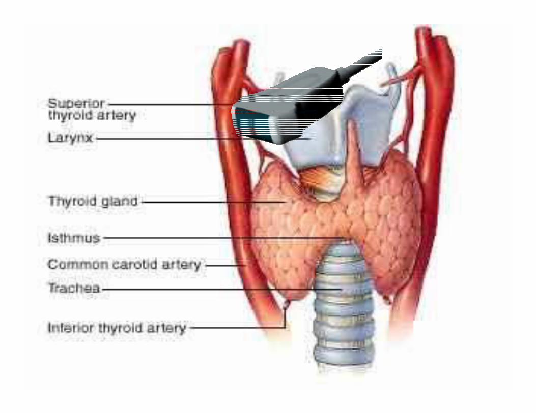
Artifacts of US Anatomy
• Air has a very high attenuation, thus the
hypoechoic appearance within the trachea.
• The carotid has a relative lack of attenuation
through the blood, thus producing a through the blood, thus producing a
hyperechoic posterior enhancement.
• Reverberation artifacts can be seen at the
trachea due to closely spaced interfaces with
significantly mismatched acoustic
impedances (tracheal rings and thyroid).
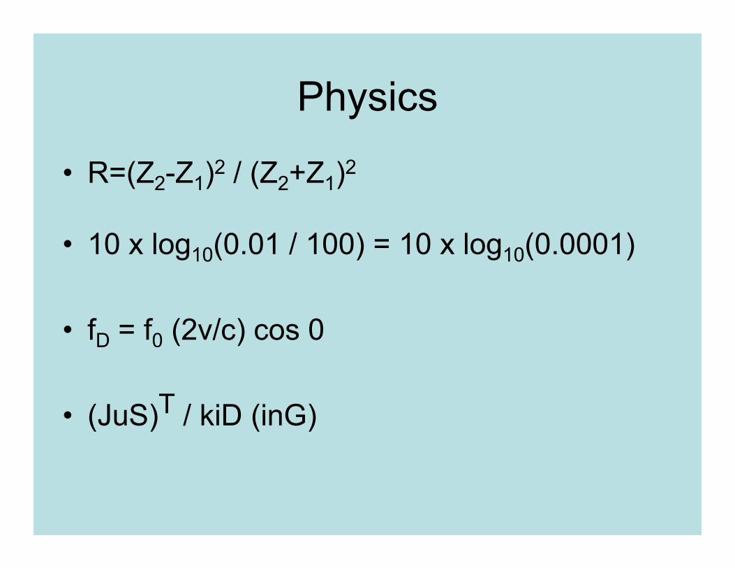
Physics
• R=(Z2-Z1)2 / (Z2+Z1)
2
• 10 x log10(0.01 / 100) = 10 x log10(0.0001)
• fD = f0 (2v/c) cos 0
• (JuS)T / kiD (inG)

Optimizing the US Exam
• The transducer:
– A “small parts” transducer at a frequency of 7.5
to 15 MHz or greater. This high frequency
results in greater resolution at the expense of results in greater resolution at the expense of
depth penetration. Lower frequencies allow
deeper penetration of the sound wave.
– The majority of anatomic structures in the neck
are within 4cm of the skin so loss of penetration
is usually of little concern.

US Features
• Gain:
–Refers to the overall brightness of the
image on the screen.
– If the gain is too high, the image is bright – If the gain is too high, the image is bright
(white)- hyperechoic.
– If the gain is too low, the image is dark –
hypoechoic.

US Features
• Color-flow Doppler:
– Identification of blood vessels vs lymph
nodes or ductsnodes or ducts
–Assessing the vascularity of structures
• Can be helpful in benign vs malignant

Performing The
UltrasoundUltrasound

Normal Head and
Neck AnatomyNeck Anatomy

Starting View

Right Lobe Transverse
Sternothyroid
Sternohyoid

Right Strap Muscles

Longitudinal Thyroid
Subcutaneous fat
Strap muscles

Left Lobe transverse
Strap muscles

Left Transverse
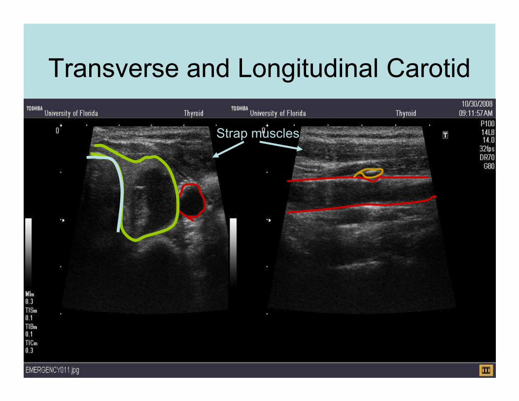
Transverse and Longitudinal Carotid
Strap muscles

Lateral to the Carotid

Parathyroid
Strap muscles
Post enhancement

Ultrasound Features of Thyroid
Nodules

Ultrasound
Anatomy QuizAnatomy Quiz

Trachea
Thyroid:
Thyroid:
Isthmus SCM
Carotid
Thyroid:
Right
Lobe Thyroid:
Left Lobe Esophagus
Adding Ultrasound to your
FNAsFNAs

Performing the FNA
• Localize the nodule
with U/S
• Skin Prep with
sterile techniquesterile technique
• Local anesthetic
Benign complex thyroid nodule

Performing the FNA
• Visualize needle
placement with U/S
guidance
• Perform aspirations • Perform aspirations
• Slide and
cytology/cell block
preparation
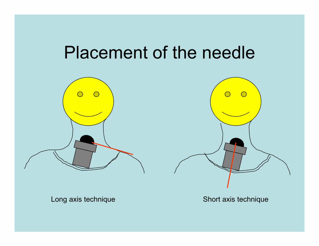
Placement of the needle
Short axis techniqueLong axis technique

Long axis technique
•Goal: Line up the
needle parallel to the
long axis of the probe
Needle
long axis of the probe
•Enter the skin in the
center of the short
axis of the probe
Transducer
long axis
Transducer short axis
Nodule

Long axis technique
• Advantages• Working in one plane
• Better visualization of entire needle• Better visualization of entire needle
• Disadvantages• Needle travels longer distance to get to the
nodule
• Adjacent structures may obstruct
• May require working in planes other than the axial plane
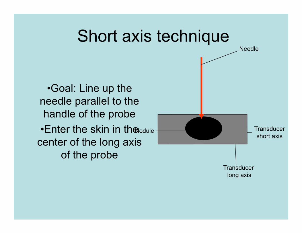
Short axis technique
•Goal: Line up the
needle parallel to the
handle of the probe
Needle
handle of the probe
•Enter the skin in the
center of the long axis
of the probeTransducer
long axis
Transducer
short axisNodule

Short axis technique
• Advantages• Needle travels shorter distance to get to the nodule
• Less likely for adjacent structures to obstruct
• Easier to access nodule under bony structure i.e. mandible or sternoclavicular bonesternoclavicular bone
• Disadvantages• More complex image-working in 2 planes
• Needle is more difficult to visualize due to less of it in the field of view and results in decreased echo due to angle of approach

Ultrasound Features of
Thyroid NodulesThyroid Nodules
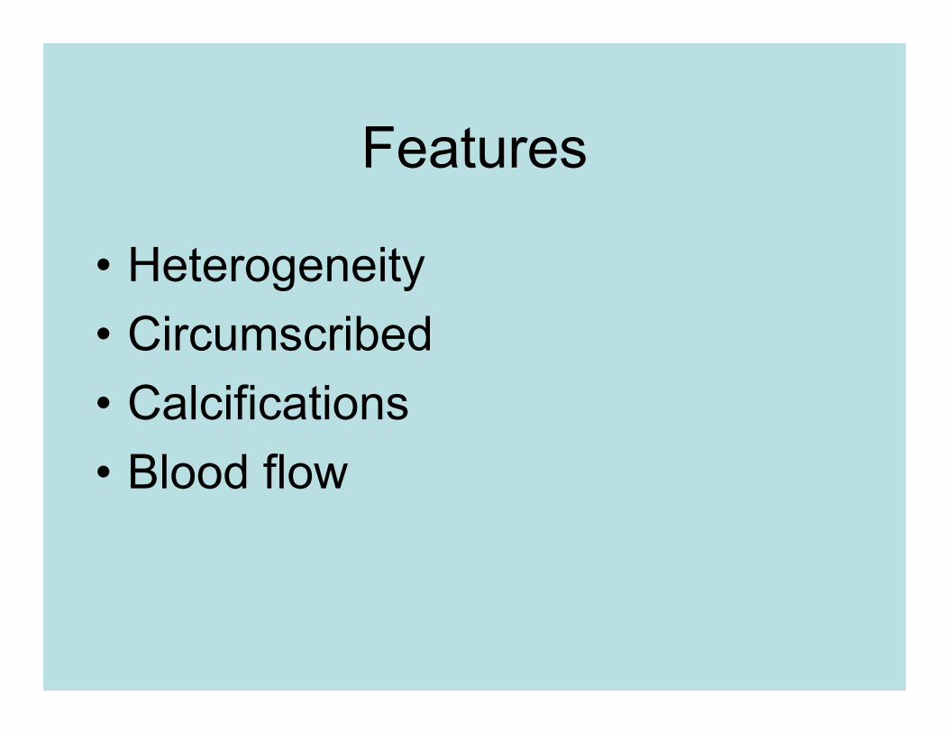
Features
• Heterogeneity
• Circumscribed
• Calcifications
• Blood flow

Heterogeneity
Endocr Pract. 2004 May-Jun;10(3):246-52.

Longitudinal view

Case 1
• Rubbery nodule in
medial left lobe
• Freely mobile upon
swallowing, well swallowing, well
delineated
• No
lymphadenopathy
• TSH 1.5
Radiology. 2005 Dec;237(3):794-800

Case 1
FNA =
Benign colloid Benign colloid
nodule
Spongiform
Nodule

Malignant Features
Endocr Pract. 2004 May-Jun;10(3):246-52.


Internal Doppler flow
Radiology. 2005 Dec;237(3):794-800

Microcalcifications
Punctate echogenicities Comet-tail artifact
Papillary thyroid carcinoma Benign nodule

Calcifications

Benign complex nodules

Hashimoto’s Thyroiditis

Multinodular goiter
• Risk of cancer in a patient with a
multinodular goiter is the same as a
patient with a solitary nodule
• Should biopsy the most suspicious
nodules, up to four or five nodules

Biopsying the largest nodule in a patient with 2 nodules would have missed
13.7% of cancers. In patients with 3 nodules, 48.2% of cancers would have
been missed by biopsying only the largest nodule.

Questions?
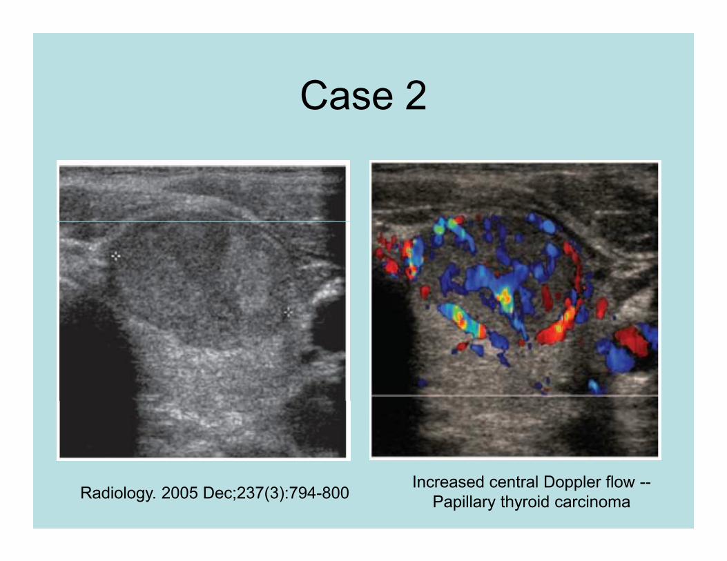
Case 2
Increased central Doppler flow --
Papillary thyroid carcinomaRadiology. 2005 Dec;237(3):794-800

Case 2
FNA =
Metastatic
Level III lymph node
Radiology. 2005 Dec;237(3):794-800
Metastatic
PTC

Case 4
FNA
Indeterminate

Summary
• Ultrasounds can add to the nodules
that cytopathologists can biopsy.
• Adds a level of comfort to knowing
you are in the lesion.
• Helps identify nodules that are
suspicious