Embed Size (px)
Citation preview
5/13/2017
1
Jeffrey A. Strakowski, MDClinical Associate Professor, Dept of PM&R
The Ohio State University
Associate Director of Medical Education, PM&R
Riverside Methodist Hospital
Director of Musculoskeletal Research,
The McConnell Spine, Sport & Joint Center
ULTRASOUND GUIDANCE FOR PERIPHERAL NERVE
PROCEDURES
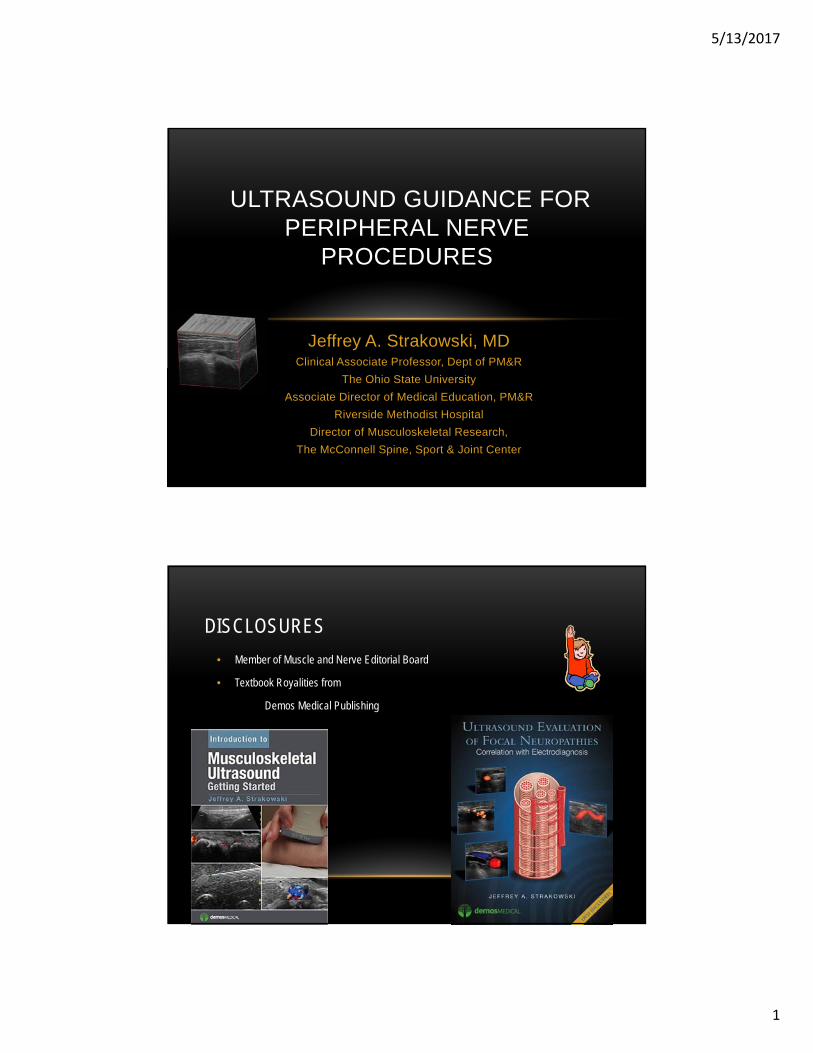
DISCLOSURES
• Member of Muscle and Nerve Editorial Board
• Textbook Royalities from
Demos Medical Publishing
5/13/2017
2
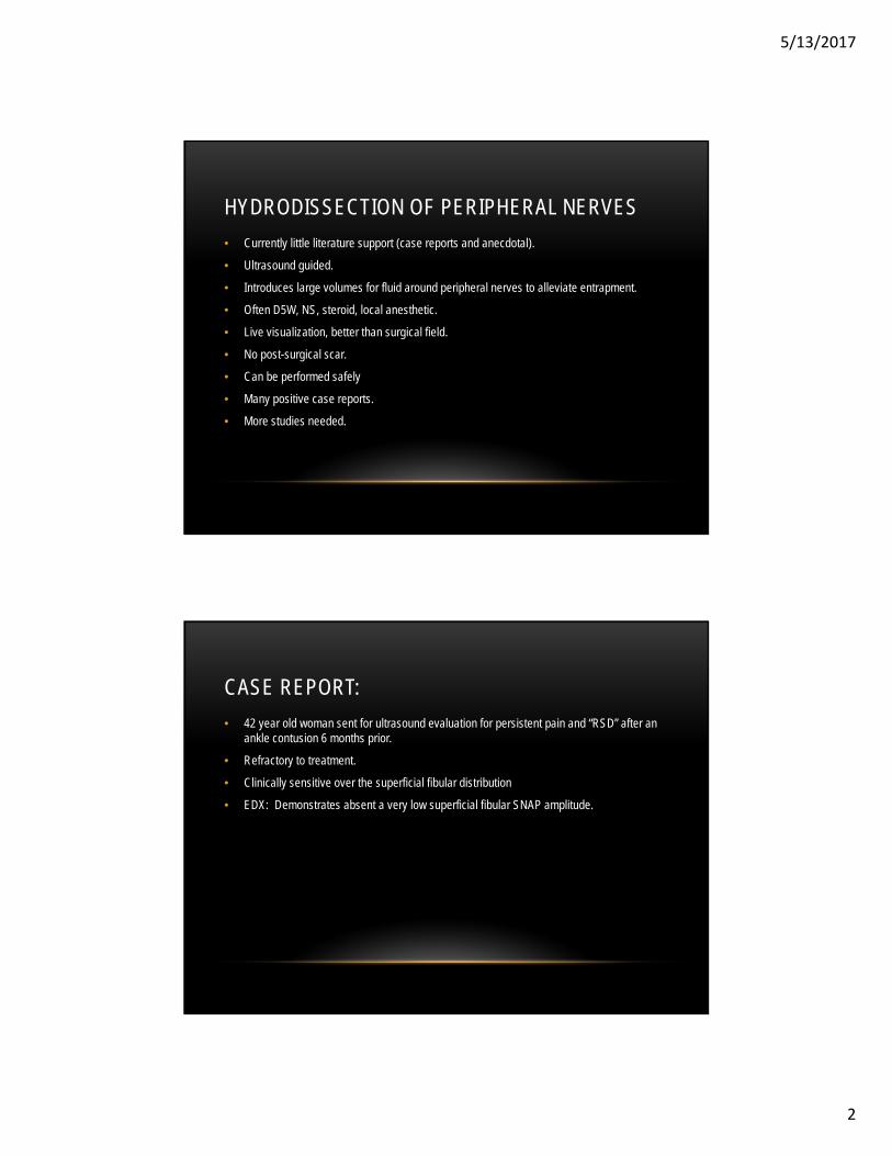
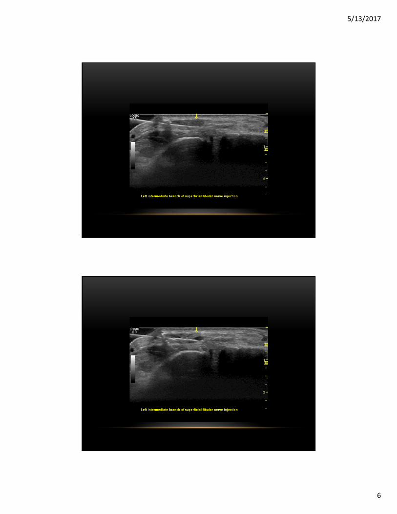
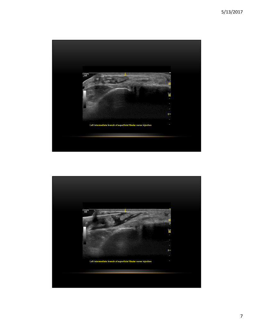
HYDRODISSECTION OF PERIPHERAL NERVES
• Currently little literature support (case reports and anecdotal).
• Ultrasound guided.
• Introduces large volumes for fluid around peripheral nerves to alleviate entrapment.
• Often D5W, NS, steroid, local anesthetic.
• Live visualization, better than surgical field.
• No post-surgical scar.
• Can be performed safely
• Many positive case reports.
• More studies needed.
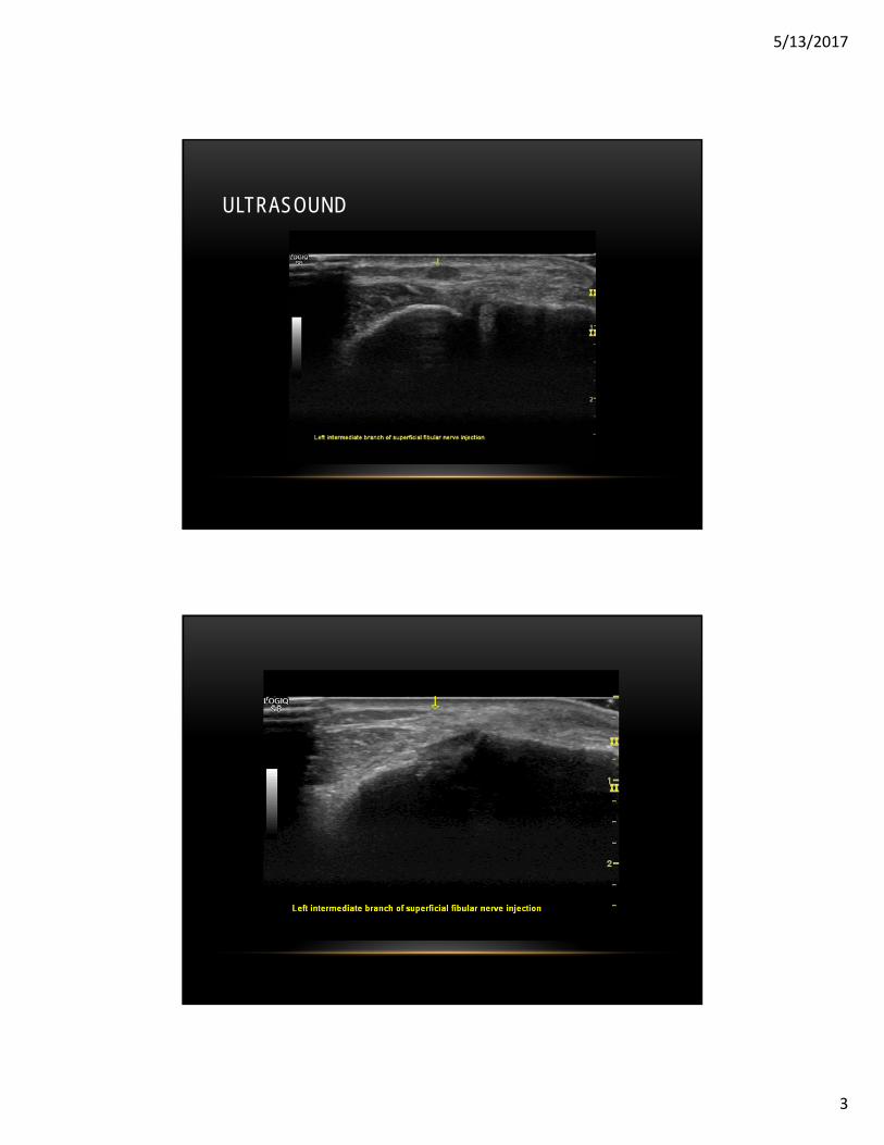
CASE REPORT:
• 42 year old woman sent for ultrasound evaluation for persistent pain and “RSD” after an ankle contusion 6 months prior.
• Refractory to treatment.
• Clinically sensitive over the superficial fibular distribution
• EDX: Demonstrates absent a very low superficial fibular SNAP amplitude.
5/13/2017
3
ULTRASOUND

5/13/2017
4
ELECTED HYDRODISSECTION

5/13/2017
5
VIDEO
5/13/2017
6
5/13/2017
7
5/13/2017
8
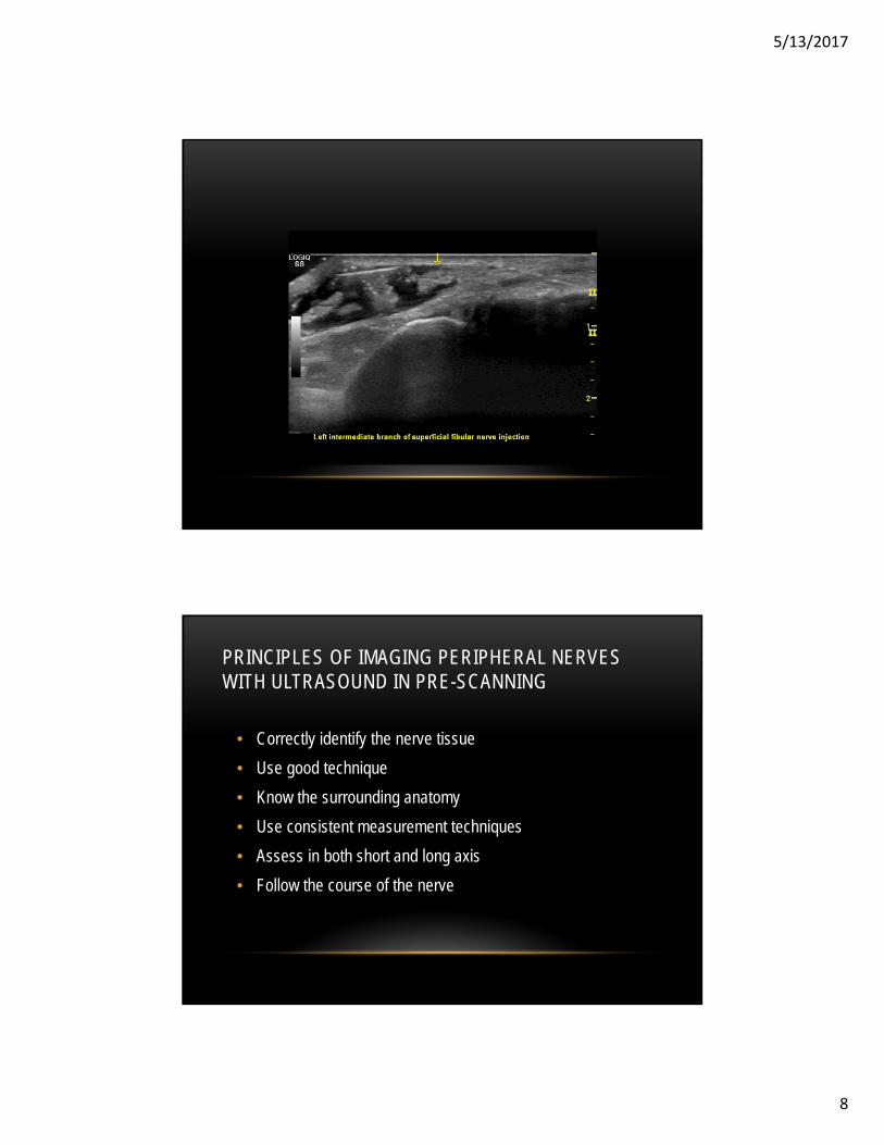
PRINCIPLES OF IMAGING PERIPHERAL NERVES WITH ULTRASOUND IN PRE-SCANNING
• Correctly identify the nerve tissue
• Use good technique
• Know the surrounding anatomy
• Use consistent measurement techniques
• Assess in both short and long axis
• Follow the course of the nerve

5/13/2017
9
US GUIDED INJECTION BASICS
• Pre-plan, pre-scan
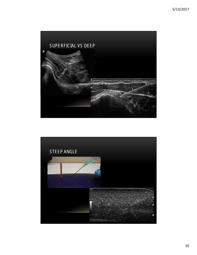
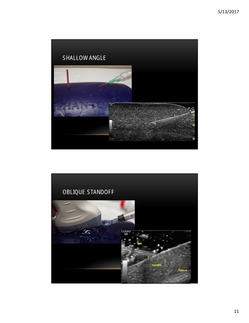
• Consider depth of the injection
• Use an oblique standoff when needed
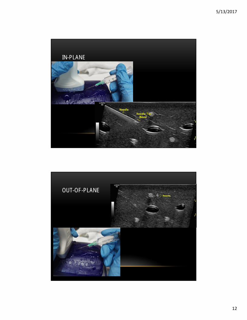
• Understand in-plane vs out-of-plane injections
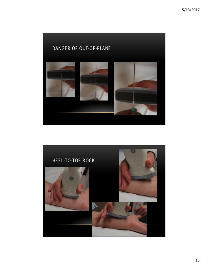
• Use toggling and heel-to-toe rocking to optimize needle conspicuity
• Be aware of needle reverberation artifact
• Avoid too many moving parts
PRESCAN
5/13/2017
10
SUPERFICIAL VS DEEP
STEEP ANGLE
5/13/2017
11
SHALLOW ANGLE
OBLIQUE STANDOFF
5/13/2017
12
IN-PLANE
OUT-OF-PLANE
5/13/2017
13
DANGER OF OUT-OF-PLANE
HEEL-TO-TOE ROCK

5/13/2017
14
EFFECT OF HEEL-TO-TOE ROCK
TOGGLE
5/13/2017
15
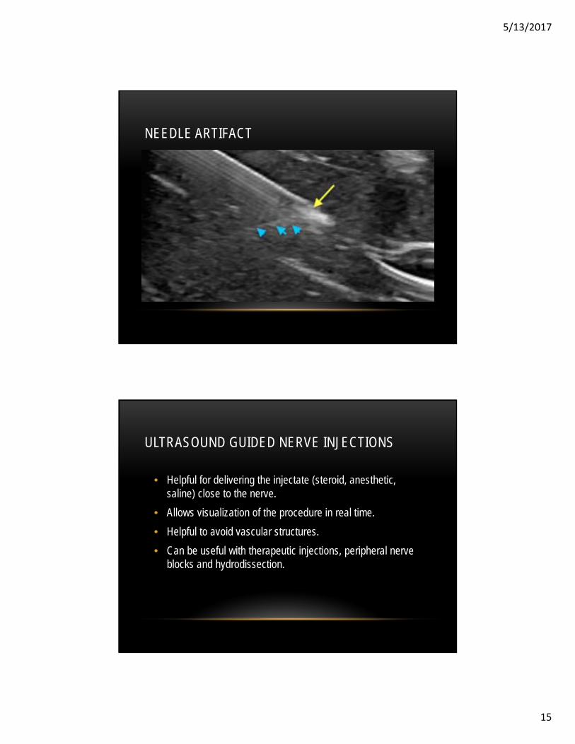
NEEDLE ARTIFACT
ULTRASOUND GUIDED NERVE INJECTIONS
• Helpful for delivering the injectate (steroid, anesthetic, saline) close to the nerve.
• Allows visualization of the procedure in real time.
• Helpful to avoid vascular structures.
• Can be useful with therapeutic injections, peripheral nerve blocks and hydrodissection.
5/13/2017
16
REMEMBER:
• Accurate identification of the nerve tissue is needed for effective injections.
• Pre-scan the surrounding region for potential areas to avoid and to facilitate the proper approach with the needle.
• Have a straight line between the patient and injection site, and ultrasound screen.
• Do a checklist for all necessary equipment in advance and have it within reach during the procedure.
NERVE SAFETY
• Transducer must be placed perpendicular to the nerve for accuracy.
• Identify the outer epineurium.
• Optimize Focal Zone.
• Optimize Gray Scale Mapping
• Set depth so target takes up majority of the screen.
• Use highest frequency with effective penetration to visualize the nerve.
• Caution with intraneural injections
• Must avoid injuring the fascicles.

5/13/2017
17
PERIPHERAL NERVE INJECTIONS: GENERAL
• Need to know course and function of the nerve.
• Do adequate pre-scan.
• High frequency linear transducer is used for most nerve injections.
• Most injections will use a short-axis view of the nerve and in-plane view of the needle.
• With hydrodissections, might also use long-axis view of needle.
• Creating a halo around the nerve with injectate will increase conspicuity.
• The patient should be positioned between the ultrasound screen to allow easy visualization of both the needle at the target site and the ultrasound image.
WRONG!
5/13/2017
18
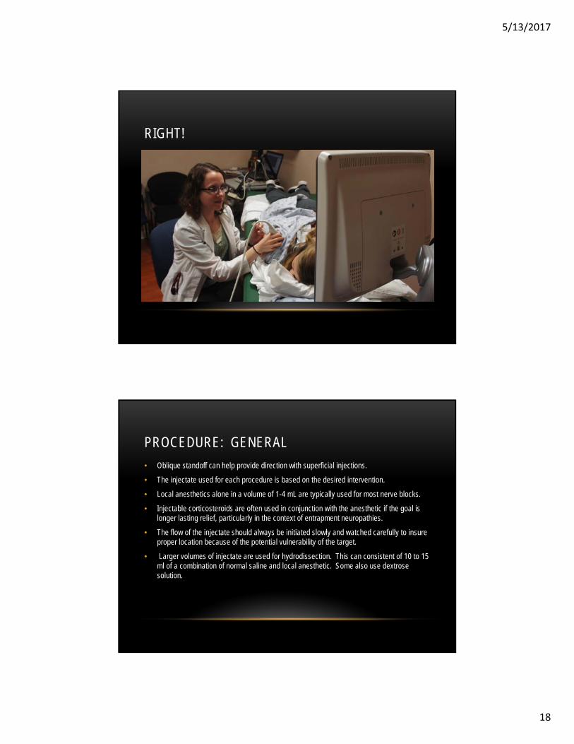
RIGHT!
PROCEDURE: GENERAL
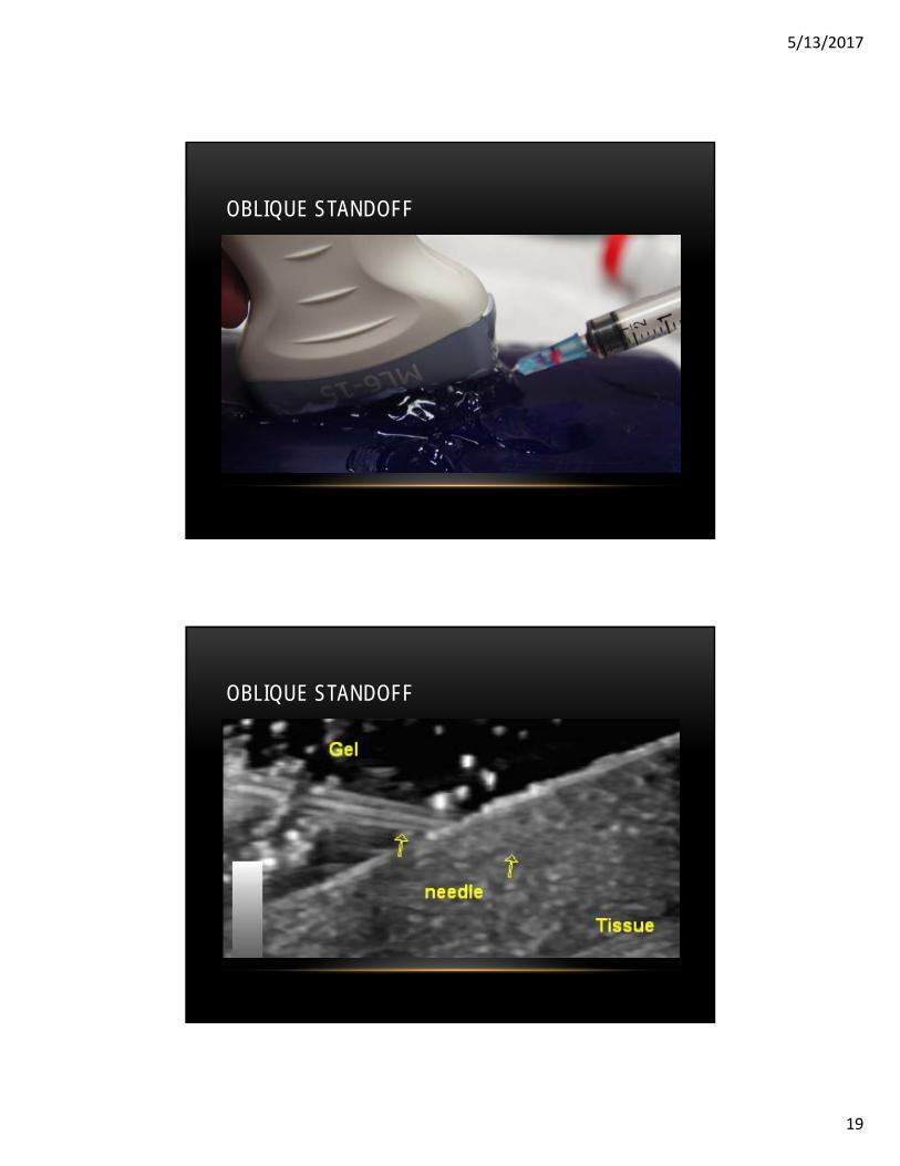
• Oblique standoff can help provide direction with superficial injections.
• The injectate used for each procedure is based on the desired intervention.
• Local anesthetics alone in a volume of 1-4 mL are typically used for most nerve blocks.
• Injectable corticosteroids are often used in conjunction with the anesthetic if the goal is longer lasting relief, particularly in the context of entrapment neuropathies.
• The flow of the injectate should always be initiated slowly and watched carefully to insure proper location because of the potential vulnerability of the target.
• Larger volumes of injectate are used for hydrodissection. This can consistent of 10 to 15 ml of a combination of normal saline and local anesthetic. Some also use dextrose solution.
5/13/2017
19
OBLIQUE STANDOFF
OBLIQUE STANDOFF
5/13/2017
20
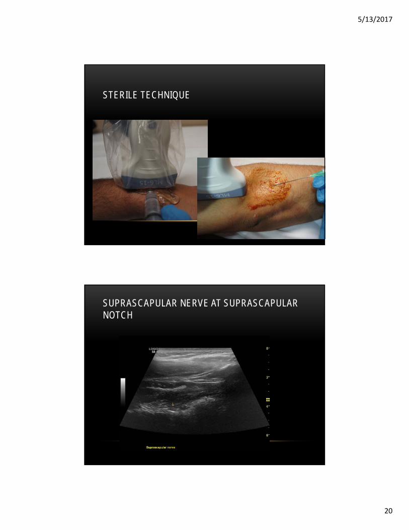
STERILE TECHNIQUE
SUPRASCAPULAR NERVE AT SUPRASCAPULARNOTCH

5/13/2017
21
SUPRASCAPULAR NERVE: INDICATIONS
• For intractable shoulder pain.
• Acutely in post-operative pain.
• Diagnostic trial with anesthetic agent.
• Longer activing agents: steroids, toxic agents such as phenol, radiofrequency ablations
• Drainage of compressive cyst
SUPRASCAPULAR NERVE: ANATOMY AND IDENTIFICATION
5/13/2017
22
SUPRASCAPULAR NERVE: ANATOMY
• Derived from C5 and C6 roots.
• Provides motor function to the supraspinatus and infraspinatus
• External rotation of the shoulder
• Sensory innervation to acromioclavicular and glenohumeral joint
SUPRASCAPULAR NERVE: SCANNING
• Visualized at the suprascapular notch and spinoglenoidforemen.
• Transducer is placed in the same plane as the spine of the scapula.
• Use internal and external rotation (and Doppler) to distinguish the artery and vein.
5/13/2017
23
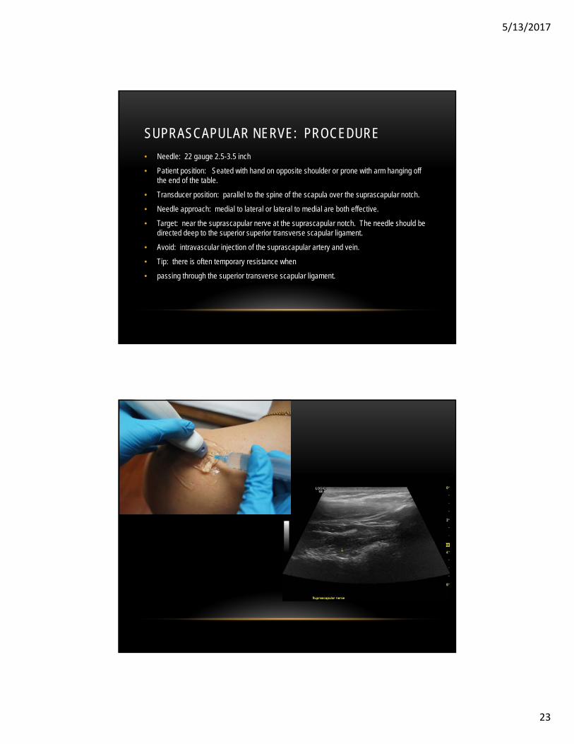
SUPRASCAPULAR NERVE: PROCEDURE
• Needle: 22 gauge 2.5-3.5 inch
• Patient position: Seated with hand on opposite shoulder or prone with arm hanging off the end of the table.
• Transducer position: parallel to the spine of the scapula over the suprascapular notch.
• Needle approach: medial to lateral or lateral to medial are both effective.
• Target: near the suprascapular nerve at the suprascapular notch. The needle should be directed deep to the superior superior transverse scapular ligament.
• Avoid: intravascular injection of the suprascapular artery and vein.
• Tip: there is often temporary resistance when
• passing through the superior transverse scapular ligament.

5/13/2017
24
EXTRINSIC GANGLIA
CONCLUSIONS
• Understanding of basic scanning and imaging techniques is needed for successful use of this modality in performing effective injection techniques.
• Injections around peripheral nerves requires reasonable caution because of the vulnerability of the targets.
• Preparation with knowledge of the course of the nerve and surrounding anatomy, adequate pre-scanning and planning preparation can lead to success.

5/13/2017
25
THANK YOU!

![NAOSITE: Nagasaki University's Academic Output SITEnaosite.lb.nagasaki-u.ac.jp/dspace/bitstream/10069/39435/...technique to create blood vessels [13], peripheral nerves [14], cartilage](https://img.pdfslide.tips/doc/110x75/5ecb29817886ea396f6715eb/naosite-nagasaki-universitys-academic-output-technique-to-create-blood-vessels.jpg)