Embed Size (px)
Citation preview
UNIVERSITA’ DEGLI STUDI DI CATANIA
FACOLTA’ DI FARMACIA
Dottorato di ricerca in Biotecnologie
XXIV ciclo
Potential involvement of gap junctions
in pathology of addiction
Dott. MASSIMO LAZZARO
Coordinatore e tutor prof. Federico Cicirata
Supervisors prof. PierVincenzo Piazza, prof. Guillaume Drutel


Ai miei genitori

Abstract
Cocaine abuse represents a huge social problem for the widespread in the world and for
the many health risks associated. Moreover, a significative percentage of cocaine users
develop an addiction with loss of control on drug intake. Several molecular
modifications occurs with cocaine use, some of which seem occur only in addicted
individuals.
Gap junctions (GJs) allow cellular communication, indeed they are fundamental to
regulate cellular synchronization, cellular growth and metabolic coordination in tissues.
We analyzed the role of GJs proteins in the cocaine’s mechanism of action and
furthermore we verified the potential role of GJs in addiction.
Our experiments show several modifications occurring at early and after prolonged
cocaine exposure. In addition are shown differential alterations depending by addiction-
like behaviour in animals, suggesting a potential involvement of GJs in transition to
addiction.
Contents
1 INTRODUCTION ....................................................................................................................................... 1
1.1 COCAINE ................................................................................................................................................... 2
1.1.1 History and administration methods ................................................................................................ 2
1.1.2 Side effects ........................................................................................................................................ 3
1.1.3 Psychological effects of cocaine ........................................................................................................ 4
1.1.4 Exposure time-dependent effects ..................................................................................................... 5
1.1.5 Addiction ........................................................................................................................................... 7
1.1.6 Molecular mechanisms of cocaine .................................................................................................... 9
1.1.6.1 Modifications in genes expression ......................................................................................................... 11
1.1.7 Neurobiological substrates of cocaine: brain reward system ......................................................... 16
1.1.7.1 Dopaminergic and glutamatergic transmission ...................................................................................... 18
1.1.8 Synaptic plasticity in the addiction ................................................................................................. 20
1.1.9 Animal models of addiction ............................................................................................................ 27
1.2 GAP JUNCTIONS ....................................................................................................................................... 29
1.2.1 Formation and structure ................................................................................................................. 29
1.2.2 Connexins ........................................................................................................................................ 32
1.2.2.1 Assembly and degradation ..................................................................................................................... 34
1.2.2.2 Gap junctions functions .......................................................................................................................... 35
1.2.2.3 Permeability and regulation ................................................................................................................... 37
1.2.3 Pannexins ........................................................................................................................................ 41
1.2.3.1 Expression .............................................................................................................................................. 41
1.2.3.2 Functions and regulation ........................................................................................................................ 42
1.2.4 Gap Junctions in nervous system .................................................................................................... 44
1.2.4.1 Pannexins in brain .................................................................................................................................. 48
1.2.5 Electrical synchronization................................................................................................................ 50
1.2.6 Gap junctions and seizures .............................................................................................................. 51
1.2.7 Gap junctions and mesocorticolimbic system ................................................................................. 52
1.2.8 Gap Junctions and cocaine abuse ................................................................................................... 53
2 AIM OF THE WORK................................................................................................................................. 55
3 MATERIALS AND METHODS ................................................................................................................... 58
3.1 COCAINE SELF-ADMINISTRATION (SA) PROCEDURES ........................................................................................ 58
3.2 QUANTITATIVE REAL-TIME PCR ................................................................................................................... 63
3.3 IMMUNOBLOTTING ................................................................................................................................... 66
3.4 STATISTICS .............................................................................................................................................. 68
4 RESULTS ................................................................................................................................................. 70
4.1 EFFECTS OF COCAINE SELF-ADMINISTRATION ON NUCLEUS ACCUMBENS GAP JUNCTIONS ........................................ 71
4.1.1 Effect on Cx36 in Nucleus Accumbens ............................................................................................ 71
4.1.2 Effect on Cx43 in Nucleus Accumbens ............................................................................................ 73
4.1.3 Effect on Cx32 in Nucleus Accumbens ............................................................................................ 75
4.1.4 Effect on Panx1 in Nucleus Accumbens .......................................................................................... 77
4.1.5 Effect on Panx2 in Nucleus Accumbens .......................................................................................... 79
4.1.6 Effects of cocaine SA on Nucleus Accumbens in other genes ......................................................... 81
4.2 EFFECTS OF COCAINE SA IN MEDIAL PREFRONTAL CORTEX GAP JUNCTIONS ........................................................... 83
4.2.1 Effect on Cx36 in medial prefrontal cortex ..................................................................................... 83
4.2.2 Effect on Cx43 in medial prefrontal cortex ..................................................................................... 85
4.2.3 Effect on Cx32 in medial prefrontal cortex ..................................................................................... 87
4.2.4 Effect on Panx1 in medial prefrontal cortex ................................................................................... 89
4.2.5 Effect on Panx2 in medial prefrontal cortex ................................................................................... 91
4.2.6 Effect of cocaine SA on other genes in medial prefrontal cortex.................................................... 93
4.3 EFFECTS OF COCAINE SA ON VENTRAL TEGMENTAL AREA GAP JUNCTIONS ............................................................. 95
4.3.1 Effect on Cx36 in ventral tegmental area ........................................................................................ 95
4.3.2 Effect on Cx43 in ventral tegmental area ........................................................................................ 97
4.3.3 Effect on Cx32 in ventral tegmental area ........................................................................................ 99
4.3.4 Effect on Panx1 in ventral tegmental area .................................................................................... 101
4.3.5 Effect on Panx2 in ventral tegmental area .................................................................................... 103
4.3.6 Effects of cocaine SA on other genes in ventral tegmental area .................................................. 105
4.4 SUMMARY OF RELEVANT MODIFICATIONS IN NAC GAP JUNCTIONS .................................................................... 107
4.5 SUMMARY OF RELEVANT MODIFICATIONS IN MPFC GAP JUNCTIONS ................................................................. 109
4.6 SUMMARY OF RELEVANT MODIFICATIONS IN VTA GAP JUNCTIONS .................................................................... 111
5 DISCUSSION ......................................................................................................................................... 114
5.1 GENERAL CONSIDERATIONS....................................................................................................................... 115
5.2 NEURONAL SYNCHRONIZATION .................................................................................................................. 116
5.3 COCAINE AND INFLAMMATION .................................................................................................................. 118
5.4 SYNAPTIC PLASTICITY ............................................................................................................................... 122
5.5 PANNEXINS ........................................................................................................................................... 123
5.6 CONCLUSIONS ........................................................................................................................................ 127
6 ABBREVIATIONS................................................................................................................................... 129
7 REFERENCES ......................................................................................................................................... 133
8 ACKNOWLEDGEMENTS ........................................................................................................................ 145
1
Introduction
1 Introduction
Drug abuse represents a serious health issue worldwide. Many are the drugs of abuse
and between these, cocaine has taken over the years a prominent place. Only in Europe,
it represents the second drug of abuse used after cannabis. It is therefore a question of
great importance and extensive studies have been carried out in order to understand the
physiological mechanisms of action and try to cure addiction. In fact, it is possible to
distinguish two types of individuals, there are the majority of users who occasionally use
cocaine for recreational purposes, but they are able to control the frequency and doses
and a second group of individuals who show a real addiction, with loss of control on
drug intake. Cocaine use is a huge social problem due to the numerous health risks it
causes; indeed, it is in relation to cardiovascular and neurological damage, and
furthermore to risk of overdose. It entails also an increase of social and health costs
sustained for actions of detoxification and rehabilitation.
Recent studies, as regards the dependency, highlight the presence of a real pathology. In
many animal models are present alterations in the limbic system at receptorial level and
in the mechanisms of signal transduction together with alteration in genes expression.
Mechanisms of long-term synaptic modifications seem also involved, including those
associated with disturbance of learning and memory.
From the foregoing, it is evident that studies are needed to understand the molecular
mechanisms and then potentially identify drug targets to treat addiction.
2
Introduction
1.1 Cocaine
1.1.1 History and administration methods
Cocaine (benzoylmethylecgonine C17H21NO4) is extracted by Erythroxylon Coca, a plant
native to the Andes and other parts of South America. For hundreds years has been used
by Andean Indians that chew the leaves to control symptoms associated with living at
high altitude. This form of administration is not very dangerous because the content of
cocaine in leaf is about 0.5%, oral absorption is low (about 1 hour) and the hepatic
metabolism degrades up to 80% of the ingested dose; so it doesn’t reach important
blood concentration, the toxicity is rare and phenomena of habituation are mild [1, 2].
The most common form of cocaine as drug of abuse is cocaine hydrochloride, produced
by dissolving the alkaloid (as free base) in hydrochloric acid. The salt that is obtained
after dehydration represents the white powder that can be taken by nasal sniffing, orally
or intravenously.
The free base is water insoluble, can be obtained by dissolution of hydrochloride in
alkaline water with use of baking soda; the crystals are vaporizable at temperature
lower than chloride and can be smoked together tobacco. In this form has termed
“crack”, for the popping sound during smoking. Crack produces, respect to
hydrochloride, an instantaneous action, high plasma levels but a short-lived euphoria
(about 15 minutes). The absorption at the pulmonary level induces, thanks to the
considerable absorption area, very high blood levels (similar to intravenous injection),
and the passage into the venous circulation produces direct availability in brain,
rendering it more addictive.
The intranasal administration of hydrochloride is characterized by low absorption from
mucosa, due also to vasoconstrictive properties of cocaine.
The behavioural effect starts after 3-5 minutes and the blood peak occurs between 10
and 20 minutes but after 1 hour, cocaine is no longer detectable. Intravenous
administration cause effects after about 1 minute and the action persists up to 20
minutes [3, 4].
After whatever form of administration, cocaine is rapidly distributed from plasma to
different district, among which the brain and the fat. Indeed, it crosses the blood brain
barrier rapidly due to its lipophilic properties, and more slowly is distributed in other
3
Introduction
compartments. 5% of cocaine is excreted unchanged in the urine (detectable after 3-6
hours of use), while 85% is metabolized by the liver and plasma esterases to give
ecgonine methyl ester and benzoylecgonine, detectable in urine for more than 14 days
after the assumption [5].
1.1.2 Side effects
Cocaine can cause many damages in brain, heart, blood vessels and lungs; moreover for
the strong involvement of vital organs, it can cause sudden death [6].
Cardiovascular system is the most common site where appear complications among
cocaine users and these can occur after acute or chronic use. Cocaine increases heart
rate, blood pressure and myocardial oxygen demand and in addition vasoconstriction
and tachycardia. The result can be infarction and ischemia, even in people without heart
disease [7, 8].
Cocaine use damages lungs and respiratory system; nose and sinuses are the first to be
affected for the direct contact with the substance, indeed a chronic use can degrade the
cartilage in the septum leading to nasal perforation. Smoking “crack” irritates the lungs
and can cause asthma, bronchospasm, dyspnoea, haemoptysis, diffuse alveolar
infiltrates pulmonary and systemic eosinophilia, chest pain, lung trauma pneumonitis,
vascular lesions and pulmonary edema [9-11].
Pathological effects of cocaine in central nervous system (CNS) may vary from
cerebrovascular effects, as intracranial haemorrhage and infarction due to rapid
increase in blood pressure and vasoconstriction, to arrive at neurological effects as
seizures, which can compare also at the first intoxication and hyperthermia. There are
also psychiatric consequences largely associated to abuse, as psychosis, schizophrenia,
depression, suicidal ideation, obsessive-compulsive disorders. To add sexual
dysfunction, even though low doses may increase sexual excitement, high doses have the
opposite effect with disinterest and impotence [12-19].
On gastrointestinal system cocaine cause constriction of blood vessels and hence a
reduction in mesenteric blood flow leading to gastritis or even perforation of the
stomach or intestines. It has been reported also hepatocellular necrosis and ischemic
hepatitis [20, 21].
4
Introduction
As regard the renal system, cocaine can cause sudden and overwhelming kidney failure
through a process called rhabdomyolysis. In people with high blood pressure a regular
cocaine use can accelerate the long-term kidney damage causing renal infarction,
glomerulosclerosis [22, 23].
1.1.3 Psychological effects of cocaine
The use of drugs of abuse may be considered as behaviour that is maintained by its own
consequences. A drug of abuse may reinforce the behaviour by inducing pleasurable
effects of intake (positive reinforcement) or putting an end to an unpleasant (negative
reinforcement). Cocaine is an addictive drug both rewarding, seen as intrinsically
positive, and has positive reinforcement effect, because pushing the individual to repeat
the intake.
The effects of cocaine can be distinct in short-term and long-term effects.
The principal effect is a powerful stimulation of nervous system. Use in acute induces a
rapid sense of euphoria, depending on the dose, by the route of administration and by
the state of individual tolerance; this euphoric state can last from about 30 minutes to
two hours. This phase, termed high, is the most widely effect recognized among the
users and hence the reason for which people taking cocaine, at least the occasional
users. It increases the alertness, the feeling of well-being and supremacy, an high sense
of energy and motor activity, feelings of competence and sexuality. Athletic performance
may be enhanced in sports. Indeed the hallmarks of cocaine users are principally high
levels of energy and activity, excitement, exuberance, dilated pupils.
The cocaine high involves psychological and physical changes. The effects of cocaine on
the brain and nervous system cause some of these, and others are due to personal
feeling of cocaine users.
This first state, characterized by a complete sense of security is followed by onset of bad
mood and anxiety, that taking the users to a binge, that is a repeated use of drug at short
intervals. With higher dose for prolonged time the effects can be the opposite of the
high, with a blunting of the emotions, sadness, anxiety, irritability, paranoia; in addition
5
Introduction
there are different physical effects as tremors, convulsions, tachycardia, hyperthermia,
as a result of an increased catecolaminergic tone [24].
1.1.4 Exposure time-dependent effects
Acute cocaine use acts in deep areas of the brain, among which areas that reward us for
"good behaviour", as those activities that lead to food, sex and healthy pleasure. The
stimulation of these areas provides a sense of well-being, but it can create a powerful
craving. The craving is the moment in which there is a strong and compelling need to
assume the substance, but this need becomes uncontrollable. It may occur even if there
is no physical dependence and may be triggered by seeing objects or experiencing
moments that are associated with the drug or usage of it. In some case, this feeling may
persist for the rest of the life.
Chronic cocaine use induces changes in brain reward centres and consequent chronic
dysphoria. This last together with the sensation of depression, felt after the initial high,
are caused by the lack of normal amounts of serotonin and dopamine in the brain. The
dysphoria magnifies craving for cocaine, because cocaine reward occurs rapidly and
improves mood, this leads the individual to continue a compulsive drug intake even if
there is a worsening of conditions. At the end, the long-term effects are opposite those
occurring after acute use.
The molecular changes occurring in the brain, after chronic use, can give the phenomena
of tolerance, dependence and withdrawal.
Tolerance is a state characterized by the need to increase the dose of drug to maintain
constant the intensity of the effect produced by it. This phenomenon is the manifestation
of the processes that occur in brain cells, in order to adapt functionally themselves to
strong imbalances of transmitter levels caused by chronic use of cocaine. The
mechanisms by which cells induce a tissue response are different and maybe are the
summa of different factors. A main factor is the modification at receptorial level that can
be obtained in different ways, with the modulation of receptor affinity for the ligands,
with mechanisms of down or up-regulation but also with modifications in intracellular
pathways or modulation of second messengers. In addition, there are losses of vesicular
6
Introduction
monoamine transporters, of neurofilament proteins and other morphological changes
that in the long-term cause damage of dopamine neurons. All these effects contribute a
rise in tolerance thus requiring a larger dosage to achieve the initial effect.
When dependence is present, stopping cocaine suddenly leads to withdrawal.
Cocaine determines the appearance of a withdrawal syndrome, characterized by an
immoderate search of the substance in order to alleviate the physical dependence that
produced the drug. The withdrawal is caused by a physical alteration in the normal
physiological state of the individual, to adapt itself to the substance. It is characterized
by deleterious symptoms that can only be offset with the additional intake of the drug.
The symptoms of cocaine withdrawal (also known as “comedown” or “crash”) range
from moderate to severe and are more psychological than physiological, so usually are
not visible physical symptoms such as those due to others drugs (like vomiting, chills,
tremors) but are present dysphoria, fatigue, difficulty concentrating depression, anxiety,
psychological and physical weakness, pain and compulsive craving. Sometimes may
cause suicidal thoughts. Some users also report formication: a feeling of a crawling
sensation on the skin also known as "coke bugs". These symptoms can last for weeks or
in some cases for months.
[25, 26]
7
Introduction
1.1.5 Addiction
Cocaine addiction is a persistent state in which drug use escapes control, even when
serious negative consequences ensue. These behavioural abnormalities develop
gradually and progressively during the course of repeated exposure to a drug of abuse
and can persist for months or years after discontinuation of drug use. Indeed, usually the
withdrawal symptoms resolve in 1-2 weeks, but craving may returns even many years
after the last use. The major stimuli that precipitate this state are three, a stressful life
event, an environmental stimulus associated with a drug taking event or a re-exposure
to the drug. As a result, drug addiction can be considered a form of drug-induced neural
plasticity [27-29].
Definition of addiction is in accord with the criteria for substance dependence contained
in the Diagnostic and Statistical Manual of Mental Disorders, Fourth Edition (DSM-IV):
substance dependence is a chronic disorder characterized by compulsion to seek and
take the drug, loss of control in limiting intake and the emergency of a negative
emotional state. The substance intake is continued despite knowledge of having a
persistent or recurrent physical or psychological problem, that is likely to have been
caused or exacerbated by the substance [30].
There is no "safe" frequency of use for cocaine. It is impossible to predict whether a
person will become physically or psychologically dependent on cocaine but only a small
proportion (15-20%) of individual become addicted. In fact, the majority of cocaine
users maintain a controlled use of the substance and although they manifest
pharmacological tolerance never develop a real addiction, contrary to what happens
with opioids.
The principal problem is to establish why some individuals move from occasional use to
a compulsive use, independently by the time of use.
Several theories attempt to explain transition to addiction; these theories are the result
of numerous and different perspectives. Despite the heterogeneity, they can be grouped
into two broad theoretical paradigms; the first is linked to the drug while the other is
linked to an individual predisposition. Theories centred on drugs are probably the most
explored. According to these theories, the transition to dependence arises from
neurobiological and psychological effects caused by prolonged use of a drug, which,
8
Introduction
through profound changes in the brain and behaviour would render the individual
addicted.
In fact neurobiological researches have shown that chronic use of a drug is associated
with profound changes at molecular, cellular or synaptic level in the brain; hence for
these theories addiction is an iatrogenic disease, a side effect of repeated application of
cocaine.
The theories centred on the individual are more recent and are based on the observation
that only a small number of people that use the cocaine for long time become dependent.
Therefore, the transition to addiction would be a pathological response to the drug and
it would depend by specific characteristics of the individual. Addiction would be indeed,
an abnormal response to the drug in some individuals and not an inevitable
consequence of prolonged use. The drug dependence, according to these theories, is a
real pathology involving presumed biological bases associated with a greater or lesser
susceptibility of individual to the drug. The biological changes that cause the
vulnerability concern many of motivational systems that control the physiological
dependence by natural rewards the vulnerable systems, but these are activated
aberrantly in response to the drug [31-34].
.
9
Introduction
1.1.6 Molecular mechanisms of cocaine
The main goal in the study of addiction is to understand how the effects of a drug of
abuse with a prolonged use, progressively lead to permanent molecular and cellular
changes.
Cocaine, at synaptic level, inhibits reuptake of dopamine (DA), norepinephrine (NE), and
serotonin (5-HT) such as inhibits the action of monoamine oxidases (MAO), enzymes
required for catabolism of these neurotransmitters (NTs). The consequence is a high
concentration of these molecules after the normal process of depolarization that causes
a prolonged activation of the sympathetic nervous system and consequently with effects
on hearth, blood pressure and other systems.
Although cocaine acts on different neurotransmitters, growing researches indicate
dopamine, as the principal amine involved in cocaine effects. Cocaine binds to dopamine
re-uptake transporters (DAT) on the pre-synaptic membranes of dopaminergic
neurones; this binding inhibits the removal of dopamine from the synaptic cleft and its
subsequent degradation by monoamine oxidase in the nerve terminal. The excess of DA
in the synaptic space makes it free to bind to its receptors on the post-synaptic
membrane, producing further nerve impulses. This increased activation of the
dopaminergic transmission pathway leads to the feelings of euphoria and the high
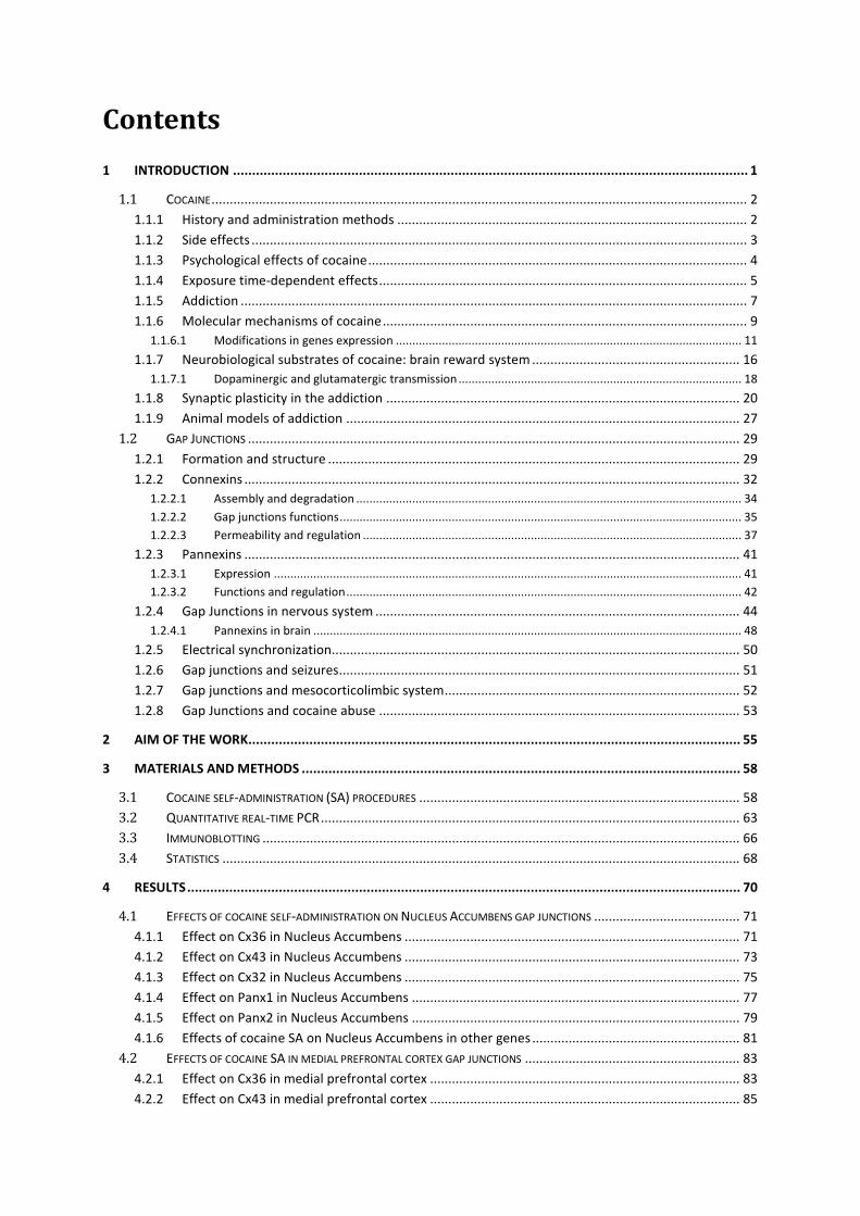
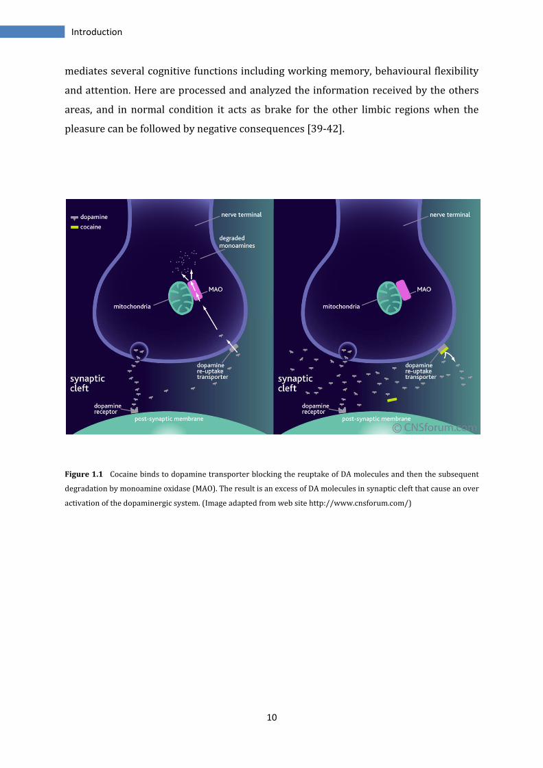
associated with cocaine use [35-38] (Figure 1.1).
The principal mechanism of action of cocaine is the activation of reward circuitry in the
brain. This circuit is principally constituted by the dopaminergic neurons of ventral
tegmental area (VTA) in the midbrain, that project to the other regions of
mesocorticolimbic system as nucleus accumbens (NAc), dorsal striatum, amygdala,
hippocampus and some regions of prefrontal cortex (PFC).
The NAc seems to be the area mostly involved in the high phase; indeed this area is
implicated in the biological base of survival and reproduction. The DAergic stimulation
in NAc induced by cocaine, causes release of DA and the consequences are the sensation
of well-being and the desire to repeat the experience pleasant. In limbic system
furthermore there are amygdala and hippocampus, memory centres that allow to
associate and remember the pleasure had with increase of DA in NAc but also everything
associated with the drug; maybe for this, is enough only a place, an image or an emotion
to desire repeat experience. The other region involved is the prefrontal cortex; this
10
Introduction
mediates several cognitive functions including working memory, behavioural flexibility
and attention. Here are processed and analyzed the information received by the others
areas, and in normal condition it acts as brake for the other limbic regions when the
pleasure can be followed by negative consequences [39-42].
Figure 1.1 Cocaine binds to dopamine transporter blocking the reuptake of DA molecules and then the subsequent
degradation by monoamine oxidase (MAO). The result is an excess of DA molecules in synaptic cleft that cause an over
activation of the dopaminergic system. (Image adapted from web site http://www.cnsforum.com/)
11
Introduction
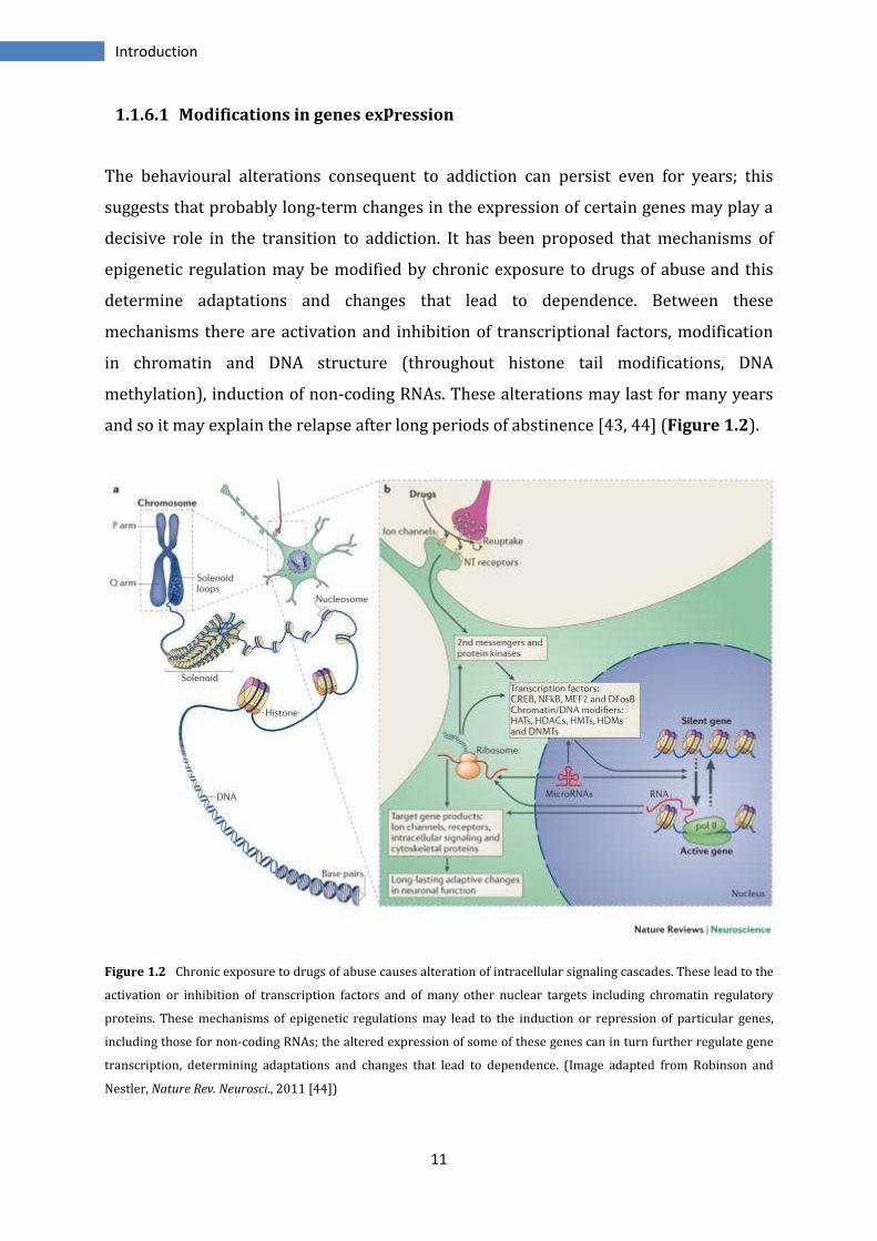
1.1.6.1 Modifications in genes expression
The behavioural alterations consequent to addiction can persist even for years; this
suggests that probably long-term changes in the expression of certain genes may play a
decisive role in the transition to addiction. It has been proposed that mechanisms of
epigenetic regulation may be modified by chronic exposure to drugs of abuse and this
determine adaptations and changes that lead to dependence. Between these
mechanisms there are activation and inhibition of transcriptional factors, modification
in chromatin and DNA structure (throughout histone tail modifications, DNA
methylation), induction of non-coding RNAs. These alterations may last for many years
and so it may explain the relapse after long periods of abstinence [43, 44] (Figure 1.2).
Figure 1.2 Chronic exposure to drugs of abuse causes alteration of intracellular signaling cascades. These lead to the
activation or inhibition of transcription factors and of many other nuclear targets including chromatin regulatory
proteins. These mechanisms of epigenetic regulations may lead to the induction or repression of particular genes,
including those for non-coding RNAs; the altered expression of some of these genes can in turn further regulate gene
transcription, determining adaptations and changes that lead to dependence. (Image adapted from Robinson and
Nestler, Nature Rev. Neurosci., 2011 [44])
12
Introduction
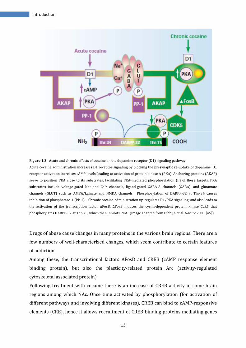
Following treatment with cocaine, amphetamine and other drugs there is an activation
of cyclic adenosine monophosphate (cAMP) pathway that can lead to different events
depending by the exposure time.
After acute administration of cocaine has been observed an increase of dopamine
receptor signaling (in particular of D1 subtype). This D1 activation increases cAMP
levels, leading to activation of protein kinase A (PKA) that finally causes an increase in
phosphorylation of DARPP-32 at Thr-34 and a decrease in phosphorylation at Thr-75 via
a signalling mechanism depending on protein phosphatase-2A (PP2A).
DARPP-32 (dopamine and cyclic AMP-regulated phosphoprotein, Mr 32kDa) is a
phosphoprotein having different phosphorylation site. The phosphorylation of Thr-34
by protein kinase A (PKA), transforms DARPP32 as a strong inhibitor of protein
phosphatase-1 (PP-1), while the phosphorylation at Thr-75 level by cyclin-dependent
kinase 5 (Cdk5), convert it as an inhibitor of PKA.
PP-1 and PKA are central proteins for the regulation of intracellular events triggered by
the activation of D1 receptors and involved in neuronal excitability. The effectors
include voltage-gated Na+ and Ca2+ channels, ligand-gated GABA-A channels and
glutamate channels such as AMPA/kainate and NMDA.
Instead, after chronic administration of cocaine there is an up-regulation of D1/PKA
signaling system and the activation of ΔFosB that cause the increase of Cdk5 expression
in caudatoputamen and NAc. This protein kinase may be involved in the locomotors
effects of cocaine through the regulation of dopamine signalling. Indeed Cdk5
phosphorylates DARPP-32, co-localized in medium spiny neurons of nucleus accumbens,
at Thr-75, becoming inhibitor of PKA.
The effects of chronic cocaine use cause activation of ΔFosB and Cdk5 and maybe these
may cause modifications in other pathways, giving stable compensatory adaptations
that lead to mechanism of drug addiction [45, 46] (Figure 1.3).
13
Introduction
Figure 1.3 Acute and chronic effects of cocaine on the dopamine receptor (D1) signaling pathway.
Acute cocaine administration increases D1 receptor signaling by blocking the presynaptic re-uptake of dopamine. D1
receptor activation increases cAMP levels, leading to activation of protein kinase A (PKA). Anchoring proteins (AKAP)
serve to position PKA close to its substrates, facilitating PKA-mediated phosphorylation (P) of these targets. PKA
substrates include voltage-gated Na+ and Ca2+ channels, ligand-gated GABA-A channels (GABA), and glutamate
channels (GLUT) such as AMPA/kainate and NMDA channels. Phosphorylation of DARPP-32 at Thr-34 causes
inhibition of phosphatase-1 (PP-1). Chronic cocaine administration up-regulates D1/PKA signaling, and also leads to
the activation of the transcription factor ΔFosB. ΔFosB induces the cyclin-dependent protein kinase Cdk5 that
phosphorylates DARPP-32 at Thr-75, which then inhibits PKA. (Image adapted from Bibb JA et al. Nature 2001 [45])
Drugs of abuse cause changes in many proteins in the various brain regions. There are a
few numbers of well-characterized changes, which seem contribute to certain features
of addiction.
Among these, the transcriptional factors ΔFosB and CREB (cAMP response element
binding protein), but also the plasticity-related protein Arc (activity-regulated
cytoskeletal associated protein).
Following treatment with cocaine there is an increase of CREB activity in some brain
regions among which NAc. Once time activated by phosphorylation (for activation of
different pathways and involving different kinases), CREB can bind to cAMP-responsive
elements (CRE), hence it allows recruitment of CREB-binding proteins mediating genes
14
Introduction
transcription. It has been shown that over expression of CREB reduces the rewarding
effects of cocaine. So it has been led the hypotheses that CREB mediates tolerance to
cocaine's positive hedonic effects, but also increases responsiveness to stress and may
mediate the negative and dysphoric aspects associated with cocaine withdrawal [47-51].
FosB belong to Fos family of transcriptional factors; after binding to Jun family proteins,
it forms a complex activator protein-1 (AP-1). This complex, by binding to AP-1 sites on
DNA, regulates transcription of some genes. Several studies show the induction of c-Fos,
FosB and others Fos family components in NAc and dorsal striatum after acute
administration of cocaine, but their levels return normal after few hours by drug intake.
Instead, after chronic cocaine administration, a many stable protein ΔFosB, derived by
alternative splicing of FosB, is induced at high levels in NAc. This protein has been linked
to typical behaviour of cocaine abuse observed in laboratory animals, as increased
locomotor sensitivity, increased conditioned place preference and increased self-
administration. In fact, all these behaviours are reduced in mice blocking this protein.
Thus ΔFosB may represent the switch from occasional use to addiction, because alone is
sufficient to sensitize the animals to reward of drugs and its long stability can persist for
months, even if this would be the starting conditions for other stable modifications [52-
56].
The increase of ΔFosB after cocaine administration causes transcriptions of some genes
among which, cyclin-dependant kinase-5, Arc and others that would mediate increasing
of dendrites spine in medium spiny neurons of NAc. In this way, NAc may receive more
information from other regions and so lead to long-lasting neurobiological effects of
addiction. ΔFosB regulates also proteins important for glutamatergic transmission and
plasticity as AMPA receptors and Ca2+/calmodulin-dependent protein kinases II
(CAMKII) [45, 57-60].
Activity-regulated cytoskeletal associated protein Arc, belongs to the class of Immediate
Early Genes. Studies show that Arc is index of neuronal activity because it plays an
important role in activity-dependent synaptic plasticity. It has also been shown that Arc
influence directly homeostasis and cellular function.
Arc encodes for a protein principally localized in the neurons at the level of dendritic
spines. It is important in the regulation of long-term depression (LTD) activated by
NMDA and metabotropic glutamate receptors. The translation of Arc is induced within
five minutes after the activation of these receptors, and leads to endocytosis of AMPAR
15
Introduction
and then to depression of excitatory synaptic transmission. Arc plays also a crucial role
in the consolidation of long-term potentiation (LTP) and then in synaptic plasticity. One
of the regulators of this process seems to be BDNF (brain-derived neurotrophic factor)
and the LTP BDNF-dependent would be associated with the transport to dendrites of
Arc mRNA. Here Arc protein participates in the organization of cytoskeleton proteins
promoting the growth of dendrites. This process is also observed in amygdala following
exposition to several drugs. So this suggest an involvement of Arc in the modulation of
others genes and proteins with use of drugs of abuse [61-63].
Many others transcriptional factors have been implicated in transition to addiction,
among these NF-κB (nuclear factor-κB), equally involved in dendrites sprout and
sensitization to drug’s reward or MEF2 (multiple myocite-specific enhancer factor 2),
involved in the structural and behavioural changes. Others evidences regard
glucocorticoid receptors, early growth response factors (EGR) and signal transducers
and activators of transcription STATs [44].
16
Introduction
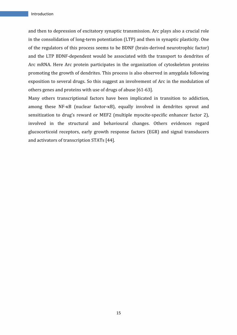
1.1.7 Neurobiological substrates of cocaine: brain reward system
Mesocorticolimbic system regulates the reward mechanisms to natural stimuli as food,
water and sex. The drugs of abuse, such as cocaine, produce their reinforcing effects
acting on the same areas of limbic system responsible for influence of motivational,
emotional and affective information.
This system includes several structures interconnected. It is constituted by
dopaminergic neurons that from VTA innervate NAc, amygdala, PFC and hippocampus.
Amygdala, hippocampus and prefrontal cortex send glutamatergic projections to NAc.
NAc send GABAergic projections to the ventral pallidum and to VTA/substantia nigra.
VTA transmits GABAergic inputs to the medial dorsal thalamus. This last sends
glutamatergic efferents to PFC [64](Figure 1.4).
The major responsible of rewarding and reinforcing effects of cocaine is the
dopaminergic system that projects from VTA to NAc and to others forebrain areas
including dorsal striatum. Beside to dopaminergic transmission cocaine affects also the
glutamatergic system, inducing long-term adaptations that lead to typical behaviour of
addiction as cocaine craving and seeking [64].
Nucleus Accumbens (NAc) is divided in two sub regions, the core and the shell, having
each one distinct function. The shell is classified as a part of limbic system and is
implicated in reward effect of cocaine, hence it regulates the response in presence of
motivational stimuli; while the core, considered part of basal ganglia, mediates seeking
and locomotor activity under stimuli linked to drug consumption. The NAc functions
translating the rewarding/reinforcing effects of drugs of abuse into drug-seeking
behaviour. It processes, consolidates and integrates information from limbic nuclei to
basal ganglia structures, including the ventral pallidum, thalamus and motor cortex.
After acute cocaine administration, there is a selective activation of DAergic
transmission in NAc shell, due to activation of Daergic projections from VTA; the huge
increase of DA returns to basal levels stopping the administration. However, a prolonged
cocaine use causes depletion of DA in NAc, PFC and cerebral cortex and in addition in
NAc there is also a decrease in glutamate levels [65].
The ventral tegmental area (VTA) is the origin of the dopaminergic cell bodies of the
mesocorticolimbic dopamine system; indeed, it is constituted by about 50-60% of DA
neurons. These neurons are stimulated by excitatory glutamatergic afferents and they
17
Introduction
are negatively regulated by GABAergic neurons. It is widely implicated in the drug and
natural reward circuitry of the brain but also in cognition and motivation [38, 64-66].
Medial prefrontal cortex (mPFC) is the region involved in higher-order cognition
(decision-making) and emotion. In drugs addiction, it has implicated with salience
attribution and inhibitory control and in compulsive behaviours, as craving and
compulsive drug intake. It receives DAergic stimulation from VTA and sends
glutamatergic projections to NAc.
The dorsal region of mPFC send efferents to NAc core, while the ventral region to the
NAc shell. After cocaine intake there is an increase in metabolic activity and blood flow
in PFC. During the phase of seeking there are changes in glutamatergic communication,
followed by an increase of Glut in NAc that may cause the motor behaviour. Maybe this is
due to the increase of DA stimulation of PFC from VTA that increase the excitability of
glutamatergic neurons [67-69].
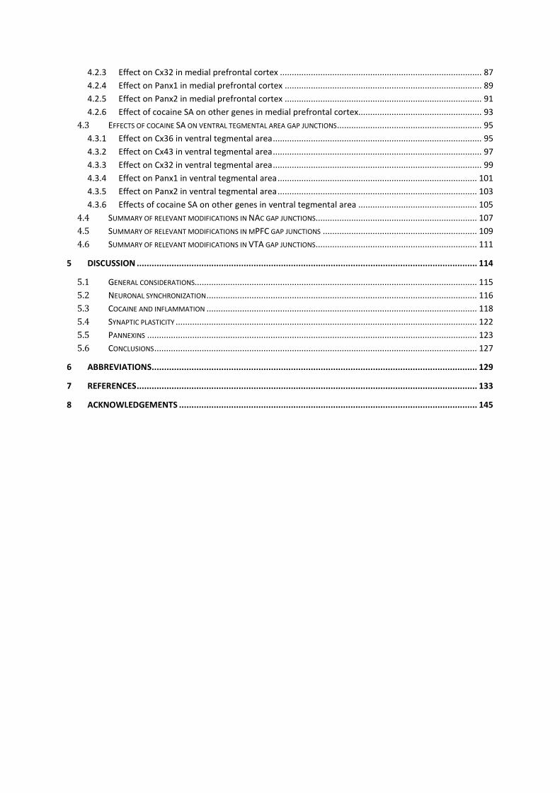
Figure 1.4 Organization of mesocorticolimbic system on rat brain (on the left) and human brain (on the right). The
medial prefrontal cortex (mPFC) sends glutamatergic afferents to the nucleus accumbens. The core and shell sub
regions of the NAc also receive excitatory glutamatergic projections from hippocampus and basolateral amygdala
nuclei. Dopaminergic projections from the VTA and substantia nigra (SN) modulate the flow of emotional, declarative,
and procedural memories. The activity of VTA and SN dopamine cells is regulated by excitatory glutamatergic
projections from the mPFC, hippocampus, and amygdala, and by inhibitory GABAergic projections from the NAc and
ventral pallidum (VP). In rat brain are indicated in green the DAergic, in red the Glutamatergic and in blue the
GABAergic projections; in human brain, in blue are indicated Daergic projections. (Adapted from Schmidt et al., Ann. N
Y Acad. Sci.; 2010 [64]).
18
Introduction
1.1.7.1 Dopaminergic and glutamatergic transmission
Dopaminergic system is the main actor of rewarding mechanisms in the brain. The
dopamine (DA) is the neurotransmitter most involved in the regulation of pleasure and
reinforcement. It mediates its physiological effects by binding with a series of five G
protein-coupled receptors. The dopamine receptors consist of the D1, D2, D3, D4, and D5
receptors and are categorized into two groups based on their properties and effects. The
D1 and D5 receptors belong to the D1-like subfamily, they are coupled to Gs protein and
increase the cellular concentrations of cAMP by the activation of the enzyme adenylate
cyclase. The D2, D3, and D4 receptors belong to the D2-like subfamily; they are coupled
to Gi/Go proteins and decrease the cellular concentrations of cAMP by inhibition of
adenylate cyclase.
Even though is clear the involvement of DAergic transmission in the behaviour of
cocaine seeking, it is still controversial which class of receptors are involved.
Some studies report that the administration of D2-like receptors agonists in blood flow
or in NAc shell, give cocaine seeking, while antagonists attenuate this behaviour. Other
works report that D1-like agonists prevent cocaine-seeking behaviour induced by
cocaine, even if there are contrasting results, indeed others report that administration of
D1 agonists into NAc shell reinstates drug seeking. These controversial results
demonstrate however a role for both D1- and D2-like receptors in cocaine seeking, even
if the dissociation of effects observed between the two types, may depend by the site of
expression [70-74].
In the last years, it has become evident a fundamental role of glutamatergic transmission
in the cocaine effects. In fact, persistent changes in this system are implicated with
cocaine craving and seeking behaviours, but also with learning and memory processes
linked to development of addiction [75, 76].
Glutamatergic system plays an important role in almost all physiological functions.
Glutamate (Glut) is the most abundant excitatory neurotransmitter in the vertebrate
nervous system, accounting over 60% of neurons and two types of receptors regulate its
action: the ionotropic and the metabotropic. The ionotropic receptors (iGluR) are ion
channels activated by glutamate. Their activation increases the influx of cations, as
sodium and potassium, causing depolarization of the membrane. They are divided into
three subtypes receptors: N-methyl-D-aspartate (NMDA), 2-amino-3-(3-hydroxy-5-
19
Introduction
methyl-isoxazol-4-yl)propanoic acid (AMPA) and kainate receptors.
The metabotropic receptors (mGluR) are eight G protein-coupled receptors and are
divided into three groups, I (mGluR1-5), II (mGluR2-3) and III (mGluR4-6-7-8), based on
sequence homology, mechanisms of signal transduction and their pharmacological
selectivity. Ionotropic receptors tend to be quicker in relaying information, but
metabotropic ones are associated with a more prolonged stimulus.
Cocaine does not act directly on glutamatergic neurons; acute administration has no
effect on glutamate levels on NAc, but during withdrawal, the glutamate levels are
reduced, perhaps due to a reduced activity of cystine-glutamate antiporter in glial cells
(plasma membrane transporter for the cellular uptake of cystine in exchange for
intracellular glutamate). Moreover it has been found that cocaine-induced reinstatement
of drug-seeking behaviour is accompanied instead by an increase in glutamate levels in
NAc, due to the glutamatergic stimulation from dorsal prefrontal cortex [77-79].
The metabotropic glutamate receptors (mGluR) are potentially involved and may be a
pharmacological target for cocaine addiction. Indeed, they are subjected to variations in
transcription and membrane trafficking during withdrawal. For example, expression of
mGluR5 and mGluR2/3 mRNAs, are increased and decreased respectively in the NAc
after 3 weeks of withdrawal. Agonists for mGluR2/3 attenuate cocaine self-
administration and the reinstatement of cocaine seeking, while mGluR5-KO mice are
insensitive to the reinforcing effects of cocaine and mGluR5 antagonists attenuate the
reinstatement [64].
As regard the ionotropic glutamate receptors , NMDAR in NAc seem to have a role in the
neuronal plasticity during addiction. The use of NMDAR agonists promote reinstatement
of cocaine seeking while antagonists decrease it.
The AMPA receptors antagonists decrease cocaine self-administration when are infused
in NAc core but not in shell. After abstinence, there is also an increase in the number of
these receptors. It seems that an increased glutamatergic transmission performed by
AMPA receptors may mediate the reinstatement of cocaine-seeking behaviour. AMPA
receptors have been also implicated in neuronal plasticity observed in several regions of
mesocorticolimbic system; in particular changes in the ratio between GluR1 and
GluR2/3 subunits (this last increases as trafficking at cell surface), would be responsible
of alterations in forms of long-term potentiation (LTP) and long-term depression (LTD),
observed after cocaine administration [64, 80-82].
20
Introduction
1.1.8 Synaptic plasticity in the addiction
Growing researches support the hypothesis that forms of long-lasting synaptic plasticity
may be implicated in mechanisms of addiction.
Synaptic plasticity is the ability of the synapse to change its strength in response to use
or disuse of transmission, over synaptic pathways. Plasticity can be divided as short-
term, lasting a few seconds or less, or long-term, which lasts from minutes to hours.
Short-term synaptic plasticity results from an increase or decrease of probability that
synaptic terminals will release transmitters in response to action potential.
Long-term potentiation (LTP) and long-term depression (LTD) are two forms of long-
term plasticity, lasting minutes or more, that occur at excitatory and inhibitory synapses.
These two forms of synaptic plasticity are principally involved in the mechanisms of
learning and memory, but seem to have also an important role in the consequences of
drug use.
LTP is a long-lasting enhancement in signal transmission between two neurons resulting
from their synchronous stimulation, while LTD is an activity-dependent reduction in the
efficacy of neuronal synapses following a long patterned stimulus.
LTP is induced by high frequency stimulation (HFS) and requires activation of NMDA
receptors through the binding of glutamate, glycine or D-serine. The opening of NMDAR
causes a strong increase of Ca2+ in the post-synaptic neuron; moreover, the strong
depolarization displaces the block of NMDAR by Mg2+, allowing more Ca2+ to enter. The
strong increase of Ca2+ causes activation of several proteins such as Ca2+/calmodulin-
dependent protein kinase II (CAMKII), protein kinase-A (PKA), mitogen-activated
protein kinase (MAPK). These kinases are strongly involved in the increased membrane
trafficking and in the activation of excitatory post-synaptic receptors, the AMPAR. These,
enhance the cations afflux in the cells, and as a results there is an enhancement of
synapse’s strength.
Other mechanisms can activate an LTP. In mossy fiber-CA3 hippocampal synapses or in
cerebellar parallel fiber synapses, LTP is induced by a rise of calcium in presynaptic
terminal. Here, Ca2+ causes the activation of calcium-stimulated adenylate cyclase, then
the rise of cAMP and the consequent activation of PKA, activates Rab3a and RIM1α,
proteins involved in the long-lasting increase of glutamate release.
21
Introduction
LTD can be induced by low frequency stimulation (LFS) with three different mechanisms.
In hippocampus LFS causes a weak depolarization that open NMDAR but determine a
low Ca2+ concentration in post-synaptic neuron; this activates different phosphatases,
such as protein phosphatases calcineurin and protein phosphatase-1 (PP1), that revert
the LTP process, inhibiting the opening and internalizing the AMPAR. In cerebellum it
has been found an LTD mGluR-dependent, in which the activation of group I of mGluR
activates the protein kinase C, taking to endocytosis of AMPAR. Another mechanism is
LTD eCB-mediated, found in striatum and neocortex, where the activation of group I of
mGluR (which leads to activation of phospholipase C) or an increase of intracellular Ca2+
in the postsynaptic neuron, initiate the synthesis of endocannabinoids (eCB). The eCB
are subsequently released from the postsynaptic neuron, travel retrogradely to bind to
presynaptic cannabinoid-1 receptors (CB1R) and this prolonged activation of CB1Rs
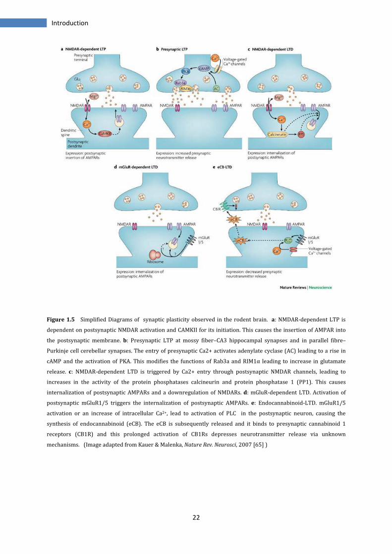
depresses neurotransmitter release [83] (Figure 1.5).
Physiologically in mesocorticolimbic system are expressed both LTP and LTD.
In VTA, the excitatory DAergic synapses exhibit LTP and LTD. For LTP are necessary
NMDAR and the consequent increase in post-synaptic calcium, instead LTD can be
inducted with two mechanisms, activation of NMDAR and voltage-dependant calcium
channels or activation of mGluR [84-87].
In NAc forms of LTP and LTD occurs in medium spiny neurons. LTD requires activation
of mGluR to increase Ca2+ and production of endocannabinoids (although exist also a
mechanism eCB-independent). As regard LTP, its activation requires NMDAR and
calcium increase [88-91].
The PFC is undergone to dynamic neuronal adaptation processes through the induction
of synaptic plasticity LTP and LTD. It has been shown that several neurochemical
substances, such as dopamine, noradrenalin, serotonin and acetylcholine, modulate
synaptic plasticity in this region. In particular serotonin, that in PFC regulates cognition
and emotion, seems to have a principal role in the LTD induction; indeed it in
collaboration with group I of mGluR, facilitates LTD induction through increase of
AMPAR internalization. Another characteristic of synaptic plasticity induction in the PFC
is its dependence from dopamine [92, 93].
22
Introduction
Figure 1.5 Simplified Diagrams of synaptic plasticity observed in the rodent brain. a: NMDAR-dependent LTP is
dependent on postsynaptic NMDAR activation and CAMKII for its initiation. This causes the insertion of AMPAR into
the postsynaptic membrane. b: Presynaptic LTP at mossy fiber–CA3 hippocampal synapses and in parallel fibre–
Purkinje cell cerebellar synapses. The entry of presynaptic Ca2+ activates adenylate cyclase (AC) leading to a rise in
cAMP and the activation of PKA. This modifies the functions of Rab3a and RIM1α leading to increase in glutamate
release. c: NMDAR-dependent LTD is triggered by Ca2+ entry through postsynaptic NMDAR channels, leading to
increases in the activity of the protein phosphatases calcineurin and protein phosphatase 1 (PP1). This causes
internalization of postsynaptic AMPARs and a downregulation of NMDARs. d: mGluR-dependent LTD. Activation of
postsynaptic mGluR1/5 triggers the internalization of postsynaptic AMPARs. e: Endocannabinoid-LTD. mGluR1/5
activation or an increase of intracellular Ca2+, lead to activation of PLC in the postsynaptic neuron, causing the
synthesis of endocannabinoid (eCB). The eCB is subsequently released and it binds to presynaptic cannabinoid 1
receptors (CB1R) and this prolonged activation of CB1Rs depresses neurotransmitter release via unknown
mechanisms. (Image adapted from Kauer & Malenka, Nature Rev. Neurosci, 2007 [65] )
23
Introduction
Under cocaine exposition occur numerous rearrangements. In VTA, only 1 or 7 days of
passive cocaine administration is enough to potentiate the glutamatergic transmission
in DAergic neurons. This potentiation is mediated by an increase in AMPAR and
particularly with the disappearance of GluR2 subtype, normally present in AMPAR, and
an increase of GluR1subunit. However, this LTP-like is transient, lasts 5 days and
disappears after 10 days, and it precludes an ulterior LTP stimulation [94-96].
Surprisingly the induced LTP AMPA-mediated last for about 3 months if the cocaine is
self-administrated, while a natural reward cause an LTP of only 7 days. From this,
appears important the involvement of mechanisms of learning and memory associated
with the drug. In addition, should be considered also the GABAergic transmission, even
influenced by drug of abuse [97, 98].
In the NAc, one single cocaine injection does not alter AMPAR activity, but it abolishes
the normal LTD eCB-mediated, probably by decreasing of mGluR5 levels. After repeated
exposure and during early withdrawal the AMPAR functions are reduced but this occurs
only in the NAc shell [99, 100].
After chronic cocaine self-administration, there is a disruption of LTP in all NAc and a
loss of LTD only in the core. The loss of LTP may be due to a reduced trafficking of
AMPAR, indeed the amount of AMPA receptors relative to NMDA receptors decrease in
medium spiny neurons. This decrease in AMPA receptors may occur through the
mechanism of LTD NMDAR-dependent, because this form of plasticity is reduced after
cocaine use [101].
In the AMPA receptors is also observed an increase of subunit GluR1 and interestingly,
the administration of interfering molecules for GluR1 and GluR2 in the NAc reduces the
cocaine reinstatement, indicating a possible involvement of the two subunits in drug-
motivation [102, 103].
Several studies show that medial PFC, after few days of cocaine administration, become
highly sensitive to the induction of LTP, due to a reduction of inhibition of pyramidal
neurons for decrease of GABA(A) receptors. Repeated cocaine administration impairs
also the LTD mGluR-mediated. In both cases is evident an involvement of DA receptors,
because their block prevent the effects, cocaine-induced on LTP and LTD [104-107].
The equip of PV Piazza in Bordeaux has developed an animal model, through which,
sessions of prolonged cocaine self-administration (SA) allow to discriminate animals
with an addiction-like behaviour from non-addict animals. Experiments carry out on
24
Introduction
slices obtained after 24 hours by the last cocaine infusion, show no modifications in LTP
or in LTD after one week of SA in NAc, while after 17 days of SA, LTD NMDAR-dependent
is completely abolished. Instead, with a prolonged cocaine exposure (50 to 72 days),
LTD returns normal in non-addict animals while in addict remains suppressed. For
contrast, the LTD mediated by mGlu2/3 receptors is not affected in all groups. For this, it
has been postulated that maybe the persistent impairment in LTD is associated with
transition to addiction even if all individuals start by the same impairment at early drug
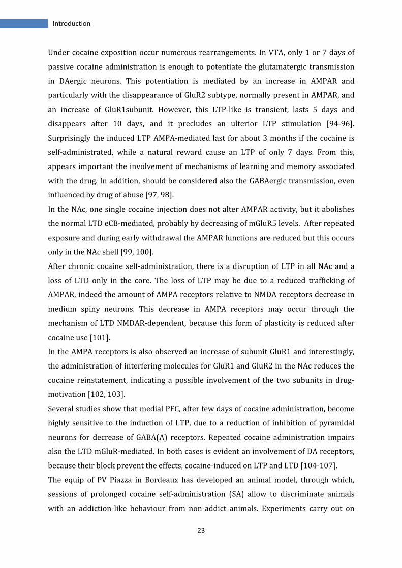
use [108, 109] (Figure 1.6).
The same experiments have been conducted on PFC slices. In this region both addict
and non-addict animals show no variations as regard LTD eCB-mediated after 17 days of
SA, but this, is suppressed in all individuals after prolonged drug exposure.
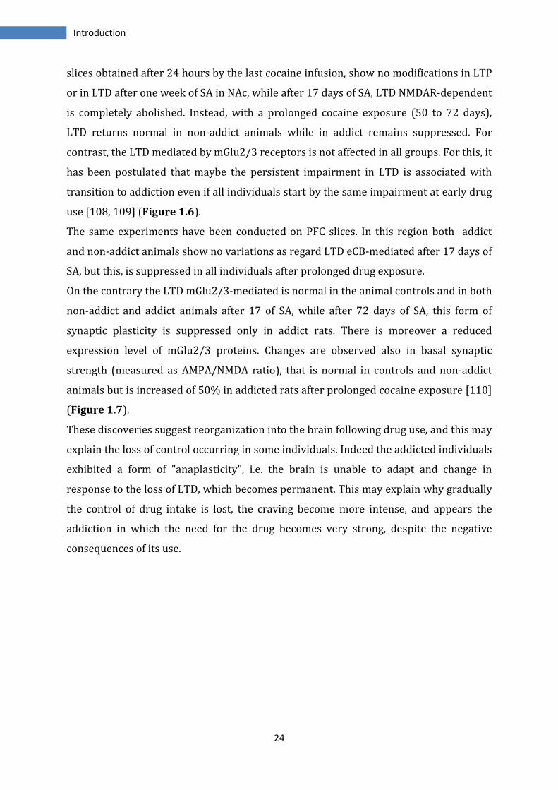
On the contrary the LTD mGlu2/3-mediated is normal in the animal controls and in both
non-addict and addict animals after 17 of SA, while after 72 days of SA, this form of
synaptic plasticity is suppressed only in addict rats. There is moreover a reduced
expression level of mGlu2/3 proteins. Changes are observed also in basal synaptic
strength (measured as AMPA/NMDA ratio), that is normal in controls and non-addict
animals but is increased of 50% in addicted rats after prolonged cocaine exposure [110]
(Figure 1.7).
These discoveries suggest reorganization into the brain following drug use, and this may
explain the loss of control occurring in some individuals. Indeed the addicted individuals
exhibited a form of "anaplasticity", i.e. the brain is unable to adapt and change in
response to the loss of LTD, which becomes permanent. This may explain why gradually
the control of drug intake is lost, the craving become more intense, and appears the
addiction in which the need for the drug becomes very strong, despite the negative
consequences of its use.
25
Introduction
Figure 1.6 NMDAR-dependent LTD is disrupted in Addict animals. In the graph are shown the averaged data of
representative excitatory postsynaptic current (EPSC) traces. LTD is induced in controls and in all animals treated for
7 days. After 17 days of cocaine SA the LTD NMDAR-dependent is suppressed in both resistant and vulnerable
animals, but after prolonged exposure to cocaine, non-addict animals restore their LTD while in addict animals
remains suppressed. (Image adapted from Kasanetz F et al. Science 2010 [109])
Figure 1.7 mGluR2/3-dependent LTD is selectively impaired in addict-like rats. After 17 days of cocaine SA, LTD was
normal in all groups, but 72 days of SA completely abolish the LTD only in addict rats (3crit), while in controls and
non-addict animals (0crit) is normal. Representative fEPSP traces recorded during baseline (1) and 35 min after LTD
induction (2) are depicted. (Image adpated from Kasanetz et al. Mol. Psychiatry 2012 [110])
26
Introduction
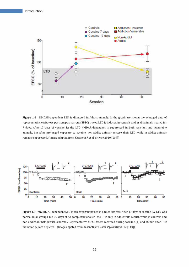
Moreover, these mechanisms of synaptic plasticity, similar to what occurs in the
mechanism of memory, are associated with formation of new connections that cause
changes in the synaptic transmission. Dopamine denervation reduces dendrites density
but repeated treatments with cocaine increase dendritic spine density and the number
of branched spines in the shell of NAc and on pyramidal cells in PFC; these alterations
last for 4 weeks after drug exposure [39, 111] (Figure 1.8).
In this way, it is possible that cocaine self-administration experience alters patterns of
synaptic connectivity within limbo cortical circuitry. These alterations may contribute to
cocaine's incentive motivational effects and have neuropathological effects in frontal
areas involved in decision-making and judgment.
Figure 1.8 Regulation of dendritic structure by drugs of abuse. The figure shows the expansion of a neuron’s
dendritic tree after chronic exposure to a drug of abuse, as it has been observed in the NAc and PFC for cocaine and
related psychostimulants. (Adapted from Nestler AJ, Nature Rev Neuroscience; 2001 [112])
27
Introduction
1.1.9 Animal models of addiction
The need to understand the mechanisms underlying the phenomenon of addiction has
led to the development of animal models that mimic the typical behaviours occurring in
humans after taking drugs. In the past years have been used principally techniques of
passive administration of the substance, procedures that showed direct results about
the pharmacology of the substance and neurobiological aspects. Other models have been
used to scan the molecular mechanisms induced by drugs and to understand why some
individuals pass from normal use to a compulsive use. However, with these models the
behavioural effects of drugs depend by several factors as the species, routes of
administration, time of administration, dose concentration. Nevertheless, differences
arise depending on type of administration, if non-contingent (passive) or contingent
(voluntary).
However, the best animal model to mimic the human behaviour seems to be the self-
administration model, in which the animals learn to achieve behaviour to obtain the
drug and after they can decide whether or not assume the drug.
Deroche-Gamonet et al. developed an experimental animal model to identify the subjects
with a predisposition for addiction. In this model, as in humans, only a small proportion
of individuals show the hallmarks characteristics of addiction. In according to substance
dependence criteria of DSM-IV, several parameters are considered, as the difficulty in
stopping drug intake, the high motivation to take the drug and the continued use of
substance despite harmful consequences.
One of the most important findings in these studies has been the demonstration that the
total amount of drug consumed by addicted rats is the same than that consumed by the
animals maintaining a perfect control of drugs use. This would demonstrate the
importance of individual vulnerability to the drugs respect to the iatrogenic theories
that predict how the amount of drug taken is directly proportional to the probability of
fall into addiction. Then these experimental animals show a real addiction, and the data
indicate that the transition would be the result from the combination of two main
factors: prolonged exposure to a substance with abuse potential and a vulnerable
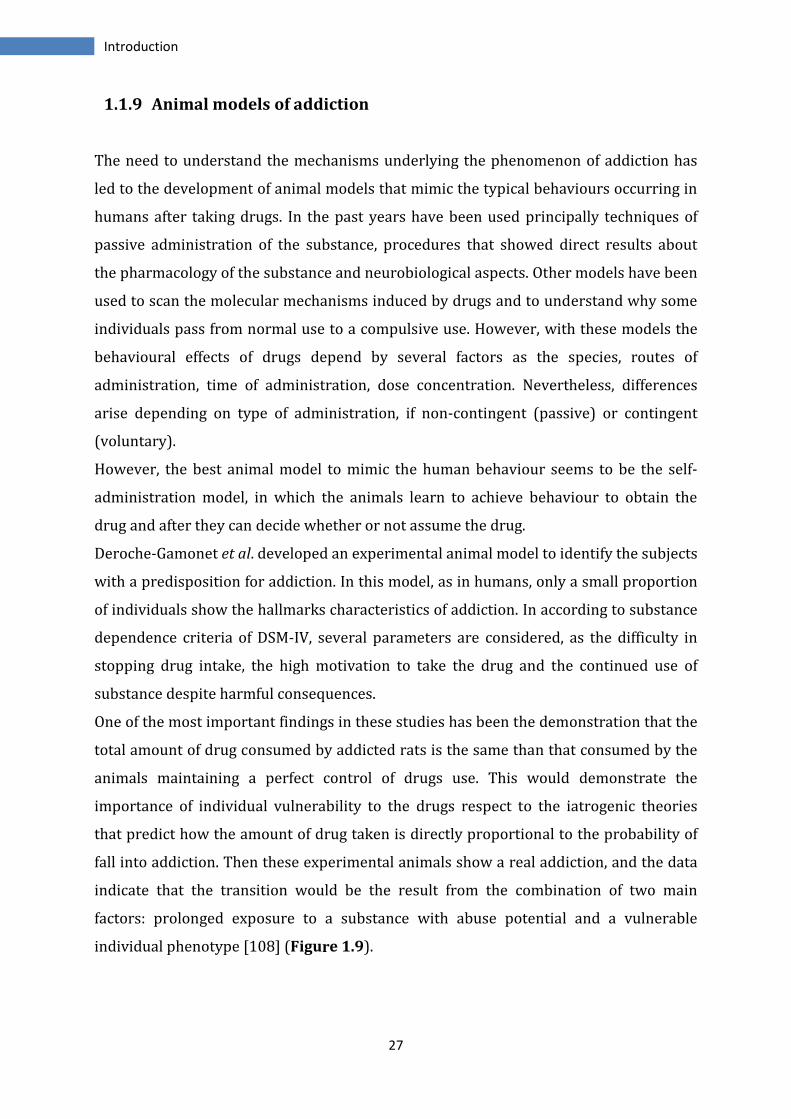
individual phenotype [108] (Figure 1.9).
28
Introduction
Figure 1.9 Intravenous self-administration apparatus used to deliver response-contingent drug infusions and collect
data during self-administration sessions. (Image adapted from Grahame NJ, Curr Protoc Neurosci. 2002 [113]
29
Introduction
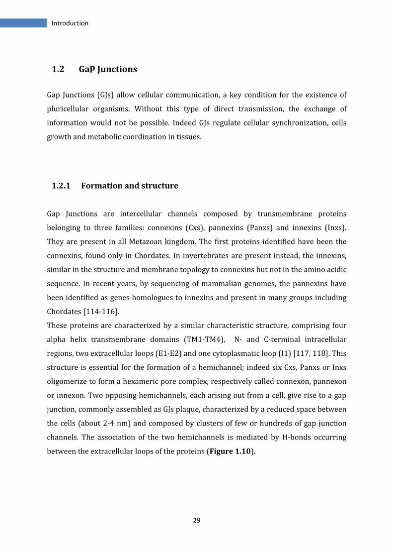
1.2 Gap Junctions
Gap Junctions (GJs) allow cellular communication, a key condition for the existence of
pluricellular organisms. Without this type of direct transmission, the exchange of
information would not be possible. Indeed GJs regulate cellular synchronization, cells
growth and metabolic coordination in tissues.
1.2.1 Formation and structure
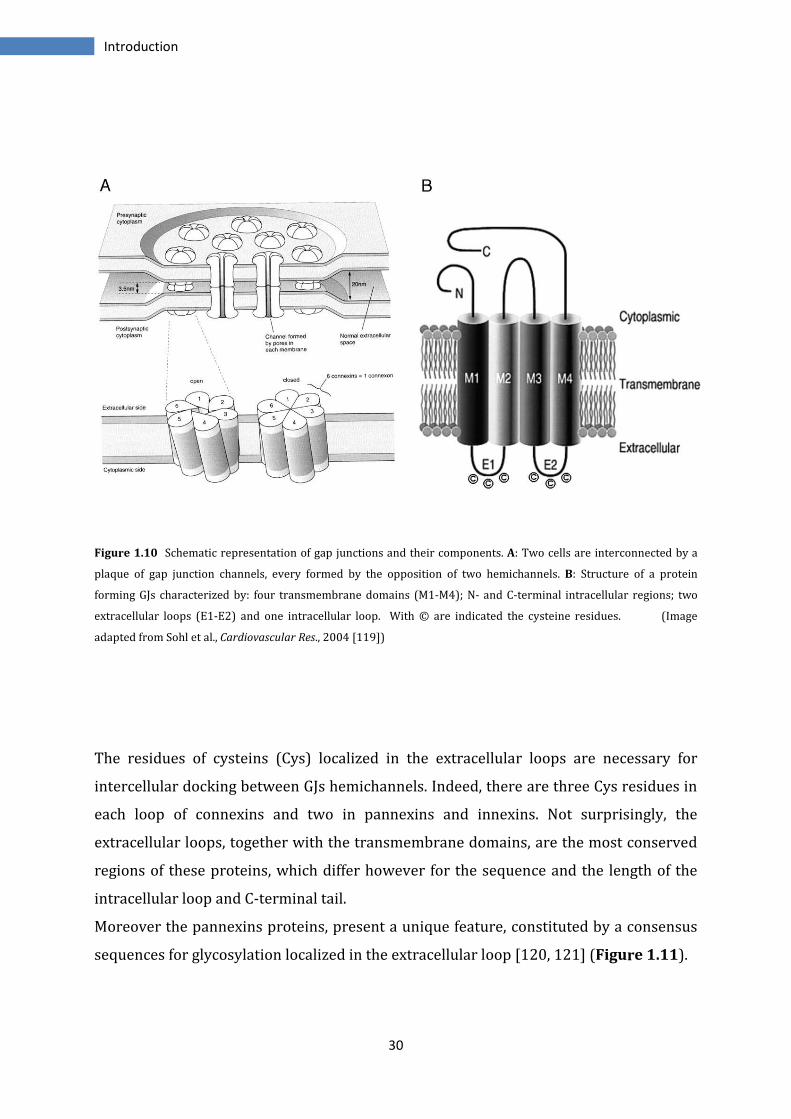
Gap Junctions are intercellular channels composed by transmembrane proteins
belonging to three families: connexins (Cxs), pannexins (Panxs) and innexins (Inxs).
They are present in all Metazoan kingdom. The first proteins identified have been the
connexins, found only in Chordates. In invertebrates are present instead, the innexins,
similar in the structure and membrane topology to connexins but not in the amino acidic
sequence. In recent years, by sequencing of mammalian genomes, the pannexins have
been identified as genes homologues to innexins and present in many groups including
Chordates [114-116].
These proteins are characterized by a similar characteristic structure, comprising four
alpha helix transmembrane domains (TM1-TM4), N- and C-terminal intracellular
regions, two extracellular loops (E1-E2) and one cytoplasmatic loop (I1) [117, 118]. This
structure is essential for the formation of a hemichannel; indeed six Cxs, Panxs or Inxs
oligomerize to form a hexameric pore complex, respectively called connexon, pannexon
or innexon. Two opposing hemichannels, each arising out from a cell, give rise to a gap
junction, commonly assembled as GJs plaque, characterized by a reduced space between
the cells (about 2-4 nm) and composed by clusters of few or hundreds of gap junction
channels. The association of the two hemichannels is mediated by H-bonds occurring
between the extracellular loops of the proteins (Figure 1.10).
30
Introduction
Figure 1.10 Schematic representation of gap junctions and their components. A: Two cells are interconnected by a
plaque of gap junction channels, every formed by the opposition of two hemichannels. B: Structure of a protein
forming GJs characterized by: four transmembrane domains (M1-M4); N- and C-terminal intracellular regions; two
extracellular loops (E1-E2) and one intracellular loop. With © are indicated the cysteine residues. (Image
adapted from Sohl et al., Cardiovascular Res., 2004 [119])
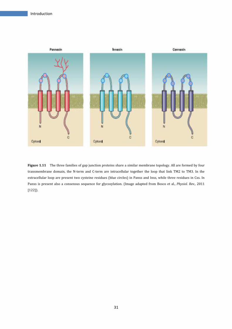
The residues of cysteins (Cys) localized in the extracellular loops are necessary for
intercellular docking between GJs hemichannels. Indeed, there are three Cys residues in
each loop of connexins and two in pannexins and innexins. Not surprisingly, the
extracellular loops, together with the transmembrane domains, are the most conserved
regions of these proteins, which differ however for the sequence and the length of the
intracellular loop and C-terminal tail.
Moreover the pannexins proteins, present a unique feature, constituted by a consensus
sequences for glycosylation localized in the extracellular loop [120, 121] (Figure 1.11).
31
Introduction
Figure 1.11 The three families of gap junction proteins share a similar membrane topology. All are formed by four
transmembrane domain, the N-term and C-term are intracellular together the loop that link TM2 to TM3. In the
extracellular loop are present two cysteine residues (blue circles) in Panxs and Inxs, while three residues in Cxs. In
Panxs is present also a consensus sequence for glycosylation. (Image adapted from Bosco et al., Physiol. Rev., 2011
[122]).
Introduction
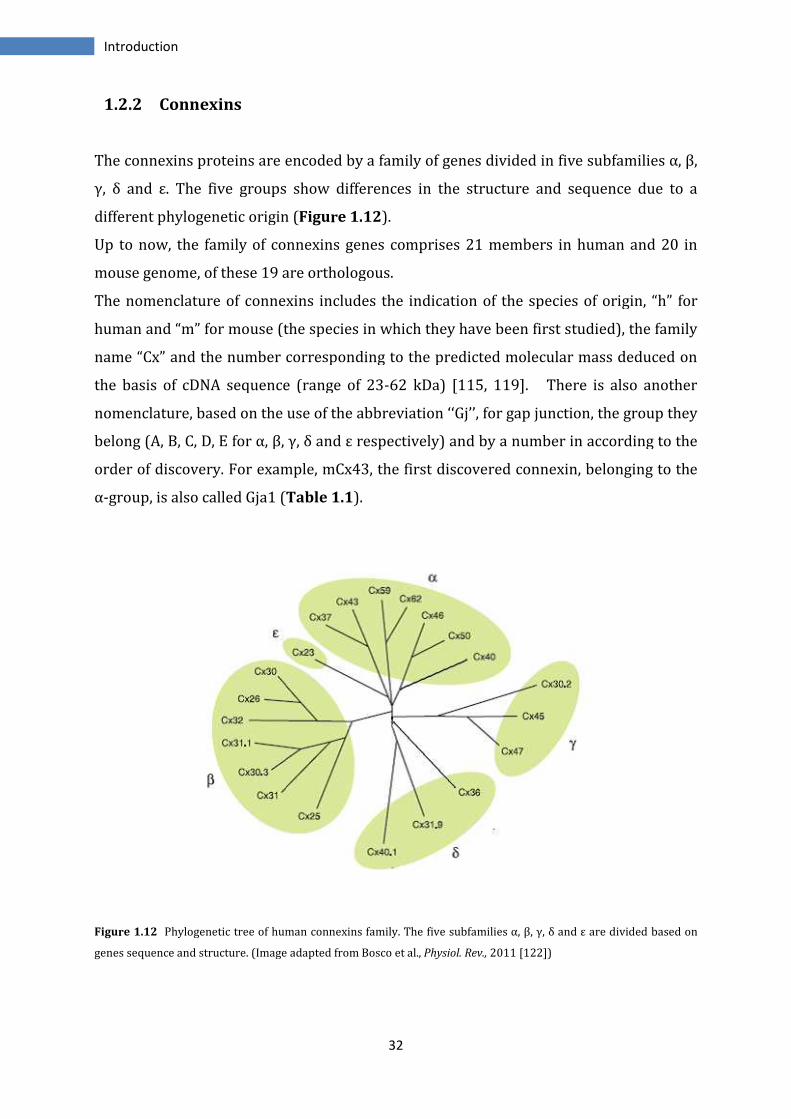
1.2.2 Connexins
The connexins proteins are en
γ, δ and ε. The five groups
different phylogenetic origin
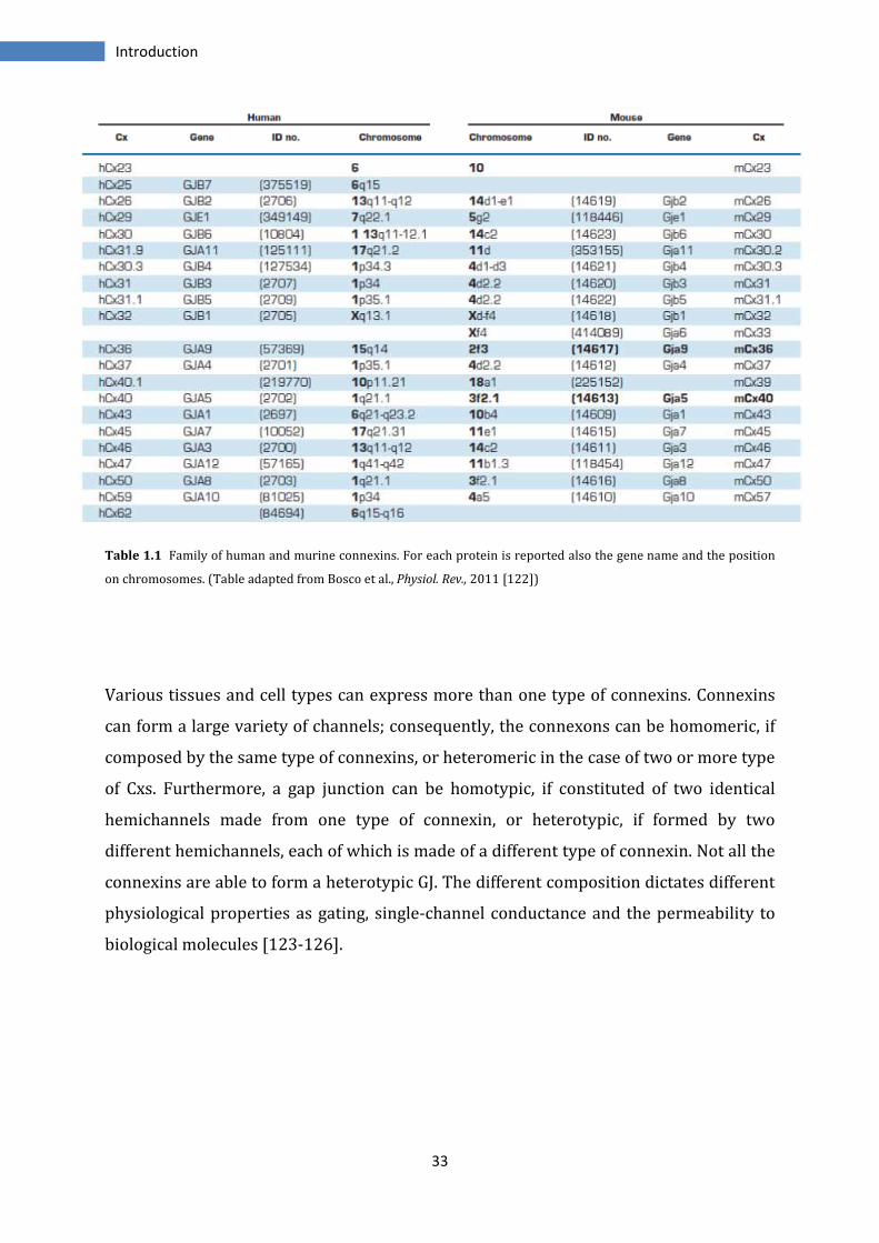
Up to now, the family of connexins
mouse genome, of these 19 are ortholog
The nomenclature of connexins
human and “m” for mouse (the species in
name “Cx” and the number corresponding to
the basis of cDNA sequence (range of 23
nomenclature, based on the use of the abbreviation
belong (A, B, C, D, E for α, β, γ, δ and ε
order of discovery. For example, mCx43, the first
α-group, is also called Gja1 (Table 1.1
Figure 1.12 Phylogenetic tree of human connexins family. The five subfamilies
genes sequence and structure. (Image a
32
encoded by a family of genes divided in five
groups show differences in the structure and sequence due to a
origin (Figure 1.12).
connexins genes comprises 21 members in human and 20 in
of these 19 are orthologous.
connexins includes the indication of the species of origin, “h” for
(the species in which they have been first studied)
the number corresponding to the predicted molecular mass deduced
cDNA sequence (range of 23-62 kDa) [115, 119]. There is also another
based on the use of the abbreviation ‘‘Gj’’, for gap junction
α, β, γ, δ and ε respectively) and by a number in according to the
order of discovery. For example, mCx43, the first discovered connexin
Table 1.1).
Phylogenetic tree of human connexins family. The five subfamilies α, β, γ, δ and ε
Image adapted from Bosco et al., Physiol. Rev., 2011 [122])
five subfamilies α, β,
show differences in the structure and sequence due to a
21 members in human and 20 in
the species of origin, “h” for
studied), the family
the predicted molecular mass deduced on
There is also another
for gap junction, the group they
a number in according to the
connexin, belonging to the
α, β, γ, δ and ε are divided based on
33
Introduction
Table 1.1 Family of human and murine connexins. For each protein is reported also the gene name and the position
on chromosomes. (Table adapted from Bosco et al., Physiol. Rev., 2011 [122])
Various tissues and cell types can express more than one type of connexins. Connexins
can form a large variety of channels; consequently, the connexons can be homomeric, if
composed by the same type of connexins, or heteromeric in the case of two or more type
of Cxs. Furthermore, a gap junction can be homotypic, if constituted of two identical
hemichannels made from one type of connexin, or heterotypic, if formed by two
different hemichannels, each of which is made of a different type of connexin. Not all the
connexins are able to form a heterotypic GJ. The different composition dictates different
physiological properties as gating, single-channel conductance and the permeability to
biological molecules [123-126].
34
Introduction
1.2.2.1 Assembly and degradation
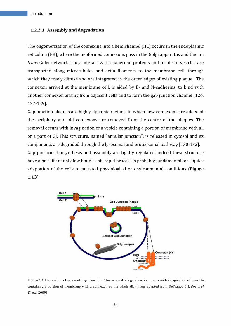
The oligomerization of the connexins into a hemichannel (HC) occurs in the endoplasmic
reticulum (ER), where the neoformed connexons pass in the Golgi apparatus and then in
trans-Golgi network. They interact with chaperone proteins and inside to vesicles are
transported along microtubules and actin filaments to the membrane cell, through
which they freely diffuse and are integrated in the outer edges of existing plaque. The
connexon arrived at the membrane cell, is aided by E- and N-cadherins, to bind with
another connexon arising from adjacent cells and to form the gap junction channel [124,
127-129].
Gap junction plaques are highly dynamic regions, in which new connexons are added at
the periphery and old connexons are removed from the centre of the plaques. The
removal occurs with invagination of a vesicle containing a portion of membrane with all
or a part of GJ. This structure, named “annular junction”, is released in cytosol and its
components are degraded through the lysosomal and proteosomal pathway [130-132].
Gap junctions biosynthesis and assembly are tightly regulated, indeed these structure
have a half-life of only few hours. This rapid process is probably fundamental for a quick
adaptation of the cells to mutated physiological or environmental conditions (Figure
1.13).
Figure 1.13 Formation of an annular gap junction. The removal of a gap junction occurs with invagination of a vesicle
containing a portion of membrane with a connexon or the whole GJ. (image adapted from DeFranco BH, Doctoral
Thesis, 2009)
35
Introduction
1.2.2.2 Gap junctions functions
Gap junctions are implicated in a large variety of functions as embryonic development,
morphogenesis, cell differentiation, cell proliferation and migration, electrical and
mechanical synchronization (cardiac, muscular and cerebral cells), transmission of
trophic or death molecules. All these functions have been discovered using targeted
mutated connexins or through the over expression of some connexins isoforms.
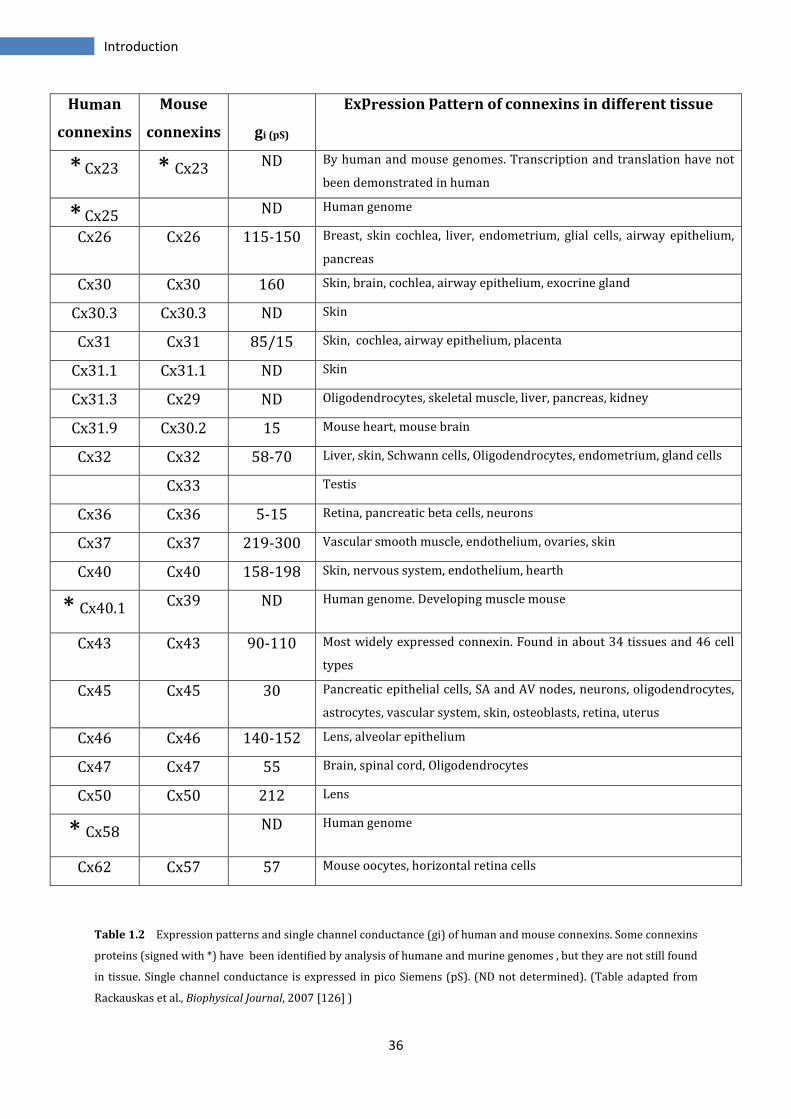
Connexins proteins are expressed in all tissue except in differentiated skeletal muscle,
erythrocytes and sperm cells. This almost ubiquitary presence is a further confirmation
of their importance for the correct functioning of the organisms (Table 1.2).
The importance of these proteins and hence, of gap junctional communication is evident
by the large number of human genetic diseases associated with connexins mutations or
with pathogenic single nucleotide polymorphisms. Among this for example the X-linked
Charcot-Marie-Tooth syndrome, a peripheral neuropathy with atrophy of distal muscles
and low number of myelinating fibers, has been linked to mutations in Cx32 gene,
suggesting its participation in myelination of peripheral nerves. Mutations in Cx43 can
cause oculodentodigital dysplasia characterized by craniofacial, neurologic, limb and
ocular abnormalities. Still mutations of Cx46 and Cx50 result in cataracts [133-137].
Recent studies show that connexons are also active in single plasma membranes as
hemichannels (HCs). HCs might be essential in intercellular signalling in different
physiological and pathological process, indeed they act in the cells of various organs in
response to extracellular signaling, injury, ischemic preconditioning and mechanical
stimulation. In contrast to GJs, they show low open probability and low permeability to
small molecules under resting conditions. HCs have been implicated in
autocrine/paracrine signalling to provide a pathway for release of ATP, glutamate, NAD+
and prostaglandins [138-140].
36
Introduction
Human
connexins
Mouse
connexins
gi (pS)
Expression pattern of connexins in different tissue
* Cx23 * Cx23 ND By human and mouse genomes. Transcription and translation have not
been demonstrated in human
* Cx25 ND Human genome
Cx26 Cx26 115-150 Breast, skin cochlea, liver, endometrium, glial cells, airway epithelium,
pancreas
Cx30 Cx30 160 Skin, brain, cochlea, airway epithelium, exocrine gland
Cx30.3 Cx30.3 ND Skin
Cx31 Cx31 85/15 Skin, cochlea, airway epithelium, placenta
Cx31.1 Cx31.1 ND Skin
Cx31.3 Cx29 ND Oligodendrocytes, skeletal muscle, liver, pancreas, kidney
Cx31.9 Cx30.2 15 Mouse heart, mouse brain
Cx32 Cx32 58-70 Liver, skin, Schwann cells, Oligodendrocytes, endometrium, gland cells
Cx33 Testis
Cx36 Cx36 5-15 Retina, pancreatic beta cells, neurons
Cx37 Cx37 219-300 Vascular smooth muscle, endothelium, ovaries, skin
Cx40 Cx40 158-198 Skin, nervous system, endothelium, hearth
* Cx40.1 Cx39 ND Human genome. Developing muscle mouse
Cx43 Cx43 90-110 Most widely expressed connexin. Found in about 34 tissues and 46 cell
types
Cx45 Cx45 30 Pancreatic epithelial cells, SA and AV nodes, neurons, oligodendrocytes,
astrocytes, vascular system, skin, osteoblasts, retina, uterus
Cx46 Cx46 140-152 Lens, alveolar epithelium
Cx47 Cx47 55 Brain, spinal cord, Oligodendrocytes
Cx50 Cx50 212 Lens
* Cx58 ND Human genome
Cx62 Cx57 57 Mouse oocytes, horizontal retina cells
Table 1.2 Expression patterns and single channel conductance (gi) of human and mouse connexins. Some connexins
proteins (signed with *) have been identified by analysis of humane and murine genomes , but they are not still found
in tissue. Single channel conductance is expressed in pico Siemens (pS). (ND not determined). (Table adapted from
Rackauskas et al., Biophysical Journal, 2007 [126] )
37
Introduction
1.2.2.3 Permeability and regulation
A gap junction forms a hydrophilic channel pore of about 100-150 Å in length and 12.5 Å
in width. It allows the passage of small molecules under 1kDa like ions, water,
nucleotides, small peptides, metabolites. In this way, GJs provide to ionic and metabolic
coupling among the cells. For some substances, this coupling is bidirectional and is
driven by an electrochemical gradient, for others GJs there is a high degree of selectivity.
Indeed the channels composed by different connexins show different permeability; for
example some channels are specific for the cations or the anions, while others are able
to discriminate between similar molecules as cyclic adenosine monophosphate (cAMP)
and cyclic guanosine monophosphate (cGMP).
The junctional conductance (g i), i.e. the passage of molecules or ions through GJ channel,
is subjected to regulation by a number of physiological factors, such as voltage,
intracellular pH and calcium, second messengers, or phosphorylation. Conductance of a
single homo-connexins channel ranges from ~10 picoSiemens (pS) to ~300 pS [125,
141-143].
The most important factor for the permeability is the structure of channel pore. Indeed
the pore width, the electrical field and the electrical charge on the pore surface affect the
permeability of ions and molecules passing through the channel.
The permeation pathway of a gap junction channel consists of an intracellular channel
entrance, a pore funnel and an extracellular cavity. The pore funnel surface is formed by
the six N-terminal regions of connexins. Because the funnel forms a constriction site at
the cytoplasmic entrance of the pore, the size and electrical character of the side-chains
in this region should have a strong effect on both the molecular cut-off size and the
charge selectivity of the channel. Indeed the substitutions or deletions of residues in N-
terminal regions can affect single channel conductance, molecular permeability and
charge selectivity [144] (Figure 1.14).
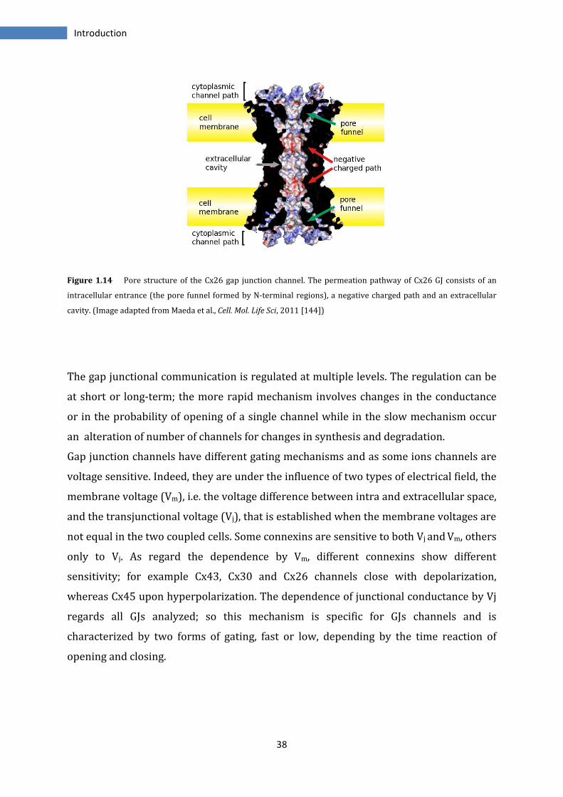
38
Introduction
Figure 1.14 Pore structure of the Cx26 gap junction channel. The permeation pathway of Cx26 GJ consists of an
intracellular entrance (the pore funnel formed by N-terminal regions), a negative charged path and an extracellular
cavity. (Image adapted from Maeda et al., Cell. Mol. Life Sci, 2011 [144])
The gap junctional communication is regulated at multiple levels. The regulation can be
at short or long-term; the more rapid mechanism involves changes in the conductance
or in the probability of opening of a single channel while in the slow mechanism occur
an alteration of number of channels for changes in synthesis and degradation.
Gap junction channels have different gating mechanisms and as some ions channels are
voltage sensitive. Indeed, they are under the influence of two types of electrical field, the
membrane voltage (Vm), i.e. the voltage difference between intra and extracellular space,
and the transjunctional voltage (Vj), that is established when the membrane voltages are
not equal in the two coupled cells. Some connexins are sensitive to both Vj and Vm, others
only to Vj. As regard the dependence by Vm, different connexins show different
sensitivity; for example Cx43, Cx30 and Cx26 channels close with depolarization,
whereas Cx45 upon hyperpolarization. The dependence of junctional conductance by Vj
regards all GJs analyzed; so this mechanism is specific for GJs channels and is
characterized by two forms of gating, fast or low, depending by the time reaction of
opening and closing.
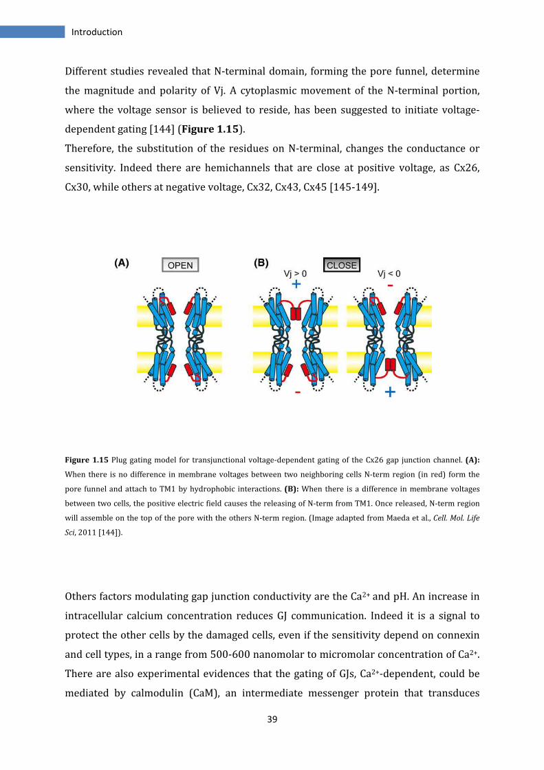
39
Introduction
Different studies revealed that N-terminal domain, forming the pore funnel, determine
the magnitude and polarity of Vj. A cytoplasmic movement of the N-terminal portion,
where the voltage sensor is believed to reside, has been suggested to initiate voltage-
dependent gating [144] (Figure 1.15).
Therefore, the substitution of the residues on N-terminal, changes the conductance or
sensitivity. Indeed there are hemichannels that are close at positive voltage, as Cx26,
Cx30, while others at negative voltage, Cx32, Cx43, Cx45 [145-149].
Figure 1.15 Plug gating model for transjunctional voltage-dependent gating of the Cx26 gap junction channel. (A):
When there is no difference in membrane voltages between two neighboring cells N-term region (in red) form the
pore funnel and attach to TM1 by hydrophobic interactions. (B): When there is a difference in membrane voltages
between two cells, the positive electric field causes the releasing of N-term from TM1. Once released, N-term region
will assemble on the top of the pore with the others N-term region. (Image adapted from Maeda et al., Cell. Mol. Life
Sci, 2011 [144]).
Others factors modulating gap junction conductivity are the Ca2+ and pH. An increase in
intracellular calcium concentration reduces GJ communication. Indeed it is a signal to
protect the other cells by the damaged cells, even if the sensitivity depend on connexin
and cell types, in a range from 500-600 nanomolar to micromolar concentration of Ca2+.
There are also experimental evidences that the gating of GJs, Ca2+-dependent, could be
mediated by calmodulin (CaM), an intermediate messenger protein that transduces
40
Introduction
calcium signals by binding calcium ions and then modifying its interactions with various
target proteins, among which there are connexins [150, 151].
Also an increase in intracellular pH decreases junctional conductance of gap junction
channels and some connexins are markedly more sensitive to acidification than others,
although it’s not still clear whether H+ acts directly on GJ channel [152-154].
An extensive way with which connexins are regulated regards several post-translational
modifications. The C-terminal region of Cxs contains serine, tyrosine and threonine,
residues that may be phosphorylated by different protein kinases. At least eight kinases,
including protein kinase A, protein kinase C, mitogen-activated protein kinase (MAPK)
and so many phosphatases control the phosphorylation/dephosphorylation of various
connexins. Except for the not phosphorylable Cx26, these modifications in Cxs are
associated with changes in gating, conductance, permeability, but this depends on
specific connexin isoforms or cell type. However, other modifications have been
identified such as hydroxylation, methylation, and acetylation. Other effects regard the
assembly into GJ plaque, their biosynthesis or their proteolytic degradation, regulating
in this way their half-life [155-158]. In addition, ubiquitination would seem an
important mechanism which connexins are regulated; the most studied Cx43 for
example, can be ubiquitinated at plasma membrane or at ER level. Recently it has been
implicated also a SUMOylation for Cx43 [159-161].
There are chemical agents able to block the GJ communication, they belong to multiple
class and structures. Some are lipophilic such as heptanol, octanol and halothane,
glycyrrhetinic acid derivatives such as carbenoxolone (CBX), quinine derivatives and
some others different for structure and action mechanism. These molecules have been
used to study the functions of GJs in vitro and in vivo, but although many of these are
used to treat common diseases, as quinine for malaria, they give a plethora of side
effects, due sometimes by blocking other channels. The development of selective
blockers will allow better understanding of GJs communication [162].
41
Introduction
1.2.3 Pannexins
Pannexins belong to a new family of proteins, also able to form GJs. They have more
similar homology with innexins than with connexins, so that initially it has been thought
they belonged to the innexins family. Many later studies showed that Panxs were a
distinct family of proteins. They do not share the strongly conserved residues of Inxs
even if as these last possess two cysteine residues in extracellular loops, and in contrast
to connexins have a larger intracellular loops (~68 aa versus ~30 aa).
1.2.3.1 Expression
In the genomes of human and rodent species, are coded three proteins, Panx1, Panx2
and Panx3. Their calculated molecular mass is respectively 48, 73 and 45 kDa. In human
genome Panx1 and Panx3 gene are located on chromosome 11 and Panx2 on
chromosome 22, while in rat genome the first two are located in chromosome 8 and
Panx2 on chromosome 7 [163, 164].
Panx1 and Panx2 transcripts are found in many rodents tissue, as brain and spinal cord,
eye, thyroid, prostate, kidney; in humans are found also in hearth, gonads and skeletal
muscle (in which there is no presence in rodents).
Panx1 protein is expressed in numerous tissues in both human and rodents. Instead
Panx2 is present in many tissues in rodents, but in humans is expressed only in the
central nervous system (CNS). The expression of Panx3 seems confined to skin and
cartilage cells, even if real-time PCR screening indicate that Panx3 mRNA is present also
in kidney, spleen and brain [165, 166].
Pannexins have been found in large accumulation in ER and Golgi apparatus, suggesting
that post-translational modifications and assembly in pannexons occur in the same way
as demonstrated for connexins.

42
Introduction
1.2.3.2 Functions and regulation
The first studies on this protein were made by Bruzzone et al. using an expression
system in Xenopus oocytes, in which they discovered that the channel formed by
pannexins, if activated by voltage, had given rise to passage of molecules. But they
concluded that Panx1 alone was able to form homomeric channels, while Panx2 could
form only heteromeric channels with Panx1 [167].
To date, increasing evidences show that also Panx2 alone is capable to form a functional
homomeric channel. Indeed the heteromeric channels Panx1/Panx2 are unstable and
disaggregate in few hours, this could explain because the heteromeric is present in
systems as Xenopus oocytes or in HEK 293, but in mammalian cells and brain tissue the
two proteins exist separately [168].
Another controversy regards the ability of Panxs to form or not gap junction channels,
but recent studies revealed evidence that they may form GJs in vitro cells, but to date it
has not been demonstrated in vertebrate.
As connexons, after activation pannexons open into large non-selective pores permeable
to ions and small molecules up to 1 kDa and under a wide range of membrane
depolarization. Panxs HCs differ from connexins HCs because they exhibit larger
currents, faster kinetics of pore opening and a conductance of over 500 pS [167].
Different stimuli are able to open pannexons, among these mechanical stress, positive
membrane potential, extracellular ATP, elevation in intracellular calcium, ischemic
insult and inflammation, while acidification of cytoplasm cause the closing of the
channels. The presence of mechanisms distinct from those of connexins, makes possible
the co-expression of the two proteins types in the same cells [169].
The pannexons may play a role in generation of oscillatory and synchronization activity
in brain. They can be involved in the propagation of calcium waves, to which normally
have been implicated the GJs connexins-formed [170]. Panxs would use a mechanism
based on release of adenosintriphosphate (ATP). This has been demonstrated in blood
endothelium, where mechanical stress or ischemia open pannexons in endothelial cells
and cause the extracellular release of ATP. The released ATP acts on purinergic
receptors P2Y, whose activation has effect on phospholipase C, increasing the IP3 and
causing the release of calcium from intercellular stores; this at the end cause a
consequent release of other ATP that can propagate using classical way of GJs or via
43
Introduction
pannexons. In this way, the propagation is ensured also without direct cell contact. This
action probably is essential for the cells in normal tissue because Panxs channels,
contrary to Cxs channels gated by cations as Ca2+ or Mg2+, are able to open when
external concentration of calcium is at physiological level.
So pannexons may propagate calcium waves and have a role in vasodilatation,
inflammatory response and ischemic death of neurons [171-173].
Other evidences of calcium waves propagation are in erythrocytes, where there is
expression only of Panx1 and not of Cx43 (the main Cx involved in calcium waves). In
these the release of ATP occurs in response to ischemia and mechanical stress and after
exhibit the same pathway observed in endothelium [174].
The Pannexins are the only ones, between the three families, to be extensively
glycosylated. There is the presence of consensus sequence on the second extracellular
loop and this modification seems important for intracellular trafficking and insertion in
the cell membrane. Moreover, the glycosylation could prevent docking with other
pannexons in adjacent cells [121, 175].
By analysis of amino acids sequence there are one phosphorylation site for Panx1 and
multiple site for Panx2 and, as in connexins, these allow the activation/deactivation of
the channels [176].
Another possible regulator of Panxs is the β-subunit of voltage-dependent potassium
channel (Kvβ3), a protein belonging to the family of regulatory beta-subunits of the
voltage-dependent potassium channels. The co-expression of Panx1 and Kvb3 has been
showed in principal neurons of the hippocampus and in Purkinje cells of the cerebellum.
The mechanism would involve the binding of Kvβ3 to the carboxy-terminus of Panx1
and maybe in response to changes of the intracellular redox potential, it would control
the inactivation mechanisms of Panx1 hemichannels [177].
44
Introduction
1.2.4 Gap Junctions in nervous system
Electrical synapses are prevalent during the early phase of neurogenesis. Studies both in
vitro and in vivo showed that gap junctions coupled all progenitor cells, neuroblasts and
proliferating cells, in order to coordinate the neuronal communication. After about two
weeks of postnatal development this communication is replaced by chemical synapses,
but in mature neurons the expression of connexins is maintained and has a role in the
coordination of neuronal activity and in mediation of synchrony and network
oscillations [178-180]. The expression of different connexins depends by developmental
stage, cell type and brain region. With electrophysiology, transgenic animals, cell
imaging and freeze-fracture replica immunolabeling (FRIL) it has been possible examine
the presence of these proteins in CNS vertebrates. Another medium available to study
gap junctions is the dye coupling. This technique allows measuring the intercellular
communication, through diffusion of a fluorescent dye, from one cell into neighbouring
cells.
The first evidence of the GJs presence in the brain was in Schwann cells of peripheral
nervous system. These cells proliferate after nerve development or after injury to
promote the myelination; maybe GJs play a role in this process, distributing and
synchronizing important signals to proliferation. Cx32 and Cx29, identified in Schwann
cells, show the same distribution pattern at paranodal regions and in Schmidt-
Lanterman incisures (small canals that interrupt the myelin sheath present in nerve
cells and allowing the nourishment of the cells isolated from the myelin).
Cx32 is required for normal functions in peripheral nerve, as demonstrated with Cx32
knocked out (KO) animals or in Charcot-Marie Tooth disease. The role of Cx29 is not
clear for controversial effects described in KO animal models [181-185].
Despite the detection of different Cxs in neurons (N), to date only Cx36 and Cx45 have
been unequivocally identified in adult rat brain. With use of FRIL, gap junctions
composed by Cx36 are detected in inferior olive, spinal cord, retina, olfactory bulb,
visual cortex, suprachiasmatic nucleus, locus coeruleus, brainstem nuclei, cerebellum,
thalamus, hypothalamus, basal forebrain, amygdala, pineal gland, hippocampus and
cortex; whereas Cx45-based neuronal gap junctions have been found in the retina and
olfactory bulb [186-190].
45
Introduction
The expression of Cx36 is maximal in prenatal period, about at P7, but after shows a
progressive decline in adult followed to decrease of dye coupling in cortex. Nevertheless
the function of Cx36 in adult brain is considered fundamental because it would mediate
the establishment of oscillatory networks between neuronal cells, as demonstrated in
Cx36 knockout mice where there is an impairment of gamma oscillations in
hippocampus [191]. The GABAergic interneurons are the population that mostly
express Cx36, where it seems responsible for oscillatory behaviour. Some evidences
support the presence of Cx36 also in dopaminergic neurons of substantia nigra and
ventral tegmental area [192-194]. Single cell quantitative PCR indicate that Cx32 is
expressed by interneurons in neocortex, although less abundantly than Cx36 [195].
In cortical astrocytes (A) have been detected Cx26, 30, 40, 43 and 45. But only Cx26, 30
and 43 have been confirmed at ultra structurally level in astroglial gap junctions. They
show extensive co-localization, but while Cx26 and Cx30 can form heteromeric and
heterotypic channels, they don’t interact with Cx43 [196]. The expression of these three
proteins is highly heterogeneous in CNS; there are some regions, as the subcortical area,
in which are all equally expressed, while in cerebral cortex there are low levels of Cx26
and 30. The Cx30 is not detected in astrocytes of white matter regions as anterior
commissure and internal capsule [197-199].
There are strong evidence that the main functional connexins in astrocytes is Cx43,
because cultured astrocytes from Cx43-deficient mice don’t form gap junctions even if
some study show an equal contribution of Cx30 [200].
Even though there are functional evidences for neuron-glia gap junctions in vivo or in
vitro, still has not be possible identify the probable candidates. Some studies show the
importance of gap junctions in astrocytes to modulate the neurons , but some groups
found no evidences about the communication via GJs through the two cell types at ultra
structural level [199, 201].
Oligodendrocytes (O) are also in communication with GJs; in these have been detected
Cx29, 32, 36, 45 and 47 [198, 202, 203]. Cx29 and Cx32 are the most expressed proteins
and have been detected in soma and in myelinated fibers. It has suggested that Cx29
with Cx32 in adult brain may contribute to connexin-mediated communication between
adjacent layers of uncompacted myelin [184].
46
Introduction
FRIL data show that oligodendrocytes are extensively coupled only with astrocytes
through Cx26, 30, 43 and 47 [204]; rather almost all gap junctions have been identified
as O/A junctions, this maybe can serve to mediate an indirect communication with the
others oligodendrocytes O/A/O such as in the proposed panglial syncytium. The panglial
syncytium is a vast network of interconnected glial cells, astrocytes, oligodendrocytes,
and ependymocytes, all extensively inter-linked by gap junctions. This structure in CNS
provides widespread metabolic and osmotic support for neuronal somata, but it is
particularly specialized for the ionic and osmotic homeostatic regulation of myelinated
axons in white matter tracts [186].
Microglial cells (MC) express low levels of Cxs 36, 43 and 45, but after activation with
cytokines the levels of Cx32 and 43 increase. In culture, electrical coupling between
microglial cells and neurons is established via GJs formed by Cx36. This coupling is very
important in neuropathology; indeed is known that microglia occurs rapidly in response
to brain insults and its activation with release of pro-inflammatory cytokines or release
of trophic factors can determine the death or the survival of neurons. So blocking of
microglial GJs could decrease the inflammatory response, limiting neurodegeneration
[205-207]. (Figure 1.16)
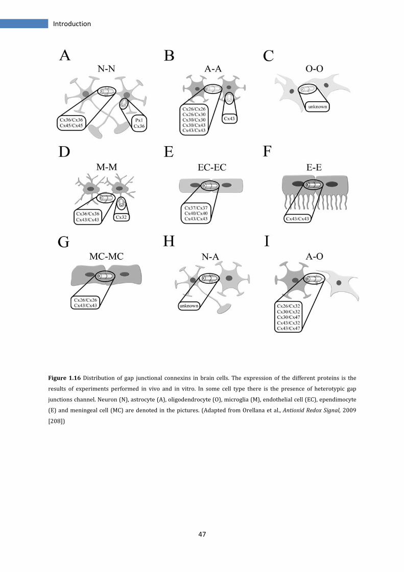
47
Introduction
Figure 1.16 Distribution of gap junctional connexins in brain cells. The expression of the different proteins is the
results of experiments performed in vivo and in vitro. In some cell type there is the presence of heterotypic gap
junctions channel. Neuron (N), astrocyte (A), oligodendrocyte (O), microglia (M), endothelial cell (EC), ependimocyte
(E) and meningeal cell (MC) are denoted in the pictures. (Adapted from Orellana et al., Antioxid Redox Signal, 2009
[208])
48
Introduction
1.2.4.1 Pannexins in brain
Panx1 and Panx2 transcripts are detected in many regions in rodents CNS as retina,
cortex, hippocampus, cerebellum, olfactory bulb, spinal cord. Panx1 is expressed
primarily in neurons and maybe in oligodendrocytes. Panx2 seems to be brain-specific,
but the two proteins are inversely regulated during the development of the rodents
brain.
Panx1 show high levels of expression in the embryonic and young postnatal brain and
decline considerably in the adult, whereas Panx2 mRNA expression is low in the
prenatal brain but increase substantially during subsequent postnatal development with
peaking at postnatal day 15 [209, 210].
Pannexins transcripts are particularly abundant in the adult cortex, in hippocampal and
neocortical pyramidal cells but also in GABAergic interneurons, (to note the presence in
both excitatory and inhibitory neurons, while Cx36 is present only into inhibitory
interneurons), reticular thalamus, the inferior olive, magnocellular hypothalamic
neurons, midbrain and brain stem motoneurons, Purkinje and Golgi cells in the
cerebellum [209].
Panx1 protein is widely expressed in mammalian tissues; as regard brain is found in
several regions including cortex, striatum, olfactory bulb, hippocampus, thalamus,
inferior olive, inferior colliculus, amygdala, spinal cord, retina, and cerebellum. At the
cellular level, Panx1 has been localized in different neuronal types, including olfactory
bulb mitral cells, Purkinje cells, dopaminergic, cholinergic and glutamatergic neurons. In
the cerebral cortex and hippocampus is localized in the postsynaptic cell membranes.
Panx1 is also detected in cultured astrocytes, immature oligodendrocytes and neurons,
using immunofluorescence and Western blot analysis, but it has not been found in the
cell surface of astrocytes [209-214].
Panx2 protein is expressed exclusively in the brain including the olfactory bulb,
hippocampus, amygdala, superior colliculus, substantia nigra, cerebellum, hypothalamus
and spinal cord. Its presence it has been confirmed in the majority of pyramidal cells and
in GABAergic interneurons but as regard the expression in glial cells there are still
contrasting opinions; under resting conditions, hippocampal astrocytes do not express
Panx2 even if it appears in hippocampal astrocytes several hours after
ischemia/reperfusion [209-212, 215].
49
Introduction
The presence in brain becomes important because pannexins are present in all regions
in which lack connexins but are alike coupled both metabolically and electrically. For
example, the abundance of Panxs in Purkinje cells or in hippocampal pyramidal cells,
that do not express Cx36 or Cx45, suggests that these are responsible for electric
coupling and generation of high-frequency oscillations. Experiments in mice KO for Cx36
show a reduction in gamma frequency, while ultrafast oscillations are not modified.
These data are consistent with the absence of Cx36, present only in hippocampal
interneurons, while other channels may determine fast oscillations occurring in
pyramidal cells [216-218]. However, the impact of Panxs channels on neuronal network
synchronization remains to demonstrate.
It remains also unknown whether pannexins form functional GJCs and/or hemichannels.
There are many controversial works regard the formation of GJs; in some systems as
Xenopus oocytes or C6 glioma cells an over-expression of Panx1, but not Panx2, mediate
intercellular coupling, while in other systems and other groups report no
communication. In contrast, functional hemichannels formed by both Panx1 and 2 are
demonstrated in different cellular system and in hippocampal neurons [211, 212, 219].
50
Introduction
1.2.5 Electrical synchronization
Electrical synapses represent ontogenetically and phylogenetically a primitive form of
intercellular communication; afterwards they have been replaced by chemical synapses
in the CNS of vertebrates. However, they represent the principal form of communication
during early development of mammalians CNS and in some less evolved organism as in
crayfish cord giant fibers. Nevertheless in the adult mammalian CNS they are of great
importance, indeed is thought that they have a role in the promotion of synchronous
activity in different areas of the brain and in retina [220].
More and more evidences correlate electrical synapses with the establishment of
oscillatory networks between neuronal cells. The neuronal synchronization occurs at
different frequency bands, in hippocampus occurs through three types, theta θ
oscillations (4-12 Hz), gamma γ waves (40-100 Hz) and high frequency oscillation (also
named ripples) (~200 Hz). The gap junctions are involved in the generation of gamma
and ripples oscillations [221].
The first demonstration of electrotonic coupling in neurons has been observed in
pyramidal cells of hippocampus. Here, high-frequency oscillations (100-200 Hz), or
ripples, are mediated by electrical coupling and occur via axo-axonal contact sites; these
results are confirmed by the inhibition of spontaneous ripples with use of gap junction
blockers, or by the maintaining of ripples in absence of chemical transmission. However,
is not clear which proteins are capable to generate this synchronous oscillatory activity
because the main player Cx36, is located only in interneurons, in fact in Cx36 KO mice
the generation of ripples is not affected. Controversial study support the possible
involvement of Cx45 or pannexins [222-225].
In hippocampus, gap junctions formed by Cx36 are present in GABAergic interneurons
of CA1 and CA3 region, where they are important components of γ-frequency oscillation,
even though in Cx36 deficient mice there is a residual gamma oscillations, suggesting the
involvement of other proteins [189, 218, 224, 226]. An important electrical
communication between GABAergic interneurons is demonstrated in various parts of
the brain, as in the cerebral cortex, thalamus, striatum and cerebellum. These seem to be
a fundamental feature for inhibitory circuits because they could facilitate synchronous
spiking of interneurons providing in this way to the control of activity in neurons [227-
230]. In the neocortex there are two networks of GABA neurons, FS (fast-spiking) cells
51
Introduction
and LTS (low threshold cells) interconnected by GABA-mediated synapses; the FS cells
are strongly interconnected by GABA-mediated synapses, while few chemical synapses
are found among LTS cells. Studies using Cx36 deficient mice show the absence of
electrical coupling or spikelet in LTS cells and a reduction in FS cells [218, 226].
Many others brain regions display an oscillatory activity mediated by gap junctions
coupling as medium spiny neurons of striatum, locus coeruleus, inferior olive, spinal
cord, hypothalamus and others [188].
1.2.6 Gap junctions and seizures
In epilepsy, many neurons are synchronized and there are several evidences that gap
junctions coupling can mediate the generation of seizures and epileptiform activity.
Several works show that seizures, induced with 4-aminopyridine or kainic acid, are
impaired using gap junctions blockers; while in Cx36 deficient mice the incidence of
seizures is reduced [218, 225, 231].
The role of GJs in seizures has been demonstrated both in vitro and in human epilepsies,
where are found increased levels of some mRNA connexins as Cx36, 32 and 43 and a
more pronounced coupling between the cells to respect normal tissue. The presence of
these connexins would suggest an increased coupling between neurons and
astrocytes/oligodendrocytes in the epileptiform discharges [232, 233].
Gap junctions would seem crucial for the synchronization in the epileptic foci because
their opening or blocking, respectively with trimethylamine and carbenoxolone,
increased or decreased the duration of the seizures [234]. Interestingly specific blockers
for Cx36 showed a reduction of the seizures only after ~30 discharges and considering
the low levels of Cx36 mRNA in neocortex would suggest a rarely interconnection with
GJs but after continued exposure to some factors this communication will increase
[235].
52
Introduction
1.2.7 Gap junctions and mesocorticolimbic system
The presence of GJs has been demonstrated with the presence of an electrotonic
coupling or by dye marker transfer between brain cells.
Intercellular channels have been identified in Nucleus Accumbens (NAc), striatum,
prefrontal cortex (PFC), hippocampus and moreover in these regions the dye transfer
can be modulated by dopamine [236-241]. Particularly, several studies show the
different action of agonists or antagonists in the two sections of NAc. The infusion of D1-
like agonists in Nac core, causes a decrease in dye coupling and the effect is blocked by
D1 or D2 antagonists; while in the NAc shell the administration of D2-like agonists
increase dye transfer independently of the state of D1 activation [242, 243]. The
mechanism by which DA modulates GJs is not known.
Also in ventral tegmental area (VTA) there is a wide network of GABAergic neurons
coupled via Cx36 GJs, and they seem modulated by DA. Similarly, blocking Cx36 GJs
increases inhibition of DAergic neurons, supporting a key role of GJs [192, 244].
Gap junctions can be also regulated by coupling with glutamate receptors, indeed in
ischemia we assist to an increase of coupling in neurons and this is regulated by
glutamate, via group II metabotropic glutamate receptors [245-247]. As the same way
GJs can regulate the release of glutamate; indeed Cx43 glial hemichannels, under
different stimuli, can release glutamate causing various effects in normal and
pathological brain [248, 249].
The nucleus accumbens receives afferents from different limbic structures, so it must
process the different input to absolve its role. It’s possible that the different input
contact distinct populations of neurons in NAc and each set could represent an ensemble
coding capable to modulate the information [250, 251].
Several works showed that NAc neurons exhibited increase or decrease in firing rate in
response to both natural (food, water) and not natural stimuli (as drugs of abuse).
However, it would seem that natural stimuli activate distinct population of NAc neurons
respect to those activated by drugs, even if is unknown the precise manner in which NAc
neurons encode differently the two types of stimuli. Moreover, after protracted
abstinence, the NAc neurons that encoded for cocaine behaviours were strongly
increase; Cameron & Carelli (2012) have showed that normal reward processing is
dysregulated and the encoding of drug related information is potentiated at the cost of
53
Introduction
natural physiological rewards. They also postulate that a prolonged drug exposure and
extended or repeated periods of abstinence could increase these effects, leading to the
loss of control characteristic of the addicted state. Maybe gap junctions could mediate
the synchronization of every ensemble coding [252-255].
Through electrical stimulation of hypothalamus, mimicking the behavioural positive
reinforcement, and subsequent infusion of carbenoxolone (GJs blocker), the neuronal
synchronization is reduced and so the stereotyped behaviours, as locomotor activity,
indicating a role for GJs in self-stimulation reward [256].
1.2.8 Gap Junctions and cocaine abuse
Previous studies show changes in connexins expression after cocaine self-
administration; this lead to support that change in neuronal gap junction
communication may be responsible for the long-term alterations observed in addiction.
Following chronic cocaine SA (for 14 days) and 2 to 7 days of withdrawal, Bennett et al.
show that the levels of Cx32 are decreased in NAc but the same decrease is not observed
in dorsal striatum. This could be important because NAc is linked to the reinforcing
effects of drug, while dorsal striatum is associated with motor behaviour. Other
important changes are observed in hippocampus, where there is a decrease in Cx32
protein expression [257].
In another work, McCracken et al. show after exposure to cocaine for 10 days, changes in
Cx36 protein expression in NAc, PFC and hippocampus. Interestingly the effects
observed are different depending if the animals are sacrificed just after last cocaine
injection or with 1 or 7 days of withdrawal; while in the first case there are no changes,
after 7 days of abstinence is showed a very huge increase of Cx36 mRNA in NAc and a
slight increase in PFC and hippocampus. This increase is not followed by the same
increase in protein expression, indeed in NAc and PFC only a slight increase is present
with 1 day of abstinence and nothing in hippocampus. Analysis of Cx32 and Cx43 in the
same regions show no differences in both regions and at any time point [258].

54
Introduction
Aim of the work
55
2 Aim of the work
The aim of this study has been investigate the role of gap junctions in the mechanism of
action of cocaine. Drug dependence is a huge social problem and cocaine has taken in the
last years a prominent place among drug of abuse. Although the psychological effects
and the risks are the same for all the users, it’s clear that the phenomenon of addiction
affects only a low, but significant, percentage of individuals. In this regard, many
theories have been postulated. Some suppose a role for cocaine that cause
neurobiological changes in brain rendering the individual addict; for some others the
transition to addiction is a pathological response to the drug that cause several
modifications in brain of addicted individuals, but it depends by specific characteristics
of the individual, i.e. there would be people more susceptible to others.
The main goal in the study of addiction is to understand how the effects of a drug of
abuse with a prolonged use, progressively lead to permanent molecular and cellular
changes.
Nucleus accumbens is thought to be the area that mediates rewarding and reinforcing
effects of cocaine and in this, distinct neuronal population seem operate and function as
ensemble coding, that are set of neurons able to receive information and after analysis
sort output to other regions. In addition, every ensemble coding seems to be
synchronized to perform its function. In the NAc and in several other regions, as
hippocampus or striatum, synchronization is mediated by electrical coupling directed by
gap junctions. Already is known the importance of GJs in developing seizures in
epilepsia, where increase in expression of some connexins genes have been found. In
vitro and in vivo experiments, it is showed also, how GJs opener or blocker, respectively
increase or reduces the duration of seizures.
The use of classic GJs blocker carbenoxolone, demonstrates to suppress the stereotyped
behaviour in animals after induction of reward and reinforcement stimuli. Moreover,
electrical coupling between brain cells is regulated by several neurotransmitters as
dopamine and glutamate.
From above, the aim of this study has been identify the identity of gap junctions proteins
involved in the mechanism of action of cocaine and furthermore see if there was an
Aim of the work
56
involvement of neuronal GJs proteins or the ones present in glial cells. As the same time,
verify the potential role of GJs in the transition to addiction.
In neurons, the only connexin certainly expressed is Cx36, but there are many evidences
as regard the presence of Panx1. Instead in astrocytes, oligodendrocytes and microglia
have been found Cx43, 32, 26, 30. For Panx2 controversial remains the exact
localization, it has been found in neurons and under pathological conditions in
astrocytes.
So connexins and pannexins mRNA and protein expression have been analyzed
quantitavely in the nucleus accumbens, in medial prefrontal cortex and in ventral
tegmental area, i.e. those brain regions of mesocorticolimbic system principally involved
in the actions of cocaine abuse. To investigate the effect of cocaine we used animals
exposed for different time to cocaine self-administration, to analyze the effects in the
short and long-term. Moreover, to evaluate the differences between the instaneous
effect of cocaine and that after one day of withdrawal, the animals were sacrificed after
40 minutes or 24 hours by the last cocaine injection.
A self-administration model has been used to discriminate the addiction-like
behaviours, from those animals that instead kept a controlled use of the drug, and so
verify if these proteins are involved in the transition to addiction.
Quantitative real-time PCR and immunoblotting have been used to analyze the mRNA
expression and proteins level expression of connexins and pannexins.

Aim of the work
57
Materials and Methods
58
3 Materials and Methods
3.1 Cocaine self-administration (SA) procedures
The protocol of cocaine self-administration and the animal model, have been carried out by
Deroche-Gamonet et al. following the protocol already described [108, 109].
Subjects
Male Sprague-Dawley rats (280-300 g) were used. Rats were single housed under a 12
hr reverse dark/light cycle (on 20h00, off 8h00). Temperature (22 ± 1°C) and humidity
(60 ± 5%) were also controlled. Animal care and use followed the directives of European
Communities Council Directive (86/609/EEC).
Drugs
Cocaine HCl (Coopération Pharmaceutique Française, Bordeaux, France) was dissolved
in 0.9% NaCl. Ketamine (Imalgene ®) and xylazine (Rompun®) were mixed for
anesthesia. Gentamicine (Gentalline®) was dissolved in 0.9% NaCl.
Surgery
A silastic catheter (internal diameter = 0.28 mm; external diameter = 0.61 mm; dead
volume = 12μl) was implanted in the right jugular vein under ketamine (100 mg/kg) +
xylazine (1 mg/kg) anaesthesia. Rats were allowed to recover for 5 to 7 days after
surgery. During the first 4 days following surgery, rats received an antibiotic treatment
(gentamicine 1 mg/kg i.p.). After surgery, catheters were flushed daily with a saline
solution containing unfractionated heparin (100 IU/ml).
Materials and Methods
59
Intravenous Self-Administration (SA) apparatus
The SA setup consisted of 48 SA chambers made of Plexiglas and metal (Imetronic,
Pessac, France). Each chamber (40 cm long x 30 cm width x 52 cm high) was located
within an opaque box equipped with exhaust fans that assured air renewal and masked
background noise. Briefly, each rat was placed daily in a SA chamber and the
intracardiac catheter connected to a pump-driven syringe (infusion speed: 20μl/sec).
Two holes, located at opposite sides of the SA chamber placed at 5 cm from the grid
floor, were used to record instrumental responding. A white house light at the top of the
chamber allowed its complete illumination. A white cue light (1.8 cm in diameter) was
located 9.5 cm above the active hole. A green cue light (1.8 cm in diameter) was located
10 cm right to the white cue light. A blue cue light (1.8 cm in diameter) was located on
the wall opposite to the one containing the active hole at 33 cm from the floor on the left
side. Experimental contingencies were controlled and data collected with PC Windows-
compatible software.
Basal training protocol
The daily SA session was composed of three drug components (40 min each) separated
by two 15 min periods during which responding in the active hole had no scheduled
consequences (no drug periods). Drug periods were signaled by the blue cue light, while
the no drug periods were signaled by illumination of the entire SA box and extinction of
the blue cue light. During the drug periods, introduction of the rat's nose into one hole
(active) turned on the white cue light located above it and then, 1 sec later, switched on
the infusion pump. The cue light remained on for a total of 4 sec. Nose-pokes in the other
hole (inactive) had no scheduled consequences. The self-infusion volume was 40 μL (2
sec infusion) and contained 0.8 mg/kg of cocaine. Each infusion was followed by a time-
out period of 40 sec. During the first 6 days, a Fixed Ratio 3 (FR3) schedule of
reinforcement was applied. Then, the FR was increased to 5 for the rest of the
experiment. Criterion for acquisition of cocaine SA was defined by a stable number of
self-infusions over at least three consecutive SA sessions (± 10%).
Materials and Methods
60
Addiction-like criteria
Cocaine use severity is assessed using three procedures resembling some of DSM-IV [30]
criteria for substance dependence:
(i) persistence in drug seeking even if the drug is not available, measured by active
responding during periods of signaled drug non-availability; (ii) high motivation for the
drug, measured by a progressive ratio schedule of reinforcement and (iii) drug use
despite negative consequences, measured by resistance to footshock-induced
punishment during cocaine SA.
Persistence of drug seeking in the absence of cocaine. This test allows assessing the
subject's difficulty to control drug seeking and drug use. It was assessed daily by
measuring the responses in the active hole during the two no drug periods of the basal
training protocol. Active responses during the two daily no drug periods were summed.
For analysis, the mean responses over two to three consecutive sessions were used as
dependent variables.
Progressive-ratio schedule. The ratio of responses per infusion was increased after
each infusion according to the following progression (10, 20, 30, 45, 65, 85, 115, 145,
185, 225, 275, 325, 385, 445, 515, 585, 665, 745, 835, 925, 1025, 1125, 1235, 1345,
1465, 1585), the last ratio completed, called “the breakpoint”, was used to measure
motivation for cocaine. The session ceased after either 5 hours or when a period of 1
hour had elapsed since the previously earned infusion.
Resistance to punishment: During this session rats were placed for 40 min in the SA
chamber. The blue cue light signaling drug availability was on. The schedule was the
following: the first response led to the illumination of the green cue light signaling the
presence of the shock. After 3 additional responses, rats received an electric footshock
(0.2 mA, 1 sec), and after the 5th response, rats received both an electric footshock (0.2
mA, 1 sec) and a cocaine infusion (0.8 mg/kg), associated with the corresponding
conditioned stimulus (white cue light). Then the green cue light was turned off. The
schedule reinitiated at the end of the time-out period, i.e. 40 sec after the infusion. If,
within a minute, rats did not complete response requirements leading to shock and
shock plus infusion respectively, the green cue light turned off and the sequence was
reinitiated, i.e. the following response turned on the green cue light. The number of
active responses earned during this test was expressed in percentage of the number of
Materials and Methods
61
active responses performed, over the same period of time, during the basal training
session conducted the day before.
Establishment of Addict-like and non Addict-like groups
The rats were classified based on their scores for each of the three addiction-like tests
and considering a rat positive for an addiction-like criterion when its score for this
behaviour was in the 35% highest percentile of the distribution. Four groups of rats are
isolated depending on the number of positive criteria met (0crit, 1crit, 2crit and 3crit). 0
and 3 criteria rats are defined as non Addict-like and Addict-like animals respectively.
1crit and 2crit rats either represent specific stable cocaine “use-related troubles” or
intermediate steps toward addiction.
The scores in the three addiction-like behaviours were linearly related to the number of
addiction-like criteria met.
Addiction score
It was calculated as the algebraic sum of standardized scores of each of the three
addiction-like criteria. Standardization consisted in subtracting the mean of the group to
each individual score and then dividing this number by the standard deviation. This
procedure yields scores which have a mean of 0 and a standard deviation of 1. The
addiction score is thus distributed along a scale from -3 to 3.
Evaluation of vulnerability to addiction-like behaviour in early cocaine use
The animal model developed allowed also to evaluate the vulnerability to addiction
during an early cocaine use (after 15 to 20 days of SA). Indeed, starting from sessions
11-13, addict rats showed a tendency to a progressive increase in response during the
last part of the non-drug periods. Then, analyzing several variables characterizing
responding during the early non-drug periods were individuated early behavioural
features that allow predicting transition in a reliable and consistent manner in early
cocaine use.
Materials and Methods
62
The variables analyzed were the number of active responses measured at early or late
minutes of no-drug periods (for example during the first 6 minutes or the last 9 minutes
of no drug periods). Rats were then ranked for these four variables. Rats with a score in
the 20th highest percentile of the population were defined as positive for this predictive
criterion. Rats with 4 positive criteria were defined Addiction Vulnerable rats, whilst
rats with 4 negative criteria were called Addiction Resistant rats.
Groups of animals analyzed
For expression of connexins and pannexins genes and proteins have been tested
different groups of animals. Three groups of rats were subjected to cocaine self-
administration for 7, 18 and 72 days. The groups of rats treated for 7 and 18 days were
ulteriorly subdivided in two independent subgroups, the first subgroup were animals
sacrificed after 40 minutes by the last cocaine injection and the second group with
animals sacrificed after 24 hours by the last cocaine injection. The groups of rats treated
for 72 days were analyzed only after 40 minutes and only for western blot analysis. Each
group was formed also by animals Controls; these were left undisturbed in the animal
house for the same time of the others. In order to obtain the same time point within
groups the start of the SA sessions of each animal was scheduled appropriately.
With 7 days of SA the rats were indicated as cocJ7.
At 18 days of cocaine SA the animals, depending behavioural features observed during
no-drug periods were divided in vulnerable (predicted 3criteria) and resistant to
addiction (predicted 0 criteria) rats.
At 72 days of cocaine SA the animals were classified as 3 criteria (addict) or 0 criteria
(non-addict) depending by their addiction score.
Brain regions dissection
Rats were anesthetized with a mix of ketamine (100mg/kg) and xylazine (1mg/kg),
followed by decapitation. Brains were rapidly removed and placed into an ice-cold
dissection plate and areas of interest were hand-dissected, then frozen on dry ice and
stored in centrifuge tubes at -80°C.
Materials and Methods
63
3.2 Quantitative real-time PCR
Samples
Three structure have been analyzed, nucleus accumbens (NAc), medial prefrontal cortex
(mPFC), ventral tegmental area (VTA) on 4 groups of rats
Every group was constituted by:
7days SA/40 minutes: n=15 (controls n=8, CocJ7/40’ n=7)
18days SA/40 minutes: n=23 (controls n=8; pred 0crit n=9; pred 3crit n=6)
7days SA/24 hours: n=14 (controls n=7, CocJ7/24h n=7)
18days SA/24hours: n=32 (controls n=10; pred 0crit n=10; pred 3crit n=12)
RNA extraction and PCR procedures
RNA was extracted with TRIzol® in according with manufacturer’s protocol (Invitrogen,
Carlsblad, CA, USA). After extraction with chloroforme and ethanol precipitation, RNA
samples were treated with a DNase inactivation reagent kit (Ambion® TURBO DNA-
free™ DNase; Ambion, Austin, TX, USA). To evaluate the purity of RNA, the ratio of
absorptions at 260 nm vs 280 nm (for contamination by other nucleic acids and protein)
and ratio 230 nm vs 260 nm (for contamination by organic compounds), were assessed
with spectrophotometer (Eppendorf AG, Hamburg, Germany).
The RNA quality was assessed by capillary electrophoresis, using the RNA 6000 Nano
Lab-on-a-Chip kit and the Bioanalyzer 2100 (Agilent Technologies, Palo Alto, CA, USA).
RNA with RIN (RNA integrity number) > 8.5] was used for the study.
cDNA was synthesized from 2 µg total RNA with Reverse Transcriptase SuperScript™ III
(Applied Biosystems, Foster City, CA, USA) according to the manufacturer's instructions,
in presence of random primer and Oligo(dT)18 primer (Fermentas, Waltham,
Massachusetts, USA). The RT-PCR amplification was carried out: 10 minutes at 25°C, 1
hour at 50°C and 15 minutes at 70°c, using a PTC-200 Biorad Thermal Cycler (BioRad,
Hercules, California, USA).
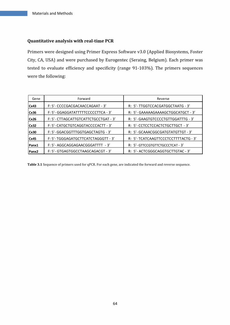
Materials and Methods
64
Quantitative analysis with real-time PCR
Primers were designed using Primer Express Software v3.0 (Applied Biosystems, Foster
City, CA, USA) and were purchased by Eurogentec (Seraing, Belgium). Each primer was
tested to evaluate efficiency and specificity (range 91-103%). The primers sequences
were the following:
Table 3.1 Sequence of primers used for qPCR. For each gene, are indicated the forward and reverse sequence.
Gene Forward Reverse
Cx43 F: 5’- CCCCGACGACAACCAGAAT - 3’ R: 5’- TTGGTCCACGATGGCTAATG - 3’
Cx36 F: 5’- GGAGGATATTTTTCCCCCTTCA - 3’ R: 5’- GAAAAAGAAAAGCTGGCATGCT - 3’
Cx26 F: 5’- CTTAGCATTGTCATTCTGCCTGAT - 3’ R: 5’- GAAGTGTCCCCTGTTGGATTTG - 3’
Cx32 F: 5’- CATGCTGTCAGGTACCCCACTT - 3’ R: 5’- CCTCCTCCACTCTGCTTGCT - 3’
Cx30 F: 5’- GGACGGTTTGGTGAGCTAGTG - 3’ R: 5’- GCAAACGGCGATGTATGTTGT - 3’
Cx45 F: 5’- TGGGAGATGCTTCATCTAGGGTT - 3’ R: 5’- TCATCAAGTTCCCTCCTTTTACTG - 3’
Panx1 F: 5’- AGGCAGGAGAACGGGATTTT - 3’ R: 5’- GTTCCGTGTTCTGCCCTCAT - 3’
Panx2 F: 5’- GTGAGTGGCCTAAGCAGACGT - 3’ R: 5’- ACTCGGGCAGGTGCTTGTAC - 3’
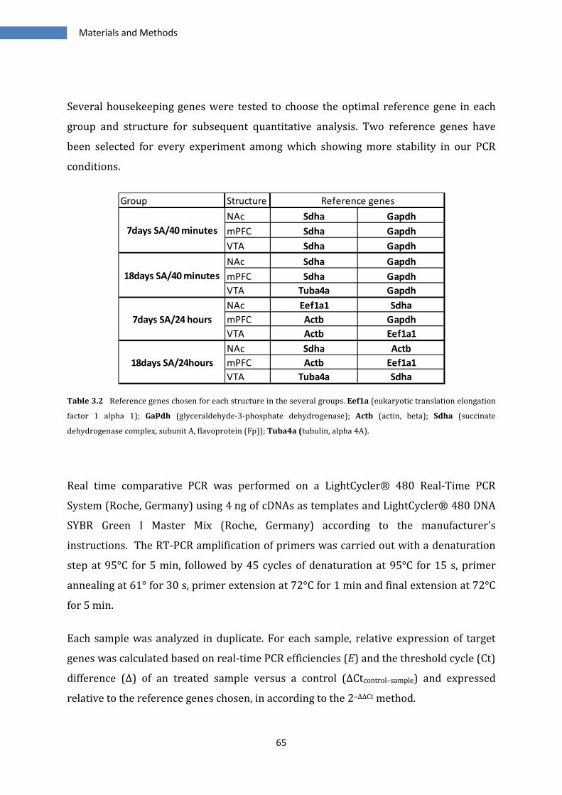
Materials and Methods
65
Several housekeeping genes were tested to choose the optimal reference gene in each
group and structure for subsequent quantitative analysis. Two reference genes have
been selected for every experiment among which showing more stability in our PCR
conditions.
Table 3.2 Reference genes chosen for each structure in the several groups. Eef1a (eukaryotic translation elongation
factor 1 alpha 1); Gapdh (glyceraldehyde-3-phosphate dehydrogenase); Actb (actin, beta); Sdha (succinate
dehydrogenase complex, subunit A, flavoprotein (Fp)); Tuba4a (tubulin, alpha 4A).
Real time comparative PCR was performed on a LightCycler® 480 Real-Time PCR
System (Roche, Germany) using 4 ng of cDNAs as templates and LightCycler® 480 DNA
SYBR Green I Master Mix (Roche, Germany) according to the manufacturer's
instructions. The RT-PCR amplification of primers was carried out with a denaturation
step at 95°C for 5 min, followed by 45 cycles of denaturation at 95°C for 15 s, primer
annealing at 61° for 30 s, primer extension at 72°C for 1 min and final extension at 72°C
for 5 min.
Each sample was analyzed in duplicate. For each sample, relative expression of target
genes was calculated based on real-time PCR efficiencies (E) and the threshold cycle (Ct)
difference (Δ) of an treated sample versus a control (ΔCtcontrol–sample) and expressed
relative to the reference genes chosen, in according to the 2–ΔΔCt method.
Group Structure
NAc Sdha Gapdh
mPFC Sdha Gapdh
VTA Sdha Gapdh
NAc Sdha Gapdh
mPFC Sdha Gapdh
VTA Tuba4a Gapdh
NAc Eef1a1 Sdha
mPFC Actb Gapdh
VTA Actb Eef1a1
NAc Sdha Actb
mPFC Actb Eef1a1
VTA Tuba4a Sdha
Reference genes
7days SA/40 minutes
18days SA/40 minutes
7days SA/24 hours
18days SA/24hours
Materials and Methods
66
3.3 Immunoblotting
Samples:
Three structures have been analyzed, nucleus accumbens (NAc), medial prefrontal
cortex (mPFC), and ventral tegmental area (VTA) on 5 groups of rats:
7days SA/40 minutes: n=15 (controls n=8, CocJ7/40’ n=7)
18days SA/40 minutes: n=23 (controls n=8; pred 0crit n=9; pred 3crit n=6)
72Days SA/40 minutes: n=31 (controls n=10; 0crit n=11; 3crit n=10)
7days SA/24 hours: n=18 (controls n=9, CocJ7/24h n=9)
18days SA/24hours: n=32 (controls n=10; pred 0crit n=10; pred 3crit n=12)
Protein extraction: Protein extracts from rat nucleus accumbens (NAc), medial
prefrontal cortex (mPFC) and ventral tegmental area (VTA) were prepared with a
homogenizing protocol using the Precellys 24 (Bertin Technologies, France). The tissues
were homogenized with ceramic CK14 beads (Bertin Technologies, France) in RIPA
buffer (Radioimmunoprecipitation assay buffer), containing DTT, protease and
phosphatase inhibitors (Sigma, St. Louis, MO, USA) at 5000 rpm 2x30 sec + 10 sec break.
After centrifugation at 10000 rpm for 10 minutes the supernatant was kept and stored
at -80 °C. Proteins concentrations were determined by Bradford assay (Bio-Rad
Laboratories, Hercules, CA, USA).
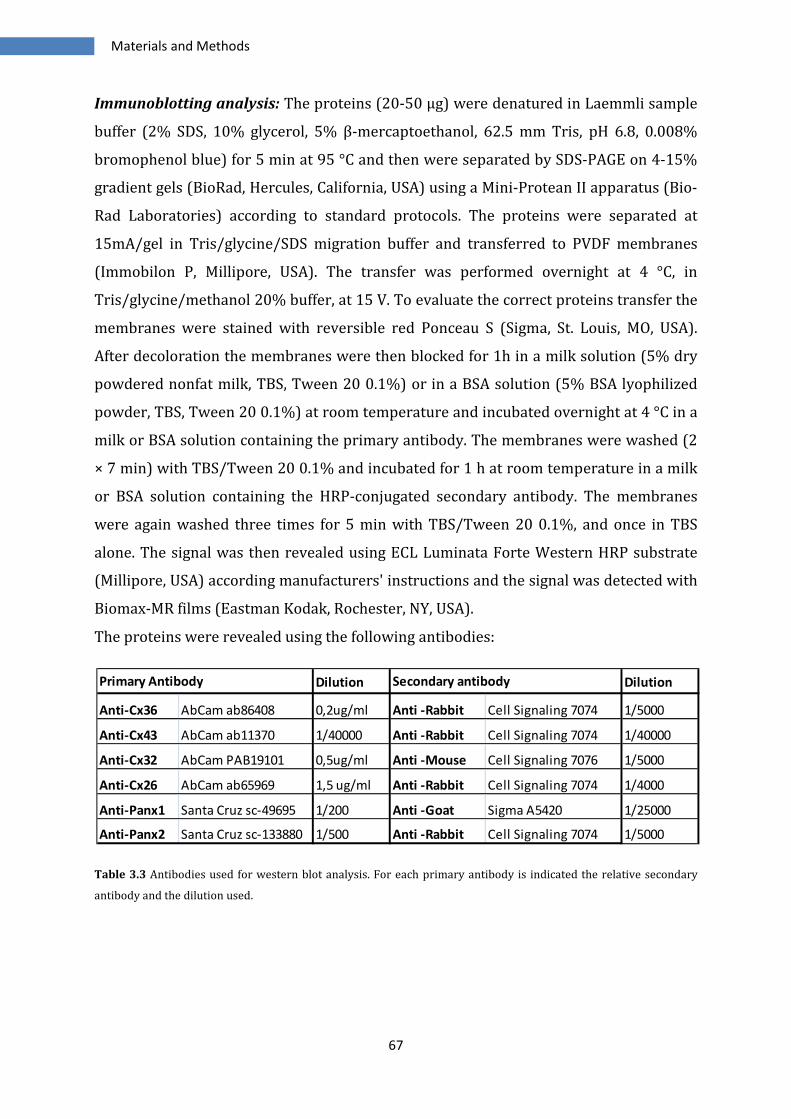
Materials and Methods
67
Immunoblotting analysis: The proteins (20-50 μg) were denatured in Laemmli sample
buffer (2% SDS, 10% glycerol, 5% β-mercaptoethanol, 62.5 mm Tris, pH 6.8, 0.008%
bromophenol blue) for 5 min at 95 °C and then were separated by SDS-PAGE on 4-15%
gradient gels (BioRad, Hercules, California, USA) using a Mini-Protean II apparatus (Bio-
Rad Laboratories) according to standard protocols. The proteins were separated at
15mA/gel in Tris/glycine/SDS migration buffer and transferred to PVDF membranes
(Immobilon P, Millipore, USA). The transfer was performed overnight at 4 °C, in
Tris/glycine/methanol 20% buffer, at 15 V. To evaluate the correct proteins transfer the
membranes were stained with reversible red Ponceau S (Sigma, St. Louis, MO, USA).
After decoloration the membranes were then blocked for 1h in a milk solution (5% dry
powdered nonfat milk, TBS, Tween 20 0.1%) or in a BSA solution (5% BSA lyophilized
powder, TBS, Tween 20 0.1%) at room temperature and incubated overnight at 4 °C in a
milk or BSA solution containing the primary antibody. The membranes were washed (2
× 7 min) with TBS/Tween 20 0.1% and incubated for 1 h at room temperature in a milk
or BSA solution containing the HRP-conjugated secondary antibody. The membranes
were again washed three times for 5 min with TBS/Tween 20 0.1%, and once in TBS
alone. The signal was then revealed using ECL Luminata Forte Western HRP substrate
(Millipore, USA) according manufacturers' instructions and the signal was detected with
Biomax-MR films (Eastman Kodak, Rochester, NY, USA).
The proteins were revealed using the following antibodies:
Table 3.3 Antibodies used for western blot analysis. For each primary antibody is indicated the relative secondary
antibody and the dilution used.
Dilution Dilution
Anti-Cx36 AbCam ab86408 0,2ug/ml Anti -Rabbit Cell Signaling 7074 1/5000
Anti-Cx43 AbCam ab11370 1/40000 Anti -Rabbit Cell Signaling 7074 1/40000
Anti-Cx32 AbCam PAB19101 0,5ug/ml Anti -Mouse Cell Signaling 7076 1/5000
Anti-Cx26 AbCam ab65969 1,5 ug/ml Anti -Rabbit Cell Signaling 7074 1/4000
Anti-Panx1 Santa Cruz sc-49695 1/200 Anti -Goat Sigma A5420 1/25000
Anti-Panx2 Santa Cruz sc-133880 1/500 Anti -Rabbit Cell Signaling 7074 1/5000
Secondary antibodyPrimary Antibody
Materials and Methods
68
Analysis of the results
The bands in western blot were quantified by densitometry using a GS-800 scanner (in
transmission mode) and the associated Quantity One software (Bio-Rad). The X-ray
films were scanned and the optical density (OD) of each pixel was measured. OD of each
band was corrected by subtracting the background OD. Identical quantities (µg) of
proteins were loaded on the gels in each experiment. The Neuronal Class III β-tubulin
(βIII-tubulin) monoclonal antibody (MMS-435P; 1/1000 dilution) used as a loading
control was purchased from Eurogentec (Seraing, Belgium).
Data were presented and analyzed as adjusted band volume (adjusted volume OD)
and/or percentage of basal values, ± SEM.
3.4 Statistics
Results are expressed as mRNA fold change expression or protein levels expression
relative to control value and are shown as mean ± S.E.M. The differences of each group
with the group of controls were assessed with Student t-test. Significance is set at
P<0.05 and is represented with symbol #.
To evaluate group differences in the expression, the values of each sample in a group
were normalized with the average of controls values, and after the groups were
analyzed with one-way ANOVA. The Newman-Keuls post hoc test was used for pair wise
comparison of means in the different groups. Significance was set at P<0.05 and is
represented with symbol *.
Statistical tests were performed with GraphPad Prism (GraphPad Software Inc., La Jolla,
CA, USA) and Statistica 10.0© (StatSoft Inc, Tulsa, OK, USA).

Materials and Methods
69
70
4 Results
The main goal of this work has been evaluate the effects of cocaine on the expression of
connexins and pannexins genes and protein levels. We wanted to investigate the
modifications after acute and after prolonged self-administration of cocaine. Moreover
we divided the different groups of animals in two subgroups, 40 minutes and 24 hours,
depending by the time elapsed by last injection at the moment of sacrifice. Another goal
of this work was establish if there were different regulations between animal showed
addiction-like behaviour and non-addict animals.
Results
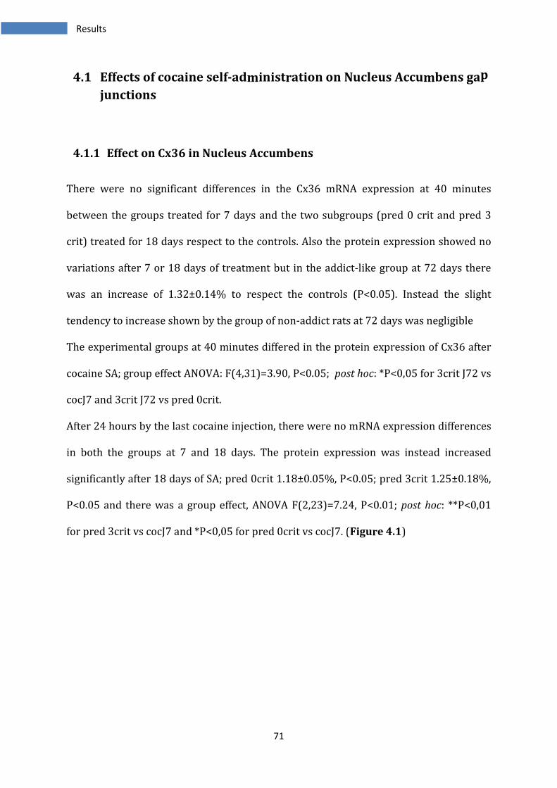
71
4.1 Effects of cocaine self-administration on Nucleus Accumbens gap
junctions
4.1.1 Effect on Cx36 in Nucleus Accumbens
There were no significant differences in the Cx36 mRNA expression at 40 minutes
between the groups treated for 7 days and the two subgroups (pred 0 crit and pred 3
crit) treated for 18 days respect to the controls. Also the protein expression showed no
variations after 7 or 18 days of treatment but in the addict-like group at 72 days there
was an increase of 1.32±0.14% to respect the controls (P<0.05). Instead the slight
tendency to increase shown by the group of non-addict rats at 72 days was negligible
The experimental groups at 40 minutes differed in the protein expression of Cx36 after
cocaine SA; group effect ANOVA: F(4,31)=3.90, P<0.05; post hoc: *P<0,05 for 3crit J72 vs
cocJ7 and 3crit J72 vs pred 0crit.
After 24 hours by the last cocaine injection, there were no mRNA expression differences
in both the groups at 7 and 18 days. The protein expression was instead increased
significantly after 18 days of SA; pred 0crit 1.18±0.05%, P<0.05; pred 3crit 1.25±0.18%,
P<0.05 and there was a group effect, ANOVA F(2,23)=7.24, P<0.01; post hoc: **P<0,01
for pred 3crit vs cocJ7 and *P<0,05 for pred 0crit vs cocJ7. (Figure 4.1)
Results
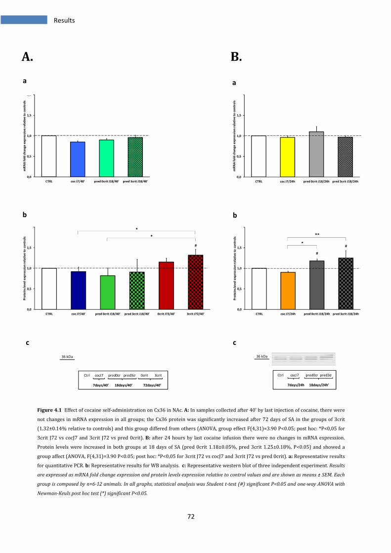
72
A. B.
Figure 4.1 Effect of cocaine self-administration on Cx36 in NAc. A: In samples collected after 40’ by last injection of cocaine, there were
not changes in mRNA expression in all groups; the Cx36 protein was significantly increased after 72 days of SA in the groups of 3crit
(1.32±0.14% relative to controls) and this group differed from others (ANOVA, group effect F(4,31)=3.90 P<0.05; post hoc: *P<0,05 for
3crit J72 vs cocJ7 and 3crit J72 vs pred 0crit). B: after 24 hours by last cocaine infusion there were no changes in mRNA expression.
Protein levels were increased in both groups at 18 days of SA (pred 0crit 1.18±0.05%, pred 3crit 1.25±0.18%, P<0.05) and showed a
group affect (ANOVA, F(4,31)=3.90 P<0.05; post hoc: *P<0,05 for 3crit J72 vs cocJ7 and 3crit J72 vs pred 0crit). a: Representative results
for quantitative PCR. b: Representative results for WB analysis. c: Representative western blot of three independent experiment. Results
are expressed as mRNA fold change expression and protein levels expression relative to control values and are shown as means ± SEM. Each
group is composed by n=6-12 animals. In all graphs, statistical analysis was Student t-test (#) significant P<0.05 and one-way ANOVA with
Newman-Keuls post hoc test (*) significant P<0.05.
0,0
0,5
1,0
1,5
2,0
CTRL coc J7/40' pred 0crit J18/40' pred 3crit J18/40'
mR
NA
fo
ld c
ha
nge
exp
ressi
on
re
lati
ve
to
co
ntr
ols
0,0
0,5
1,0
1,5
2,0
CTRL coc J7/24h pred 0crit J18/24h pred 3crit J18/24h
mR
NA
fo
ld c
ha
ng
e e
xp
ressi
on
re
lati
ve
to
co
ntr
ols
0,0
0,5
1,0
1,5
2,0
CTRL coc J7/40' pred 0crit J18/40' pred 3crit J18/40' 0crit J72/40' 3crit J72/40'
Pro
tein
s le
ve
l e
xp
ress
ion
re
lati
ve
to
co
ntr
ols
*
*
#
0,0
0,5
1,0
1,5
2,0
CTRL coc J7/24h pred 0crit J18/24h pred 3crit J18/24h
Pro
tein
s le
ve
l e
xp
ressi
on
re
lati
ve
to
co
ntr
ols
*
**
#
#
36 kDa
Ctrl cocJ7 pred0cr pred3cr 0crit 3crit
7days/40’ 18days/40’ 72days/40’
Ctrl cocJ7 pred0cr pred3cr
7days/24h 18days/24h’
36 kDa
a a
b b
c c
Results
73
4.1.2 Effect on Cx43 in Nucleus Accumbens
After 40’ the mRNA expression of Cx43 did not show changes, both the groups subjected
to 7 and 18 days of SA. On the contrary the protein expression showed significant
changes: after 7 days of treatment there was a decrease of 0.70±0.22%, P<0.05; at 18
days both the groups of resistant and vulnerable rats showed a decrease of 0.57±0.10%
and 0.58±0.11% respectively (t-test: pred 0crit P<0.001; pred 3crit P<0.01).
Interestingly after 72 days of cocaine SA, the 0crit group restored the protein expression
to level comparable to controls, but the 3crit animals showed a reduction of 0.64±0.06%
with a P<0.05. There was also a group affect, as analyzed with ANOVA F(4,30)=4.55
P<0.01; post hoc: *P<0,05 for 0crit J72 vs cocJ7, pred 0crit, pred 3crit and 3crit J72.
After 24 hours there were still no changes in the mRNA expression in all the groups
analyzed. Moreover there were no variations in the protein expression after 7 days.
After 18 days there was a slight reduction in Cx43 expression in predicted 3 criteria
group 0.81±0.14%, P<0.05.
The levels of protein expression, decreased at any time point in the animals with drug on
board were restored after one days of abstinence, even though the groups of vulnerable
animals at 18 days/24 hours started to show the reduction that they would stably
maintained. (Figure 4.2)
Results
74
A. B.
Figure 4.2 Effect of cocaine self administration on Cx43 in Nac. A: 7 or 18 days of SA did not influence expression of Cx43 mRNA in
samples at 40’ while the protein showed a reduction at any time point; after 7 days of SA there was significant decrease in Cx43 protein
(0.70±0.22% P<0.05); after 18 days both pred 0crit and pred 3crit showed the same level of decrease, 0.57±0.1% and P<0.001 for pred
0crit and 0.58±0.11% P<0.01 for pred 3crit. After 72 days, 0crit restored levels than to controls 1.13±0.16%, while 3 crit animals
exhibited low levels of Cx43 protein (0.64±0.06% P<0.05). The groups differed accordingly ANOVA : F(4,30)=4.55 P<0.01; post hoc:
*P<0.05 for 0crit 72J/40’ vs all the other 4 groups cocJ7, pred 0crit J18, pred 3crit J18 and 3crit J72. B: mRNA expression was not
modified in the samples collected after 24 hours in both groups at 7 and 18 days. 24 hours of abstinence restored the normal levels of
protein at 7 days, but after 18 days a small decrease was detected in the predicted 3 criteria group (0.81±0.14% P<0.05). a:
Representative results for quantitative PCR. b: Representative results for WB analysis. c: Representative western blot of three
independent experiment. Results are expressed as mRNA fold change expression and protein levels expression relative to control values and
are shown as means ± SEM. Each group is composed by n=6-12 animals. In all graphs, statistical analysis was Student t-test (#) significant
P<0.05 and one-way ANOVA with Newman-Keuls post hoc test (*) significant P<0.05.
0,0
0,5
1,0
1,5
2,0
CTRL coc J7/40' pred 0crit J18/40' pred 3crit J18/40'
mR
NA
fo
ld c
ha
nge
exp
ress
ion
re
lati
ve
to
co
ntr
ols
0,0
0,5
1,0
1,5
2,0
CTRL coc J7/24h pred 0crit J18/24h pred 3crit J18/24h
mR
NA
fo
ld c
ha
nge
exp
ress
ion
re
lati
ve
to
co
ntr
ols
0,0
0,5
1,0
1,5
2,0
CTRL coc J7/40' pred 0crit J18/40' pred 3crit J18/40' 0crit J72/40' 3crit J72/40'
Pro
tein
s le
ve
l e
xp
ressi
on
re
lati
ve
to
co
ntr
ols
*
*
* *
#
######
0,0
0,5
1,0
1,5
2,0
CTRL coc J7/24h pred 0crit J18/24h pred 3crit J18/24h
Pro
tein
s le
ve
l e
xp
ressi
on
re
lati
ve
to
co
ntr
ols
#
43 kDa
Ctrl cocJ7 pred0cr pred3cr 0crit 3crit
7days/40’ 18days/40’ 72days/40’
43 kDa
Ctrl cocJ7 pred0cr pred3cr
7days/24h 18days/24h’
a a
b b
c c
Results
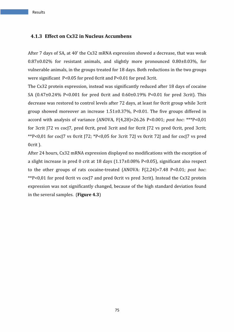
75
4.1.3 Effect on Cx32 in Nucleus Accumbens
After 7 days of SA, at 40’ the Cx32 mRNA expression showed a decrease, that was weak
0.87±0.02% for resistant animals, and slightly more pronounced 0.80±0.03%, for
vulnerable animals, in the groups treated for 18 days. Both reductions in the two groups
were significant P<0.05 for pred 0crit and P<0.01 for pred 3crit.
The Cx32 protein expression, instead was significantly reduced after 18 days of cocaine
SA (0.47±0.24% P<0.001 for pred 0crit and 0.60±0.19% P<0.01 for pred 3crit). This
decrease was restored to control levels after 72 days, at least for 0crit group while 3crit
group showed moreover an increase 1.51±0.37%, P<0.01. The five groups differed in
accord with analysis of variance (ANOVA, F(4,28)=26.26 P<0.001; post hoc: ***P<0,01
for 3crit J72 vs cocJ7, pred 0crit, pred 3crit and for 0crit J72 vs pred 0crit, pred 3crit;
**P<0,01 for cocJ7 vs 0crit J72; *P<0,05 for 3crit 72J vs 0crit 72J and for cocJ7 vs pred
0crit ).
After 24 hours, Cx32 mRNA expression displayed no modifications with the exception of
a slight increase in pred 0 crit at 18 days (1.17±0.08% P<0.05), significant also respect
to the other groups of rats cocaine-treated (ANOVA: F(2,24)=7.48 P<0.01; post hoc:
**P<0,01 for pred 0crit vs cocJ7 and pred 0crit vs pred 3crit). Instead the Cx32 protein
expression was not significantly changed, because of the high standard deviation found
in the several samples. (Figure 4.3)
Results
76
A. B.
Figure 4.3 Effect of cocaine self administration on Cx32 in NAc. A: Slight decrease at 18 days of SA in both predicted 0 and pred 3criteria
(0.87±0.02 P<0.05 for pred 0crit and 80±3 P<0.01 for pred 3crit). In protein expression this decrease is reinforced, pred 0crit
0.47±0.24% with P<0.001 and pred 3crit 0.60±0.19% P<0.01. 72 days of self-administration caused an increase in Cx32 expression, a
weak, not significant, increase for 0 crit and an increase of 1.51±0.37% in 3 crit group with a t-test P<0.01 compared to controls. The
ANOVA showed strong differences between groups, F(4,28)=26.26 P<0.001; post hoc: ***P<0,01 for 3crit J72 vs cocJ7, pred 0crit, pred
3crit and for 0crit J72 vs pred 0crit, pred 3crit; **P<0,01 for cocJ7 vs 0crit J72; *P<0,05 for 3crit 72J vs 0crit 72J and for cocJ7 vs pred
0crit. B: cocJ7 and pred 3crit at 18 days revealed no differences in Cx32 gene expression, only a weak increase in pred 0 crit 1.17±0.08%
P<0.01, confirmed moreover by ANOVA for effect group, F(2,24)=7.48 P<0.01 post hoc: **P<0,01 for pred 0crit vs cocJ7 and pred 0crit vs
pred 3crit. The proteins at 24 hours showed no significant differences. a: Representative results for quantitative PCR. b: Representative
results for WB analysis. c: Representative western blot of three independent experiment. Results are expressed as mRNA fold change
expression and protein levels expression relative to control values and are shown as means ± SEM. Each group is composed by n=6-12
animals. In all graphs, statistical analysis was Student t-test (#) significant P<0.05 and one-way ANOVA with Newman-Keuls post hoc test (*)
significant P<0.05.
0,0
0,5
1,0
1,5
2,0
CTRL coc J7/40' pred 0crit J18/40' pred 3crit J18/40'
mR
NA
fo
ld c
ha
nge
exp
ressi
on
re
lati
ve
to
co
ntr
ols
# ##
0,0
0,5
1,0
1,5
2,0
CTRL coc J7/24h pred 0crit J18/24h pred 3crit J18/24h
mR
NA
fo
ld c
ha
nge
exp
ress
ion
re
lati
ve
to
co
ntr
ols
****
#
0,0
0,5
1,0
1,5
2,0
2,5
CTRL coc J7/40' pred 0crit J18/40' pred 3crit J18/40' 0crit J72/40' 3crit J72/40'
Pro
tein
s le
ve
l e
xp
ress
ion
re
lati
ve
to
co
ntr
ols
***
***
***
**
*** *
*
**
#####
##
0,0
0,5
1,0
1,5
2,0
CTRL coc J7/24h pred 0crit J18/24h pred 3crit J18/24h
Pro
tein
s le
ve
l e
xp
ressi
on
re
lati
ve
to
co
ntr
ols
32 kDa
Ctrl cocJ7 pred0cr pred3cr 0crit 3crit
7days/40’ 18days/40’ 72days/40’
32 kDa
Ctrl cocJ7 pred0cr pred3cr
7days/24h 18days/24h’
a a
b b
c c
Results
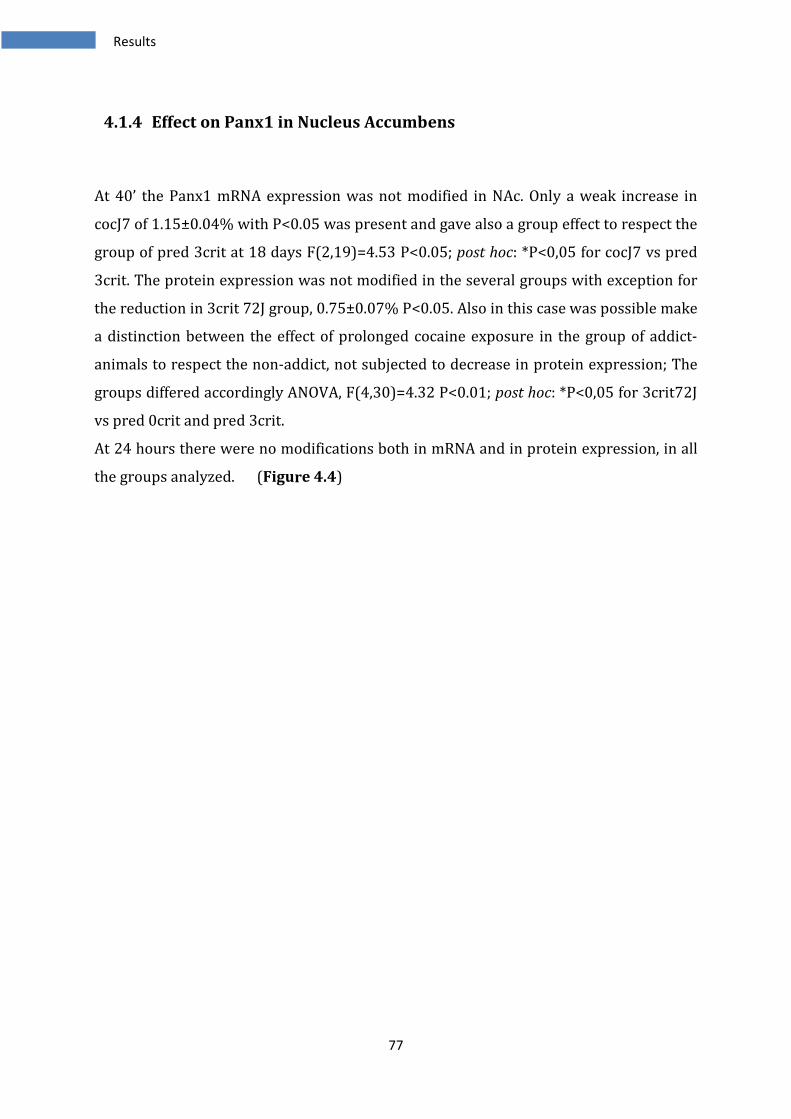
77
4.1.4 Effect on Panx1 in Nucleus Accumbens
At 40’ the Panx1 mRNA expression was not modified in NAc. Only a weak increase in
cocJ7 of 1.15±0.04% with P<0.05 was present and gave also a group effect to respect the
group of pred 3crit at 18 days F(2,19)=4.53 P<0.05; post hoc: *P<0,05 for cocJ7 vs pred
3crit. The protein expression was not modified in the several groups with exception for
the reduction in 3crit 72J group, 0.75±0.07% P<0.05. Also in this case was possible make
a distinction between the effect of prolonged cocaine exposure in the group of addict-
animals to respect the non-addict, not subjected to decrease in protein expression; The
groups differed accordingly ANOVA, F(4,30)=4.32 P<0.01; post hoc: *P<0,05 for 3crit72J
vs pred 0crit and pred 3crit.
At 24 hours there were no modifications both in mRNA and in protein expression, in all
the groups analyzed. (Figure 4.4)
Results
78
A. B.
Figure 4.4 Effect of cocaine self administration on Panx1 in NAc. A: the Panx1 mRNA showed a weak increase after 7 days of SA
1.15±0.04% P<0.05 and a group effect characterized by ANOVA F(2,19)=4.53 P<0.05; post hoc: *P<0,05 for cocJ7 vs pred 3crit. For
protein expression there was a decrease at 72 days in addicted animals 0.75±0.07% P<0.05, while the other groups displayed no changes
or slight non significant changes. The groups differed accordingly ANOVA, F(4,30)=4.32 P<0.01; post hoc: *P<0,05 for 3crit72J vs pred
0crit and vs pred 3crit. B: Panx1 mRNA and protein were not modified in the groups of samples collected after 24 hours. a:
Representative results for quantitative PCR. b: Representative results for WB analysis. c: Representative western blot of three
independent experiment. Results are expressed as mRNA fold change expression and protein levels expression relative to control values and
are shown as means ± SEM. Each group is composed by n=6-12 animals. In all graphs, statistical analysis was Student t-test (#) significant
P<0.05 and one-way ANOVA with Newman-Keuls post hoc test (*) significant P<0.05.
0,0
0,5
1,0
1,5
2,0
CTRL coc J7/40' pred 0crit J18/40' pred 3crit J18/40'
mR
NA
fo
ld c
ha
ng
e e
xp
ressi
on
re
lati
ve
to
co
ntr
ols
*
#
0,0
0,5
1,0
1,5
2,0
CTRL coc J7/24h pred 0crit J18/24h pred 3crit J18/24h
mR
NA
fo
ld c
ha
nge
exp
ress
ion
re
lati
ve
to
co
ntr
ols
0,0
0,5
1,0
1,5
2,0
CTRL coc J7/40' pred 0crit J18/40' pred 3crit J18/40' 0crit J72/40' 3crit J72/40'
Pro
tein
s le
ve
l e
xp
ress
ion
re
lati
ve
to
co
ntr
ols
*
*
#
0,0
0,5
1,0
1,5
2,0
CTRL coc J7/24h pred 0crit J18/24h pred 3crit J18/24h
Pro
tein
s le
ve
l e
xp
ress
ion
re
lati
ve
to
co
ntr
ols
48 kDa
Ctrl cocJ7 pred0cr pred3cr 0crit 3crit
7days/40’ 18days/40’ 72days/40’
48 kDa
Ctrl cocJ7 pred0cr pred3cr
7days/24h 18days/24h’
a a
b b
c c
Results
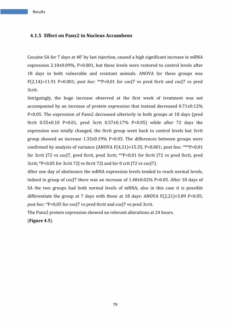
79
4.1.5 Effect on Panx2 in Nucleus Accumbens
Cocaine SA for 7 days at 40’ by last injection, caused a high significant increase in mRNA
expression 2.10±0.09%, P<0.001, but these levels were restored to control levels after
18 days in both vulnerable and resistant animals. ANOVA for these groups was
F(2,14)=11.91 P<0.001; post hoc: **P<0,01 for cocJ7 vs pred 0crit and cocJ7 vs pred
3crit.
Intriguingly, the huge increase observed at the first week of treatment was not
accompanied by an increase of protein expression that instead decreased 0.71±0.12%
P<0.05. The expression of Panx2 decreased ulteriorly in both groups at 18 days (pred
0crit 0.55±0.10 P<0.01, pred 3crit 0.57±0.17% P<0.05) while after 72 days the
expression was totally changed, the 0crit group went back to control levels but 3crit
group showed an increase 1.33±0.19% P<0.05. The differences between groups were
confirmed by analysis of variance (ANOVA F(4,31)=15.35, P<0.001; post hoc: ***P<0,01
for 3crit J72 vs cocJ7, pred 0crit, pred 3crit; **P<0,01 for 0crit J72 vs pred 0crit, pred
3crit; *P<0,05 for 3crit 72J vs 0crit 72J and for 0 crit J72 vs cocJ7).
After one day of abstinence the mRNA expression levels tended to reach normal levels,
indeed in group of cocJ7 there was an increase of 1.48±0.02% P<0.05. After 18 days of
SA the two groups had both normal levels of mRNA; also in this case it is possible
differentiate the group at 7 days with those at 18 days: ANOVA F(2,21)=3.89 P<0.05;
post hoc: *P<0,05 for cocJ7 vs pred 0crit and cocJ7 vs pred 3crit.
The Panx2 protein expression showed no relevant alterations at 24 hours.
(Figure 4.5)
Results
80
A. B.
Figure 4.5 Effect of cocaine self administration on Panx2 in NAc. A: The mRNA expression after 7 days of cocaine SA at 40’ showed a
huge increase 2.10±0.09%, P<0.001. At 18 days the levels returned to control levels in predicted addict and non-addict animals. ANOVA
was F(2,14)=11.91 P<0.001; post hoc: **P<0,01 for cocJ7 vs pred 0crit and cocJ7 vs pred 3crit. The Panx2 protein expression showed a
different profile: 7 days of cocaine SA caused a decrease 0.71±0.12% P<0.05. The expression of Panx2 decreased ulteriorly in both
groups at 18 days (pred 0crit 0.55±0.10 P<0.01, pred 3crit 0.57±0.17% P<0.05). After 72 days the 0crit group returned to normal levels
while 3crit group showed a significant increase 1.33±0.19% P<0.05. These differences were confirmed by analysis of variance (ANOVA
F(4,31)=15.35 P<0.001; post hoc: ***P<0,01 for 3crit J72 vs cocJ7, pred 0crit, pred 3crit; **P<0,01 for 0crit J72 vs pred 0crit, pred 3crit;
*P<0,05 for 3crit 72J vs 0crit 72J and for 0 crit J72 vs cocJ7. B: After 24 hours the effect on Panx 2 genes were attenuated; in cocJ7
group there was an increase 1.48±0.02% P<0.05 but after 18 days the two groups had both normal levels of mRNA. The groups differed
accordingly (ANOVA F(2,21)=3.89 P<0.05; post hoc: *P<0,05 for cocJ7 vs pred 0crit and cocJ7 vs pred 3crit). The Panx2 protein
expression at 24 hours showed no modifications. a: Representative results for quantitative PCR. b: Representative results for WB
analysis. c: Representative western blot of three independent experiment. Results are expressed as mRNA fold change expression and
protein levels expression relative to control values and are shown as means ± SEM. Each group is composed by n=6-12 animals. In all graphs,
statistical analysis was Student t-test (#) significant P<0.05 and one-way ANOVA with Newman-Keuls post hoc test (*) significant P<0.05.
0,0
0,5
1,0
1,5
2,0
2,5
CTRL coc J7/40' pred 0crit J18/40' pred 3crit J18/40'
mR
NA
fo
ld c
ha
nge
exp
ress
ion
re
lati
ve
to
co
ntr
ols
**
**
###
0,0
0,5
1,0
1,5
2,0
CTRL coc J7/24h pred 0crit J18/24h pred 3crit J18/24h
mR
NA
fo
ld c
ha
nge
ex
pre
ssio
n r
ela
tive
to
co
ntr
ols
*
*
#
0,0
0,5
1,0
1,5
2,0
CTRL coc J7/40' pred 0crit J18/40' pred 3crit J18/40' 0crit J72/40' 3crit J72/40'
Pro
tein
s le
ve
l e
xp
ressi
on
re
lati
ve
to
co
ntr
ols
***
***
***
* *
**
**
#
###
#
0,0
0,5
1,0
1,5
2,0
CTRL coc J7/24h pred 0crit J18/24h pred 3crit J18/24h
Pro
tein
s le
ve
l e
xp
ressi
on
re
lati
ve
to
co
ntr
ols
70 kDa
Ctrl cocJ7 pred0cr pred3cr 0crit 3crit
7days/40’ 18days/40’ 72days/40’
70 kDa
Ctrl cocJ7 pred0cr pred3cr
7days/24h 18days/24h’
a a
b b
c c
Results
81
4.1.6 Effects of cocaine SA on Nucleus Accumbens in other genes
In nucleus accumbens, we analyzed also the mRNA expression of Cx26, Cx30 and Cx45.
It has been analyzed also the Cx26 protein expression, but this was undetectable with
western-blot technique in this area.
Analysis of cx26 mRNA expression in NAc revealed no relevant differences. only the
group of predicted 0crit at 18j/40’ showed a weak increase, 1.37±0.12%, that was not
significant respect to the controls group but there was a group effect, accordingly with
ANOVA F(2,17)=3.91 p<0.05; post hoc: *p<0.05 for pred 0crit vs cocj70,41.
The groups analyzed at 24 hours showed no alterations except of a very slight, not
significant, tendency to decrease in pred 3 crit group.
In cx30 mRNA expression there were no appreciable variations at 40’ but after 24 hours
the group of resistant to addiction animals, exhibited an increase of 1.26±0.11% p<0.05.
This increase gave also a discrete group effect by revealed with ANOVA F(2,22)=4.57
p<0.05; post hoc: *p<0,05 for pred 0crit vs cocj7 and pred 0crit vs pred 3crit.
The Cx45 gene, showed no modifications in expression in all the groups analyzed.
(Figure 4.6)
Results
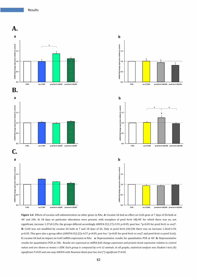
82
A.
B.
C.
Figure 4.6 Effects of cocaine self-administration on other genes in NAc. A: Cocaine SA had no effect on Cx26 gene at 7 days of SA both at
40’ and 24h. At 18 days no particular alterations were present, with exception of pred 0crit 18J/40’ for which there was an, not
significant, increase 1.37±0.12%; the groups differed accordingly ANOVA F(2,17)=3.91 p<0.05; post hoc: *p<0.05 for pred 0crit vs cocj7.
B: Cx30 was not modified by cocaine SA both at 7 and 18 days of SA. Only in pred 0crit j18/24h there was an increase 1.26±0.11%
p<0.05. This gave also a group effect (ANOVA F(2,22)=4.57 p<0.05; post hoc: *p<0,05 for pred 0crit vs cocj7 and pred 0crit vs pred 3crit).
C: cocaine SA had no impact on Cx45 mRNA expression in NAc. a: Representative results for quantitative PCR at 40’. b: Representative
results for quantitative PCR at 24h. Results are expressed as mRNA fold change expression and protein levels expression relative to control
values and are shown as means ± SEM. Each group is composed by n=6-12 animals. In all graphs, statistical analysis was Student t-test (#)
significant P<0.05 and one-way ANOVA with Newman-Keuls post hoc test (*) significant P<0.05.
0,0
0,5
1,0
1,5
2,0
CTRL coc J7/40' pred 0crit J18/40' pred 3crit J18/40'
mR
NA
fo
ld c
ha
ng
e e
xp
ressi
on
re
lati
ve
to
co
ntr
ols
*
0,0
0,5
1,0
1,5
2,0
CTRL coc J7/24h pred 0crit J18/24h pred 3crit J18/24h
mR
NA
fo
ld c
ha
ng
e e
xp
ressi
on
re
lati
ve
to
co
ntr
ols
0,0
0,5
1,0
1,5
2,0
CTRL coc J7/40' pred 0crit J18/40' pred 3crit J18/40'
mR
NA
fo
ld c
ha
ng
e e
xp
ressi
on
re
lati
ve
to
co
ntr
ols
0,0
0,5
1,0
1,5
2,0
CTRL coc J7/24h pred 0crit J18/24h pred 3crit J18/24h
mR
NA
fo
ld c
ha
nge
exp
ress
ion
re
lati
ve
to
co
ntr
ols
* *
#
0,0
0,5
1,0
1,5
2,0
CTRL coc J7/40' pred 0crit J18/40' pred 3crit J18/40'
mR
NA
fo
ld c
ha
ng
e e
xp
ressi
on
re
lati
ve
to
co
ntr
ols
0,0
0,5
1,0
1,5
2,0
CTRL coc J7/24h pred 0crit J18/24h pred 3crit J18/24h
mR
NA
fo
ld c
ha
ng
e e
xp
ressi
on
re
lati
ve
to
co
ntr
ols
a
a
a
b
b
b
Results
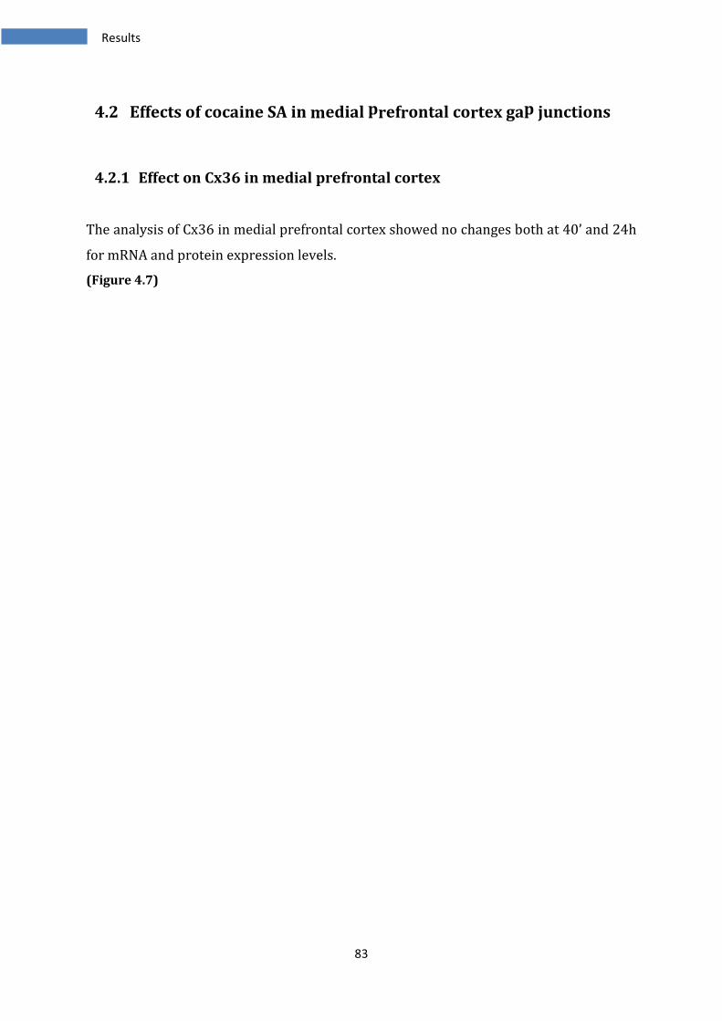
83
4.2 Effects of cocaine SA in medial prefrontal cortex gap junctions
4.2.1 Effect on Cx36 in medial prefrontal cortex
The analysis of Cx36 in medial prefrontal cortex showed no changes both at 40’ and 24h
for mRNA and protein expression levels.
(Figure 4.7)
Results
84
A. B.
Figure 4.7 Effect of cocaine self-administration on Cx36 in mPFC. A: the self-administration of cocaine produced no alterations in all the
groups analyzed both for mRNA and protein expression of Cx36 in rats with drug on board. B: Identical situation in animals analyzed at
24 hours. a: Representative results for quantitative PCR. b: Representative results for WB analysis. c: Representative western blot of
three independent experiment. Results are expressed as mRNA fold change expression and protein levels expression relative to control
values and are shown as means ± SEM. Each group is composed by n=6-12 animals. In all graphs, statistical analysis was Student t-test (#)
significant P<0.05 and one-way ANOVA with Newman-Keuls post hoc test (*) significant P<0.05.
0,0
0,5
1,0
1,5
2,0
CTRL coc J7/40' pred 0crit J18/40' pred 3crit J18/40'
mR
NA
fo
ld c
ha
ng
e e
xp
ressi
on
re
lati
ve
to
co
ntr
ols
0,0
0,5
1,0
1,5
2,0
CTRL coc J7/24h pred 0crit J18/24h pred 3crit J18/24h
mR
NA
fo
ld c
ha
nge
exp
ress
ion
re
lati
ve
to
co
ntr
ols
0,0
0,5
1,0
1,5
2,0
CTRL coc J7/40' pred 0crit J18/40' pred 3crit J18/40' 0crit J72/40' 3crit J72/40'
Pro
tein
s le
ve
l e
xp
ress
ion
re
lati
ve
to
co
ntr
ols
0,0
0,5
1,0
1,5
2,0
CTRL coc J7/24h pred 0crit J18/24h pred 3crit J18/24h
Pro
tein
s le
ve
l e
xp
ressi
on
re
lati
ve
to
co
ntr
ols
36 kDa
Ctrl cocJ7 pred0cr pred3cr 0crit 3crit
7days/40’ 18days/40’ 72days/40’
36 kDa
Ctrl cocJ7 pred0cr pred3cr
7days/24h 18days/24h’
a a
b b
c c
Results
85
4.2.2 Effect on Cx43 in medial prefrontal cortex
In the nucleus accumbens Cx43 showed no differences as regard the mRNA expression
respect to the controls, but protein levels were differentially regulated.
There were no changes in mRNA expression after 7 or 18 days of SA at 40 minutes.
The protein expression showed instead some differences: 7 days of SA were not
sufficient to give rise modifications, even though it seemed to be a tendency to increase.
18 days of SA caused a decrease in protein expression, mostly for the predicted 3 criteria
group, in which there was a reduction of 0.54±0.31%, P<0.05. A prolonged exposure
instead showed a different behaviour to respect the same protein analyzed in NAc; the 0
crit group had protein levels comparable to controls (even if the SEM was high) but the 3
crit group showed an increase in Cx43 expression of 1.40±0.34% P<0.01. The groups
differed accordingly ANOVA F(4,31)=6.79 P<0.001; post hoc: ***P<0,001 for 3crit J72 vs
pred 3crit; **P<0,01 for 3crit J72 vs pred 0crit and for pred 3crit vs cocJ7; *P<0,05 for
pred 3crit vs 0crit J72.
One day of abstinence caused no modifications in Cx43 gene expression, but the
treatment for 18 days caused a reduction in both vulnerable and resistant to addiction
animal groups, pred 0crit 0.80±0.22% P<0.05, pred 3crit 0.80±0.17% P<0.05.
(Figure 4.8)
Results
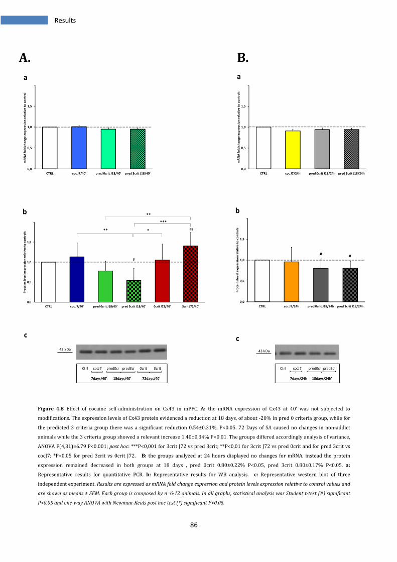
86
A. B.
Figure 4.8 Effect of cocaine self-administration on Cx43 in mPFC. A: the mRNA expression of Cx43 at 40’ was not subjected to
modifications. The expression levels of Cx43 protein evidenced a reduction at 18 days, of about -20% in pred 0 criteria group, while for
the predicted 3 criteria group there was a significant reduction 0.54±0.31%, P<0.05. 72 Days of SA caused no changes in non-addict
animals while the 3 criteria group showed a relevant increase 1.40±0.34% P<0.01. The groups differed accordingly analysis of variance,
ANOVA F(4,31)=6.79 P<0.001; post hoc: ***P<0,001 for 3crit J72 vs pred 3crit; **P<0,01 for 3crit J72 vs pred 0crit and for pred 3crit vs
cocJ7; *P<0,05 for pred 3crit vs 0crit J72. B: the groups analyzed at 24 hours displayed no changes for mRNA, instead the protein
expression remained decreased in both groups at 18 days , pred 0crit 0.80±0.22% P<0.05, pred 3crit 0.80±0.17% P<0.05. a:
Representative results for quantitative PCR. b: Representative results for WB analysis. c: Representative western blot of three
independent experiment. Results are expressed as mRNA fold change expression and protein levels expression relative to control values and
are shown as means ± SEM. Each group is composed by n=6-12 animals. In all graphs, statistical analysis was Student t-test (#) significant
P<0.05 and one-way ANOVA with Newman-Keuls post hoc test (*) significant P<0.05.
0,0
0,5
1,0
1,5
2,0
CTRL coc J7/40' pred 0crit J18/40' pred 3crit J18/40'
mR
NA
fo
ld c
ha
ng
e e
xp
ressi
on
re
lati
ve
to
co
ntr
ols
0,0
0,5
1,0
1,5
2,0
CTRL coc J7/24h pred 0crit J18/24h pred 3crit J18/24h
mR
NA
fo
ld c
ha
ng
e e
xp
ressi
on
re
lati
ve
to
co
ntr
ols
0,0
0,5
1,0
1,5
2,0
CTRL coc J7/40' pred 0crit J18/40' pred 3crit J18/40' 0crit J72/40' 3crit J72/40'
Pro
tein
s le
ve
l e
xp
ress
ion
re
lati
ve
to
co
ntr
ols *
**
**
***
#
##
0,0
0,5
1,0
1,5
2,0
CTRL coc J7/24h pred 0crit J18/24h pred 3crit J18/24h
Pro
tein
s le
ve
l e
xp
ressi
on
re
lati
ve
to
co
ntr
ols
##
43 kDa
Ctrl cocJ7 pred0cr pred3cr 0crit 3crit
7days/40’ 18days/40’ 72days/40’
43 kDa
Ctrl cocJ7 pred0cr pred3cr
7days/24h 18days/24h’
a a
b b
c c
Results
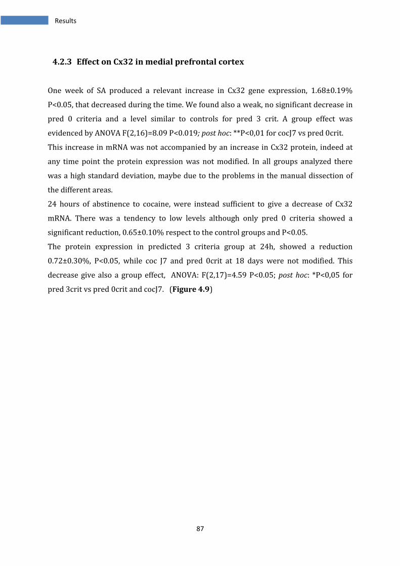
87
4.2.3 Effect on Cx32 in medial prefrontal cortex
One week of SA produced a relevant increase in Cx32 gene expression, 1.68±0.19%
P<0.05, that decreased during the time. We found also a weak, no significant decrease in
pred 0 criteria and a level similar to controls for pred 3 crit. A group effect was
evidenced by ANOVA F(2,16)=8.09 P<0.019; post hoc: **P<0,01 for cocJ7 vs pred 0crit.
This increase in mRNA was not accompanied by an increase in Cx32 protein, indeed at
any time point the protein expression was not modified. In all groups analyzed there
was a high standard deviation, maybe due to the problems in the manual dissection of
the different areas.
24 hours of abstinence to cocaine, were instead sufficient to give a decrease of Cx32
mRNA. There was a tendency to low levels although only pred 0 criteria showed a
significant reduction, 0.65±0.10% respect to the control groups and P<0.05.
The protein expression in predicted 3 criteria group at 24h, showed a reduction
0.72±0.30%, P<0.05, while coc J7 and pred 0crit at 18 days were not modified. This
decrease give also a group effect, ANOVA: F(2,17)=4.59 P<0.05; post hoc: *P<0,05 for
pred 3crit vs pred 0crit and cocJ7. (Figure 4.9)
Results
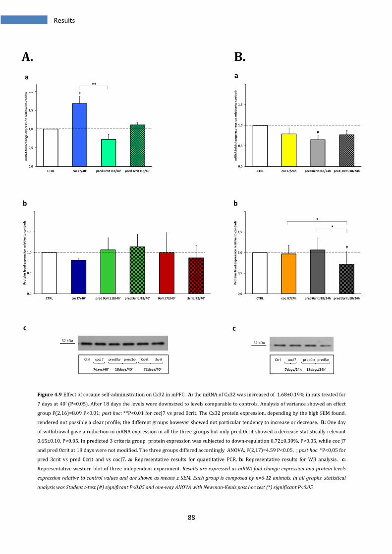
88
A. B.
Figure 4.9 Effect of cocaine self-administration on Cx32 in mPFC. A: the mRNA of Cx32 was increased of 1.68±0.19% in rats treated for
7 days at 40’ (P<0.05). After 18 days the levels were downsized to levels comparable to controls. Analysis of variance showed an effect
group F(2,16)=8.09 P<0.01; post hoc: **P<0,01 for cocJ7 vs pred 0crit. The Cx32 protein expression, depending by the high SEM found,
rendered not possible a clear profile; the different groups however showed not particular tendency to increase or decrease. B: One day
of withdrawal gave a reduction in mRNA expression in all the three groups but only pred 0crit showed a decrease statistically relevant
0.65±0.10, P<0.05. In predicted 3 criteria group protein expression was subjected to down-regulation 0.72±0.30%, P<0.05, while coc J7
and pred 0crit at 18 days were not modified. The three groups differed accordingly ANOVA, F(2,17)=4.59 P<0.05, ; post hoc: *P<0,05 for
pred 3crit vs pred 0crit and vs cocJ7. a: Representative results for quantitative PCR. b: Representative results for WB analysis. c:
Representative western blot of three independent experiment. Results are expressed as mRNA fold change expression and protein levels
expression relative to control values and are shown as means ± SEM. Each group is composed by n=6-12 animals. In all graphs, statistical
analysis was Student t-test (#) significant P<0.05 and one-way ANOVA with Newman-Keuls post hoc test (*) significant P<0.05.
0,0
0,5
1,0
1,5
2,0
CTRL coc J7/40' pred 0crit J18/40' pred 3crit J18/40'
mR
NA
fo
ld c
ha
ng
e e
xp
ressi
on
re
lati
ve
to
co
ntr
ols
**
#
0,0
0,5
1,0
1,5
2,0
CTRL coc J7/24h pred 0crit J18/24h pred 3crit J18/24h
mR
NA
fo
ld c
ha
nge
exp
ress
ion
re
lati
ve
to
co
ntr
ols
#
0,0
0,5
1,0
1,5
2,0
CTRL coc J7/40' pred 0crit J18/40' pred 3crit J18/40' 0crit J72/40' 3crit J72/40'
Pro
tein
s le
ve
l e
xp
ress
ion
re
lati
ve
to
co
ntr
ols
0,0
0,5
1,0
1,5
2,0
CTRL coc J7/24h pred 0crit J18/24h pred 3crit J18/24h
Pro
tein
s le
ve
l e
xp
ressi
on
re
lati
ve
to
co
ntr
ols
*
*
#
32 kDa
Ctrl cocJ7 pred0cr pred3cr 0crit 3crit
7days/40’ 18days/40’ 72days/40’
32 kDa
Ctrl cocJ7 pred0cr pred3cr
7days/24h 18days/24h’
a a
b b
c c
Results
89
4.2.4 Effect on Panx1 in medial prefrontal cortex
Panx1 mRNA expression, exhibited no differences both at 7 and 18 days of cocaine self-
administration in the animals with drug on board.
Instead the Panx1 protein showed several alterations; 7 days of exposure to cocaine
there were not changes but during the time, the levels were reduced. Only pred 0crit
exhibited an increase 1.36±0.33 P<0.05, while both pred 3 crit and the groups at 72 days
of SA showed a decrease in the protein levels: non-addict rats 0.74±0.25%, addicted
0.70±0.24%, for both P<0.05. This determined an effect groups revealed by analysis of
variance, ANOVA F(4,32)=11.83 P<0.001; post hoc: ***P<0,05 for pred 0crit vs pred
3crit, 0crit J72, 3crit J72; **P<0,01 for pred 0crit vs cocJ7; *P<0,05 for cocJ7 vs pred
3crit, 3crit J72.
The samples collected with 24 hours of abstinence, showed no modifications (with a
slight exception for cocJ7/24h group in which Panx1 mRNA was weakly, but
significantly, reduced 0.86±0.02% P<0.05 and this gave also a group effect to respect the
pred 0crit at 24h, F(2,26)=4.00 P<0.05; post hoc: *P<0,05 for cocJ7 vs pred 0crit.
The Cx32 protein was unchanged in all three groups of animals in which the mPFC area
was collected after 24 hours by the last cocaine injection. (Figure 4.10)
Results
90
A. B.
Figure 4.10 Effect of cocaine self-administration on Panx1 in mPFC. A: 7 or 18 days of cocaine SA had no effects on Panx1 gene
expression. All groups were unmodified respect the controls. Pannexin 1 protein was diminished during the time of cocaine use. In
predicted 0crit samples the protein levels were increased 1.36±0.33 P<0.05 After 72 days of SA, 0 crit animals had reduced levels
0.74±0.25 P<0.05%, such as the 3 criteria group 0.70±0.24%, for both P<0.05. The groups differed accordingly statistical analysis of
variance (ANOVA: F(4,32)=11.83 P<0.001; post hoc: ***P<0,05 for pred 0crit vs pred 3crit, 0crit J72, 3crit J72; **P<0,01 for pred 0crit vs
cocJ7; *P<0,05 for cocJ7 vs pred 3crit and vs 3crit J72. B: Samples analyzed at 24 hours by last cocaine infusion showed no relevant
modifications at any time point both for gene and protein expression. Unique exception was the slight effect of reduction in CocJ7 group
of 0.86±0.02% P<0.05 and a group effect revealed by ANOVA, F(2,26)=4.00 P<0.05; post hoc: *P<0,05 for cocJ7 vs pred 0crit. a:
Representative results for quantitative PCR. b: Representative results for WB analysis. c: Representative western blot of three
independent experiment. Results are expressed as mRNA fold change expression and protein levels expression relative to control values and
are shown as means ± SEM. Each group is composed by n=6-12 animals. In all graphs, statistical analysis was Student t-test (#) significant
P<0.05 and one-way ANOVA with Newman-Keuls post hoc test (*) significant P<0.05.
0,0
0,5
1,0
1,5
2,0
CTRL coc J7/40' pred 0crit J18/40' pred 3crit J18/40'
mR
NA
fo
ld c
ha
ng
e e
xp
ressi
on
re
lati
ve
to
co
ntr
ols
0,0
0,5
1,0
1,5
2,0
CTRL coc J7/24h pred 0crit J18/24h pred 3crit J18/24h
mR
NA
fo
ld c
ha
nge
exp
ress
ion
re
lati
ve
to
co
ntr
ols
*
#
0,0
0,5
1,0
1,5
2,0
2,5
CTRL coc J7/40' pred 0crit J18/40' pred 3crit J18/40' 0crit J72/40' 3crit J72/40'
Pro
tein
s le
ve
l e
xp
ress
ion
re
lati
ve
to
co
ntr
ols
***
***
***
*
*
**
#
#
#
0,0
0,5
1,0
1,5
2,0
CTRL coc J7/24h pred 0crit J18/24h pred 3crit J18/24h
Pro
tein
s le
ve
l e
xp
ressi
on
re
lati
ve
to
co
ntr
ols
48 kDa
Ctrl cocJ7 pred0cr pred3cr 0crit 3crit
7days/40’ 18days/40’ 72days/40’
48 kDa
Ctrl cocJ7 pred0cr pred3cr
7days/24h 18days/24h’
a a
b b
c c
Results
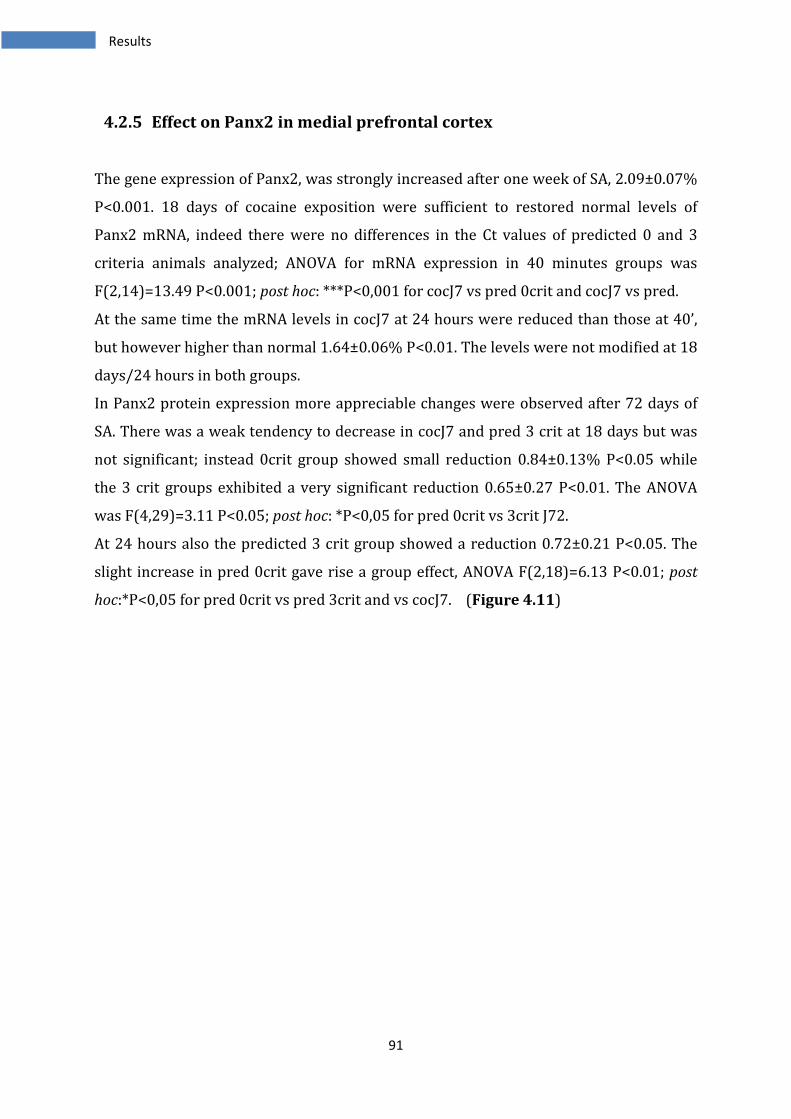
91
4.2.5 Effect on Panx2 in medial prefrontal cortex
The gene expression of Panx2, was strongly increased after one week of SA, 2.09±0.07%
P<0.001. 18 days of cocaine exposition were sufficient to restored normal levels of
Panx2 mRNA, indeed there were no differences in the Ct values of predicted 0 and 3
criteria animals analyzed; ANOVA for mRNA expression in 40 minutes groups was
F(2,14)=13.49 P<0.001; post hoc: ***P<0,001 for cocJ7 vs pred 0crit and cocJ7 vs pred.
At the same time the mRNA levels in cocJ7 at 24 hours were reduced than those at 40’,
but however higher than normal 1.64±0.06% P<0.01. The levels were not modified at 18
days/24 hours in both groups.
In Panx2 protein expression more appreciable changes were observed after 72 days of
SA. There was a weak tendency to decrease in cocJ7 and pred 3 crit at 18 days but was
not significant; instead 0crit group showed small reduction 0.84±0.13% P<0.05 while
the 3 crit groups exhibited a very significant reduction 0.65±0.27 P<0.01. The ANOVA
was F(4,29)=3.11 P<0.05; post hoc: *P<0,05 for pred 0crit vs 3crit J72.
At 24 hours also the predicted 3 crit group showed a reduction 0.72±0.21 P<0.05. The
slight increase in pred 0crit gave rise a group effect, ANOVA F(2,18)=6.13 P<0.01; post
hoc:*P<0,05 for pred 0crit vs pred 3crit and vs cocJ7. (Figure 4.11)
Results

92
A. B.
Figure 4.11 Effect of cocaine SA on Panx2 in mPFC. A: after 1 week of SA, group of cocJ7 showed an increase of 2.09±0.07% P<0.001
while 18 days caused no variations. The groups differed accordingly ANOVA, F(2,14)=13.49 P<0.001; post hoc: ***P<0,001 for cocJ7 vs
pred 0crit and cocJ7 vs pred 3crit. The protein levels were slightly, but not significantly, reduced at 7 days and in predicted vulnerable
animals. At 72 days of cocaine SA non-addict animals had a decrease in Panx2 protein of 0.84±0.13% P<0.05 while the animals showing
addiction-like behavior had higher reduction 0.65±0.27 P<0.01. The analysis of variance was F(4,29)=3.11 P<0.05, post hoc: *P<0,05 for
pred 0crit vs 3crit J72. B: one week of SA and 24h of abstinence presented still high levels 1.64±0.06% P<0.01, but proceeding with
cocaine assumption the levels were totally restored. In withdrawal animals a reduction in Panx2 protein was present in vulnerable to
addiction animals 0.65±0.27 P<0.01 and there was a group effect given by weak, not significant, increase in pred 0crit, ANOVA
F(2,18)=6.13 P<0.01; post hoc: *P<0,05 for pred 0crit vs pred 3crit and vs cocJ7. a: Representative results for quantitative PCR. b:
Representative results for WB analysis. c: Representative western blot of three independent experiment. Results are expressed as mRNA
fold change expression and protein levels expression relative to control values and are shown as means ± SEM. Each group is composed by
n=6-12 animals. In all graphs, statistical analysis was Student t-test (#) significant P<0.05 and one-way ANOVA with Newman-Keuls post hoc
test (*) significant P<0.05.
0,0
0,5
1,0
1,5
2,0
2,5
CTRL coc J7/40' pred 0crit J18/40' pred 3crit J18/40'
mR
NA
fo
ld c
ha
ng
e e
xp
ress
ion
re
lati
ve
to
co
ntr
ols
***
***
###
0,0
0,5
1,0
1,5
2,0
CTRL coc J7/24h pred 0crit J18/24h pred 3crit J18/24h
mR
NA
fo
ld c
ha
nge
exp
ress
ion
re
lati
ve
to
co
ntr
ols ##
0,0
0,5
1,0
1,5
2,0
CTRL coc J7/40' pred 0crit J18/40' pred 3crit J18/40' 0crit J72/40' 3crit J72/40'
Pro
tein
s le
ve
l e
xp
ress
ion
re
lati
ve
to
co
ntr
ols
*
###
0,0
0,5
1,0
1,5
2,0
CTRL coc J7/24h pred 0crit J18/24h pred 3crit J18/24h
Pro
tein
s le
ve
l e
xp
ressi
on
re
lati
ve
to
co
ntr
ols
* *
#
70 kDa
Ctrl cocJ7 pred0cr pred3cr 0crit 3crit
7days/40’ 18days/40’ 72days/40’
70 kDa
Ctrl cocJ7 pred0cr pred3cr
7days/24h 18days/24h’
a a
b b
c c
Results
93
4.2.6 Effect of cocaine SA on other genes in medial prefrontal cortex
In medial PFC the analysis of Cx26 gene during cocaine SA, showed a strong effect after
18 days and 40’ in the 0crit group, indeed in this last there was an increase of
2.36±0.54% P<0.05. In addition, a weak tendency to decrease, but not significant, was
found in cocJ7/40’ group. The three groups differed depending on analysis of ANOVA
F(2,17)=8.95 P<0.01; post hoc: **P<0,01 for cocJ7 vs pred 0crit and *P<0,05 for pred
0crit vs pred 3crit. In contrast all the three groups analyzed after 24 hours by the last
cocaine injection were decreased respect to the controls, cocJ7 0.68±0.08% P<0.05; pred
0crit 18J 0.62±0.12% P<0.05; pred 3crit 18J 0.46±0.12% P<0.05.
Analysis of Cx30 revealed no modifications in mRNA expression in all the analyzed
samples.
The identical situation was found in expression of Cx45 gene, in which only a weak
decrease in cocJ7/24hours, 0.80±0.05% P<0.01, gave a group effect in the animals
subjected to abstinence (ANOVA F(2,26)=4,93 P<0,05; post hoc: *P<0,05 for cocJ7 vs
pred 0crit and cocJ7 vs pred 3crit).
As in the NAc the Cx26 was undetectable in this region.
(Figure 4.12)
Results
94
A.
B.
C.
Figure 4.12 Effects of cocaine self-administration on other genes in mPFC. A: Analysis of Cx26 gene, showed a strong effect after 18 days
and 40’ in the 0crit group with an increase of 2.36±0.54% P<0.05. The three groups differed, depending on ANOVA F(2,17)=8.95 P<0.01;
post hoc: **P<0,01 for cocJ7 vs pred 0crit and *P<0,05 for pred 0crit vs pred 3crit. The three groups at 24 hours showed all a decrease,
cocJ7 0.68±0.08% P<0.05; pred 0crit 18J 0.62±0.12% P<0.05; pred 3crit 18J 0.46±0.12% P<0.05. B: Analysis of Cx30 revealed no
modifications in mRNA expression in all analyzed groups. C: Cx45 gene showed a slight decrease in cocJ7/24hours, 0.80±0.05% P<0.01; a
group effect was present in the groups at 24h (ANOVA F(2,26)=4.93 P<0.05; post hoc: *P<0,05 for cocJ7 vs pred 0crit and cocJ7 vs pred
3crit). a: Representative results for quantitative PCR at 40’. b: Representative results for quantitative PCR at 24h. Results are expressed as
mRNA fold change expression and protein levels expression relative to control values and are shown as means ± SEM. Each group is
composed by n=6-12 animals. In all graphs, statistical analysis was Student t-test (#) significant P<0.05 and one-way ANOVA with Newman-
Keuls post hoc test (*) significant P<0.05.
0,0
0,5
1,0
1,5
2,0
2,5
3,0
CTRL coc J7/40' pred 0crit J18/40' pred 3crit J18/40'
mR
NA
fo
ld c
ha
nge
exp
ressi
on
re
lati
ve
to
co
ntr
ols
***
#
0,0
0,5
1,0
1,5
2,0
CTRL coc J7/24h pred 0crit J18/24h pred 3crit J18/24h
mR
NA
fo
ld c
ha
nge
exp
ress
ion
re
lati
ve
to
co
ntr
ols
#
##
0,0
0,5
1,0
1,5
2,0
CTRL coc J7/40' pred 0crit J18/40' pred 3crit J18/40'
mR
NA
fo
ld c
ha
ng
e e
xp
ressi
on
re
lati
ve
to
co
ntr
ols
0,0
0,5
1,0
1,5
2,0
CTRL coc J7/24h pred 0crit J18/24h pred 3crit J18/24h
mR
NA
fo
ld c
ha
ng
e e
xp
ressi
on
re
lati
ve
to
co
ntr
ols
0,0
0,5
1,0
1,5
2,0
CTRL coc J7/40' pred 0crit J18/40' pred 3crit J18/40'
mR
NA
fo
ld c
ha
ng
e e
xp
ressi
on
re
lati
ve
to
co
ntr
ols
0,0
0,5
1,0
1,5
2,0
CTRL coc J7/24h pred 0crit J18/24h pred 3crit J18/24h
mR
NA
fo
ld c
ha
nge
exp
ress
ion
re
lati
ve
to
co
ntr
ols
*
*
##
a
a
a
b
b
b
Results
95
4.3 Effects of cocaine SA on ventral tegmental area gap junctions
The VTA is the first brain region involved in the cocaine effect. This in fact stimulates the
dopaminergic transmission and then the communications with the other parts of
mesocorticolimbic system.
4.3.1 Effect on Cx36 in ventral tegmental area
Cx36 mRNA expression was not influenced by cocaine SA at 7 or 18 days. Moreover also
with an abstinence of 24 hours there were no changes in gene expression.
Similarly, the protein was not influenced; there was only a low increase in pred 3crit
18J/40’ group 1.27±0.19% P<0.05
But nothing else variations in the other groups. (Figure 4.13)
Results
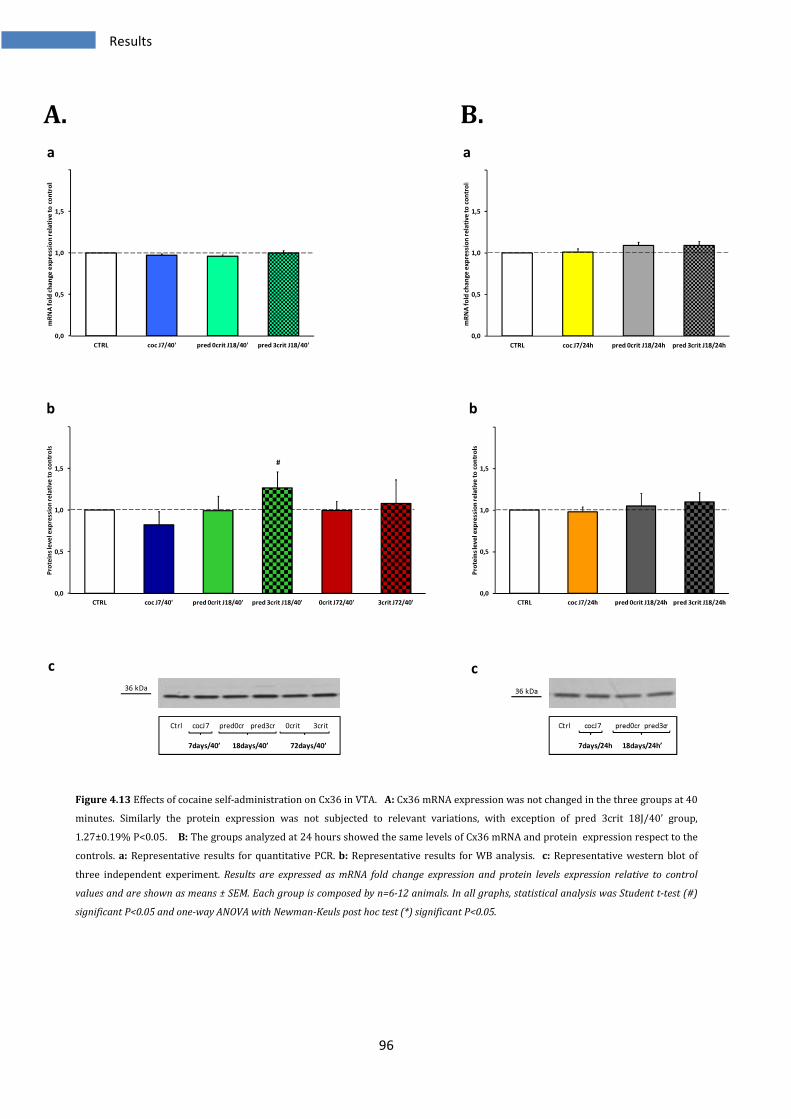
96
A. B.
Figure 4.13 Effects of cocaine self-administration on Cx36 in VTA. A: Cx36 mRNA expression was not changed in the three groups at 40
minutes. Similarly the protein expression was not subjected to relevant variations, with exception of pred 3crit 18J/40’ group,
1.27±0.19% P<0.05. B: The groups analyzed at 24 hours showed the same levels of Cx36 mRNA and protein expression respect to the
controls. a: Representative results for quantitative PCR. b: Representative results for WB analysis. c: Representative western blot of
three independent experiment. Results are expressed as mRNA fold change expression and protein levels expression relative to control
values and are shown as means ± SEM. Each group is composed by n=6-12 animals. In all graphs, statistical analysis was Student t-test (#)
significant P<0.05 and one-way ANOVA with Newman-Keuls post hoc test (*) significant P<0.05.
0,0
0,5
1,0
1,5
2,0
CTRL coc J7/40' pred 0crit J18/40' pred 3crit J18/40'
mR
NA
fo
ld c
ha
nge
exp
ress
ion
re
lati
ve
to
co
ntr
ols
0,0
0,5
1,0
1,5
2,0
CTRL coc J7/24h pred 0crit J18/24h pred 3crit J18/24h
mR
NA
fo
ld c
ha
ng
e e
xp
ress
ion
re
lati
ve
to
co
ntr
ols
0,0
0,5
1,0
1,5
2,0
CTRL coc J7/40' pred 0crit J18/40' pred 3crit J18/40' 0crit J72/40' 3crit J72/40'
Pro
tein
s le
ve
l e
xp
ress
ion
re
lati
ve
to
co
ntr
ols
#
0,0
0,5
1,0
1,5
2,0
CTRL coc J7/24h pred 0crit J18/24h pred 3crit J18/24h
Pro
tein
s le
ve
l e
xp
ressi
on
re
lati
ve
to
co
ntr
ols
36 kDa
Ctrl cocJ7 pred0cr pred3cr 0crit 3crit
7days/40’ 18days/40’ 72days/40’
36 kDa
Ctrl cocJ7 pred0cr pred3cr
7days/24h 18days/24h’
a a
b b
c c
Results
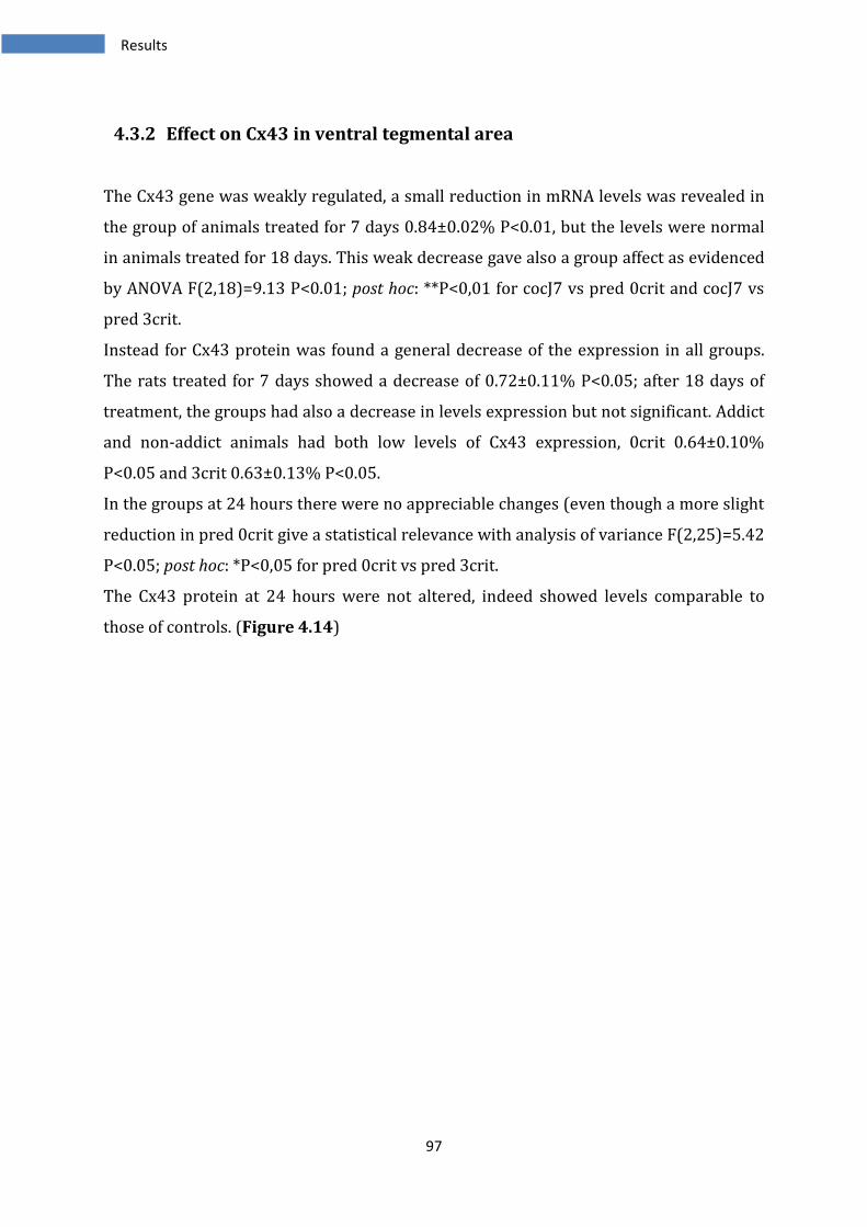
97
4.3.2 Effect on Cx43 in ventral tegmental area
The Cx43 gene was weakly regulated, a small reduction in mRNA levels was revealed in
the group of animals treated for 7 days 0.84±0.02% P<0.01, but the levels were normal
in animals treated for 18 days. This weak decrease gave also a group affect as evidenced
by ANOVA F(2,18)=9.13 P<0.01; post hoc: **P<0,01 for cocJ7 vs pred 0crit and cocJ7 vs
pred 3crit.
Instead for Cx43 protein was found a general decrease of the expression in all groups.
The rats treated for 7 days showed a decrease of 0.72±0.11% P<0.05; after 18 days of
treatment, the groups had also a decrease in levels expression but not significant. Addict
and non-addict animals had both low levels of Cx43 expression, 0crit 0.64±0.10%
P<0.05 and 3crit 0.63±0.13% P<0.05.
In the groups at 24 hours there were no appreciable changes (even though a more slight
reduction in pred 0crit give a statistical relevance with analysis of variance F(2,25)=5.42
P<0.05; post hoc: *P<0,05 for pred 0crit vs pred 3crit.
The Cx43 protein at 24 hours were not altered, indeed showed levels comparable to
those of controls. (Figure 4.14)
Results
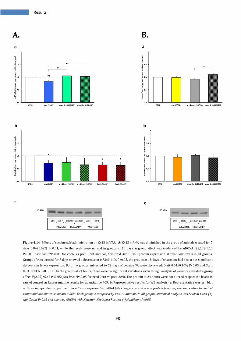
98
A. B.
Figure 4.14 Effects of cocaine self-administration on Cx43 in VTA. A: Cx43 mRNA was diminished in the group of animals treated for 7
days 0.84±0.02% P<0.01, while the levels were normal in groups at 18 days. A group affect was evidenced by ANOVA F(2,18)=9.13
P<0.01; post hoc: **P<0,01 for cocJ7 vs pred 0crit and cocJ7 vs pred 3crit. Cx43 protein expression showed low levels in all groups.
Groups of rats treated for 7 days showed a decrease of 0.72±0.11% P<0.05, the groups at 18 days of treatment had also a not significant
decrease in levels expression. Both the groups subjected to 72 days of cocaine SA were decreased, 0crit 0.64±0.10% P<0.05 and 3crit
0.63±0.13% P<0.05. B: In the groups at 24 hours, there were no significant variations, even though analysis of variance revealed a group
effect, F(2,25)=5.42 P<0.05; post hoc: *P<0,05 for pred 0crit vs pred 3crit. The protein at 24 hours were not altered respect the levels in
rats of control. a: Representative results for quantitative PCR. b: Representative results for WB analysis. c: Representative western blot
of three independent experiment. Results are expressed as mRNA fold change expression and protein levels expression relative to control
values and are shown as means ± SEM. Each group is composed by n=6-12 animals. In all graphs, statistical analysis was Student t-test (#)
significant P<0.05 and one-way ANOVA with Newman-Keuls post hoc test (*) significant P<0.05.
0,0
0,5
1,0
1,5
2,0
CTRL coc J7/40' pred 0crit J18/40' pred 3crit J18/40'
mR
NA
fo
ld c
ha
nge
exp
ress
ion
re
lati
ve
to
co
ntr
ols
**
**
##
0,0
0,5
1,0
1,5
2,0
CTRL coc J7/24h pred 0crit J18/24h pred 3crit J18/24h
mR
NA
fo
ld c
ha
nge
ex
pre
ssio
n r
ela
tive
to
co
ntr
ols
*
0,0
0,5
1,0
1,5
2,0
CTRL coc J7/40' pred 0crit J18/40' pred 3crit J18/40' 0crit J72/40' 3crit J72/40'
Pro
tein
s le
ve
l e
xp
ress
ion
re
lati
ve
to
co
ntr
ols
#
# #
0,0
0,5
1,0
1,5
2,0
CTRL coc J7/24h pred 0crit J18/24h pred 3crit J18/24h
Pro
tein
s le
ve
l e
xp
ressi
on
re
lati
ve
to
co
ntr
ols
43 kDa
Ctrl cocJ7 pred0cr pred3cr 0crit 3crit
7days/40’ 18days/40’ 72days/40’
43 kDa
Ctrl cocJ7 pred0cr pred3cr
7days/24h 18days/24h’
a a
b b
c c
Results
99
4.3.3 Effect on Cx32 in ventral tegmental area
Cx32 gene expression was not modified in all the groups analyzed. All the Ct values of
several samples were completely comparable to the Ct values of the controls. It was
evident that the drug assumption had no effects on this gene, either after 40’ than after
24 hours of abstinence. Interestingly the protein showed instead a different regulation,
Initially cocaine diminished the expression of Cx32, indeed cocJ7 at 40 minutes had
0.74±0.12 P<0.05, but following a more long exposure there was a difference; the
resistant to addiction group of animals had normal levels of Cx32 protein but the group
of vulnerable animals showed a significant increase 1.63±0.37% P<0.05. At 72 days of
cocaine exposition both the groups presented the same levels of protein than control
levels. The five groups differed accordingly analysis of variance F(4,30)=6.42 P<0.001;
post hoc: ***P<0,001 for pred 3crit J18 vs pred ocrit and vs cocJ7; **P<0,01 for pred 3crit
vs 0crit and vs 3crit.
At 24 hours the levels of the Cx32 protein were normal in all groups. (Figure 4.15)
Results
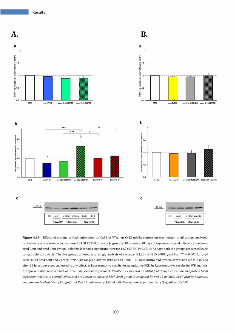
100
A. B.
Figure 4.15 Effects of cocaine self-administration on Cx32 in VTA. A: Cx32 mRNA expression was normal in all groups analyzed.
Protein expression revealed a decrease 0.74±0.12 P<0.05 in cocJ7 group at 40 minutes; 18 days of exposure showed differences between
pred 0crit and pred 3crit groups, only this last had a significant increase 1.63±0.37% P<0.05. At 72 days both the groups presented levels
comparable to controls. The five groups differed accordingly analysis of variance F(4,30)=6.42 P<0.001; post hoc: ***P<0,001 for pred
3crit J18 vs pred ocrit and vs cocJ7; **P<0,01 for pred 3crit vs 0crit and vs 3crit. B: Both mRNA and protein expression of Cx32 in VTA
after 24 hours were not subjected to any effect. a: Representative results for quantitative PCR. b: Representative results for WB analysis.
c: Representative western blot of three independent experiment. Results are expressed as mRNA fold change expression and protein levels
expression relative to control values and are shown as means ± SEM. Each group is composed by n=6-12 animals. In all graphs, statistical
analysis was Student t-test (#) significant P<0.05 and one-way ANOVA with Newman-Keuls post hoc test (*) significant P<0.05.
0,0
0,5
1,0
1,5
2,0
CTRL coc J7/40' pred 0crit J18/40' pred 3crit J18/40'
mR
NA
fo
ld c
ha
nge
exp
ress
ion
re
lati
ve
to
co
ntr
ols
0,0
0,5
1,0
1,5
2,0
CTRL coc J7/24h pred 0crit J18/24h pred 3crit J18/24h
mR
NA
fo
ld c
ha
nge
exp
ress
ion
re
lati
ve
to
co
ntr
ols
0,0
0,5
1,0
1,5
2,0
2,5
CTRL coc J7/40' pred 0crit J18/40' pred 3crit J18/40' 0crit J72/40' 3crit J72/40'
Pro
tein
s le
ve
l e
xp
ressio
n r
ela
tive
to
co
ntr
ols
#
#
**
**
***
***
0,0
0,5
1,0
1,5
2,0
CTRL coc J7/24h pred 0crit J18/24h pred 3crit J18/24h
Pro
tein
s le
ve
l e
xp
ressi
on
re
lati
ve
to
co
ntr
ols
32 kDa
Ctrl cocJ7 pred0cr pred3cr 0crit 3crit
7days/40’ 18days/40’ 72days/40’
32 kDa
Ctrl cocJ7 pred0cr pred3cr
7days/24h 18days/24h’
a a
b b
c c
Results
101
4.3.4 Effect on Panx1 in ventral tegmental area
Panx1 mRNA was not modified in rats analyzed with drug on board while with 24 hours
of abstinence we assisted to a small decrease only in cocJ7 group 0.87±0.03% P<0.05
and a modest increase in vulnerable group at 18 days 1.29±0.08% P<0.05. ANOVA for
these groups was F(2,25)=8.13 P<0.01; post hoc: **P<0,01 for cocJ7 vs pred 3crit and
*P<0,05 for pred 0crit vs pred 3crit.
The protein at 40 minutes was not modified, but seemed to be a general tendency to a
diminution, as observed also in Cx43 protein, but only the group of cocJ7 showed a
significant decrease 0.71±0.14 P<0.05. As for this protein, 24 hours of abstinence did not
modify the levels or restored the slight differences observed in proteins at 40 minutes.
(Figure 4.1)
Results
102
A. B.
Figure 4.16 Effects of cocaine self-administration on Panx1 in VTA. A: Panx1 mRNA levels were no modified in regions analyzed after 40
minutes. The protein at 40 minutes showed a general tendency to a decrease but only cocJ7 group showed a significant decrease
0.71±0.14 P<0.05. B: 24 hours of abstinence caused a weak decrease in cocJ7 group of 0.87±0.03% P<0.05 and a modest increase
in vulnerable group at 18 days 1.29±0.08%. The groups analyzed differed according to ANOVA F(2,25)=8.13 P<0.01; post hoc: **P<0,01
for cocJ7 vs pred 3crit and *P<0,05 for pred 0crit vs pred 3crit. Protein levels expression measured at 24 hours were no altered. a:
Representative results for quantitative PCR. b: Representative results for WB analysis. c: Representative western blot of three
independent experiment. Results are expressed as mRNA fold change expression and protein levels expression relative to control values and
are shown as means ± SEM. Each group is composed by n=6-12 animals. In all graphs, statistical analysis was Student t-test (#) significant
P<0.05 and one-way ANOVA with Newman-Keuls post hoc test (*) significant P<0.05.
0,0
0,5
1,0
1,5
2,0
CTRL coc J7/40' pred 0crit J18/40' pred 3crit J18/40'
mR
NA
fo
ld c
ha
nge
exp
ress
ion
re
lati
ve
to
co
ntr
ols
0,0
0,5
1,0
1,5
2,0
CTRL coc J7/24h pred 0crit J18/24h pred 3crit J18/24h
mR
NA
fo
ld c
ha
nge
ex
pre
ssio
n r
ela
tive
to
co
ntr
ols
*
**
#
#
0,0
0,5
1,0
1,5
2,0
CTRL coc J7/40' pred 0crit J18/40' pred 3crit J18/40' 0crit J72/40' 3crit J72/40'
Pro
tein
s le
ve
l e
xp
ressi
on
re
lati
ve
to
co
ntr
ols
#
0,0
0,5
1,0
1,5
2,0
CTRL coc J7/24h pred 0crit J18/24h pred 3crit J18/24h
Pro
tein
s le
ve
l e
xp
ressi
on
re
lati
ve
to
co
ntr
ols
48 kDa
Ctrl cocJ7 pred0cr pred3cr 0crit 3crit
7days/40’ 18days/40’ 72days/40’
48 kDa
Ctrl cocJ7 pred0cr pred3cr
7days/24h 18days/24h’
a a
b b
c c
Results
103
4.3.5 Effect on Panx2 in ventral tegmental area
Even in this structure, the profile for Panx2 mRNA was exactly the same of that
evidenced in nucleus accumbens and medial prefrontal cortex.
There was a huge increase at the first week of treatment 2.46±0.02% P<0.001 that went
back to normal levels with 18 days of SA. The groups differed accordingly ANOVA
F(2,13)=10,02 P<0,01; post hoc: **P<0,01 for cocJ7 vs pred 0crit and cocJ7 vs pred 3crit
24 hours of no-cocaine administration allowed to Panx2 mRNA to restore levels, indeed
cocJ7/24h was increased of 1.42±0.06% P<0.05 while groups at 18 days had levels
comparable to control group. Despite this strong increase in mRNA expression, the
Panx2 protein was not subjected to the same regulation; indeed the analysis of the
protein revealed no modifications at any time point analyzed. (Figure 4.17)
Results
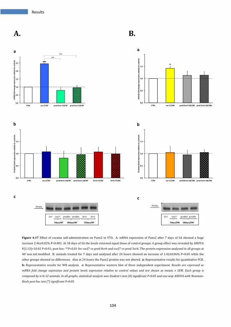
104
A. B.
Figure 4.17 Effect of cocaine self-administration on Panx2 in VTA. A: mRNA expression of Panx2 after 7 days of SA showed a huge
increase 2.46±0.02% P<0.001. At 18 days of SA the levels returned equal those of control groups. A group effect was revealed by ANOVA
F(2,13)=10.02 P<0.01; post hoc: **P<0,01 for cocJ7 vs pred 0crit and cocJ7 vs pred 3crit. The protein expression analyzed in all groups at
40’ was not modified. B: animals treated for 7 days and analyzed after 24 hours showed an increase of 1.42±0.06% P<0.05 while the
other groups showed no differences. Also at 24 hours the Panx2 protein was not altered. a: Representative results for quantitative PCR.
b: Representative results for WB analysis. c: Representative western blot of three independent experiment. Results are expressed as
mRNA fold change expression and protein levels expression relative to control values and are shown as means ± SEM. Each group is
composed by n=6-12 animals. In all graphs, statistical analysis was Student t-test (#) significant P<0.05 and one-way ANOVA with Newman-
Keuls post hoc test (*) significant P<0.05.
0,0
0,5
1,0
1,5
2,0
2,5
CTRL coc J7/40' pred 0crit J18/40' pred 3crit J18/40'
mR
NA
fo
ld c
ha
nge
ex
pre
ssio
n r
ela
tive
to
co
ntr
ols
**
**
###
0,0
0,5
1,0
1,5
2,0
CTRL coc J7/24h pred 0crit J18/24h pred 3crit J18/24h
mR
NA
fo
ld c
ha
nge
exp
ress
ion
re
lati
ve
to
co
ntr
ols
#
0,0
0,5
1,0
1,5
2,0
CTRL coc J7/40' pred 0crit J18/40' pred 3crit J18/40' 0crit J72/40' 3crit J72/40'
Pro
tein
s le
ve
l e
xp
ress
ion
re
lati
ve
to
co
ntr
ols
0,0
0,5
1,0
1,5
2,0
CTRL coc J7/24h pred 0crit J18/24h pred 3crit J18/24h
Pro
tein
s le
ve
l e
xp
ressi
on
re
lati
ve
to
co
ntr
ols
70 kDa
Ctrl cocJ7 pred0cr pred3cr 0crit 3crit
7days/40’ 18days/40’ 72days/40’
70 kDa
Ctrl cocJ7 pred0cr pred3cr
7days/24h 18days/24h’
a a
b b
c c
Results
105
4.3.6 Effects of cocaine SA on other genes in ventral tegmental area
In VTA, equally to the other two structures analyzed, with western-blot technique was
not possible reveal the Cx26 protein.
Analysis with real-time PCR evidenced a decrease in mRNA expression of Cx26 in cocJ7
group analyzed at 40 minutes of 0.71±0.03 P<0.05, while at 18 days the levels were
restored equal to controls mRNA levels. An effect group was present, indeed ANOVA
gave F(2,17)=9.25 P<0.01; post hoc: **P<0,01 for coc J7 vs pred 0crit and cocJ7 vs pred
3crit. At 24 hours there were no relevant modifications but a slight increase in
predicted 3crit gave a group effect , ANOVA F(2,25)=4.43 P<0.05; post hoc: *P<0,05 for
cocJ7 vs pred 3crit.
Cx30 mRNA expression exhibited no differences in the groups at 40’. A weak increase
was present in pred 3crit group at 24 hours 1.19±0.04 P<0.05 and analysis of variance
between the three groups revealed an effect group F(2,25)=6.15 P<0.01; post hoc:
**P<0,01 for cocJ7 vs pred 3crit and *P<0,05 for cocJ7 vs pred 3crit.
Also Cx45 gene showed no relevant alterations. A slight increase in both groups treated
for 18 days and sacrificed at 40 minutes evidenced a group effect if compared to groups
of animals treated for 7 days ANOVA F(2,18)=4.46 P<0.05; post hoc: *P<0,05 for coc J7 vs
pred 0crit and cocJ7 vs pred 3crit. Instead at 24 h was found only a slight, but significant
increase in pred 3crit group 1.19±0.07 P<0.05. (Figure 4.18)
Results
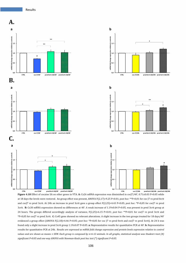
106
A.
B.
C.
Figure 4.18 Effect of cocaine SA on other genes in VTA. A: Cx26 mRNA expression was diminished in cocJ7/40’, 0.71±0.03 P<0.05 while
at 18 days the levels were restored. An group effect was present, ANOVA F(2,17)=9.25 P<0.01; post hoc: **P<0,01 for coc J7 vs pred 0crit
and cocJ7 vs pred 3crit. At 24h an increase in pred 3crit gave a group effect F(2,25)=4.43 P<0.05; post hoc: *P<0,05 for cocJ7 vs pred
3crit. B: Cx30 mRNA expression showed no differences at 40’. A weak increase of 1.19±0.04 P<0.05, was present in pred 3crit group at
24 hours. The groups differed accordingly analysis of variance, F(2,25)=6.15 P<0.01; post hoc: **P<0,01 for cocJ7 vs pred 3crit and
*P<0,05 for cocJ7 vs pred 3crit. C: Cx45 gene showed no relevant alterations. A slight increase in the two groups treated for 18 days/40’
evidenced a group effect (ANOVA F(2,18)=4.46 P<0.05; post hoc: *P<0,05 for coc J7 vs pred 0crit and cocJ7 vs pred 3crit). At 24 h was
found only a slight increase in pred 3crit group 1.19±0.07 P<0.05. a: Representative results for quantitative PCR at 40’. b: Representative
results for quantitative PCR at 24h. Results are expressed as mRNA fold change expression and protein levels expression relative to control
values and are shown as means ± SEM. Each group is composed by n=6-12 animals. In all graphs, statistical analysis was Student t-test (#)
significant P<0.05 and one-way ANOVA with Newman-Keuls post hoc test (*) significant P<0.05.
0,0
0,5
1,0
1,5
2,0
CTRL coc J7/40' pred 0crit J18/40' pred 3crit J18/40'
mR
NA
fo
ld c
ha
ng
e e
xp
ressi
on
re
lati
ve
to
co
ntr
ols
**
**
#
0,0
0,5
1,0
1,5
2,0
CTRL coc J7/24h pred 0crit J18/24h pred 3crit J18/24h
mR
NA
fo
ld c
ha
ng
e e
xp
ressi
on
re
lati
ve
to
co
ntr
ols
*
0,0
0,5
1,0
1,5
2,0
CTRL coc J7/40' pred 0crit J18/40' pred 3crit J18/40'
mR
NA
fo
ld c
ha
ng
e e
xp
ress
ion
re
lati
ve
to
co
ntr
ols
0,0
0,5
1,0
1,5
2,0
CTRL coc J7/24h pred 0crit J18/24h pred 3crit J18/24h
mR
NA
fo
ld c
ha
ng
e e
xp
ress
ion
re
lati
ve
to
co
ntr
ols
*
**
#
0,0
0,5
1,0
1,5
2,0
CTRL coc J7/40' pred 0crit J18/40' pred 3crit J18/40'
mR
NA
fo
ld c
ha
nge
exp
ress
ion
re
lati
ve
to
co
ntr
ols
*
*
0,0
0,5
1,0
1,5
2,0
CTRL coc J7/24h pred 0crit J18/24h pred 3crit J18/24h
mR
NA
fo
ld c
ha
nge
exp
ress
ion
re
lati
ve
to
co
ntr
ols
#
a
a
a
b
b
b
Results
107
4.4 Summary of relevant modifications in NAc gap junctions
Nucleus accumbens is the regions mostly involved in the rewarding and reinforcing
effects of cocaine following the activation of dopaminergic system in VTA. After acute
cocaine administration there is a huge increase of DA in NAc shell but blocking the
administration, the DA levels return to basal levels. With a prolonged exposure to
cocaine, there is instead depletion of DA and glutamate. In NAc has been demonstrated
the presence of gap junctions and their regulation by dopamine, depending by DA
receptorial types and area interested. We analyzed the whole NAc and we observed a
decreased expression of the principal connexins and pannexins, indeed Cx43, Cx32 and
Panx2 at 7 and 18 days of cocaine SA showed low protein expression levels respect to
the animal controls. But with a prolonged exposure the differences in expression of
connexins and pannexins, between addict and non-addict animals became more
pronounced. Indeed, we observed for Cx36 that just to 3 weeks of SA did not affect the
protein expression, but with 72 days of SA there were increased levels only in 3 criteria
animals while non-addicted animals had normal levels of this protein.
Cx32 and Panx2 exhibited the same profile; after a general decrease in the expression at
18 days, in which both groups vulnerable and resistant to addiction rats, exhibited the
same diminution in the protein levels, 72 days of cocaine exposition allowed to 0 criteria
animals restored normal protein levels while in addicted rats region there were raised
levels. The Cx43 showed instead low levels in addicted animals, in accord to those
observed at 18 days of SA but interestingly also in this case was possible to observe a
difference with non addict animals that restored protein levels comparable to controls.
At 24 hours of withdrawal, instead we observed a tendency to restore normal protein
levels to respect the changes observed at 40 minutes. An exception was present in Cx36
protein expression, in which we observed an increase, although weak, in both predicted
0 and 3 criteria at 18 days of SA. Low levels were already stable in vulnerable animals
for Cx43 at 18 days. In contrast, for Panx2 we found increased protein levels only in
0criteria at 18 days/24 hours. (Table 4.1)
Results
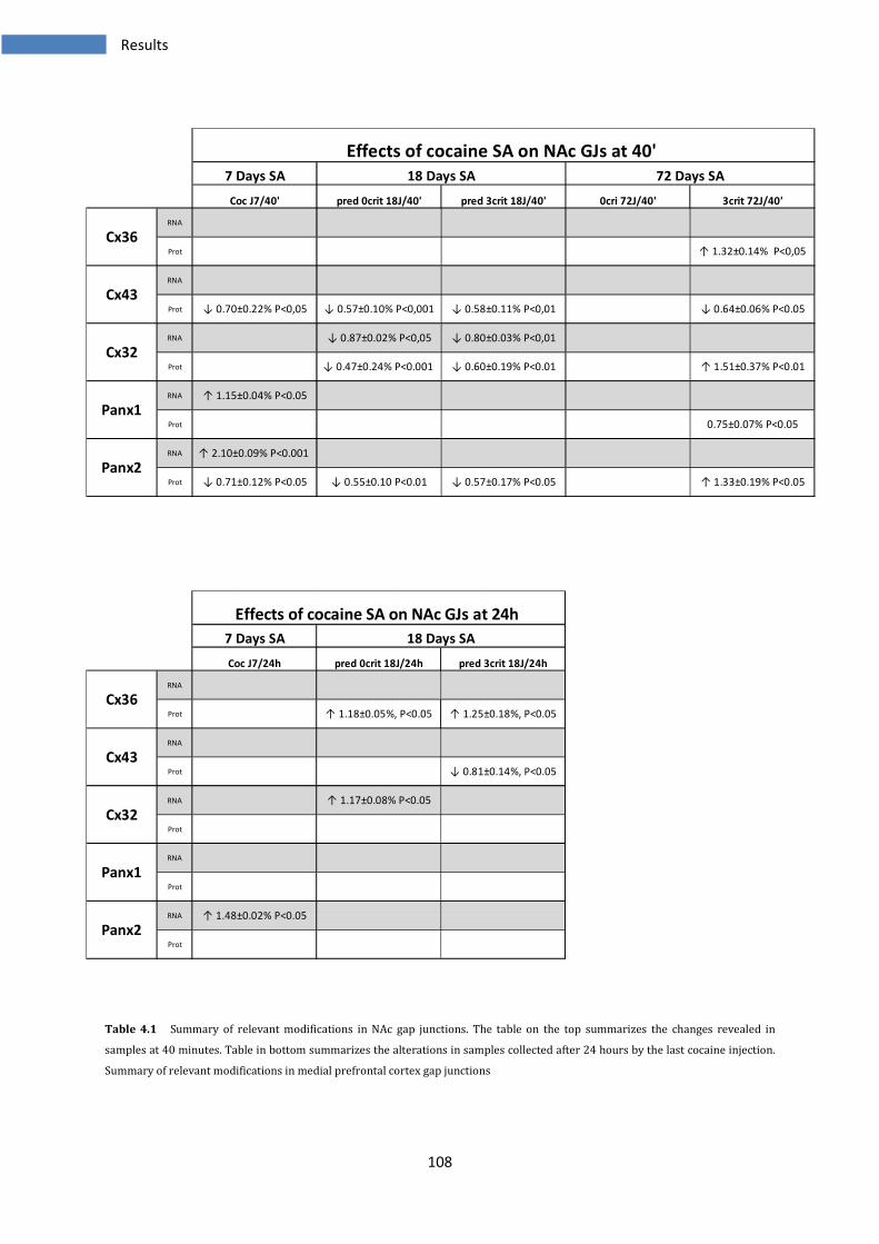
108
Table 4.1 Summary of relevant modifications in NAc gap junctions. The table on the top summarizes the changes revealed in
samples at 40 minutes. Table in bottom summarizes the alterations in samples collected after 24 hours by the last cocaine injection.
Summary of relevant modifications in medial prefrontal cortex gap junctions
Effects of cocaine SA on NAc GJs at 40'
↓ 0.58±0.11% P<0,01 ↓ 0.64±0.06% P<0.05
↑ 1.32±0.14% P<0,05
72 Days SA
↓ 0.57±0.17% P<0.05
↓ 0.60±0.19% P<0.01
↓ 0.80±0.03% P<0,01
↑ 1.51±0.37% P<0.01
↑ 1.33±0.19% P<0.05
0.75±0.07% P<0.05
↓ 0.70±0.22% P<0,05
↓ 0.55±0.10 P<0.01
↓ 0.47±0.24% P<0.001
↓ 0.87±0.02% P<0,05
↓ 0.57±0.10% P<0,001
↓ 0.71±0.12% P<0.05
↑ 2.10±0.09% P<0.001
↑ 1.15±0.04% P<0.05
RNA
Prot
RNA
Prot
RNA
pred 0crit 18J/40' pred 3crit 18J/40' 0cri 72J/40' 3crit 72J/40'
7 Days SA
Coc J7/40'
18 Days SA
Prot
RNA
Prot
RNA
Prot
Cx36
Cx43
Cx32
Panx1
Panx2
Effects of cocaine SA on NAc GJs at 24h
Prot
Panx2
RNA
Prot
Panx1
RNA
Prot
Cx32
RNA
Prot
Cx43
RNA
7 Days SA 18 Days SA
Coc J7/24h pred 0crit 18J/24h pred 3crit 18J/24h
Prot
Cx36
RNA
↑ 1.48±0.02% P<0.05
↑ 1.17±0.08% P<0.05
↓ 0.81±0.14%, P<0.05
↑ 1.25±0.18%, P<0.05↑ 1.18±0.05%, P<0.05
Results
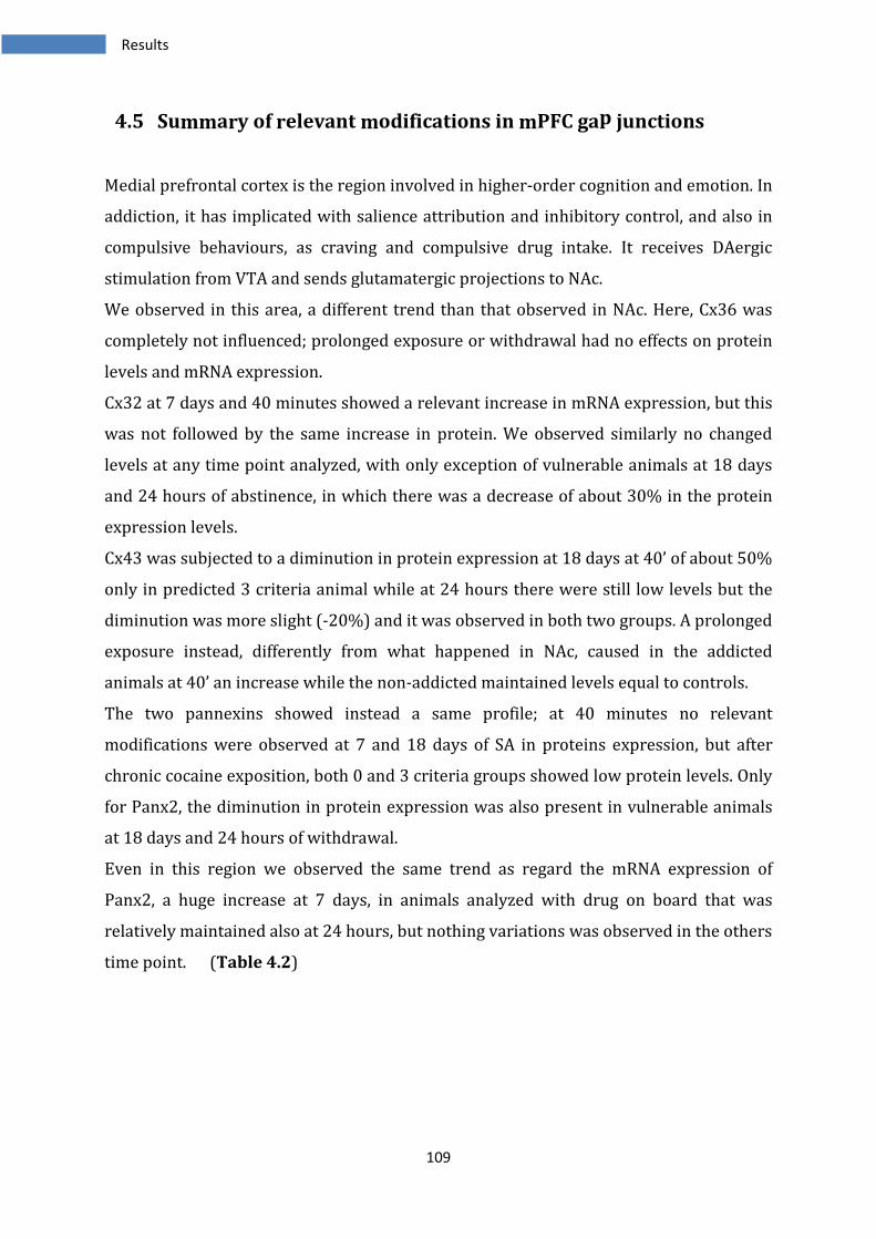
109
4.5 Summary of relevant modifications in mPFC gap junctions
Medial prefrontal cortex is the region involved in higher-order cognition and emotion. In
addiction, it has implicated with salience attribution and inhibitory control, and also in
compulsive behaviours, as craving and compulsive drug intake. It receives DAergic
stimulation from VTA and sends glutamatergic projections to NAc.
We observed in this area, a different trend than that observed in NAc. Here, Cx36 was
completely not influenced; prolonged exposure or withdrawal had no effects on protein
levels and mRNA expression.
Cx32 at 7 days and 40 minutes showed a relevant increase in mRNA expression, but this
was not followed by the same increase in protein. We observed similarly no changed
levels at any time point analyzed, with only exception of vulnerable animals at 18 days
and 24 hours of abstinence, in which there was a decrease of about 30% in the protein
expression levels.
Cx43 was subjected to a diminution in protein expression at 18 days at 40’ of about 50%
only in predicted 3 criteria animal while at 24 hours there were still low levels but the
diminution was more slight (-20%) and it was observed in both two groups. A prolonged
exposure instead, differently from what happened in NAc, caused in the addicted
animals at 40’ an increase while the non-addicted maintained levels equal to controls.
The two pannexins showed instead a same profile; at 40 minutes no relevant
modifications were observed at 7 and 18 days of SA in proteins expression, but after
chronic cocaine exposition, both 0 and 3 criteria groups showed low protein levels. Only
for Panx2, the diminution in protein expression was also present in vulnerable animals
at 18 days and 24 hours of withdrawal.
Even in this region we observed the same trend as regard the mRNA expression of
Panx2, a huge increase at 7 days, in animals analyzed with drug on board that was
relatively maintained also at 24 hours, but nothing variations was observed in the others
time point. (Table 4.2)
Results
110
Table 4.2 Summary of relevant modifications in mPFC gap junctions. The table on the top summarizes the changes revealed in
samples at 40 minutes. Table in bottom summarizes the alterations in samples collected after 24 hours by the last cocaine injection.
↓ 0.65±0.27 P<0.01
Panx2
RNA ↑ 2.09±0.07% P<0.001
Prot ↓ 0.84±0.13% P<0.05
↓ 0.70±0.24% P<0.05
Panx1
RNA
Prot ↑ 1.36±0.33 P<0.05 ↓ 0.74±0.25% P<0.05
Cx32
RNA ↑ 1.68±0.19% P<0.05
Prot
Cx36
Prot ↓ 0.54±0.31% P<0.05 ↑ 1.40±0.34% P<0.01
pred 0crit 18J/40' pred 3crit 18J/40' 0cri 72J/40' 3crit 72J/40'
Cx43
RNA
Effects of cocaine SA on mPFC GJs at 40'
7 Days SA 18 Days SA 72 Days SA
Prot
RNA
Coc J7/40'
Prot ↓ 0.72±0.21 P<0.05
Panx2
RNA ↑ 1.64±0.06% P<0.01
Prot
Panx1
RNA ↓ 0.86±0.02% P<0.05
Prot ↓ 0.72±0.30% P<0.05
Cx32
RNA ↓ 0.65±0.10% P<0.05
Prot ↓ 0.80±0.22% P<0.05 ↓ 0.80±0.17% P<0.05
Prot
Cx43
RNA
Coc J7/24h
RNA
Effects of cocaine SA on mPFC GJs at 24h
7 Days SA 18 Days SA
pred 0crit 18J/24h pred 3crit 18J/24h
Cx36
Results
111
4.6 Summary of relevant modifications in VTA gap junctions
Ventral tegmental area is a part of midbrain constituted by 60% of DAergic neurons;
inhibitory GABAergic and excitatory Glutamatergic neurons regulate these. This is the
first region to be affected by cocaine; in VTA cocaine causes increase of dopamine and
the consequent increased Daergic transmission.
In this area, we observed the smallest effects in connexins and pannexins after cocaine
self-administration.
For first however, Panx2 mRNA showed identical profile to that observed in accumbens
and prefrontal cortex. As in NAc, the first week of cocaine treatment caused a decrease
in the proteins expression of Cx43, Cx32 and Panx1.
The more evident changes were observed in Cx43 and Cx32 protein, analyzed after 40
minutes by last cocaine injection. Cx32 was slightly diminished at the first week of
treatment but intriguingly a relevant increase was evident in predicted 3 criteria at 18
days of SA, but nothing else variations was seen with prolonged exposure in all groups
analyzed. The Cx43 showed a general decrease in protein expression at any time point
analyzed and moreover was not possible discriminate between addicted and non-
addicted animals at 72 days because was present the same level of reduction in protein
expression. Others changes were revealed as weak increase in Cx36 only in predicted 3
criteria at 18 days/40’ or increase in Panx2 in the same group but analyzed at 24 hours.
(Table 4.3)
Results

112
Table 4.3 Summary of relevant modifications in VTA gap junctions. The table on the top summarizes the changes revealed in
samples at 40 minutes. Table in bottom summarizes the alterations in samples collected after 24 hours by the last cocaine injection.
Panx2
RNA ↑ 2.46±0.02% P<0.001
Prot
Panx1
RNA
Prot ↓ 0.71±0.14 P<0.05
Cx32
RNA
Prot ↓ 0.74±0.12 P<0.05 ↑ 1.63±0.37% P<0.05
Cx36
Prot ↓ 0.72±0.11% P<0.05 ↓ 0.64±0.10% P<0.05 ↓ 0.63±0.13% P<0.05
pred 0crit 18J/40' pred 3crit 18J/40' 0cri 72J/40' 3crit 72J/40'
Cx43
RNA ↓ 0.84±0.02% P<0.01
Effects of cocaine SA on VTA GJs at 40'
7 Days SA 18 Days SA 72 Days SA
Prot ↑ 1.27±0.19% P<0.05
RNA
Coc J7/40'
Prot
Panx2
RNA ↑ 1.42±0.06% P<0.05
Prot
Panx1
RNA ↓ 0.87±0.03% P<0.05 ↑ 1.29±0.08% P<0.05
Prot
Cx32
RNA
Prot
Prot
Cx43
RNA
Coc J7/24h
RNA
Effects of cocaine SA on VTA GJs at 24h
7 Days SA 18 Days SA
pred 0crit 18J/24h pred 3crit 18J/24h
Cx36
Results

113
Discussion
114
5 Discussion
Cocaine abuse causes alterations in brain cells and in synaptic transmission. Many
modifications have been found under acute use of cocaine and many more after
prolonged exposure. Interestingly, cocaine use can give dependency only in a small
percentage of individuals. Many theories have been postulated to explain how, the
progressive use of cocaine would lead to permanent changes in brain and in particular
in mesocorticolimbic system [31-34].
Modifications in transcriptional factors expression, changes in chromatin and DNA
structure, induction of non-coding sRNA, but also down- or up-regulation of several
receptors, alterations in intracellular pathways, are all effects observed in CNS [43, 44].
Moreover have been demonstrated alterations in synaptic plasticity in addicted
individuals, suggesting a different susceptibility in individuals. This is in contrast to
traditional point of view that hypothesize a principal role of cocaine to cause
neurobiological and behavioural changes rendering an individual addicted [97, 110].
The presence of intercellular gap junctions has been extensively demonstrated in
nucleus accumbens, prefrontal cortex and ventral tegmental area. GJs can be modulated
by several neurotransmitters as dopamine or glutamate, indeed has been showed
changes in the coupling of the cells depending on administration of specific agonists or
antagonists [189, 233-244]. Few works demonstrated an alteration in gap junctional
communication after cocaine self-administration. In particular Bennett [257] and
McCracken [258], showed modifications in Cx36 and Cx32 mRNA and proteins
expression after several days of withdrawal. However, in these works, the animals were
subjected only to 10 or 14 days of SA but with a longer availability of the drug during the
day and with an infusion of cocaine of 1.5 mg/Kg weight (double respect to that used in
our animal model).
For this work has been used an experimental animal model subjected to daily SA session
composed of three drug components (40 min each) separated by two no drug periods of
15 min. The self-infusion volume was 40 μL (2 sec infusion) and contained 0.8 mg/kg of
cocaine. Moreover, this model allows to discriminate addiction-like behaviour in rats,
similar to those observed in human. Indeed with a prolonged exposure (72 days), was
Discussion
115
possible to distinguish, depending of some parameters as difficulty in stopping drug
intake, high motivation and the continued use of cocaine despite negative consequences,
animals showed an addiction-like behaviour (termed also 3 criteria positive) from
animals maintaining a controlled use of the substance (termed 0 criteria positive). This
distinction could be also made after 18 days, even if at this time point we speak of
predictive 0 criteria (or resistant to addiction) and predictive 3 criteria (or vulnerable to
addiction), because still there is only an indication of the future state progression.
So with the work we wanted analyze the expression of connexins and pannexins for
their potential involvement in the effects of cocaine in mesocorticolimbic system and
moreover to analyze the probable role in the transition to addiction.
With this analysis we wanted discriminate between:
- the effects observed in the short and long term of cocaine SA (7, 18 and 72 days);
- the effects on expression between animals with drug on board and with one day
of abstinence (after 40 minutes or 24 hours by the last cocaine injection)
- the effects on addicted and non-addicted animals.
5.1 General considerations
An evidence of considerable interest is the different regulation of mRNA and protein
expression of GJs observed in these experiments. In fact, mRNA expression is not
subjected to changes, with some exceptions as Panx2, Cx26, Cx32, while proteins
expression is more modified in the experiments analyzed.
This indicates us three things:
i. Cocaine does not induce changes in genes expression of Cx36, Cx43 and Panx1 in the
structures analyzed. Neither doses in acute nor in chronic, nor abstinence cause
changes in the mRNA of these. Cx32 and Cx26 show some modifications in some
groups of individuals but without a clear relationship between exposure times.
Panx2 mRNA instead is modified by early cocaine use, showing a huge increase with
7 days of SA and normal levels with prolonged exposition to cocaine.
Discussion
116
ii. The proteins expression is subjected to more modifications. This is an indication that
expression of connexins and pannexins is tightly regulated by the cellular and
extracellular environment. Many works show as these proteins are regulated by a
plethora of substances. So at any time connexins and pannexins proteins are
subjected to modifications by the different events occurring in brain cells as a result
of alterations cocaine-induced, rather than be regulated by the cell at transcriptional
level.
iii. The regulation of GJs or HCs is made at the translational level or post-translational
rather than at the transcriptional level. Indeed, multiple works show changes in
proteins without changes in mRNA or vice versa, sometimes with a trend completely
opposite between the two levels [246, 258-262].
GJs biosynthesis and assembly are tightly regulated, indeed these have a half-life of only
few minutes or hours. Moreover the regulation can occur at different levels; they are
continously assembly and degraded and the post-transdutional modifications have a
strong role in these processes. This rapid process is probably fundamental for a quick
adaptation of these cells to mutated physiological or environmental conditions.
5.2 Neuronal synchronization
Cx36 is the principal connexins found in neurons. Gap junctions formed by Cx36 have
been found extensively in almost whole brain and the population that mostly express
this connexin are GABAergic interneurons, even if there are evidences regard its
presence in DAergic neurons [189-192]. The function of Cx36 is considered fundamental
for the brain, because this mediates the establishment of oscillatory networks between
neurons; indeed genetic ablation of Cx36 gene, causes reduced synchronization between
interneurons, impaired cerebellar motor learning, reduced γ- and high frequency
oscillation in hippocampus and impairment of learning and memory. In particular, the
electrical interconnection between GABAergic interneurons is a fundamental feature,
because it facilitates the synchronization and then the activity of inhibitory circuits [218,
222, 224-226].
Discussion
117
Also normal synaptic plasticity is impaired in absence of Cx36, indeed in hippocampus
Cx36 KO mice show an impairment of learning and memory, probably due to reduction
of LTP; an effect observed also in visual cortex [263, 264].
Another important role ascribed to Cx36, is the involvement in the seizures in epylepsy.
Indeed increased levels of Cx36, but also of Cx43 and Cx32 mRNAs are found in seizures
model and the administration of GJs blockers reduces the epyleptiform activity [234].
In NAc we found no modifications in Cx36 mRNA, but after prolonged exposure to
cocaine, there was a weak increase in protein expression in both groups at 18 days of SA
and 24 hours of abstinence, while after 72 days of SA only the addict animals showed an
increase of 32% in the protein expression.
For contrast in the others two structures, mPFC and VTA, the Cx36 did not show any
modification, neither at mRNA level nor at protein level. This indicates that in these
structures the neuronal communication via Cx36 GJs is not modified, even though there
might be modifications at post-translational level regulating the conductance or
permeability.
The increase of Cx36 protein expression found in NAc of addicted animals may be
instead the result of an increased neuronal synchronization. It has been postulated that
neurons in NAc, may function as distinct set of neurons, termed ensemble coding, having
each a specific function. Indeed is showed as drugs of abuse stimulate specific ensemble
coding, but different from those activated by natural stimuli, even if the different
populations have not been identified [250-253]. Moreover, GJs blocker carbenoxolone
reduces neuronal synchronization and then the stereotyped behaviours in a positive
reinforcement model [256].
The increase found at 18 days and 24 hours but not at 40 minutes, may indicate that an
increased synchronization in cells population in NAc could be established as an effect of
withdrawal. It has been found that Cx36 GJs coupling is increased by activation of group
II mGluRs, following increase of glutamate induced by brain injury [246]. In fact, after
prolonged cocaine exposition, withdrawal leads to a reduced glutamate levels; however
in animals showing a cocaine-induced reinstatement drug-seeking behaviour, occurs
instead an elevation in NAc glutamate levels, due to glutamatergic stimulation of NAc by
PFC [77-79]. So, after 18 days of SA, could be present high glutamate levels in both
vulnerable and resistant to addiction animals, causing then the increase of Cx36 protein
Discussion
118
observed. But proceeding with drug exposure, the modifications in glutamatergic
transmission and the differences between addict and non-addict rats would become
more strong, and the increase of glutamate would occur only in 3 criteria rats, i.e. those
showing a cocaine-induced reinstatement drug-seeking behaviour. In this way the
increase of Cx36 protein, become a permanent alteration only in addicted animals. It is
also possible that this increase in Cx36 expression reinforces specifically the
synchronization in the ensemble coding related with rewarding and reinforcing effects
of cocaine.
Cx45 is the other connexin localized at neuronal levels, even though this has a main role
during brain development while in adult brain seems have a marginal function. Similarly
to Cx36 mRNA expression also the mRNA expression of Cx45 is not modified by cocaine
in the three structure analyzed.
5.3 Cocaine and inflammation
Inflammation and oxidative stress occur during cocaine use. Several studies show
increased expression of genes, such as tumor necrosis factor-α (TNF-α), interleukin-1β
(IL-1β), interleukin-8 (IL-8), nuclear factor-κ B (NF-κB) and interferon-regulating genes,
consistent with activation of inflammatory and immune responses [265]. The
inflammatory response also causes increase of oxidative stress and may trigger
neurotoxic condition [266].
Microglia is the first system to be activated after brain injury caused by several acute
insults, as hypoxia, glutamate, LPS and cytokines [208, 267]. Activated microglia,
releases large amounts of glutamate, that causes neurotoxicity by NMDA receptor
signaling, and pro-inflammatory cytokines, such as IL-1β, IL-6, IFN-γ and TNF-α, which
also promote neuronal damage [207].
In activated microglia the passage of neurotoxic compounds, as glutamate, occurs
throughout HCs or GJs. Indeed, blocking GJs with carbenoxolone (CBX), cause reduction
of neurotoxic effects, decreasing of inflammatory response and limited
neurodegeneration. TNF-α released by microglia has a double role; it induces
neurodegeneration through the silencing of cell survival signals and caspase dependent
Discussion
119
cascades, even if direct neurotoxicity seems counterbalanced by the activation of
neuroprotective factors, including expression of MAPK and NF-κB. Nevertheless, TNF-α
also stimulates extensively microglial glutamate release, in an autocrine manner by up-
regulating glutaminase, to cause excitoneurotoxicity. Glutamate originating from
microglia, is released principally through the Cx32 GJs [207].
Beyond the direct effect of microglial cells on neurons with the release of neurotoxic
substances, they can also act indirectly, promoting astroglial activation.
Astrocytes can be activated by various inflammatory mediators, but are particularly
sensitive to pro-inflammatory cytokines released by microglia. Astrocytes are highly
coupled via GJs, forming in this way a wide network able to cooperate and to exert
effects on neurons. One of their main roles is transfer glutamine via Cx43 GJs to neurons
(that use this to synthesize glutamate and GABA). Beyond this, astrocytes release several
gliotransmitters as glutamate, GABA, ATP, adenosine, D-Serine, and there are
demonstrations that many pass through GJs. Another important role for astrocytes is
the propagation of calcium waves; indeed they can propagate calcium signals through
transfer of IP3 or ATP via Cx43 GJs. Several studies showed also as, during inflammation
or other brain injuries, the astrocytes enhance the communication via HCs and reduced
that via GJs. It seems that in this way they may exert an opposite action, passing from
neurotrophic actions with GJs to a neurotoxic action, because through HCs would be
mediate the passage of signals death [208, 267].
Cx43 is the main functional connexin present in astrocytes. In these cells are present
also Cx26 and Cx30 but it has been reported that these are expressed at very low levels
and that mice deficient for Cx43 are unable to form gap junctions. So the contribute of
Cx26 and Cx30 is minimum [197-199].
Cx32 instead is one of the most expressed connexin in oligodendrocytes and in
interneurons, even though at lower levels than Cx36. Cx32 together with Cx43, Cx30
and Cx26, form also the extensive communication between oligodendrocytes and
astrocytes to form panglial syncitium [184, 186, 202-204].
Cx32 and Cx43 are expressed in microglial cells. After activation induced by
inflammation, these cells show increased levels of Cx32 and Cx43 [205-207].
In our work found an initial decrease in Cx43 protein expression at 7 or 18 days of SA in
NAc. But, prolonged exposure had different effects; indeed non-addict animals restored
normal levels while the addict ones maintained low levels of Cx43 protein. In addition,
Discussion
120
24 hours of withdrawal caused at 18 days of SA, a diminution of Cx43 protein levels in
predicted addict animals.
Some works show as TNF-α and IL-1β reduce the total levels of Cx43 in glial cells by
increasing internalization and degradation. The same cytokines induce activation of p38
MAPK in astrocytes, which in turn can induce the expression of NOS and then increase
NO production. Also this oxidant compound seems participate in the reduction of Cx43
protein, even if the mechanism is unknown [268, 269] .
The down-regulation of Cx43 protein, observed both in NAc and VTA could be due
therefore to the inflammatory environment rich in cytokines. However, while in VTA the
effect of reduction seems due to a cocaine effect, because was found in both groups of
animals, in NAc the effect seem specific for addiction. It could be interesting to know
why non-addict animals showed normal Cx43 levels in NAc. In addition, changes in
dopamine levels may have consequences on decreasing of Cx43, indeed the action of
agonists and antagonists for DA receptors have effects on dye coupling in NAc, although
it depends by the interested NAc area and receptorial type [233-240].
In mPFC, Cx43 protein exhibited an opposite trend after prolonged cocaine exposure
respect to NAc. The non-addict maintained equally normal protein expression level
while addict showed an increase. During withdrawal at 18 days instead, there were
diminished levels in both groups analyzed. The increased Cx43 levels may be due to an
effect mediated by CNTF. Indeed after brain injury the astrocytes release ciliary
neurotrophic factor (CNTF, a member of Il-6 family) that is a potent survival factor for
neurons, oligodendrocytes, microglia and may be relevant in reducing tissue destruction
during inflammatory attacks. It has been showed that CNTF infusion within its soluble
receptors CNTFRα cause an increase of Cx43 protein of 70%, probably mediated by
JAK/STAT pathway [270]. In addition cocaine exposure, causes activation of JAK/STAT
pathway, rendering the system more responsiveness to action of CNTF [271].
Furthermore, three putative CNTF-response elements have been found in the promoter
region of Cx43. Therefore, it would be to verify if between addict and non-addict animals
there are different levels of CNTF or however differences in the activation of JAK/STAT
pathway.
The Cx32 showed in NAc an initial decrease in both mRNA and protein expression, but
after 72 days of SA there was an increase in protein expression of 50% only in addict
animals, while non-addict rats showed normal protein levels. This increase may be due
Discussion
121
to activation of microglia and release of TNF-α. It has been demonstrated that glutamate
is released from activated microglia through Cx32 gap junctions, even more significantly
than by glutamate transporters such as excitatory amino acids transporter (EAAT). As
the same time, TNF-α released from microglia, enhances the surface expression of Cx32
hemichannels [207]. Then, the increase of Cx32 may be the consequence of the
persistent inflammatory state cocaine-induced and it may enhance the release of
neurotoxic compounds from microglia. Why this increase occurs only in addict is at the
moment unknown, but maybe is caused by different cellular and extracellular conditions
in addict and non-addict rats.
An identical situation to that observed in our work, is showed by studies on global
ischemia. Indeed, it has been found an up-regulation of Cx36 and Cx32 proteins in
hippocampal GABAergic inhibitory interneurons after ischemia and these changes occur
in the absence of detectable changes in the corresponding mRNAs. Furthermore, Cx32-
null mice exhibit enhanced vulnerability to the effects of ischemia.
These evidences are consistent with a role for Cx32 GJs in neuroprotection of
hippocampal interneurons against ischemia-induced cell death. Indeed, while astrocytic
GJs seem to be important for the propagation and amplification of injury, the GJs
between interneurons may promote inhibitory transmission and thereby may afford
protection against neuronal injury after global ischemia [261].
Therefore, it would be interesting observe in NAc where this Cx32 increasing occur, to
establish if it has neuroprotective or neurotoxic function. However, this is a specific
effect for rats showing an addiction-like behaviour.
In mPFC, 7 days of cocaine SA caused an increase of ~ 70% in Cx32 mRNA expression in
rats sacrificed after 40 minutes, even if this was not followed by modifications in protein
expression at any time point analyzed. Only at 18 days of SA and 24 hours of withdrawal
there was a decrease in Cx32 expression in predicted 3 criteria animals. Also this
decrease may be explained as a consequence of inflammation. Indeed studies on
endothelial cells culture, treated with pro-inflammatory substances as TNF-α, show as a
decrease of Cx32 protein is related with an aggravation of inflammation and raising the
levels of pro-inflammatory cytokines, while the over-expression of Cx32 exerts the
opposite effect [272].
Discussion
122
Then, it seems that TNF-α can exert an opposite effect on Cx32, maybe for the
involvement of other proteins or pathways, differently activated by this factor in the
different regions and also in the different cell types.
Numerous evidences show also an increase of Cx36 protein expression following
neuronal injury as ischemia, trauma, inflammation. In these conditions Cx36 GJs would
be responsible of neuronal death, indeed their presence amplify the NMDAR-mediated
excitotoxicity. As the same time, the activation of mGlu2/3 receptors, caused by release
injury-mediated of glutamate, has been found induce synthesis of new neuronal GJs that
spread the neuronal death [246].
5.4 Synaptic plasticity
In physiological conditions, astroglial network communication is crucial for precise
synaptic information processing and in fact, a potential role is to contribute to synaptic
plasticity. Astrocytes become reactive during pathological states and exposure to drugs
of abuse such as cocaine lead to reactive astrocytosis and altered glial fibrillary (GFAP)
expression. Dysfunction of astroglial networks over time instead, should result in an
alteration of normal processes regulating synaptic functions.
Astrocytes act as recycler for many NTs (glutamate with enzyme glutamine synthetase);
they exhibit biochemical excitability that is manifest as Ca2+ oscillations able to
propagate from an astrocyte to another. The elevation of Ca2+ is also necessary for the
release of chemical transmitters, occurring between astrocytes, neurons and microglia;
indeed various substances cross through astrocytic gap junction channels, as
neurotransmitters and ions, important to regulate basal synaptic activity [273].
Astrocytes, beyond glutamate, release D-serine. This acts as NMDAR co-agonist and in
several cerebral regions D-Ser is the endogenous ligand for the glycine-binding site of
the NMDA receptors. In this way in some regions the availability of astrocytic D-Ser is
critical for controlling NMDA receptors activity and as a consequence determines
whether stimuli that induce synaptic plasticity lead to long-term potentiation or long-
term depression [274].
Discussion
123
Given the strong involvement of astrocytes in regulate some brain function, this
highlight the possibility that they can mediate or modulate the plastic events occurring
in response to drugs of abuse.
Kasanetz et al. [109] have showed a suppression of LTD NMDAR-mediated in NAc after
18 days of SA in both vulnerable and resistant animals, while after a prolonged exposure
to cocaine, the suppression of LTD occurred only in addict animals. Non-addict animals
instead in the same condition restore the ability of LTD induction.
The analysis of Cx43 protein expression in NAc follows the same profile of LTD
regulation observed by Kasanetz et al. in NAc. Then, if the expression of Cx43 is low, also
the gliotransmission is impaired because GJs formed by Cx43 are main channels used for
the release of gliotransmitters. In this way, the glutamate and D-Serine levels decrease
and probably this is the cause of failure of activation NMDA receptors, suppressing the
ability of synapses to induce LTD. Indeed, Cx43 was decreased at 18 days in all cocaine-
treated individuals, such as suppression of LTD. But, after 72 days of SA, interestingly
the non-addict individuals restored both normal levels of Cx43 protein and LTD
induction while addict-like rats showed still diminution of Cx43 expression and
suppressed LTD. It has been postulate that only addict individuals exhibit this form of
anaplasticity, i.e. the inability of brain to adapt and change in response to synaptic
alterations. Maybe this is due to permanent modifications in the normal astrocytic
pathway, causing alterations in release of gliotransmitters through GJs.
5.5 Pannexins
A most interesting aspect in this work was the profile of pannexins genes and proteins
expression. Panx2 mRNA expression in all the three structures analyzed was subjected
to the same regulation, indeed we observed a huge increase at the first week of
treatment, of about 110% in NAc and mPFC and of 146% in VTA in samples analyzed
with drug on board. Animals underwent to the same time of SA, but analyzed after 24
hours by the last cocaine injection still showed high levels of Panx2 mRNA 48-62% more
than levels found in controls. With a prolonged exposure to cocaine the levels of mRNA
Discussion
124
Panx2 returned to normal levels both at 18 or 72 days of SA and indifferently in rats
analyzed with drug on board or with 24 hours of abstinence.
The protein expression instead did not exhibit the same profile but there were different
regulation depending by the structure and by the time of exposition to cocaine.
Interestingly, it was possible to observe a general diminution of protein expression in
NAc and in mPFC but while in mPFC low levels were present at 72 days indiscriminately
in addicted and non-addicted animals, on the NAc was present an increase of Panx2
protein only in addicted animals.
Panx1 instead, seemed less influenced by the cocaine action. In NAc, nothing effect was
present in mRNA expression and the protein expression was slightly decreased only in
addicted animals at 72 days /40 minutes. In medial PFC this diminution in protein levels
was exhibited in both 0 and 3 criteria rats as yet observed for Panx2. In VTA, a decrease
was present at 7 days and 40’ and an increase in predicted 3 criteria animals at 18 days
and one day of withdrawal.
Little is known about these proteins. Surely, many evidences demonstrate that they act
as HCs, and about the possibility to form GJs there are contrasting results. In addition,
the right localization is controversy: Panx1 has been found exclusively in neurons but
other works describe it in glia. Also Panx2 seems present in neurons even if the
transcript is also identified in glial cells and the proteins appear in hippocampal
astrocytes after ischemia/reperfusion [215].
Pannexin1 has been implicated in propagation of calcium waves; they in fact release ATP
that, by binding with purinergic receptor (in particular P2X7), stimulates IP3 and then
increases Ca2+. This increase causes release of other ATP, which can continue the cycle
[167].
Several studies show a huge increase in Panx1 mRNA expression following treatment
with IFN, LPS and TNF-α. It has been demonstrated that in macrophages and astrocytes
cultures, Panx1 linked to P2X7 has a role in the inflammatory state. Indeed P2X7,
activated by ATP, causes opening of large pore (pannexons) throughout may pass ATP
and calcium. The interaction between Panx1 and P2X7 seems moreover essential for
activation of caspase-1, (incorporated into large protein complexes termed
inflammasomes), and then for processing pro-IL-1β. Indeed knock-down of pannexin-1
attenuates the release of IL-1 β from stimulated macrophages [169, 174, 275, 276]. It
has been proposed also a link between astrocytic GJs channel and pannexons in neurons.
Discussion
125
Initially, under brain insults, the activation of microglia causes release of pro-
inflammatory cytokines (TNF-α/IL-1β), which increase astroglial hemichannel activity.
Then, astrocytes release glutamate and ATP via Cx43 hemichannels, which activate
opening of Panx1 hemichannels in neurons, acting respectively on NMDAR and
purinergic receptors. ATP released, as a result of Panx1 hemichannel opening, could
contribute in the progression and activation of intracellular neurotoxic cascades, for
their contribute to intracellular Ca2+ overload [267].
Now it’s possible that Panx2, less known than Panx1, can absolve the same role of the
homologue, and this may explain the huge increase of Panx2 mRNA expression observed
in all the structures after 1 week of SA, such as observed with increase of Panx1 mRNA
following pro-inflammatory stimuli. It is strange as in these structures the Panx1 gene is
not affected, but maybe in different sites the functions and the regulations of the two
pannexins are interchangeable. But this increase in the mRNA expression is differently
regulated by the cell, indeed we didn’t find the same regulation at protein levels.
In NAc we observed that the increase of Panx2 protein level occurs only after prolonged
exposure and only in addict-animals; while at the first weeks of treatment there was a
general decrease in the expression followed by a restore of normal level in non-addict.
Then, we observed an increase on NAc of addict animals in three proteins, Cx36
normally present in neurons, Cx32 also found in interneurons and Panx2 expressed in
neurons; So, it’s possible that this increase in the communication, is due to an increased
cellular synchronization, mediating the rewarding-linked effects of cocaine.
In mPFC, there was a decrease in the protein expression of Panx1 and Panx2 in both
groups after prolonged cocaine exposure.
Astrocytes, activated by microglial cytokines, are stimulated to release several
molecules, which modulate neuronal excitability and synaptic strenght. They have a role
in LTP that depend by the secretion of D-serine, which in turns bind to glycine-site in
NMDAR and carry out their role in LTP. Astrocytes also release TNF-α, known as
enhancer of synaptic strenght because it increases the surface expression of AMPAR.
It has been found in hippocampus that a critical factor for the LTP HFS-induced seems to
depend by activation of microglial P2X7 receptors with ATP released by astrocytes,
stimulating IL-1 production. This microglial IL-1, together with TNF-α can facilitate
NMDAR activation and increasing AMPAR expression in neurons [277].
Discussion
126
This occurs in physiological conditions, but an increase of IL-1, IL-6 and TNF-α can
instead impair the induction and maintenance of LTP, maybe due to an increase in SOD
activity and ROS levels, along the activation of the MAP kinase cascade, stress-related
MAPK c-jun JNK and p38 MAPK and activation of NF-kB [277].
In contrast it is found that in hippocampus the p38 MAPK pathway mediates the LTD
mGluR-dependent, indeed inhibition of p38 MAPK decreases LTD and have no
consequences in hippocampal LTP [278].
Blocking of P2X7 receptors has been shown prevent the release of IL-1β and TNF-α, but
the activation of P2X7 receptors by ATP, is linked with the increase of p38 MAPK
phosphorylation [279].
We found a decrease in both pannexins after prolonged exposure, even if Panx1
expression was decreased at the same level in addict and non-addict, while Panx2
showed lower levels in addict than in non-addict. If pannexons and P2X7 are in
relationship to mediate processing of IL-1β, likely a decrease in pannexins expression is
accompanied by a decrease of purinergic receptors. This phenomenon prevents the
release of IL-1β and TNF-α, then inhibits the activation of p38MAPK necessary for LTD
mGluR-mediated, that is showed to be suppressed in addict animals [110]. This
mechanism may contribute to the loss of plasticity observed in addicted.
Discussion
127
5.6 Conclusions
Our work shows the modifications occurring in connexins and pannexins with cocaine
SA. Several modifications occur at early cocaine use and many more after prolonged
exposure. Moreover some proteins are differently regulated depending by addiction-like
behaviour in animals.
• NAc and mPFC are the regions in which the connexins and pannexins are mostly
modified at early and prolonged cocaine exposure, while in VTA there are fewer
alterations, principally observed during early cocaine exposition. These
modifications would be linked with the main role of NAc and mPFC in the aspects
cocaine-related regarding the reward and the phenomenon of learning.
• The withdrawal restores the alterations in the proteins to normal levels,
occurring at 7 and 18 days of SA.
• The protein most influenced is Cx43, the main astrocytic connexin, with
modifications occurring in all the analyzed structures. This leads to suppose a
strong involvement of astrocytes in the mesocorticolimbic system after cocaine
use.
• The increased expression of neuronal Cx36 in NAc, but also of Cx32, presumably
present in interneurons, and Panx2, could be linked to an increased neuronal
synchronization.
• The effects are different depending on exposure time and the specific protein
type. Remarkable points are the differences observed in these proteins between
addict and non-addict animals, supporting a potential role in the transition to
addiction.
Hence, it would be interesting clarify the exact role of these proteins in the cocaine
effects, i.e. if the modifications occurring in GJs proteins actively participate to cause
alterations in brain with modifications at molecular or at synaptic level leading to
addiction.
Further investigation with KO models will be essential for better understanding the role
played by these proteins and could give us indications about their use as target/marker
of addiction pathology.

Discussion
128
Abbreviations
129
6 Abbreviations
0crit 0 criteria
3crit 3 criteria
5-HT 5-Hydroxytryptamine
AMPA 2-amino-3-(3-hydroxy-5-methyl-isoxazol-4-yl)
AP-1 activator protein-1
Arc activity-regulated cytoskeletal associated protein
ATP adenosintriphosphate
BDNF brain-derived neurotrophic factor
CAMKII Ca2+/calmodulin-dependent protein kinases II
cAMP cyclic adenosine monophosphate
CBX carbenoxolon
Cdk5 cyclin-dependent kinase 5
CNS central nervous system
CNTF ciliary neurotrophic factor
CREB cAMP response element binding protein
Cx connexin
Cys cysteine
DA dopamine
DARPP-32 dopamine and cyclic AMP-regulated phosphoprotein, Mr 32kDa
DAT dopamine active transporter
DNA Deoxyribonucleic acid
DSM-IV Diagnostic and Statistical Manual of Mental Disorders, 4th Edition
Abbreviations
130
eCB endocannabinoids
ER endoplasmic reticulum
FRIL freeze-fracture replica immunolabeling
GJ gap junction
Glut glutamate
HC hemichannel
HFS high frequency stimulation
IFN interferon
IL-1β interleukin-1β
Inx innexin
KO knock-out
LFS low frequency stimulation
LTD long-term depression
LTP long-term potentiation
MAO monoamine oxidase
MAPK mitogen-activated protein kinase
MEF2 multiple myocite-specific enhancer factor 2
mGluR metabotropic receptors
mPFC medial prefrontal cortex
NAc nucleus accumbens
NE norepinephrine
NF-κB nuclear factor-κB
NMDA N-methyl-D-aspartate
NT neurotransmitter
Panx pannexin
Abbreviations
131
PFC prefrontal cortex
PKA protein kinase A
PP-1 protein phosphatase-1
PP2A protein phosphatase-2A
RNA ribonucleic acid
SA self-administration
TNF-α tumor necrosis factor-α
VTA ventral tegmental area

Abbreviations
132
References
133
7 References
1. Brownlow, H.A. and J. Pappachan, Pathophysiology of cocaine abuse. Eur J Anaesthesiol, 2002. 19(6):
p. 395-414.
2. Karch, S.B., Cocaine: history, use, abuse. J R Soc Med, 1999. 92(8): p. 393-7.
3. Carrera, M.R., M.M. Meijler, and K.D. Janda, Cocaine pharmacology and current pharmacotherapies
for its abuse. Bioorg Med Chem, 2004. 12(19): p. 5019-30.
4. Goldstein, R.A., C. DesLauriers, and A.M. Burda, Cocaine: history, social implications, and toxicity--a
review. Dis Mon, 2009. 55(1): p. 6-38.
5. Fleming, J.A., R. Byck, and P.G. Barash, Pharmacology and therapeutic applications of cocaine.
Anesthesiology, 1990. 73(3): p. 518-31.
6. Jentzen, J., Medical complications of cocaine abuse. Am J Clin Pathol, 1993. 100(5): p. 475-6.
7. Frishman, W.H., et al., Cardiovascular manifestations of substance abuse part 1: cocaine. Heart Dis,
2003. 5(3): p. 187-201.
8. Rump, A.F., M. Theisohn, and W. Klaus, The pathophysiology of cocaine cardiotoxicity. Forensic Sci Int,
1995. 71(2): p. 103-15.
9. Baldwin, G.C., et al., Evidence of chronic damage to the pulmonary microcirculation in habitual users of
alkaloidal ("crack") cocaine. Chest, 2002. 121(4): p. 1231-8.
10. Restrepo, C.S., et al., Pulmonary complications from cocaine and cocaine-based substances: imaging
manifestations. Radiographics, 2007. 27(4): p. 941-56.
11. Terra Filho, M., et al., Pulmonary alterations in cocaine users. Sao Paulo Med J, 2004. 122(1): p. 26-31.
12. Roy, A., Characteristics of cocaine-dependent patients who attempt suicide. Am J Psychiatry, 2001.
158(8): p. 1215-9.
13. Harris, D. and S.L. Batki, Stimulant psychosis: symptom profile and acute clinical course. Am J Addict,
2000. 9(1): p. 28-37.
14. Herning, R.I., et al., The regulation of cerebral blood flow during intravenous cocaine administration in
cocaine abusers. Ann N Y Acad Sci, 1999. 890: p. 489-94.
15. Koppel, B.S., L. Samkoff, and M. Daras, Relation of cocaine use to seizures and epilepsy. Epilepsia,
1996. 37(9): p. 875-8.
16. Konkol, R.J., et al., Seizures induced by the cocaine metabolite benzoylecgonine in rats. Epilepsia, 1992.
33(3): p. 420-7.
17. Brady, K.T., et al., Cocaine-induced psychosis. J Clin Psychiatry, 1991. 52(12): p. 509-12.
18. Green, R.M., et al., Multiple intracerebral hemorrhages after smoking "crack" cocaine. Stroke, 1990.
21(6): p. 957-62.
19. Cocores, J.A., et al., Sexual dysfunction in abusers of cocaine and alcohol. Am J Drug Alcohol Abuse,
1988. 14(2): p. 169-73.
20. Brown, D.N., M.J. Rosenholtz, and J.B. Marshall, Ischemic colitis related to cocaine abuse. Am J
Gastroenterol, 1994. 89(9): p. 1558-61.
21. Van Thiel, D.H. and J.A. Perper, Gastrointestinal complications of cocaine abuse. Recent Dev Alcohol,
1992. 10: p. 331-4.
22. Gitman, M.D. and P.C. Singhal, Cocaine-induced renal disease. Expert Opin Drug Saf, 2004. 3(5): p. 441-
8.
23. Roth, D., et al., Acute rhabdomyolysis associated with cocaine intoxication. N Engl J Med, 1988.
319(11): p. 673-7.
24. Frank, R.A., et al., Cocaine euphoria, dysphoria, and tolerance assessed using drug-induced changes in
brain-stimulation reward. Pharmacol Biochem Behav, 1992. 42(4): p. 771-9.
25. Hammer, R.P., Jr., Y. Egilmez, and M.W. Emmett-Oglesby, Neural mechanisms of tolerance to the
effects of cocaine. Behav Brain Res, 1997. 84(1-2): p. 225-39.
26. Nestler, E.J., Cellular responses to chronic treatment with drugs of abuse. Crit Rev Neurobiol, 1993.
7(1): p. 23-39.
27. Sinha, R., D. Catapano, and S. O'Malley, Stress-induced craving and stress response in cocaine
dependent individuals. Psychopharmacology (Berl), 1999. 142(4): p. 343-51.
28. Jaffe, J.H., et al., Cocaine-induced cocaine craving. Psychopharmacology (Berl), 1989. 97(1): p. 59-64.
References
134
29. de Wit, H. and J. Stewart, Reinstatement of cocaine-reinforced responding in the rat.
Psychopharmacology (Berl), 1981. 75(2): p. 134-43.
30. . Diagnostic and Statistical Manual of Mental Disorders, Fourth Edition, Text Revision (DSM-IV-TR®).
2000: American Psychiatric Association.
31. Koob, G.F. and M. Le Moal, Drug abuse: hedonic homeostatic dysregulation. Science, 1997. 278(5335):
p. 52-8.
32. Robinson, T.E. and K.C. Berridge, Addiction. Annu Rev Psychol, 2003. 54: p. 25-53.
33. Hyman, S.E., R.C. Malenka, and E.J. Nestler, Neural mechanisms of addiction: the role of reward-
related learning and memory. Annu Rev Neurosci, 2006. 29: p. 565-98.
34. Belin, D., et al., High impulsivity predicts the switch to compulsive cocaine-taking. Science, 2008.
320(5881): p. 1352-5.
35. Prakash, A. and G. Das, Cocaine and the nervous system. Int J Clin Pharmacol Ther Toxicol, 1993.
31(12): p. 575-81.
36. Quinn, D.I., A. Wodak, and R.O. Day, Pharmacokinetic and pharmacodynamic principles of illicit drug
use and treatment of illicit drug users. Clin Pharmacokinet, 1997. 33(5): p. 344-400.
37. Ritz, M.C., E.J. Cone, and M.J. Kuhar, Cocaine inhibition of ligand binding at dopamine, norepinephrine
and serotonin transporters: a structure-activity study. Life Sci, 1990. 46(9): p. 635-45.
38. Schmitt, K.C. and M.E. Reith, Regulation of the dopamine transporter: aspects relevant to
psychostimulant drugs of abuse. Ann N Y Acad Sci, 2010. 1187: p. 316-40.
39. Hyman, S.E. and R.C. Malenka, Addiction and the brain: the neurobiology of compulsion and its
persistence. Nat Rev Neurosci, 2001. 2(10): p. 695-703.
40. Kalivas, P.W. and K. McFarland, Brain circuitry and the reinstatement of cocaine-seeking behavior.
Psychopharmacology (Berl), 2003. 168(1-2): p. 44-56.
41. Koob, G.F., P.P. Sanna, and F.E. Bloom, Neuroscience of addiction. Neuron, 1998. 21(3): p. 467-76.
42. Nestler, E.J., Molecular neurobiology of addiction. Am J Addict, 2001. 10(3): p. 201-17.
43. LaPlant, Q. and E.J. Nestler, CRACKing the histone code: cocaine's effects on chromatin structure and
function. Horm Behav, 2011. 59(3): p. 321-30.
44. Robison, A.J. and E.J. Nestler, Transcriptional and epigenetic mechanisms of addiction. Nat Rev
Neurosci, 2011. 12(11): p. 623-37.
45. Bibb, J.A., et al., Effects of chronic exposure to cocaine are regulated by the neuronal protein Cdk5.
Nature, 2001. 410(6826): p. 376-80.
46. White, F.J. and D.C. Cooper, The vicious cyclin of addiction. Nat Med, 2001. 7(4): p. 416-7.
47. Vialou, V., et al., Serum response factor and cAMP response element binding protein are both required
for cocaine induction of DeltaFosB. J Neurosci, 2012. 32(22): p. 7577-84.
48. Larson, E.B., et al., Overexpression of CREB in the nucleus accumbens shell increases cocaine
reinforcement in self-administering rats. J Neurosci, 2011. 31(45): p. 16447-57.
49. Edwards, S., et al., Region-specific tolerance to cocaine-regulated cAMP-dependent protein
phosphorylation following chronic self-administration. Eur J Neurosci, 2007. 25(7): p. 2201-13.
50. Barrot, M., et al., CREB activity in the nucleus accumbens shell controls gating of behavioral responses
to emotional stimuli. Proc Natl Acad Sci U S A, 2002. 99(17): p. 11435-40.
51. Pluzarev, O. and S.C. Pandey, Modulation of CREB expression and phosphorylation in the rat nucleus
accumbens during nicotine exposure and withdrawal. J Neurosci Res, 2004. 77(6): p. 884-91.
52. Colby, C.R., et al., Striatal cell type-specific overexpression of DeltaFosB enhances incentive for cocaine.
J Neurosci, 2003. 23(6): p. 2488-93.
53. Kelz, M.B., et al., Expression of the transcription factor deltaFosB in the brain controls sensitivity to
cocaine. Nature, 1999. 401(6750): p. 272-6.
54. Larson, E.B., et al., Striatal regulation of DeltaFosB, FosB, and cFos during cocaine self-administration
and withdrawal. J Neurochem, 2010. 115(1): p. 112-22.
55. Nestler, E.J., Review. Transcriptional mechanisms of addiction: role of DeltaFosB. Philos Trans R Soc
Lond B Biol Sci, 2008. 363(1507): p. 3245-55.
56. Wallace, D.L., et al., The influence of DeltaFosB in the nucleus accumbens on natural reward-related
behavior. J Neurosci, 2008. 28(41): p. 10272-7.
57. Norrholm, S.D., et al., Cocaine-induced proliferation of dendritic spines in nucleus accumbens is
dependent on the activity of cyclin-dependent kinase-5. Neuroscience, 2003. 116(1): p. 19-22.
58. Russo, S.J., et al., The addicted synapse: mechanisms of synaptic and structural plasticity in nucleus
accumbens. Trends Neurosci, 2010. 33(6): p. 267-76.
References
135
59. Wolf, M.E. and C.R. Ferrario, AMPA receptor plasticity in the nucleus accumbens after repeated
exposure to cocaine. Neurosci Biobehav Rev, 2010. 35(2): p. 185-211.
60. McClung, C.A. and E.J. Nestler, Regulation of gene expression and cocaine reward by CREB and
DeltaFosB. Nat Neurosci, 2003. 6(11): p. 1208-15.
61. Bramham, C.R., et al., The Arc of synaptic memory. Exp Brain Res, 2010. 200(2): p. 125-40.
62. Pandey, S.C., et al., Central and medial amygdaloid brain-derived neurotrophic factor signaling plays a
critical role in alcohol-drinking and anxiety-like behaviors. J Neurosci, 2006. 26(32): p. 8320-31.
63. Messaoudi, E., et al., Sustained Arc/Arg3.1 synthesis controls long-term potentiation consolidation
through regulation of local actin polymerization in the dentate gyrus in vivo. J Neurosci, 2007. 27(39):
p. 10445-55.
64. Schmidt, H.D. and R.C. Pierce, Cocaine-induced neuroadaptations in glutamate transmission: potential
therapeutic targets for craving and addiction. Ann N Y Acad Sci, 2010. 1187: p. 35-75.
65. Kauer, J.A. and R.C. Malenka, Synaptic plasticity and addiction. Nat Rev Neurosci, 2007. 8(11): p. 844-
58.
66. Jones, S. and A. Bonci, Synaptic plasticity and drug addiction. Curr Opin Pharmacol, 2005. 5(1): p. 20-5.
67. Capriles, N., et al., A role for the prefrontal cortex in stress- and cocaine-induced reinstatement of
cocaine seeking in rats. Psychopharmacology (Berl), 2003. 168(1-2): p. 66-74.
68. Del Arco, A. and F. Mora, Prefrontal cortex-nucleus accumbens interaction: in vivo modulation by
dopamine and glutamate in the prefrontal cortex. Pharmacol Biochem Behav, 2008. 90(2): p. 226-35.
69. Sun, W. and G.V. Rebec, The role of prefrontal cortex D1-like and D2-like receptors in cocaine-seeking
behavior in rats. Psychopharmacology (Berl), 2005. 177(3): p. 315-23.
70. Khroyan, T.V., et al., Dopamine D1- and D2-like receptor mechanisms in relapse to cocaine-seeking
behavior: effects of selective antagonists and agonists. J Pharmacol Exp Ther, 2000. 294(2): p. 680-7.
71. Kleven, M.S., et al., Effects of repeated injections of cocaine on D1 and D2 dopamine receptors in rat
brain. Brain Res, 1990. 532(1-2): p. 265-70.
72. Schmidt, H.D., S.M. Anderson, and R.C. Pierce, Stimulation of D1-like or D2 dopamine receptors in the
shell, but not the core, of the nucleus accumbens reinstates cocaine-seeking behaviour in the rat. Eur J
Neurosci, 2006. 23(1): p. 219-28.
73. Schmidt, H.D. and R.C. Pierce, Cooperative activation of D1-like and D2-like dopamine receptors in the
nucleus accumbens shell is required for the reinstatement of cocaine-seeking behavior in the rat.
Neuroscience, 2006. 142(2): p. 451-61.
74. Self, D.W., et al., Opposite modulation of cocaine-seeking behavior by D1- and D2-like dopamine
receptor agonists. Science, 1996. 271(5255): p. 1586-9.
75. Cornish, J.L., P. Duffy, and P.W. Kalivas, A role for nucleus accumbens glutamate transmission in the
relapse to cocaine-seeking behavior. Neuroscience, 1999. 93(4): p. 1359-67.
76. Uys, J.D. and R.T. LaLumiere, Glutamate: the new frontier in pharmacotherapy for cocaine addiction.
CNS Neurol Disord Drug Targets, 2008. 7(5): p. 482-91.
77. Kozell, L.B. and C.K. Meshul, Nerve terminal glutamate immunoreactivity in the rat nucleus accumbens
and ventral tegmental area after a short withdrawal from cocaine. Synapse, 2004. 51(4): p. 224-32.
78. McFarland, K., C.C. Lapish, and P.W. Kalivas, Prefrontal glutamate release into the core of the nucleus
accumbens mediates cocaine-induced reinstatement of drug-seeking behavior. J Neurosci, 2003. 23(8):
p. 3531-7.
79. Pierce, R.C., et al., Repeated cocaine augments excitatory amino acid transmission in the nucleus
accumbens only in rats having developed behavioral sensitization. J Neurosci, 1996. 16(4): p. 1550-60.
80. Isaac, J.T., M.C. Ashby, and C.J. McBain, The role of the GluR2 subunit in AMPA receptor function and
synaptic plasticity. Neuron, 2007. 54(6): p. 859-71.
81. Kerchner, G.A. and R.A. Nicoll, Silent synapses and the emergence of a postsynaptic mechanism for
LTP. Nat Rev Neurosci, 2008. 9(11): p. 813-25.
82. Thomas, M.J., et al., Long-term depression in the nucleus accumbens: a neural correlate of behavioral
sensitization to cocaine. Nat Neurosci, 2001. 4(12): p. 1217-23.
83. Malenka, R.C. and M.F. Bear, LTP and LTD: an embarrassment of riches. Neuron, 2004. 44(1): p. 5-21.
84. Argilli, E., et al., Mechanism and time course of cocaine-induced long-term potentiation in the ventral
tegmental area. J Neurosci, 2008. 28(37): p. 9092-100.
85. Bellone, C. and C. Luscher, mGluRs induce a long-term depression in the ventral tegmental area that
involves a switch of the subunit composition of AMPA receptors. Eur J Neurosci, 2005. 21(5): p. 1280-8.
86. Bonci, A. and R.C. Malenka, Properties and plasticity of excitatory synapses on dopaminergic and
GABAergic cells in the ventral tegmental area. J Neurosci, 1999. 19(10): p. 3723-30.
References
136
87. Nugent, F.S., et al., High-frequency afferent stimulation induces long-term potentiation of field
potentials in the ventral tegmental area. Neuropsychopharmacology, 2008. 33(7): p. 1704-12.
88. Pennartz, C.M., et al., Synaptic plasticity in an in vitro slice preparation of the rat nucleus accumbens.
Eur J Neurosci, 1993. 5(2): p. 107-17.
89. Kombian, S.B., et al., Dopamine and adenosine mediate substance P-induced depression of evoked
IPSCs in the rat nucleus accumbens in vitro. Eur J Neurosci, 2003. 18(2): p. 303-11.
90. Kombian, S.B. and R.C. Malenka, Simultaneous LTP of non-NMDA- and LTD of NMDA-receptor-
mediated responses in the nucleus accumbens. Nature, 1994. 368(6468): p. 242-6.
91. Schotanus, S.M. and K. Chergui, Dopamine D1 receptors and group I metabotropic glutamate receptors
contribute to the induction of long-term potentiation in the nucleus accumbens. Neuropharmacology,
2008. 54(5): p. 837-44.
92. Goto, Y., C.R. Yang, and S. Otani, Functional and dysfunctional synaptic plasticity in prefrontal cortex:
roles in psychiatric disorders. Biol Psychiatry, 2010. 67(3): p. 199-207.
93. Zhong, P., et al., Serotonin facilitates long-term depression induction in prefrontal cortex via p38
MAPK/Rab5-mediated enhancement of AMPA receptor internalization. J Physiol, 2008. 586(Pt 18): p.
4465-79.
94. Luu, P. and R.C. Malenka, Spike timing-dependent long-term potentiation in ventral tegmental area
dopamine cells requires PKC. J Neurophysiol, 2008. 100(1): p. 533-8.
95. Saal, D., et al., Drugs of abuse and stress trigger a common synaptic adaptation in dopamine neurons.
Neuron, 2003. 37(4): p. 577-82.
96. Ungless, M.A., et al., Single cocaine exposure in vivo induces long-term potentiation in dopamine
neurons. Nature, 2001. 411(6837): p. 583-7.
97. Chen, B.T., et al., Cocaine but not natural reward self-administration nor passive cocaine infusion
produces persistent LTP in the VTA. Neuron, 2008. 59(2): p. 288-97.
98. Nugent, F.S. and J.A. Kauer, LTP of GABAergic synapses in the ventral tegmental area and beyond. J
Physiol, 2008. 586(6): p. 1487-93.
99. Fourgeaud, L., et al., A single in vivo exposure to cocaine abolishes endocannabinoid-mediated long-
term depression in the nucleus accumbens. J Neurosci, 2004. 24(31): p. 6939-45.
100. Schramm-Sapyta, N.L., C.M. Olsen, and D.G. Winder, Cocaine self-administration reduces excitatory
responses in the mouse nucleus accumbens shell. Neuropsychopharmacology, 2006. 31(7): p. 1444-51.
101. Martin, M., et al., Cocaine self-administration selectively abolishes LTD in the core of the nucleus
accumbens. Nat Neurosci, 2006. 9(7): p. 868-9.
102. Moussawi, K., et al., N-Acetylcysteine reverses cocaine-induced metaplasticity. Nat Neurosci, 2009.
12(2): p. 182-9.
103. Famous, K.R., et al., Phosphorylation-dependent trafficking of GluR2-containing AMPA receptors in the
nucleus accumbens plays a critical role in the reinstatement of cocaine seeking. J Neurosci, 2008.
28(43): p. 11061-70.
104. Volkow, N.D., et al., Role of dopamine, the frontal cortex and memory circuits in drug addiction: insight
from imaging studies. Neurobiol Learn Mem, 2002. 78(3): p. 610-24.
105. Lasseter, H.C., et al., Prefrontal cortical regulation of drug seeking in animal models of drug relapse.
Curr Top Behav Neurosci, 2010. 3: p. 101-17.
106. Huang, C.C., et al., Repeated cocaine administration impairs group II metabotropic glutamate receptor-
mediated long-term depression in rat medial prefrontal cortex. J Neurosci, 2007. 27(11): p. 2958-68.
107. Huang, C.C., H.J. Lin, and K.S. Hsu, Repeated cocaine administration promotes long-term potentiation
induction in rat medial prefrontal cortex. Cereb Cortex, 2007. 17(8): p. 1877-88.
108. Deroche-Gamonet, V., D. Belin, and P.V. Piazza, Evidence for addiction-like behavior in the rat. Science,
2004. 305(5686): p. 1014-7.
109. Kasanetz, F., et al., Transition to addiction is associated with a persistent impairment in synaptic
plasticity. Science, 2010. 328(5986): p. 1709-12.
110. Kasanetz, F., et al., Prefrontal synaptic markers of cocaine addiction-like behavior in rats. Mol
Psychiatry, 2012.
111. Robinson, T.E., et al., Cocaine self-administration alters the morphology of dendrites and dendritic
spines in the nucleus accumbens and neocortex. Synapse, 2001. 39(3): p. 257-66.
112. Nestler, E.J., Molecular basis of long-term plasticity underlying addiction. Nat Rev Neurosci, 2001. 2(2):
p. 119-28.
113. Grahame, N.J. and C.L. Cunningham, Intravenous self-administration of ethanol in mice. Curr Protoc
Neurosci, 2002. Chapter 9: p. Unit 9 11.
References
137
114. Scemes, E., D.C. Spray, and P. Meda, Connexins, pannexins, innexins: novel roles of "hemi-channels".
Pflugers Arch, 2009. 457(6): p. 1207-26.
115. Willecke, K., et al., Structural and functional diversity of connexin genes in the mouse and human
genome. Biol Chem, 2002. 383(5): p. 725-37.
116. Phelan, P., Innexins: members of an evolutionarily conserved family of gap-junction proteins. Biochim
Biophys Acta, 2005. 1711(2): p. 225-45.
117. Kumar, N.M. and N.B. Gilula, The gap junction communication channel. Cell, 1996. 84(3): p. 381-8.
118. Yeager, M., V.M. Unger, and M.M. Falk, Synthesis, assembly and structure of gap junction intercellular
channels. Curr Opin Struct Biol, 1998. 8(4): p. 517-24.
119. Sohl, G. and K. Willecke, Gap junctions and the connexin protein family. Cardiovasc Res, 2004. 62(2): p.
228-32.
120. Unger, V.M., et al., Three-dimensional structure of a recombinant gap junction membrane channel.
Science, 1999. 283(5405): p. 1176-80.
121. Boassa, D., et al., Pannexin1 channels contain a glycosylation site that targets the hexamer to the
plasma membrane. J Biol Chem, 2007. 282(43): p. 31733-43.
122. Bosco, D., J.A. Haefliger, and P. Meda, Connexins: key mediators of endocrine function. Physiol Rev,
2011. 91(4): p. 1393-445.
123. Elenes, S., et al., Heterotypic docking of Cx43 and Cx45 connexons blocks fast voltage gating of Cx43.
Biophys J, 2001. 81(3): p. 1406-18.
124. Falk, M.M., et al., Cell-free synthesis and assembly of connexins into functional gap junction membrane
channels. EMBO J, 1997. 16(10): p. 2703-16.
125. Harris, A.L., Connexin channel permeability to cytoplasmic molecules. Prog Biophys Mol Biol, 2007.
94(1-2): p. 120-43.
126. Rackauskas, M., et al., Gating properties of heterotypic gap junction channels formed of connexins 40,
43, and 45. Biophys J, 2007. 92(6): p. 1952-65.
127. Gaietta, G., et al., Multicolor and electron microscopic imaging of connexin trafficking. Science, 2002.
296(5567): p. 503-7.
128. Laird, D.W., Life cycle of connexins in health and disease. Biochem J, 2006. 394(Pt 3): p. 527-43.
129. Lauf, U., et al., Dynamic trafficking and delivery of connexons to the plasma membrane and accretion
to gap junctions in living cells. Proc Natl Acad Sci U S A, 2002. 99(16): p. 10446-51.
130. Falk, M.M., et al., Gap junction turnover is achieved by the internalization of small endocytic double-
membrane vesicles. Mol Biol Cell, 2009. 20(14): p. 3342-52.
131. Kjenseth, A., et al., Regulation of gap junction intercellular communication by the ubiquitin system. Cell
Signal, 2010. 22(9): p. 1267-73.
132. Jordan, K., et al., The origin of annular junctions: a mechanism of gap junction internalization. J Cell Sci,
2001. 114(Pt 4): p. 763-73.
133. Dobrowolski, R. and K. Willecke, Connexin-caused genetic diseases and corresponding mouse models.
Antioxid Redox Signal, 2009. 11(2): p. 283-95.
134. Firouzi, M., et al., Polymorphisms in human connexin40 gene promoter are associated with increased
risk of hypertension in men. J Hypertens, 2006. 24(2): p. 325-30.
135. Gerido, D.A. and T.W. White, Connexin disorders of the ear, skin, and lens. Biochim Biophys Acta, 2004.
1662(1-2): p. 159-70.
136. Simon, A.M. and D.A. Goodenough, Diverse functions of vertebrate gap junctions. Trends Cell Biol,
1998. 8(12): p. 477-83.
137. White, T.W. and D.L. Paul, Genetic diseases and gene knockouts reveal diverse connexin functions.
Annu Rev Physiol, 1999. 61: p. 283-310.
138. Ebihara, L., Physiology and biophysics of hemi-gap-junctional channels expressed in Xenopus oocytes.
Acta Physiol Scand, 2003. 179(1): p. 5-8.
139. Goodenough, D.A. and D.L. Paul, Beyond the gap: functions of unpaired connexon channels. Nat Rev
Mol Cell Biol, 2003. 4(4): p. 285-94.
140. Kar, R., et al., Biological role of connexin intercellular channels and hemichannels. Arch Biochem
Biophys, 2012. 524(1): p. 2-15.
141. Goldberg, G.S., P.D. Lampe, and B.J. Nicholson, Selective transfer of endogenous metabolites through
gap junctions composed of different connexins. Nat Cell Biol, 1999. 1(7): p. 457-9.
142. Valiunas, V., E.C. Beyer, and P.R. Brink, Cardiac gap junction channels show quantitative differences in
selectivity. Circ Res, 2002. 91(2): p. 104-11.
References
138
143. Weber, P.A., et al., The permeability of gap junction channels to probes of different size is dependent
on connexin composition and permeant-pore affinities. Biophys J, 2004. 87(2): p. 958-73.
144. Maeda, S. and T. Tsukihara, Structure of the gap junction channel and its implications for its biological
functions. Cell Mol Life Sci, 2011. 68(7): p. 1115-29.
145. Gonzalez, D., J.M. Gomez-Hernandez, and L.C. Barrio, Molecular basis of voltage dependence of
connexin channels: an integrative appraisal. Prog Biophys Mol Biol, 2007. 94(1-2): p. 66-106.
146. Revilla, A., M.V. Bennett, and L.C. Barrio, Molecular determinants of membrane potential dependence
in vertebrate gap junction channels. Proc Natl Acad Sci U S A, 2000. 97(26): p. 14760-5.
147. Bukauskas, F.F. and V.K. Verselis, Gap junction channel gating. Biochim Biophys Acta, 2004. 1662(1-2):
p. 42-60.
148. Verselis, V.K., C.S. Ginter, and T.A. Bargiello, Opposite voltage gating polarities of two closely related
connexins. Nature, 1994. 368(6469): p. 348-51.
149. Musa, H., et al., Amino terminal glutamate residues confer spermine sensitivity and affect voltage
gating and channel conductance of rat connexin40 gap junctions. J Physiol, 2004. 557(Pt 3): p. 863-78.
150. Neyton, J. and A. Trautmann, Physiological modulation of gap junction permeability. J Exp Biol, 1986.
124: p. 993-114.
151. Peracchia, C., Chemical gating of gap junction channels; roles of calcium, pH and calmodulin. Biochim
Biophys Acta, 2004. 1662(1-2): p. 61-80.
152. Gonzalez-Nieto, D., et al., Regulation of neuronal connexin-36 channels by pH. Proc Natl Acad Sci U S A,
2008. 105(44): p. 17169-74.
153. Harris, A.L., Emerging issues of connexin channels: biophysics fills the gap. Q Rev Biophys, 2001. 34(3):
p. 325-472.
154. Trexler, E.B., et al., Rapid and direct effects of pH on connexins revealed by the connexin46
hemichannel preparation. J Gen Physiol, 1999. 113(5): p. 721-42.
155. Moreno, A.P. and A.F. Lau, Gap junction channel gating modulated through protein phosphorylation.
Prog Biophys Mol Biol, 2007. 94(1-2): p. 107-19.
156. Saez, J.C., et al., Regulation of gap junctions by protein phosphorylation. Braz J Med Biol Res, 1998.
31(5): p. 593-600.
157. Shah, M.M., A.M. Martinez, and W.H. Fletcher, The connexin43 gap junction protein is phosphorylated
by protein kinase A and protein kinase C: in vivo and in vitro studies. Mol Cell Biochem, 2002. 238(1-2):
p. 57-68.
158. Solan, J.L. and P.D. Lampe, Connexin phosphorylation as a regulatory event linked to gap junction
channel assembly. Biochim Biophys Acta, 2005. 1711(2): p. 154-63.
159. Leithe, E. and E. Rivedal, Ubiquitination of gap junction proteins. J Membr Biol, 2007. 217(1-3): p. 43-
51.
160. Su, V. and A.F. Lau, Ubiquitination, intracellular trafficking, and degradation of connexins. Arch
Biochem Biophys, 2012. 524(1): p. 16-22.
161. Kjenseth, A., et al., The gap junction channel protein connexin 43 is covalently modified and regulated
by SUMOylation. J Biol Chem, 2012. 287(19): p. 15851-61.
162. Juszczak, G.R. and A.H. Swiergiel, Properties of gap junction blockers and their behavioural, cognitive
and electrophysiological effects: animal and human studies. Prog Neuropsychopharmacol Biol
Psychiatry, 2009. 33(2): p. 181-98.
163. Kim, S.K., et al., Natural killer activity and antibody-dependent cellular cytotoxicity in patients with
primary lung cancer. Yonsei Med J, 1992. 33(1): p. 41-7.
164. Panchin, Y., et al., A ubiquitous family of putative gap junction molecules. Curr Biol, 2000. 10(13): p.
R473-4.
165. Baranova, A., et al., The mammalian pannexin family is homologous to the invertebrate innexin gap
junction proteins. Genomics, 2004. 83(4): p. 706-16.
166. Panchin, Y.V., Evolution of gap junction proteins--the pannexin alternative. J Exp Biol, 2005. 208(Pt 8):
p. 1415-9.
167. Bruzzone, R., et al., Pharmacological properties of homomeric and heteromeric pannexin hemichannels
expressed in Xenopus oocytes. J Neurochem, 2005. 92(5): p. 1033-43.
168. Ambrosi, C., et al., Pannexin1 and Pannexin2 channels show quaternary similarities to connexons and
different oligomerization numbers from each other. J Biol Chem, 2010. 285(32): p. 24420-31.
169. Locovei, S., J. Wang, and G. Dahl, Activation of pannexin 1 channels by ATP through P2Y receptors and
by cytoplasmic calcium. FEBS Lett, 2006. 580(1): p. 239-44.
References
139
170. Charles, A.C., et al., Intercellular calcium signaling via gap junctions in glioma cells. J Cell Biol, 1992.
118(1): p. 195-201.
171. Boitano, S., E.R. Dirksen, and M.J. Sanderson, Intercellular propagation of calcium waves mediated by
inositol trisphosphate. Science, 1992. 258(5080): p. 292-5.
172. Dietrich, H.H., et al., Red blood cell regulation of microvascular tone through adenosine triphosphate.
Am J Physiol Heart Circ Physiol, 2000. 278(4): p. H1294-8.
173. Harrington, L.S. and J.A. Mitchell, Novel role for P2X receptor activation in endothelium-dependent
vasodilation. Br J Pharmacol, 2004. 143(5): p. 611-7.
174. Locovei, S., L. Bao, and G. Dahl, Pannexin 1 in erythrocytes: function without a gap. Proc Natl Acad Sci
U S A, 2006. 103(20): p. 7655-9.
175. Boassa, D., et al., Trafficking dynamics of glycosylated pannexin 1 proteins. Cell Commun Adhes, 2008.
15(1): p. 119-32.
176. Barbe, M.T., H. Monyer, and R. Bruzzone, Cell-cell communication beyond connexins: the pannexin
channels. Physiology (Bethesda), 2006. 21: p. 103-14.
177. Bunse, S., et al., The potassium channel subunit Kvbeta3 interacts with pannexin 1 and attenuates its
sensitivity to changes in redox potentials. FEBS J, 2009. 276(21): p. 6258-70.
178. Dermietzel, R., et al., Differential expression of three gap junction proteins in developing and mature
brain tissues. Proc Natl Acad Sci U S A, 1989. 86(24): p. 10148-52.
179. Gibson, J.R., M. Beierlein, and B.W. Connors, Functional properties of electrical synapses between
inhibitory interneurons of neocortical layer 4. J Neurophysiol, 2005. 93(1): p. 467-80.
180. Kandler, K. and L.C. Katz, Neuronal coupling and uncoupling in the developing nervous system. Curr
Opin Neurobiol, 1995. 5(1): p. 98-105.
181. Balice-Gordon, R.J., L.J. Bone, and S.S. Scherer, Functional gap junctions in the schwann cell myelin
sheath. J Cell Biol, 1998. 142(4): p. 1095-104.
182. Kleopa, K.A., J. Orthmann-Murphy, and I. Sargiannidou, Gap junction disorders of myelinating cells. Rev
Neurosci, 2010. 21(5): p. 397-419.
183. Meier, C., et al., Connexin32-containing gap junctions in Schwann cells at the internodal zone of partial
myelin compaction and in Schmidt-Lanterman incisures. J Neurosci, 2004. 24(13): p. 3186-98.
184. Nagy, J.I., et al., Connexin29 and connexin32 at oligodendrocyte and astrocyte gap junctions and in
myelin of the mouse central nervous system. J Comp Neurol, 2003. 464(3): p. 356-70.
185. Scherer, S.S., et al., Transgenic expression of human connexin32 in myelinating Schwann cells prevents
demyelination in connexin32-null mice. J Neurosci, 2005. 25(6): p. 1550-9.
186. Rash, J.E., et al., Grid-mapped freeze-fracture analysis of gap junctions in gray and white matter of
adult rat central nervous system, with evidence for a "panglial syncytium" that is not coupled to
neurons. J Comp Neurol, 1997. 388(2): p. 265-92.
187. Rash, J.E., T. Yasumura, and F.E. Dudek, Ultrastructure, histological distribution, and freeze-fracture
immunocytochemistry of gap junctions in rat brain and spinal cord. Cell Biol Int, 1998. 22(11-12): p.
731-49.
188. Bennett, M.V. and R.S. Zukin, Electrical coupling and neuronal synchronization in the Mammalian
brain. Neuron, 2004. 41(4): p. 495-511.
189. Belluardo, N., et al., Expression of connexin36 in the adult and developing rat brain. Brain Res, 2000.
865(1): p. 121-38.
190. Condorelli, D.F., et al., Expression of Cx36 in mammalian neurons. Brain Res Brain Res Rev, 2000. 32(1):
p. 72-85.
191. Buhl, D.L., et al., Selective impairment of hippocampal gamma oscillations in connexin-36 knock-out
mouse in vivo. J Neurosci, 2003. 23(3): p. 1013-8.
192. Vandecasteele, M., J. Glowinski, and L. Venance, Electrical synapses between dopaminergic neurons of
the substantia nigra pars compacta. J Neurosci, 2005. 25(2): p. 291-8.
193. Buzsaki, G. and A. Draguhn, Neuronal oscillations in cortical networks. Science, 2004. 304(5679): p.
1926-9.
194. Traub, R.D., et al., High-frequency population oscillations are predicted to occur in hippocampal
pyramidal neuronal networks interconnected by axoaxonal gap junctions. Neuroscience, 1999. 92(2):
p. 407-26.
195. Venance, L., et al., Connexin expression in electrically coupled postnatal rat brain neurons. Proc Natl
Acad Sci U S A, 2000. 97(18): p. 10260-5.
196. Nagy, J.I. and J.E. Rash, Connexins and gap junctions of astrocytes and oligodendrocytes in the CNS.
Brain Res Brain Res Rev, 2000. 32(1): p. 29-44.
References
140
197. Nagy, J.I., et al., Connexin26 in adult rodent central nervous system: demonstration at astrocytic gap
junctions and colocalization with connexin30 and connexin43. J Comp Neurol, 2001. 441(4): p. 302-23.
198. Rash, J.E., et al., Identification of cells expressing Cx43, Cx30, Cx26, Cx32 and Cx36 in gap junctions of
rat brain and spinal cord. Cell Commun Adhes, 2001. 8(4-6): p. 315-20.
199. Rash, J.E., et al., Cell-specific expression of connexins and evidence of restricted gap junctional coupling
between glial cells and between neurons. J Neurosci, 2001. 21(6): p. 1983-2000.
200. Naus, C.C., et al., Altered gap junctional communication, intercellular signaling, and growth in cultured
astrocytes deficient in connexin43. J Neurosci Res, 1997. 49(5): p. 528-40.
201. Rash, J.E., et al., Immunogold evidence that neuronal gap junctions in adult rat brain and spinal cord
contain connexin-36 but not connexin-32 or connexin-43. Proc Natl Acad Sci U S A, 2000. 97(13): p.
7573-8.
202. Dermietzel, R., et al., Oligodendrocytes express gap junction proteins connexin32 and connexin45. Glia,
1997. 20(2): p. 101-14.
203. Kunzelmann, P., et al., Coexpression of connexin45 and -32 in oligodendrocytes of rat brain. J
Neurocytol, 1997. 26(1): p. 17-22.
204. Li, X., et al., Connexin47, connexin29 and connexin32 co-expression in oligodendrocytes and Cx47
association with zonula occludens-1 (ZO-1) in mouse brain. Neuroscience, 2004. 126(3): p. 611-30.
205. Dobrenis, K., et al., Human and mouse microglia express connexin36, and functional gap junctions are
formed between rodent microglia and neurons. J Neurosci Res, 2005. 82(3): p. 306-15.
206. Eugenin, E.A., et al., Microglia at brain stab wounds express connexin 43 and in vitro form functional
gap junctions after treatment with interferon-gamma and tumor necrosis factor-alpha. Proc Natl Acad
Sci U S A, 2001. 98(7): p. 4190-5.
207. Takeuchi, H., et al., Tumor necrosis factor-alpha induces neurotoxicity via glutamate release from
hemichannels of activated microglia in an autocrine manner. J Biol Chem, 2006. 281(30): p. 21362-8.
208. Orellana, J.A., et al., Modulation of brain hemichannels and gap junction channels by pro-inflammatory
agents and their possible role in neurodegeneration. Antioxid Redox Signal, 2009. 11(2): p. 369-99.
209. Ray, A., et al., Site-specific and developmental expression of pannexin1 in the mouse nervous system.
Eur J Neurosci, 2005. 21(12): p. 3277-90.
210. Vogt, A., S.G. Hormuzdi, and H. Monyer, Pannexin1 and Pannexin2 expression in the developing and
mature rat brain. Brain Res Mol Brain Res, 2005. 141(1): p. 113-20.
211. Bruzzone, R., et al., Pannexins, a family of gap junction proteins expressed in brain. Proc Natl Acad Sci
U S A, 2003. 100(23): p. 13644-9.
212. Huang, Y., et al., Pannexin1 is expressed by neurons and glia but does not form functional gap
junctions. Glia, 2007. 55(1): p. 46-56.
213. Zappala, A., et al., Expression of pannexin1 in the CNS of adult mouse: cellular localization and effect of
4-aminopyridine-induced seizures. Neuroscience, 2006. 141(1): p. 167-78.
214. Zoidl, G., et al., Localization of the pannexin1 protein at postsynaptic sites in the cerebral cortex and
hippocampus. Neuroscience, 2007. 146(1): p. 9-16.
215. Zappala, A., et al., Expression of pannexin2 protein in healthy and ischemized brain of adult rats.
Neuroscience, 2007. 148(3): p. 653-67.
216. Pakhotin, P. and A. Verkhratsky, Electrical synapses between Bergmann glial cells and Purkinje
neurones in rat cerebellar slices. Mol Cell Neurosci, 2005. 28(1): p. 79-84.
217. Ray, A., et al., Pannexin expression in the cerebellum. Cerebellum, 2006. 5(3): p. 189-92.
218. Hormuzdi, S.G., et al., Impaired electrical signaling disrupts gamma frequency oscillations in connexin
36-deficient mice. Neuron, 2001. 31(3): p. 487-95.
219. Lai, C.P., et al., Tumor-suppressive effects of pannexin 1 in C6 glioma cells. Cancer Res, 2007. 67(4): p.
1545-54.
220. Zoidl, G. and R. Dermietzel, On the search for the electrical synapse: a glimpse at the future. Cell Tissue
Res, 2002. 310(2): p. 137-42.
221. LeBeau, F.E., et al., Fast network oscillations induced by potassium transients in the rat hippocampus in
vitro. J Physiol, 2002. 542(Pt 1): p. 167-79.
222. Draguhn, A., et al., Electrical coupling underlies high-frequency oscillations in the hippocampus in vitro.
Nature, 1998. 394(6689): p. 189-92.
223. Schmitz, D., et al., Axo-axonal coupling. a novel mechanism for ultrafast neuronal communication.
Neuron, 2001. 31(5): p. 831-40.
References
141
224. Traub, R.D. and A. Bibbig, A model of high-frequency ripples in the hippocampus based on synaptic
coupling plus axon-axon gap junctions between pyramidal neurons. J Neurosci, 2000. 20(6): p. 2086-
93.
225. Maier, N., et al., Reduction of high-frequency network oscillations (ripples) and pathological network
discharges in hippocampal slices from connexin 36-deficient mice. J Physiol, 2002. 541(Pt 2): p. 521-8.
226. Deans, M.R., et al., Synchronous activity of inhibitory networks in neocortex requires electrical
synapses containing connexin36. Neuron, 2001. 31(3): p. 477-85.
227. Landisman, C.E. and B.W. Connors, Long-term modulation of electrical synapses in the mammalian
thalamus. Science, 2005. 310(5755): p. 1809-13.
228. Landisman, C.E., et al., Electrical synapses in the thalamic reticular nucleus. J Neurosci, 2002. 22(3): p.
1002-9.
229. Mann-Metzer, P. and Y. Yarom, Electrotonic coupling synchronizes interneuron activity in the cerebellar
cortex. Prog Brain Res, 2000. 124: p. 115-22.
230. Venance, L., J. Glowinski, and C. Giaume, Electrical and chemical transmission between striatal
GABAergic output neurones in rat brain slices. J Physiol, 2004. 559(Pt 1): p. 215-30.
231. Szente, M., et al., Involvement of electrical coupling in the in vivo ictal epileptiform activity induced by
4-aminopyridine in the neocortex. Neuroscience, 2002. 115(4): p. 1067-78.
232. Naus, C.C., J.F. Bechberger, and D.L. Paul, Gap junction gene expression in human seizure disorder. Exp
Neurol, 1991. 111(2): p. 198-203.
233. Li, J., et al., Upregulation of gap junction connexin 32 with epileptiform activity in the isolated mouse
hippocampus. Neuroscience, 2001. 105(3): p. 589-98.
234. Gajda, Z., et al., Involvement of gap junctions in the manifestation and control of the duration of
seizures in rats in vivo. Epilepsia, 2003. 44(12): p. 1596-600.
235. Gajda, Z., et al., Quinine, a blocker of neuronal cx36 channels, suppresses seizure activity in rat
neocortex in vivo. Epilepsia, 2005. 46(10): p. 1581-91.
236. Velazquez, J.L., D. Han, and P.L. Carlen, Neurotransmitter modulation of gap junctional communication
in the rat hippocampus. Eur J Neurosci, 1997. 9(12): p. 2522-31.
237. Rorig, B., G. Klausa, and B. Sutor, Dye coupling between pyramidal neurons in developing rat prefrontal
and frontal cortex is reduced by protein kinase A activation and dopamine. J Neurosci, 1995. 15(11): p.
7386-400.
238. O'Donnell, P. and A.A. Grace, Different effects of subchronic clozapine and haloperidol on dye-coupling
between neurons in the rat striatal complex. Neuroscience, 1995. 66(4): p. 763-7.
239. Onn, S.P. and A.A. Grace, Dye coupling between rat striatal neurons recorded in vivo: compartmental
organization and modulation by dopamine. J Neurophysiol, 1994. 71(5): p. 1917-34.
240. Hampson, E.C., D.I. Vaney, and R. Weiler, Dopaminergic modulation of gap junction permeability
between amacrine cells in mammalian retina. J Neurosci, 1992. 12(12): p. 4911-22.
241. Cepeda, C., et al., Dye-coupling in the neostriatum of the rat: I. Modulation by dopamine-depleting
lesions. Synapse, 1989. 4(3): p. 229-37.
242. Onn, S.P. and A.A. Grace, Repeated treatment with haloperidol and clozapine exerts differential effects
on dye coupling between neurons in subregions of striatum and nucleus accumbens. J Neurosci, 1995.
15(11): p. 7024-36.
243. O'Donnell, P. and A.A. Grace, Dopaminergic modulation of dye coupling between neurons in the core
and shell regions of the nucleus accumbens. J Neurosci, 1993. 13(8): p. 3456-71.
244. Allison, D.W., et al., Mefloquine effects on ventral tegmental area dopamine and GABA neuron
inhibition: a physiologic role for connexin-36 GAP junctions. Synapse, 2011. 65(8): p. 804-13.
245. Belousov, A.B., The regulation and role of neuronal gap junctions during development. Commun Integr
Biol, 2011. 4(5): p. 579-81.
246. Wang, Y., et al., Neuronal gap junction coupling is regulated by glutamate and plays critical role in cell
death during neuronal injury. J Neurosci, 2012. 32(2): p. 713-25.
247. Alev, C., et al., The neuronal connexin36 interacts with and is phosphorylated by CaMKII in a way
similar to CaMKII interaction with glutamate receptors. Proc Natl Acad Sci U S A, 2008. 105(52): p.
20964-9.
248. Ye, Z.C., et al., Functional hemichannels in astrocytes: a novel mechanism of glutamate release. J
Neurosci, 2003. 23(9): p. 3588-96.
249. Jiang, S., et al., Glutamate release through connexin 43 by cultured astrocytes in a stimulated
hypertonicity model. Brain Res, 2011. 1392: p. 8-15.
References
142
250. Pennartz, C.M., H.J. Groenewegen, and F.H. Lopes da Silva, The nucleus accumbens as a complex of
functionally distinct neuronal ensembles: an integration of behavioural, electrophysiological and
anatomical data. Prog Neurobiol, 1994. 42(6): p. 719-61.
251. O'Donnell, P., et al., Modulation of cell firing in the nucleus accumbens. Ann N Y Acad Sci, 1999. 877: p.
157-75.
252. Cameron, C.M. and R.M. Carelli, Cocaine abstinence alters nucleus accumbens firing dynamics during
goal-directed behaviors for cocaine and sucrose. Eur J Neurosci, 2012. 35(6): p. 940-51.
253. Carelli, R.M., The nucleus accumbens and reward: neurophysiological investigations in behaving
animals. Behav Cogn Neurosci Rev, 2002. 1(4): p. 281-96.
254. Lee, R.S., G.F. Koob, and S.J. Henriksen, Electrophysiological responses of nucleus accumbens neurons
to novelty stimuli and exploratory behavior in the awake, unrestrained rat. Brain Res, 1998. 799(2): p.
317-22.
255. Schultz, W., Multiple reward signals in the brain. Nat Rev Neurosci, 2000. 1(3): p. 199-207.
256. Kokarovtseva, L., et al., Excitability and gap junction-mediated mechanisms in nucleus accumbens
regulate self-stimulation reward in rats. Neuroscience, 2009. 159(4): p. 1257-63.
257. Bennett, S.A., et al., Long-term changes in connexin32 gap junction protein and mRNA expression
following cocaine self-administration in rats. Eur J Neurosci, 1999. 11(9): p. 3329-38.
258. McCracken, C.B., et al., Extended cocaine self-administration and deprivation produces region-specific
and time-dependent changes in connexin36 expression in rat brain. Synapse, 2005. 58(3): p. 141-50.
259. Matesic, D.F., et al., Changes in gap-junction permeability, phosphorylation, and number mediated by
phorbol ester and non-phorbol-ester tumor promoters in rat liver epithelial cells. Mol Carcinog, 1994.
10(4): p. 226-36.
260. Nakata, Y., et al., Prolonged decrease in hepatic connexin32 in chronic liver injury induced by carbon
tetrachloride in rats. J Hepatol, 1996. 25(4): p. 529-37.
261. Oguro, K., et al., Global ischemia-induced increases in the gap junctional proteins connexin 32 (Cx32)
and Cx36 in hippocampus and enhanced vulnerability of Cx32 knock-out mice. J Neurosci, 2001. 21(19):
p. 7534-42.
262. Temme, A., O. Traub, and K. Willecke, Downregulation of connexin32 protein and gap-junctional
intercellular communication by cytokine-mediated acute-phase response in immortalized mouse
hepatocytes. Cell Tissue Res, 1998. 294(2): p. 345-50.
263. Postma, F., et al., Electrical synapses formed by connexin36 regulate inhibition- and experience-
dependent plasticity. Proc Natl Acad Sci U S A, 2011. 108(33): p. 13770-5.
264. Wang, Y. and A.B. Belousov, Deletion of neuronal gap junction protein connexin 36 impairs
hippocampal LTP. Neurosci Lett, 2011. 502(1): p. 30-2.
265. Crawford, F.C., et al., Cocaine induced inflammatory response in human neuronal progenitor cells. J
Neurochem, 2006. 97(3): p. 662-74.
266. Poon, H.F., et al., Cocaine-induced oxidative stress precedes cell death in human neuronal progenitor
cells. Neurochem Int, 2007. 50(1): p. 69-73.
267. Orellana, J.A., et al., ATP and glutamate released via astroglial connexin 43 hemichannels mediate
neuronal death through activation of pannexin 1 hemichannels. J Neurochem, 2011. 118(5): p. 826-40.
268. Orellana, J.A., et al., Hypoxia in high glucose followed by reoxygenation in normal glucose reduces the
viability of cortical astrocytes through increased permeability of connexin 43 hemichannels. Glia, 2010.
58(3): p. 329-43.
269. Retamal, M.A., et al., Cx43 hemichannels and gap junction channels in astrocytes are regulated
oppositely by proinflammatory cytokines released from activated microglia. J Neurosci, 2007. 27(50):
p. 13781-92.
270. Ozog, M.A., et al., The complex of ciliary neurotrophic factor-ciliary neurotrophic factor receptor alpha
up-regulates connexin43 and intercellular coupling in astrocytes via the Janus tyrosine kinase/signal
transducer and activator of transcription pathway. Mol Biol Cell, 2004. 15(11): p. 4761-74.
271. Berhow, M.T., et al., Influence of cocaine on the JAK-STAT pathway in the mesolimbic dopamine
system. J Neurosci, 1996. 16(24): p. 8019-26.
272. Okamoto, T., et al., Connexin32 protects against vascular inflammation by modulating inflammatory
cytokine expression by endothelial cells. Exp Cell Res, 2011. 317(3): p. 348-55.
273. Haydon, P.G., et al., Astrocytic control of synaptic transmission and plasticity: a target for drugs of
abuse? Neuropharmacology, 2009. 56 Suppl 1: p. 83-90.
274. Panatier, A., et al., Glia-derived D-serine controls NMDA receptor activity and synaptic memory. Cell,
2006. 125(4): p. 775-84.
References
143
275. Kanneganti, T.D., et al., Pannexin-1-mediated recognition of bacterial molecules activates the cryopyrin
inflammasome independent of Toll-like receptor signaling. Immunity, 2007. 26(4): p. 433-43.
276. Pelegrin, P. and A. Surprenant, Pannexin-1 mediates large pore formation and interleukin-1beta
release by the ATP-gated P2X7 receptor. EMBO J, 2006. 25(21): p. 5071-82.
277. Yirmiya, R. and I. Goshen, Immune modulation of learning, memory, neural plasticity and
neurogenesis. Brain Behav Immun, 2011. 25(2): p. 181-213.
278. Papp, L., E.S. Vizi, and B. Sperlagh, P2X7 receptor mediated phosphorylation of p38MAP kinase in the
hippocampus. Biochem Biophys Res Commun, 2007. 355(2): p. 568-74.
279. Chu, Y.X., et al., Involvement of microglial P2X7 receptors and downstream signaling pathways in long-
term potentiation of spinal nociceptive responses. Brain Behav Immun, 2010. 24(7): p. 1176-89.

Acknowledgements
144
Acknowledgements
145
8 Acknowledgements
Un grande ringraziamento va al prof. Federico Cicirata, coordinatore del dottorato e mio
tutor. Egli mi ha permesso di approfondire varie tematiche scientifiche e conoscere
anche varie realtà grazie agli stages che ha organizzato in questi anni.
Grazie al prof. PierVincenzo Piazza per l’opportunità datami di svolgere il mio lavoro di
tesi nel suo laboratorio al Neurocentre Magendie di Bordeaux.
Un sincero ringraziamento non può che andare poi al prof. Guillaume Drutel, per avermi
seguito durante tutta la parte sperimentale e nella stesura della tesi, per le interessanti
conversazioni scientifiche e per la disponibilità mostratami durante la mia permanenza
a Bordeaux.
Vorrei ringraziare tutti i componenti dell’equipe Piazza che sono stati fondamentali
affinché potessi portare a compimento la mia ricerca.
La mia gratitudine va inoltre al prof. Massimo Alessio per avermi accolto nel suo
laboratorio all’unità di Biochimica del Proteoma del San Raffaele di Milano. Oltre alla sua
grande professionalità ho apprezzato molto la sua disponibilità e l’avermi fatto sentire
sempre parte del gruppo. Grazie anche a Marco e Sheila per i piacevoli momenti.
Grazie alla dr.ssa Stefania Aurino e agli altri ragazzi del TIGEM di Napoli; grazie a loro ho
appreso la maggior parte delle tecniche di cui sono a conoscenza.
Grazie alla dr.ssa Zappalà e ai colleghi di dottorato per i gradevoli anni passati insieme in
dipartimento.
Il mio più grande ringraziamento non può che andare però alla mia famiglia e ai miei
genitori in particolare. Senza di loro non sarei mai arrivato a questo punto e a essere
quello che sono. Anche nei mille momenti di difficoltà ho sempre saputo di poter avere
un appoggio e un aiuto, e questo è stato fondamentale per proseguire nella mia strada.
Infine, ma non per importanza, grazie a te Imma. Sei stata la prima a insegnarmi le basi
delle metodiche di laboratorio; quando arrivai a Napoli, non sapevo come muovermi in
un lab di biologia molecolare. Grazie per essermi stata vicina anche nei momenti più
difficili e per aver resistito a questi lunghi periodi di lontananza. E’ stata dura ma
insieme ce l’abbiamo fatta.

Acknowledgements
146