Embed Size (px)
Citation preview
Genska terapija
Maja Čemažar
Institute of Oncology Ljubljana

Genska terapija – DNA vakcinacija
• Vnos genskega materiala v celice s terapevtskim
namenom

Genska terapija raka
Klinične študije: več kot 1703, od tega prb. 1098 za zdravljenje raka (65%)
Journal of Gene Medicine http://www.wiley.com/legacy/wileychi/
genmed/clinical/
Zahteve: • Direkten, tkivno specifičen vnos z
visokim nivojem transfekcije
• Regulacija ekspresije vnešenega
gena
• Terapevtska učinkovitost
• Minimalna toksičnost, brez imunske
reakcije

Genska terapija - strategije
GENE THERAPY STRATEGY
GENES
Genetic replacement or correction therapy p53, CTS1, MDA7, Bcl-2-, BclXL-,
survivin- antisense…
Suicide gene
therapy
(gene
chemotherapy)
Endogenous
precursors iNOS, p21
Exogenous
precursors
HSV-tk, CD, CD/HSV-tk fusion; HRP,
IAA
Gene based immunotherapy TNF-α, IFN-γ, IL-12…, tumor associated
antigens (PSA)
Vascular-targeted gene therapy VEGF-antisense, soluble Flt-1,
endostatin, angiostatin, vazostatin, TNF-
α, IL-12
CTS1, Chimeric Tumor Suppressor 1(synthetic variant of wild-type p53); HSV-tk, Herpes Simplex Virus thymidine kinase; CD, Cytosine
Deaminase; HRP, Horseradish Peroxidase; IAA, Indol-3-Acetic Acid; iNOS, inducible Nitric Oxide Synthase; TNF- α, Tumor Necrosis
Factor-α; IL-12, Interleukin-12; PSA, Prostate Specific Antigen; VEGF, Vascular Endothelial Growth Factor.

Ustavitev rasti celice
Smrt celice
Normalna celica – ni učinka
Tumorska celica
Terapevtski gen
inhibira
delovanje
onkogena
KOMPENZACIJA MUTACIJE ONKOGENOV

Ustavitev rasti celice
Smrt celice
Normalna celica – ni učinka
Tumorska celica
Terapevtski
gen
KOMPENZACIJA MUTACIJE TUMORSKIH SUPRESORSKIH GENOV
Učinek na sosednje celice in
tumorsko žilje
Terapevtski protein

Terapevtski
gen-encim
MOLEKULARNA KEMOTERAPIJA
Aktivno zdravilo Neaktivno zdravilo Tumorska celica
Encim

Tumorske vakcine

Tumorska vakcina
Koža
Imunske celice
bezgavka
Aktivirane imunske celice
Imunske celice ubijejo
tumorske celice
TERAPEVTSKE TUMORSKE VAKCINE

Tumorska vakcina
Koža
Imunske celice
bezgavka
Aktivirane imunske celice
Imunske celice in protitelesa ubijejo
tumorske celice
PREVENTIVNE TUMORSKE VAKCINE
protitelesa
Zdrave celice

Načini dostavljanja
genskega materiala
- vektorji
Vektorji:
• virusni vektorji
• nevirusni načini vnosa
• liposomi
• lipopleksi - polimeri
• fizikalne metode - elektroporacija

Virusni vektorji
Retrovirusi Adenovirusi Herpesvirusi AAV
+ Vključevanje v gostiteljev
genom; dolgotrajne
genetske spr.
Okužba delečih in nedelečih
se c., učinkovit prenos
genov, poceni
produkcija
Okužba delečih in nedelečih
se c; potencialno podaljšana
ekspresija genov
Okužba delečih in
nedelečih c., nizko
imunogeni
- Okužba samo delečih se
celic, majhna količina
genetskega materiala,
insercijska mutageneza
Prehodna ekspresija (se ne
vključijo v gostiteljev
genom); antiadenovirusni
odg. gostitelja
Toksičnost povezana z
litično infekcijo
Majhna količina
genetskega materiala;
za replikacijo
potrebujejo “helper”
viruse

Nevirusni načini vnosa
• Direktno injiciranje gole DNK
• Hidrodinamično injiciranje
• Liposomi
• Peptidi
• Ultrazvok
• Laser
• Biobalistični način
• Elektroporacija

Electroporation based gene therapy: principle
Pulse After pulse
Insertion Translocation Expression
Time
Permeabilization and electrophoresis
Tumor
Electrodes
Plasmid DNA injection
DNA injection
Expression of GFP
tumor muscle
Electric pulses
generator
Electric
pulses
generator

Optimization of electrotransfection
•Comparison to other methods
•Time dependence of transfection
•Different electrical parameters
•Timing of the procedure
•Tumor histological properties
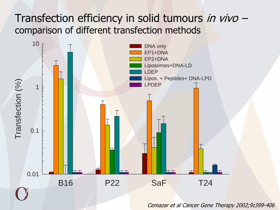
Transfection efficiency in solid tumours in vivo – comparison of different transfection methods
B16 P22 SaF T24
Tra
nsfe
ction (
%)
0.01
0.1
1
10 DNA only
EP1+DNA
EP2+DNA
Liposomes+DNA-LD
LDEP
Lipos. + Peptides+ DNA-LPD
LPDEP
Cemazar et al Cancer Gene Therapy 2002;9:399-406

Electroporation (600 V/cm, 5 ms, 1 Hz, 8 pulses) of tumours increases GFP (40 g pEGFP-N1) expression
Pre x 4
Pre EP epi x 4
2 days epi x 4
2 days epi x 20 Rat No. 7
Intravital miscroscopy – spatial and time dependent distribution

Electrotransfection in B16 melanoma and SA-1 sarcoma –
comparison of different electric pulse protocols
Luciferase activity as a function of LV pulse amplitude
1HV+8LV; HV: 1x0.1 ms, 1200 V/cm; LV: 8x 50 ms, increasing V
8x5 m
s 600V/cm
ŘÁ��
80V/cm
100V/cm
120V/cm
140V/cm
160V/cm
Lucifera
se a
ctivity (
pg luc/m
g t
um
our)
0
50
100
150
200
250
300
Control L
uc only
8x5 m
s 600V/cm
8x0.1
ms 1
300V/cm
80V/cm
120V/cm
140V/cm
160V/cm
B16 SA-1
Cliniporator EU project

Transfection efficiency in solid tumours in vivo – timing of the procedure
Cemazar et al Current Drug Delivery 2006;3:77-81; Mesojednik et al, Gene Therapy 2007; 14(17):1261-1269
Time of DNA injection prior to electroporation (min)
control - 60 - 30 - 15 - 10 - 5 -0.5
Tra
nsfe
ctio
n (
pg
Lu
cife
rase
/ m
g t
um
or)
0.001
0.01
1
10
100LPB
SA-1
EAT
B16F1
* * *

Electrotransfection of mouse muscle
in vivo non-invasive imaging
ex vivo spatial distribution on frozen muscle sections
Injection (25µl) od DNA in the
tibialis cranialis muscle
Application of electric pulses

Parameters of electrotransfection
Electric parameters pEGFP-N1dose/20 l Time lag
1 HV (600 V/cm, 100 s) + 1 LV (80 V/cm, 400 ms, 1Hz) 20 g 10 min
1 HV (600 V/cm, 100 s) + 4 LV (80 V/cm, 100 ms, 1Hz) 20 g 10 min
1 HV (600 V/cm, 100 s) + 8 LV (80 V/cm, 50 ms, 2Hz) 20 g 10 min
6 LV (100 V/cm, 60 ms, 1Hz) 20 g 10 min
8 LV (200 V/cm, 20 ms, 1Hz) 20 g 10 min
1 HV + 4 LV 20 g 5 s
1 HV + 4 LV 20 g 1 min
1 HV + 4 LV 20 g 3 min
1 HV + 4 LV 20 g 5 min
1 HV + 4 LV 20 g 10 min
1 HV + 4 LV 20 g 20 min
1 HV + 4 LV 20 g 30 min
1 HV + 4 LV 20 g 60 min
1 HV + 4 LV 20 g 120 min
1 HV + 4 LV 1 g 5 s
1 HV + 4 LV 5 g 5 s
1 HV + 4 LV 10 g 5 s
1 HV + 4 LV 20 g 5 s
1 HV + 4 LV 30 g 5 s

Electrotransfection of mouse muscle – influence of timing
Time after transfection (weeks)
0 10 20 30 40
Norm
ali
sed
mea
n f
luore
scen
ce (
a.u
.)
0
1
2
3
4
Background fluorescence intensity
5s
1min
3min
5min
10 min
20min
30min
60min
120min
*
Time lag between DNA injection and application of electric pulses (min)
020406080100120
Mea
n a
rea
of
tra
nsf
ecti
on
(%
)
0
5
10
15
20
EP
*
Tevz et al.TCRT 2008; 7(2): 91-106

Electrotransfection of mouse muscle – dependence on DNA amount injected
Dose of plasmid DNA (g)
0 5 10 15 20 25 30
Mea
n a
rea
of
tra
nsf
ecti
on
(%
)
0
5
10
15
20
25
30
Time after transfection (week)
0 10 20 30 40 50 60
No
rmal
ized
mea
n f
luo
resc
ence
(a.
u.)
0
1
2
3
41ug
5 ug
10 ug
20 ug
30 ug
Background fluorescence intensity
*
Tevz et al.TCRT 2008; 7(2): 91-106
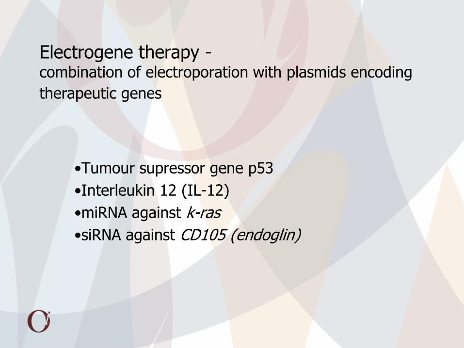
Electrogene therapy - combination of electroporation with plasmids encoding
therapeutic genes
•Tumour supressor gene p53
•Interleukin 12 (IL-12)
•miRNA against k-ras
•siRNA against CD105 (endoglin)

p53 is a sensor of different forms of stress
Stress conditions
Non-genotoxic stress: hypoxia, temp. changes, depletion of ribonucleotides, growth factors, microtubules; redox changes, cytokines
Genotoxic stress: UV, X, g rays,
carcinogens, chemotherapuetic drugs
Oncogenic stress: oncogenes
Modulation of p53 activity
p53
p53 p53
Induction of target genes Binding to proteins
Cell cycle Angiogenesis Apoptosis DNA repair
Genomic integrity Growth control
Senescence

Repetitive electrogenetherapy with p53wt in a mouse models
Time after treatment (days)
0 2 4 6 8 10 12 14 16 18 20 22
Tum
our
volu
me (
mm
3)
30
50
80
300
500
800
100
Control
EP(EGT)
p53
pCMV
EGTpCMV
EGTp53
Time after treatment (days)
0 2 4 6 8 10 12 14 16 18
30
50
80
300
500
800
100
Control
EP(EGT)
pCMV
p53
EGTpCMV
EGTp53
21.4% tumour cures 12.5 % tumour cures
Grošel et al DNA Cell Biol 2006,25:674-683
LPB – wt p53 SA-1 – mt p53

Electrogenetherapy with p53wt of human tumor xenografts
Days after treatment
0 2 4 6 8 10 12 14 16 18 20 22 24 26 28 30 32 34
Tum
our
volu
me (
mm
3)
30
50
80
200
300
500
100
ControlElectric pulses
p53wt
Electrogenetherapy with p53wt
pCMV
Electrogenetherapy with pCMV
Treatment of human tumor xenograft PC3 with electrogenetherapy with p53wt resulted in pronounced antitumour effect.
PC3 prostatic carcinoma
Cemazar et al DNA Cell Biol 2003; 12 765-75

Interleukin-12 antitumor effect
IL-12 has direct cytotoxic
effect, activates potent anti-
angiogenic mechanisms
Il-12 Induces responses of
Th1 cells and cytotoxic T
lymphocytes, NK cells

Recombinant mIL-12 has no cytotoxic effect on tumor cells in vitro
0 mg/m
l
1 pg/ml
10 pg/ml
100 pg/ml
1 ng/ml
10 ng/ml
100 ng/ml
Su
rviv
al f
ract
ion
0,01
0,1
1
SA-1
LBP

Intramuscular mIL-12 gene electrotransfer has good antitumor effect on subcutaneous solid tumors
Time after gene electrotransfer (days)
0 5 10 15 20 25 30 35 40
Tum
or
vo
lum
e (
mm
3)
25
35
50
75
250
350
10
100Control
EP
pORF-mIL12
EGT (13/18)
EGT (5/18)
Time after gene electrotransfer (days)
0 5 10 15 20 25 30T
um
or
vo
lum
e (
mm
3)
25
35
50
75
250
350
10
100
Control
EP
pORF-mIL12
EGT (24/28)
EGT (4/28)
SA-1 LPB
28% CR 13% CR
TREATMENT 1 WEEK 2 WEEKS 4 WEEKS

Antitumor effectiveness of mIL-12 gene electrotransfer – intratumoral vs. peritumoral
Intratumoral EGT resulted in high level of complete responses (18/20 tumors) with significant inhibition of tumor growth in the remaining 2 tumors.
A - INTRATUMORAL DELIVERY
TIME AFTER GENE ELECTROTRANSFER (days)
0 5 10 15 20 25 30 35 40 45 50 55 60
TU
MO
R V
OLU
ME
(m
m3 )
7
20
30
50
70
200
300
500
10
100
Control i.t.
EP i.t.
DNA i.t.
EGT i.t. (animals with CR - 18/20)
EGT i.t. (animals without CR - 2/20)
B - PERITUMORAL DELIVERY
TIME AFTER GENE ELECTROTRANSFER (days)
0 5 10 15 20 25 30 35 40 45 50 55 60T
UM
OR
VO
LUM
E (
mm
3 )
7
20
30
50
70
200
300
500
10
100
Control p.t.
EP p.t.
DNA p.t.
EGT p.t. (animals withCR - 3/19)
EGT p.t. (animals without CR - 16/19)
Pavlin et al. Cancer Biol Ther 2009; 8:2112-2120
90% CR 16% CR
Peritumoral EGT resulted in lower complete response rate (3/19 tumors), with remaining 16/19 showing significant delay in tumor growth.

Gene electrotransfer induces local and systemic release of IL-12 and IFN-γ and has antitumor effect on distant untreated tumors
Control EP i.t. EP p.t. DNA i.t. DNA p.t. EGT i.t. EGT p.t.
SE
RU
M C
ON
CE
TR
AT
ION
(pg
/ml)
0
20
40
60
80
100
120
IL-12
IFN-g
*
*
*
*
Control EP i.t. EP p.t. DNA i.t. DNA p.t. EGT i.t. EGT p.t.
INT
RA
TU
MO
RA
L C
ON
CE
NT
RA
TIO
N(p
g/m
g of
tum
or ti
ssue
)
0
20000
40000
60000
80000
IL-12
IFN-g *
*
*
*
SERUM
TUMOR
TIME AFTER TREATMENT (days)
0 5 10 15 20 25 30 35 40
TU
MO
R V
OLU
ME
(m
m3 )
7
30
50
70
300
500
10
100
Control i.t.
Control p.t.
EP i.t.
EP p.t.
DNA i.t.
DNA p.t.
EGT i.t.
EGT p.t.
Control i.t.
Control p.t.
EP i.t.
EP p.t.
DNA i.t.
DNA p.t.
EGT i.t.
EGT p.t.
Treated tumors Untreated tumors
Growth curves of distant untreated tumors
Pavlin et al. Cancer Biol Ther 2009; 8:2112-2120

Systemic mIL-12 release (electrotransfection of muscle) and local tumor irradiation
Local mIL-12 release (electrotransfection of tumor) and local tumor irradiation
IL-12 gene therapy combined with tumor irradiation

Time after gene electrotransfer (days)
0 5 10 15 20 25 30 35 100
Per
cen
t o
f cu
res
(%)
0
20
40
60
80
100
GEP
IR
Time after gene electrotransfer (days)
0 5 10 15 20 25 30 100
Per
cen
t o
f cu
res
(%)
0
20
40
60
80
100 Control
EP
pORF-mIL-12
GET
IR 10Gy
EP+IR
mIL-12+IR
GET+IR
IR
GEP
SA-1
LPB
Intramuscular mIL-12 gene electrotransfer has radiosensitizing effect on solid subcutaneous tumors
Tevž et al. J Gene Med 2009; 11:1125-1137
Day 0 Day 7 Day 14

mIL-12 gene electrotransfer has radiosensitizing effect on induced lung metastases
Control EP
pORF-mIL12
-24h GETIR 4 Gy
GET+IR
Per
cen
t o
f m
etas
tase
s (%
)
0
20
40
60
80
100
120
Control EGT +IR
19% 30%
0.5% *
PF = 1.3
IR 4 Gy

Intratumoral IL-12 gene electrotransfer combined with irradiation comparison between 1x EGT vs 3x EGT

Single intratumoral mIL-12 electrogene therapy has antitumor and also radiosensitizing effect on SA-1 sarcoma
Three intratumoral mIL-12 electrogene therapies have better antitumor and radiosensitizing effect on SA-1 sarcoma compared to single treatment

Improvement in therapeutic index, as a result of adding single intratumoral electrogene therapy with mIL-12 (20µg of plasmid DNA) to radiotherapy, for treatment of murine SA-1 sarcoma.
Dry desquamation < 20 % of irradiated area
Dose modifying factor (DMF) of single mIL-12 electrogene therapy on SA-1 sarcoma is
2,17
At the same level of skin damage, dry desquamation, in combined therapy a 44% higher probability of local tumor control than with irradiation alone was observed.
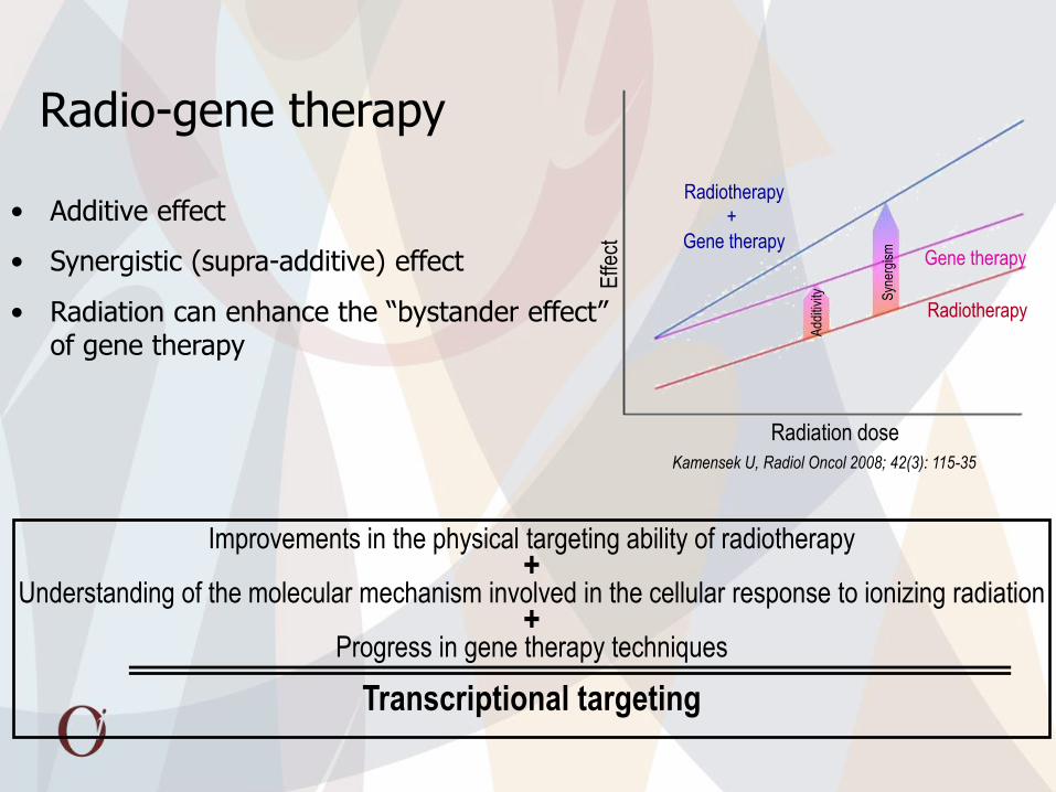
Improvements in the physical targeting ability of radiotherapy +
Understanding of the molecular mechanism involved in the cellular response to ionizing radiation +
Progress in gene therapy techniques
Transcriptional targeting
Radio-gene therapy
• Additive effect
• Synergistic (supra-additive) effect
• Radiation can enhance the “bystander effect” of gene therapy
Kamensek U, Radiol Oncol 2008; 42(3): 115-35
Add
itivi
ty
Syn
ergi
sm
Radiation dose
Effe
ct
Radiotherapy
+
Gene therapy
Radiotherapy
Gene therapy

Tumor irradiation combined with IL-12 gene therapy controlled by p21 radio-inducible promoter
Promoters of radiation inducible genes, that are activated by clinically relevant
doses of irradiation, can be exploited to control transgenes expression
spatially and temporally within the irradiated tumor tissue.
Promoter of gene p21 + gene for IL-12
Stimulation of immune response

Tumor growth delay after radio-gene therapy with p21-mIL12 or pORFmIL12 plasmid
Time (days)
0 5 10 15 20 25 30
Tum
or v
olum
e (m
m3 )
40
60
80
200
400
100
Control
p21+EP
p21+EP+IR
pORF+EP
pORF+EP+IR
Time (days)
0 5 10 15 20 25 30
Tum
or v
olum
e (m
m3 )
40
60
80
200
400
100
Control
p21+EP+IR
pORF+EP
pORF+EP+IR
EP
IR
EP+IR
p21
p21+IR
pORF
pORF+IR
RESULTS
EP electroporation IR irradiation p21 intratumoral injection of p21-mIL12 plasmid pORF intratumoral injection of pORF-mIL12 plasmid

Treatment of colon carcinoma by miRNA for K-ras by gene electrotransfer
miRNA against k-ras

IZBIRA PRIMERNE SEKVENCE siRNA
LIPOFEKCIJA Z siRNA-KRAS (3)
siRNA-KRAS 53
CCU UGA CGA UAC AGC UAA U
AUU AGC UGU AUC GUC AAG G
siRNA-KRAS 111
GGA UUC CUA CAG GAA GCA A
UUG CUU CCU GUA GGA AUC C
siRNA-KRAS 393
GGA CUU AGC AAG AAG UUA U
AUA ACU UCU UGC UAA GUC C
siRNA-53 siRNA-111 siRNA-393
K-r
as
mR
NA
/ 1
8S
mR
NA
(%
)
0
20
40
60
80
100
120
LF
LF + siRNA-K-ras
LF + siRNA-ctrl
***
*
*
*
*
*
**
qRT-PCR analiza

LF
LF + siRNA-K-ras53
LF + siRNA-ctrl53
LF + siRNA-K-ras393
LF + siRNA-ctrl393
De
lež p
reživ
elih
ce
lic
0,0
0,2
0,4
0,6
0,8
1,0
1,2
*
IZBIRA PRIMERNE SEKVENCE siRNA
WESTERN BLOT ANALIZA TEST KLONOGENOSTI
1 Kontrola
2 LF
3 LF + siRNA-K-ras53
4 LF + siRNA-ctrl53
5 LF + siRNA-K-ras393
6 LF + siRNA-ctrl393
K-ras
Aktin

PRIPRAVA PLAZMIDNE DNA, KI KODIRA miRNA-K-ras
Dobre lastnosti:
SPECIFIČNO DELUJOČE
STABILNE
NI STRANSKIH UČINKOV
IN VITRO ELEKTROTRANSFEKCIJA

IN VITRO ELEKTROTRANSFEKCIJA CELIC LoVo S PLAZMIDNO DNA, KI KODIRA miRNA-K-ras
pmiRNA-K-ras
pmiRNA-ctrl EP
pmiRNA-K-ras + EP
pmiRNA-ctrl + EP
mR
NA
K-r
as / 1
8S
mR
NA
(%
)
0
20
40
60
80
100
120
pmiRNA-K-ras
pmiRNA-ctrl EP
pmiRNA-K-ras + EP
pmiRNA-ctrl + EP
De
lež p
reživ
elih
ce
lic
0,0
0,2
0,4
0,6
0,8
1,0
1,2
**
qRT-PCR ANALIZA TEST KLONOGENOSTI
WESTERN BLOT
1 Kontrola
2 pmiRNA-K-ras
3 pmiRNA-ctrl
4 EP
5 pmiRNA-K-ra + EP
6 pmiRNA-ctrl + EP
K-ras
Aktin

IN VIVO ELEKTROTRANSFEKCIJA TUMORJEV LoVo S PLAZMIDNO DNA, KI KODIRA miRNA-K-ras
Dnevi
0 5 10 15 20 25 30
Vo
lum
en
tu
mo
rja
(m
m3
)
30
40
50
60
80
200
300
100
Kontrola
EP
pmiRNA-K-ras
pmiRNA-ctrl
pmiRNA-K-ras + EP
pmiRNA-ctrl + EP
pmiRNA-K-ras
pmiRNA-ctrl EP
pmiRNA-K-ras + EP
pmiRNA-ctrl + EP
K-r
as m
RN
A / 1
8 S
mR
NA
(%
)
0
20
40
60
80
100
120
*
qRT-PCR ANALIZA
1 Kontrola
2 EP
3 pmiRNA-K-ras
4 pmiRNA-ctrl
5 pmiRNA-K-ra + EP
6 pmiRNA-ctrl + EP
K-ras
Aktin

Utišanje endoglina (CD105) v humanih endotelnih celicah in mišjih tumorjih TS/A
• Endoglin je homodimernih membranski glikoprotein
• Je koreceptor TGFb receptorskega sistema
• Njgova ekspresija je povišana v aktiviranih endotelnih celicah v čvrstih tumorjih
LF
siRNA 5
29
siRNA 2
40
siRNA 2
41
siRNA C
trl
endoglin
mR
NA
/ P
OL2 m
RN
A
0,0
0,2
0,4
0,6
0,8
1,0
1,2
*
* *
*
qRT-PCR
Survival
LF
siRNA 5
29
siRNA 2
40
siRNA 2
41
siRNA C
trl
surv
ival (%
of
contr
ol)
0
20
40
60
80
100
1203th day after lipofection
7th day after lipofection
**
*

Vpliv 1x in 3x elektroprenosa siRNA proti endoglinu na rast podkožnih TS/A tumorjev

Število tumorskih kapilar je po 3x terapiji zmanjšano

Veterinary oncology
Human clinical trials
Electrogene therapy – clinical experience

Intramuscular delivery of plasmid, encoding hIL-12 in canine patients with various spontaneously arising tumors
Patients and methods 7 patients with different histological types of tumors:
• Mast cell tumors (Mct) (3 pts.)
• Mammary adenocarcinoma (2 pts.)
• Osteosarcoma (1 pt)
• Malignant histiocytosis (1 pt) Plasmid:
• pORF-hIL12 (Invivogen, Toulouse, France)
• 1 mg/i.m. delivery
EP delivery:
• 1 high voltage pulse (600 V/cm, 100 μs) + 4 low voltage pulses (80 V/cm, 100 ms, 1 Hz))
• Electric pulses generator Cliniporator® (Igea s.r.l., Italy)
Follow-up:
• 7, 14, 28 days after procedure

Results
patient Patient data Tumor type Therapy hIL-12 conc.
cIFN-γ conc. Outcome
1 Boxer, 7 years, male
Mast cell tumor
Gradus III
Surgery
Chemotherapy
EGT
17 pg/mL
246.8 pg/mL CR
(o.p. 36 months)
2
Boxer, 3 years, female
Mast cell tumor
Gradus II
Surgery
EGT
n.d. 6.5 pg/ml CR
(o.p. 36 months)
3 Lhasa-apso,
14 years, female
Mast cell tumor
Gradus III
Recurrence after surgery
Chemotherapy
EGT
n.d. 80 pg/ml
PD
euth. 6 months after the procedure
4 Bernese mountain dog
6 years, male
Malignant hystiocytosis Chemotherapy
EGT
n.d. 104,2 pg/ml
SD
euth. 7 months after the procedure
Average survival after surgical therapy in MCT: Gradus II - 7 – 18 months Gradus III – 3 - 4 months
- Survival times of all of these patients was significantly longer than reported in literature for respective tumor types (but very low no. of patients)
- IL-12 and/or IFN-γ was detected in serum samples of 4 patients at various time points after the procedure
Average survival after surgical therapy in mal. hystiocyosis after chemotherapy: 4 months

Intratumoral delivery plasmid, encoding hIL-12 in canine patients with mast cell tumors
Patients and methods
11 Mct nodules in 8 dogs
No. of performed EGT sessions:
• 1xEGT in 3/11 nodules
• 2xEGT in 5/11 nodules
• 3xEGT in 1/11 nodules
• 4xEGT in 2/11 nodules
Measurements of tumor size
Determination of hIL12 and cIFN-γ in serum
1, 2 and 4 week after EGT
Surgical removal of tumors in 5/8 dogs →
histology

Patient Nodule T. volume before EGT
(cm3)
clinical stage Post EGT therapy
Follow up after 1st EGT (mon.)
Response at the end of follow up
1 1 0.25 I / 36 SD Euth., not related to MCT
2 1 2.3 I Surgery 24 CR without recurrence
3 1 3.2 II CCNU
(4 cycles) 12 SD stable disease
4 1 0.6 II ECT 13 CR without recurrence
2 1.2 Surgery CR without recurrence
5 1 2.9 III Surgery 10 CR without recurrence
6 1 0.03 II / 44 SD stable disease
2 0.27 / SD
7 1 25.4 III Surgery 2 PD Euth. due to PD
8 1 0.45 III CCNU
(3 cycles) 5 PD Euth. due to PD
2 0.03
Results
• Reduction in tumor size in 9/11 tumors for 25-85% of tumor volume
• Detection of both measured cytokines in multiple serum samples:
IL-12 (3/8 patients): 1-12 pg/mL
IFN-γ (3/8 patients): 74-815 pg/mL

Antitumor effectiveness of hIL-12 therapy
Boxer, dog #2

Tumor histology
a. MCT before EGT
(orange arrow: mast cells)
b. MCT after EGT:
-Reduction in no. of mast cells
-Infiltration with inflammatory cells

First electrogene clinical trial with IL-12 on subcutaneous melanoma metastases

Antitumor effectiveness of treated and non-treated melanoma metastases

Preparations for gene electrotransfer clinical trial
Angioskin EU FP6 project

Transfection efficiency Therapeutic outcome
Electric field Distribution
Plasmid construction
and administration
Tissue properties
Inflammation; Immune response
Blood flow modification
Tissue damage
Electrode design
Electric pulse parameters
Factors influencing transfection efficiency and therapeutic outcome of in vivo electrotransfection
Cemazar et al. Curr Pharm Design 2006; 12 (29): 3817-3825