Embed Size (px)
Citation preview

TYPE – IV HYPER SENSITIVITY
Moderator: Dr. Sanchita GhoshSpeaker: Dipayan Das

INTRODUCTION
Immunity is the balanced state having adequate biological defense against infections and diseases while having adequate tolerance to inflammation, allergy and autoimmune diseases.
The response is how our body recognizes and defends itself against foreign and harmful substances.

Hypersensitivity reactions are a set of undesirable reactions produced by normal immune system and the resulting diseases are called hypersensitivity diseases.
Persons who mount immune responses against an antigen are said to be “sensitized” to that antigen.
So pathologic or excessive reactions represent a “hypersensitive” state.
INTRODUCTION

FEATURES OF HYPERSENSITIVITY Elicited by exogenous environmental antigens
(microbial and nonmicrobial) or endogenous self antigens.
Allergy : caused by environmental antigens. Autoimmune diseases : Immune responses
against self or autologous antigens.
It results from an imbalance between the effector mechanisms of immune responses and the control mechanisms that serve to normally limit such responses.
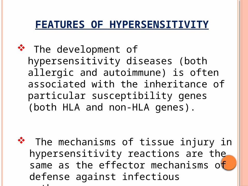
The development of hypersensitivity diseases (both allergic and autoimmune) is often associated with the inheritance of particular susceptibility genes (both HLA and non-HLA genes).
The mechanisms of tissue injury in
hypersensitivity reactions are the same as the effector mechanisms of defense against infectious pathogens.
FEATURES OF HYPERSENSITIVITY
CLASSIFICATION
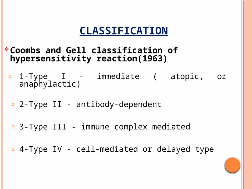
Coombs and Gell classification of hypersensitivity reaction(1963)
o 1-Type I - immediate ( atopic, or anaphylactic)
o 2-Type II - antibody-dependent
o 3-Type III - immune complex mediated
o 4-Type IV - cell-mediated or delayed type


TYPE-IV HYPERSENSITIVITY
Caused by inflammation resulting from cytokines produced by CD4+ T cells and cell killing by CD8+ T cells.
Also known as T cell mediated hyper sensitivity.
CD4+T cell mediated hypersensitivity: Major role in autoimmunity.
CD8+T cell mediated hypersensitivity : Predominantly acts against viral infection, tumor cells and graft rejection.

CD4+T CELL MEDIATED HYPERSENSITIVITY
In CD4+ T cell–mediated hypersensitivity reactions, cytokines produced by the T cells induce inflammation that may be chronic and destructive.
The prototype of T cell–mediated inflammation is delayed-type hypersensitivity(DTH).
Both Th1 and Th17 are involved in immune mediated destruction.

The inflammatory reactions stimulated by CD4+ T cells can be divided into two stages :
Activation of CD4+T cells.
Responses of differentiated effector T cells.

CD4+T cells recognize peptides displayed by
APC
Activation of CD4+T cells :
IL-2 secreted by CD4+T cells
Proliferation of CD4+T cells and
activation of APC(macrophages & dendritic cells)

Production of cytokines by the APCs(IL-12,6,23)
IL-12
Differentiation of CD4+T
cells into Th1 cells
Differentiation of CD4+T
cells into Th17 cells
IL-6, IL-23
Secretion of INF-γ
Further Th1 development

Responses of Differentiated Effector T Cells :
Th1 secretes INF-γ
Activation of macrophage
(Classical pathway)
Increased ability to
phagocytose and kill
microorganism
Expression of more MHC-II
Facilitating further antigenpresentation
IL-12
Amplification of the TH1 response
TNF, IL-1, and chemokines
Promote inflammati
on

Responses of Differentiated Effector T Cells :
Th17 secretes IL-17, IL-22,
chemokines & other cytokines
Recruitment of neutrophils and
monocytes
Promote inflammati
on


CLINICAL EXAMPLES OF CD4+ T CELL– MEDIATED HYPERSENSITIVITY :
Tuberculin reaction : Intracutaneous injection of PPD in a previously sensitized individual
Accumulation of mononuclear cells(CD4+T cells and macrophages) around the venules
Perivascular cuffing Release of cytokines Hyprtrophy of venules Reddening and induration of the site in 8-12 hrs Peak in 24hrs and thereafter subside slowly

Delayed hypersensitivity reaction in the skin. A, Perivascular accumulation (“cuffing”) of mononuclear inflammatory cells (lymphocytes and macrophages), with associated dermal edema and fibrin deposition.B, Immunoperoxidase staining reveals a predominantly perivascular cellular infiltrate that marks positively with anti-CD4 antibodies.

Granulomatous inflammation : CD4+T cells(Th1 & Th2) releases high level of INF-Y Sustained activation of macrophages Transformed to epithelioid cells Granuloma formation
CLINICAL EXAMPLES OF CD4+ T CELL– MEDIATED HYPERSENSITIVITY :

Contact dermatitis : Contact with urushiol, antigenic component of poison ivy or oak Bind and structurally modifies some self protiens and/or HLA molecules These altered protien are recognized as foreign by CD4+T cells Activation and cytokine production Inflammation Vesicular dermatitis
Rheumatoid arthritis : Th17 acts against collagen and citrullinated self protien leading to chronic arthrits with inflammation and destruction of articular cartilage.
CLINICAL EXAMPLES OF CD4+ T CELL– MEDIATED HYPERSENSITIVITY :

Multiple sclerosis : Th1 and Th17 act against myelin causing demyelination in CNS with perivascular inflammation and paralysis.
Inflammatory bowel disease : Th1 and Th17 act against enteric bacteria or a self antigen leading to chronic intestinal inflammation and obstruction.
Psoriasis : Here CD4+T cells get activated against unknown antigen and differentiated into Th17 which prdoces destructive plaques in the skin.
CLINICAL EXAMPLES OF CD4+ T CELL– MEDIATED HYPERSENSITIVITY :

CD8+T CELL MEDIATED HYPERSENSITIVITY
EFFECTOR FUNCTIONS OF CD8+T CELLS :
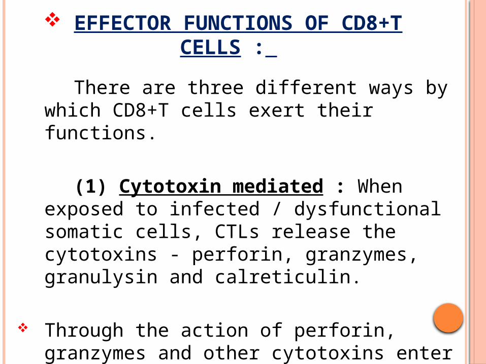
There are three different ways by which CD8+T cells exert their functions.
(1) Cytotoxin mediated : When
exposed to infected / dysfunctional somatic cells, CTLs release the cytotoxins - perforin, granzymes, granulysin and calreticulin.
Through the action of perforin, granzymes and other cytotoxins enter into the cytoplasm of the target cells.

EFFECTOR FUNCTIONS CONT…
(a) Granzymes : Granzymes have two subunits – Granzyme A and Granzyme B.
Granzyme B is a serine protease which triggers the caspase cascade, which is a series of cysteine proteases that eventually lead to apoptosis.

EFFECTOR FUNCTIONS CONT…Granzyme A is a tryptase and induces
caspase-independent cell death.
It concentrates in the nucleus of the target cells and degrades histone H1 into small fragments.
Histone digestion provides a mechanism for unfolding compacted chromatin and facilitating endogenous DNAase access to DNA during T cell granule-mediated apoptosis.

EFFECTOR FUNCTIONS CONT…
(b) Perforin (Cytolysin) : Perforin is a 65 to 75 kD glycoprotein with homology to C9 complement component.
It is synthesized as an inactive precursor, which is cleaved to yield a 60 kD active form.

EFFECTOR FUNCTIONS CONT…
The C-terminal portion of this active form is cleaved by proteolytic enzymes, activating the C2 domains for phospholipid binding.
The N-terminal is involved in interaction with the membrane and polymerization.
Finally, aggregation of such 10-20 perforin monomers forms polyperforin pores.

EFFECTOR FUNCTIONS CONT…
(c) Granulysin : It is active against Gram-positive and Gram-negative bacteria, fungi and parasites.
It disrupts artificial liposomes.
Damages mitochondria.
Activates caspase 9 to induce apoptosis.It kills extracellular M. tuberculosis and decreases their viability inside the cells.

EFFECTOR FUNCTIONS CONT…
(d) Calreticulin : It is a Ca2+ binding protein and acts as a chaperone for perforin. It protects CTL from autodigestion during biogenesis of the granules.
(e) Other components : Chondroitin sulfate is a negatively charged molecule which helps in delivery of granzymes into the target cells.
EFFECTOR FUNCTIONS CONT…
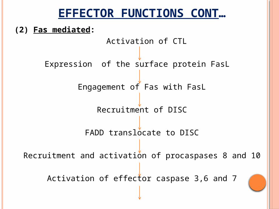
(2) Fas mediated: Activation of CTL
Expression of the surface protein FasL
Engagement of Fas with FasL
Recruitment of DISC
FADD translocate to DISC
Recruitment and activation of procaspases 8 and 10
Activation of effector caspase 3,6 and 7

EFFECTOR FUNCTIONS CONT…(2) Fas mediated:
Cleavage of death substrates such as laminin A, B1 & B2
Apoptosis of target cells

EFFECTOR FUNCTIONS CONT…
(3) Cytokine mediated : CTLs also produce cytokines, notably IFN-Y following viral infections and exposure to some contact sensitizing agents. INF-Y is produced in two pathways:
(a) Major pathway : The majority of INF-Y is produced when TCR of CTL bind with an anitgen. So it is antigen-dependent pathway.
(b) Minor pathway : The innate cytokines type-1 INF and IL-12 can also induce INF-Y production through STAT-4. It is antigen-independent and so it makes CTL able to take part in innate immunity.

CLINICAL EXAMPLES OF CD8+ T CELL– MEDIATED HYPERSENSITIVITY :
(1) Graft versus Host Disease and Graft versus Leukemia Reaction : CTL is implicated for both graft vs host disease and graft vs leukemia reaction.
The first one is attributed to both perforin and Fas pathway but later is mediated predominantly by perforin pathway.
(2) Type-I Diabetes Mellitus : Here CTLs act against antigens of pancreatic β cells(insulin, glutamic acid decarboxylase) leading to inflammation and destruction of islet cells.

CLINICAL EXAMPLES OF CD8+ T CELL– MEDIATED HYPERSENSITIVITY :
(3) Familial hemophagocytic lymphohistiocytosis :
Perforin prevents uncontrolled expansion and activation of T cells.
Homozygous loss of function defect in perforin gene (PRF1) causes familial hemophagocytic lymphohistiocytosis(FHL2).
It is due to uncontrolled activation of T cells and macrophages with overproduction of inflammatory cytokines.
.

CLINICAL EXAMPLES OF CD8+ T CELL– MEDIATED HYPERSENSITIVITY :
The disease is mapped on chromosome 10q21-22 (i.e., the location of PRF1).

CLINICAL EXAMPLES OF CD8+ T CELL– MEDIATED HYPERSENSITIVITY :
(4) Hepatic injury during HBV infection : CTL kills the infected hepatocytes leading to liver injury.
(5) Arthritis : CTL causes depletion of articular cartilage macromolecule, glycosaminoglycan.

TAKE HOME MESSAGE Hypersensitivity is of four types: Type-I, Type-II, Type-III
and Type-IV.
Type-IV Hypersensitivity is mediated by CD4+T cells and CD8+T cells.
In CD4+T cell mediated hypersensitivity, CD4+T cells differentiate into Th1 and Th17 cells which exerts their effect by producing cytokines.
In CD8+T cell mediated hypersensitivity, CD8+T cells produce their effect by three pathways cytotoxin mediated, fas mediated and cytokine mediated.
The major role of CD4+T cell mediated hypersensitivity is in autoimmunity while CD8+T cell mediated hypersensitivity act against viral infection, tumor cells and graft rejection.


![[Young marketers 2013] Final - Mbood type](https://img.pdfslide.tips/doc/110x75/546ea432af79598c6d8b4c18/young-marketers-2013-final-mbood-type.jpg)