Embed Size (px)
Citation preview

01/05/2023 drAR 1
LUMBAR PUNCTURE(Lumbar)

01/05/2023 drAR 2
Indication• Diagnosis :– CNS infection, eg. meningitis, encephalitis and CNS syphilis– Subarachnoid hemorrhage (SAH)– Normal pressure hydrocephalus
• Evaluation and diagnosis of demyelinating or inflammatory CNS processes– Multiple sclerosis– Guillain-Barré syndrome– Paraneoplastic syndromes
• Infusion of anesthetic, chemotherapy, or contrast agents into the spinal canal
• Treatment of idiopathic intracranial hypertension

01/05/2023 drAR 3
Contraindication1. Idiopathic or Suspicion of increased ICP d/t cerebral
mass • Brain tumor• Cerebral hemorrhage• Abscesses• Epidural or subdural hematomas
– Rationale: LP in increased ICP may cause uncal herniation– Exception: Therapeutic use of LP to reduce ICP– Precaution
• CT brain is advocated by some, especially in the following situations– Age >65– Reduced GCS– Recent history of seizure– Focal neurological signs
• Ophthalmoscopy for papilledema
Signs of Increase ICP:• Decreased levels of consciousness• Focal neurological signs • Papilloedema
Low-pressure shunt is formed LP site where CSF can escape. As the CSF pressure drops in the spinal column, CSF and brain mass may then shift towards the low-pressure outlet (the LP site). This cause either trans-tentorial or uncal herniation & acute neurological deterioration.

01/05/2023 drAR 4

01/05/2023 drAR 5
Increase ICP
Mass effect Obstruction to
CSF flow or absorption
Generalized brain swelling
Increase in Venous
pressure

01/05/2023 drAR 6
2. Bleeding diathesis• Uncorrected Coagulopathy• Clotting defects such as disseminated intravascular coagulation, hemophilia or
thrombocytopenia (<50 x 109/L)• Heparin, Warfarin
– Reversal of warfarin with Vitamin K or fresh frozen plasma– Replacement of a hemophiliac's clotting factors – Transfusion of platelets to the thrombocytopenic patient
3. Skin infection– Skin infection near the site of the lumbar puncture > increases the risk of
carrying the infection into the CSF with the LP needle >sepsis4. Abnormal respiratory pattern5. Hypertension with bradycardia and deteriorating consciousness.6. Vertebral deformities (scoliosis or kyphosis), 7. Acute spinal trauma8. Obtunded state with poor peripheral perfusion or hypotension9. Seizures-prolonged or recent (within 30 minutes) 10. Inexperienced physician
Tr(x) for safe LP

01/05/2023 drAR 7
Anatomy• At birth inferior end of the spinal cord is opposite the
body of the third lumbar vertebrae (L3)• As the child grows, the vertebral column grows much
faster than the spinal cord itself.• Adulthood the spinal cord only reaches the inferior
border of the L1 vertebra, or the superior aspect of L2. In order to avoid transfixing the spinal cord during LP, the needle is placed distal to L2. This means the needle enters the subarachnoid space at the level of the mobile cauda equina.

01/05/2023 drAR 8

01/05/2023 drAR 9

01/05/2023 drAR 10
WHERE TO INSERT THE NEEDLE ??• The imaginary line that crosses the lumbar
region of the back joining the posterior superior iliac crests will cross the L3-L4 interspace

01/05/2023 drAR 11
• The tissues pierced are (in order): skin, subcutaneous tissue, supraspinal ligament, interspinal ligament, ligamentum flavum, dura mater, the arachnoid mater and into the subarachnoid space.

01/05/2023 drAR 12
THE LP KIT• three cleaning sponges• a 20 gauge spinal needle• a 25 gauge and a 20 gauge needle for anesthetic
infiltration• a 3cc syringe• a vial of 1% lidocaine for anesthesia• a pressure manometer with tubing• four collection vials and a Band-Aid. The other tools you may need include sterile gloves,
gown, proviodine cleaning solution

01/05/2023 drAR 13
EquipmentsA spinal or lumbar puncture tray should
include the following items:• Sterile dressing• Sterile gloves• Sterile drape• Antiseptic solution with skin swabs• Lidocaine 1% without epinephrine• Syringe, 3 mL• Needles, 20 and 25 gauge• Spinal needles, 20 and 22 gauge• Three-way stopcock• Manometer• Four plastic test tubes, numbered 1-4,
with caps• Syringe, 10 mL (optional)

01/05/2023 drAR 14

01/05/2023 drAR 15
Patient Preparation• Local anesthesia is
employed for lumbar puncture
• The patient is placed in the lateral recumbent position with the hips, knees, and chin flexed toward the chest so as to open the inter-laminar spaces or interspinous distance
• A pillow may be used to support the head
• Inter-laminar space• Inter-spinous diameter

Lateral Decubitus Position
• Apply topical anesthetic 30-45 min prior to procedure• Spinal cord ends at L1-L2, so sites for puncture are located at
L3-L4 or L4-L5• Restrain patient in lateral decubitus position
– Maximally flex spine without compromising airway– Keep alignment of feet, knees and hips– Position head to left if right handed or vice versa

Sitting Position
• Restrain infant in the seated position with maximal spinal flexion– Hold infant’s hands between flexed
legs with one hand and flex head with the other hand
• Drape patient below buttocks and fenestrated drape opening over puncture site
• Insert needle so bevel is parallel to spinal cord (Bevel left or right)
• Cannot measure pressure accurately in this position

Paramedian (Lateral) Approach
• Use for patients who have calcifications from repeated LPs or anatomic abnormalities
• Needle passes through erector spinae muscles, and ligamentum flavum– Bypasses supraspinal
and interspinal ligaments
• Less incidence of spinal headache

Equipment
• Spinal needle, usually 22 gauge– 1.5 in for < 1 yr– 2.5 in for 1 year to
middle childhood– 3.5 in for older
children and adolescents
– Larger for large adolescents
• Atraumatic needles, less spinal headaches

01/05/2023 drAR 20
Procedure• Cleanse skin with povidone iodine from puncture
site radially out to 10 cm and ALLOW TO DRY• Drape below patient and around site with
fenestrated drape• Anesthetize with lidocaine if topical not used by:– Intradermally raising a wheal at needle insertion site– Advance needle through wheal to desired interspace
• Careful not to inject into a blood vessel or spinal canal
• Insert spinal needle with stylet with bevel up to keep cutting edge parallel with nerve and ligament fibers

Procedure
• Aim towards umbilicus directing needle slightly cephalad
• Hold needle firmly

Procedure
• A “pop” of sudden decrease in resistance indicates that ligamentum flavum and dura are punctured
• Remove stylet and check for flow of spinal fluid

01/05/2023 drAR 23
Procedure• If no fluid, then:
– Rotate needle 90°– Reinsert stylet and advance needle slowly checking frequently for CSF
• Jugular vein compression can increase CSF pressure in low flow situations
• If bony resistance is felt immediately then you are not in the spinal interspace
• If bony resistance is felt deeply, then withdraw needle to the skin surface and redirect more cephalad and increase patient flexion
• If bloody fluid that does not clear or that clots results, then withdraw needle and reattempt at a different interspace

01/05/2023 drAR 24
Manometry
• When CSF flows, attach manometer to obtain opening pressure if desired
• Pressure can only be accurately measured in lateral decubitus position and in the relaxed patient
• Attach manometer with a 3-way stopcock when free flow of CSF is obtained
• Read column when highest level is achieved and respiratory variation is noted

01/05/2023 drAR 25
PROCEDURE1. Positioning 2. Landmark
LP site 3. Marking LP
site
4. Tray opening & set-up
5. Prep & drape
6. Cutaneous anaesthesia
7. Deep anaesthesia
8. Lumbar puncture
9. Attach manometer
& obtain opening pressure
10. Collect CSF
11. Finishing up

01/05/2023 drAR 26
What to do with the CSF ??• The recommended studies include (this order can be
changed): Collect 1ml of CSF in each of vialso Tube#1 : Gram stain, Culture and Sensitivityo Tube#2 : Glucose, Proteino Tube#3 : Cell Count and Differentialso Tube#4 : Any special studies you require
(fungal/viral/chemical studies)• Check closing pressure with manometer, if desired• Reinsert stylet and remove needle in one quick motion• Cleanse back and cover puncture site

01/05/2023 drAR 27
Opening pressure 90-180 mm H2O / 6.6 – 13.2 mmHg (with patient lying in lateral position)
Appearance and color Clear, colorlessBlood cell count and differential • White blood cells: < 5 (all mononuclear)
• Red blood cells: 0
Glucose 2.8 – 4.4 mmol/L, 50 - 80 mg/dL (or greater than two-thirds of blood glucose)
Total protein 15-45 mg/dLBacteria (Gram stain, culture, VDRL) NegativepH 7.28-7.32Antibodies, viral DNA NoneLactate dehydrogenase (LDH) < 2.0-7.2 U/mLLactate • Newborns 10-40 mg/dL
• Older children and adults 10-25 mg/dL
Chloride 110-125 mmol/LCancerous cells NoneCryptococcal antigen NoneGlutamine 6-15 mg/dL
Normal CSF Parameter

01/05/2023 drAR 28
CSF Analysis

01/05/2023 drAR 29

01/05/2023 drAR 30
Study Bacterial Meningitis Viral Meningitis SAH
Opening Pressure Often elevated Normal to mildly
elevated Often elevated
Appearance Clear to turbid Often clear Clear to bloody Xanthochromia Negative Negative Often present RBC's <5 per mm3 <5 per mm3 >50 per mm3
WBC's Elevated. Many PMNs
Elevated. Many lymphocytes Slightly increased
Glucose Low Normal Normal Protein Elevated Elevated Elevated
Gram Stain May show organisms Normal Normal
A review of the CSF values in common disorders shows:

01/05/2023 drAR 31

01/05/2023 drAR 32
Conditions associated with a reactive CSF lymphocytosis include the following:
• Meningitis• Syphilitic meningoencephalitis• Parasitic CNS infection• Multiple sclerosis• Guillain-Barré syndrome• Polyneuritis
Conditions associated with CSF monocytosis include the following:
• Chronic or treated bacterial meningitis• Syphilitic, viral, fungal, amebic meningitis• Intracranial hemorrhage• Cerebral infarct• CNS malignancy

01/05/2023 drAR 33
Conditions associated with increased CSF polymorphonuclear neutrophils include the following:
• Bacterial meningitis• Acute viral meningitis• Tuberculous and fungal meningitis• Amebic encephalomyelitis• Brain abscess• Subdural empyema• CNS hemorrhage• Cerebral infarct• Malignancies• Previous lumbar puncture• Intrathecal chemotherapy• Seizure

01/05/2023 drAR 34
Complication of Lumbar Puncture1. Post LP headache (most common)
• Usually begins 24-48 hours after the procedure• Probable etiology is continued leakage of CSF• Bilateral pressure or throbbing that is intensified in the upright position• Self-limited (≤7 days), uncommon less than 10 yo
2. Post LP back pain• Occasionally with short-lived referred limp• Disc herniation if needle advanced too far
3. Bloody tap / Dry tap• Bloody: Micro-trauma caused by spinal needle• Dry: misplacement, dehydrated patient (low csf)
4. Infection to the CSF• Can occur with breaks in sterile technique, use of contaminated equipment
and placement of the needle through infected skin• Cellulitis, skin abscesses, epidural abscesses, spinal abscesses
5. Hemorrhage (low platelets counts /coagulopathies)6. Dysesthesia (impairement of sensation)7. Post–dural puncture cerebral herniation (most serious but very rare)
• risk of herniation is 0 –5 % in those patients who are known to have intracranial masses

01/05/2023 drAR 35
Spinal Headache• Most common complication• Risk factors:
– female– age 18-30– lower BMI – hx of Headache– prior spinal headache
• Bilateral Headache, improves when supine• Can last hours to weeks• Treatment
– Supine position for at least 2 hours – Hydration– Caffeine either PO or IV– Epidural blood patch
• Prevention: – Can avoid by:
• Passing needle bevel parallel to longitudinal fibers of dura• Replacing stylet before removing needle• Using small diameter needles• Using atraumatic needles
– Bed rest or PO intake after LP does not reduce incidence of headache

01/05/2023 drAR 36
Nerve Root Trauma/Irritation
• Can feel electric shocks or dysesthesias• Back pain can persist for months– Consider disc herniation
• Rarely permanent• Withdraw needle immediately• If pain or motor weakness persists, start
corticosteroids• Electromyogram/nerve conduction velocity studies
should be scheduled if pain persists

01/05/2023 drAR 37
Herniation
• Manifests initially as altered mental status, followed by cranial nerve abnormalities and Cushing triad
• May be rapidly fatal.• Immediately remove needle and raise the head of
bed to 30-45° improve venous return from the brain.
• Mannitol or 3% Saline• Intubate patient and hyperventilate• Emergent neurosurgical consult
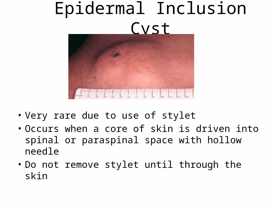
Epidermal Inclusion Cyst
• Very rare due to use of stylet• Occurs when a core of skin is driven into
spinal or paraspinal space with hollow needle• Do not remove stylet until through the skin

Failure of Procedure• If sample of CSF is critical several alternatives are
available:– Have someone else try• Anesthesia• Neurology
– Bedside ultrasound for difficult LPs – Radiographic guided procedure• Fluoroscopy• Ultrasound• CT
– Cisterna Magna tap