Embed Size (px)
Citation preview

S1
Supplementary information
Synthetic glycopeptide as a designated standard in the focused glycoproteomics to
discover serum cancer biomarkers
Yogesh K. V.,1 Toshiya Kamiyama,2 Chikara Ohyama,3 Tohru Yoneyama,3 Kazuhiro Nouso,4 Satoshi Kimura,5
Hiroshi Hinou,1,6 and Shin-Ichiro Nishimura*1,6
1Division of Drug Discovery Research, Faculty of Advanced Life Science and Graduate School of Life Science,
Hokkaido University, N21, W11, Kita-ku, Sapporo 001-0021, Japan
2Department of Gastroenterological Surgery I, Graduate School of Medicine, Hokkaido University, N15, W7,
Kita-ku, Sapporo 060-8638, Japan
3Department of Urology, Graduate School of Medicine, Hirosaki University, Hirosaki 036-8562, Japan
4Department of Gastroenterology and Hepatology, Graduate School of Medicine, Dentistry, and Pharmaceutical
Sciences, Okayama University, Okayama 700-8530, Japan
5Department of Laboratory Medicine and Central Clinical Laboratory, Showa University, Northern Yokohama
Hospital, Yokohama 224-8503, Japan
6Medicinal Chemistry Pharmaceuticals, Co. Ltd., N9, W15, Chuo-ku, Sapporo 060-0009, Sapporo, Japan
Corresponding author: Shin-Ichiro Nishimura, e-mail: [email protected]
Electronic Supplementary Material (ESI) for MedChemComm.This journal is © The Royal Society of Chemistry 2018

S2
Table of Contents
Experimental procedure
I. General methods and materials……...……………………………………………………….….S3
II. Synthesis and characterization…………………………………………………………….…….S4
II-I. Synthesis of a disaccharide intermediate (4)
II-II. Synthesis of an asialo-triantennary decasaccharide (16)
II-III. Construction of AGP glycopeptide (1) by trans glycosylation with mutant endo-M-N175Q
III. SRM-based targeted glycoproteomics…………………………………………………………S17
III-I. SRM/MRM channel setting for the synthetic AGP glycopeptide (1)
III-II. Targeted glycoproteomics by SRM assay focusing human serum AGP glycopeptide (1)
IV. References for the Supporting Information……………………………………………………S25
Proton NMR and HSQC spectra (Figure S1~S20)……………………………………………….S26

S3
Experimental Procedure
I. General methods and materials
All chemical reactions with dry or semi-dry solvents were performed under nitrogen or argon atmosphere unless
otherwise noted. Thin layer chromatography (TLC) was performed on Merck precoated plates (20 cm × 20 cm;
layer thickness, 0.25 mm; Silica Gel 60F254); spots were visualized by spraying a solution of 90:5:5 (v/v/v)
MeOH-p-anisaldehyde-concentrated sulfuric acid and heating at 250°C for ca. 1/2 min, a solution of 95: 5 (v/v)
MeOH-concentrated sulfuric acid and heating at 180°C for ca. 1/2 min, and by UV light (256 or 365 nm) when
applicable. was made on Merck TLC Silica Gel 60 G F254 Glass plate. Flash column chromatography was done
with Silica Gel N60 spherical type, particle size 40-50 μm (Kanto Chemical Co.) or Wakogel® 60N 38-100 μm
(Wako Pure Chemical Industries, Ltd.). Solvent systems are mentioned in v/v. Proton and carbon NMR was
recorded with Varian UnityInova 500 MHz (Agilent Inc., USA; 1H: 500 MHz, 13C: 125 MHz) and Bruker
AVANCE 600 MHz (Bruker Biospin Co., Germany; 1H: 600 MHz, 13C: 150 MHz). Chemical shifts are given
in ppm and referenced to internal TMS (δH 0.00 in CDCl3), CHCl3 (δH 7.26 in CDCl3) or CDCl3 (δC 77.00).
Assignments in 1H NMR were made by first-order analysis of the spectra by using ACD/NMR processor
software (Advanced Chemistry Development, inc.) and were verified by H−H COSY and HSQC experiments.
NMR spectra is assigned with ACD/NMR processor (Advanced Chemistry Development, Inc.). Matrix Assisted
Laser Desorption Ionization Time of Flight Mass Spectrometry (MALDI-TOFMS) spectra were obtained with
Bruker ultraflex I, and III and autoflex III. Reverse phase HPLC (RP HPLC) analysis was conducted with Hitachi
system. Separation was performed with L-6250 intelligent pump, L-7400 UV detector and C18 column: Intersil
ODS-3 250×20 mm I.D. (GL Sciences Inc.). Analysis was performed with L-7100 pump, L-7405 UV detector,
and C18 column: Intersil ODS-3 250×4.6 mm I.D. (GL Sciences Inc.).
Locust bean gum Ceratonia siliqua seeds (G0753), Pectinase from Aspergillus aculeatus (P2611), and
recombinant human β1,4-galactosyltransferase were purchased from Sigma-Aldrich Chemical Co.
Glycosynthase (Endo-M-N175Q) (G0365) from Mucor hiemalis was purchased from Tokyo Chemical Industry
Co., Ltd. Compounds 5 and 6 were synthesized according to the procedures described in our previous paper.S1
SRM parameters for the glycopeptide 1 were optimized using 4000 QTRAP® triple quadrupole mass
spectrometer (AB Sciex Pte. Ltd.) with UltiMate™ 3000 HPLC (Thermo Fisher Scientific Inc.)S2 Detail
procedures for the LC/MS/MS analysis were described in the following section.
Informed consent for the use of clinical data was obtained from all patients. The study protocol conformed
to the ethical guidelines of the World Medical Association Declaration of Helsinki and was approved by the
institutional review board.

S4
II. Synthesis and characterization
II-I. Synthesis of a disaccharide intermediate (4)
Benzyl 2,3,4,6-tetra-O-acetyl-β-D-mannopyranosyl-(1→4)-3,6-di-O-acetyl-2-azido-2-deoxy-β-D-
glucopyranoside (8).
8
2-Azido derivative 7 obtained readily by azidonitration of glycalS1 (4.64 g, 6.9 mmol) dissolved in dry
acetonitrile was added lithium bromide (3.03g, 34.9 mmol) and stirred at r.t. for 3 h. The reaction mixture was
diluted with ethyl acetate and extracted with water, washed with brine solution, dried with Na2SO4 and
concentrated. The crude product was purified by flash column chromatography (hexane/EtOAc= 10:90) and pure
2-azidobromide (3.47g, 5.09 mmol) was co-evaporated with toluene two times, added 3.47 g of molecular sieves
and once more co-evaporated with toluene, dried under vacuum for 12 h. To the residue, added dry
dichloromethane (51 mL) and stirred at r.t for 15 min. To the resulting solution was added iodine (2.6 g, 20.3
mmol) and the solution was stirred at r.t. for 1 h and then silver carbonate (2.8 g, 10.1 mmol) and benzyl alcohol
(1.58 mL, 15.2 mmol) were added and stirred at r.t. for 16 h. The mixture was diluted with chloroform followed
by celite filtration, organic layer was washed with Na2S2O3 solution, NaHCO3 solution, brine solution, and dried
over Na2SO4. The residual product was purified by flash column chromatography (hexane/EtOAc=65:35) to
give benzyl glycoside 8 (1.8 g, 61%) as a white powder.
1H NMR (500 MHz, CDCl3, 25°C, TMS): δ 7.37-7.31 (m, 5 H), 5.37 (d, J = 3.16 Hz, 1 H), 5.19 (t, J = 9.9 Hz,
1 H), 5.00 (dd, J = 3.44 Hz, 2.87 Hz, 1 H), 4.96- 4.89 (m, 2 H; H-3), 4.68 (d, J = 12.05 Hz, 1 H), 4.63 (s, 1 H),
4.41-4.36 (m, 2 H), 4.33 (dd, J = 5.45 Hz, 5.17 Hz, 1 H), 4.23 (dd, J = 4.02 Hz, 4.30 Hz, 1 H), 4.11 (dd, J = 2.58
Hz, 1.15 Hz, 1 H), 3.77 (t, J = 9.76 Hz, 1 H), 3.61 (m, 1 H), 3.55 (m, 1 H), 3.45 (t, J = 9.04 Hz, 1 H), 2.155,
2.145, 2.14, 2.08, 2.04, 1.98 (s each, 3 H each); see, Figure S1.
13C NMR (125 MHz, CDCl3, 25°C, TMS): δ 128.4, 100.2, 97.4, 74.7, 72.6, 72.5, 71.5, 71.2, 71.2, 70.7, 68.2,
65.8, 63.9, 62.3, 62.3, 62.3, 62.3, 20.8, 20.8, 20.8, 20.8, 20.8, 20.8; see, Figure S2.
MALDI-TOFMS: m/z calculated for C31H39N3NaO16, [M+Na]+ = 732.222, found 732.248.

S5
Benzyl (2,3:4,6-di-O-benzylidine-β-D-mannopyranosyl)-(1→4)-2-azido-2-deoxy-β-D-glucopyranoside (9).
9
To a solution of 8 (8.1 g, 11.46 mmol) in dry methanol (114 mL, 0.1 M) was added sodium methoxide (309 mg,
5.73 mmol) and the solution was stirred at r.t. for 12 h. Then the solution was neutralized with Dowex 50W-X8
[H+] resin. After the filtration, the solution was concentrated and dried in a vaccum for 3 h. The residue dissolved
in dry N,N-dimethylformamide (114 mL, 0.1 M) was added benzaldehyde dimethyl acetal (5.1 mL, 34.39 mmol)
and (±)-10-camphorsulfonic acid (799 mg, 3.43 mmol) under nitrogen gas. The mixture was stirred at 70oC
under reduced pressure for 3 h and then added aq. NaHCO3 slowly at r.t., and extracted with EtOAc. The organic
phase was dried over Na2SO4 and concentrated. The crude residue was purified by flash column chromatography
(hexane/EtOAc=72:28) to give a diastereomeric mixture 9 (3.32 g, 45%) as a white powder. Characterization
data of the pure endo-isomer isolated were listed as follows.
9 (endo): 1H NMR (500 MHz, CDCl3, 25°C, TMS): δ 7.52-7.48 (m, 2 H), 7.47-7.43 (m, 2 H), 7.41-7.31 (m, 11
H), 6.30 (s, 1 H), 5.61(s, 1 H), 5.05 (d, J = 2.63 Hz, 1 H), 4.90 (d, J = 11.96 Hz, 1 H), 4.72 (d, J = 11.96 Hz, 1
H), 4.58 (dd, J = 6.13 Hz, 5.83 Hz, 1 H), 4.46-4.40 (m, 3 H), 4.12 (dd, J = 7.88 Hz, 1 H), 3.89-3.93 (m, 1 H),
3.86 (t, J = 10.50 Hz, 1 H), 3.76-3.80 (m, 1 H), 3.73 (t, J = 9.33 Hz, 1 H), 3.64-3.55 (m, 2 H), 3.47-3.35 (m, 2
H), 2.35 (s, 1 H), 1.90 (dd, J = 4.38 Hz, 4.67 Hz, 1 H) ; see, Figure S3.
13C NMR (125 MHz, CDCl3, 25°C, TMS): δ 128.3, 126.2, 126.2, 104.7, 101.9, 100.7, 100.5, 81.2, 76.8, 76.7,
74.3, 73.8, 73.3, 71.7, 71.6, 68.7, 68.7, 65.7, 65.6, 61.2, 61.2; see, Figure S4.
MALDI-TOFMS: m/z calculated for C33H35N3NaO10, [M+Na] + = 656.221, found 655.878.

S6
Benzyl (2,3:4,6-di-O-benzylidine-β-D-mannopyranosyl)-(1→4)-2-azido-3,6-di-O-benzyl-2-deoxy-β-D-
glucopyranoside (10).
10
To a solution of the diastereomixture 9 (3.32g, 5.23 mmol) in dry N,N-dimethyl formamide (52 mL, 0.1 M),
added 50% sodium hydride (377 mg, 15.7 mmol) at -15oC and stirred for 15 min. Then added benzyl bromide
(1.86 mL, 15.7 mmol) and stirred for o.n., added aq. NaHCO3 was slowly and extracted with EtOAc. The organic
phase was washed with water, brine, dried (Na2SO4) and concentrated. The crude residue was purified by flash
column chromatography to give diastereomeric mixture (hexane/EtOAc=85:15) to give a white powdery 10 as
a diastereo-mixture (3.59 g, 84%). Characterization data of the pure endo-isomer isolated were listed as follows.
10 (endo): 1H NMR (500 MHz, CDCl3, 25°C, TMS): δ 7.53-7.49 (m, 2 H), 7.46-7.43 (m, 2 H), 7.42-7.26 (m, 21
H), 5.98 (s, 1 H), 5.28 (s, 1 H), 5.0 (d, J = 10.81 Hz, 1 H), 4.96-4.91 (m, 2 H), 4.80-4.72 (m, 2 H), 4.68 (d, J =
11.97 Hz, 1 H), 4.46 (d, J = 11.97 Hz, 1 H), 4.29 (d, J = 8.18 Hz, 1 H), 4.23 (t, J = 6.72 Hz, 1 H), 4.14-4.03 (m,
2 H; H-4), 4.04 (dd, J = 2.04 Hz, 1 H), 3.81 (dd, J = 3.21 Hz, 3.92 Hz, 1 H), 3.73-3.67 (m, 2 H), 3.52 (t, J = 9.35
Hz, 1 H), 3.45-3.36 (m, 2 H), 3.32 (t, J = 10.51 Hz, 1 H), 3.16 (m, 1 H) ; see, Figure S5.
13C-NMR (125 MHz, CDCl3, 25°C, TMS): δ 129.4, 128.3, 127.9, 127.7, 126.6, 126.1, 105.0, 101.6, 100.8,
98.5, 81.1, 79.8, 76.8, 76.4, 76.1, 75.0, 75.0, 74.6, 73.8, 73.8, 71.1, 71.1, 68.6, 68.6, 67.9, 67.9, 65.9, 65.4; see,
Figure S6.
MALDI-TOFMS: m/z calculated for C47H47N3NaO10, [M+Na]+ = 836.315, found 835.698.

S7
Benzyl (2-O-benzyl-4,6-O-benzylidine-β-D-mannopyranosyl)-(1→4)-2-azido-3,6-di-O-benzyl-2-deoxy-β-
D-glucopyranoside (4).
4
To a solution of a diastereomixture 10 (1.0 g, 1.22 mmol) in toluene (6.14 mL, 0.2 M), added 1 M DIBAL (540
µL, 0.54 mmol) in toluene and stirred at -300C for 2 h. Then, added 1 M DIBAL (360 µL, 0.36 mmol) in toluene
and stirred at -300C for another 3 h. The reaction mixture was quenched with 10% aq. KOH at 00C and extracted
with diethyl ether, dried (MgSO4) and concentrated. The crude residue was purified by flash column
chromatography (hexane/EtOAc = 80:20) to give 4 (640 mg, 63%) as a white powder.
1H-NMR (500 MHz, CDCl3, 25°C, TMS): δ 7.45 (d, J = 7.26 Hz, 2 H; H-Ar), 7.43-7.25 (m, 23 H; H-Ar), 5.45
(s, 1 H; H-CPh), 5.05 (d, J = 10.45 Hz, 1 H; H2CPh), 4.96-4.89 (t, J = 11.61 Hz, 10.16 Hz, 2 H; H2CPh), 4.77-
4.67 (t, J = 13.93 Hz, 13.06 Hz, 2 H; H2CPh), 4.67-4.61 (t, J = 11.03 Hz, 10.16 Hz, 2 H; H2CPh), 4.47 (d, J =
11.9 Hz, 1 H; H2CPh), 4.59 (s, 1 H; H’-1), 4.3 (d, J = 8.13 Hz, 1 H; H-1), 4.10-4.05 (dd, J = 4.64 Hz, 4.64 Hz,
1 H; H’-6a), 4.01 (t, J = 9.43 Hz, 1 H; H-4), 3.74-3.64 (m, 4 H; H’-2, H’-4, H-6ab), 3.57-3.43 (m, 3 H; H-2, H’-
3, H’-6b), 3.35-3.30 (m, 2H; H-3, H-5), 3.08 (m, 1H; H’-5), 2.32 (d, J = 8.42 Hz, 1 H; OH) ; see, Figure S7.
13C-NMR (125 MHz, CDCl3, 25°C, TMS): δ 128.2, 127.7, 126.2, 101.9, 101.7, 100.5, 81.6, 79.1, 79.0, 77.6,
75.9, 75.9, 75.3, 75.3, 74.9, 73.9, 73.9, 71.0, 70.9, 70.9, 68.4, 68.3, 67.0, 65.8; see, Figure S8.
MALDI-TOFMS: m/z calculated for C47H49N3NaO10, [M+Na]+ = 838.331, found 838.414.

S8
II-II. Synthesis of an asialo-triantennary decasaccharide (16)
Benzyl[(3,4,6-tri-O-acetyl-2-deoxy-2-(2,2,2-trichloroethoxycarbonylamino)-β-D-glucopyranosyl-(1→2)-
3,4,6-tri-O-acetyl-α-D-mannopyranosyl)-(1→3)-2-O-benzyl-4,6-O-benzylidine-β-D-mannopyranosyl]-
(1→4)-2-azido-2-deoxy-3,6-di-O-benzyl-β-D-glucopyranoside (11).
11
A glycosyl donor 5S1 (830 mg, 912 µmol) and glycosyl acceptor 4 (496 mg, 608 µmol) were dissolved and co-
evaporated with toluene two times, and once more in the presence of magnetic stirrer and molecular sieves (830
mg), and dried under a vacuum for 12 h. The residue was dissolved in dry dichloromethane (6 mL, 0.1 M) and
cooled to -15°C. After 10 min, the solution was added TMSOTf (11.8 μL, 60 μmol) at -15oC and the reaction
mixture was stirred at -15oC for 1 h and then quenched by addition with triethylamine. The mixture was diluted
with EtOAc, washed with sat. NaHCO3, brine, dried over Na2SO4, filtered and concentrated. The crude product
was purified by flash column chromatography on silica gel (toluene/EtOAc=70:20) to give compound 11 (845
mg, 88%).
1H NMR (600 MHz, CDCl3, 25oC): δ ppm 7.52 (s, 5 H), 7.42-7.25 (m, 20 H), 5.42 (s, 1 H), 5.21 (t, 1 H, J = 9.7
Hz), 5.06 (d, 1 H, J = 10.0 Hz), 5.01-4.99 (m, 2 H), 4.94 (d, 1 H, J = 12.0 Hz), 4.85 (t, 1 H, J = 9.4 Hz), 4.82-
4.62 (m, 8 H), 4.58 (s, 1 H), 4.50 (d, 1 H, J = 11.7 Hz), 4.32 (d, 1 H, J = 7.7 Hz), 4.21 (brd, 1 H, J = 8.8 Hz),
4.16-4.01 (m, 8 H), 3.84 (d, 1 H, J = 12.0 Hz), 3.77-3.63 (m, 6 H), 3,57 (m, 1 H), 3,51 (t, 1 H, J = 8.8 Hz), 3.42-
3.34 (m, 3 H), 3.03 (m, 1 H), 2.74 (brd, 1 H, J = 8.8 Hz), 2.07-2.06 (m, 9 H), 2.04 (s, 3 H), 2.01 (s, 3 H), 2.00 (s,
3 H) ; see, Figure S9.
13C NMR (150 MHz, CDCl3, 25oC): δ ppm 128.8, 128.4, 128.2, 126.9, 102.3, 101.0, 100.6, 98.7, 81.7, 78.8,
77.7, 76.6, 76.6, 75.7, 75.4, 75.2, 75.1, 74.7, 74.1, 73.9, 71.9, 70.9, 70.9, 71.2, 69.9, 68.8, 68.5, 68.4, 68.3, 68.3,
68.2, 66.9, 65.9, 65.8, 62.8, 61.6, 61.6, 55.4, 20.9, 20.9; see, Figure S10.
MALDI-TOFMS: m/z calculated for C74H83Cl3N4NaO27, [M+Na]+ = 1587.420, found 1587.654.

S9
Benzyl [(3,4,6-tri-O-acetyl-2-deoxy-2-(2,2,2-trichloroethoxycarbonylamino)-β-D-glucopyranosyl-(1→2)-
3,4,6-tri-O-acetyl-α-D-mannopyranosyl)-(1→3)-2-O-benzyl-β-D-mannopyranosyl]-(1→4)-2-azido-2-
deoxy-3,6-di-O-benzyl-β-D-glucopyranoside (12).
12
A solution of compound 11 (400 mg) in 12 mL (acetic acid: water: ethanol = 4:1:1) was stirred at 80oC. After
reaction completion confirmed by TLC, the reaction mixture was evaporated and the crude residue was purified
by flash column chromatography on silica gel (hexane/EtOAc=40:60) to give diol 12 (300 mg, 80%).
1H NMR (600 MHz, CDCl3, 25oC): δ ppm 7.40-7.25 (m, 20 H), 5.34 (brs, 1 H), 5.13 (t, 1 H, J = 9.0 Hz), 5.08
(d, 1 H, J = 9.5 Hz), 5.03 (d, 1 H, J = 10.8 Hz), 4.96 (d, 1 H, J = 11.3 Hz), 4.95-4.89 (m, 3 H), 4.86 (brd, 1 H, J
= 9.8 Hz), 4.73-4.64 (m, 4 H), 4.60 (d, 1 H, J = 12.1 Hz), 4.55-4.53 (m, 3 H), 4.29 (d, 1 H, J = 8.1 Hz), 4.19 (brd,
1 H, J = 12.1 Hz), 4.13-3.93 (m, 8 H), 3.76-3.66 (m, 4 H), 3.56 (m, 1 H), 3.49-3.46 (m, 3 H), 3.36 (d, 1 H, J =
9.5 Hz), 3.29-3.26 (m, 2 H), 3.21 (brs, 1 H), 3.06 (m, 1 H), 2.09 (s, 3 H), 2.04 (s, 6 H), 2.02 (s, 9 H) ; see, Figure
S11.
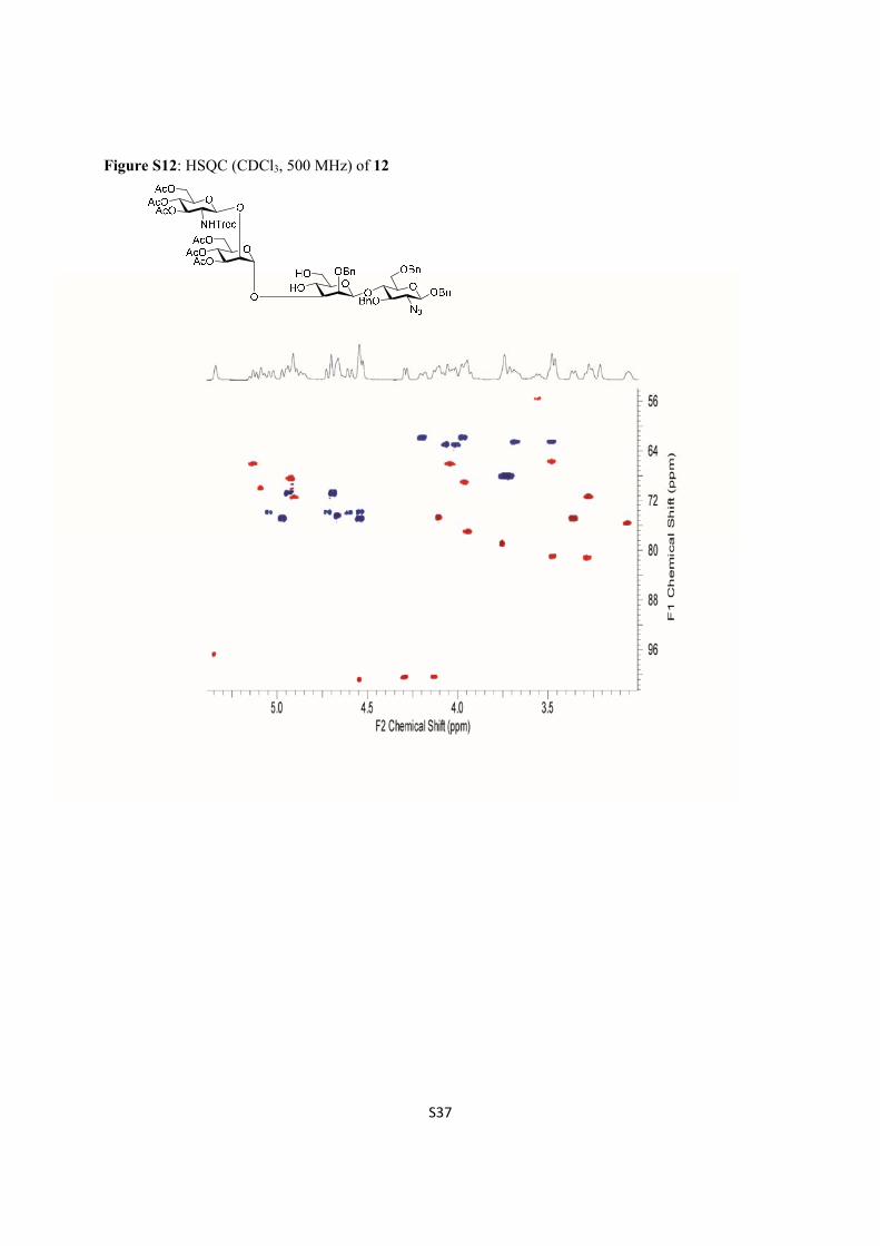
13C NMR (150 MHz, CDCl3, 25oC): δ ppm 128.3, 128.2, 127.7, 126.7, 100.9, 100.4, 100.4, 96.8, 81.3, 81.0,
79.1, 77.1, 75.7, 75.1, 75.0, 75.0, 74.9, 74.5, 74.1, 73.9, 73.9, 73.9, 71.4, 71.4, 70.9, 70.8, 70.2, 70.1, 70.1, 69.6,
69.1, 68.6, 68.3, 66.9, 66.3, 66.3, 65.8, 64.1, 63.3, 63.1, 62.7, 62.6, 62.0, 62.0, 55.5, 20.8, 20.8; see, Figure S12.
MALDI-TOFMS: m/z calculated for C67H79Cl3N4O27, [M+H]+ = 1477.407, C67H79Cl3N4NaO27, [M+Na]+ =
1499.389, found 1476.030 and 1502.174.

S10
Benzyl {(3,4,6-tri-O-acetyl-2-deoxy-2-(2,2,2-trichloroethoxycarbonylamino)-β-D-glucopyranosyl-(1→2)-
3,4,6-tri-O-acetyl-α-D-mannopyranosyl)-(1→3)-[(3,4,6-tri-O-acetyl-2-deoxy-2-(2,2,2-
trichloroethoxycarbonylamino)-β-D-glucopyranosyl-(1→2)-(3,4,6-tri-O-acetyl-2-deoxy-2-(2,2,2-
trichloroethoxycarbonylamino)-β-D-glucopyranosyl)-(1→6)]-3,4-di-O-acetyl-α-D-mannopyranosyl)-
(1→6)-2-O-benzyl-β-D-mannopyranosyl}-(1→4)-2-azido-2-deoxy-3,6-di-O-benzyl-β-D-glucopyranoside
(13).
13
A solution of diol as a glycosyl acceptor 12 (236 mg, 160 μmol) and a glycosyl donor 6S1 (319 mg, 239 µmol)
was co-evaporated with toluene twice and once more in presence of molecular sieves 4 Ǻ (320mg), magnetic
stirrer and dried under vacuum for 12 h. Dry DCM (32 mL, 0.005 M) was added and cooled to -35°C. After 30
min, BF3ꞏEt2O (8 μL, 63 μmol) was added at -35oC and the reaction mixture was stirred at -35°C for 2 h, and
then the mixture was filtered, diluted with EtOAc and washed with sat. NaHCO3, brine, dried over Na2SO4,
filtered and concentrated. The crude residue was purified by column chromatography on silica gel
(toluene/EtOAc=65:35) to give the heptasaccharide 13 (387 mg, 90%).
1H NMR (600 MHz, CDCl3, 25oC): δ ppm 7.40-7.15 (m, 20 H), 6.27 (brs, 1 H), 5.83 (d, 1 H, J = 9.5 Hz), 5.54
(t, 1 H, J = 9.2 Hz), 5.35 (brs, 1 H), 5.54 (t, 1 H, J = 10.1 Hz), 5.20-4.52 (m, 30H), 4.31-4.26 (m, 3 H), 4.21-4.06
(m, 11 H), 4.02 (brd, 1 H, J = 7.8 Hz), 3.98 (d, 1 H, J = 9.5 Hz), 3.95-3.89 (m, 5 H), 3.80 (m, 1 H), 3.74-3.58
(m, 9 H), 3.52-3.46 (m, 2 H), 3.37-3.28 (m, 4 H), 3.22 (m, 1 H), 3.15-3.07 (m, 2 H), 2.95 (brd, 1 H, J = 10.4 Hz),
2.11 (s, 3 H), 2.09 (s, 6 H), 2.04-1.99 (m, 36 H) ; see, Figure S13.
13C NMR (150 MHz, CDCl3, 25oC): δ ppm 128.4, 128.2, 128.2, 128.0, 126.5, 101.9, 100.5, 100.3, 100.3, 97.3,
96.8, 96.5, 81.0, 81.0, 78.8, 76.7, 74.9, 74.9, 74.8, 74.7, 74.6, 74.2, 74.0, 73.9, 73.9, 73.8, 73.2, 72.9, 71.9, 71.8,
71.6, 71.3, 71.1, 71.0, 71.0, 70.9, 70.6, 70.1, 70.1, 70.0, 69.3, 69.2, 69.2, 68.6, 68.4, 68.3, 67.6, 67.5, 67.4, 67.4,
66.9, 66.0, 65.9, 65.8, 62.2, 62.1, 62.1, 61.8, 61.7, 56.3, 56.1, 56.0, 55.7, 21.0, 20.8, 20.8; see, Figure S14.
MALDI-TOFMS: m/z calculated for C107H129Cl9N6NaO52, [M+Na]+ = 2667.472, found 2665.197.
OOBnO
OO
O
OBnBnO
HO OBn
N3
O
O
O
AcOAcO
OAcO
AcOAcO
NHTrocO
AcOAcO
AcO
NHTroc
OAcO
O
AcOAcO
OAcO
AcOAcO
NHTroc
S11
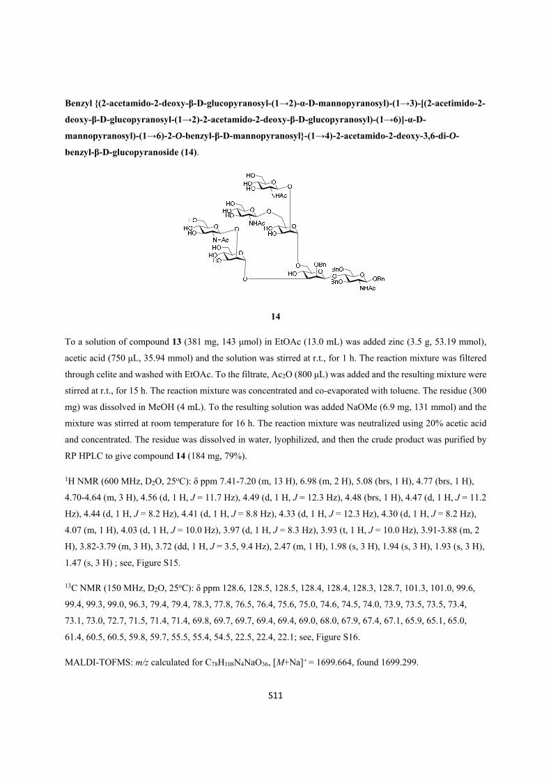
Benzyl {(2-acetamido-2-deoxy-β-D-glucopyranosyl-(1→2)-α-D-mannopyranosyl)-(1→3)-[(2-acetimido-2-
deoxy-β-D-glucopyranosyl-(1→2)-2-acetamido-2-deoxy-β-D-glucopyranosyl)-(1→6)]-α-D-
mannopyranosyl)-(1→6)-2-O-benzyl-β-D-mannopyranosyl}-(1→4)-2-acetamido-2-deoxy-3,6-di-O-
benzyl-β-D-glucopyranoside (14).
14
To a solution of compound 13 (381 mg, 143 μmol) in EtOAc (13.0 mL) was added zinc (3.5 g, 53.19 mmol),
acetic acid (750 μL, 35.94 mmol) and the solution was stirred at r.t., for 1 h. The reaction mixture was filtered
through celite and washed with EtOAc. To the filtrate, Ac2O (800 μL) was added and the resulting mixture were
stirred at r.t., for 15 h. The reaction mixture was concentrated and co-evaporated with toluene. The residue (300
mg) was dissolved in MeOH (4 mL). To the resulting solution was added NaOMe (6.9 mg, 131 mmol) and the
mixture was stirred at room temperature for 16 h. The reaction mixture was neutralized using 20% acetic acid
and concentrated. The residue was dissolved in water, lyophilized, and then the crude product was purified by
RP HPLC to give compound 14 (184 mg, 79%).
1H NMR (600 MHz, D2O, 25oC): δ ppm 7.41-7.20 (m, 13 H), 6.98 (m, 2 H), 5.08 (brs, 1 H), 4.77 (brs, 1 H),
4.70-4.64 (m, 3 H), 4.56 (d, 1 H, J = 11.7 Hz), 4.49 (d, 1 H, J = 12.3 Hz), 4.48 (brs, 1 H), 4.47 (d, 1 H, J = 11.2
Hz), 4.44 (d, 1 H, J = 8.2 Hz), 4.41 (d, 1 H, J = 8.8 Hz), 4.33 (d, 1 H, J = 12.3 Hz), 4.30 (d, 1 H, J = 8.2 Hz),
4.07 (m, 1 H), 4.03 (d, 1 H, J = 10.0 Hz), 3.97 (d, 1 H, J = 8.3 Hz), 3.93 (t, 1 H, J = 10.0 Hz), 3.91-3.88 (m, 2
H), 3.82-3.79 (m, 3 H), 3.72 (dd, 1 H, J = 3.5, 9.4 Hz), 2.47 (m, 1 H), 1.98 (s, 3 H), 1.94 (s, 3 H), 1.93 (s, 3 H),
1.47 (s, 3 H) ; see, Figure S15.
13C NMR (150 MHz, D2O, 25oC): δ ppm 128.6, 128.5, 128.5, 128.4, 128.4, 128.3, 128.7, 101.3, 101.0, 99.6,
99.4, 99.3, 99.0, 96.3, 79.4, 79.4, 78.3, 77.8, 76.5, 76.4, 75.6, 75.0, 74.6, 74.5, 74.0, 73.9, 73.5, 73.5, 73.4,
73.1, 73.0, 72.7, 71.5, 71.4, 71.4, 69.8, 69.7, 69.7, 69.4, 69.4, 69.0, 68.0, 67.9, 67.4, 67.1, 65.9, 65.1, 65.0,
61.4, 60.5, 60.5, 59.8, 59.7, 55.5, 55.4, 54.5, 22.5, 22.4, 22.1; see, Figure S16.
MALDI-TOFMS: m/z calculated for C78H108N4NaO36, [M+Na]+ = 1699.664, found 1699.299.

S12
Benzyl {(β-D-galactopyranosyl-(1→4)-2-acetamido-2-deoxy-β-D-glucopyranosyl-(1→2)-α-D-
mannopyranosyl)-(1→3)-[(β-D-galactopyranosyl-(1→4)-2-acetimido-2-deoxy-β-D-glucopyranosyl-
(1→2)-β-D-galactopyranosyl-(1→4)-2-acetamido-2-deoxy--β-D-glucopyranosyl)-(1→6)]-α-D-
mannopyranosyl)-(1→6)-2-O-benzyl-β-D-mannopyranosyl}-(1→4)-2-acetamido-2-deoxy-3,6-di-O-
benzyl-β-D-glucopyranoside (15).
15
To a solution of compound 14 (3.7 mg, 2.2 μmol) in 110.2 μL of water (20 mM; theoretical concentration),
added a mixture of 1 M Tris buffer (pH 7.5) (24 μL), 1 M MnCl2 (2.4 μL), 200 mM UDP-galactose (90 μL), 4
units/mL recombinant human β1,4-galactosyltransferase (GalT) (24 μL) and water (99.6 μL). After incubation
at 250C for 24 h, the crude product was purified by RP-HPLC (column: Inertile ODS-3, 20x250 mm) eluted with
H2O and CH3CN to afford the decasaccharide 15 (2.4 mg, 93%).
1H NMR (600 MHz, D2O, 25oC): δ ppm 7.41-7.19 (m, 13 H), 6.97 (m, 2 H), 5.08 (brs, 1 H), 4.70-4.64 (m, 3 H),
4.56 (d, 1 H, J = 11.7 Hz), 4.49 (d, 1 H, J = 12.6 Hz), 4.47-4.45 (m, 3 H), 4.43 (d, 1 H, J = 8.3 Hz), 4.37 (d, 1 H,
J = 7.8 Hz), 4.36 (d, 1 H, J = 7.8 Hz),4.32 (d, 1 H, J = 7.9 Hz), 4.30 (d, 1 H, J = 7.7 Hz), 4.07 (m, 1 H), 4.04 (d,
1 H, J = 9.3 Hz), 3.99 (m, 1 H), 3.97 (d, 1 H, J = 8.3 Hz), 3.93 (t, 1 H, J = 9.9 Hz), 3.92-3.86 (m, 4 H), 3.82-3.79
(m, 5 H), 3.75 (m, 1 H), 2.56 (m, 1 H), 1.97 (s, 3 H), 1.93 (s, 6 H), 1.47 (s, 3 H) ; see, Figure S17.
13C NMR (150 MHz, D2O, 25oC): δ ppm 128.6, 128.5, 128.5, 128.4, 127.6, 102.8, 102.8, 102.6, 101.2, 101.0,
99.5, 99.4, 99.2, 98.9, 96.2, 79.4, 79.3, 78.2, 77.8, 77.6, 76.4, 76.3, 75.2, 74.6, 74.5, 74.4, 74.0, 73.9, 73.9, 73.5,
73.1, 73.0, 72.5, 71.4, 71.3, 70.9, 69.9, 69.9, 69.3, 68.4, 67.9, 67.8, 67.4, 67.0, 66.0, 65.1, 65.0, 61.4, 61.4, 60.9,
60.0, 59.9, 59.2, 59.2, 54.9, 54.7, 22.5, 22.5, 22.2; see, Figure S18.
MALDI-TOFMS: m/z calculated for C96H138N4NaO51, [M+Na]+ = 2185.822, found 2186.200.
O OO
OHO
O
OBn
O
O
O
HO
NHAcHO
HOHO
OHO
OBn
NHAc
OBnBnO
O
O
O
HO
NHAc
HOHO
OHO
O O
HO
NHAc
OHO
OOHOH
HOOH
OOHOH
HOOH
OOHOH
HOOH

S13
β-D-galactopyranosyl-(1→4)-2-acetimido-2-deoxy-β-D-glucopyranosyl-(1→2)-α-D-mannopyranosyl-
(1→3)-[(β-D-galactopyranosyl-(1→4)-2-acetimido-2-deoxy-β-D-glucopyranosyl-(1→2)-β-D-
galactopyranosyl-(1→4)-2-acetimido-2-deoxy--β-D-glucopyranosyl)-(1→6)]-α-D-mannopyranosyl)-
(1→6)-β-D-mannopyranosyl-(1→4)-2-acetamido-deoxy-D-glucopyranose (16).
16
To a solution of compound 15 (9.0 mg, 3.6 μmol) in 50% AcOH (aq.) (4.0 mL), added 20% Pd(OH)2/C (5 mg)
and stirred for 4 h under H2 atmosphere. The reaction mixture was filtered through a celite pad, the filtrate was
concentrated and then the residue was dissolved in water and lyophilized. The crude product was purified on a
Sephadex G15 column by elution with H2O. Fractions containing the product were pooled and lyophilized to
give the free decasaccharide 16 (7.3 mg, 95%) as white solid.
1H NMR (600 MHz, D2O, 25oC): δ ppm 5.10 (d, 1 H, J = 3.2 Hz), 5.02 (brs, 1 H), 4.77 (brs, 1 H), 4.67-4.61 (m,
1 H), 4.50-4.47 (m, 2 H), 4.45 (d, 1 H, J = 8.2 Hz), 4.38-4.35 (m, 3 H), 4.15 (m, 1 H), 4.11-4.09 (m, 2 H), 3.99
(m, 1 H), 3.30 (m, 1 H), 1.95 (s, 6 H), 1.94 (brs, 6 H) ; see, Figure S19.
13C NMR (150 MHz, D2O, 25oC): δ ppm 102.8, 102.8, 102.8, 101.4, 100.1, 99.3, 99.3, 97.0, 94.8, 90.4, 80.2,
79.6, 78.3, 76.4, 76.2, 75.2, 74.5, 72.4, 71.9, 70.9, 70.2, 70.1, 68.4, 67.4, 67.2, 65.5, 65.3, 65.3, 61.6, 61.6, 61.0,
60.0, 59.8, 54.9, 53.5, 23.3, 23.3, 22.5, 22.3; see, Figure S20.
MALDI-TOF MS: m/z calculated for C68H114N4NaO51, [M+Na]+ = 1825.634, found 1826.191.

S14
II-III. Construction of AGP glycopeptide (1) by trans glycosylation with mutant endo-M-N175Q
Synthesis of AGP 24mer peptide fragment (3) carrying a GlcNAc at Asn54 residue.
3
2-Chlorotrityl chloride resin (1.58 mmol/g, 100 mg, 158 μmol), Fmoc-amino acids (96 μmol, 4.0 equiv), and
Fmoc-Asn(OAc3-β-GlcNAc)-OH (189.6 μmol, 1.2 equiv) were used. Fmoc amino acids used are Fmoc-
Arg(Pbf)-OH, Fmoc-Leu-OH, Fmoc-Phe-OH, Fmoc-Asp(OtBu)-OH, Fmoc-Asn(Trt)-OH, Fmoc-Pro-OH,
Fmoc-Lys(Boc)-OH, Fmoc-Tyr(t-Bu)-OH, Fmoc-Gln(Trt)-OH, Fmoc-Val-OH, Fmoc-Ser(t-Bu)-OH, Fmoc-
Glu(OtBu)-OH, Fmoc-Ala-OH, Fmoc-Ile-OH and Fmoc-Thr(t-Bu)-OH. Fifty mg of 2-chloro trityl chloride resin
was placed in a 10 mL Libra tube and allowed to swell in DCM for a period of 2 h at r.t., removed the DCM
after that. Very first Fmoc amino acid was coupled to the resin by adding a base, DIEA (144 µmol, 6 eq.) under
microwave irradiation for 9 min and methanol capping was performed for 10 sec. at room temperature. From 2nd
Fmoc amino acids coupling onwards, each corresponding Fmoc amino acid (632 μmol, 4.0 equiv) dissolved in
a mixture of HBTU, HOBt in DMF (96 μmol, 4.0 equiv), and DIEA (144 μmol, 6.0 equiv) (final concentration
of amino acid, 0.4 M) was added to the resin and the mixture was shaken under microwave irradiation for 10
min. For an introduction of the Fmoc-glycosylated amino acid, Fmoc-Asn(OAc3-β-GlcNAc)-OH (36 μmol, 1.5
equiv) dissolved in a mixture of PyBOP, HOBT in DMF (36 μmol, 1.5 equiv), and DIEA (72 μmol, 3 equiv)
(final concentration of amino acid, 0.2 M) was treated with a resin under microwave irradiation for 15 min. After
coupling of each amino acid, acetyl capping of unreacted amino acids was performed with a solution of
Ac2O:DIEA:DMF (1.0:0.5:8.5) followed by the Fmoc removal reaction conducted with 20% piperidine in DMF
(2 mL) and the mixture was shaken under microwave irradiation for 3 min. Following filtration and washing
with DMF and DCM (3 mL, three times each), Fmoc-removal, coupling, and capping procedures as described
above were carried out repeatedly. After completion of the synthesis, the glycopeptidyl-resin was treated with
TFA:H2O:TIS (95.0:2.5:2.5) (2.0 mL) at r.t. for 2 h and the resin was filtered. The resin was washed twice with
the same cocktail and the filtrates were combined and concentrated by streaming of nitrogen gas. The
glycopeptides was precipitated by adding cold tert-butylmethylether, discarded the liquid which is separated
after centrifugation and the residue obtained was dissolved in 50% aq. acetonitrile and lyophilized. Then, the
residue was dissolved in methanol (5.0 mL) and the pH was adjusted to 12.8 using 1 N sodium hydroxide, stirred
at r.t. for 2 h. The mixture was neutralized by an addition of 1 N acetic acid and evaporated. The crude material
was purified by RP-HPLC to yield pure compound 3 (1.6 mg, 5.4% overall yield calculated from the resin
employed, 9.6 µmole). HPLC profile and MALDI-TOFMS were shown in Figure S21.

S15
A
B
Figure S21. (A) Reverse-phase analytical HPLC analysis of the compound 3, and (B) MALDI-TOFMS
representing a peak at m/z 3098.520 (m/z 3098.536 as [C143H213N32O45+H] +).

S16
Sythesis of AGP glycopeptide having asialo-triantennary N-glycan at Asn54 residue: Recombinant endo-
M (N175Q) catalyzed trans-glycosylation between decasaccharide oxazoline (2) and AGP 24mer peptide
fragment carrying a GlcNAc residue (3).
To a solution of decasaccharide 16 (7.3 mg, 3.4 μmol) in H2O (1.0 mL) was added Et3N (43 μL, 305 μmol) and
2-chloro-1,3-dimethylimidazolinium chloride (DMC) (22 mg, 119 μmol). The solution was stirred at 00C for 40
min, then the reaction mixture was purified directly by gel filtration on a Sephadex G10 column eluted with
0.5% aq. NH3. The fractions containing the product was evaporated in vacuo and the residue was lyophilized to
afford crude decasaccharide oxazoline 2 (approximately 7.0 mg) as a white solid. According to the general
method reported previously,S3 unstable oxazoline derivative 2 was used directly for the next step without further
purification. To a solution of 2 μL of 6.5 mM AGP glycopeptide 3 in Milli Q H2O, was added 2.6 μL (3eq.) of 15
mM decasaccharide oxazoline 2 dissolved in 25 mM phosphate buffer (pH6.0) and mutant endo-M-N175Q (50
mU/mL), and the mixture was incubated at 300C for 10 h to provide compound 1 in 20% yield estimated from the
reverse-phase analytical HPLC. As shown in Figure 2A in the main text, trans-glycosylation of an oxazoline 2 to the
acceptor 3 monitored by reverse-phase analytical HPLC indicated that the reaction becomes an optimal at around 10
h and longer incubation appears to reduce the yield of the product. HPLC profile of the isolated AGP glycopeptide 1
was shown in Figure 2B in the main text.
MALDI-TOFMS: m/z calculated for C211H325N36O95, [M+H] + = 4883.170, found = 4883.150 [see, Figure 2C in the
main text].

S17
III. SRM-based targeted glycoproteomics
III-I. SRM channel for the synthetic AGP glycopeptide (1)
Optimization of SRM/MRM parameters for synthetic AGP glycopeptide 1.
The accurate concentration of compound 1 was defined on the basis of the results of amino acid analysis as
summarized in Table S1. A solution of synthetic AGP glycopeptide 1 (800 fmol/µL) prepared in 1:1 ratio of
0.1% formic acid aq. and acetonitrile was used to analyze its mass and fragments by infusion method using
4000Q-TRAP mass spectrometry and syringe pump with a flow of 5µL/min using 1mL syringe of a diameter of
4.61 mm. The EMS (Enhanced mass) mode measurement showed m/z 1630.0 which is believed to be of the
synthetic AGP glycopeptide 1; ER (Enhanced Resolution) mode of measurement which is usually gives higher
resolution compared to EMS mode of measurement and hence found the m/z 1628.500 its isotopomer found in
the TIC (Total Ion Chromatogram) are 1628.5, 1628.8, 1629.2, 1629.5, 1629.8, 1630.2, 1630.5, 1630.9 shown
in Figure 3A in the main text. These isotopomer with a difference of almost 0.3 Da indicates the charge of the
glycopeptide 1 is 3+. Herein the highest intensity of isotopomer observed at m/z 1629.2 was selected as a
precursor ion (Q1) of glycopeptide fragment 1 of AGP. Thus, the isotopomer of m/z 1629.2 was selected to
analyze fragmentation by collision with inert N2 gas at Q2 quadrupole in EPI (Enhanced Product Ion) mode of
measurement at collision energy of 80 V and found that the fragments detected are of m/z 366.2, 649.6, 1448.5,
1550.4, 1652.0, 1733.0, 1814.2, 1894.9, 1996.6, 2077.6 as shown in Figure 3B in the main text.
The selected fragments of glycopeptide 1 with m/z 1629.200 as Q1 for an optimization of MRM parameters
were of m/z 1550.400, 366.200, 2077.600, 1996.600, 1894.900, 1814.200, 1733.000, 1652.000, 1448.500 and
649.600 (Figure 3B in the main text). Out of which, fragments of m/z 366.200, 2077.600, 1996.000, 1550.400,
649.000 showed better intensity in cps with an increasing order of almost 366.200 > 2077.600 > 1996.000 >
1550.400 > 649.000 with optimized collision energy listed in Table S2.

S18
Table S1. Amino acid analysis of synthetic AGP glycopeptide 1.
Final concentration of the stock solution of AGP glycopeptide 1 was estimated as 35.2 nmol/mL (172µg/mL) by calculated from the above total concentration of the tested solution (4.41 nmol/40 µL).
VIS1 (570 nm) No.
Elution time (min)
Name of amino acid
Peak height Peak area Concentration in nmole/40 µL
1
16.587
Asp
33245
1420379
0.36622
2 20.133 Thr 53760 2558219 0.64245
3 21.807 Ser 18137 953817 0.22957
4 27.100 Glu 49141 3019402 0.72700
5 39.360 Gly 5089 374586 0.09312
6 42.547 Ala 11122 853469 0.20598
7 46.027 Val 24471 776801 0.18551
8 51.693 Ile 32585 1379442 0.33574
9 53.320 Leu 16945 785332 0.19209
10 55.153 Nle 93790 4253808 1.05682
11 56.627 Tyr 18197 705397 0.17840
12 58.420 Phe 70987 2617778 0.67015
15 68.347 Lys 21701 877271 0.19496
18 71.127 His 1669 71335 0.01761
19 73.873 NH3 200436 15110489 4.64983
20 85.120 Arg 10151 656013 0.16964
VIS2 (440 nm) No.
Elution time(min)
Name of amino acid
Peak height Peak area Concentration in nmole/40 µL
5 29.473 Pro 2561 177471 0.20194
Total 4.41039

S19
Table S2. SRM/MRM channel candidates with optimized parameters for Q1 (m/z 1629.200).
Q1 Q3 DP (V) CE (V) CXP (V) EP (V)
1629.200 366.200 186.0 57.0 8.0 10.0
1629.200 2077.600 186.0 57.0 54.0 10.0
1629.200 1996.600 186.0 63.0 50.0 10.0
1629.200 1550.400 186.0 65.0 40.0 10.0
1629.200 649.600 186.0 111.0 16.0 10.0
Manual CE (collision energy) optimization was performed for a few parameters by step of 1 CE difference ramping. Finally, SRM/MRM transition of 1629.2/366.2 was obtained as an optimized channel for the quantitation of AGP glycopeptide 1. DP (declustering potential), CXP (collision cell exit potential), and EP (entrance potential).

S20
III-II. Targeted glycoproteomics by SRM assay focusing human serum AGP glycopeptide (1)
Tryptic digestion of whole serum glycoproteins to release glycopeptide fragments.
Ten micro liter (10 µL) of human serum was added to 0.33 M ammonium bicarbonate (15 µL) and Milli Q H2O
(30 µL) and the solution was incubated at 60°C for 10 min. To the solution was added 120 mM dithiothreitol
(DTT) (5 µL) and incubated at 60°C for 30 min, subsequently added 123 mM iodoacetamide (IAA) (10 µL) and
incubated for 1 h in a dark condition at room temp. Then, the mixture was added 5 µL of trypsin (40 U/µL) in 1
mM HCl and incubated at 370C for 16 h. The reaction mixture was heated at 900C for 10 min to inactivate an
enzyme. Finally, the mixture was dried in a SpeedVac concentrator.
SRM/MRM-based LC-MS/MS quantification of serum AGP glycopeptide fragment (1).
LC-MS/MS analysis was conducted with Dionex UltimateTM 3000 HPLC and AB Sciex 4000Q Trap®
TurboIonSpray system. Separation was performed with LPG-3×00 pump, WPS-3000 auto sampler, FLM-3100
column component and WVD-3400 detector under the control of software: Chromeleon 6.80. Column: Inertsil
Diol 4.6×250 mm and Inertsil ODS-3 2.1×150 mm (GL Sciences Inc.). Acquired data was analyzed by a series
of software: Analyst 1.5 and MultiQuant 1.1.0.26.
LC condition was optimized for the SRM/MRM channel of Q1/Q3 (1629.2/366.2) for the synthetic AGP
glycopeptide fragment 1 in advance with a concentration of 1000 fmole/µL: flow of 200 µL/min; multi-step
gradient; (A):(B) = 0.1% formic acid in aqueous solution: 0.1% formic acid in acetonitrile; (A)/(B), 0 min :
97/3→18 min: 72/28→19 min: 10/90→23 min: 10/90→23.1 min: 97/3→30 min: 97/3; column temperature:
60°C; inject volume: 1.5 µL, respectively. As a result, Q1/Q3 = 1629.2/366.2 gave satisfactory S/N spectra at
elution time of 17.35 ± 0.05 min. A series of samples of the concentrations of 40, 80, 150, 300, 600 and 1600
fmole/µL was prepared from above mentioned stock solution of 35.2 nmole/mL (172µg/mL) of the synthetic AGP
glycopeptide 1 as internal standards with 0.1% formic acid in Milli Q H2O. Pretreated human serum samples were
dissolved in 30 µL of 0.1% formic acid in Milli Q H2O, separately. Finally, 1.5 µL of all standard samples of the
synthetic AGP glycopeptide 1 and 10 µL of all pretreated human serum samples of healthy, HCC and RCC patients
were submitted to the SRM-based LC-MS/MS quantification and the total ion chromatogram (TIC) of acquired
quantification data were shown in Figure S22-S25.

S21
Figure S22. TIC of synthetic glycopeptide 1 at 6 different concentrations for the calibration curve: (A) 1600 fmole/L,
(B) 600 fmole/L, (C) 300 fmole/L, (D) 150 fmole/L, (E) 80 fmole/L, and (F) 40 fmole/L, respectively.

S22
Figure S23. TIC of serum AGP glycopeptide 1 derived from sera of 5 different healthy controls A~E, respectively.

S23
Figure S24. TIC of serum AGP glycopeptide 1 derived from sera of 5 different HCC patients A~E, respectively.

S24
Figure S25. TIC of serum AGP glycopeptide 1 derived from sera of 5 different RCC patients A~E, respectively.

S25
IV. References for the Supporting Information
S1. Ravi, K. H. V.; Naruchi, K.; Miyoshi, R.; Hinou, H.; Nishimura, S. -I. A new approach for the synthesis of
hyperbranched N-glycan core structures from locust bean gum. Org. Lett. 2013, 15, 6278-6281.
S2. Kurogochi, M.; Matsushita, T.; Amano, M.; Furukawa, J.; Shinohara, Y.; Aoshima, M.; Nishimura, S. -I. Sialic
acid-focused quantitative mouse serum glycoproteomics by multiple reaction monitoring assay. Mol. Cell.
Proteomics 2010, 9, 2354-2368.
S3. Umekawa, M.; Li, C.; Higashiyama, T.; Huang, W.; Ashida, H.; Yamamoto, K.; Wang, L-X. fficient
glycosynthase mutant derived from Mucor hiemalis endo-β-N-acetylglucosaminidase capable of transferring
oligosaccharide from both sugar oxazoline and natural N-glycan. J. Biol. Chem. 2010, 285, 511-521.

S26
Figure S1: 1H-NMR (CDCl3, 500 MHz) of 8

S27
Figure S2: HSQC (CDCl3, 500 MHz) of 8

S28
Figure S3: 1H-NMR (CDCl3, 500 MHz) of 9

S29
Figure S4: HSQC (CDCl3, 500 MHz) of 9

S30
Figure S5: 1H-NMR (CDCl3, 500 MHz) of 10

S31
Figure S6: HSQC (CDCl3, 500 MHz) of 10

S32
Figure S7: 1H-NMR (CDCl3, 500 MHz) of 4

S33
Figure S8: HSQC (CDCl3, 500 MHz) of 4

S34
Figure S9: 1H-NMR (CDCl3, 500 MHz) of 11

S35
Figure S10: HSQC (CDCl3, 500 MHz) of 11

S36
Figure S11: 1H-NMR (CDCl3, 500 MHz) of 12
S37
Figure S12: HSQC (CDCl3, 500 MHz) of 12

S38
Figure S13: 1H-NMR (CDCl3, 500 MHz) of 13

S39
Figure S14: HSQC (CDCl3, 500 MHz) of 13

S40
Figure S15: 1H-NMR (D2O, 600 MHz) of 14

S41
Figure S16: HSQC (D2O, 600 MHz) of 14

S42
Figure S17: 1H-NMR (D2O, 600 MHz) of 15

S43
Figure S18: HSQC (D2O, 600 MHz) of 15

S44
Figure S19: 1H-NMR (D2O, 600 MHz) of 16

S45
Figure S20: HSQC (D2O, 600 MHz) of 16