Embed Size (px)
Citation preview
An Introduction to the Muscular System
The Muscular System
Consists only of skeletal muscles
Muscle Organization and Function
Muscle organization affects power, range, and speed
of muscle movement
Fascicles
Muscle cells (fibers) are organized in bundles (fascicles)
Fascicle Arrangement
Classification of Skeletal Muscles
By the way fascicles are organized
By relationships of fascicles to tendons
Organization of Skeletal Muscle Fibers
Four patterns of fascicle organization
1. Parallel
2. Convergent
3. Pennate
4. Circular
Fascicle Arrangement
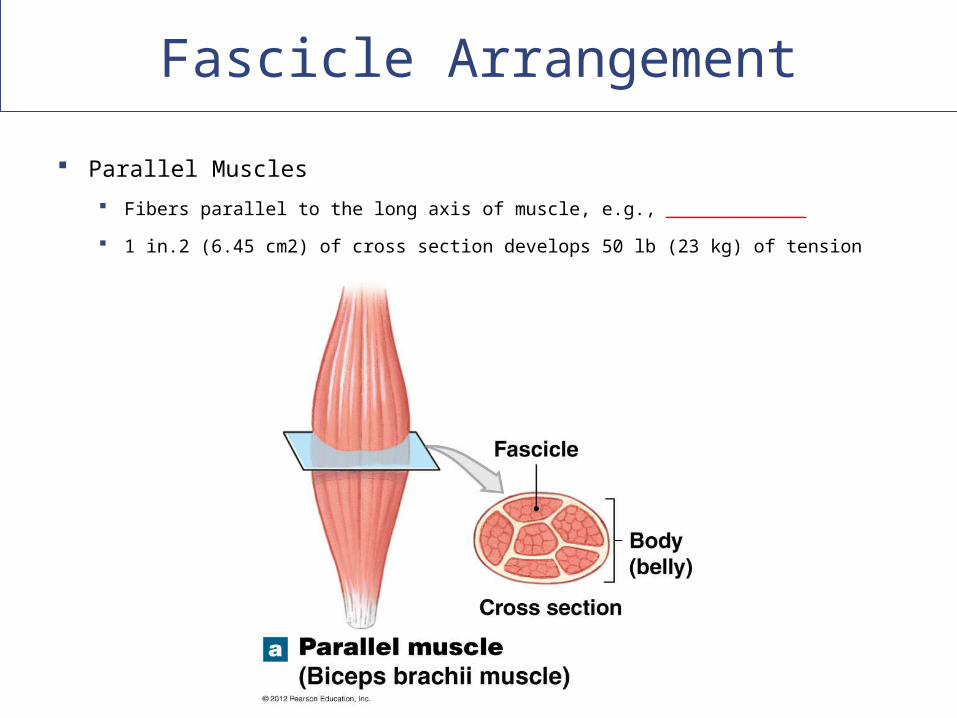
Parallel Muscles
Fibers parallel to the long axis of muscle, e.g., ___________________
1 in.2 (6.45 cm2) of cross section develops 50 lb (23 kg) of tension
Fascicle Arrangement
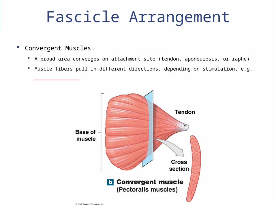
Convergent Muscles
A broad area converges on attachment site (tendon, aponeurosis, or raphe)
Muscle fibers pull in different directions, depending on stimulation, e.g.,
_______________________
Fascicle Arrangement
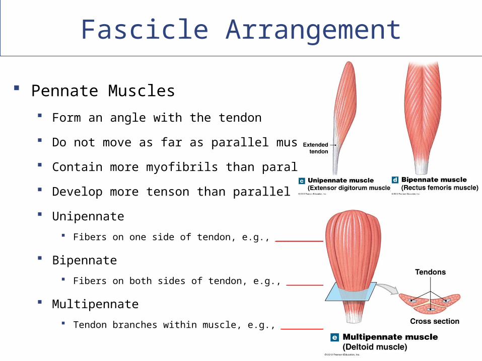
Pennate Muscles
Form an angle with the tendon
Do not move as far as parallel muscles
Contain more myofibrils than parallel muscles
Develop more tenson than parallel muscles
Unipennate
Fibers on one side of tendon, e.g., ________________
Bipennate
Fibers on both sides of tendon, e.g., _________________
Multipennate
Tendon branches within muscle, e.g., __________________
Fascicle Arrangement
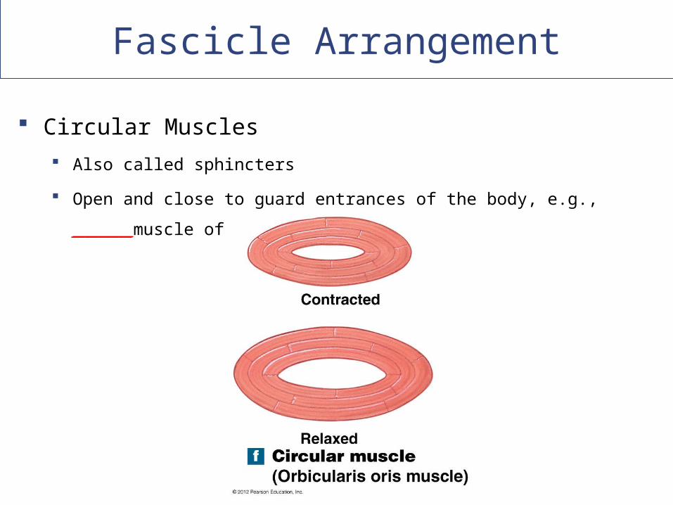
Circular Muscles
Also called sphincters
Open and close to guard entrances of the body, e.g., _________muscle
of the mouth
Muscle Attachments to Other Tissues
Origins and Insertions
Muscles have one fixed point of attachment
(______) and one moving point of attachment
(______________)
Most muscles originate or insert on the
skeleton
_________ is usually __________ to insertion
Muscle Attachments to Other Tissues
Actions
Movements produced by muscle contraction
Body movements
For example, flexion, extension, adduction, etc.
Described in terms of bone, joint, or region
Muscle Attachments to Other Tissues
Muscle Opposition
Agonists and antagonists work in pairs:
When one contracts, the other stretches
Such as flexors–extensors, abductors–adductors,
etc.
Naming Skeletal Muscles
Descriptive Names for Skeletal Muscles
Location in the body
Origin and insertion
Fascicle organization
Relative position
Structural characteristics
Action
Naming Skeletal Muscles
Location in the Body
Identifies body regions
For example, temporalis muscle
Origin and Insertion
First part of name indicates origin
Second part of name indicates insertion
For example, genioglossus muscle
Naming Skeletal Muscles
Fascicle Organization
Describes fascicle orientation within muscle
i.e., rectus (straight), transversus, oblique
Naming Skeletal Muscles
Relative Position Externus (superficialis)
Visible at body surface
Internus (profundus) Deep muscles
Extrinsic Muscles outside an organ
Intrinsic Muscles inside an organ
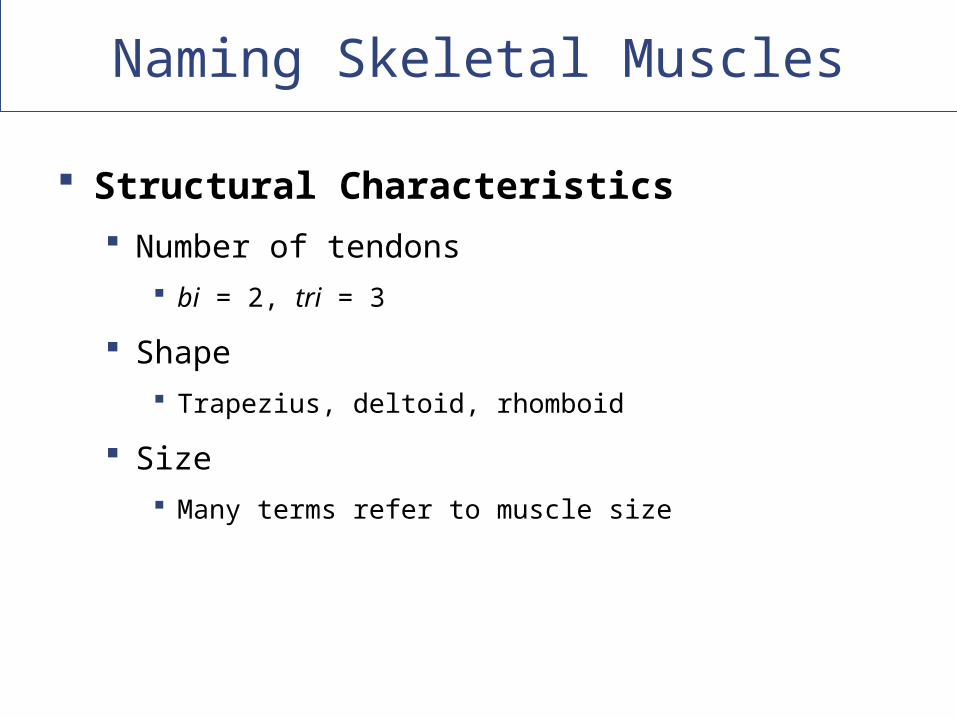
Naming Skeletal Muscles
Structural Characteristics
Number of tendons
bi = 2, tri = 3
Shape
Trapezius, deltoid, rhomboid
Size
Many terms refer to muscle size
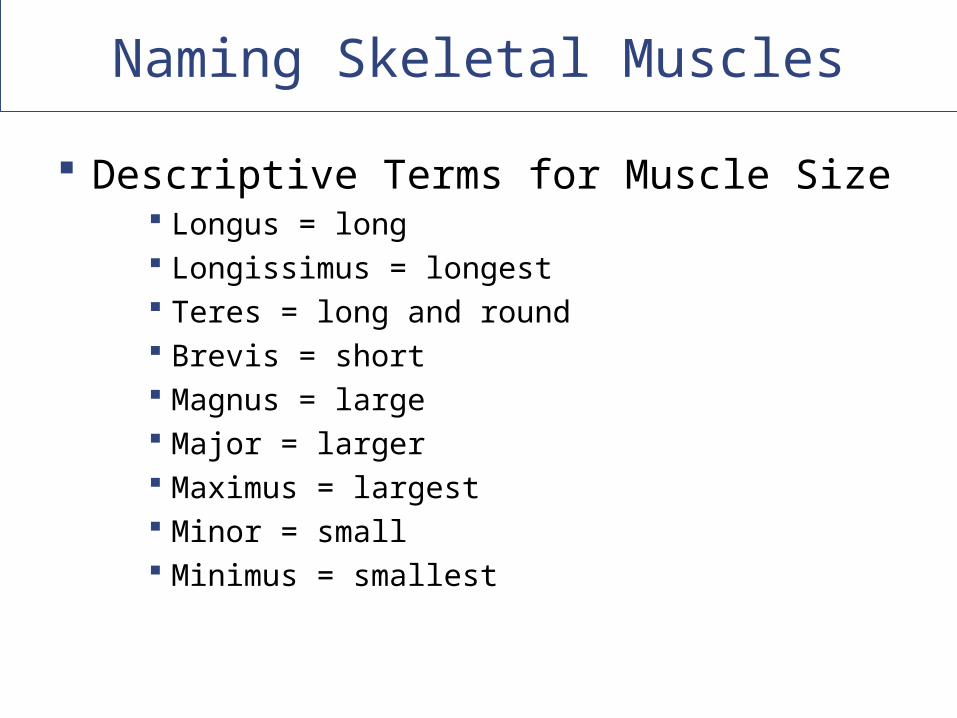
Naming Skeletal Muscles
Descriptive Terms for Muscle Size Longus = long Longissimus = longest Teres = long and round Brevis = short Magnus = large Major = larger Maximus = largest Minor = small Minimus = smallest
Naming Skeletal Muscles
Action
Movements
For example, flexor, extensor, retractor
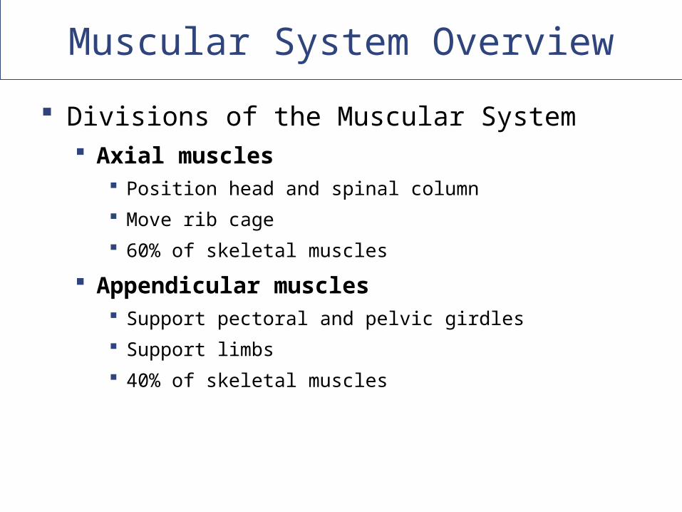
Muscular System Overview
Divisions of the Muscular System Axial muscles
Position head and spinal column
Move rib cage
60% of skeletal muscles
Appendicular muscles Support pectoral and pelvic girdles
Support limbs
40% of skeletal muscles
Copyright © 2009 Pearson Education, Inc., publishing as Pearson Benjamin Cummings
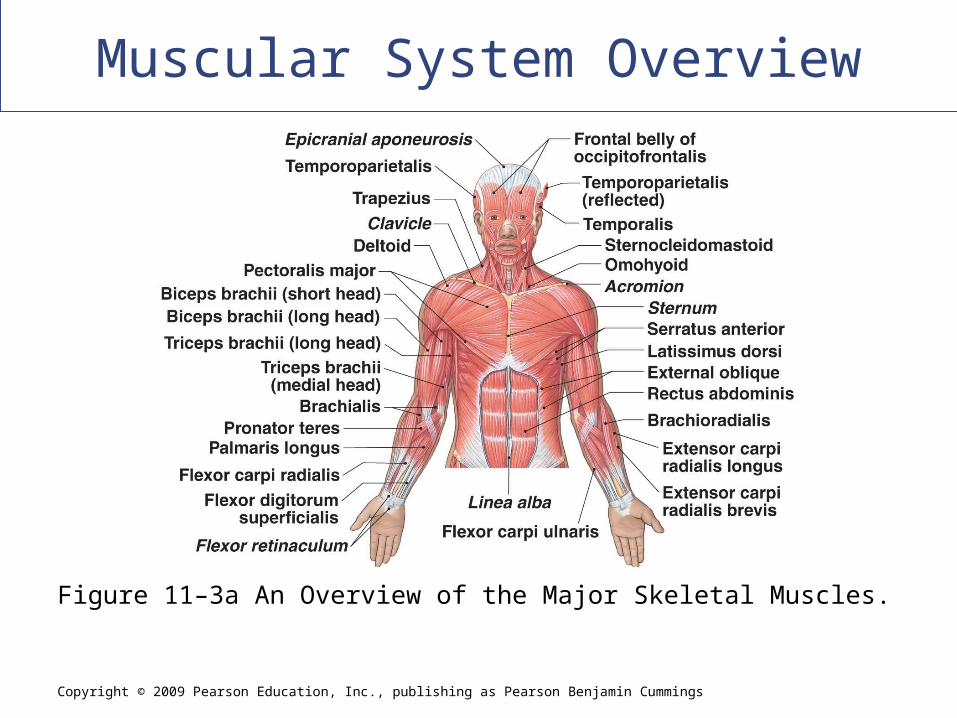
Muscular System Overview
Figure 11–3a An Overview of the Major Skeletal Muscles.
Copyright © 2009 Pearson Education, Inc., publishing as Pearson Benjamin Cummings
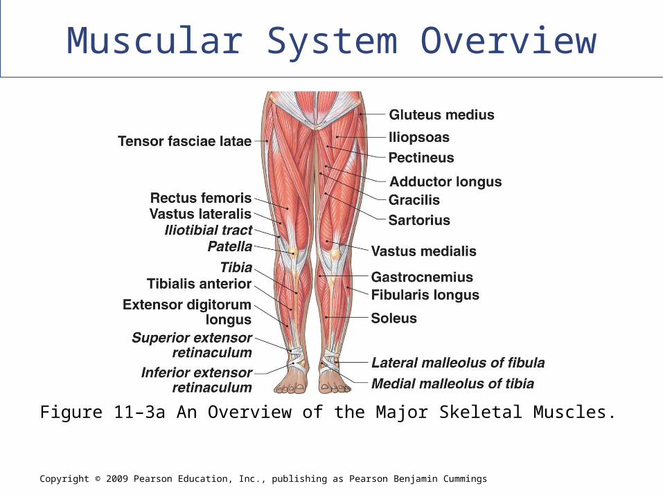
Muscular System Overview
Figure 11–3a An Overview of the Major Skeletal Muscles.
Copyright © 2009 Pearson Education, Inc., publishing as Pearson Benjamin Cummings
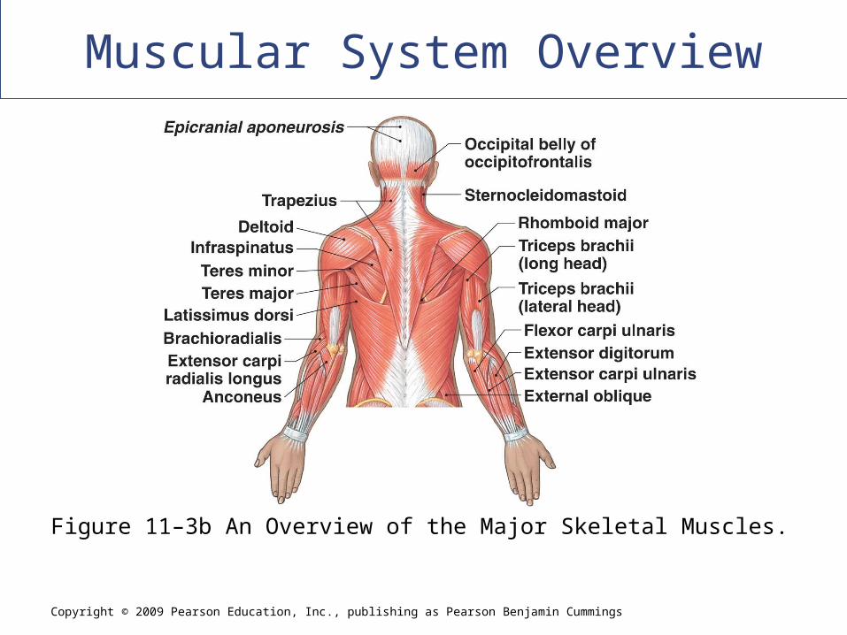
Muscular System Overview
Figure 11–3b An Overview of the Major Skeletal Muscles.
Copyright © 2009 Pearson Education, Inc., publishing as Pearson Benjamin Cummings
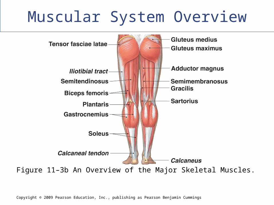
Muscular System Overview
Figure 11–3b An Overview of the Major Skeletal Muscles.
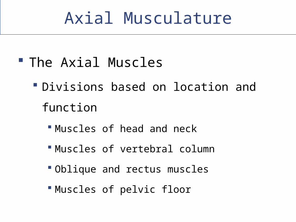
Axial Musculature
The Axial Muscles
Divisions based on location and function
Muscles of head and neck
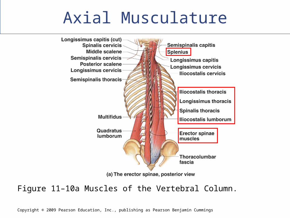
Muscles of vertebral column
Oblique and rectus muscles
Muscles of pelvic floor
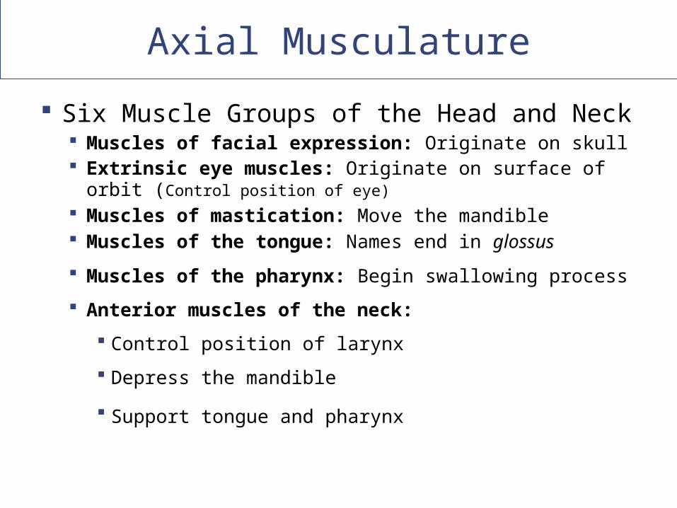
Axial Musculature
Six Muscle Groups of the Head and Neck Muscles of facial expression: Originate on skull Extrinsic eye muscles: Originate on surface of orbit
(Control position of eye)
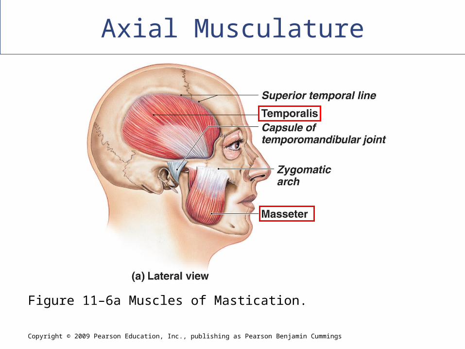
Muscles of mastication: Move the mandible Muscles of the tongue: Names end in glossus
Muscles of the pharynx: Begin swallowing process
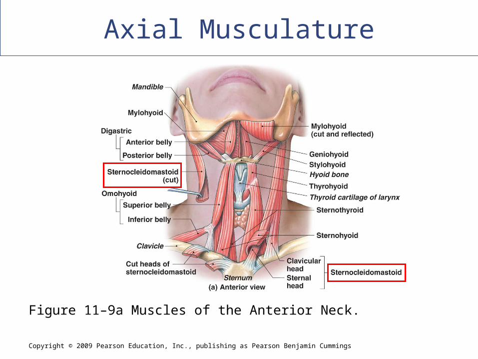
Anterior muscles of the neck:
Control position of larynx
Depress the mandible
Support tongue and pharynx
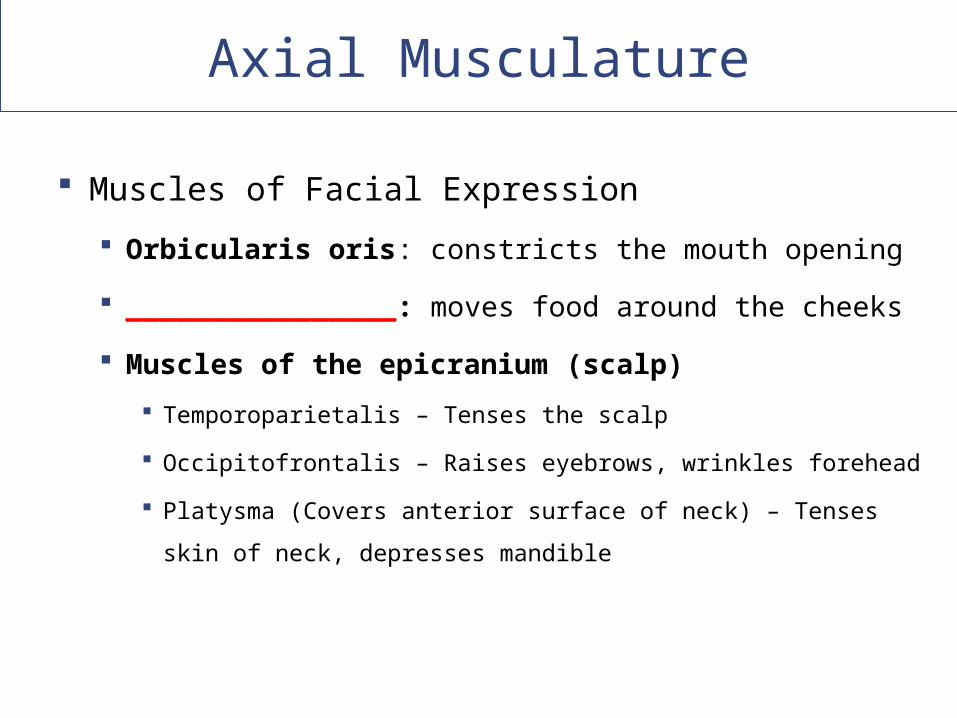
Axial Musculature
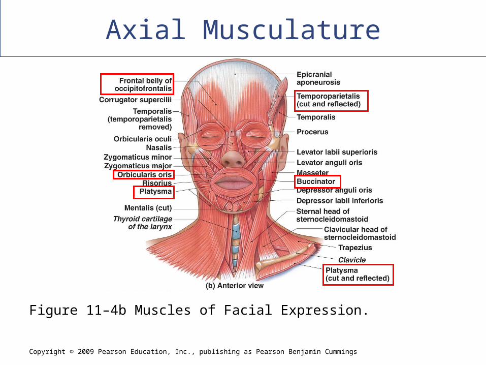
Muscles of Facial Expression
Orbicularis oris: constricts the mouth opening
________________: moves food around the cheeks
Muscles of the epicranium (scalp)
Temporoparietalis – Tenses the scalp
Occipitofrontalis – Raises eyebrows, wrinkles forehead
Platysma (Covers anterior surface of neck) – Tenses skin of
neck, depresses mandible
Copyright © 2009 Pearson Education, Inc., publishing as Pearson Benjamin Cummings
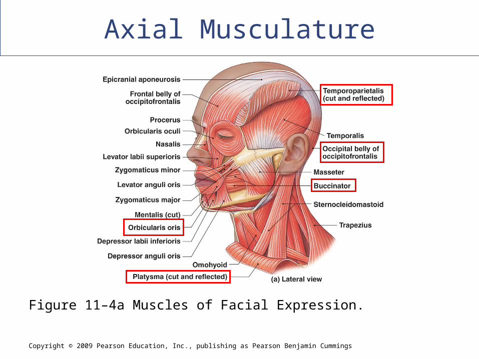
Axial Musculature
Figure 11–4a Muscles of Facial Expression.
Copyright © 2009 Pearson Education, Inc., publishing as Pearson Benjamin Cummings
Axial Musculature
Figure 11–4b Muscles of Facial Expression.
Axial Musculature
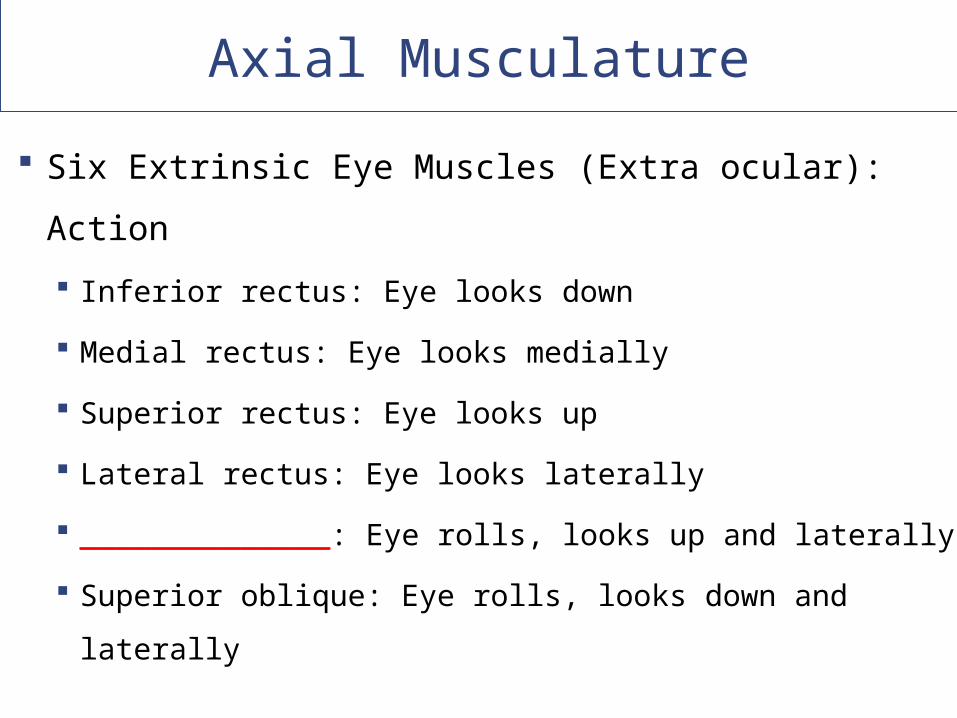
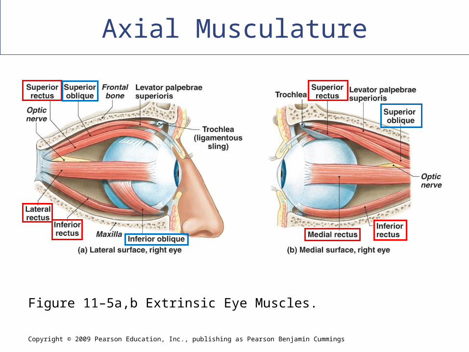
Six Extrinsic Eye Muscles (Extra ocular): Action
Inferior rectus: Eye looks down
Medial rectus: Eye looks medially
Superior rectus: Eye looks up
Lateral rectus: Eye looks laterally
______________: Eye rolls, looks up and laterally
Superior oblique: Eye rolls, looks down and laterally
Copyright © 2009 Pearson Education, Inc., publishing as Pearson Benjamin Cummings
Axial Musculature
Figure 11–5a,b Extrinsic Eye Muscles.
Copyright © 2009 Pearson Education, Inc., publishing as Pearson Benjamin Cummings
Axial Musculature
Figure 11–5c, d Extrinsic Eye Muscles.
Copyright © 2009 Pearson Education, Inc., publishing as Pearson Benjamin Cummings
Axial Musculature
Figure 11–6a Muscles of Mastication.
Copyright © 2009 Pearson Education, Inc., publishing as Pearson Benjamin Cummings
Axial Musculature
Figure 11–9a Muscles of the Anterior Neck.
Copyright © 2009 Pearson Education, Inc., publishing as Pearson Benjamin Cummings
Axial Musculature
[INSERT FIG. 11.10a]
Figure 11–10a Muscles of the Vertebral Column.
Copyright © 2009 Pearson Education, Inc., publishing as Pearson Benjamin Cummings
Axial Musculature
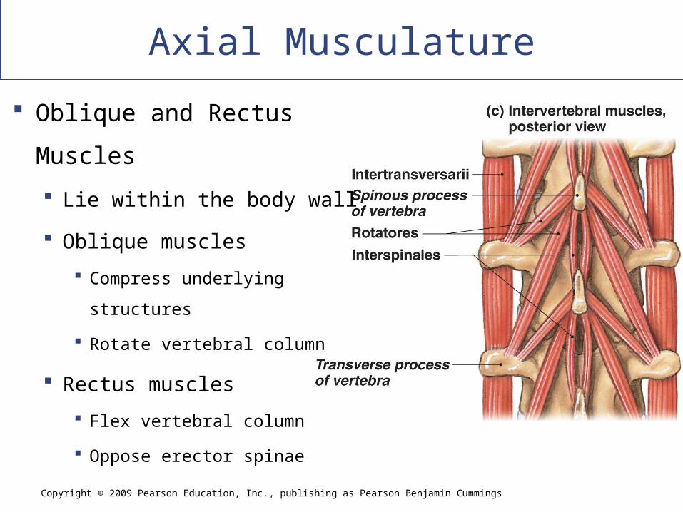
Oblique and Rectus
Muscles
Lie within the body wall
Oblique muscles
Compress underlying structures
Rotate vertebral column
Rectus muscles
Flex vertebral column
Oppose erector spinae
Appendicular Musculature
Position and stabilize pectoral and pelvic
girdles
Move upper and lower limbs
Divisions of Appendicular Muscles
Muscles of the shoulders and upper limbs
Muscles of the pelvis and lower limbs
Copyright © 2009 Pearson Education, Inc., publishing as Pearson Benjamin Cummings
Appendicular Musculature
[INSERT FIG. 11.13a]
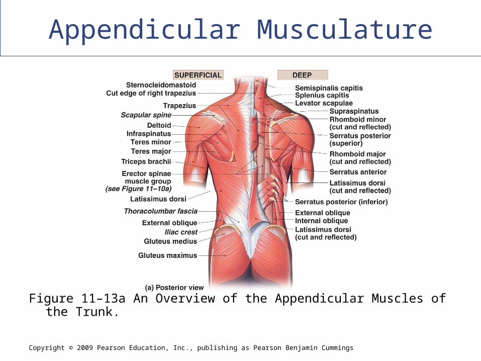
Figure 11–13a An Overview of the Appendicular Muscles of the Trunk.
Copyright © 2009 Pearson Education, Inc., publishing as Pearson Benjamin Cummings
Appendicular Musculature
[INSERT FIG. 11.13b]
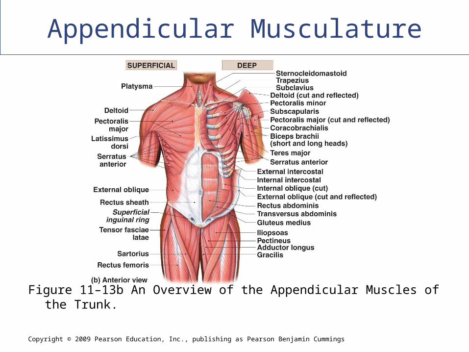
Figure 11–13b An Overview of the Appendicular Muscles of the Trunk.
Appendicular Musculature
Muscles of the Shoulders and Upper
Limbs
Position the pectoral girdle
Move the arm
Move the forearm and hand
Move the hand and fingers
Appendicular Musculature
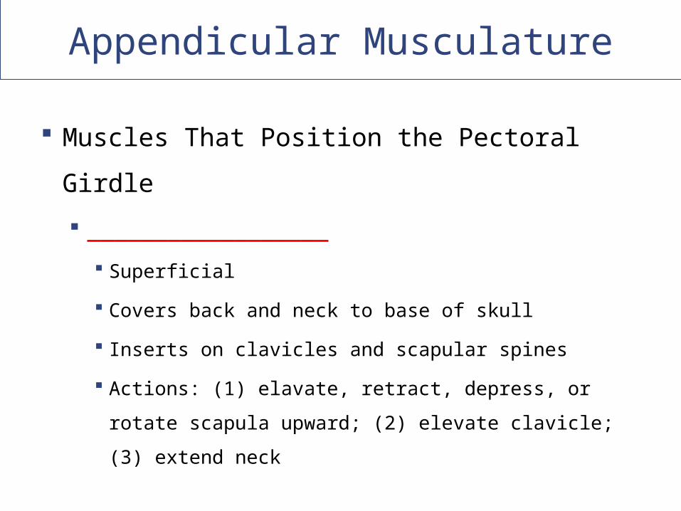
Muscles That Position the Pectoral Girdle
__________________
Superficial
Covers back and neck to base of skull
Inserts on clavicles and scapular spines
Actions: (1) elavate, retract, depress, or rotate
scapula upward; (2) elevate clavicle; (3) extend
neck
Appendicular Musculature
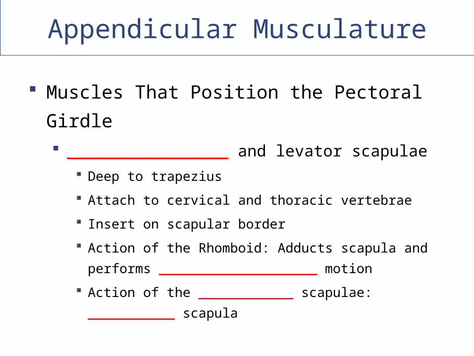
Muscles That Position the Pectoral Girdle
_________________ and levator scapulae
Deep to trapezius
Attach to cervical and thoracic vertebrae
Insert on scapular border
Action of the Rhomboid: Adducts scapula and performs
____________________ motion
Action of the ____________ scapulae: ___________
scapula
Copyright © 2009 Pearson Education, Inc., publishing as Pearson Benjamin Cummings
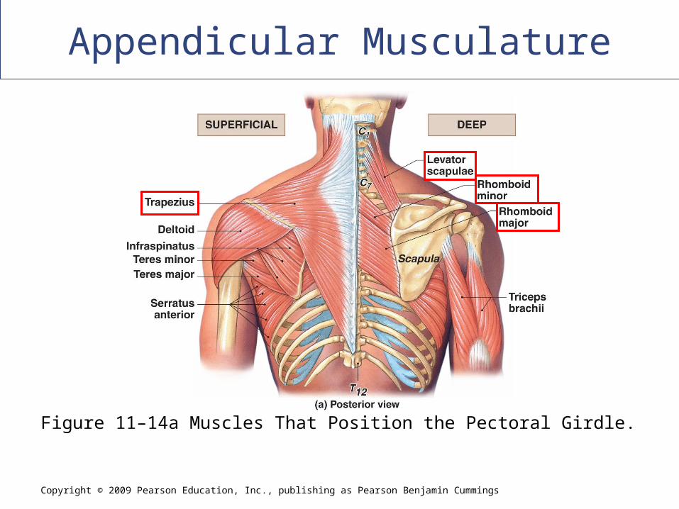
Appendicular Musculature
[INSERT FIG. 11.15a]
Figure 11–14a Muscles That Position the Pectoral Girdle.
Appendicular Musculature
Other Muscles That Position the Pectoral Girdle
Serratus anterior: Protracts shoulder, rotates scapula
On the chest
Originates along ribs
Inserts on anterior scapular margin
Subclavius: Depresses and protracts shoulder
Originates on ribs
Inserts on clavicle
Pectoralis minor: Depresses and protracts shoulder,
rotates scapula, elevates ribs
Attaches to scapula

Copyright © 2009 Pearson Education, Inc., publishing as Pearson Benjamin Cummings
Appendicular Musculature
[INSERT FIG. 11.15b]
Figure 11–14b Muscles That Position the Pectoral Girdle.
Appendicular Musculature
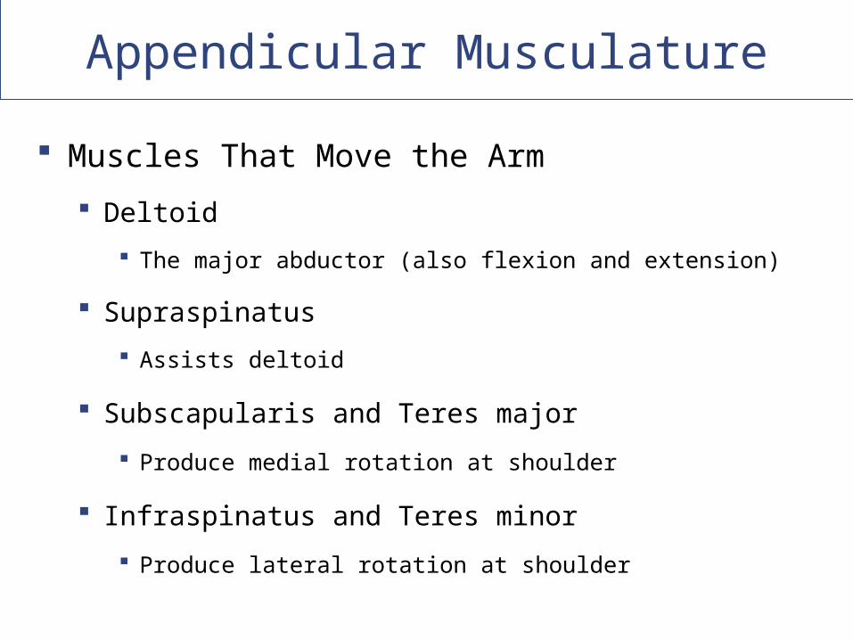
Muscles That Move the Arm
Deltoid
The major abductor (also flexion and extension)
Supraspinatus
Assists deltoid
Subscapularis and Teres major
Produce medial rotation at shoulder
Infraspinatus and Teres minor
Produce lateral rotation at shoulder
Copyright © 2009 Pearson Education, Inc., publishing as Pearson Benjamin Cummings
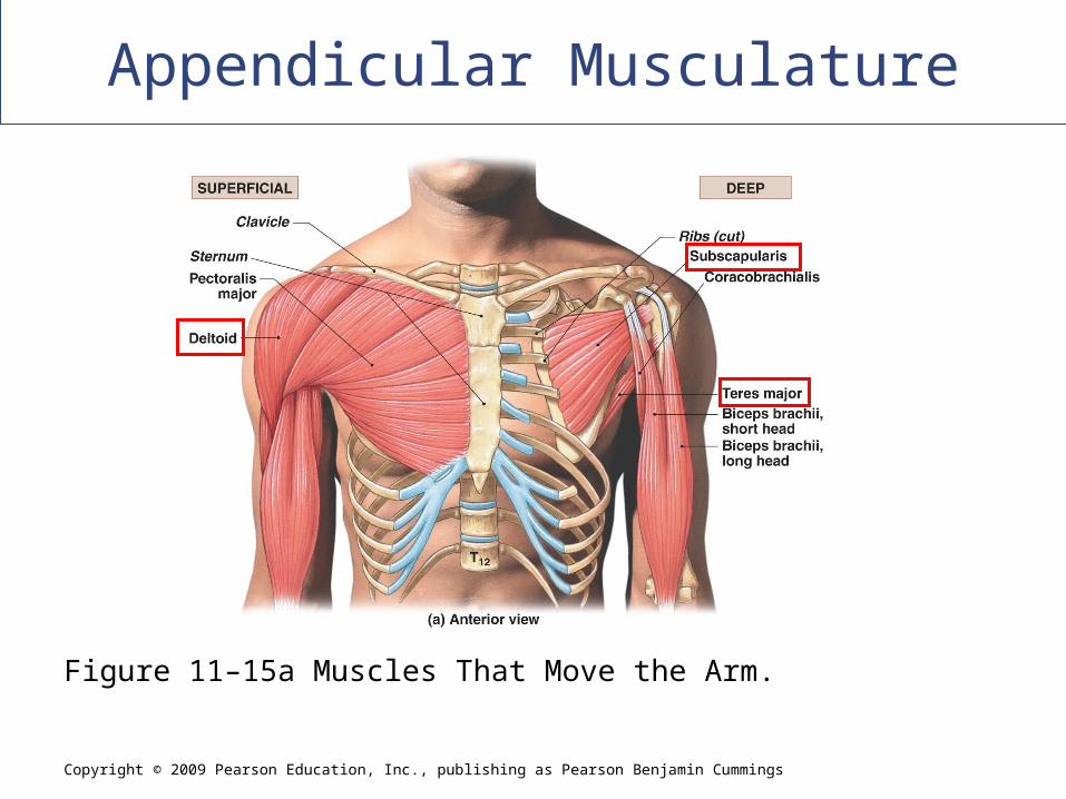
Appendicular Musculature
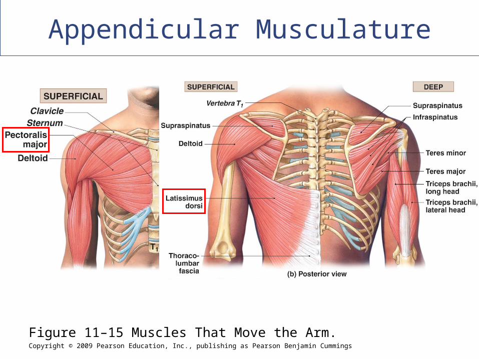
Figure 11–15a Muscles That Move the Arm.
Copyright © 2009 Pearson Education, Inc., publishing as Pearson Benjamin Cummings
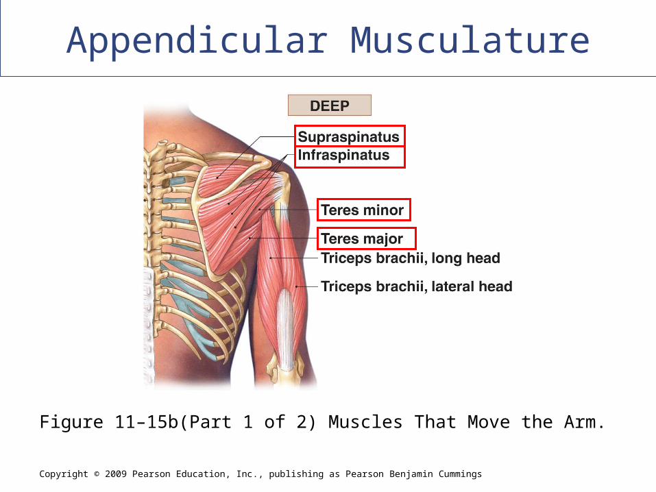
Appendicular Musculature
Figure 11–15b(Part 1 of 2) Muscles That Move the Arm.
Appendicular Musculature
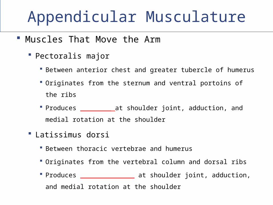
Muscles That Move the Arm
Pectoralis major
Between anterior chest and greater tubercle of humerus
Originates from the sternum and ventral portoins of the ribs
Produces ________ at shoulder joint, adduction, and medial
rotation at the shoulder
Latissimus dorsi
Between thoracic vertebrae and humerus
Originates from the vertebral column and dorsal ribs
Produces ______________ at shoulder joint, adduction, and
medial rotation at the shoulder
Copyright © 2009 Pearson Education, Inc., publishing as Pearson Benjamin Cummings
Appendicular Musculature
Figure 11–15 Muscles That Move the Arm.
Appendicular Musculature
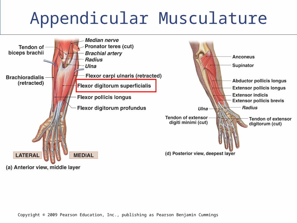
Muscles That Move the Forearm and Hand
Originate on humerus and insert on forearm
Exceptions:
The major flexor (biceps brachii)
The major extensor (triceps brachii)
Copyright © 2009 Pearson Education, Inc., publishing as Pearson Benjamin Cummings
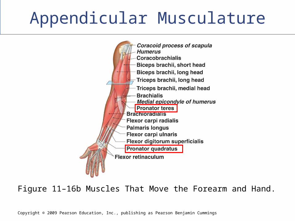
Appendicular Musculature
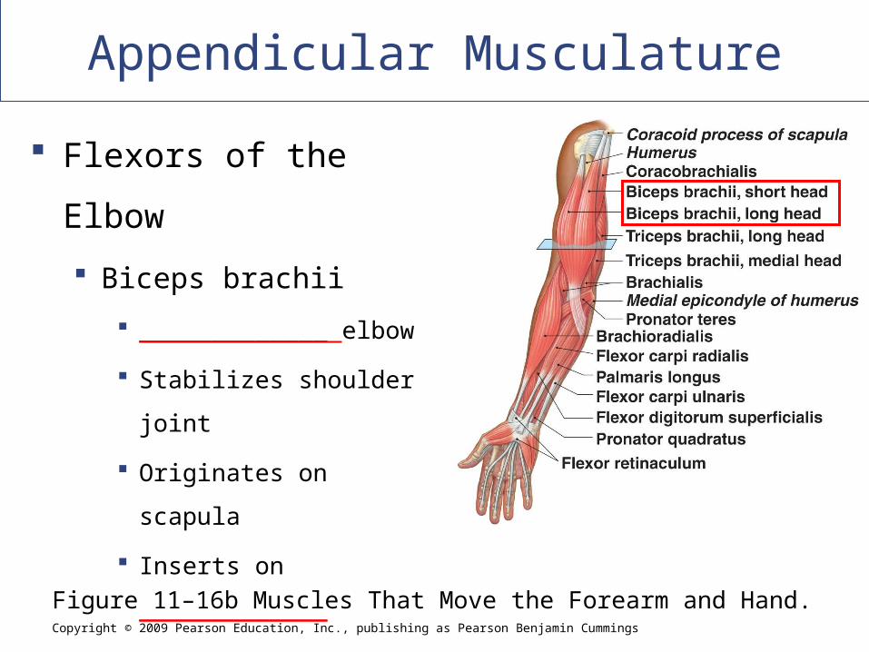
Figure 11–16b Muscles That Move the Forearm and Hand.
Flexors of the Elbow
Biceps brachii
_____________ elbow
Stabilizes shoulder joint
Originates on scapula
Inserts on _____________
Copyright © 2009 Pearson Education, Inc., publishing as Pearson Benjamin Cummings
Appendicular Musculature
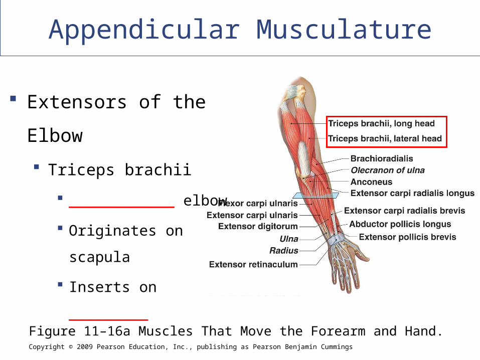
Figure 11–16a Muscles That Move the Forearm and Hand.
Extensors of the Elbow
Triceps brachii
____________ elbow
Originates on scapula
Inserts on _________
Appendicular Musculature
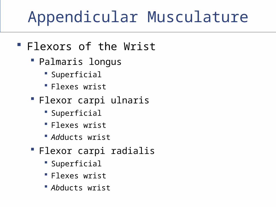
Flexors of the Wrist Palmaris longus
Superficial
Flexes wrist
Flexor carpi ulnaris Superficial
Flexes wrist
Adducts wrist
Flexor carpi radialis Superficial
Flexes wrist
Abducts wrist
Appendicular Musculature
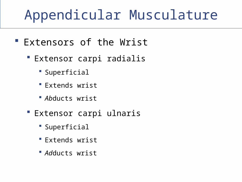
Extensors of the Wrist
Extensor carpi radialis
Superficial
Extends wrist
Abducts wrist
Extensor carpi ulnaris
Superficial
Extends wrist
Adducts wrist
Copyright © 2009 Pearson Education, Inc., publishing as Pearson Benjamin Cummings
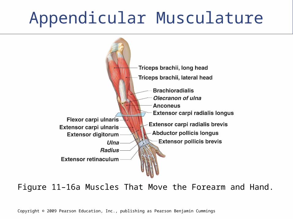
Appendicular Musculature
Figure 11–16a Muscles That Move the Forearm and Hand.

Copyright © 2009 Pearson Education, Inc., publishing as Pearson Benjamin Cummings
Appendicular Musculature
Figure 11–16b Muscles That Move the Forearm and Hand.
Appendicular Musculature
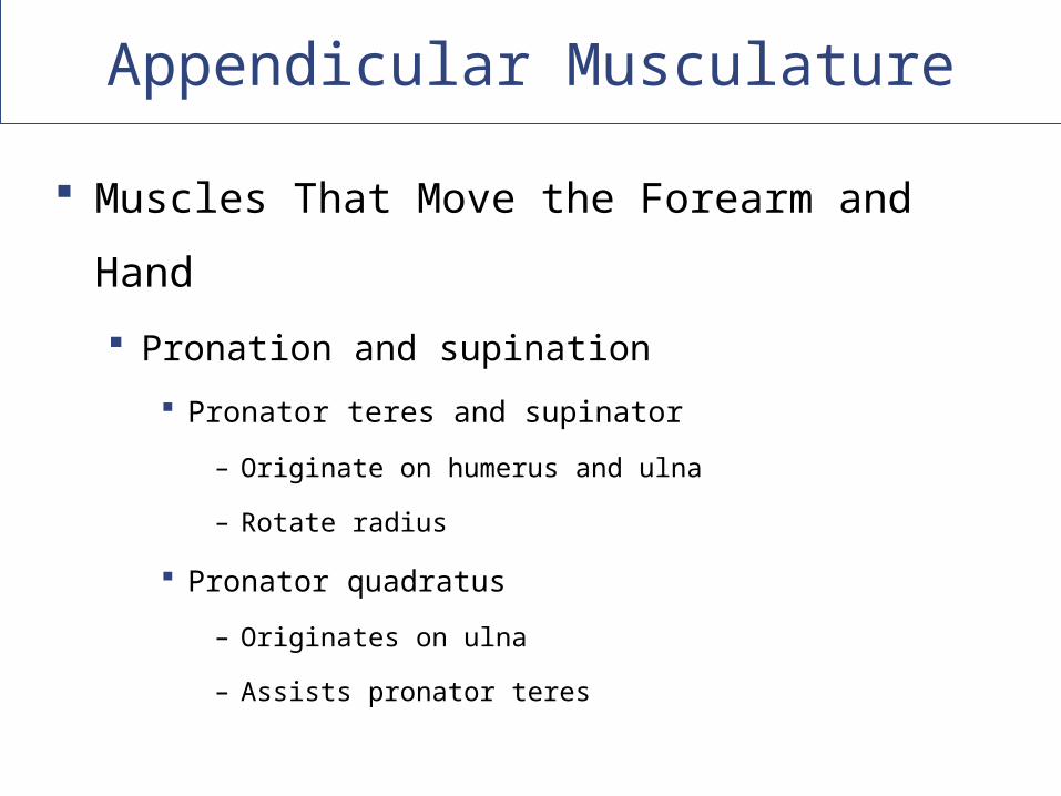
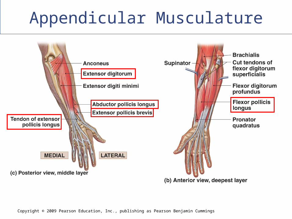
Muscles That Move the Forearm and Hand
Pronation and supination
Pronator teres and supinator
– Originate on humerus and ulna
– Rotate radius
Pronator quadratus
– Originates on ulna
– Assists pronator teres
Copyright © 2009 Pearson Education, Inc., publishing as Pearson Benjamin Cummings
Appendicular Musculature
Figure 11–16b Muscles That Move the Forearm and Hand.
Appendicular Musculature
Muscles That Move the Hand and Fingers
Also called extrinsic muscles of the hand
Lie entirely within forearm
Only ____________ cross wrist (in synovial
tendon sheaths)
Appendicular Musculature
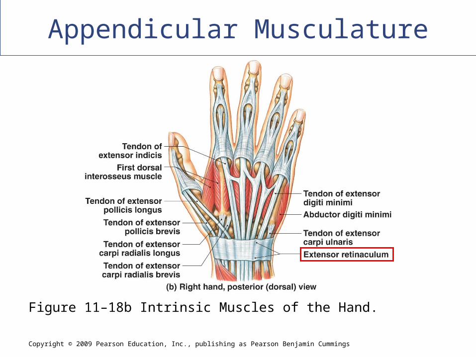
Muscles that Move the Hand and Fingers
Tendon sheaths
Extensor retinaculum
– Wide band of connective tissue
– Posterior surface of wrist
– Stabilizes tendons of extensor muscles
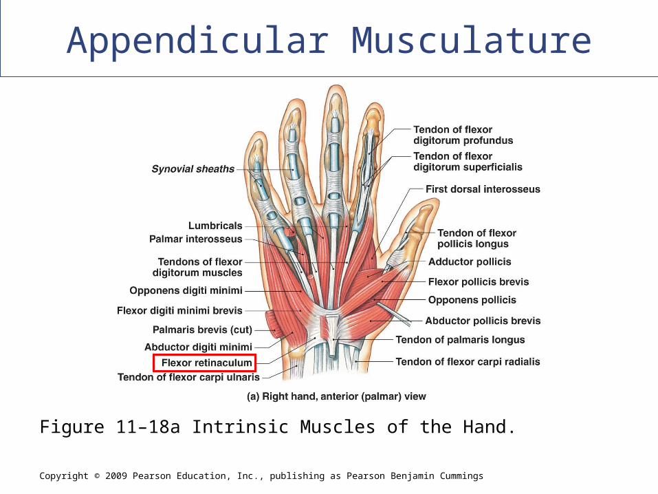
Flexor retinaculum:
Anterior surface of wrist
Stabilizes tendons of flexor muscles
Copyright © 2009 Pearson Education, Inc., publishing as Pearson Benjamin Cummings
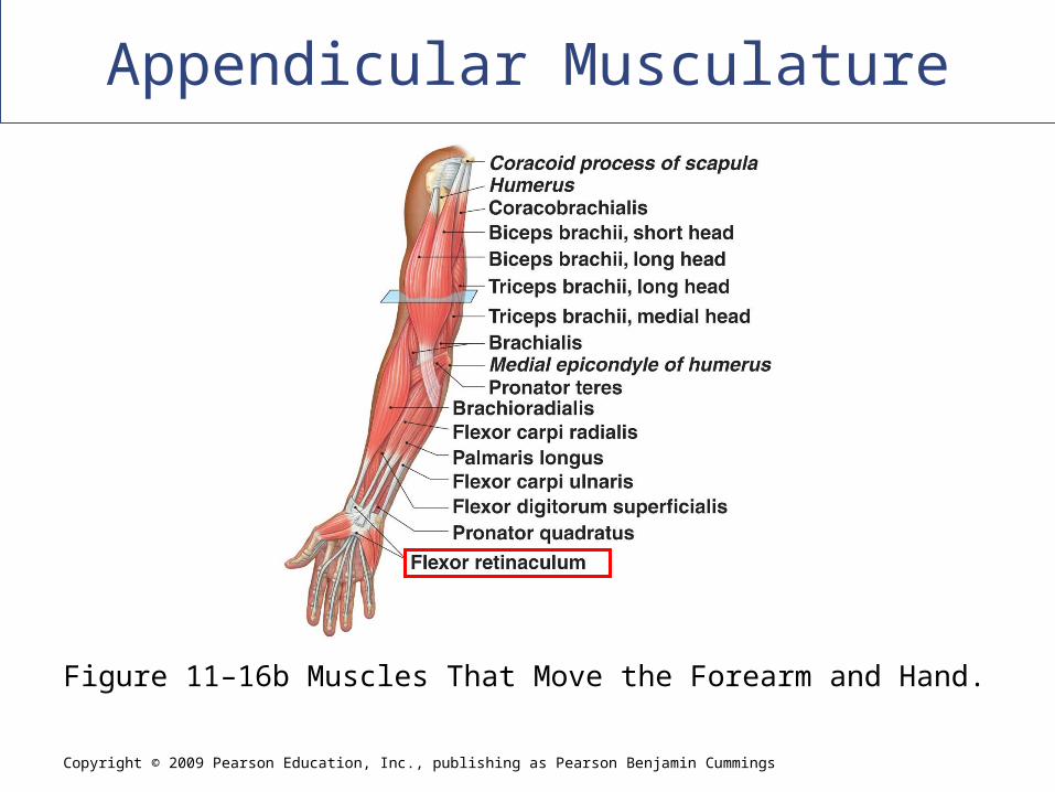
Appendicular Musculature
Figure 11–16b Muscles That Move the Forearm and Hand.
Copyright © 2009 Pearson Education, Inc., publishing as Pearson Benjamin Cummings
Appendicular Musculature
Figure 11–18b Intrinsic Muscles of the Hand.
Copyright © 2009 Pearson Education, Inc., publishing as Pearson Benjamin Cummings
Appendicular Musculature
Figure 11–18a Intrinsic Muscles of the Hand.
Copyright © 2009 Pearson Education, Inc., publishing as Pearson Benjamin Cummings
Appendicular Musculature
Copyright © 2009 Pearson Education, Inc., publishing as Pearson Benjamin Cummings
Appendicular Musculature
Appendicular Musculature
Muscles of the Lower Limbs
36 muscles move the bones of the lower
extremities:
Muscles that move the thigh
Muscles that move the leg
Muscles that move the foot and toes

Appendicular Musculature
Muscles That Move the Thigh Gluteal muscles
Lateral rotators
Adductors
Iliopsoas
Appendicular Musculature
Muscles That Move the Thigh: Gluteal Muscles Cover lateral surfaces of ilia
Gluteus maximus Largest, most posterior gluteal muscle
Produces extension and lateral rotation at hip
Tensor fasciae latae Works with gluteus maximus
Stabilizes iliotibial tract
Gluteus medius and gluteus minimus Originate anterior to gluteus maximus
Insert on trochanter
Copyright © 2009 Pearson Education, Inc., publishing as Pearson Benjamin Cummings
Appendicular Musculature
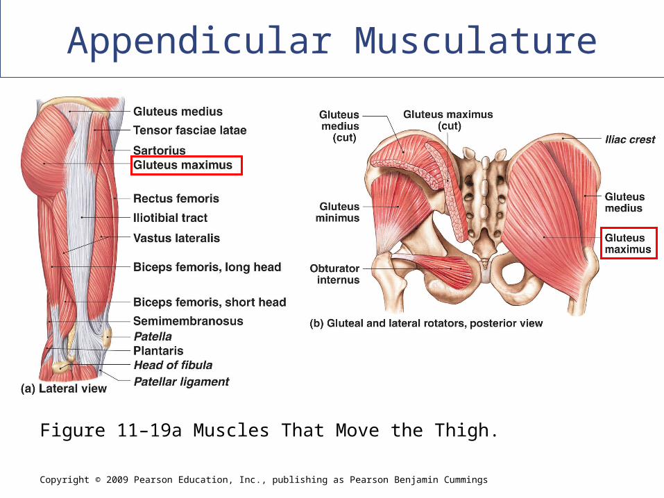
Figure 11–19a Muscles That Move the Thigh.
Appendicular Musculature
Muscles That Move the Thigh: Lateral rotators
Group of six muscles, including:
– Piriformis
» Also hip abduction
– Obturator (externus and internus)
– Gemilli (superior and inferior)
– Quadratus femoris
Appendicular Musculature
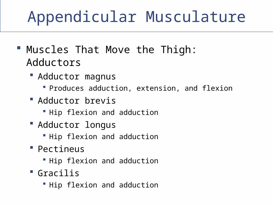
Muscles That Move the Thigh: Adductors Adductor magnus
Produces adduction, extension, and flexion
Adductor brevis Hip flexion and adduction
Adductor longus Hip flexion and adduction
Pectineus Hip flexion and adduction
Gracilis Hip flexion and adduction
Appendicular Musculature
Muscles That Move the Thigh: Iliopsoas
Two hip flexors insert on the same tendon
Psoas major
Iliacus
Appendicular Musculature
Muscles That Move the Leg
__________________ of the knee
Originate on the ______________________
__________________ of the knee
Originate on the _________________ surface
Insert on the patella
Copyright © 2009 Pearson Education, Inc., publishing as Pearson Benjamin Cummings
Appendicular Musculature
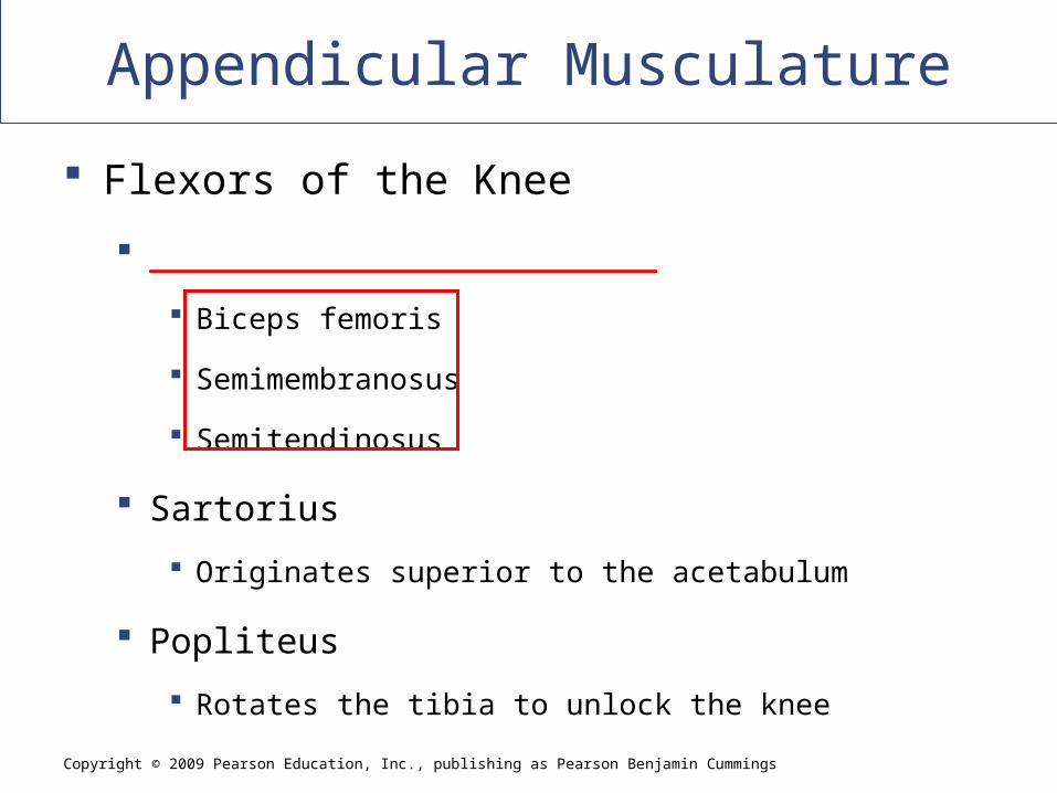
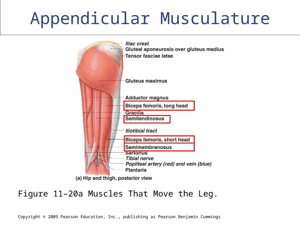
Flexors of the Knee
________________________
Biceps femoris
Semimembranosus
Semitendinosus
Sartorius
Originates superior to the acetabulum
Popliteus
Rotates the tibia to unlock the knee
Copyright © 2009 Pearson Education, Inc., publishing as Pearson Benjamin Cummings
Appendicular Musculature
Figure 11–20a Muscles That Move the Leg.
Appendicular Musculature
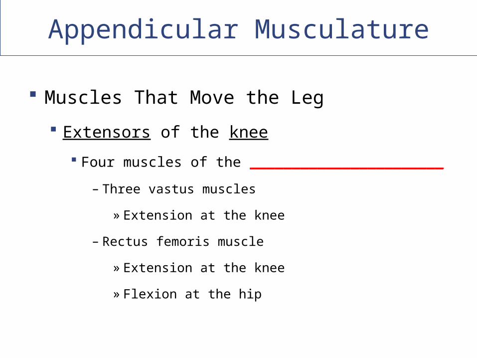
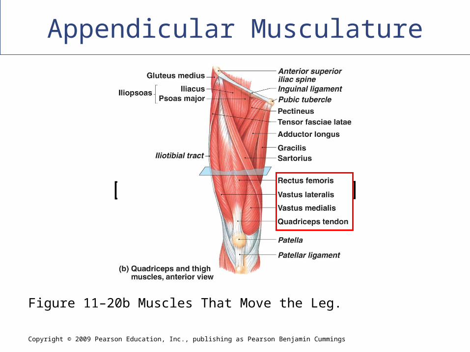
Muscles That Move the Leg
Extensors of the knee
Four muscles of the _______________________
– Three vastus muscles
» Extension at the knee
– Rectus femoris muscle
» Extension at the knee
» Flexion at the hip
Copyright © 2009 Pearson Education, Inc., publishing as Pearson Benjamin Cummings
Appendicular Musculature
[INSERT FIG. 11.21b]
Figure 11–20b Muscles That Move the Leg.
Appendicular Musculature
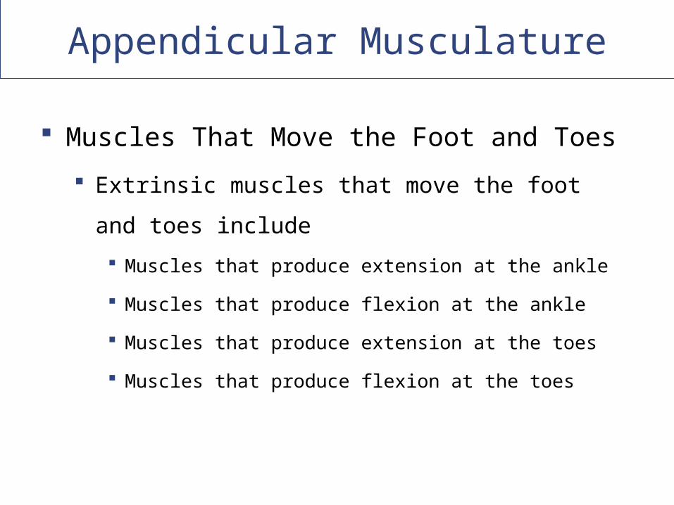
Muscles That Move the Foot and Toes
Extrinsic muscles that move the foot and toes include
Muscles that produce extension at the ankle
Muscles that produce flexion at the ankle
Muscles that produce extension at the toes
Muscles that produce flexion at the toes
Appendicular Musculature
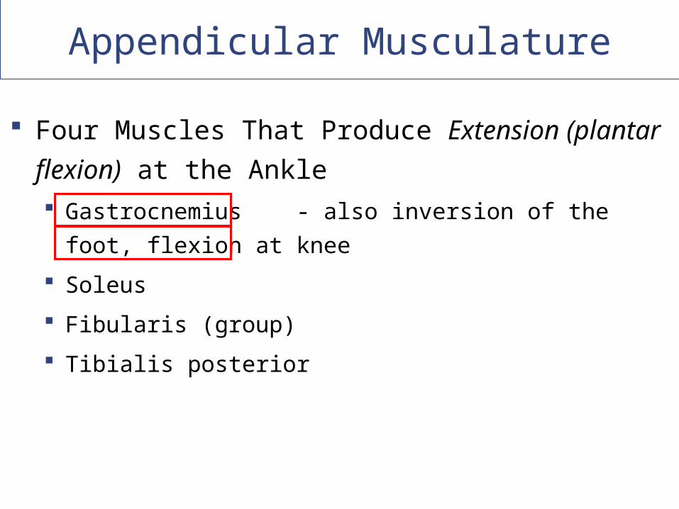
Four Muscles That Produce Extension (plantar flexion)
at the Ankle Gastrocnemius - also inversion of the foot, flexion at knee
Soleus
Fibularis (group)
Tibialis posterior
Copyright © 2009 Pearson Education, Inc., publishing as Pearson Benjamin Cummings
Appendicular Musculature
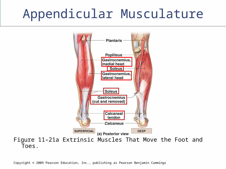
Figure 11–21a Extrinsic Muscles That Move the Foot and Toes.
Appendicular Musculature
Muscles That Move the Foot and Toes
The Achilles Tendon
The calcaneal tendon (Achilles tendon)
– Shared by the gastrocnemius and soleus
Appendicular Musculature
Muscles That Produce Flexion
(Dorsiflexsion) at the Ankle
Tibialis anterior
Opposes the gastrocnemius (extensor)
Except both are involved with inversion of the foot
Appendicular Musculature
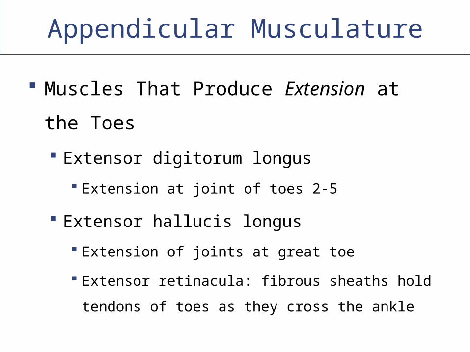
Muscles That Produce Extension at the
Toes
Extensor digitorum longus
Extension at joint of toes 2-5
Extensor hallucis longus
Extension of joints at great toe
Extensor retinacula: fibrous sheaths hold tendons
of toes as they cross the ankle
Copyright © 2009 Pearson Education, Inc., publishing as Pearson Benjamin Cummings
Appendicular Musculature
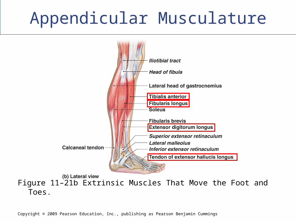
[INSERT FIG. 11.22b]
Figure 11–21b Extrinsic Muscles That Move the Foot and Toes.
Copyright © 2009 Pearson Education, Inc., publishing as Pearson Benjamin Cummings
Appendicular Musculature
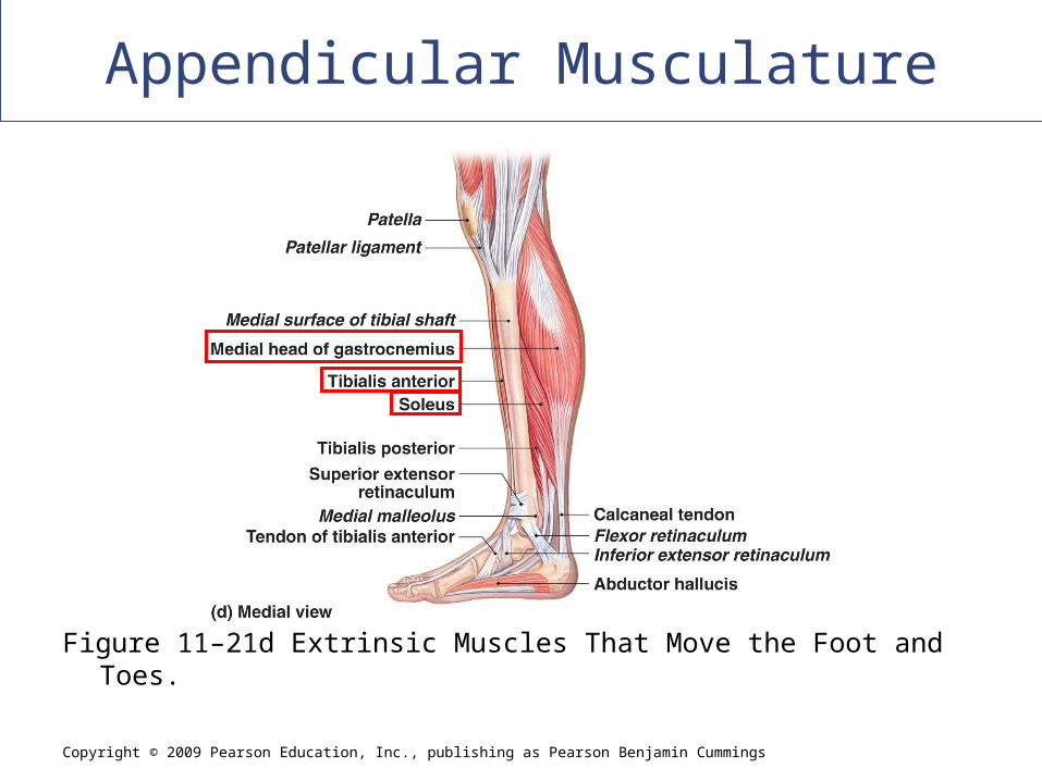
[INSERT FIG. 11.22d]
Figure 11–21d Extrinsic Muscles That Move the Foot and Toes.
Appendicular Musculature
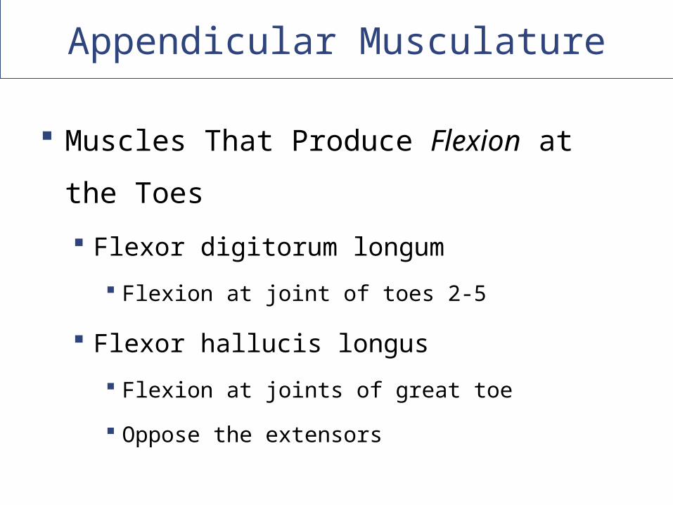
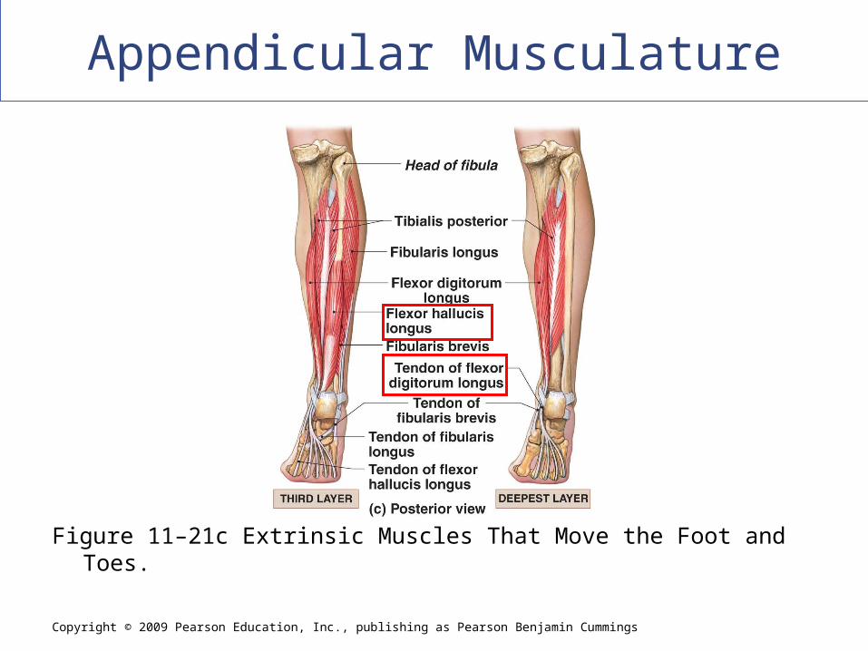
Muscles That Produce Flexion at the Toes
Flexor digitorum longum
Flexion at joint of toes 2-5
Flexor hallucis longus
Flexion at joints of great toe
Oppose the extensors
Copyright © 2009 Pearson Education, Inc., publishing as Pearson Benjamin Cummings
Appendicular Musculature
Figure 11–21c Extrinsic Muscles That Move the Foot and Toes.
Appendicular Musculature
The Intrinsic Muscles of the Foot
Muscles that move the tarsals, metatarsals,
and phalanges and originate and insert only
on those bones
Copyright © 2009 Pearson Education, Inc., publishing as Pearson Benjamin Cummings
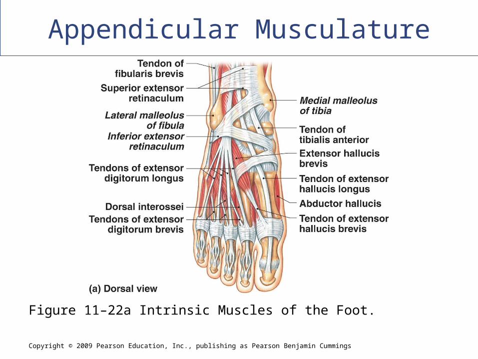
Appendicular Musculature
Figure 11–22a Intrinsic Muscles of the Foot.
Copyright © 2009 Pearson Education, Inc., publishing as Pearson Benjamin Cummings
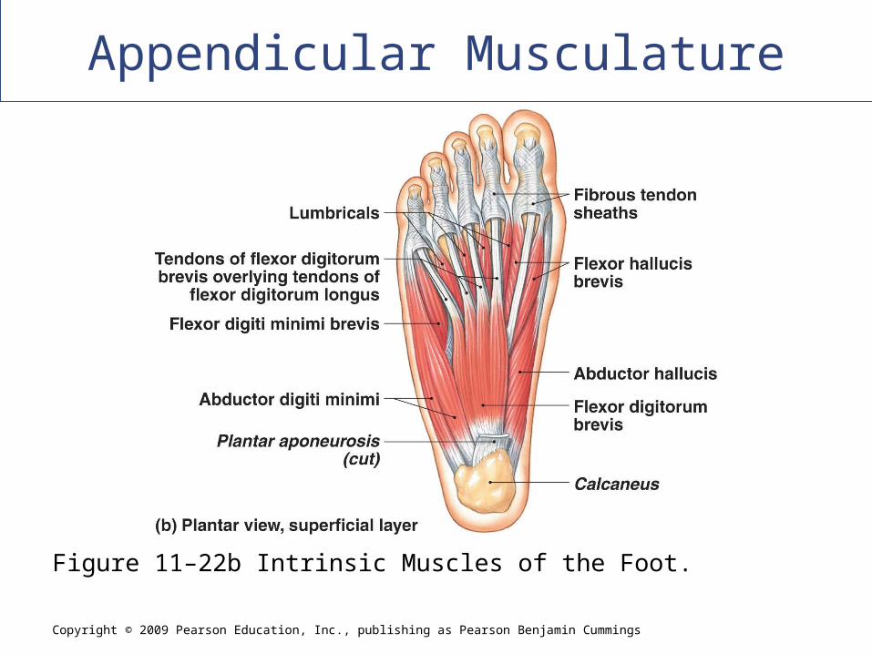
Appendicular Musculature
[INSERT FIG. 11.23b]
Figure 11–22b Intrinsic Muscles of the Foot.
Copyright © 2009 Pearson Education, Inc., publishing as Pearson Benjamin Cummings
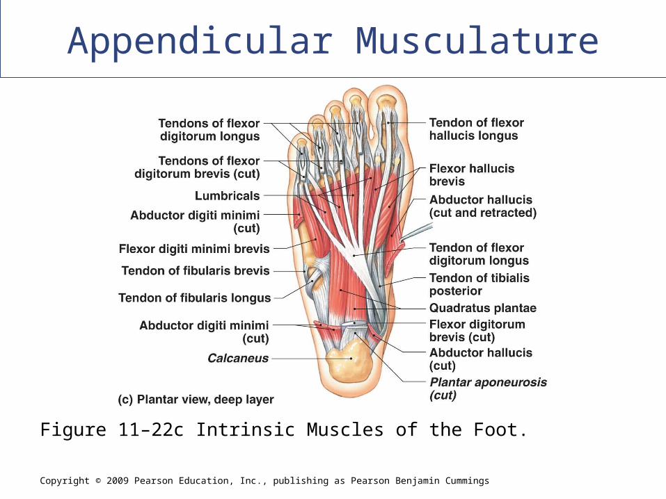
Appendicular Musculature
[INSERT FIG. 11.23c]
Figure 11–22c Intrinsic Muscles of the Foot.
Effects of Aging on the Muscular System
Skeletal muscle fibers become smaller in diameter
Skeletal muscles become less elastic Develop increasing amounts of fibrous tissue
(fibrosis)
Decreased tolerance for exercise
Decreased ability to recover from muscular injuries
Integration with Other Systems
Cardiovascular system Delivers oxygen and fuel Removes carbon dioxide and wastes
Respiratory system Responds to oxygen demand of muscles
Integumentary system Disperses heat from muscle activity
Nervous and endocrine systems Direct responses of all systems
Copyright © 2009 Pearson Education, Inc., publishing as Pearson Benjamin Cummings
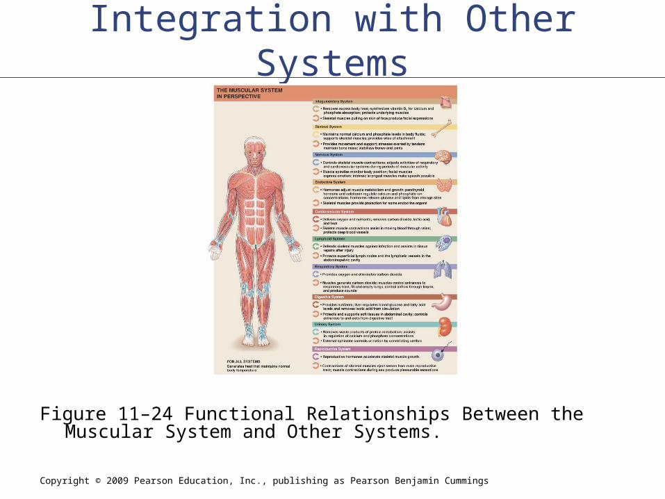
Integration with Other Systems
Figure 11–24 Functional Relationships Between the Muscular System and Other Systems.