Embed Size (px)
Citation preview

Muscle Physiology

Sherwood et. al. (2005)

Fig 8.2 Levels of organization in a skeletal muscle. (a) Enlargement of a cross section of a whole muscle.
Sherwood et. al. (2005)

Fig 8.2 Levels of organization in a skeletal muscle. (b) Enlargement of a myofibril within a muscle fiber.
Sherwood et. al. (2005)

Fig 8.2 Levels of organization in a skeletal muscle. Sherwood et. al. (2005)

Fig 8.3 Light-microscope view of skeletal muscle components.(a) High-power light-microscope view of a myofibril.
Sherwood et. al. (2005)
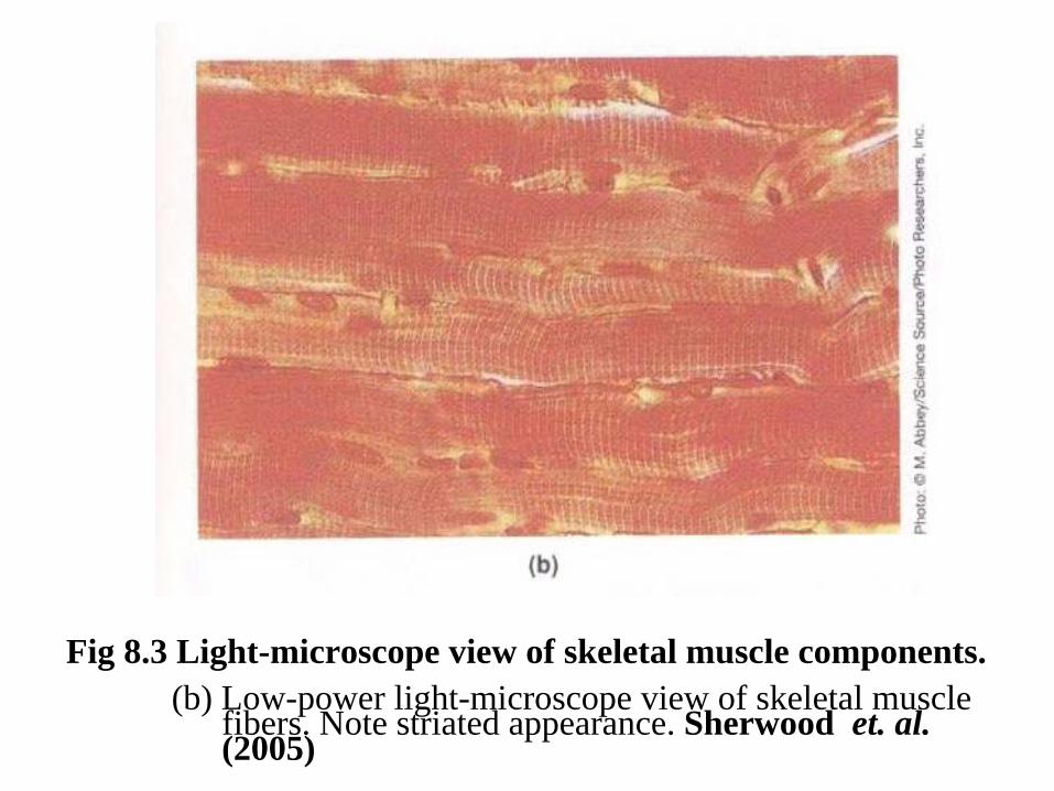
Fig 8.3 Light-microscope view of skeletal muscle components.(b) Low-power light-microscope view of skeletal muscle
fibers. Note striated appearance. Sherwood et. al. (2005)

Fig 8.4 Cross-sectional arrangement of thick and thin filaments.(a) Electron micrograph cross section through the A band in
the region of thick and thin filament overlap. Note the fine cross bridges extending from the thick filaments.
(b) Schematic representation of the geometric relation among thick and thin filaments and cross bridges.
Sherwood et. al. (2005)

Fig 8.5 Structure of myosin molecules and their organization within a thick filament. (a) Myosin molecule. Each myosin molecule consists of two identical golf club-shaped subunits with their tails intertwined and their globular heads, each of which contains an actin- binding site and a myosin ATPase site, projecting out at one end. Sherwood et. al. (2005)

Fig 8.5 Structure of myosin molecules and their organization within a thick filament. (b) Thick filament. A thick filament is made up of myosin molecules lying lengthwise parallel to each other. Half are oriented in one direction and half in the opposite direction. The globular heads, which protrude at regular intervals along the thick filament, form the cross bridges. Sherwood et. al. (2005)

Sherwood et. al. (2005)

Figure 8-7 Role of calcium in turning on cross bridges. Sherwood et. al. (2005)

Fig 8.8 Changes in banding pattern during shortening.
Sherwood et. al. (2005)

Fig 8.9 Cross-bridge activity.(a) During each cross-bridge cycle, the cross bridge binds with an actin molecule, bends to pull the thin filament inward during the power stroke, then detaches and returns to its resting conformation, ready to repeat the cycle. Sherwood et. al. (2005)

Fig 8.9 Cross-bridge activity.(b) The power strokes of all cross bridges extending form a thick filament are directed toward the center of the thick filament.
Sherwood et. al. (2005)

Fig 8.9 Cross-bridge activity.(c) Each thick filament is surrounded on each end by six thin filaments, all of which are pulled inward simultaneously through cross-bridge cycling during muscle contraction. Sherwood et. al. (2005)

Fig 8.10 The T tubules and sarcoplasmic reticulum in relationship to the myofibrils. Sherwood et. al. (2005)

Sherwood et. al. (2005)

Fig 8.12 Calcium release in excitation-contraction coupling.Sherwood et. al. (2005)

Fig 8-13. Cross-bridge cycle. Sherwood et. al. (2005)

Fig 8-13. Cross-bridge cycle. Sherwood et. al. (2005)


Fig 8.14 Relationship of an action potential to the resultant muscle twitch. Sherwood et. al. (2005)
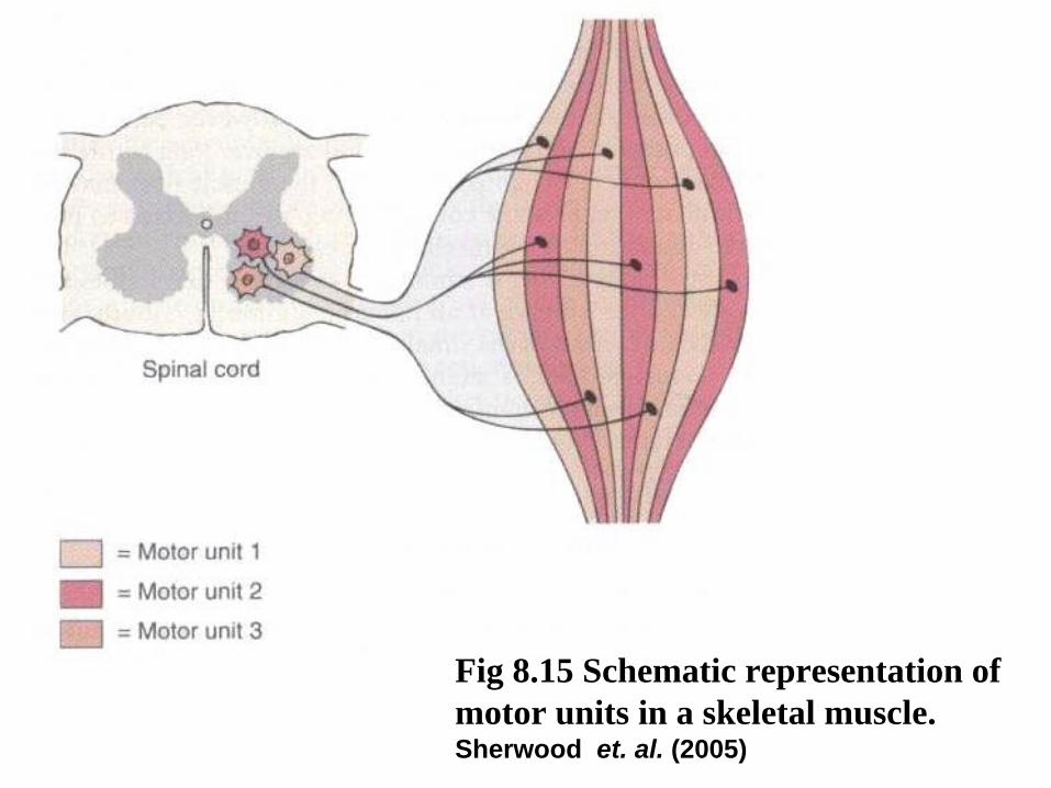
Fig 8.15 Schematic representation of motor units in a skeletal muscle. Sherwood et. al. (2005)

Fig 8.16 Comparison of motor unit recruitment in muscles with
small motor units and muscles with large motor units. Sherwood et. al. (2005)

Sherwood et. al. (2005)

Fig 8.18 Length-tension relationship. Sherwood et. al. (2005)
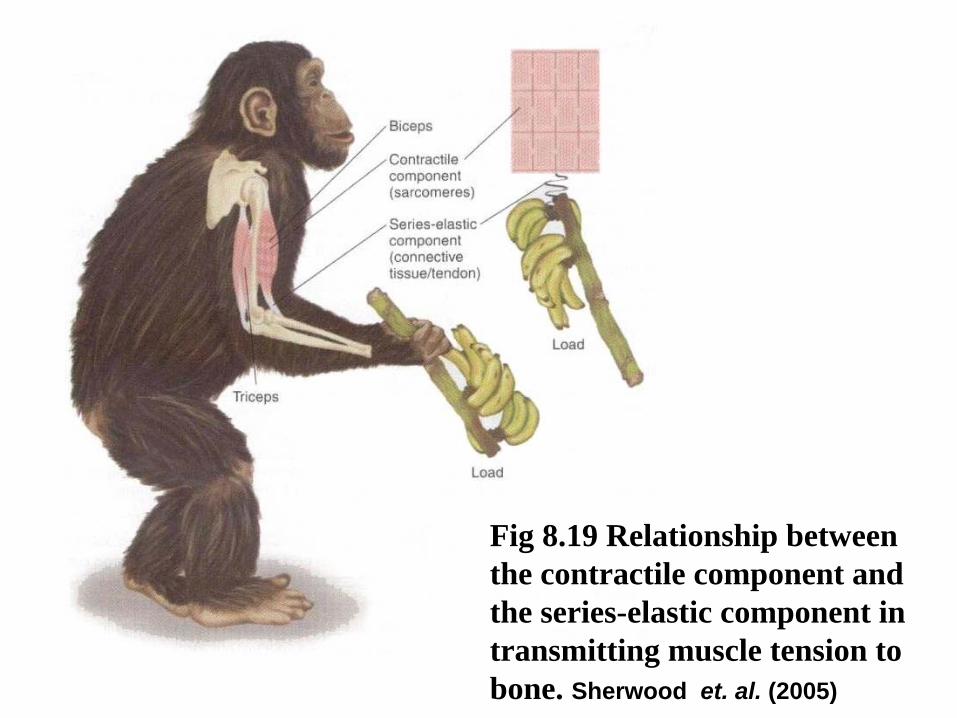
Fig 8.19 Relationship between the contractile component and the series-elastic component in transmitting muscle tension to bone. Sherwood et. al. (2005)

Sherwood et. al. (2005)

Fig 8.21 Lever systems of muscles, bones, and joints. Sherwood et. al. (2005)

Fig 8.21 Lever systems of muscles, bones, and joints.Sherwood et. al. (2005)

Fig 8.22 Metabolic pathways producing ATP used during muscle contraction and relaxation.
Sherwood et. al. (2005)


Sherwood et. al. (2005)
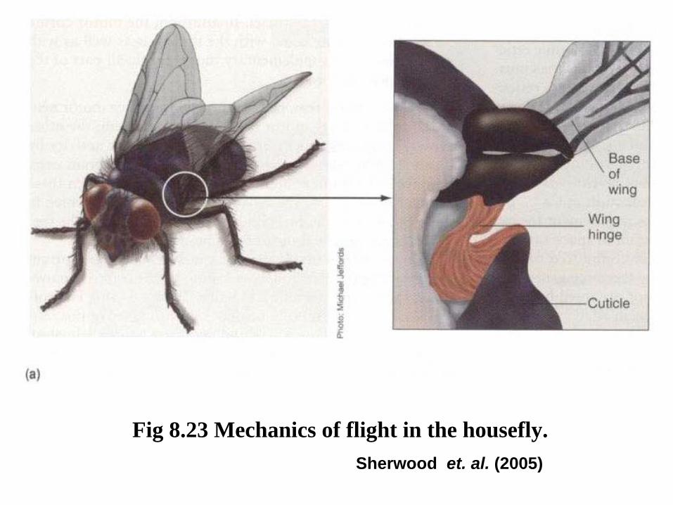
Fig 8.23 Mechanics of flight in the housefly. Sherwood et. al. (2005)

Fig 8.23 Mechanics of flight in the housefly. Sherwood et. al. (2005)

Sherwood et. al. (2005)

Fig 8.25 Muscle spindle. Sherwood et. al. (2005)

Fig 8.26 Muscle spindle function. Sherwood et. al. (2005)
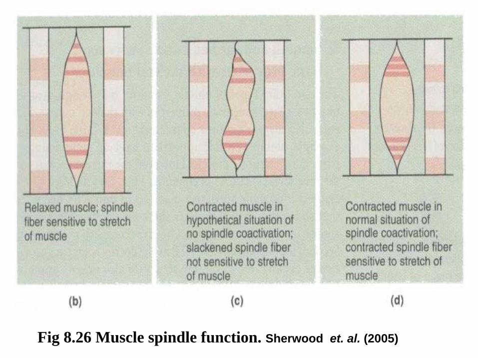
Fig 8.26 Muscle spindle function. Sherwood et. al. (2005)

Fig 8.27 Patellar tendon reflex (a stretch reflex). Sherwood et. al. (2005)


Fig 8.28 Microscopic view of smooth muscle cell. Sherwood et. al. (2005)

Fig 8.28 Microscopic view of smooth muscle cell. Sherwood et. al. (2005)

Fig 8.29 Schematic representation of the arrangement of thick and thin filaments in a smooth muscle cell in contracted and relaxed states. (a) Relaxed smooth muscle cell. Sherwood et. al. (2005)

Fig 8.29 Schematic representation of the arrangement of thick and thin filaments in a smooth muscle cell in contracted and relaxed states. (b) Contracted smooth muscle cell. Sherwood et. al. (2005)

Fig 8.30 Calcium activation of myosin in smooth muscle.Sherwood et. al. (2005)

Fig 8.31 Self-generated electrical activity in smooth muscle.

Fig 8.31 Self-generated electrical activity in smooth muscle.