Embed Size (px)
Citation preview

Assessment of Left Ventricular Volume
by Volume Imaging Ultrasound System and
Full-automated Volumetric Analysis Software
compared to Magnetic Resonance Imaging
Department of Cardiology, Sakakibara Heart Institute
Hirotsugu Mihara, Hiroyuki Watanabe, Masaru Aikawa,
Tetsuya Tobaru, Nobuo Iguchi, Masatoshi Nagayama,
Ryuta Asano, Morimasa Takayama, Jun Umemura,
Tetsuya Sumiyoshi

Background
Left ventricular (LV) volume measurement by 3D
echocardiography (3DE) is more accurate than 2D
echocardiographyGopal AS, et al. J Am Soc Echocardiogr 1992;5:115–24
Hozumi T, et al. Am J Cardiol 1996;78:1077–80
Semiautomated volumetric analysis by 3DE is feasible
and allows fast measurement of LV volumeJacobs LD, et al. Eur Heart J 2006;27:460–8
Corsi C, et al. Circulation 2005;112:1161–70

90 degrees x 90 degrees full-volume data
acquisition in a single heart beat
This system contains full-automated
volumetric analysis software
Volume Imaging Ultrasound System
Auto Auto Autoautomatic
standard planesborder detection 3D analysis

Objectives
The aim of this study was to validate
full-automated LV volume measurements
compared to cardiac magnetic resonance
(CMR) measurements

Subject
We enrolled 31 consecutive patients with coronary
and valvular heart diseases who performed 3DE
and CMR in several days at Sakakibara Heart
Institute
31 patients were analyzed
age 61±14 years
sex male 20pts , female 11pts

LV volume measurement by 3DE
Equipment: Siemens ACUSON SC2000
Probe: 4Z1c
Analysis software: Siemens Syngo SC2000 workplace
15 volume/sec
Method

LV volume measurement by CMR
Equipment: Siemens MAGNETOM Sonata 1.5T
Analysis software: Siemens Syngo Console VA 30 Argus
20 frame/beat
Method

Result
Disease
n=31
Rythme
Sinus rythme
Atrial fibrillation
97%
3%
Coronary heart disease
Cardiomyopathy
Vulvular heart disease
Others
29%
16%48%
6% n=31

EF
Full-automated
correlation difference
Y=0.45X+27, r=0.60 Mean difference (3DE - MRI)
= -0.8±11 %
MRI
3D
E
+2SD
-2SD
mean
(%)
Mean (3DE,MRI)
Dif
fere
nce
(3D
E -
MR
I)
(%)
(%) (%)

EF
Manual modified
Y=0.76X+19, r=0.89
correlation
Mean difference (3DE - MRI)
= 2.2±6.6 %
MRI
3D
E
(%)
Mean (3DE,MRI)
Dif
fere
nce
(3D
E -
MR
I)
+2SD
-2SD
mean
(%)
(%) (%)
difference
LV volume by 3DE vs MRI
Full-automated
Y=0.64X+24, r=0.83
Y=0.52X+19, r=0.78
-18Mean difference (3DE - MRI)
= -14±23ml
correlation
MRI
3D
E
Mean (3DE,MRI)
Dif
fere
nce
(3D
E -
MR
I) +2SD
-2SD
mean
(ml)
(ml) (ml)
(ml)difference
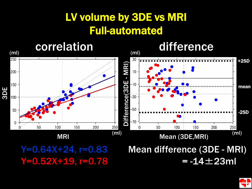
LV volume by 3DE vs MRI
Manual modified
Y=0.96X+3.5, r=0.96
Y=0.85X+5.1, r=0.95
Mean difference (3DE - MRI)
= -3.1±11 ml
correlation
MRI
3D
E
Mean (3DE,MRI)
Dif
fere
nce
(3D
E -
MR
I)
+2SD
-2SD
mean
(ml)
(ml) (ml)
(ml)difference

Conclusions
Automated LV volume measurement system
using single-beat full-volume acquisition of
real- time echocardiography provides quick
and accurate measurement
It is a new objective solution for noninvasive
LV volume analysis