Embed Size (px)
Citation preview
Case ReportGranuloma Caused by Carbon Deposition in the Dermis
Rintaro Shibuya, Yuichiro Endo, Akihiro Fujisawa, Miki Tanioka, and Yoshiki Miyachi
Department of Dermatology, Graduate School of Medicine, Kyoto University, 54 Shogoin, Kawahara-cho, Sakyo-ku,Kyoto 606-8507, Japan
Correspondence should be addressed to Miki Tanioka; [email protected]
Received 5 December 2013; Accepted 16 January 2014; Published 20 February 2014
Academic Editors: S. Inui and E. Schmidt
Copyright © 2014 Rintaro Shibuya et al.This is an open access article distributed under theCreative CommonsAttribution License,which permits unrestricted use, distribution, and reproduction in any medium, provided the original work is properly cited.
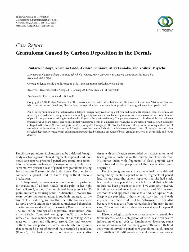
Pencil core granuloma is characterized by a delayed foreign-body reaction against retained fragments of pencil lead. Previous casereports presented pencil core granuloma resemblingmalignant melanoma, haemangioma, or soft tissue sarcoma.We present a caseof pencil core granuloma arising from the palm 25 years after the initial injury.The patient presented a bluish nodule that had beenpresent over 25 years before. The nodule initially measured 5mm in diameter. However, five years before presentation, it suddenlyenlarged to the size of 30mmduring sixmonths. Computed tomography (CT) of the lesion revealed a linear radiopaque structure of8mm long with a mass on its distal end. Surgical resection revealed a bluish muddymass and pencil lead. Histological examinationrevealed degenerative tissue with calcification surrounded by massive amounts of black granular material in the middle and lowerdermis.
Pencil core granuloma is characterized by a delayed foreign-body reaction against retained fragments of pencil lead. Pre-vious case reports presented pencil core granuloma resem-bling malignant melanoma, haemangioma, or soft tissuesarcoma. We present a case of pencil core granuloma arisingfrom the palm 25 years after the initial injury.The granulomacontained a pencil lead of 8mm long without obviousbreakdown.
A 67-year-old woman was referred to our departmentfor evaluation of a bluish nodule on the palm of her righthand (Figure 1, arrow). The nodule had been present for 25years, initially measuring 5mm in diameter. However, fiveyears before her presentation, it suddenly enlarged to thesize of 30mm during six months. Then, the tumor ceasedits rapid growth and its size remained unchanged thereafter.The tumorwas solid and hard,measuring 30mm in diameter.The rest of the findings of her physical examination wereunremarkable. Computed tomography (CT) of the lesionrevealed a linear radiopaque structure of 8mm long with amass on its distal end (Figure 2, arrow). The total excisionof the tumor was performed, revealing a bluish muddy massthat contained a piece of material that resembled pencil lead(Figure 3). Histological examination revealed degenerative
tissue with calcification surrounded by massive amounts ofblack granular material in the middle and lower dermis.Histiocytes laden with fragments of black graphite werealso observed at the peripheral of the degenerative tissue(Figure 4).
Pencil core granuloma is characterized by a delayedforeign-body reaction against retained fragments of pencillead. In our case, the patient reported that she had stuckher hand with a pencil 25 years before and that a bluishnodule had been present since then. Five years ago, however,it suddenly started to enlarge to the size of 30mm oversix months and appeared similar to a nodular type of MM.Despite the past history that she had stuck her hand witha pencil, the lesion could not be distinguished from MMbecause MMmay arise from various kinds of injuries. In ourcase, CT was useful since it showed a clear shape of the pencillead [1].
Histopathological study of our case revealed a remarkabletissue necrosis and disintegration of pencil lead with scantyinfiltration of giant cells or lymphocytes. However, someexaminations reported that many giant cells and epithelioidcells were observed in pencil core granuloma [2, 3]. Hatanoet al. attributed this difference to granulomatous reactions at
Hindawi Publishing CorporationCase Reports in Dermatological MedicineVolume 2014, Article ID 686489, 2 pageshttp://dx.doi.org/10.1155/2014/686489

2 Case Reports in Dermatological Medicine
Figure 1: A bluish tumor of 30mm in diameter was observed on thepalm of her right hand.
Figure 2: Contrast-enhanced CT showing a linear radiopaquestructure of 8mm long with a mass on its distal end.
Figure 3: A black-pigmented fragment of 28 × 30mm along witha lead was removed.
different stages [4]. Although pencil lead generally inducesa nonallergic granulomatous reaction, it might induce tran-sient activation of macrophages during the rapid growthphase of the tumor. This implies that a high-turnover phasemay precede the establishment of the quiescent lesion. Inour case, the lesion was excited five years after the rapid
Figure 4:HE stain of the specimen revealed degenerative tissuewithcalcification and granulomas associated with histiocytes laden withfragments of black graphite (Hematoxylin eosin stain, ×100).
growth phase, reflecting the scanty infiltration of giant cells orlymphocytes. Further evaluation is required for the detailedmechanism of activation of macrophages that triggers therapid growth of the granuloma.
Conflict of Interests
The authors declare that there is no conflict of interestsregarding the publication of this paper.
References
[1] T. E.Herman,G.D. Shackelford, andL. Tychsen, “Unrecognisedretention of intraorbital graphite pencil fragments: the role ofcomputerized tomography,” Pediatric Radiology, vol. 25, no. 7,pp. 535–537, 1995.
[2] M. S. Granick, E. R. Erickson, and M. P. Solomon, “Pencil-coregranuloma,” Plastic and Reconstructive Surgery, vol. 89, no. 1, pp.136–138, 1992.
[3] S. Yoshitatsu and T. Takagi, “A case of giant pencil-coregranuloma,” Journal of Dermatology, vol. 27, no. 5, pp. 329–332,2000.
[4] Y. Hatano, Y. Asada, S. Komada, S. Fujiwara, and S. Takayasu, “Acase of pencil core granulomawith an unusual temporal profile,”Dermatology, vol. 201, no. 2, pp. 151–153, 2000.

Submit your manuscripts athttp://www.hindawi.com
Stem CellsInternational
Hindawi Publishing Corporationhttp://www.hindawi.com Volume 2014
Hindawi Publishing Corporationhttp://www.hindawi.com Volume 2014
MEDIATORSINFLAMMATION
of
Hindawi Publishing Corporationhttp://www.hindawi.com Volume 2014
Behavioural Neurology
EndocrinologyInternational Journal of
Hindawi Publishing Corporationhttp://www.hindawi.com Volume 2014
Hindawi Publishing Corporationhttp://www.hindawi.com Volume 2014
Disease Markers
Hindawi Publishing Corporationhttp://www.hindawi.com Volume 2014
BioMed Research International
OncologyJournal of
Hindawi Publishing Corporationhttp://www.hindawi.com Volume 2014
Hindawi Publishing Corporationhttp://www.hindawi.com Volume 2014
Oxidative Medicine and Cellular Longevity
Hindawi Publishing Corporationhttp://www.hindawi.com Volume 2014
PPAR Research
The Scientific World JournalHindawi Publishing Corporation http://www.hindawi.com Volume 2014
Immunology ResearchHindawi Publishing Corporationhttp://www.hindawi.com Volume 2014
Journal of
ObesityJournal of
Hindawi Publishing Corporationhttp://www.hindawi.com Volume 2014
Hindawi Publishing Corporationhttp://www.hindawi.com Volume 2014
Computational and Mathematical Methods in Medicine
OphthalmologyJournal of
Hindawi Publishing Corporationhttp://www.hindawi.com Volume 2014
Diabetes ResearchJournal of
Hindawi Publishing Corporationhttp://www.hindawi.com Volume 2014
Hindawi Publishing Corporationhttp://www.hindawi.com Volume 2014
Research and TreatmentAIDS
Hindawi Publishing Corporationhttp://www.hindawi.com Volume 2014
Gastroenterology Research and Practice
Hindawi Publishing Corporationhttp://www.hindawi.com Volume 2014
Parkinson’s Disease
Evidence-Based Complementary and Alternative Medicine
Volume 2014Hindawi Publishing Corporationhttp://www.hindawi.com