Embed Size (px)
Citation preview
AP Biology 2007-2008
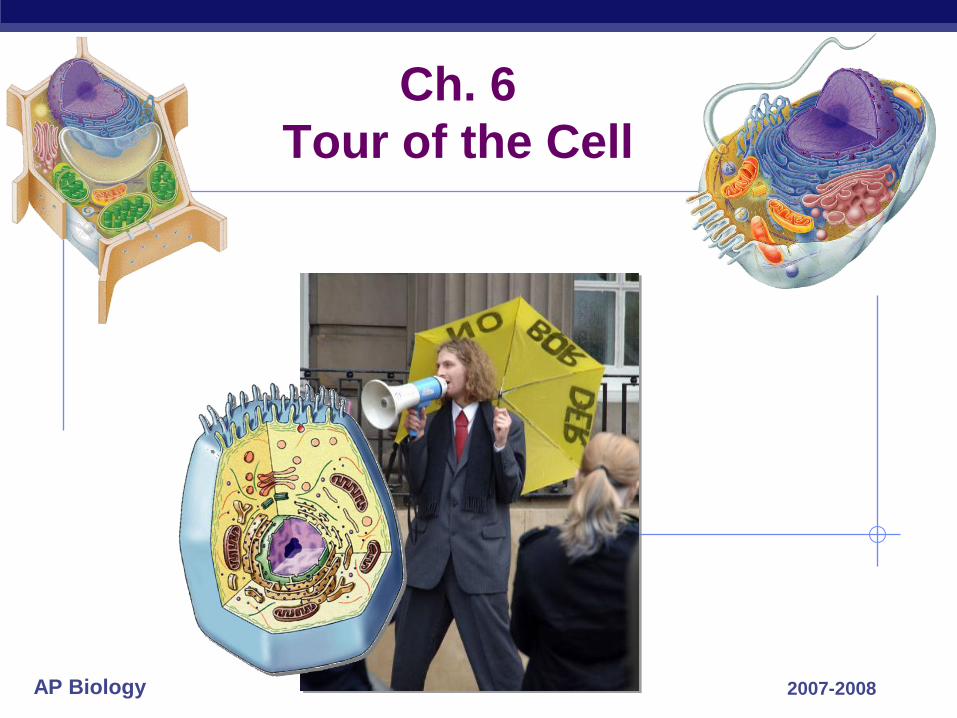
Ch. 6
Tour of the Cell
AP Biology
Microscopy
Scientists use microscopes to visualize cells too small to see with the naked eye
In a light microscope (LM), visible light is passed through a specimen and then through glass lenses
Lenses refract (bend) the light, so that the image is magnified
© 2011 Pearson Education, Inc.
AP Biology
Three important parameters of microscopy
Magnification, the ratio of an object’s image size to its real size
Resolution, the measure of the clarity of the image, or the minimum distance of two distinguishable points
Contrast, visible differences in parts of the sample
© 2011 Pearson Education, Inc.
AP Biology
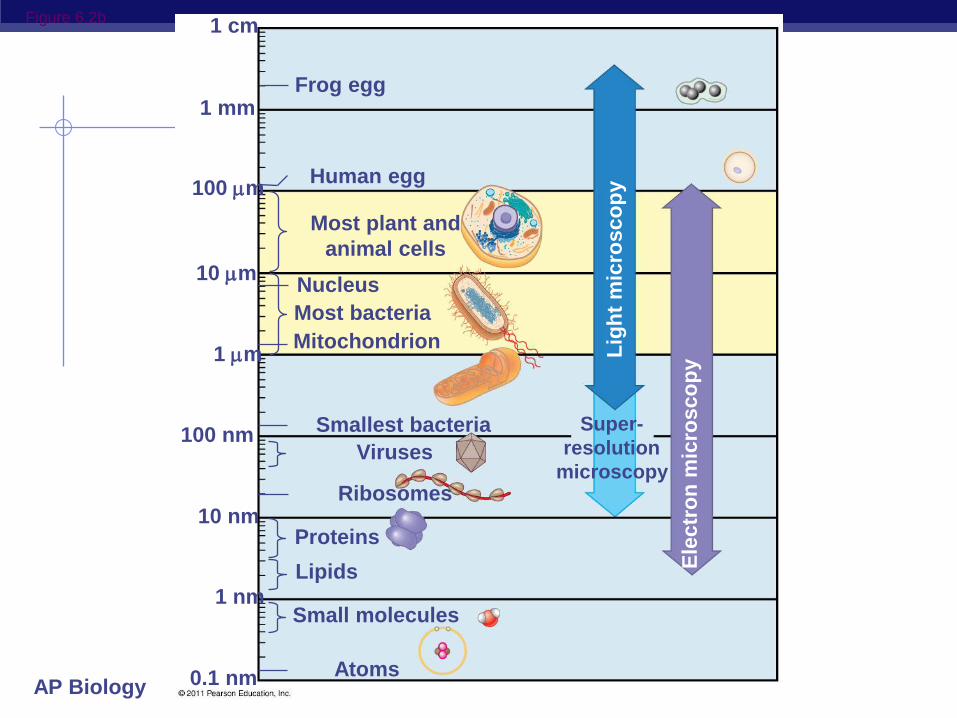
Figure 6.2b
1 mm
100 m
10 m
1 m
100 nm
10 nm
1 nm
0.1 nm Atoms
Small molecules
Lipids
Proteins
Ribosomes
Viruses
Smallest bacteria
Mitochondrion
Most bacteria
Nucleus
Most plant and
animal cells
Human egg
Lig
ht
mic
rosco
py
Ele
ctr
on
mic
rosco
py
Super-
resolution
microscopy
1 cm
Frog egg
AP Biology
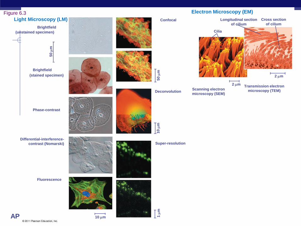
Brightfield
(unstained specimen)
Brightfield
(stained specimen)
50
m
Confocal
Differential-interference-
contrast (Nomarski)
Fluorescence
10 m
Deconvolution
Super-resolution
Scanning electron
microscopy (SEM)
Transmission electron
microscopy (TEM)
Cross section
of cilium
Longitudinal section
of cilium
Cilia
Electron Microscopy (EM)
1
m
10
m
5
0
m
2 m
2 m
Light Microscopy (LM)
Phase-contrast
Figure 6.3
AP Biology
LMs can magnify effectively to about 1,000 times the size of the actual specimen
Various techniques enhance contrast and enable cell components to be stained or labeled
Most subcellular structures, including organelles (membrane-enclosed compartments), are too small to be resolved by an LM
© 2011 Pearson Education, Inc.
AP Biology
Two basic types of electron microscopes
(EMs) are used to study subcellular
structures
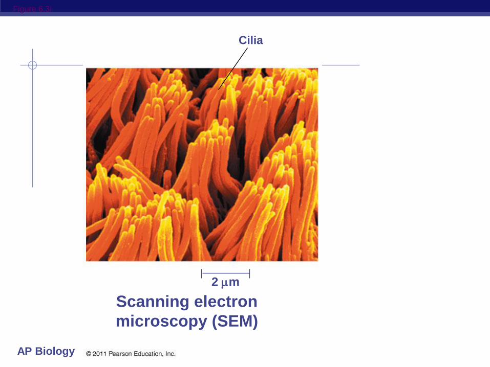
Scanning electron microscopes (SEMs) focus
a beam of electrons onto the surface of a
specimen, providing images that look 3-D
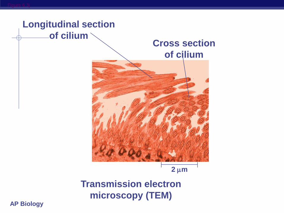
Transmission electron microscopes (TEMs)
focus a beam of electrons through a
specimen
TEMs are used mainly to study the internal
structure of cells
© 2011 Pearson Education, Inc.
AP Biology
Recent advances in light microscopy
Confocal microscopy and deconvolution
microscopy provide sharper images of three-
dimensional tissues and cells
New techniques for labeling cells improve
resolution
© 2011 Pearson Education, Inc.
AP Biology
Figure 6.3i
Cilia
2 m
Scanning electron
microscopy (SEM)
AP Biology
Figure 6.3j
Longitudinal section
of cilium Cross section
of cilium
2 m
Transmission electron
microscopy (TEM)

AP Biology
AP Biology
AP Biology
AP Biology
AP Biology
AP Biology
AP Biology
AP Biology
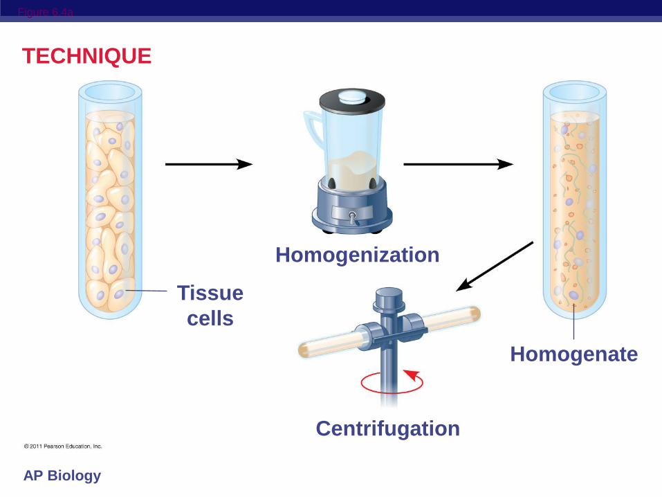
Cell Fractionation
Cell fractionation takes cells apart and
separates the major organelles from one
another
Centrifuges fractionate cells into their
component parts
Cell fractionation enables scientists to
determine the functions of organelles
Biochemistry and cytology help correlate cell
function with structure
© 2011 Pearson Education, Inc.
AP Biology
Figure 6.4a
TECHNIQUE
Homogenization
Tissue
cells
Homogenate
Centrifugation
AP Biology
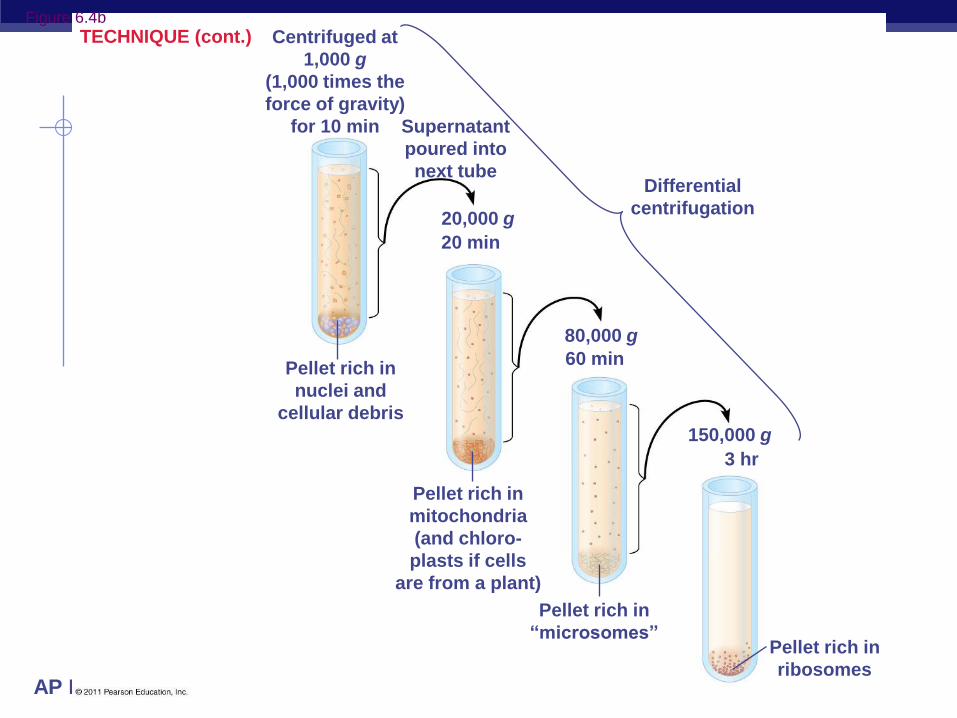
Differential
centrifugation
Centrifuged at
1,000 g
(1,000 times the
force of gravity)
for 10 min Supernatant
poured into
next tube
20 min
60 min Pellet rich in
nuclei and
cellular debris
3 hr
Pellet rich in
mitochondria
(and chloro-
plasts if cells
are from a plant)
Pellet rich in
“microsomes” Pellet rich in
ribosomes
20,000 g
80,000 g
150,000 g
TECHNIQUE (cont.) Figure 6.4b
AP Biology
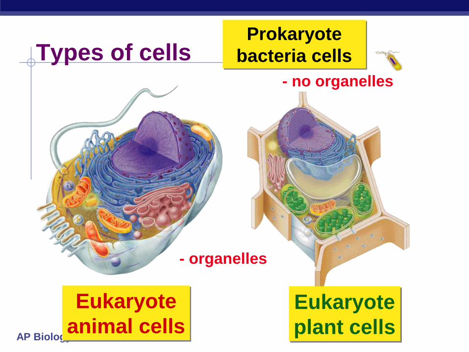
Prokaryote
bacteria cells Types of cells
Eukaryote
animal cells
- no organelles
- organelles
Eukaryote
plant cells
AP Biology
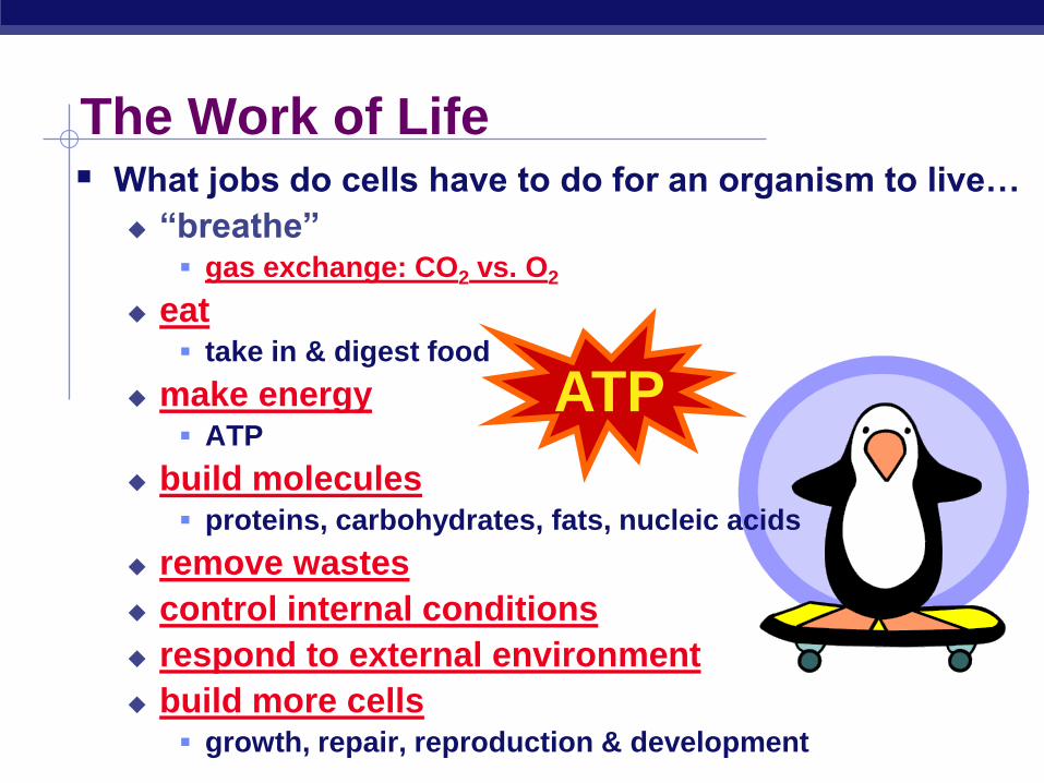
What jobs do cells have to do for an organism to live…
“breathe” gas exchange: CO2 vs. O2
eat take in & digest food
make energy ATP
build molecules proteins, carbohydrates, fats, nucleic acids
remove wastes
control internal conditions
respond to external environment
build more cells growth, repair, reproduction & development
The Work of Life
ATP
AP Biology
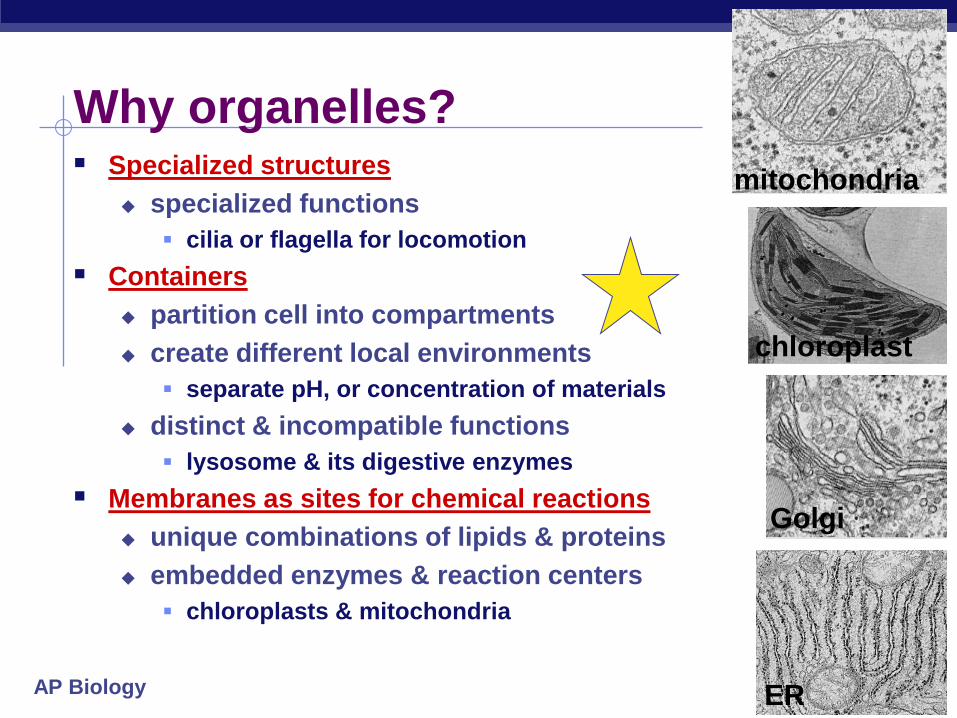
Why organelles? Specialized structures
specialized functions
cilia or flagella for locomotion
Containers
partition cell into compartments
create different local environments
separate pH, or concentration of materials
distinct & incompatible functions
lysosome & its digestive enzymes
Membranes as sites for chemical reactions
unique combinations of lipids & proteins
embedded enzymes & reaction centers
chloroplasts & mitochondria
mitochondria
chloroplast
Golgi
ER

AP Biology
Cells gotta work to live! What jobs do cells have to do?
make proteins
proteins control every
cell function
make energy
for daily life
for growth
make more cells
growth
repair
renewal
AP Biology
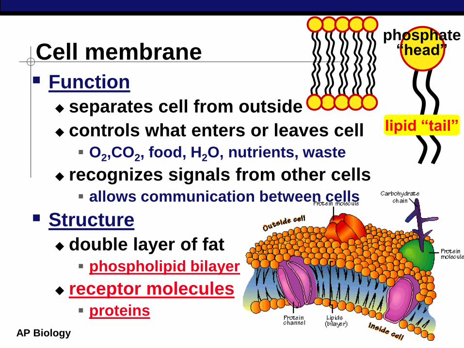
Cell membrane
Function
separates cell from outside
controls what enters or leaves cell O2,CO2, food, H2O, nutrients, waste
recognizes signals from other cells allows communication between cells
Structure
double layer of fat phospholipid bilayer
receptor molecules proteins
lipid “tail”
phosphate “head”
AP Biology
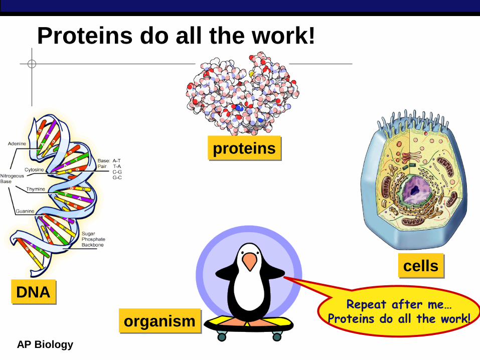
Proteins do all the work!
cells
DNA
proteins
organism Repeat after me…
Proteins do all the work!
AP Biology
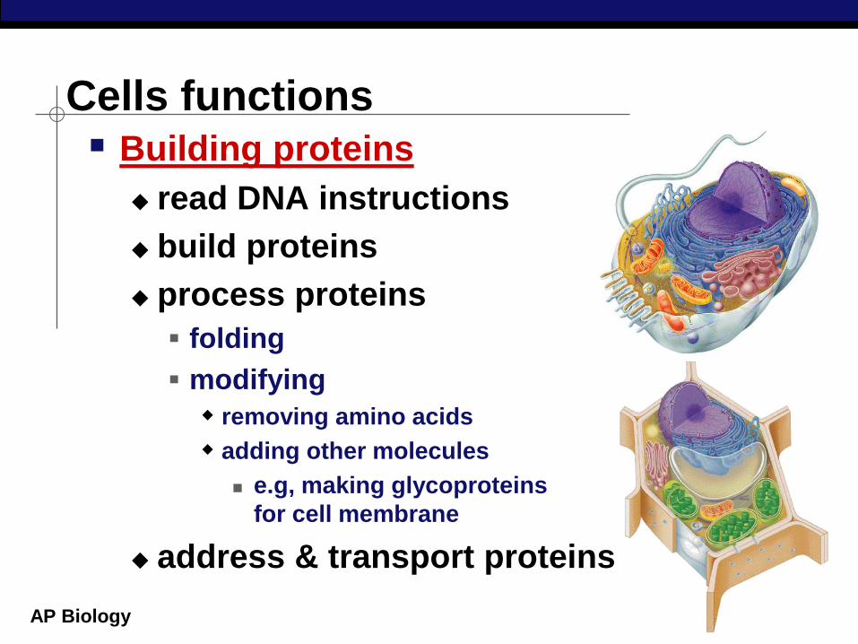
Cells functions Building proteins
read DNA instructions
build proteins
process proteins
folding
modifying
removing amino acids
adding other molecules
e.g, making glycoproteins
for cell membrane
address & transport proteins
AP Biology
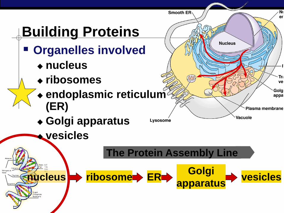
Building Proteins
Organelles involved
nucleus
ribosomes
endoplasmic reticulum (ER)
Golgi apparatus
vesicles
nucleus ribosome ER Golgi
apparatus vesicles
The Protein Assembly Line
AP Biology
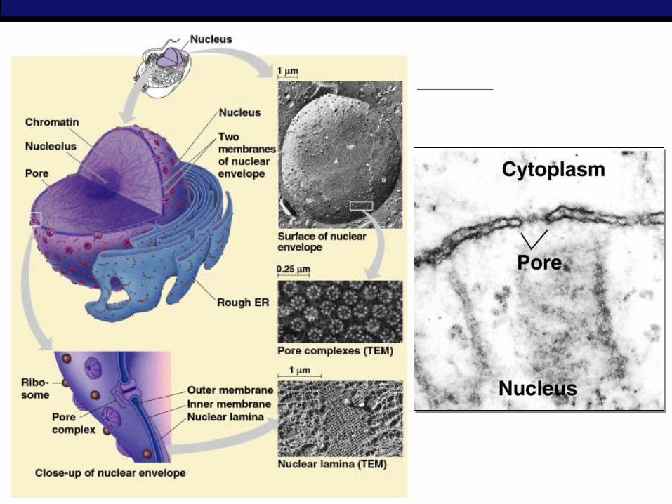
nuclear pores
nuclear pore
nuclear envelope
nucleolus
histone protein
chromosome
DNA
Function
protects DNA
Structure
nuclear envelope
double membrane
membrane fused in spots to create pores
allows large macromolecules to pass through
Nucleus
What kind of molecules need to
pass through?
AP Biology
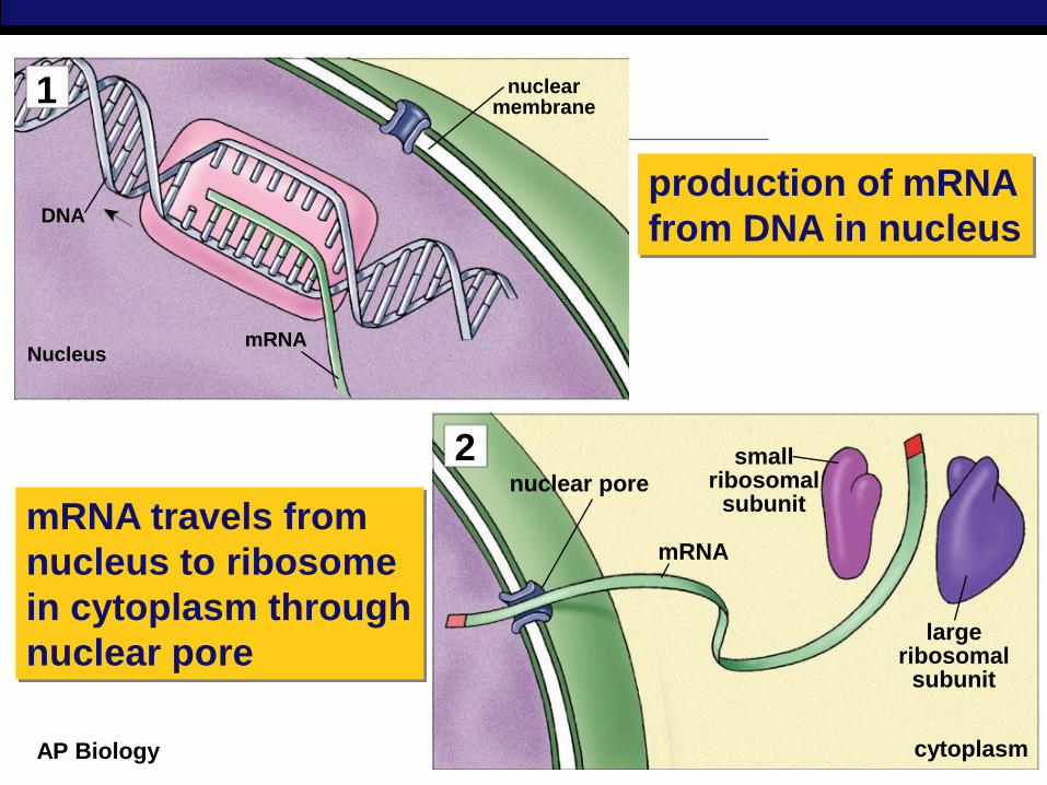
DNA
Nucleus mRNA
nuclear membrane
small ribosomal
subunit
large ribosomal
subunit
cytoplasm
mRNA
nuclear pore
production of mRNA
from DNA in nucleus
mRNA travels from
nucleus to ribosome
in cytoplasm through
nuclear pore
1
2
AP Biology
AP Biology
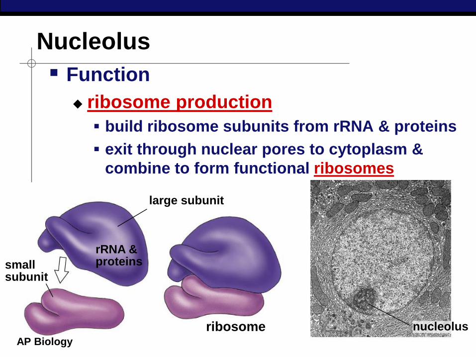
Nucleolus
Function
ribosome production
build ribosome subunits from rRNA & proteins
exit through nuclear pores to cytoplasm &
combine to form functional ribosomes
small subunit
large subunit
ribosome
rRNA & proteins
nucleolus
AP Biology
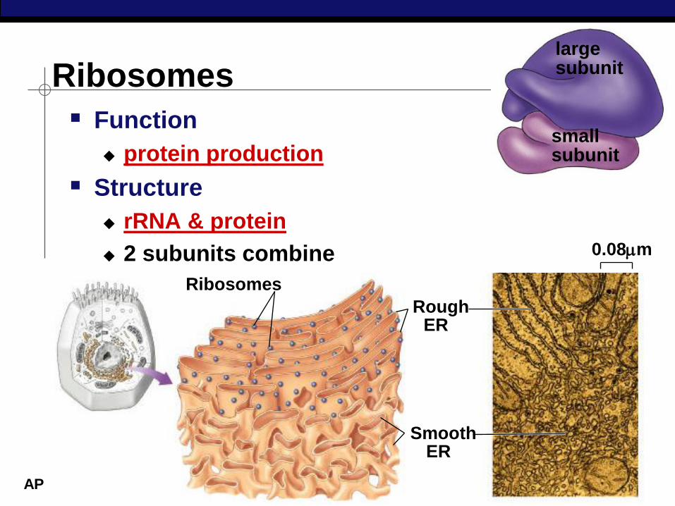
small subunit
large subunit Ribosomes
Function
protein production
Structure
rRNA & protein
2 subunits combine 0.08m
Ribosomes
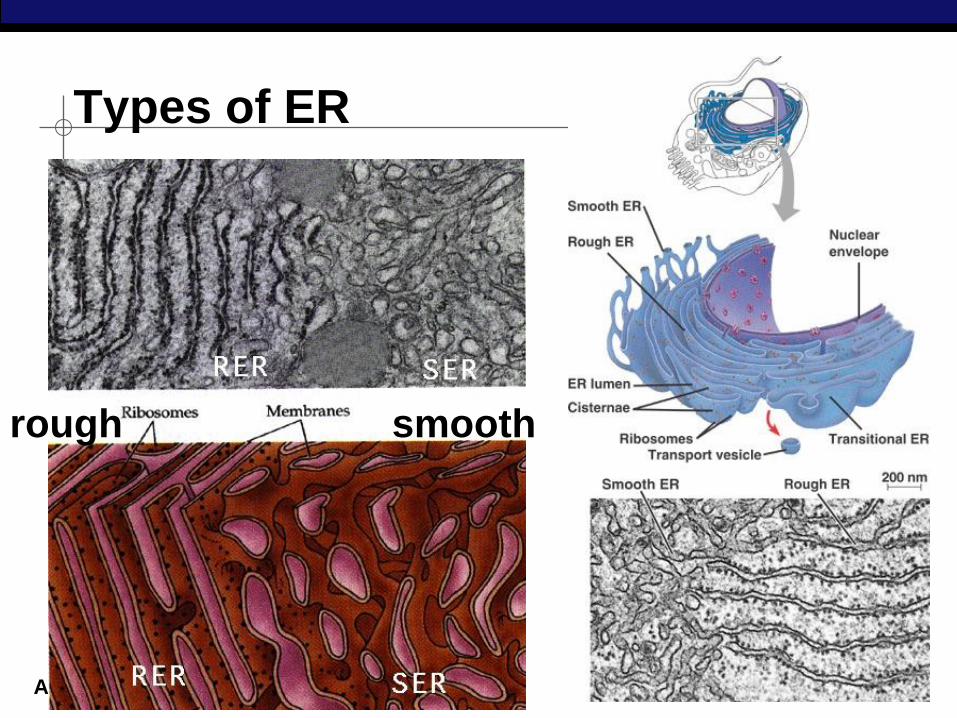
Rough ER
Smooth ER
AP Biology membrane proteins
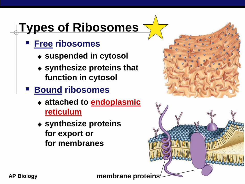
Types of Ribosomes
Free ribosomes
suspended in cytosol
synthesize proteins that
function in cytosol
Bound ribosomes
attached to endoplasmic
reticulum
synthesize proteins
for export or
for membranes
AP Biology
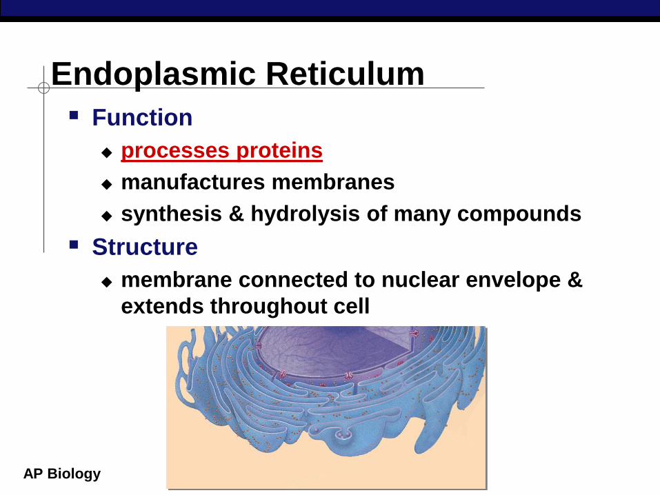
Endoplasmic Reticulum
Function
processes proteins
manufactures membranes
synthesis & hydrolysis of many compounds
Structure
membrane connected to nuclear envelope &
extends throughout cell
AP Biology
Types of ER
rough smooth
AP Biology
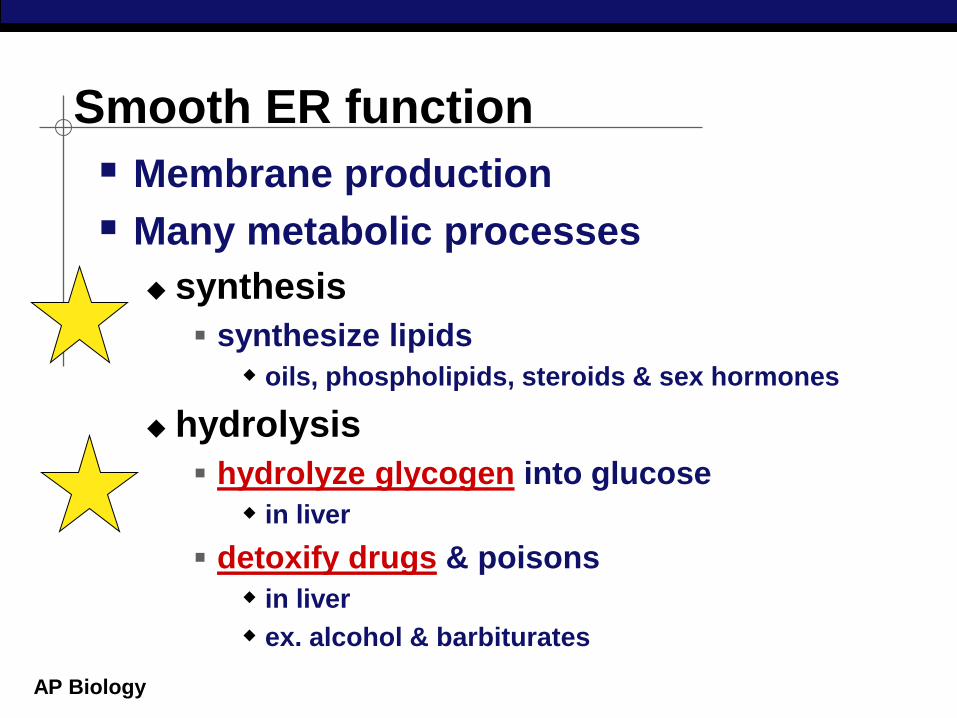
Smooth ER function
Membrane production
Many metabolic processes
synthesis
synthesize lipids
oils, phospholipids, steroids & sex hormones
hydrolysis
hydrolyze glycogen into glucose
in liver
detoxify drugs & poisons
in liver
ex. alcohol & barbiturates
AP Biology
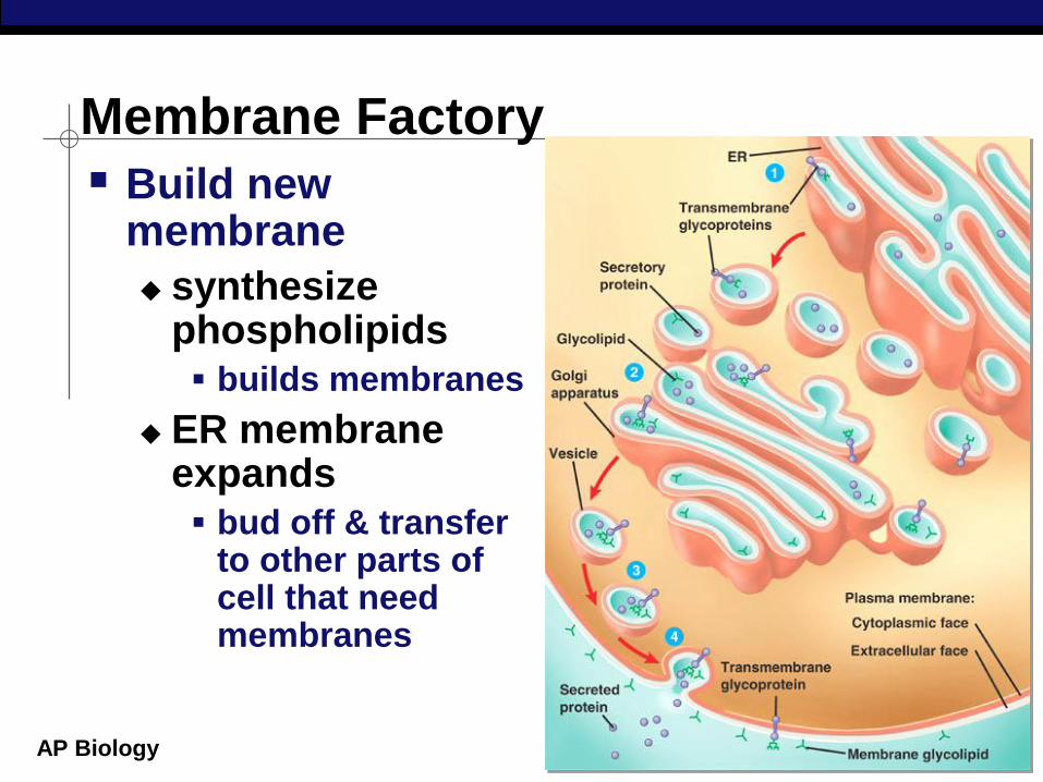
Membrane Factory
Build new membrane
synthesize phospholipids builds membranes
ER membrane expands bud off & transfer
to other parts of cell that need membranes
AP Biology
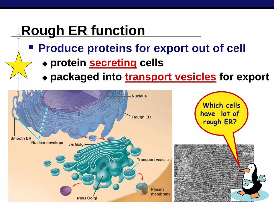
Rough ER function
Produce proteins for export out of cell
protein secreting cells
packaged into transport vesicles for export
Which cells have lot of rough ER?
AP Biology
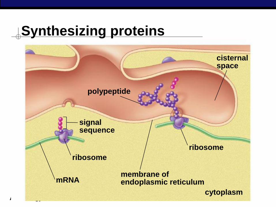
Synthesizing proteins
cytoplasm
cisternal space
mRNA
ribosome
membrane of endoplasmic reticulum
polypeptide
signal sequence
ribosome
AP Biology
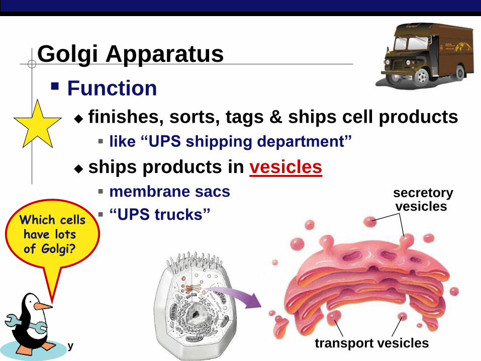
Golgi Apparatus
Which cells have lots of Golgi?
transport vesicles
secretory vesicles
Function
finishes, sorts, tags & ships cell products
like “UPS shipping department”
ships products in vesicles
membrane sacs
“UPS trucks”
AP Biology
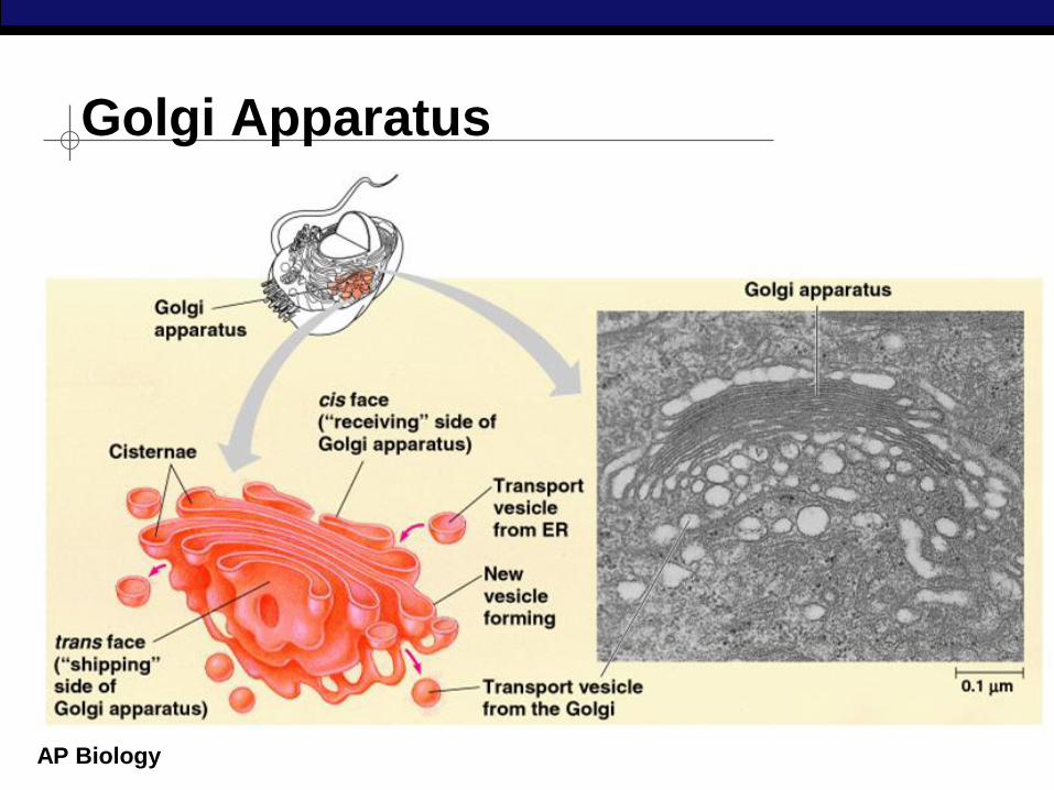
Golgi Apparatus
AP Biology
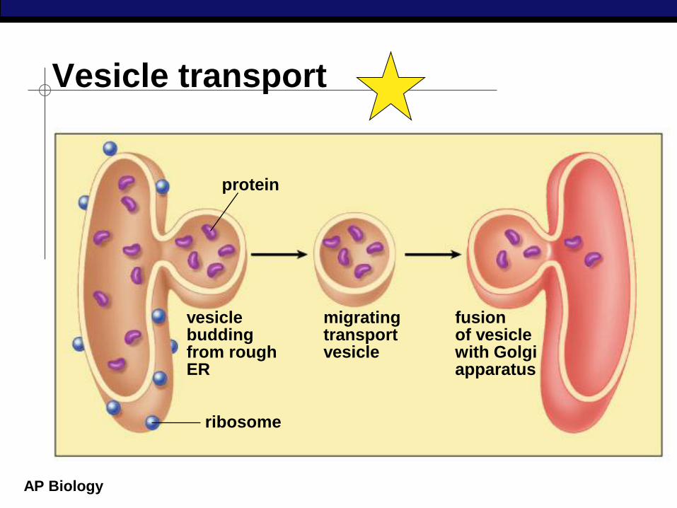
Vesicle transport
vesicle budding from rough ER
fusion of vesicle with Golgi apparatus
migrating transport vesicle
protein
ribosome
Regents Biology
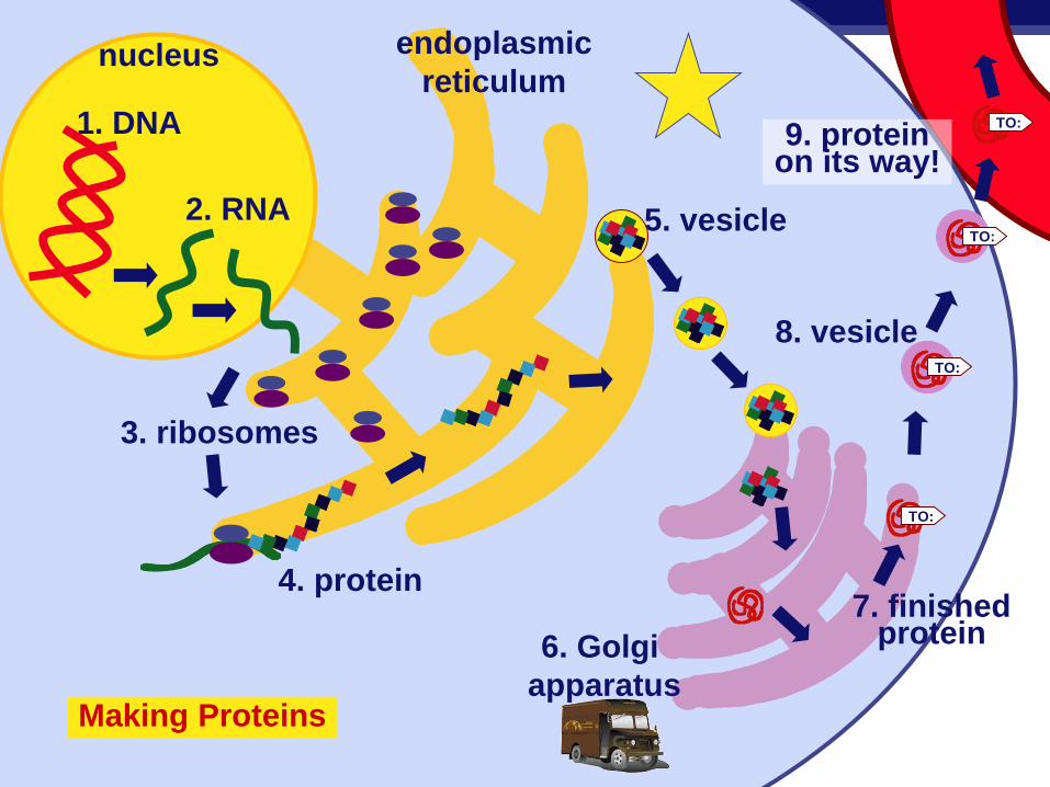
1. DNA
2. RNA
3. ribosomes
endoplasmic
reticulum
5. vesicle
6. Golgi
apparatus
8. vesicle
9. protein on its way!
4. protein 7. finished
protein
Making Proteins
TO:
TO:
TO:
TO:
nucleus

AP Biology
proteins
transport vesicle
Golgi apparatus
vesicle
smooth ER
rough ER
nuclear pore nucleus
ribosome
cell membrane protein secreted
cytoplasm
Making proteins Putting it together…
AP Biology
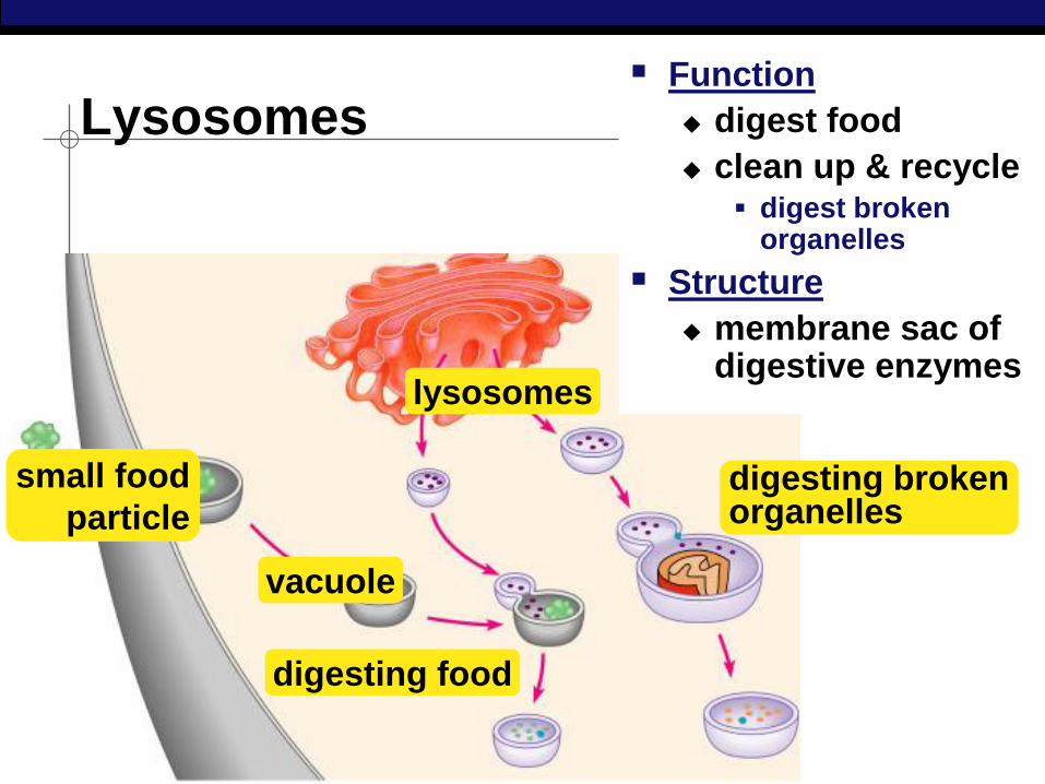
Lysosomes
small food
particle
vacuole
digesting food
lysosomes
Function
digest food
clean up & recycle digest broken
organelles
Structure
membrane sac of digestive enzymes
digesting broken organelles
AP Biology
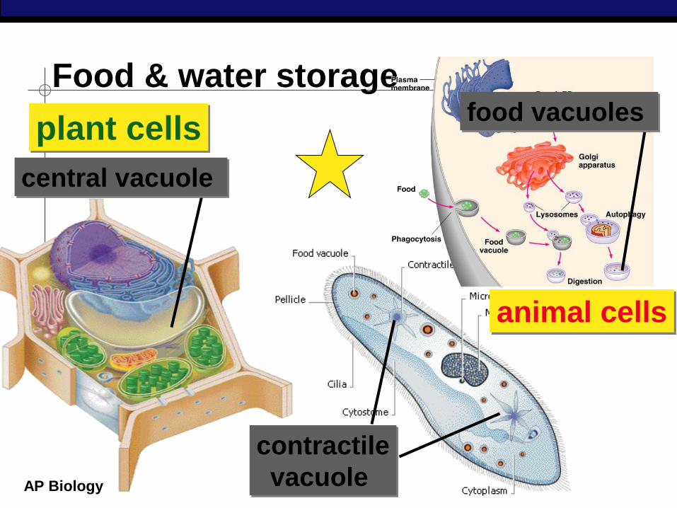
Food & water storage
plant cells
contractile
vacuole
animal cells
central vacuole
food vacuoles
AP Biology
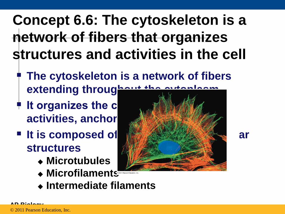
Concept 6.6: The cytoskeleton is a
network of fibers that organizes
structures and activities in the cell
The cytoskeleton is a network of fibers
extending throughout the cytoplasm
It organizes the cell’s structures and
activities, anchoring many organelles
It is composed of three types of molecular
structures Microtubules
Microfilaments
Intermediate filaments
© 2011 Pearson Education, Inc.
AP Biology
Roles of the Cytoskeleton:
Support and Motility The cytoskeleton helps to support the cell
and maintain its shape
It interacts with motor proteins to produce motility
Inside the cell, vesicles can travel along “monorails” provided by the cytoskeleton
Recent evidence suggests that the cytoskeleton may help regulate biochemical activities
© 2011 Pearson Education, Inc.
AP Biology
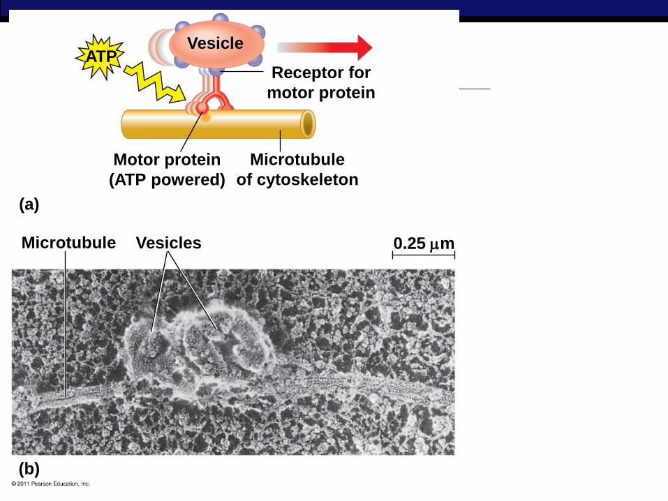
Figure 6.21
ATP Vesicle
(a)
Motor protein
(ATP powered)
Microtubule
of cytoskeleton
Receptor for
motor protein
0.25 m Vesicles Microtubule
(b)
AP Biology
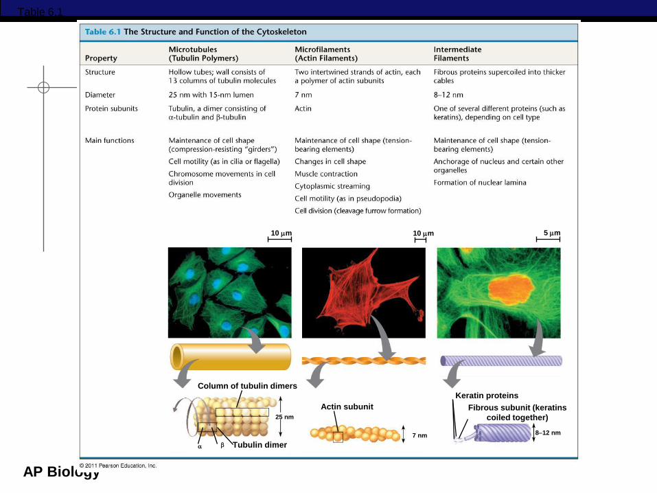
Components of the Cytoskeleton
Three main types of fibers make up the
cytoskeleton
Microtubules are the thickest of the three
components of the cytoskeleton
Microfilaments, also called actin filaments,
are the thinnest components
Intermediate filaments are fibers with
diameters in a middle range
© 2011 Pearson Education, Inc.
AP Biology
Column of tubulin dimers
Tubulin dimer
25 nm
Actin subunit
7 nm
Keratin proteins
812 nm
Fibrous subunit (keratins
coiled together)
10 m 10 m 5 m
Table 6.1
AP Biology
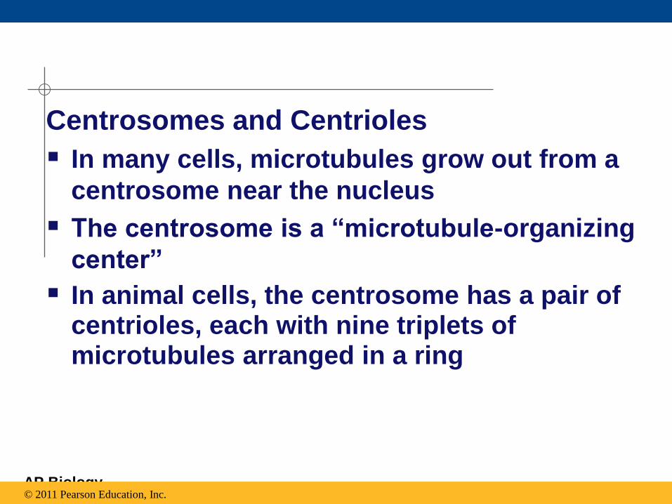
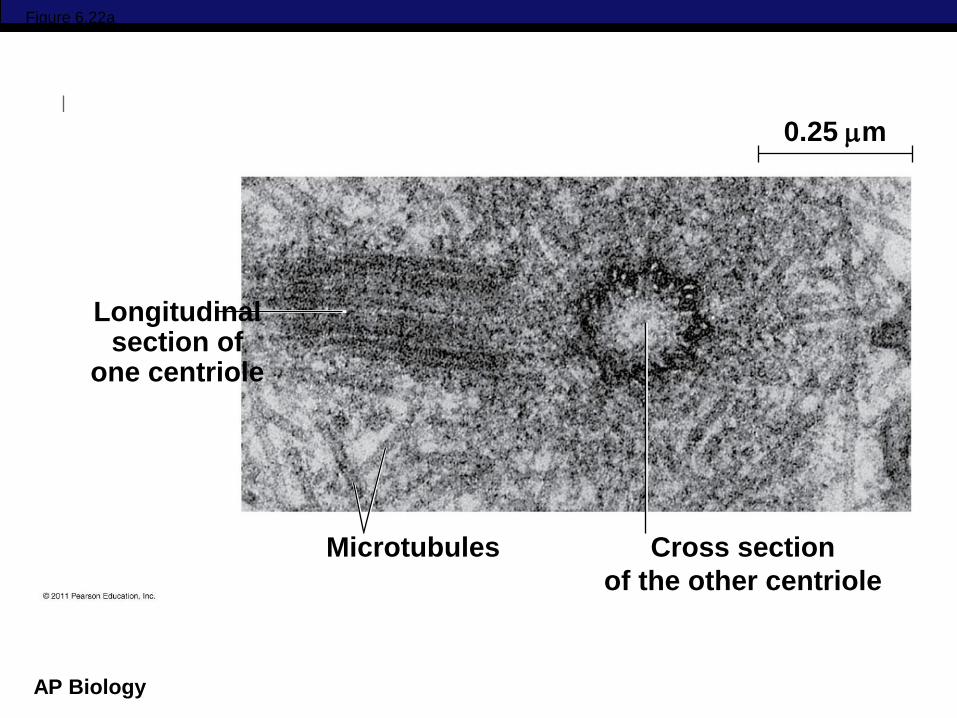
Centrosomes and Centrioles
In many cells, microtubules grow out from a
centrosome near the nucleus
The centrosome is a “microtubule-organizing
center”
In animal cells, the centrosome has a pair of centrioles, each with nine triplets of microtubules arranged in a ring
© 2011 Pearson Education, Inc.
AP Biology
Centrosome
Longitudinal section of
one centriole
Centrioles
Microtubule
0.25 m
Microtubules Cross section
of the other centriole
Figure 6.22
AP Biology
Figure 6.22a
Longitudinal section of
one centriole
0.25 m
Microtubules Cross section
of the other centriole
AP Biology
Cilia and Flagella Microtubules control the beating of cilia and
flagella, locomotor appendages of some cells
Cilia and flagella differ in their beating patterns
© 2011 Pearson Education, Inc.
AP Biology © 2011 Pearson Education, Inc.
Video: Chlamydomonas
AP Biology © 2011 Pearson Education, Inc.
Video: Paramecium Cilia
AP Biology
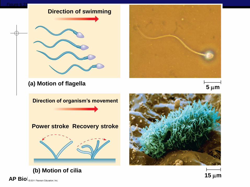
Direction of swimming
(b) Motion of cilia
Direction of organism’s movement
Power stroke Recovery stroke
(a) Motion of flagella 5 m
15 m
Figure 6.23
AP Biology
Microfilaments (Actin Filaments)
Microfilaments are solid rods about 7 nm in
diameter, built as a twisted double chain of
actin subunits
The structural role of microfilaments is to bear tension, resisting pulling forces within the cell
They form a 3-D network called the cortex just inside the plasma membrane to help support the cell’s shape
Bundles of microfilaments make up the core of microvilli of intestinal cells
© 2011 Pearson Education, Inc.
AP Biology
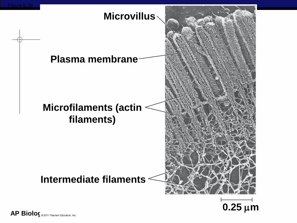
Figure 6.26
Microvillus
Plasma membrane
Microfilaments (actin
filaments)
Intermediate filaments
0.25 m
AP Biology
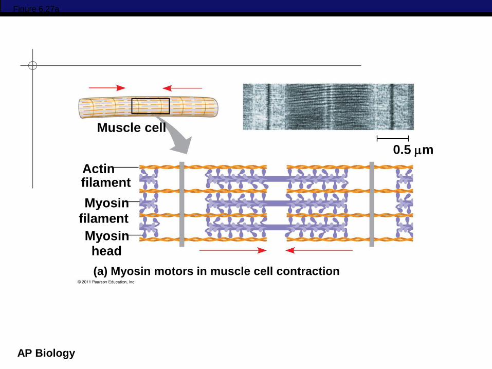
Microfilaments that function in cellular
motility contain the protein myosin in
addition to actin
In muscle cells, thousands of actin filaments
are arranged parallel to one another
Thicker filaments composed of myosin
interdigitate with the thinner actin fibers
© 2011 Pearson Education, Inc.
AP Biology
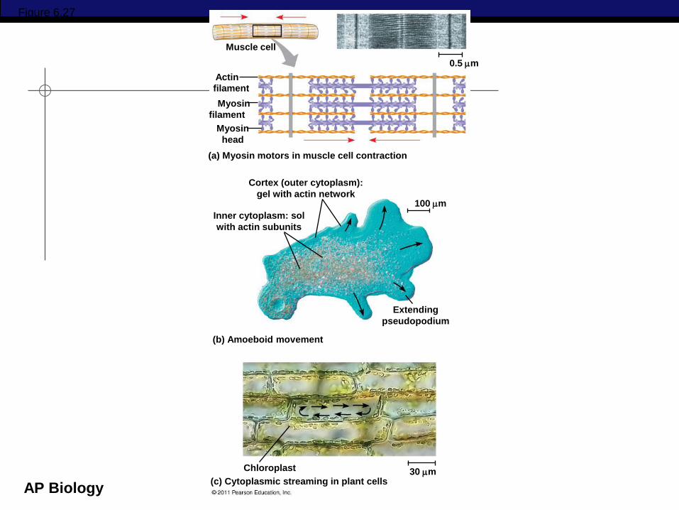
Figure 6.27
Muscle cell
Actin
filament
Myosin
Myosin
filament
head
(a) Myosin motors in muscle cell contraction
0.5 m
100 m
Cortex (outer cytoplasm):
gel with actin network
Inner cytoplasm: sol
with actin subunits
(b) Amoeboid movement
Extending
pseudopodium
30 m (c) Cytoplasmic streaming in plant cells
Chloroplast
AP Biology
Figure 6.27a
Muscle cell
Actin filament
Myosin
Myosin
filament
(a) Myosin motors in muscle cell contraction
0.5 m
head
AP Biology
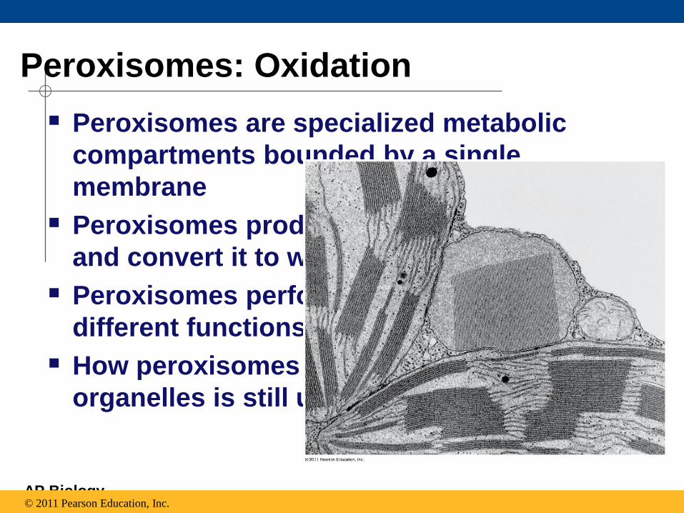
Peroxisomes: Oxidation
Peroxisomes are specialized metabolic
compartments bounded by a single
membrane
Peroxisomes produce hydrogen peroxide
and convert it to water
Peroxisomes perform reactions with many
different functions
How peroxisomes are related to other
organelles is still unknown
© 2011 Pearson Education, Inc.
AP Biology
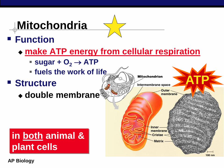
Function
make ATP energy from cellular respiration sugar + O2 ATP
fuels the work of life
Structure
double membrane
Mitochondria
in both animal &
plant cells
ATP
AP Biology
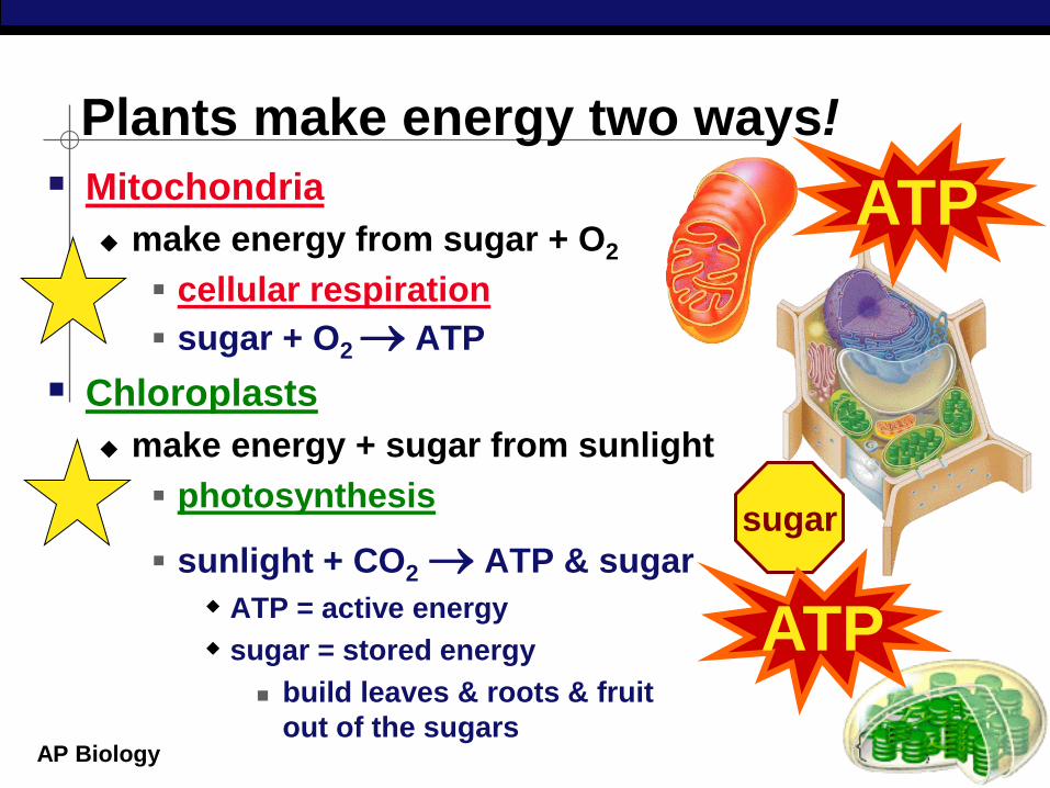
Mitochondria
make energy from sugar + O2
cellular respiration
sugar + O2 ATP
Chloroplasts
make energy + sugar from sunlight
photosynthesis
sunlight + CO2 ATP & sugar
ATP = active energy
sugar = stored energy
build leaves & roots & fruit
out of the sugars
Plants make energy two ways!
ATP
sugar
ATP
AP Biology
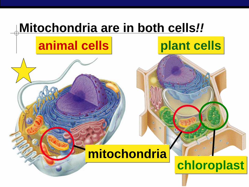
Mitochondria are in both cells!!
animal cells plant cells
mitochondria chloroplast
AP Biology
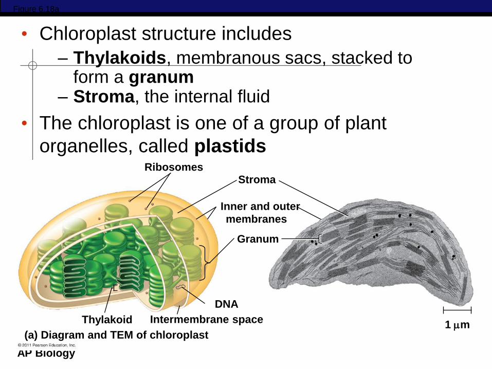
Figure 6.18a
Ribosomes Stroma
Inner and outer membranes
Granum
1 m Intermembrane space Thylakoid
(a) Diagram and TEM of chloroplast
DNA
• Chloroplast structure includes
– Thylakoids, membranous sacs, stacked to form a granum
– Stroma, the internal fluid
• The chloroplast is one of a group of plant
organelles, called plastids
AP Biology
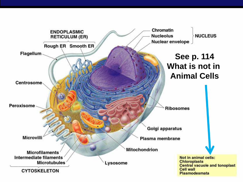
See p. 114
What is not in
Animal Cells
AP Biology
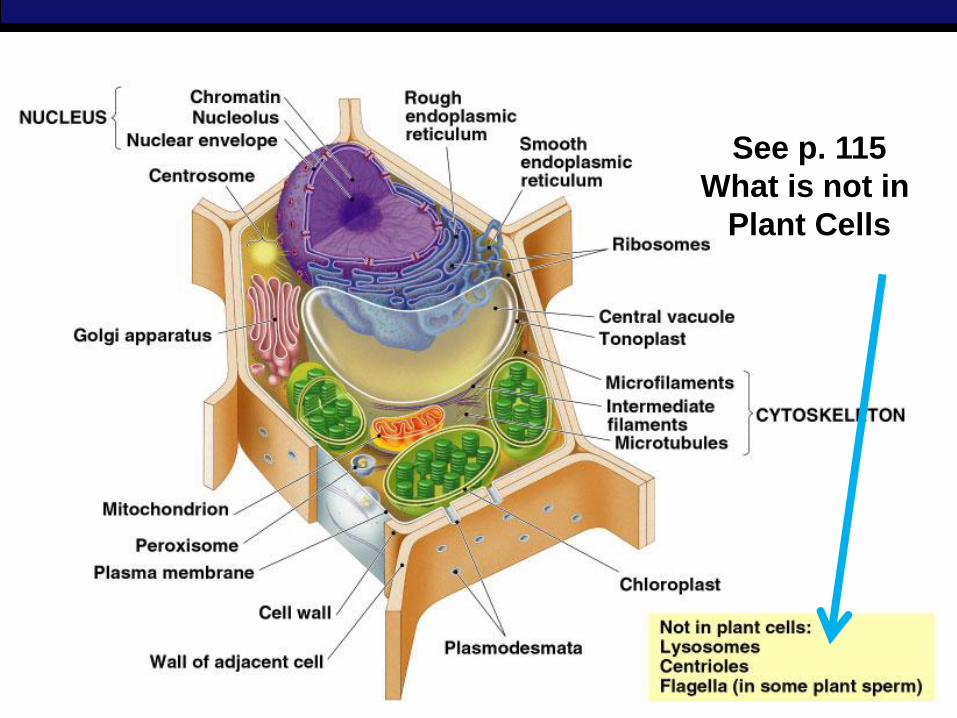
See p. 115
What is not in
Plant Cells
AP Biology
Concept 6.7: Extracellular components
and connections between cells help
coordinate cellular activities
Most cells synthesize and secrete materials
that are external to the plasma membrane
These extracellular structures include
Cell walls of plants
The extracellular matrix (ECM) of animal
cells
Intercellular junctions
© 2011 Pearson Education, Inc.
AP Biology
Cell Walls of Plants
The cell wall is an extracellular structure that
distinguishes plant cells from animal cells
Prokaryotes, fungi, and some protists also
have cell walls
The cell wall protects the plant cell,
maintains its shape, and prevents excessive
uptake of water
Plant cell walls are made of cellulose fibers
embedded in other polysaccharides and
protein
© 2011 Pearson Education, Inc.

AP Biology
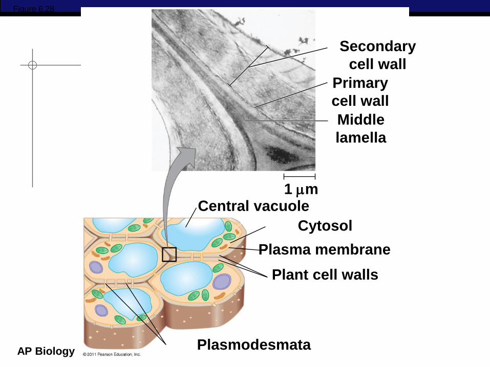
Plant cell walls may have multiple layers
Primary cell wall: relatively thin and flexible
Middle lamella: thin layer between primary walls of adjacent cells
Secondary cell wall (in some cells): added between the plasma membrane and the primary cell wall
Plasmodesmata are channels between adjacent plant cells
© 2011 Pearson Education, Inc.
AP Biology
Secondary
cell wall
Primary
cell wall
Middle
lamella
Central vacuole
Cytosol
Plasma membrane
Plant cell walls
Plasmodesmata
1 m
Figure 6.28
AP Biology
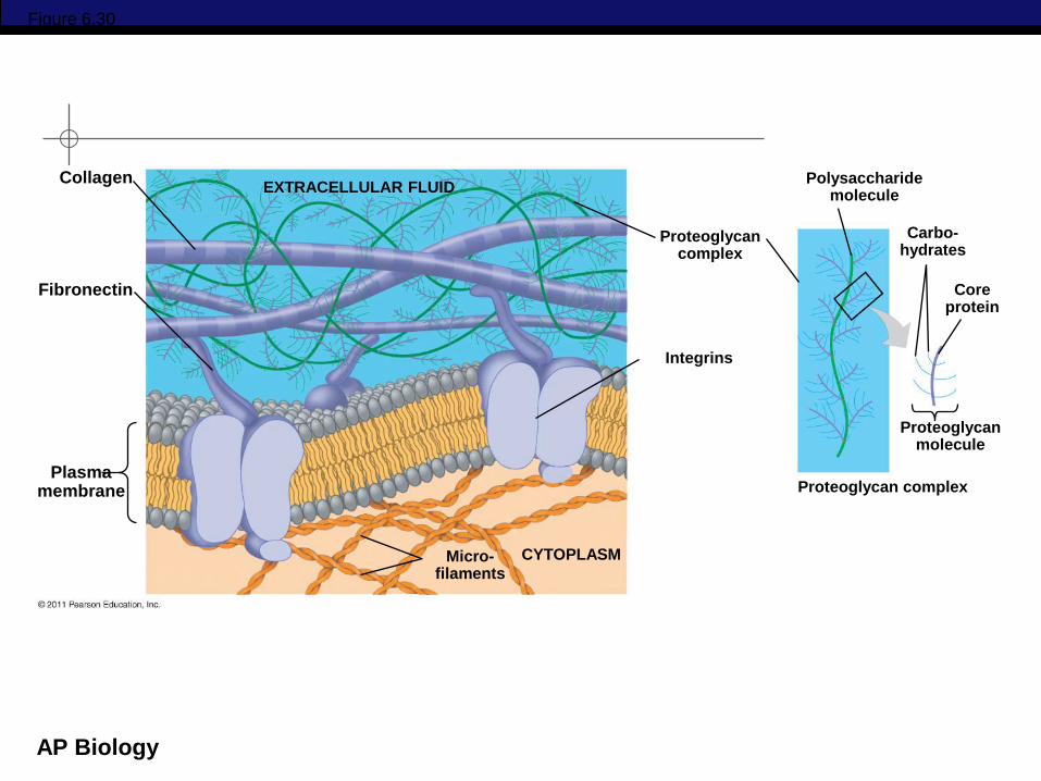
The Extracellular Matrix (ECM) of
Animal Cells Animal cells lack cell walls but are covered
by an elaborate extracellular matrix (ECM)
The ECM is made up of glycoproteins such as
collagen, proteoglycans, and fibronectin
ECM proteins bind to receptor proteins in the
plasma membrane called integrins
© 2011 Pearson Education, Inc.
AP Biology
Figure 6.30
EXTRACELLULAR FLUID Collagen
Fibronectin
Plasma membrane
Micro- filaments
CYTOPLASM
Integrins
Proteoglycan complex
Polysaccharide molecule
Carbo- hydrates
Core protein
Proteoglycan molecule
Proteoglycan complex
AP Biology
Tight Junctions, Desmosomes, and
Gap Junctions in Animal Cells
At tight junctions, membranes of neighboring
cells are pressed together, preventing leakage
of extracellular fluid
Desmosomes (anchoring junctions) fasten
cells together into strong sheets
Gap junctions (communicating junctions)
provide cytoplasmic channels between
adjacent cells
© 2011 Pearson Education, Inc.
AP Biology
http://aimediaserver4.com/studi
odaily/videoplayer/?src=ai4/harv
ard/harvard.swf&width=640&hei
ght=520
Cellular Visions:
Inner Life of the Cell