Embed Size (px)
Citation preview

Chest x ray how to solve
it
Chest x ray how to solve
it
Dr/ Allam Elsayed Allam
Dr/ Allam Elsayed Allam

Aim of presentation 1. to know the main anatomical structures.
2. main different views, when you call ,when you call.
3. adequate or not adequate film
4. Main and the commonest pathology

introduction• What are x-rays?
• X-rays: a form of electromagnetic energy• Travel at the speed of light• Electromagnetic spectrum
– Gamma Rays– X-rays– Visible light– Infrared light– Microwaves– Radio waves

Three things can happen• X-rays can:
– Pass all the way through the body– Be deflected or scattered– Be absorbed

X-rays Passing Through Tissue
• Depends on the energy of the x-ray and the atomic number of the tissue
• Higher energy x-ray - more likely to pass through• Higher atomic number - more likely to absorb the
x-raySo• X-rays that pass through the body to the film
render the film dark (black)• X-rays that are totally blocked do not reach the
film and render the film light (white)• Air = low atomic # = x-rays get through = image
is dark• Metal = high atomic # = x-rays blocked = image
is light (white)

5 Basic Radiographic Densities
• Air• Fat• Soft tissue/fluid• Mineral• Metal

Main anatomical points


Standard technquine

Standard technquine

Other technquine

P-A vs. A-P view

Technically Adequate Chest X-ray
1. Penetration 2. Inspiration3. Rotation4. angulation

Penetration
Adequate: should see the thoracic spine through the heart
If under-penetration: left hemi-diaphragm / lung base is not visible and increased pulmonary vascular marking
If over-penetration: the verse

Inspiration
adequate: Should 10 posterior ribs is visible if poor inspiration:

Common pathology
cardiomegally pleural effusion. PneumothoraxPulmonary disease.
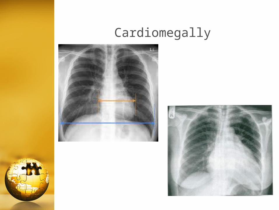
Cardiomegally

Causes of false cardiomegally
A-P film. pregnancy. Ascitis.Pecuts excavatum

Pleural effusion
the Commenst 2 forms:
1. Blunted costo-phrenic angle.
2. Meniscus sign .


Pneumothorax


Pulmonary disease(airspace vs interstitial )
airspace disease: Soft tissue opacity Indistinct borders Respect segmental or lobar boundary May shows air-bronchogram
Interstitial disease: Inhomogeneous Linear( reticular) / dots( nodular) opacities or mixed of
both ( reticulo-nodular) Not respect lobar boundaries No air-bronchogram


Common causes of airspace disease
pneumonia : ----inflammatory exudates.Pulmonary edema : --- transudatePulmonary hemorrhage: blood

Interstitial disease


Common causes
Pulmonary fibrosis Sarcoidosis Cystic fibrosis Metastatic Lung carcinoma Tubercoloma Hamartoma

QUIZZES

Technical error

Technical error

diagnosis

diagnosis

diagnosis

diagnosis