Embed Size (px)
Citation preview
540 Biophysical Journal Volume 96 January 2009 540–551
Clustering of a-Synuclein on Supported Lipid Bilayers: Role of AnionicLipid, Protein, and Divalent Ion Concentration
Anjan P. Pandey,† Farzin Haque,† Jean-Christophe Rochet,‡* and Jennifer S. Hovis†*†Department of Chemistry and ‡Department of Medicinal Chemistry and Molecular Pharmacology, Purdue University, West Lafayette, Indiana
ABSTRACT a-Synuclein is the major component of Lewy body inclusions found in the brains of patients with Parkinson’sdisease. Several studies indicate that a-synuclein binds to negatively charged phospholipid bilayers. We examined the bindingof a-synuclein to membranes containing different amounts of negatively charged lipids using supported lipid bilayers, epifluor-escence microscopy, fluorescence recovery after photobleaching, and bulk fluorescence techniques. The membranes containedphosphatidylcholine and phosphatidylglycerol. In the absence of protein, these lipids mix uniformly. Our results show that thepropensity of a-synuclein to cluster on the membrane increases as the concentration of anionic lipid and/or protein increases.Regions on the lipid bilayer where a-synuclein is clustered are enriched in phosphatidylglycerol. We also observe divalent metalions stimulate protein cluster formation, primarily by promoting lipid demixing. The importance of protein structure, lipid demixing,and divalent ions, as well as the physiological implications, will be discussed. Because membrane-bound a-synuclein assem-blies may play a role in neurotoxicity, it is of interest to determine how membranes can be used to tune the propensity ofa-synuclein to aggregate.
INTRODUCTION
a-Synuclein has received considerable attention recently
because of its connection to Parkinson’s disease (PD). A
neuropathological hallmark of PD is the presence of
cytosolic inclusions named Lewy bodies (1–3) in some of
the surviving neurons; these Lewy bodies consist largely
of fibrillar a-synuclein (4–7). Three a-synuclein mutants—
A53T (8), A30P (9), and E46K (10)—are associated with
familial PD, a rare, early-onset form of the disease inherited
in an autosomal dominant manner. All three mutants have
a greater tendency to self-associate than wild-type a-synu-
clein (11–16). All of these observations have led to the
hypothesis that the aggregation of a-synuclein plays a role
in the pathogenesis of PD (reviewed in Dev et al. (17)).
a-Synuclein is thought to be involved in regulating the
size of synaptic vesicle pools (18,19) and the release of dopa-
mine (20). The protein contains 140 residues and is ‘‘natively
unfolded’’ in solution (21). It binds to phospholipid
membranes through the N-terminal region; upon binding it
adopts an a-helical structure that sits on top of the membrane
(22), whereas the C-terminal region remains unstructured
(23). The N-terminal region (residues 1–95) consists of
seven degenerate, 11-residue repeats, six of which contain
a highly conserved motif: ‘‘KTK(E/Q)GV’’ (22,24). This
is consistent with an amphipathic helical domain with the
polar face having a net positive charge (23,25–27). Unsur-
prisingly, the binding of a-synuclein is enhanced when
anionic lipids are present (22,28,29). Whether the conversion
of a-synuclein to b-sheet structures is promoted (30–32) or
inhibited (32–34) upon binding to membranes is at present
Submitted August 12, 2008, and accepted for publication October 9, 2008.
*Correspondence: [email protected] or jennifer.hovis@gmail.
com
Editor: William C. Wimley.
� 2009 by the Biophysical Society
0006-3495/09/01/0540/12 $2.00
an open question. It is clear that a-synuclein avidly interacts
with lipids; however, further studies of this interaction are
needed to understand both the native function of a-synuclein
and the mechanism by which it becomes toxic.
In this study, supported lipid bilayers and epifluorescence
microscopy were used to examine the interaction of a-synu-
clein with membranes. Supported lipid bilayers can be
formed on appropriately treated solid supports by either
vesicle fusion or Langmuir-Blodgett/Langmuir-Schaffer
transfer (35). In either method, a thin water layer is trapped
between the bilayer and the support, allowing the lipids to
retain their natural fluidity (36). By labeling the lipids and
the protein, it is possible to monitor the spatial distribution
of both macromolecules simultaneously. In the experiments
shown herein, the lipid bilayer was composed of
phosphatidylglycerol (PG) and phosphatidylcholine (PC) in
varying ratios. These lipids were chosen because they give
an anionic/zwitterionic combination wherein the lipids mix
nearly ideally in the absence of protein or divalent ions
(37,38). Herein we examine the effect of anionic lipid
concentration, protein concentration, and divalent ion
concentration on the formation of protein clusters. The
biophysics and physiological implications of the results are
discussed.
MATERIALS AND METHODS
Stock solutions of L-a-phosphatidylcholine (egg PC), L-a-phosphatidylgly-
cerol (egg PG), and 1-palmitoyl-2-[6-[(7-nitro-2-1,3-benzoxadiazol-4-yl)
amino]hexanoyl]-sn-glycero-3-phosphocholine (NBD-PC) in chloroform
were purchased from Avanti Polar Lipids (Birmingham, AL) and used
without further purification. Ethylenediaminetetra-acetic acid (EDTA) was
purchased from Sigma Chemical (St. Louis, MO). Sodium chloride (NaCl),
sodium hydroxide (NaOH), and 4-(2-hydroxyethyl)-1-piperazineethanessul-
fonic acid (HEPES, free acid) were purchased from Mallinckrodt Chemicals
doi: 10.1016/j.bpj.2008.10.011
Clustering of a-Synuclein
(Philipsburg, NJ). Alexa Fluor 647 carboxylic acid, succinimidyl ester was
purchased from Invitrogen (Carlsbad, CA). The Superdex 200 gel-filtration
column was purchased from GE Healthcare Life Sciences (Piscataway,
NJ), and the Microcon YM-100 centrifugal filter units were obtained from
Millipore (Bedford, MA).
Vesicle and supported lipid bilayer preparation
Lipid stock solutions in chloroform were mixed in the appropriate molar
ratios, dried under a stream of nitrogen, and placed under vacuum for 1 h.
After drying, 30, 40, and 50 mol % PG lipid films were rehydrated in
100 mM NaCl solution, and 0% PG was rehydrated in 50 mM HEPES,
0.1 mM EDTA, and 100 mM NaCl buffer, pH 7.4. Large unilamellar vesi-
cles were prepared by extruding the solution 21 times through 100 nm poly-
carbonate membranes. The vesicle solution was then centrifuged for 5 min at
14,000 rpm (Eppendorf Minispin Plus). The vesicles were stored at room
temperature and shielded from light. Supported lipid bilayers were formed
by vesicle fusion inside a 60 mL perfusion chamber (Molecular Probes,
Eugene, OR) on appropriately treated glass slides. PG vesicles (30, 40,
and 50 mol %) were mixed with 750 mM NaCl solution at a 1:1 ratio before
fusion. After 15 min, excess vesicles were removed from the perfusion
chamber using 750 mM NaCl solution; the pH was adjusted using 50 mM
HEPES, 0.1 mM EDTA, and 750 mM NaCl, pH 7.4 buffer; and the slide
was finally rinsed with 50 mM HEPES, 0.1 mM EDTA, pH 7.4 buffer to re-
move salt. Before fusion, 0 mol % PG vesicles were mixed with 50 mM
HEPES, 0.1 mM EDTA, pH 7.4 buffer at a 1:1 ratio, and the excess vesicles
were removed with the same buffer. Before bilayer formation, glass cover-
slips were washed in ICN 7X detergent (VWR International, Chicago, IL),
rinsed profusely in deionized water, dried with a stream of nitrogen, and
baked at 450�C for 4 h. The slides and vesicles were used within a day of
preparation. At least 3 mL of buffer were passed through the perfusion
chamber to ensure complete exchange. The bilayers were kept hydrated in
50 mM HEPES, 0.1 mM EDTA, pH 7.4 buffer before protein addition.
Apart from using buffers with no EDTA, all other conditions were kept
the same in the experiments involving calcium.
Expression, purification, and labelingof a-synuclein
Human wild-type a-synuclein was expressed in Escherichia coli and puri-
fied as described previously (12). The protein was preferentially labeled
at the N-terminus: monomeric a-synuclein (0.56 mM in 225 mL PBS,
pH 7.0) was mixed with Alexa Fluor 647 carboxylic acid, succinimidyl ester
(0.25 mg in 25 mL dimethyl sulfoxide (DMSO)). The mixture was placed on
a Microplate shaker at 800 rpm for 2 h. The protein solution was then loaded
onto a Superdex 200 gel-filtration column and eluted in 20 mM HEPES, pH
7.4. The protein concentration and degree of labeling were determined using
ultraviolet-visible absorbance measurements (the degree of labeling of the
protein was typically 20%, mol/mol). The protein was then aliquoted and
stored at �20�C. Before each experiment, an aliquot was thawed and centri-
fuged through a Microcon YM-100 centrifugal filter unit with a molecular-
weight cutoff of 100 kDa to remove any aggregates.
Calculating the amount of a-synuclein adsorbed
A Nikon TE2000 fluorescence microscope equipped with a 40� oil immer-
sion objective, an Alexa filter set (Chroma Technology, Rockingham, VT)
and a silicon avalanche photodiode (APD) single photon counting module
(SPCM-AQR-16-FC, PerkinElmer, Vaudreuil, Quebec, Canada) was used
to focus, collect, and count the emitted fluorescence. An arc lamp was
used to monitor the protein and a LabVIEW program was used to acquire
the counts from the APD. To minimize photobleaching of the fluorophore
during the counting period, the lamp intensity was reduced using a 5X (focal
transmission of 1 � 105) neutral density filter (NE50B; Thorlabs, Newton,
NJ). a-Synuclein (2.6 mM, 60 mL) was added to phospholipid bilayers
and incubated for 15 min. Protein counts were obtained before and after
the unbound protein was rinsed off with 2.5 mL of 50 mM HEPES,
0.1 mM EDTA, pH 7.4 buffer.
Imaging of supported lipid bilayers and protein
A Nikon (Tokyo, Japan) TE2000 fluorescence microscope equipped with
a Cascade 512B CCD camera (Roper Scientific, Tucson, AZ) was used.
An X-Cite 120 arc lamp (EXFO) was used as a light source. For imaging,
0.25 mol % tail-labeled NBD-PC was present in the bilayer and the protein
was labeled with Alexa Fluor 647. The NBD and Alexa fluorophores were
imaged using NBD and Alexa 647 filter sets, respectively (Chroma Tech-
nology). Images were acquired using a 100�, 1.30 NA objective. The
images were contrasted in each case to make any features appear clearly.
For quantitative comparisons, linescans are also provided. From all the line-
scans, appropriate backgrounds (dark noise of the camera, glass autofluores-
cence, etc.) were subtracted off using counts obtained from a bilayer without
the NBD fluorophore.
Fluorescence recovery after photobleaching
The fluorescence recovery after photobleaching (FRAP) measurement
system is discussed in detail elsewhere (39). Briefly, a Nikon TE2000 fluo-
rescence microscope equipped with a 40� oil immersion objective, an NBD
filter set (Chroma Technology) and an APD single photon counting module
(SPCM-AQR-16-FC, PerkinElmer, Vaudreuil, Canada) was used to focus,
collect, and count the emitted fluorescence. A 15 mW Argon ion laser
(488 nm Melles Griot) was used to both bleach and monitor the lipid bilayer.
To reduce further photobleaching of the fluorophore during the recovery
period, the laser intensity was reduced 100,000-fold using a 5X (focal trans-
mission of 1� 105) neutral density filter (NE50B, Thorlabs, Newton, NJ). A
LabVIEW program was used to acquire the counts from the APD, control
the filter wheel, and trigger the shutter (Uniblitz; VA, Rochester, NY).
RESULTS
Some reports in the literature suggest that membranes
promote a-synuclein aggregation (30–32), whereas others
suggest that membranes inhibit a-synuclein aggregation
(32–34). Herein we describe three control parameters that
can be used to adjust the propensity of a-synuclein to adopt
an aggregate-like appearance: anionic lipid concentration,
protein concentration, and divalent ion concentration.
Supported lipid bilayers prepared by vesicle fusion were
used as a model membrane system. The bilayers were
imaged using epifluorescence microscopy, and the fluidity
of each bilayer was checked with FRAP. The bilayers were
composed of varying amounts of egg PG and egg PC. Egg
PG is prepared from egg PC by the action of phospholipase
D; therefore, the tail compositions were identical for the
phospholipids used in this study. After varying concentra-
tions of a-synuclein (in 50 mM HEPES, 0.1 mM EDTA,
pH 7.4) were added to the bulk solution, the bilayer was
incubated for 15 min. Unbound protein was then rinsed off
with buffer, and images of the protein and lipid were
acquired. The bilayers contained 0.25 mol % NBD-PC,
and ~20% of the protein was labeled with Alexa 647. The
excitation and emission spectra of the fluorophores are far
enough apart that fluorescence resonance energy transfer is
not a concern.
Biophysical Journal 96(2) 540–551
541
542 Pandey et al.
Anionic lipid
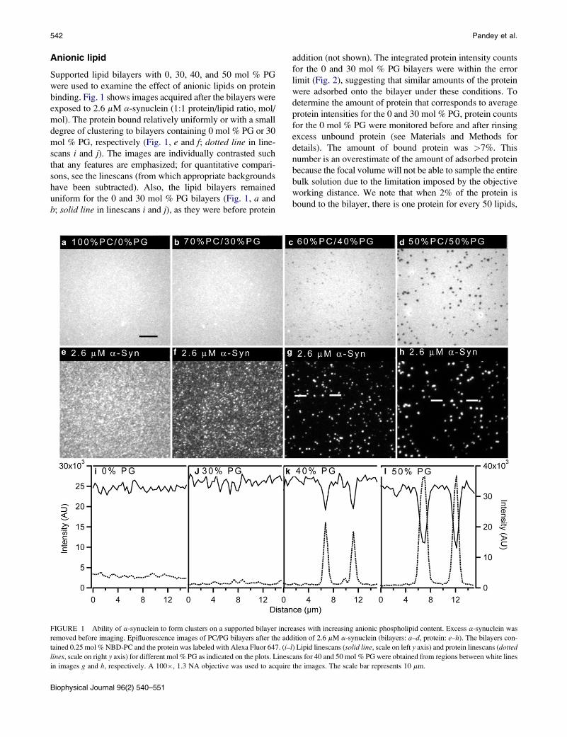
Supported lipid bilayers with 0, 30, 40, and 50 mol % PG
were used to examine the effect of anionic lipids on protein
binding. Fig. 1 shows images acquired after the bilayers were
exposed to 2.6 mM a-synuclein (1:1 protein/lipid ratio, mol/
mol). The protein bound relatively uniformly or with a small
degree of clustering to bilayers containing 0 mol % PG or 30
mol % PG, respectively (Fig. 1, e and f; dotted line in line-
scans i and j). The images are individually contrasted such
that any features are emphasized; for quantitative compari-
sons, see the linescans (from which appropriate backgrounds
have been subtracted). Also, the lipid bilayers remained
uniform for the 0 and 30 mol % PG bilayers (Fig. 1, a and
b; solid line in linescans i and j), as they were before protein
addition (not shown). The integrated protein intensity counts
for the 0 and 30 mol % PG bilayers were within the error
limit (Fig. 2), suggesting that similar amounts of the protein
were adsorbed onto the bilayer under these conditions. To
determine the amount of protein that corresponds to average
protein intensities for the 0 and 30 mol % PG, protein counts
for the 0 mol % PG were monitored before and after rinsing
excess unbound protein (see Materials and Methods for
details). The amount of bound protein was >7%. This
number is an overestimate of the amount of adsorbed protein
because the focal volume will not be able to sample the entire
bulk solution due to the limitation imposed by the objective
working distance. We note that when 2% of the protein is
bound to the bilayer, there is one protein for every 50 lipids,
FIGURE 1 Ability of a-synuclein to form clusters on a supported bilayer increases with increasing anionic phospholipid content. Excess a-synuclein was
removed before imaging. Epifluorescence images of PC/PG bilayers after the addition of 2.6 mM a-synuclein (bilayers: a–d, protein: e–h). The bilayers con-
tained 0.25 mol % NBD-PC and the protein was labeled with Alexa Fluor 647. (i–l) Lipid linescans (solid line, scale on left y axis) and protein linescans (dottedlines, scale on right y axis) for different mol % PG as indicated on the plots. Linescans for 40 and 50 mol % PG were obtained from regions between white lines
in images g and h, respectively. A 100�, 1.3 NA objective was used to acquire the images. The scale bar represents 10 mm.
Biophysical Journal 96(2) 540–551
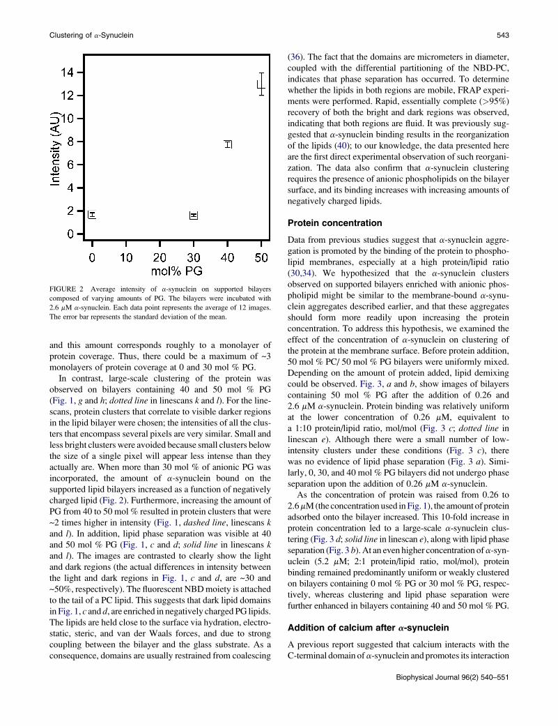
and this amount corresponds roughly to a monolayer of
protein coverage. Thus, there could be a maximum of ~3
monolayers of protein coverage at 0 and 30 mol % PG.
In contrast, large-scale clustering of the protein was
observed on bilayers containing 40 and 50 mol % PG
(Fig. 1, g and h; dotted line in linescans k and l). For the line-
scans, protein clusters that correlate to visible darker regions
in the lipid bilayer were chosen; the intensities of all the clus-
ters that encompass several pixels are very similar. Small and
less bright clusters were avoided because small clusters below
the size of a single pixel will appear less intense than they
actually are. When more than 30 mol % of anionic PG was
incorporated, the amount of a-synuclein bound on the
supported lipid bilayers increased as a function of negatively
charged lipid (Fig. 2). Furthermore, increasing the amount of
PG from 40 to 50 mol % resulted in protein clusters that were
~2 times higher in intensity (Fig. 1, dashed line, linescans kand l). In addition, lipid phase separation was visible at 40
and 50 mol % PG (Fig. 1, c and d; solid line in linescans kand l). The images are contrasted to clearly show the light
and dark regions (the actual differences in intensity between
the light and dark regions in Fig. 1, c and d, are ~30 and
~50%, respectively). The fluorescent NBD moiety is attached
to the tail of a PC lipid. This suggests that dark lipid domains
in Fig. 1, c and d, are enriched in negatively charged PG lipids.
The lipids are held close to the surface via hydration, electro-
static, steric, and van der Waals forces, and due to strong
coupling between the bilayer and the glass substrate. As a
consequence, domains are usually restrained from coalescing
FIGURE 2 Average intensity of a-synuclein on supported bilayers
composed of varying amounts of PG. The bilayers were incubated with
2.6 mM a-synuclein. Each data point represents the average of 12 images.
The error bar represents the standard deviation of the mean.
Clustering of a-Synuclein
(36). The fact that the domains are micrometers in diameter,
coupled with the differential partitioning of the NBD-PC,
indicates that phase separation has occurred. To determine
whether the lipids in both regions are mobile, FRAP experi-
ments were performed. Rapid, essentially complete (>95%)
recovery of both the bright and dark regions was observed,
indicating that both regions are fluid. It was previously sug-
gested that a-synuclein binding results in the reorganization
of the lipids (40); to our knowledge, the data presented here
are the first direct experimental observation of such reorgani-
zation. The data also confirm that a-synuclein clustering
requires the presence of anionic phospholipids on the bilayer
surface, and its binding increases with increasing amounts of
negatively charged lipids.
Protein concentration
Data from previous studies suggest that a-synuclein aggre-
gation is promoted by the binding of the protein to phospho-
lipid membranes, especially at a high protein/lipid ratio
(30,34). We hypothesized that the a-synuclein clusters
observed on supported bilayers enriched with anionic phos-
pholipid might be similar to the membrane-bound a-synu-
clein aggregates described earlier, and that these aggregates
should form more readily upon increasing the protein
concentration. To address this hypothesis, we examined the
effect of the concentration of a-synuclein on clustering of
the protein at the membrane surface. Before protein addition,
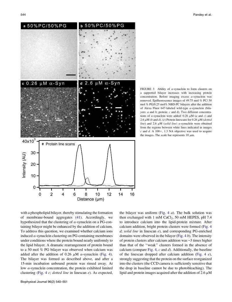
50 mol % PC/ 50 mol % PG bilayers were uniformly mixed.
Depending on the amount of protein added, lipid demixing
could be observed. Fig. 3, a and b, show images of bilayers
containing 50 mol % PG after the addition of 0.26 and
2.6 mM a-synuclein. Protein binding was relatively uniform
at the lower concentration of 0.26 mM, equivalent to
a 1:10 protein/lipid ratio, mol/mol (Fig. 3 c; dotted line in
linescan e). Although there were a small number of low-
intensity clusters under these conditions (Fig. 3 c), there
was no evidence of lipid phase separation (Fig. 3 a). Simi-
larly, 0, 30, and 40 mol % PG bilayers did not undergo phase
separation upon the addition of 0.26 mM a-synuclein.
As the concentration of protein was raised from 0.26 to
2.6 mM (the concentration used in Fig. 1), the amount of protein
adsorbed onto the bilayer increased. This 10-fold increase in
protein concentration led to a large-scale a-synuclein clus-
tering (Fig. 3 d; solid line in linescan e), along with lipid phase
separation (Fig. 3 b). At an even higher concentration of a-syn-
uclein (5.2 mM; 2:1 protein/lipid ratio, mol/mol), protein
binding remained predominantly uniform or weakly clustered
on bilayers containing 0 mol % PG or 30 mol % PG, respec-
tively, whereas clustering and lipid phase separation were
further enhanced in bilayers containing 40 and 50 mol % PG.
Addition of calcium after a-synuclein
A previous report suggested that calcium interacts with the
C-terminal domain of a-synuclein and promotes its interaction
Biophysical Journal 96(2) 540–551
543
with a phospholipid bilayer, thereby stimulating the formation
of membrane-bound aggregates (41). Accordingly, we
hypothesized that the clustering of a-synuclein on a PG-con-
taining bilayer might be enhanced by the addition of calcium.
To address this question, we examined whether calcium ions
induced a-synuclein clustering on PG-containing membranes
under conditions where the protein bound nearly uniformly to
the lipid bilayer. A dramatic rearrangement of protein bound
to a 50 mol % PG bilayer was observed when calcium was
added after the addition of 0.26 mM a-synuclein (Fig. 4).
The bilayer was formed as described above, and after a
15-min incubation unbound protein was rinsed away. At
low a-synuclein concentration, the protein exhibited limited
clustering (Fig. 4 c; dotted line in linescan e). As expected,
the bilayer was uniform (Fig. 4 a). The bulk solution was
then exchanged with 1 mM CaCl2, 50 mM HEPES, pH 7.4
to introduce calcium into the lipid-protein mixture. After
calcium addition, bright protein clusters were formed (Fig. 4
d; solid line in linescan e), and corresponding PG-enriched
domains were observed in the bilayer (Fig. 4 b). The intensity
of protein clusters after calcium addition was ~3 times higher
than that of the ‘‘weak’’ clusters formed in the absence of
calcium (compare Fig. 4, c and d). Additionally, the baseline
of the linescan dropped after calcium addition (Fig. 4 e),
strongly suggesting that the protein on the surface reorganized
into the clusters (the CCD camera is sufficiently sensitive that
the drop in baseline cannot be due to photobleaching). The
lipid and protein images acquired after the addition of 2.6 mM
FIGURE 3 Ability of a-synuclein to form clusters on
a supported bilayer increases with increasing protein
concentration. Before imaging excess a-synuclein was
removed. Epifluorescence images of 49.75 mol % PC/ 50
mol % PG/0.25 mol% NBD-PC bilayers after the addition
of Alexa Fluor 647-labeled wild-type a-synuclein (bila-
yers: a and b; protein: c and d). Two different concentra-
tions of a-synuclein were added: 0.26 mM (a and c) and
2.6 mM (b and d). (e) Protein linescans for 0.26 mM (dottedline) and 2.6 mM (solid line) a-synuclein were obtained
from the regions between white lines indicated in images
c and d. A 100�, 1.3 NA objective was used to acquire
the images. The scale bar represents 10 mm.
Biophysical Journal 96(2) 540–551
544 Pandey et al.
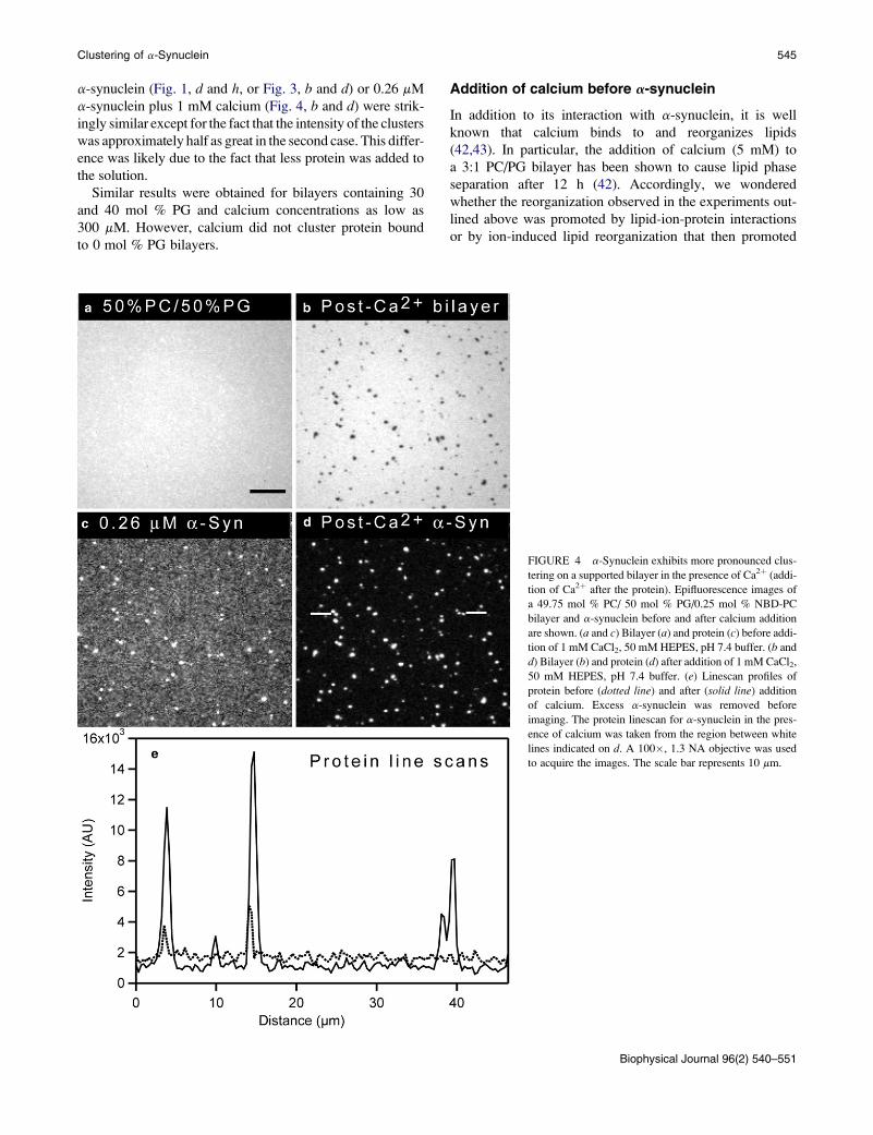
a-synuclein (Fig. 1, d and h, or Fig. 3, b and d) or 0.26 mM
a-synuclein plus 1 mM calcium (Fig. 4, b and d) were strik-
ingly similar except for the fact that the intensity of the clusters
was approximately half as great in the second case. This differ-
ence was likely due to the fact that less protein was added to
the solution.
Similar results were obtained for bilayers containing 30
and 40 mol % PG and calcium concentrations as low as
300 mM. However, calcium did not cluster protein bound
to 0 mol % PG bilayers.
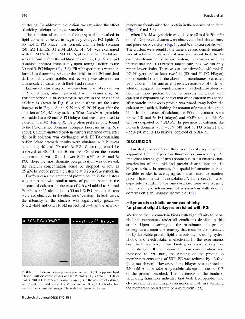
Addition of calcium before a-synuclein
In addition to its interaction with a-synuclein, it is well
known that calcium binds to and reorganizes lipids
(42,43). In particular, the addition of calcium (5 mM) to
a 3:1 PC/PG bilayer has been shown to cause lipid phase
separation after 12 h (42). Accordingly, we wondered
whether the reorganization observed in the experiments out-
lined above was promoted by lipid-ion-protein interactions
or by ion-induced lipid reorganization that then promoted
FIGURE 4 a-Synuclein exhibits more pronounced clus-
tering on a supported bilayer in the presence of Ca2þ (addi-
tion of Ca2þ after the protein). Epifluorescence images of
a 49.75 mol % PC/ 50 mol % PG/0.25 mol % NBD-PC
bilayer and a-synuclein before and after calcium addition
are shown. (a and c) Bilayer (a) and protein (c) before addi-
tion of 1 mM CaCl2, 50 mM HEPES, pH 7.4 buffer. (b and
d) Bilayer (b) and protein (d) after addition of 1 mM CaCl2,
50 mM HEPES, pH 7.4 buffer. (e) Linescan profiles of
protein before (dotted line) and after (solid line) addition
of calcium. Excess a-synuclein was removed before
imaging. The protein linescan for a-synuclein in the pres-
ence of calcium was taken from the region between white
lines indicated on d. A 100�, 1.3 NA objective was used
to acquire the images. The scale bar represents 10 mm.
Biophysical Journal 96(2) 540–551
Clustering of a-Synuclein 545
clustering. To address this question, we examined the effect
of adding calcium before a-synuclein.
The addition of calcium before a-synuclein resulted in
lipid domains enriched in negatively charged PG lipids. A
30 mol % PG bilayer was formed, and the bulk solution
(50 mM HEPES, 0.1 mM EDTA, pH 7.4) was exchanged
with 1 mM CaCl2, 50 mM HEPES, pH 7.4 buffer. The bilayer
was uniform before the addition of calcium, Fig. 5 a. Lipid
domains appeared immediately upon adding calcium to the
30 mol % PG bilayer (Fig. 5 b). FRAP experiments were per-
formed to determine whether the lipids in the PG-enriched
dark domains were mobile, and recovery was observed on
a timescale consistent with fluid-fluid separation.
Enhanced clustering of a-synuclein was observed on
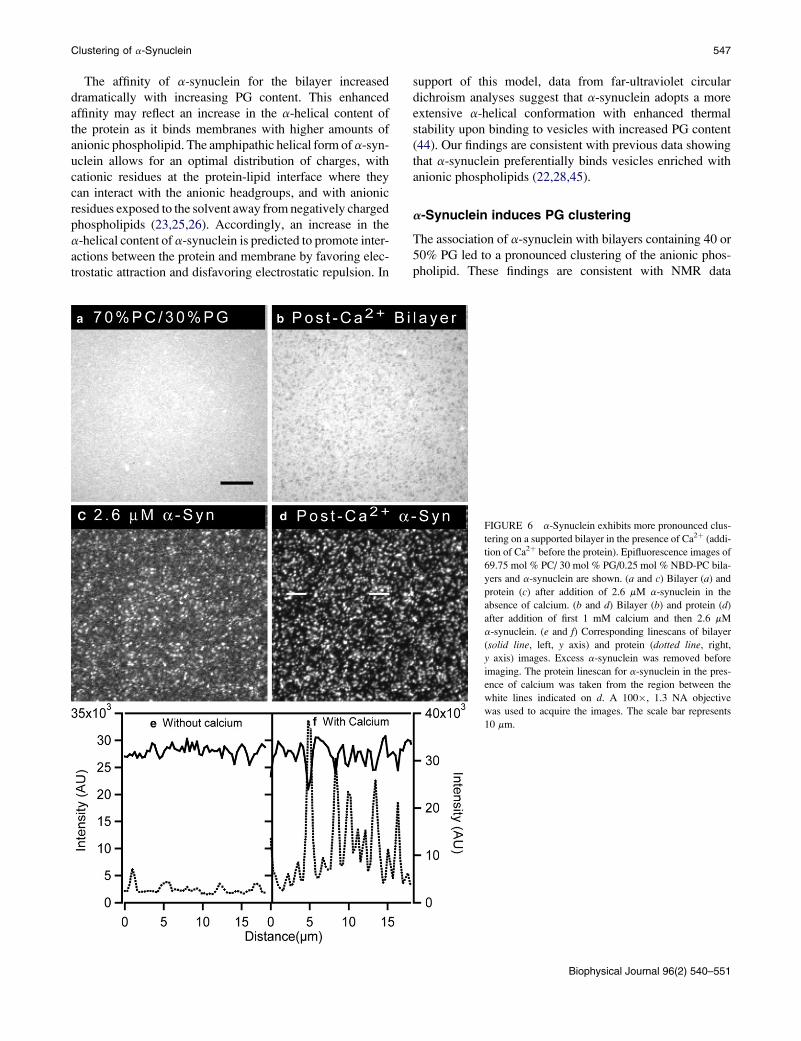
a PG-containing bilayer pretreated with calcium (Fig. 6).
For comparison, a bilayer-protein system in the absence of
calcium is shown in Fig. 6, a and c (these are the same
images as in Fig. 1, b and f, 30 mol % PG bilayer after the
addition of 2.6 mM a-synuclein). When 2.6 mM a-synuclein
was added to a 30 mol % PG bilayer that was preexposed to
calcium (1 mM) (Fig. 6 d), the protein preferentially bound
to the PG-enriched domains (compare linescans in Fig. 6, eand f). Calcium-induced protein clusters remained even after
the bulk solution was exchanged with EDTA-containing
buffer. More dramatic results were obtained with bilayers
containing 40 and 50 mol % PG. Clustering could be
observed at 30, 40, and 50 mol % PG when the protein
concentration was 10-fold lower (0.26 mM). At 50 mol %
PG, where the most dramatic reorganization was observed,
the calcium concentration could be dropped as low as
25 mM to induce protein clustering at 0.26 mM a-synuclein.
For four cases the amount of protein bound in the clusters
was compared with similar areas of protein bound in the
absence of calcium. In the case of 2.6 mM added to 30 mol
% PG and 0.26 mM added to 50 mol % PG, protein clusters
were not observed in the absence of calcium. In both cases,
the intensity in the clusters was significantly greater—
6(52)-fold and 6(51)-fold respectively—than the approxi-
FIGURE 5 Calcium causes phase separation in a PC/PG supported lipid
bilayer. Epifluorescence images of a 69.75 mol % PC/ 30 mol % PG/0.25
mol % NBD-PC bilayer are shown. Bilayer (a) in the absence of calcium,
and (b) after the addition of 1 mM calcium. A 100�, 1.3 NA objective
was used to acquire the images. The scale bar represents 10 mm.
Biophysical Journal 96(2) 540–551
546
mately uniformly adsorbed protein in the absence of calcium
(Figs. 1 f and 3 c).
When 2.6 mM a-synuclein was added to 40 mol % PG or 50
mol % PG, protein clusters were observed in both the absence
and presence of calcium (Fig. 1, g and h, and data not shown).
The clusters were roughly the same area and density regard-
less of whether protein or calcium was added first. In the
case of calcium added before protein, the clusters were so
intense that the CCD camera maxed out; thus, we can only
report lower limits. There was at least threefold (40 mol %
PG bilayer) and at least twofold (50 mol % PG bilayer)
more protein bound in the clusters of membranes pretreated
with calcium. The similar end result, regardless of order of
addition, suggests that equilibrium was reached. The observa-
tion that more protein bound to bilayers pretreated with
calcium is explained by the fact that when calcium was added
after protein, the excess protein was rinsed away before the
calcium was added, limiting the amount of protein that could
bind. In the absence of calcium, the PG-rich domains were
~30% (40 mol % PG bilayer) and ~50% (50 mol % PG
bilayer) depleted of NBD-PC. In presence of calcium, the
PG-rich domains were ~37% (40 mol % PG bilayer) and
~55% (50 mol % PG bilayer) depleted of NBD-PC.
DISCUSSION
In this study we monitored the adsorption of a-synuclein on
supported lipid bilayers via fluorescence microscopy. An
important advantage of this approach is that it enables char-
acterization of the lipid and protein distributions on the
bilayer surface. In contrast, this spatial information is inac-
cessible to classic averaging techniques used to monitor
protein-lipid interactions in solution. A fluorescence micros-
copy setup similar to the one described here was recently
used to analyze interactions of a-synuclein with discrete
domains on giant unilamellar vesicles (28).
a-Synuclein exhibits enhanced affinityfor phospholipid bilayers enriched with PG
We found that a-synuclein binds with high affinity to phos-
pholipid membranes under all conditions detailed in this
article. Upon adsorbing to the membrane, the protein
undergoes a decrease in entropy that must be compensated
for by favorable protein-lipid interactions, including hydro-
phobic and electrostatic interactions. In the experiments
described here, a-synuclein binding occurred at very low
ionic strength. If the monovalent ion concentration was
increased to 750 mM, the binding of the protein to
membranes consisting of 50% PG was reduced by ~3-fold
(data not shown). However, if the bilayer was exposed to
750 mM solution after a-synuclein adsorption, then <10%
of the protein desorbed. This hysteresis in the binding-
unbinding transition indicates that both hydrophobic and
electrostatic interactions play an important role in stabilizing
the membrane-bound state of a-synuclein (29).
Pandey et al.
The affinity of a-synuclein for the bilayer increased
dramatically with increasing PG content. This enhanced
affinity may reflect an increase in the a-helical content of
the protein as it binds membranes with higher amounts of
anionic phospholipid. The amphipathic helical form of a-syn-
uclein allows for an optimal distribution of charges, with
cationic residues at the protein-lipid interface where they
can interact with the anionic headgroups, and with anionic
residues exposed to the solvent away from negatively charged
phospholipids (23,25,26). Accordingly, an increase in the
a-helical content of a-synuclein is predicted to promote inter-
actions between the protein and membrane by favoring elec-
trostatic attraction and disfavoring electrostatic repulsion. In
support of this model, data from far-ultraviolet circular
dichroism analyses suggest that a-synuclein adopts a more
extensive a-helical conformation with enhanced thermal
stability upon binding to vesicles with increased PG content
(44). Our findings are consistent with previous data showing
that a-synuclein preferentially binds vesicles enriched with
anionic phospholipids (22,28,45).
a-Synuclein induces PG clustering
The association of a-synuclein with bilayers containing 40 or
50% PG led to a pronounced clustering of the anionic phos-
pholipid. These findings are consistent with NMR data
FIGURE 6 a-Synuclein exhibits more pronounced clus-
tering on a supported bilayer in the presence of Ca2þ (addi-
tion of Ca2þ before the protein). Epifluorescence images of
69.75 mol % PC/ 30 mol % PG/0.25 mol % NBD-PC bila-
yers and a-synuclein are shown. (a and c) Bilayer (a) and
protein (c) after addition of 2.6 mM a-synuclein in the
absence of calcium. (b and d) Bilayer (b) and protein (d)
after addition of first 1 mM calcium and then 2.6 mM
a-synuclein. (e and f) Corresponding linescans of bilayer
(solid line, left, y axis) and protein (dotted line, right,
y axis) images. Excess a-synuclein was removed before
imaging. The protein linescan for a-synuclein in the pres-
ence of calcium was taken from the region between the
white lines indicated on d. A 100�, 1.3 NA objective
was used to acquire the images. The scale bar represents
10 mm.
Biophysical Journal 96(2) 540–551
Clustering of a-Synuclein 547
suggesting that a-synuclein induces the formation of PG-rich
domains upon binding to PC-PG vesicles (40). One driving
force for this lipid reorganization may be the neutralization
of charges on helical a-synuclein by PG. The segment of the
protein spanning residues 1–95 has 12 cationic residues
(all lysines) and nine anionic residues (eight glutamates and
one aspartate). When a-synuclein adopts a helical conforma-
tion, the lysine residues are predominantly found at the interfa-
cial region, whereas the anionic residues are exclusively
located on the solvent-exposed face (23,25,26). The average
charge density of 12 lysine residues aligned on the same face
of an amphipathic helix with a length of ~15 nm and a width
of 1 nm is ~0.8/nm2. Assuming that all of the phospholipids
have a cross-sectional surface area of 65 A2 and they are hexag-
onally packed, the average charge density of a PC-PG bilayer
with 30, 40, or 50 mol % PG is ~0.38/nm2, ~0.50/nm2, or
~0.63/nm2, respectively. To meet the conditions of charge
neutrality, the lipids must reorganize to create zones of high
local PG concentration in response to protein adsorption.
Another driving force for PG clustering may be the ener-
getic benefit associated with PC-PG demixing. In the absence
of lipid-lipid interactions, entropy drives the mixing of lipids.
Because PC and PG have different physicochemical proper-
ties (46,47), PG-PG and PC-PC interactions are likely to be
more favorable than PC-PG interactions. Although chain-
matched PC and PG have approximately identical Tm values,
PC-PG interactions may nevertheless be less favorable than
PC-PC or PG-PG interactions. Despite the decrease in
entropy, the free energy may be reduced upon PC-PG demix-
ing. Although PC-PG demixing is normally suppressed at pH
7.4 by electrostatic repulsion between the PG lipids, presum-
ably this inhibitory effect is relieved by neutralization of the
negative charge on PG after a-synuclein binding.
Because we failed to detect PG clustering after a-synuclein
adsorption to the 30 mol % PG bilayer, we infer that the
protein only induces lipid reorganization in membranes where
the anionic phospholipid content is above some threshold
value (30 < x % 40 mol % PG in PC; Fig. 7). The lower
the level of PG in the membrane, the more reorganization is
required for charge neutralization. Consequently, there may
be a threshold where the entropic cost of reorganization is
too great. Another possibility is that the threshold for immis-
cibility of PG in PC has not been crossed. In a standard model
of mixing (e.g., oil in water), molecule A has some solubility
in molecule B, and demixing does not occur until a threshold
value is reached.
The absence of lipid reorganization in 30 mol % PG bila-
yers implies that a-synuclein adopts a less a-helical structure
upon binding to membranes with lower amounts of anionic
phospholipid (Fig. 7). In the absence of PG clustering, the
charge density associated with the aligned lysine residues of
helical a-synuclein will not be neutralized, and therefore the
extended amphipathic helix will be unstable. Under these
conditions, a-synuclein may bind to the bilayer as a largely
unfolded conformer with relatively little a-helical structure.
Biophysical Journal 96(2) 540–551
548
This mode of binding would involve primarily hydrophobic
interactions between the protein and lipids. Anionic lipid clus-
tering would not be necessary to achieve charge neutrality in
this case because the lysine residues would be distributed
throughout the unfolded a-synuclein structure rather than
being aligned on the same face of a helix, and therefore the
positive charge density associated with the lysines would be
substantially diminished. Normally, electrostatic repulsion
between negatively charged residues and anionic phospho-
lipids should favor conversion of membrane-bound, unfolded
a-synuclein to the helical form, with anionic residues placed
on the solvent face away from the membrane. However, this
repulsive effect is expected to play a lesser role in driving
a-synuclein helix formation in bilayers containing lower
amounts of anionic lipid.
a-Synuclein forms clusters on membranesenriched with anionic phospholipid
The binding of a-synuclein to PC-PG bilayers resulted in the
formation of protein clusters that coincide with PG rich-
domains. The fluorescence intensity of the a-synuclein clus-
ters increased relative to background with increasing PG
content and protein concentration. These findings imply
that favorable protein-protein contacts occur in discrete
regions of the bilayer enriched with anionic phospholipid
(Fig. 7). This behavior may be attributed in part to the migra-
tion of PG-associated a-synuclein molecules into PG-rich
clusters during phospholipid demixing (47,48). Another
explanation may be that the protein adopts a more extensive
a-helical structure upon binding to membranes with higher
amounts of PG (see above) (44). Although the formation
of an amphipathic a-helix is predicted to enhance the affinity
of a-synuclein for the membrane, it may also be disfavored
by the exposure of several hydrophobic residues on the polar
face of the helix. Assuming an a11/3 helical structure, these
residues include Met5, Val16, Leu38, Val49, Val71, Val82, and
Phe94 (26,29). Two of these residues, Val71 and Val82, are
part of a hydrophobic, 12-residue segment that is essential
for a-synuclein self-assembly in solution (49). Of impor-
tance, the exposed hydrophobic residues may be sequestered
from the solvent upon binding of additional a-synuclein
molecules (originating from elsewhere on the bilayer or
from the bulk solvent) to the membrane-bound, helical
protein (Fig. 7). The burial of these residues, coupled with
the energetically favorable release of solvating water mole-
cules, may be a major driving force for a-synuclein cluster
formation. This model is consistent with the results of
a computational study showing that the dimerization of
helical a-synuclein on a bilayer surface is energetically
favorable (29). In addition, a-synuclein forms helical oligo-
mers in solutions containing fluorinated alcohols, which
have low dielectric constants that mimic the nonpolar envi-
ronment at the surface of a phospholipid membrane
(J.-C. Rochet, unpublished data) (50).
Pandey et al.
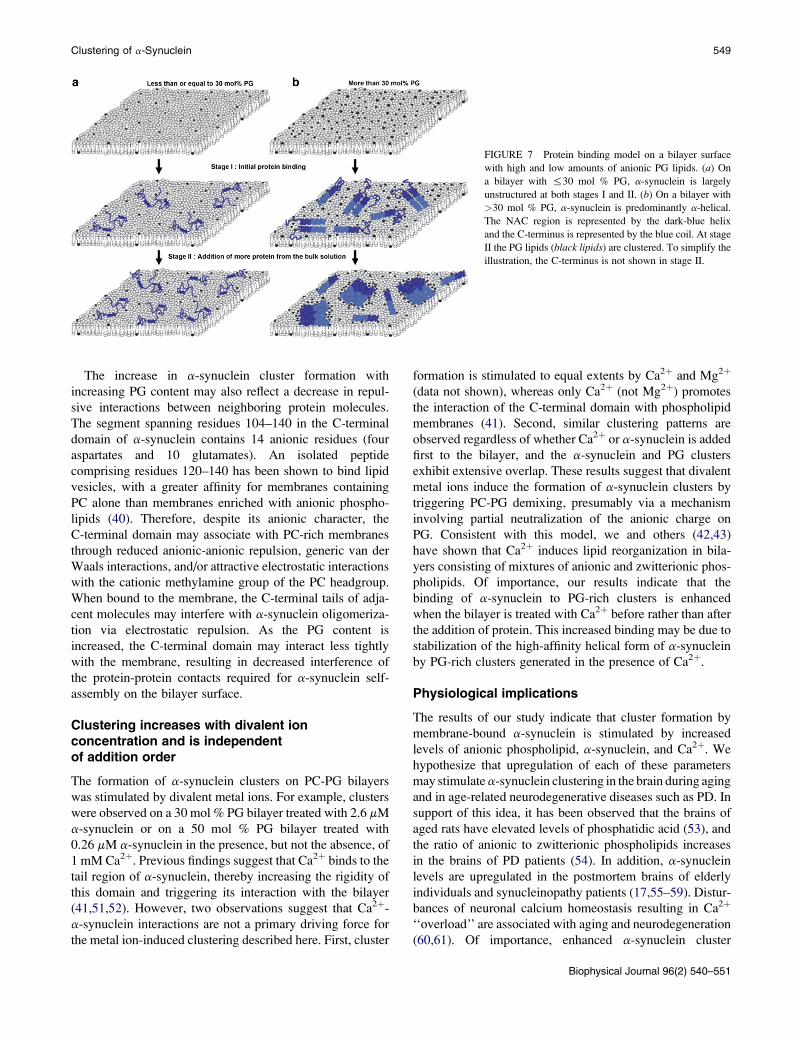
FIGURE 7 Protein binding model on a bilayer surface
with high and low amounts of anionic PG lipids. (a) On
a bilayer with %30 mol % PG, a-synuclein is largely
unstructured at both stages I and II. (b) On a bilayer with
>30 mol % PG, a-synuclein is predominantly a-helical.
The NAC region is represented by the dark-blue helix
and the C-terminus is represented by the blue coil. At stage
II the PG lipids (black lipids) are clustered. To simplify the
illustration, the C-terminus is not shown in stage II.
Clustering of a-Synuclein 549
The increase in a-synuclein cluster formation with
increasing PG content may also reflect a decrease in repul-
sive interactions between neighboring protein molecules.
The segment spanning residues 104–140 in the C-terminal
domain of a-synuclein contains 14 anionic residues (four
aspartates and 10 glutamates). An isolated peptide
comprising residues 120–140 has been shown to bind lipid
vesicles, with a greater affinity for membranes containing
PC alone than membranes enriched with anionic phospho-
lipids (40). Therefore, despite its anionic character, the
C-terminal domain may associate with PC-rich membranes
through reduced anionic-anionic repulsion, generic van der
Waals interactions, and/or attractive electrostatic interactions
with the cationic methylamine group of the PC headgroup.
When bound to the membrane, the C-terminal tails of adja-
cent molecules may interfere with a-synuclein oligomeriza-
tion via electrostatic repulsion. As the PG content is
increased, the C-terminal domain may interact less tightly
with the membrane, resulting in decreased interference of
the protein-protein contacts required for a-synuclein self-
assembly on the bilayer surface.
Clustering increases with divalent ionconcentration and is independentof addition order
The formation of a-synuclein clusters on PC-PG bilayers
was stimulated by divalent metal ions. For example, clusters
were observed on a 30 mol % PG bilayer treated with 2.6 mM
a-synuclein or on a 50 mol % PG bilayer treated with
0.26 mM a-synuclein in the presence, but not the absence, of
1 mM Ca2þ. Previous findings suggest that Ca2þ binds to the
tail region of a-synuclein, thereby increasing the rigidity of
this domain and triggering its interaction with the bilayer
(41,51,52). However, two observations suggest that Ca2þ-
a-synuclein interactions are not a primary driving force for
the metal ion-induced clustering described here. First, cluster
formation is stimulated to equal extents by Ca2þ and Mg2þ
(data not shown), whereas only Ca2þ (not Mg2þ) promotes
the interaction of the C-terminal domain with phospholipid
membranes (41). Second, similar clustering patterns are
observed regardless of whether Ca2þ or a-synuclein is added
first to the bilayer, and the a-synuclein and PG clusters
exhibit extensive overlap. These results suggest that divalent
metal ions induce the formation of a-synuclein clusters by
triggering PC-PG demixing, presumably via a mechanism
involving partial neutralization of the anionic charge on
PG. Consistent with this model, we and others (42,43)
have shown that Ca2þ induces lipid reorganization in bila-
yers consisting of mixtures of anionic and zwitterionic phos-
pholipids. Of importance, our results indicate that the
binding of a-synuclein to PG-rich clusters is enhanced
when the bilayer is treated with Ca2þ before rather than after
the addition of protein. This increased binding may be due to
stabilization of the high-affinity helical form of a-synuclein
by PG-rich clusters generated in the presence of Ca2þ.
Physiological implications
The results of our study indicate that cluster formation by
membrane-bound a-synuclein is stimulated by increased
levels of anionic phospholipid, a-synuclein, and Ca2þ. We
hypothesize that upregulation of each of these parameters
may stimulate a-synuclein clustering in the brain during aging
and in age-related neurodegenerative diseases such as PD. In
support of this idea, it has been observed that the brains of
aged rats have elevated levels of phosphatidic acid (53), and
the ratio of anionic to zwitterionic phospholipids increases
in the brains of PD patients (54). In addition, a-synuclein
levels are upregulated in the postmortem brains of elderly
individuals and synucleinopathy patients (17,55–59). Distur-
bances of neuronal calcium homeostasis resulting in Ca2þ
‘‘overload’’ are associated with aging and neurodegeneration
(60,61). Of importance, enhanced a-synuclein cluster
Biophysical Journal 96(2) 540–551
formation may contribute to the pathogenesis of age-related
neurologic disorders via diverse mechanisms. For example,
the sequestration of a-synuclein in clusters may disrupt inter-
actions between the protein and the dopamine transporter
(20). In turn, the resulting upregulation of dopamine trans-
porter activity may play a role in dopaminergic cell death.
The formation of membrane-bound a-synuclein clusters
may also promote conversion of the protein to potentially
toxic, high-molecular-weight assemblies (30), especially
under conditions that trigger dissociation of the protein
from the membrane and/or b-sheet formation (e.g., oxidative
stress or elevated Ca2þ concentrations) (41,62).
CONCLUSIONS
The results and discussion presented here lead to the
following conclusions: 1), the increased affinity of a-synu-
clein for membranes with higher amounts of anionic phos-
pholipid may reflect an increase in the a-helical content of
the protein; 2), the lipid demixing observed upon a-synu-
clein membrane binding may be driven by the need to
neutralize the charges on helical a-synuclein and/or relieve
unfavorable PC-PG interactions; 3), a-synuclein clustering
may be driven by the requirement to bury hydrophobic resi-
dues that are exposed when the protein adopts an a-helical
conformation; and 4), divalent metal ions may stimulate
cluster formation by promoting PC-PG demixing. Because
the formation of membrane-bound a-synuclein assemblies
may play a role in neurotoxicity, strategies to interfere
with clustering may prove beneficial for the treatment of
PD and other synucleinopathy disorders.
We thank Jagadish Kumar Hindupur and John Hulleman for assistance with
a-synuclein expression, purification, and labeling.
This work was supported in part by National Institutes of Health grant R01
NS049221 and the Showalter Trust.
REFERENCES
1. Pollanen, M. S., D. W. Dickson, and C. Bergeron. 1993. Pathology andbiology of the Lewy body. J. Neuropathol. Exp. Neurol. 52:183–191.
2. Forno, L. S. 1996. Neuropathology of Parkinson’s disease. J. Neuropa-thol. Exp. Neurol. 55:259–272.
3. Takahashi, H., and K. Wakabayashi. 2001. The cellular pathology ofParkinson’s disease. Neuropathology. 21:315–322.
4. Spillantini, M. G., M. L. Schmidt, V. M. -Y. Lee, J. Q. Trojanowski,R. Jakes, et al. 1997. a-Synuclein in Lewy bodies. Nature. 388:839–840.
5. Wakabayashi, K., K. Matsumoto, K. Takayama, M. Yoshimoto, and H.Takahashi. 1997. NACP, a presynaptic protein, immunoreactivity inLewy bodies in Parkinson’s disease. Neurosci. Lett. 239:45–48.
6. Baba, M., S. Nakajo, P. -H. Tu, T. Tomita, K. Nakaya, et al. 1998.Aggregation of a-synuclein in Lewy bodies of sporadic Parkinson’sdisease and dementia with Lewy bodies. Am. J. Pathol. 152:879–884.
7. Spillantini, M. G., R. A. Crowther, R. Jakes, M. Hasegawa, and M. Goe-dert. 1998. a-Synuclein in filamentous inclusions of Lewy bodies fromParkinson’s disease and dementia with Lewy bodies. Proc. Natl. Acad.Sci. USA. 95:6469–6473.
Biophysical Journal 96(2) 540–551
550
8. Polymeropoulos, M. H., C. Lavedan, E. Leroy, S. E. Ide, A. Dehejia,et al. 1997. Mutation in the a-synuclein gene identified in familieswith Parkinson’s disease. Science. 276:2045–2047.
9. Kruger, R., W. Kuhn, T. Muller, D. Woitalla, M. Graeber, et al. 1998.Ala30Pro mutation in the gene encoding a-synuclein in Parkinson’sdisease. Nat. Genet. 18:106–108.
10. Zarranz, J. J., J. Alegre, J. C. Gomez-Esteban, E. Lezcano, R. Ros, et al.2004. The new mutation, E46K, of a-synuclein causes Parkinson andLewy body dementia. Ann. Neurol. 55:164–173.
11. Narhi, L., S. J. Wood, S. Steavenson, Y. Jiang, G. M. Wu, et al. 1999.Both familial Parkinson’s disease mutations accelerate a-synucleinaggregation. J. Biol. Chem. 274:9843–9846.
12. Conway, K. A., S. -J. Lee, J. -C. Rochet, T. T. Ding, R. E. Williamson,et al. 2000. Acceleration of oligomerization, not fibrillization, is a sharedproperty of both a-synuclein mutations linked to early-onset Parkin-son’s disease: implications for pathogenesis and therapy. Proc. Natl.Acad. Sci. USA. 97:571–576.
13. Li, J., V. N. Uversky, and A. L. Fink. 2001. Effect of familial Parkin-son’s disease point mutations A30P and A53T on the structural proper-ties, aggregation, and fibrillation of human a-synuclein. Biochemistry.40:11604–11613.
14. Fredenburg, R. A., C. Rospigliosi, R. K. Meray, J. C. Kessler, H. A. La-shuel, et al. 2007. The impact of the E46K mutation on the properties ofa-synuclein in its monomeric and oligomeric states. Biochemistry.46:7107–7118.
15. Greenbaum, E. A., C. L. Graves, A. J. Mishizen-Eberz, M. A. Lupoli,D. R. Lynch, et al. 2005. The E46K mutation in a-synuclein increasesamyloid fibril formation. J. Biol. Chem. 280:7800–7807.
16. Choi, W., S. Zibaee, R. Jakes, L. C. Serpell, B. Davletov, et al. 2004.Mutation E46K increases phospholipid binding and assembly into fila-ments of human a-synuclein. FEBS Lett. 576:363–368.
17. Dev, K. K., K. Hofele, S. Barbieri, V. L. Buchman, and H. van der Put-ten. 2003. Part II: a-Synuclein and its molecular pathophysiological rolein neurodegenerative disease. Neuropharmacology. 45:14–44.
18. Murphy, D. D., S. M. Rueter, J. Q. Trojanowski, and V. M. Y. Lee.2000. Synucleins are developmentally expressed, and a-synuclein regu-lates the size of the presynaptic vesicular pool in primary hippocampalneurons. J. Neurosci. 20:3214–3220.
19. Cabin, D. E., K. Shimazu, D. Murphy, N. B. Cole, W. Gottschalk, et al.2002. Synaptic vesicle depletion correlates with attenuated synapticresponses to prolonged repetitive stimulation in mice lacking a-synu-clein. J. Neurosci. 22:8797–8807.
20. Sidhu, A., C. Wersinger, and P. Vernier. 2004. a-Synuclein regulationof the dopaminergic transporter: a possible role in the pathogenesis ofParkinson’s disease. FEBS Lett. 565:1–5.
21. Weinreb, P. H., W. G. Zhen, A. W. Poon, K. A. Conway, and P. T.Lansbury. 1996. NACP, a protein implicated in Alzheimer’s diseaseand learning, is natively unfolded. Biochemistry. 35:13709–13715.
22. Davidson, W. S., A. Jonas, D. F. Clayton, and J. M. George. 1998.Stabilization of a-synuclein secondary structure upon binding tosynthetic membranes. J. Biol. Chem. 273:9443–9449.
23. Jao, C. C., A. Der-Sarkissian, J. Chen, and R. Langen. 2004. Structureof membrane-bound a-synuclein studied by site-directed spin labeling.Proc. Natl. Acad. Sci. USA. 101:8331–8336.
24. Ramakrishnan, M., P. H. Jensen, and D. Marsh. 2003. a-Synucleinassociation with phosphatidylglycerol probed by lipid spin labels.Biochemistry. 42:12919–12926.
25. Bussell, R., T. F. Ramlall, and D. Eliezer. 2005. Helix periodicity,topology, and dynamics of membrane-associated a-synuclein. ProteinSci. 14:862–872.
26. Bussell, R., Jr., and D. Eliezer. 2003. A structural and functional role for11-mer repeats in a-synuclein and other exchangeable lipid bindingproteins. J. Mol. Biol. 329:763–778.
27. Chandra, S., X. C. Chen, J. Rizo, R. Jahn, and T. C. Sudhof. 2003.A broken a-helix in folded a-synuclein. J. Biol. Chem. 278:15313–15318.
Pandey et al.
28. Stockl, M., P. Fischer, E. Wanker, and A. Herrmann. 2008. a-Synucleinselectively binds to anionic phospholipids embedded in liquid-disor-dered domains. J. Mol. Biol. 375:1394–1404.
29. Mihajlovic, M., and T. Lazaridis. 2008. Membrane-bound structure andenergetics of a-synuclein. Proteins. 70:761–778.
30. Lee, H. -J., C. Choi, and S. -J. Lee. 2002. Membrane-bound a-synucleinhas a high aggregation propensity and the ability to seed the aggregationof the cytosolic form. J. Biol. Chem. 277:671–678.
31. Necula, M., C. N. Chirita, and J. Kuret. 2003. Rapid anionic micelle-medi-ated a-synuclein fibrillization in vitro. J. Biol. Chem. 278:46674–46680.
32. Beyer, K. 2007. Mechanistic aspects of Parkinson’s disease: a-synu-clein and the biomembrane. Cell Biochem. Biophys. 47:285–299.
33. Narayanan, V., and S. Scarlata. 2001. Membrane binding and self-asso-ciation of a-synucleins. Biochemistry. 40:9927–9934.
34. Zhu, M., and A. L. Fink. 2003. Lipid binding inhibits a-synuclein fibrilformation. J. Biol. Chem. 278:16873–16877.
35. Sackmann, E. 1996. Supported membranes: scientific and practicalapplications. Science. 271:43–48.
36. Seu, K. J., A. P. Pandey, F. Haque, E. A. Proctor, A. E. Ribbe, et al.2007. Effect of surface treatment on diffusion and domain formationin supported lipid bilayers. Biophys. J. 92:2445–2450.
37. Findlay, E. J., and P. G. Barton. 1978. Phase behavior of synthetic phos-phatidylglycerols and binary-mixtures with phosphatidylcholines inpresence and absence of calcium-ions. Biochemistry. 17:2400–2405.
38. Garidel, P., C. Johann, L. Mennicke, and A. Blume. 1997. The mixingbehavior of pseudobinary phosphatidylcholine phosphatidylglycerolmixtures asa function of pH and chain length. Eur. Biophys. J.26:447–459.
39. Seu, K., L. R. Cambrea, R. M. Everly, and J. S. Hovis. 2006. Influenceof lipid chemistry on membrane fluidity: tail and headgroup interac-tions. Biophys. J. 91:3727–3735.
40. Madine, J., A. J. Doig, and D. A. Middleton. 2006. A study of theregional effects of a-synuclein on the organization and stability of phos-pholipid bilayers. Biochemistry. 45:5783–5792.
41. Tamamizu-Kato, S., M. G. Kosaraju, H. Kato, V. Raussens, J. M. Ruys-schaert, et al. 2006. Calcium-triggered membrane interaction of thea-synuclein acidic tail. Biochemistry. 45:10947–10956.
42. Evert, L. L., D. Leckband, and J. N. Israelachvili. 1994. Structure anddynamics of ion-induced domains in free and supported monolayersand bilayers. Langmuir. 10:303–315.
43. Lamberson, E. R., L. R. Cambrea, and J. S. Hovis. 2007. Controlling thecharge and organization of anionic lipid bilayers: effect of monovalentand divalent ions. J. Phys. Chem. B. 111:13664–13667.
44. Zakharov, S. D., J. D. Hulleman, E. A. Dutseva, Y. N. Antonenko, J. C.Rochet, et al. 2007. Helical a-synuclein forms highly conductive ionchannels. Biochemistry. 46:14369–14379.
45. Jo, E., J. McLaurin, C. M. Yip, P. St. George-Hyslop, and P. E. Fraser.2000. a-Synuclein membrane interactions and lipid specificity. J. Biol.Chem. 275:34328–34334.
46. Hinderliter, A., P. F. Almeida, C. E. Creutz, and R. L. Biltonen. 2001.Domain formation in a fluid mixed lipid bilayer modulated throughbinding of the C2 protein motif. Biochemistry. 40:4181–4191.
Clustering of a-Synuclein
47. Mbamala, E. C., A. Ben-Shaul, and S. May. 2005. Domain formationinduced by the adsorption of charged proteins on mixed lipidmembranes. Biophys. J. 88:1702–1714.
48. May, S., D. Harries, and A. Ben-Shaul. 2000. Lipid demixing andprotein-protein interactions in the adsorption of charged proteins onmixed membranes. Biophys. J. 79:1747–1760.
49. Giasson, B. I., I. V. J. Murray, J. Q. Trojanowski, and V. M. Y. Lee.2001. A hydrophobic stretch of 12 amino acid residues in the middleof a-synuclein is essential for filament assembly. J. Biol. Chem.276:2380–2386.
50. Munishkina, L. A., C. Phelan, V. N. Uversky, and A. L. Fink. 2003.Conformational behavior and aggregation of a-synuclein in organicsolvents: modeling the effects of membranes. Biochemistry. 42:2720–2730.
51. Nielsen, M. S., H. Vorum, E. Lindersson, and P. H. Jensen. 2001. Ca2þ
binding to a-synuclein regulates ligand binding and oligomerization. J.Biol. Chem. 276:22680–22684.
52. de Laureto, P. P., L. Tosatto, E. Frare, O. Marin, V. N. Uversky, et al.2006. Conformational properties of the SDS-bound state of a-synucleinprobed by limited proteolysis: unexpected rigidity of the acidicC-terminal tail. Biochemistry. 45:11523–11531.
53. Giusto, N. M., G. A. Salvador, P. I. Castagnet, S. J. Pasquare, and M. G.Ilincheta de Boschero. 2002. Age-associated changes in central nervoussystem glycerolipid composition and metabolism. Neurochem. Res.27:1513–1523.
54. Riekkinen, P., U. K. Rinne, T. T. Pelliniemi, and V. Sonninen.1975. Interaction between dopamine and phospholipids. Studies ofthe substantia nigra in Parkinson disease patients. Arch. Neurol.32:25–27.
55. Dawson, T. M., and V. L. Dawson. 2003. Molecular pathways of neuro-degeneration in Parkinson’s disease. Science. 302:819–822.
56. Singleton, A. B., M. Farrer, J. Johnson, A. Singleton, S. Hague, et al.2003. a-Synuclein locus triplication causes Parkinson’s disease.Science. 302:841.
57. Li, W. X., C. Lesuisse, Y. Q. Xu, J. C. Troncoso, D. L. Price, et al. 2004.Stabilization of a-synuclein protein with aging and familial Parkinson’sdisease-linked A53T mutation. J. Neurosci. 24:7400–7409.
58. Chu, Y., and J. H. Kordower. 2007. Age-associated increases of a-syn-uclein in monkeys and humans are associated with nigrostriatal dopa-mine depletion: is this the target for Parkinson’s disease? Neurobiol.Dis. 25:134–149.
59. Kovacs, G. G., I. J. Milenkovic, M. Preusser, and H. Budka. 2008.Nigral burden of a-synuclein correlates with striatal dopamine deficit.Mov. Disord. 23:1608–1612.
60. Toescu, E. C., and A. Verkhratsky. 2000. Parameters of calcium homeo-stasis in normal neuronal ageing. J. Anat. 197:563–569.
61. Mattson, M. P. 2007. Calcium and neurodegeneration. Aging Cell.6:337–350.
62. Hodara, R., E. H. Norris, B. I. Giasson, A. J. Mishizen-Eberz, D. R.Lynch, et al. 2004. Functional consequences of a-synuclein tyrosinenitration: diminished binding to lipid vesicles and increased fibrilformation. J. Biol. Chem. 279:47746–47753.
551
Biophysical Journal 96(2) 540–551