Embed Size (px)
Citation preview
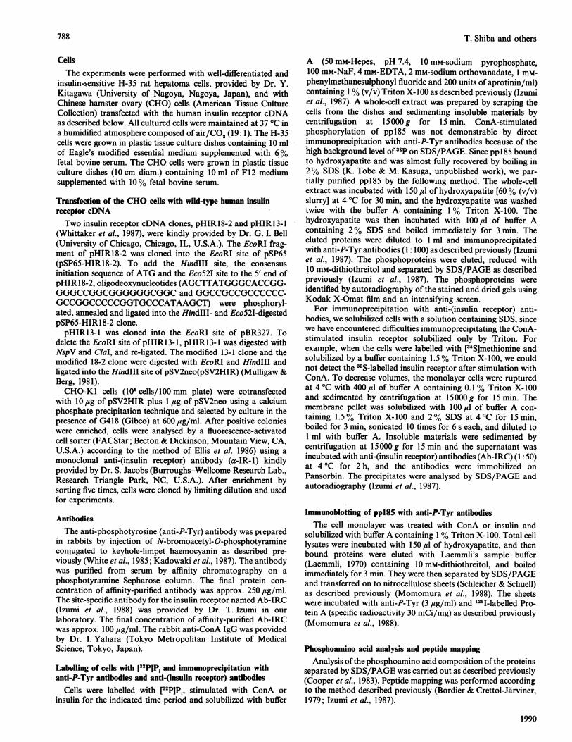
Biochem. J. (1990) 267, 787-794 (Printed in Great Britain)
Concanavalin A-induced receptor aggregation stimulates thetyrosine kinase activity of the insulin receptor in intact cellsTeruo SHIBA,* Kazuyuki TOBE,* Osamu KOSHIO,t Ritsuko YAMAMOTO,* Yoshikazu SHIBASAKI,*Naoki MATSUMOTO,4 Satoshi TOYOSHIMA,4 Toshiaki OSAWA,4 Yasuo AKANUMA,tFumimaro TAKAKU* and Masato KASUGA*§*Third Department of Internal Medicine, Faculty of Medicine, University of Tokyo, 7-3-1 Hongo, Bunkyo-ku, Tokyo,Japan 113, tInstitute of Diabetes Care and Research, Asahi Life Foundation, 1-6-1 Marunouchi, Chiyoda-ku, Tokyo,Japan 100, and tDivision of Chemical Toxicology and Immunochemistry, Faculty of Pharmaceutical Sciences,University of Tokyo, 7-3-1 Hongo, Bunkyo-ku, Tokyo, Japan 113
Concanavalin A (ConA) stimulated the phosphorylation of the f-subunit of the insulin receptor and an Mr-185000protein on serine and tyrosine residues in intact H-35 rat hepatoma cells. This Mr-185000 protein whose phosphorylationwas stimulated by ConA was identical to ppl85, a protein reported previously to be a putative endogenous substrate forthe insulin receptor tyrosine kinase in rat hepatoma cells. In Chinese hamster ovary (CHO) cells transfected with cDNAof the human insulin receptor, tyrosine-phosphorylation of ppl85 was strongly enhanced by ConA compared with thecontrols, suggesting that the induction of tyrosine-phosphorylation ofppl85 was due to stimulation of the insulin receptorkinase by ConA. Moreover, monovalent ConA only slightly induced the tyrosine-phosphorylation of ppl85, which was
enhanced by the addition of anti-ConA IgG, suggesting that ConA stimulated the insulin receptor kinase mainly by thereceptor cross-linking or aggregation in intact cells. These data suggest that the insulin-mimetic action ofConA is relatedto the autophosphorylation and activation of the insulin receptor tyrosine kinase, as well as the subsequentphosphorylation of ppl85 in intact cells.
INTRODUCTION
The insulin receptor is a membrane-spanning glycoproteincomposed of a- and f-subunits (Kasuga et al., 1982a). Insulinbinding to the a-subunit stimulates the autophosphorylation andactivation of the intrinsic tyrosine-specific protein kinase activityof the fl-subunit (Kasuga et al., 1982b, 1983; Roth & Cassell,1983). Activation of the tyrosine kinase appears to be a pre-requisite for many responses to insulin (Ellis et al., 1986; Ebinaet al., 1987; Chou et al., 1987; McClain et al., 1987; Morgan &Roth, 1987; Izumi et al., 1988). Several proteins have beenreported to be putative substrates for the insulin receptor kinase(White et al., 1985; Haring et al., 1987; Perrotti et al., 1987;Bernier et al., 1987; Yu et al., 1987). In particular, an M,-185 000protein (ppI85), or a related protein of similar Mr is found inmany cell types (Gibbs et al., 1986; Kadowaki et al., 1987; Whiteet al., 1987; Shemer et al., 1987) and is a common substrate forboth insulin and insulin-like growth factor I (IGF-I) receptorkinases (Izumi et al., 1987). This protein is regarded as one of theimportant putative substrates in signal transduction by insulin(White et al., 1988).
Several substances have been shown to mimic the actions ofinsulin. Concanavalin A (ConA), a plant lectin derived fromjackbean, binds to cell surface glycoproteins with a high specificityfor gluco- and manno-pyranosides. It has biological andmorphological actions (Bittigen & Schnebli, 1976; Wakshull &Wharton, 1985; Faltynek et al., 1988) and also mimics manyactions of insulin (Kahn et al., 1981). ConA stimulates thephosphorylation of the insulin receptor fl-subunit in a cell-freesystem (Roth et al., 1983) without inhibition of insulin binding(Kahn et al., 1981; Katzen et al., 1981), suggesting that it mayexert its insulin-mimetic effects in a manner different frominsulin. In this study, we show that ConA stimulates insulin
receptor autophosphorylation and the tyrosine-phosphorylationof ppl85 by cross-linking or aggregation of receptors in intactcells. Thus the use of ConA further supports the role of tyrosinephosphorylation in the mechanism of insulin action.
EXPERIMENTAL
MaterialsThe following materials were obtained from the indicated
sources. [32P]Pi and Triton X-100 were from New EnglandNuclear, 1251I-labelled Protein A was from Amersham, pig insulinwas kindly provided by Eli Lilly, tissue culture medium and fetalbovine serum were from Gibco or Flow, and all tissue culturesupplies were from Nunc or Costar. Other reagents were obtainedfrom the same sources as described previously (Izumi et al., 1987,1988; Kadowaki et al., 1987).
Preparation of monovalent ConAMonovalent ConA was prepared by the subunit-exchange
method as described previously (Osawa & Beppu, 1987). Theyield of monovalent hybrids was approx. 10% of the cross-linked dimers. The valency of monovalent ConA was confirmedby equilibrium dialysis against a-methyl a-[14C]mannoside atpH 7.4. The binding activity and the carbohydrate specificityof this monovalent ConA were verified by the examination of125I-labelled monovalent ConA binding to H-35 cells andits displacement by native (tetravalent) ConA and methyl a-
mannopyranoside (Sigma). The minimum concentrations forhaemagglutininating activity of the monovalent and the tetra-valent ConA against mouse erythrocytes were about 200,cug/mland 3.5 ,ug/ml respectively.
Vol. 267
787
Abbreviations used: IGF, insulin-like growth factor; ConA, concanavalin A; WGA, wheat-germ agglutinin; CHO, Chinese hamster ovary; anti-P-Tyr, anti-phosphotyrosine; PNPP, p-nitrophenyl phosphate.
§ To whom correspondence should be addressed.
T. Shiba and others
CellsThe experiments were performed with well-differentiated and
insulin-sensitive H-35 rat hepatoma cells, provided by Dr. Y.Kitagawa (University of Nagoya, Nagoya, Japan), and withChinese hamster ovary (CHO) cells (American Tissue CultureCollection) transfected with the human insulin receptor cDNAas described below. All cultured cells were maintained at 37 °C ina humidified atmosphere composed of air/CO2 (19: 1). The H-35cells were grown in plastic tissue culture dishes containing 10 mlof Eagle's modified essential medium supplemented with 6%fetal bovine serum. The CHO cells were grown in plastic tissueculture dishes (10 cm diam.) containing 10 ml of F12 mediumsupplemented with 10% fetal bovine serum.
Transfection of the CHO cells with wild-type human insulinreceptor cDNATwo insulin receptor cDNA clones, pHIR18-2 and pHIR13-1
(Whittaker et al., 1987), were kindly provided by Dr. G. I. Bell(University of Chicago, Chicago, IL, U.S.A.). The EcoRI frag-ment of pHIR18-2 was cloned into the EcoRI site of pSP65(pSP65-HIR18-2). To add the HindIII site, the consensusinitiation sequence of ATG and the Eco52I site to the 5' end ofpHIR18-2, oligodeoxynucleotides (AGCTTATGGGCACCGG-GGGCCGGCGGGGGGCGGC and GGCCGCCGCCCCCC-GCCGGCCCCCGGTGCCCATAAGCT) were phosphoryl-ated, annealed and ligated into the HindlIl- and Eco52I-digestedpSP65-HIR18-2 clone.pHIR13-1 was cloned into the EcoRI site of pBR327. To
delete the EcoRI site of pHIRl3-1, pHIR13-1 was digested withNspV and ClaI, and re-ligated. The modified 13-1 clone and themodified 18-2 clone were digested with EcoRI and HindlIl andligated into the HindIII site of pSV2neo(pSV2HIR) (Mulligaw &Berg, 1981).CHO-KI cells (106 cells/100 mm plate) were cotransfected
with 10 ,ug of pSV2HIR plus 1 ,ug of pSV2neo using a calciumphosphate precipitation technique and selected by culture in thepresence of G418 (Gibco) at 600 ,g/ml. After positive colonieswere enriched, cells were analysed by a fluorescence-activatedcell sorter (FACStar; Becton & Dickinson, Mountain View, CA,U.S.A.) according to the method of Ellis et al. 1986) using amonoclonal anti-(insulin receptor) antibody (a-IR-1) kindlyprovided by Dr. S. Jacobs (Burroughs-Wellcome Research Lab.,Research Triangle Park, NC, U.S.A.). After enrichment bysorting five times, cells were cloned by limiting dilution and usedfor experiments.
AntibodiesThe anti-phosphotyrosine (anti-P-Tyr) antibody was prepared
in rabbits by injection of N-bromoacetyl-O-phosphotyramineconjugated to keyhole-limpet haemocyanin as described pre-viously (White et al., 1985; Kadowaki et al., 1987). The antibodywas purified from serum by affinity chromatography on aphosphotyramine-Sepharose column. The final protein con-centration of affinity-purified antibody was approx. 250 ,ug/ml.The site-specific antibody for the insulin receptor named Ab-IRC(Izumi et al., 1988) was provided by Dr. T. Izumi in ourlaboratory. The final concentration of affinity-purified Ab-IRCwas approx. 100 ,ug/ml. The rabbit anti-ConA IgG was providedby Dr. I. Yahara (Tokyo Metropolitan Institute of MedicalScience, Tokyo, Japan).
Labelling of cells with 132PIP; and immunoprecipitation withanti-P-Tyr antibodies and anti-(insulin receptor) antibodies
Cells were labelled with [32P]P , stimulated with ConA orinsulin for the indicated time period and solubilized with buffer
A (50 mM-Hepes, pH 7.4, 10 mM-sodium pyrophosphate,100 mM-NaF, 4 mM-EDTA, 2 mM-sodium orthovanadate, 1 mM-phenylmethanesulphonyl fluoride and 200 units of aprotinin/ml)containing 1 % (v/v) Triton X-100 as described previously (Izumiet al., 1987). A whole-cell extract was prepared by scraping thecells from the dishes and sedimenting insoluble materials bycentrifugation at 15000 g for 15 min. ConA-stimulatedphosphorylation of ppl85 was not demonstrable by directimmunoprecipitation with anti-P-Tyr antibodies because of thehigh background level of 32p on SDS/PAGE. Since pp185 boundto hydroxyapatite and was almost fully recovered by boiling in2% SDS (K. Tobe & M. Kasuga, unpublished work), we par-tially purified pp185 by the following method. The whole-cellextract was incubated with 150 ,1 of hydroxyapatite [60% (v/v)slurry] at 4 °C for 30 min, and the hydroxyapatite was washedtwice with the buffer A containing 1% Triton X-100. Thehydroxyapatite was then incubated with 100 ,1 of buffer Acontaining 2% SDS and boiled immediately for 3 min. Theeluted proteins were diluted to I ml and immunoprecipitatedwith anti-P-Tyr antibodies (1: 100) as described previously (Izumiet al., 1987). The phosphoproteins were eluted, reduced with10 mM-dithiothreitol and separated by SDS/PAGE as describedpreviously (Izumi et al., 1987). The phosphoproteins wereidentified by autoradiography of the stained and dried gels usingKodak X-Omat film and an intensifying screen.For immunoprecipitation with anti-(insulin receptor) anti-
bodies, we solubilized cells with a solution containing SDS, sincewe have encountered difficulties immunoprecipitating the ConA-stimulated insulin receptor solubilized only by Triton. Forexample, when the cells were labelled with [35S]methionine andsolubilized by a buffer containing 1.5 % Triton X-100, we couldnot detect the 35S-labelled insulin receptor after stimulation withConA. To decrease volumes, the monolayer cells were rupturedat 4 °C with 400 ,ul of buffer A containing 0.1 % Triton X- 100and sedimented by centrifugation at 15000 g for 15 min. Themembrane pellet was solubilized with 100 ,l of buffer A con-taining 1.5% Triton X-100 and 2% SDS at 4°C for 15min,boiled for 3 min, sonicated 10 times for 6 s each, and diluted to1 ml with buffer A. Insoluble materials were sedimented bycentrifugation at 15000 g for 15 min and the supernatant wasincubated with anti-(insulin receptor) antibodies (Ab-IRC) (1: 50)at 4 °C for 2 h, and the antibodies were immobilized onPansorbin. The precipitates were analysed by SDS/PAGE andautoradiography (Izumi et al., 1987).
Immunoblotting of pp185 with anti-P-Tyr antibodiesThe cell monolayer was treated with ConA or insulin and
solubilized with buffer A containing 1 % Triton X- 100. Total celllysates were incubated with 150 4t1 of hydroxyapatite, and thenbound proteins were eluted with Laemmli's sample buffer(Laemmli, 1970) containing 10 mM-dithiothreitol, and boiledimmediately for 3 min. They were then separated by SDS/PAGEand transferred on to nitrocellulose sheets (Schleicher & Schuell)as described previously (Momomura et al., 1988). The sheetswere incubated with anti-P-Tyr (3 ,ug/ml) and 125I-labelled Pro-tein A (specific radioactivity 30 mCi/mg) as described previously(Momomura et al., 1988).
Phosphoamino acid analysis and peptide mappingAnalysis ofthe phosphoamino acid composition ofthe proteins
separated by SDS/PAGE was carried out as described previously(Cooper et at., 1983). Peptide mapping was performed accordingto the method described previously (Bordier & Crettol-Jarviner,1979; Izumi et al., 1987).
1990
788
Concanavalin A-induced tyrosine phosphorylation
185-
.. .... ...
4* ...
a b c d
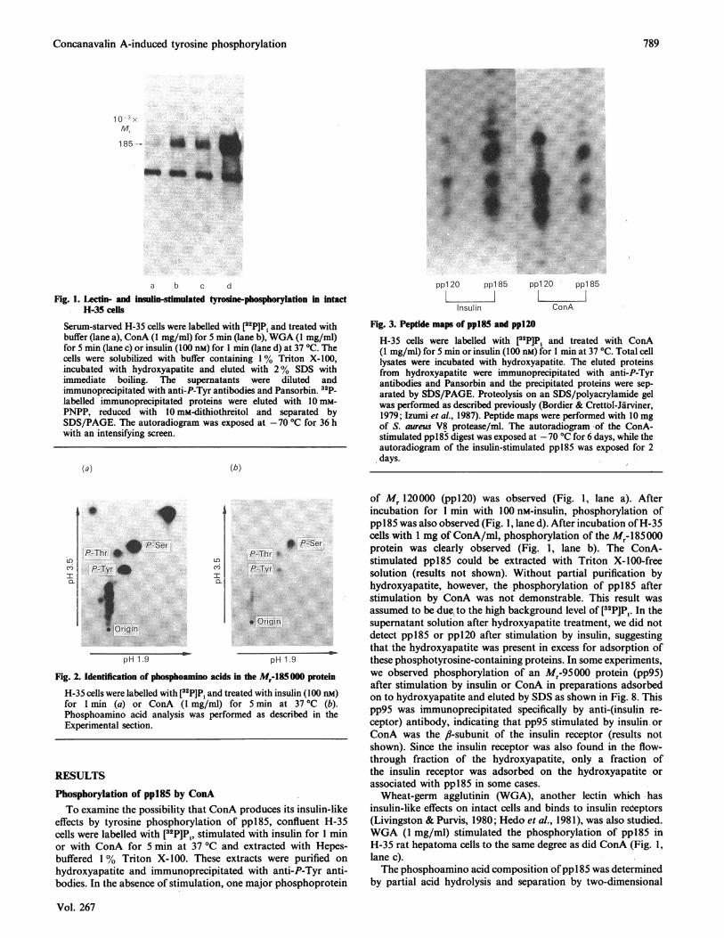
Fig. 1. Lectin- and insulin-stimulated tyrosne-phosphorylation in intactH-35 cells
Serum-starved H-35 cells were labelled with [32P]Pi and treated withbuffer (lane a), ConA (1 mg/ml) for 5 min (lane b), WGA (1 mg/ml)for 5 min (lane c) or insulin (100 nM) for I min (lane d) at 37 'C. Thecells were solubilized with buffer containing 1% Triton X-100,incubated with hydroxyapatite and eluted with 2 % SDS withimmediate boiling. The supernatants were diluted andimmunoprecipitated with anti-P-Tyr antibodies and Pansorbin. 3aP-labelled immunoprecipitated proteins were eluted with 10 mm-PNPP, reduced with 10 mM-dithiothreitol and separated bySDS/PAGE. The autoradiogram was exposed at -70 'C for 36 hwith an intensifying screen.
(a) (b)
ppl20 ppl 85
L.Insulin
ppl20 ppl 85
LJConA
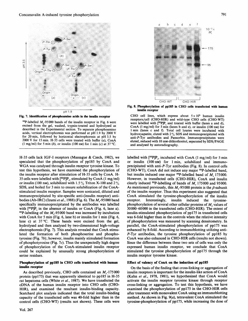
Fig. 3. Peptide maps of ppl85 and ppl20
H-35 cells were labelled with [32P]Pi and treated with ConA(1 mg/ml) for 5 min or insulin (100 nM) for I min at 37 'C. Total celllysates were incubated with hydroxyapatite. The eluted proteinsfrom hydroxyapatite were immunoprecipitated with anti-P-Tyrantibodies and Pansorbin and the precipitated proteins were sep-arated by S-DS/PAGE. Proteolysis on an SDS/polyacrylamide gelwas performed as described previously (Bordier & Crettbl-Jarviner,1979; Izumi et al., 1987). Peptide maps were performed with 10 mgof S. aureus V8 protease/ml. The autoradiogram of the ConA-stimulated pp185 digest was exposed at -70 'C for 6 days, while theautoradiogram of the insulin-stimulated ppl85 was exposed for 2days.
I
9
_P-SerP-Thr *
S
P-Tyr
* Origin
LOc'-iIn
P-Thr i
P-Tyr, ,w
.O:J...
*~Origin.
pH 1.9
* P-Ser
pH 1.9
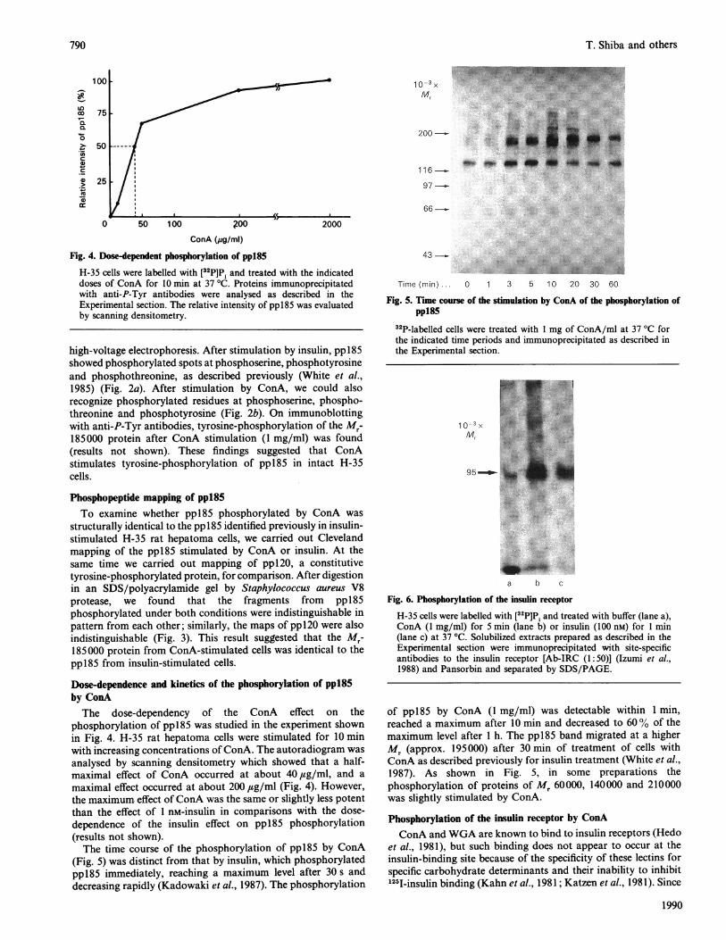
Fig. 2. Identification of phosphoamino acids in the Mr-185 000 proteinH-35 cells were labelled with [32PJP1 and treated with insulin (100 nM)for 1 min (a) or ConA (I mg/ml) for 5 min at 37 °C (b).Phosphoamino acid analysis was performed as described in theExperimental section.
RESULTS
Phosphorylation of ppl85 by ConATo examine the possibility that ConA produces its insulin-like
effects by tyrosine phosphorylation of ppl85, confluent H-35cells were labelled with [32P]P1, stimulated with insulin for 1 minor with ConA for 5 min at 37 °C and extracted with Hepes-buffered 1% Triton X-100. These extracts were purified onhydroxyapatite and immunoprecipitated with anti-P-Tyr anti-bodies. In the absence of stimulation, one major phosphoprotein
of Mr 120000 (ppl20) was observed (Fig. 1, lane a). Afterincubation for 1 min with 100 nM-insulin, phosphorylation ofppl85 was also observed (Fig. 1, lane d). After incubation ofH-35cells with 1 mg of ConA/ml, phosphorylation of the M,-485000protein was clearly observed (Fig. 1, lane b). The ConA-stimulated ppl85 could be extracted with Triton X-100-freesolution (results not shown). Without partial purification byhydroxyapatite, however, the phosphorylation of ppl85 afterstimulation by ConA was not demonstrable. This result wasassumed to be-dce, to the high background level of [32P]P1. In thesupernatant solution after hydroxyapatite treatment, we did notdetect ppl85 or ppl2O after stimulation by insulin, suggestingthat the hydroxyapatite was present in excess for adsorption ofthese phosphotyrosine-containing proteins. In some experiments,we observed phosphorylation of an M,-95000 protein (pp95)after stimulation by insulin or ConA in preparations adsorbedon to hydroxyapatite and eluted by SDS as shown in Fig. 8. Thispp95 was immunoprecipitated specifically by anti-(insulin re-ceptor) antibody, indicating that pp95 stimulated by insulin.orConA was the fl-subunit of the insulin receptor (results notshown). Since the insulin receptor was also found in the flow-through fraction of the hydroxyapatite, only a fraction ofthe insulin receptor was adsorbed on the hydroxyapatite orassociated with ppl85 in some cases.Wheat-germ agglutinin (WGA), another lectin which has
insulin-like effects on intact cells and binds to insulin redeptors(Livingston & Purvis, 1980; Hedo et al., 1981), was also studied.WGA (1 mg/ml) stimulated the phosphorylation of ppl85 inH-35 rat hepatoma cells to the same degree as did ConA (Fig. 1,lane c).The phosphoamino acid composition ofpp1 85 was determined
by partial acid hydrolysis and separation by two-dimensional
Vol. 267
789
T. Shiba -and others
LC)co
0L
CLCu0
a,
Cu3
10-3 xMr
200
116-.
97
66_
p00ConA (jug/ml)
Fig. 4. Dose-dependent phosphorylation of ppl85
H-35 cells were labelled with [32P]Pi and treated with the indicateddoses of ConA for 10 min at 37 'C. Proteins immunoprecipitatedwith anti-P-Tyr antibodies were analysed as described in theExperimental section. The relative intensity of ppl85 was evaluatedby scanning densitometry.
high-voltage electrophoresis. After stimulation by insulin, pp185showed phosphorylated spots at phosphoserine, phosphotyrosineand phosphothreonine, as described previously (White et al.,1985) (Fig. 2a). After stimulation by ConA, we could alsorecognize phosphorylated residues at phosphoserine, phospho-threonine and phosphotyrosine (Fig. 2b). On immunoblottingwith anti-P-Tyr antibodies, tyrosine-phosphorylation of the Mr-185 000 protein after ConA stimulation (1 mg/ml) was found(results not shown). These findings suggested that ConAstimulates tyrosine-phosphorylation of pp185 in intact H-35cells.
Phosphopeptide mapping of ppl85To examine whether ppl85 phosphorylated by ConA was
structurally identical to the ppl85 identified previously in insulin-stimulated H-35 rat hepatoma cells, we carried out Clevelandmapping of the pp185 stimulated by ConA or insulin. At thesame time we carried out mapping of pp120, a constitutivetyrosine-phosphorylated protein, for comparison. After digestionin an SDS/polyacrylamide gel by Staphylococcus aureus V8protease, we found that the fragments from ppl85phosphorylated under both conditions were indistinguishable inpattern from each other; similarly, the maps of pp120 were alsoindistinguishable (Fig. 3). This result suggested that the Mr-185000 protein from ConA-stimulated cells was identical to theppl85 from insulin-stimulated cells.
Dose-dependence and kinetics of the phosphorylation of ppl85by ConAThe dose-dependency of the ConA effect on the
phosphorylation of pp185 was studied in the experiment shownin Fig. 4. H-35 rat hepatoma cells were stimulated for 10 minwith increasing concentrations of ConA. The autoradiogram wasanalysed by scanning densitometry which showed that a half-maximal effect of ConA occurred at about 40,ug/ml, and amaximal effect occurred at about 200 ,ug/ml (Fig. 4). However,the maximum effect of ConA was the same or slightly less potentthan the effect of 1 nM-insulin in comparisons with the dose-dependence of the insulin effect on pp185 phosphorylation(results not shown).The time course of the phosphorylation of pp185 by ConA
(Fig. 5) was distinct from that by insulin, which phosphorylatedpp185 immediately, reaching a maximum level after 30 s anddecreasing rapidly (Kadowaki et al., 1987). The phosphorylation
43 -.......... :::....:.::.::::.:::
.......... i....
Time (min)... 0 1 3 5 10 20 30 60
Fig. 5. Time course of the stimulation by ConA of the phosphorylation ofpp185
32P-labelled cells were treated with 1 mg of ConA/ml at 37 °C forthe indicated time periods and immunoprecipitated as described inthe Experimental section.
10-3 X
Mr
95-
a b c
Fig. 6. Phosphorylation of the insulin receptor
H-35 cells were labelled with [32P]Pi and treated with buffer (lane a),ConA (1 mg/ml) for 5 min (lane b) or insulin (100 nM) for 1 min(lane c) at 37 'C. Solubilized extracts prepared as described in theExperimental section were immunoprecipitated with site-specificantibodies to the insulin receptor [Ab-IRC (1:50)] (Izumi et al.,1988) and Pansorbin and separated by SDS/PAGE.
of pp185 by ConA (1 mg/ml) was detectable within 1 min,reached a maximum after 10 min and decreased to 60 % of themaximum level after 1 h. The pp185 band migrated at a higherMr (approx. 195000) after 30 min of treatment of cells withConA as described previously for insulin treatment (White et al.,1987). As shown in Fig. 5, in some preparations thephosphorylation of proteins of Mr 60000, 140000 and 210000was slightly stimulated by ConA.
Phosphorylation of the insulin receptor by ConAConA and WGA are known to bind to insulin receptors (Hedo
et al., 1981), but such binding does not appear to occur at theinsulin-binding site because of the specificity of these lectins forspecific carbohydrate determinants and their inability to inhibit'251-insulin binding (Kahn et al., 1981; Katzen et al., 1981). Since
1990
.... .....
790
Concanavalin A-induced tyrosine phosphorylation
a b c d e f
10M3x
' P- SerP-Thr si -
P-Tyr,#/(b)
Ser lP-SerP-ThrC)' -Thr
Origin r-I -....._...., ~
P-Tyr ;-'
bt
pH1 .9Fig. 7. Identification of phosphoamino acids in the insulin receptor
32P-labelled Mr-95000 bands of the insulin receptor in Fig. 6 wereexcised from the gel, washed, trypsin-treated and hydrolysed asdescribed in the Experimental section. To separate phosphoaminoacids, vertical electrophoresis was performed at pH 1.9 by 2000 Vfor 20 min, followed by horizontal electrophoresis at pH 3.5 by2000 V for 15 min. H-35 cells were treated with buffer (a), ConA(1 mg/ml) for 5 min (b), or insulin (100 nM) for 1 min (c) at 37 'C.
.. .. ..;..:....6.. ,.S t< S . tf 0 1t *1754:-M ....... ..
0 95
CHO-WT CHO HIR
Fig. 8. Phosphorylation ofpp:85 in CHO cells transfected with humaninsulin receptor
CHO cell lines, which express about 5 x l0' human insulinreceptors/cell (CHO-HIR) and wild-type CHO cells (CHO-RT)were labelled with [32PIP, and treated with buffer (lanes a and d),ConA (1 mg/ml) for 5 min (lanes b and e), or insulin (100 nM) for1 min (lanes c and f). Total cell lysates were incubated withhydroxyapatite, eluted with 2% SDS and immunoprecipitated withanti-P-Tyr antibodies and Pansorbin. Immunoprecipitates wereeluted, reduced with 10 mM-dithiothreitol, separated by SDS/PAGEand analysed by autoradiography.
H-35 cells lack IGF-I receptors (Massague & Czech, 1982), we
speculated that the phosphorylation of pp185 by ConA andWGA was catalysed through insulin receptor tyrosine kinase. Totest this hypothesis, we have examined the phosphorylation ofthe insulin receptor after stimulation of H-35 cells by ConA. H-35 cells were labelled with [32P]P , stimulated by ConA (1 mg/ml)or insulin (100 nM), solubilized with 1.5 % Triton X-100 and 2%SDS, and boiled for 3 min to ensure solubilization of the ConA-stimulated insulin receptor. Samples were sonicated, diluted andimmunoprecipitated by site-specific anti-(insulin receptor) anti-bodies (Ab-IRC) (Izumi et al., 1988) (Fig. 6). The Mr-95 000 bandspecifically immunoprecipitated by the antibodies was labelledwith [32P]Pi in the absence of insulin or ConA (Fig. 6, lane a).32P-labelling of the Mr-95000 band was increased by incubationwith ConA for 5 min (Fig. 6, lane b) or insulin for 1 min (Fig. 6,lane c) at 37 'C. These bands were excised from the gel,hydrolysed and then analysed by two-dimensional high-voltageelectrophoresis (Fig. 7). This analysis revealed that ConA stimu-lated the formation of both phosphoserine and phospho-tyrosine (Fig. 7b); however, insulin mainly stimulated formationof phosphotyrosine (Fig. 7c). Thus the unexpectedly high degreeof phosphorylation of the ConA-stimulated insulin receptorcould be explained by unusually strong phosphorylation ofserine residues.
Phosphorylation of ppl85 in CHO cells transfected with humaninsulin receptorAs described previously, CHO cells contained an Mr-175000
protein (ppl75) that was apparently identical to pp185 in H-35rat hepatoma cells (White et al., 1987). We have transfected thecDNA of the human insulin receptor into CHO cells (CHO-HIR), and examined the resultant insulin-binding capacity.Scatchard plot analysis indicated that the total insulin-bindingcapacity of the transfected cells was 40-fold higher than in thecontrol cells (CHO-WT) (results not shown). These cells were
labelled with [32P]P , incubated with ConA (1 mg/ml) for 5 minor insulin (100 nM) for 1 min, solubilized and immuno-precipitated with anti-P-Tyr antibodies (Fig. 8). In control cells(CHO-WT), ConA did not induce any major 32P-labelled band,but insulin induced one major 32P-labelled band of Mr 175000.However, in transfected cells (CHO-HIR), ConA and insulinclearly induced 32P-labelling of bands of M, 175000 and 95000.As mentioned previously, this Mr-95 000 protein is the fl-subunitof the insulin receptor. Thus this experiment also suggested thatConA stimulated the tyrosine-phosphorylation of the insulinreceptor. Interestingly, insulin induced the tyrosine-phosphorylation of several other cellular proteins ofMr values of30000-60000 in the transfected cells (CHO-HIR). The extent ofinsulin-stimulated phosphorylation of ppl75 in transfected cellswas 6-fold higher than in the controls when the relative intensityof phosphorylation was measured by scanning densitometry. Inparallel, the ConA-stimulated phosphorylation of ppl75 was
enhanced by 8-fold. According to immunoblotting utilizing anti-P-Tyr antibodies, the tyrosine phosphorylation of pp185 byConA was also enhanced in CHO-HIR cells (results not shown).Since the difference between these two sets of cells was only theexpressed human insulin receptor, we conclude that ConAstimulated the tyrosine phosphorylation of pp175 through theinsulin receptor tyrosine kinase.
Effect of valency of ConA on the induction of ppl85On the basis of the finding that cross-linking or aggregation of
insulin receptors is important for the insulin-like action ofConA(Kahn et al., 1978, 1981), we hypothesized that ConA wouldactivate the insulin receptor tyrosine kinase through receptorcross-linking or aggregation. To test this hypothesis, we haveexamined the phosphorylation of ppl75 in the CHO-HIR cellsafter treatment with monovalent ConA using an immunoblottingmethod. As shown in Fig. 9(a), tetravalent ConA stimulated thetyrosine-phosphorylation of ppl175, while increasing the dose of
Vol. 267
LnC')In
Origin.4.
Origin(a) . (c)
791
T. Shiba and others
(a)b c d e f 10-3
x M
Dose (pg/ml) ... 0 50 200 0 12.5 50 100
Fig. 9. Effect of valency of ConA on the induction of ppl75
(a) CHO cells transfected with the cDNA of the human insulin receptor were treated with the indicated dose of monovalent (lanes a-c) ortetravalent (native) (lanes d-g) ConA for 5 min at 37 'C. Total cell lysates were incubated with hydroxyapatite and eluted with Laemmli (1970)sample buffer with 10 mM-dithiothreitol. The samples were separated by SDS/PAGE and analysed by immunoblotting utilizing anti-P-Tyrantibodies as described in the Experimental section. (b) CHO-HIR cells were exposed to monovalent ConA (25 ,g/ml) for 30 min at 37 'C, andthen rabbit anti-ConA IgG (200 #sg/ml; lane a) or control rabbit IgG (200 ,ug/ml; lane b) was added and incubated for 20 min at 37 'C. Thesamples were analysed by immunoblotting utilizing anti-P-Tyr antibodies as described in the Experimental section.
monovalent ConA only weakly phosphorylated pp 175. Theextent of phosphorylation of ppl75 by 200 ,ug of monovalentConA/ml was clearly less than that by 50 ug of tetravalentConA/ml, which should have the same number of binding sitesas 200 ,ug of monovalent ConA/ml; in fact the phosphorylationwas also less than that caused by 12.5 ,ug of tetravalent ConA/ml.The weak phosphorylation of ppl75 by the monovalent ConApreparation might be attributable to its residual aggregatingactivity, since our monovalent ConA fraction had 1.7-2.0% ofthe haemagglutinating activity against mouse erythrocyte oftetravalent ConA (results not shown). The weak residual aggre-gating activity of the monovalent derivative could be explainedby several possibilities as discussed previously (Beppu et al.,1979).To explore the role of valency in stimulating the insulin
receptor kinase, reconstitution of valency was attempted byexposing the receptor to the monovalent ConA and then cross-linking them by addition of anti-ConA antibodies in intact cells.As shown in Fig. 9(b), addition of antibodies against ConAclearly enhanced the ability of monovalent ConA to stimulatethe tyrosine phosphorylation of ppl75 in CHO-HIR cells,although the lower-Mr band, which presumably represents IgG,is labelled less by 125I-Protein A in the presence of anti-ConAIgG. These data suggest that ConA stimulates the insulin receptorkinase mainly by the receptor cross-linking or aggregation inintact cells.
DISCUSSION
The insulin receptor has a tyrosine-specific protein kinaseactivity associated with the cytoplasmic domain of its fl-subunit.Recent studies utilizing site-directed mutagenesis of tyrosineresidues or the ATP-binding site (Ebina et al., 1987; Chou et al.,1987; McClain et al., 1987) and tyrosine kinase inhibitoryantibodies (Morgan & Roth, 1987; Izumi et al., 1988) indicatethat the activation of the tyrosine kinase is a key step in insulinsignal transduction. Furthermore, it has been hypothesized that
this signal transduction may occur in the intact cell by a cascadeof substrate phosphorylation steps. The ppl85 protein is knownas one of the putative important endogenous substrates specificfor the insulin and IGF-I receptor kinase (White et al., 1985,1987, 1988; Gibbs et al., 1986; Kadowaki et al., 1987; Izumiet al., 1987; Shemer et al., 1987). In this present study, we havedemonstrated that ConA stimulated the tyrosine-phosphoryl-ation of an Mr-185000 protein in intact H-35 rat hepatoma cells.This phosphoprotein ofMr 185 000 shared the following commoncharacteristics with the pp 185 observed during the initial responseof H-35 cells to insulin. First, the phosphopeptide map of theConA-stimulated Mr-185000 protein was identical to that of theinsulin-stimulated ppl85. Secondly, prolonged exposure of cellsto ConA also caused retarded migration of this protein inSDS/PAGE and decreased its phosphorylation. Thirdly, thisprotein could be extracted by detergent-free solution andadsorbed on hydroxyapatite.
In this study, we have also demonstrated that ConA stimulatedtyrosine-phosphorylation of the ,-subunit of the insulin receptor,and it also stimulated serine-phosphorylation of the insulinreceptor. Interestingly, the degree of serine-phosphorylation wasstimulated more by ConA than by insulin, suggesting that ConAstimulated several cellular serine kinases or decreased phosphat-ase activities specific for serine residues in intact cells. This maybe explained by the fact that ConA interacts with many kindsof glycoproteins on the cell surface, and the insulin receptoris only one of the glycoproteins to which it binds (Bittigen &Schnebli, 1976). This fact also made it difficult to identify thetyrosine kinase(s) responsible for the tyrosine-phosphorylationof ppl85. However, our finding that the tyrosine-phosphory-lation of ppl85 induced by ConA was demonstrated only'in theCHO cells transfected with human insulin- receptor cDNAsuggested that the insulin receptor kinase was responsible for thetyrosine-phosphorylation of ppl85 induced by ConA.Next we investigated the role of the valency of ConA in the
activation of the receptor kinase. For this purpose, we producedmonovalent ConA, and confirmed its valency by equilibriumdialysis and it& binding activity to glycoproteins using an25I-labelled fraction. The monovalent ConA fraction was less
1990
(b)a b
10-3x Mr
-200.-175
116
-97
-66
*..... w
- 43
*0#1* * *_ 175
-116
-97
-66
- 43
792
Concanavalin A-induced tyrosine phosphorylation
potent in inducing the tyrosine-phosphorylation of ppl85 thanwas tetravalent ConA. The slight phosphorylation of ppl85induced by monovalent ConA may be explained by the residualaggregating activity of monovalent ConA (Beppu et al., 1979).Reconstitution of valency by the addition of the anti-ConAantibodies enhanced the ability of monovalent ConA to stimulatethe tyrosine phosphorylation of pp 175 in CHO-HIR cells. ThusConA may activate the tyrosine-kinase activity of the insulinreceptor by aggregation or cross-linking of the receptor. How-ever, it cannot be fully discounted that the monovalent andtetravalent ConA act differently on the tyrosine-phosphorylationof ppl85, since the direct effects of ConA on receptor auto-phosphorylation cannot be demonstrated for technical reasons.
In a Triton-solubilized system, Heffetz & Zick (1986)demonstrated that receptor cross-linking or aggregationconstituted a sufficient trigger for activation of the insulinreceptor kinase using monovalent and divalent anti-receptor Fabfragments. Thus receptor aggregation may activate the tyrosinekinase activity of the receptor both in Triton-solubilized fractionsand in the intact membrane. From studies of truncated receptors(Ellis et al., 1987) or trypsin-treated receptors (Shoelson et al.,1988), it was proposed that the a-subunit of the insulin receptorinhibited the kinase activity of the fl-subunit. That is, eliminationof inhibitory constraints imposed by the a-subunit might permita conformational relaxation that enables the fl-subunit to adoptan unconstrained, uninhibited, and thus activated conformation(Shoelson et al., 1988). According to this hypothesis, receptoraggregation may also release the kinase activity of the fl-subunitfrom inhibitory control.
Half-maximal stimulation of pp 185 phosphorylation by ConAoccurred at about 40,ug of ConA/ml. This value was slightlyhigher than that reported previously for glucose oxidation of ratadipocytes treated by ConA (Kahn et al., 1981) and for inductionof tyrosine aminotransferase activity by WGA (Smith & Liu,1981), and was similar to the value for ConA-stimulated auto-phosphorylation of the insulin receptor in a cell-free system(Roth et al., 1983). Furthermore, Kahn et al. (1981) havecharacterized the role of valency and receptor aggregation in theinsulin-like effects of ConA in rat adipocytes and found thatmonovalent ConA totally lacked insulin-like effects. Consideringthese previous data, our present results, including the data withmonovalent ConA, raised the possibility that ConA producesinsulin-like effects through activation of the insulin receptorkinase and the tyrosine-phosphorylation of ppl85 initiated bythe aggregation or cross-linking of the receptor.
In conclusion, ConA stimulated the phosphorylation of theinsulin receptor on tyrosine residues and stimulated tyrosinekinase activity, and it resulted in tyrosine phosphorylation ofppl85, a putative cellular substrate for the insulin-stimulatedinsulin receptor kinase; these steps occurred in a dose- and time-dependent manner in intact cells. The mechanism of ConAactivation of insulin receptor kinase is suggested to be mainly bycross-linking or aggregation of the receptors in intact cells.
We thank Dr. Kenneth M. Yamada (National Cancer Institute,National Institutes of Health, Bethesda, MD, U.S.A.) and M. F. White(Joslin Diabetes Center, Boston, MA, U.S.A.) for their critical reading ofthe manuscript, Dr. G. I. Bell (University of Chicago, Chicago, IL,U.S.A.) for insulin receptor cDNA, Dr. S. Jacobs (Burroughs WellcomeResearch Lab., Research Triangle Park, NC, U.S.A.) for the monoclonalantibody against the insulin receptor, Dr. I. Yahara (The TokyoMetropolitan Institute of Medical Science, Tokyo, Japan) for anti-ConAantibodies, Dr. T. Izumi in our laboratory for site-specific antibody forthe insulin receptor (Ab-IRC) and Dr. M. Morioka and personnel in theRadioisotope Center at the University of Tokyo and K. Aihara in theRadioisotope Center at the University of Tokyo Hospital for support.We also thank Dr. Y. Kanazawa and Dr. S. Takayama-Hasumi forgenerous support and useful discussions and M. Nishio, K. Tanaka and
Vol. 267
K. Toyoshima for technical and secretarial assistance. This work hasbeen supported by a Grant-in-Aid for Developmental Scientific Researchfrom the Ministry of Education, Science and Culture of Japan, a Grantfrom Juvenile Diabetes Foundation International, a Grant from theNaito Foundation and a Grant from the Uehara Memorial Foundation.
REFERENCES
Beppu, M., Terao, T. & Osawa, T. (1979) J. Biochem. (Tokyo) 85,1275-1287
Bernier, M., Laird, D. M. & Lane, M. D. (1987) Proc. Natl. Acad. Sci.U.S.A. 84, 1844-1848
Bittigen, H. & Schnebli, H. P. (eds.) (1976) Concanavalin A as a Tool,Wiley, New York
Bordier, C. & Crettol-Jarviner, A. (1979) J. Biol. Chem. 254, 2565-2567Chou, C. K., Dull, T. J., Russell, D. S., Gherzi, R., Lebwohl, D., Ullrich,
A. & Rosen, 0. M. (1987) J. Biol. Chem. 259, 5277-5286Cooper, J. A., Sefton, B. M, & Hunter, T. (1983) Methods Enzymol. 99,
387-402Ebina, Y., Araki, E., Taira, M., Shimada, F., Mori, M., Craik, C. S.,
Siddle, K., Pierce, S. B., Roth, R. A. & Rutter, W. J. (1987) Proc. Natl.Acad. Sci. U.S.A. 83, 328-332
Ellis, L., Clauser, E., Morgan, D. O., Edery, M., Roth, R. A. & Rutter,W. J. (1986) Cell 45, 721-732
Ellis, L., Morgan, D. O., Clauser, E., Roth, R. A. & Rutter, W. J. (1987)Mol. Endocrinol. 1, 15-24
Faltynek, C. R., Princler, G. L., Ruscetti, F. W. & Birchenall-Sparks, M.(1988) J. Biol. Chem. 263, 7112-7117
Gibbs, E. M., Allard, W. J. & Lienhard, G. E. (1986) J. Biol. Chem. 261,16597-16603
HWring, H. U., White, M. F., Machicao, F., Erme, B., Schleicher, E. &Obermaier, B. (1987) Proc. Natl. Acad. Sci. U.S.A. 84, 113-117
Hedo, J. A., Harrison, L. C. & Roth, J. (1981) Biochemistry 20,3385-3393
Heffetz, D. & Zick, Y. (1986) J. Biol. Chem. 261, 889-894Izumi, T., White, M. F., Kadowaki, T., Takaku, F., Akanuma, Y. &
Kasuga, M. (1987) J. Biol. Chem. 262, 1282-1287Izumi, T., Saeki, Y., Akanuma, Y., Takaku, F. & Kasuga, M. (1988) J.
Biol. Chem. 263, 10386-10393Kadowaki, T., Koyasu, S., Nishida, E., Tobe, K., Izumi, T., Takaku, F.,
Saeki, H., Yahara, I. & Kasuga, M. (1987) J. Biol. Chem. 262,7342-7350
Kahn, C. R., Baird, K. L., Jarrett, D. B. & Flier, J. S. (1978) Proc. Natl.Acad. Sci. U.S.A. 75, 4209-4213
Kahn, C. R., Baird, K. L. & Van Obberghen, E. (1981) FEBS Lett. 129,131-134
Kasuga, M., Hedo, J.-A., Yamada, K. M. & Kahn, C. R. (1982a) J. Biol.Chem. 257, 10392-10399
Kasuga, M., Karlsson, F. A. & Kahn, C. R. (1982b) Science 215, 185-187Kasuga, M., Fujita-Yamaguchi, Y., Blithe, D. & Kahn, C. R. (1983)
Proc. Natl. Acad. Sci. U.S.A. 80, 2137-2141Katzen, H. M., Solderman, D. D. & Green, B. G. (1981) Biochem.
Biophys. Res. Commun. 115, 410-416Laemmli, U. K. (1970) Nature (London) 227, 680-685Livingston, J. N. & Purvis, B. J. (1980) Am. J. Physiol. 238, E267-E275Massague, J. & Czech, M. P. (1982) J. Biol. Chem. 257, 5038-5045McClain, D. A., Maegawa, H., Lee, J., Dull, T. J., Ullrich, A. & Olefsky,
J. M. (1987) J. Biol. Chem. 262, 14663-14671Momomura, K., Tobe, K., Seyama, Y., Takaku, F. & Kasuga, M. (1988)
Biochem. Biophys. Res. Commun. 155, 1181-1186Morgan, D. 0. & Roth, R. A. (1987) Proc. Natl. Acad. Sci. U.S.A. 89,41-45
Mulligaw, R. C. & Berg, P. (1981) Proc. Natl. Acad. Sci. U.S.A. 78,2072-2076
Osawa, T. & Beppu, M. (1987) Methods Enzymol. 150, 17-28Perrotti, N., Accili, D., Marcus-Samuels, B., Rees-Jones, R. W. & Taylor,
S. I. (1987) Proc. Natl. Acad. Sci. U.S.A. 84, 3137-3140Roth, R. A. & Cassell, D. J. (1983) Science 219, 299-301Roth, R. A., Cassell, D. J., Maddux, B. A. & Goldfine, I. D. (1983)
Biochem. Biophys. Res. Commun. 115, 245-252Shemer, J., Adamo, M., Wilson, G. L., Heffez, D., Zick, Y. & LeRoith,D. (1987) J. Biol. Chem. 262, 15476-15482
Shoelson, S. E., White, M. F. & Kahn, C. R. (1988) J. Biol. Chem. 263,4852-4860
Smith, J. D. & Liu, A. Y.-C. (1981) Science 214, 799-800
793
T. Shiba and others
Wakshull, E. M. & Wharton, W. (1985) Proc. Natl. Acad. Sci. U.S.A. 82,8513-8517
White, M. F., Maron, R. & Kahn, C. R. (1985) Nature (London) 318,183-186
White, M. F., Stegmann, E. W., Dull, T. J., Ullrich, A. & Kahn, C. R.(1987) J. Biol. Chem. 262, 9769-9777
Received 27 September 1989/16 January 1990; accepted 26 January 1990
White, M. F., Livingston, J. N., Backer, J. M., Lauris, V., Dull, T. J.,Ullrich, A. & Kahn, C. R. (1988) Cell 54, 641-649
Whittaker, J., Okamoto, A. K., Thys, R., Bell, G. I., Steiner, D. F. &Hoffman, C. A. (1987) Proc. Natl. Acad. Sci. U.S.A. 84, 5237-5241
Yu, K. T., Khalaf, N. & Czech, M. P. (1987) J. Biol. Chem. 262,7865-7873
199X
794