Embed Size (px)
Citation preview
Contributions of Asparagine atR97 to the Cooperative Oxygenation Process ofHemoglobin†
Hyun-Won Kim,‡,§ Tong-Jian Shen,‡ Nancy T. Ho,‡ Ming Zou,‡ Ming F. Tam,| and Chien Ho*,‡
Department of Biological Sciences, Carnegie Mellon UniVersity, 4400 Fifth AVenue, Pittsburgh, PennsylVania 15213, andInstitute of Molecular Biology, Academia Sinica, Taipei, Taiwan
ReceiVed October 23, 1995; ReVised Manuscript ReceiVed February 8, 1996X
ABSTRACT: According to the X-ray crystallographic results from human deoxyhemoglobin,â99Asp at theR1â2 interface forms hydrogen bonds withR42Tyr andR97Asn. To clarify the structural and functionalroles of the hydrogen bond betweenR97Asn andâ99Asp, we have engineered a recombinant hemoglobinin whichR97Asn is replaced by Ala, and have investigated its oxygen-binding properties, and have usedproton nuclear magnetic resonance spectroscopy to determine the structural consequences of the mutation.Recombinant Hb (R97AsnfAla) shows a milder alteration of functional properties compared to the severelyimpairedâ99 mutants of the human abnormal hemoglobins. The addition of inositol hexaphosphate, anallosteric effector, causes recovery of the functional properties of recombinant Hb (R97AsnfAla) almostto the level of human normal adult hemoglobin without this allosteric effector. r Hb (R97AsnfAla)shows very similar tertiary structure around the heme pockets and quaternary structure in theR1â2 interfacecompared to those of human normal adult hemoglobin. The proton nuclear magnetic resonance spectrumof the deoxy form of this recombinant hemoglobin shows the existence of an altered hydrogen bondwhich is believed to be betweenR42Tyr andâ99Asp at theR1â2 interface. Thus, the present resultssuggest that the intersubunit hydrogen bond betweenR97Asn andâ99Asp at theR1â2 interface is not ascrucial as the one betweenR42Tyr andâ99Asp in the deoxy quaternary structure. Preliminary moleculardynamics simulations have been used to calculate the contributions of specific interactions of severalamino acid residues in r Hb (R97AsnfAla) to the free energy of cooperativity of this recombinanthemoglobin. The results of these calculations are consistent with the experimental results.
X-ray crystallographic analyses of oxy- and deoxyhemo-globin (Hb)1 have shown that the intersubunit contacts atthe R1â2 and R2â1 subunit interfaces undergo a majoralteration when the oxygenation state of the Hb moleculechanges (Perutz, 1970; Baldwin & Chothia, 1979; Shaanan,1983; Fermi et al., 1984). Human abnormal Hbs with aminoacid substitutions at theR1â2 subunit interface generally showvery high oxygen affinity and greatly reduced cooperativityin O2 binding [as measured by the Hill coefficient (nmax)]compared to those of human normal adult hemoglobin (HbA) (Dickerson & Geis, 1983; Bunn & Forget, 1986). X-raycrystallographic studies of Hb A (Fermi et al., 1984) haveshown thatâ99Asp is hydrogen-bonded to bothR42Tyr andR97Asn in the R1â2 subunit interface of deoxy-Hb A,suggesting that the essential role ofâ99Asp is to stabilize
the deoxy-Hb molecule by forming intersubunit hydrogenbonds and thus to provide the free energy of cooperativityin binding oxygen. The importance ofâ99Asp in thecooperative oxygenation has also been demonstrated by thefunctional defects of human mutant Hbs with an amino acidsubstitution at theâ99Asp position, such as Hb Kempsey(â99AspfAsn) (Reed et al., 1968), Hb Yakima(â99AspfHis) (Jones et al., 1967), Hb Radcliffe(â99AspfAla) (Weatherall et al., 1977), Hb Hoˆtel Dieu(â99AspfGly) (Thillet et al., 1981), and Hb Ypsilanti(â99AspfTyr) (Glynn et al., 1968), which possess greatlyreduced cooperativity and increased oxygen affinity relativeto those exhibited by Hb A.
To investigate the functional role ofR42Tyr at theR1â2
subunit interface of deoxy-Hb, two recombinant Hb (r Hb)mutants, r Hb (R42TyrfHis) and r Hb (R42TyrfPhe),have been constructed (Ishimori et al., 1989; Imai et al.,1991). r Hb (R42TyrfPhe) exhibits essentially no coop-erativity in binding oxygen (n) 1.2 at pH 6.8) and possessesvery high oxygen affinity. However, r Hb (R42TyrfHis)exhibits considerable cooperativity (n ) 2 at pH 6.8) andmoderate oxygen affinity. These authors have attributed themilder functional impairment of r Hb (R42TyrfHis) to thepresence of a new weak hydrogen bond betweenR42Hisandâ99Asp in the deoxy state of r Hb (R42TyrfHis) butnot in deoxy-r Hb (R42TyrfPhe), suggesting that thehydrogen bond betweenR42Tyr andâ99Asp in deoxy-HbA plays a key role in stabilizing the deoxy quaternarystructure and consequently in the cooperative oxygen-bindingprocess of Hb.
† Supported by a grant from the National Institutes of Health (HL-24525 to C.H.).* Address all correspondence to this author. Tel: (412) 268-3395.
FAX: (412) 268-7083. E-mail: [email protected].‡ Carnegie Mellon University.§ Present address: Department of Biochemistry, Yonsei University
Wonju College of Medicine, 162 Ilsan-dong, Wonju, Kangwon-do220-701, Korea.
| Academia Sinica.X Abstract published inAdVance ACS Abstracts,May 1, 1996.1 Abbreviations: Hb, hemoglobin; Hb A, human normal adult
hemoglobin; r Hb, recombinant hemoglobin; deoxy-Hb, deoxyhemo-globin; HbCO, carbonmonoxyhemoglobin; met-Hb, methemoglobin;IHP, inositol hexaphosphate; MD, molecular dynamics; MAP, me-thionine aminopeptidase; N-terminal, amino-terminal; Tris, tris[hy-droxymethyl]aminomethane; TETA, triethylenetetraamine hydrochlo-ride; EDTA, ethylenediaminetetraacetate; Mb, myoglobin; DSS, 2,2-dimethyl-2-silapentane-5-sulfonate.
6620 Biochemistry1996,35, 6620-6627
S0006-2960(95)02518-9 CCC: $12.00 © 1996 American Chemical Society
+ +
+ +
We have constructed a plasmid (pHE2) (Shen et al., 1993)in which synthetic humanR- andâ-globin genes (Hoffmanet al., 1990) are coexpressed with theEscherichia colimethionine aminopeptidase (MAP) gene (Ben-Bassat et al.,1987) under the control of separatetac promoters. InE.coli cells harboring the pHE2 plasmid, the amino-terminal(N-terminal) methionine residues of the expressed Hb A havebeen effectively cleaved by the coexpressed MAP, and thisexpressed Hb A lacking an N-terminal methionine is identicalto the native Hb A in a number of structural and functionalproperties (Shen et al., 1993). Recently, we have usedmolecular dynamics (MD) simulations to design compensa-tory amino acid substitutions in an abnormal Hb, HbKempsey (â99AspfAsn), which substantially restore itsallosteric properties (Kim et al., 1994). Hb Kempsey(â99AspfAsn) has a high oxygen affinity and exhibitsessentially no cooperativity in binding oxygen (Reed et al.,1968; Bunn et al., 1974). Computer simulations indicate thata new hydrogen bond involvingâ99Asn can be induced byreplacingR42Tyr by a stronger hydrogen-bond acceptor suchas Asp. The resulting double-mutant recombinant hemo-globin, r Hb (â99AspfAsn,R42TyrfAsp), was producedby site-directed mutagenesis (Kim et al., 1994). The oxygenaffinity of r Hb (â99AspfAsn,R42TyrfAsp), while stillhigh, is significantly lower than that of Hb Kempsey, andvery substantial cooperativity has been restored, showing thatthe hydrogen bond involving theR42 andâ99 amino acidpositions in theR1â2 interface is essential for the functionalproperties of Hb.Although there are two hydrogen bonds involvingâ99Asp
in theR1â2 interface, so far most of the investigations havebeen focused only on the hydrogen bond between theR42andâ99 positions. Hb Dallas (R97AsnfLys), which is theonly known human mutant Hb with a mutation at theR97position, exhibits very high oxygen affinity (Dysert et al.,1982). However, the introduction of a charged amino acid,lysine, at theR97 position may not be suitable for theinvestigation of the hydrogen bond betweenR97Asn andâ99Asp. Thus, to investigate the specific functional role(s)of the hydrogen bond betweenR97Asn andâ99Asp, we haveapplied site-directed mutagenesis to produce a new recom-binant hemoglobin, r Hb (R97AsnfAla), which disrupts thehydrogen bond betweenR97Asn andâ99Asp but is foundto cause only minimal structural perturbations.In the present work, we have determined the oxygen-
binding properties of r Hb (R97AsnfAla) and have used1H-NMR spectroscopy to investigate the tertiary structurearound the heme groups as well as the quaternary structure.Preliminary MD simulations have been used to calculate thefree energy difference between native Hb A and r Hb(R97AsnfAla). By using a thermodynamic integrationmethod (Kirkwood, 1935), the contributions of specificinteractions to the free energy change of cooperativity areestimated.
EXPERIMENTAL PROCEDURES
Plasmids, Strains, and Media.The Hb expression plasmidpHE2 (Shen et al., 1993) containing appropriately arrangedand modified expression cassettes of syntheticR- andâ-globin genes (kindly provided by Somatogen as pDLIII-13e) and theE. coli MAP gene (kindly provided by Cetusas pSYC1174) was used (after appropriate modifications)
to produce mutant Hbs. Phagemid pTZ18U andE. coliJM109 were obtained from Bio-Rad and Promega, respec-tively. E. coli cells were grown in 2× YT medium (Miller,1972) supplemented with 50µg of ampicillin/mL. TBmedium used for the expression of Hb contained bactotryp-tone (1.2%), bactoyeast extract (2.4%), glycerol (0.4%), KH2-PO4 (17 mM), K2HPO4 (72 mM), and 100µg of ampicillin/mL.Site-Directed Mutagenesis.Somatogen’s pDLIII-13e was
inserted into phagemid pTZ18U (Bio-Rad). Site-direct-ed mutagenesis was performed as previously described(Kunkel, 1985; Shen et al., 1993). Synthetic oligonucleotide5′CAGTTTGAAGGCAACCGGATC3′ was used as a primerto introduce the mutation,R97AsnfAla. The human normalR-globin gene in plasmid pHE2 was then replaced by themutatedR-globin gene to produce pHE226.Growth of Cells. The plasmid pHE226 was transformed
into E. coli BL21-DR, which is derived from strain BL21-DE3 (kindly provided by Dr. Maureen Gilmore-Hebert), isresistant to common phages, and contains more copies ofIq, and the cells were grown in TB medium in a 10-LMicroferm fermentor (New Brunswick Scientific, modelBioFlo 3000) at 30°C until the optical density at 600 nmreached 10.Expression of r Hb (R97AsnfAla) was induced by adding
isopropylâ-thiogalactopyranoside to 0.2 mM. The culturewas then supplemented with hemin (50 mg/L), and thegrowth was continued for at least another 4 h. The cellswere harvested by centrifugation and stored frozen at-80°C until needed for purification (Shen et al., 1993).Purification of Recombinant Hb.The r Hb (R97AsnfAla)
was purified as previously described (Shen et al., 1993;Looker et al., 1994) with minor modifications. The frozencell paste was suspended in lysis buffer [40 mM Tris-[hydroxymethyl]aminomethane (Tris)-base (Sigma)/1 mMbenzamidine hydrochloride (Sigma)] at 3 mL/g of cell pasteand stirred until the mixture was smooth. Lysozyme[(Sigma) 1 mg/g of cell paste in 40 mM Tris-HCl at pH8.0] was added. MgCl2 and MnCl2 to final concentrationsof 1 and 0.1 mM, respectively, and DNAseI [(ICN Bio-chemicals) 2-3 µg/mL] were added to the cell paste priorto sonication. After sonification, the sonicate was saturatedwith CO gas and centrifuged to pellet cell membranes, etc.The supernatant was adjusted to pH 8.0 with 1 M Tris-base,and polyethyleneimine (Sigma) was added to a final con-centration of 0.3%-0.5% to precipitate nucleic acids. Aftercentrifugation, the supernatant was put through a MilliporeMinitan Acrylic Ultrafiltration System to concentrate theprotein. Then, the sample was dialyzed in 20 mM Tris-HCl/0.1 mM triethylenetetraamine hydrochloride (TETA; Sigma)at pH 8.3 overnight with one change of buffer. The samplewas kept at 4°C throughout the above procedures andmaintained in the CO form throughout. We used twocolumns in the final purification process: (i) a Q-Sepharosefast-flow column (Pharmacia anion exchanger) was used tobind Hb. After the sample was loaded onto the column, itwas washed with the running buffer (20 mM Tris-HCl/0.1mM TETA at pH 8.3), and the pass through was monitoredat 260 nm until the contaminating nucleic acids had eluted.Then, the Hb fraction was eluted from the column with 20mM Tris-HCl/0.1 mM TETA at pH 7.2. After concentration,the Hb fraction was oxidized and reduced as described inShen et al. (1993). (ii) a Mono S column (Pharmacia cation
Roles of the H Bond betweenR97Asn andâ99Asp atR1â2 Interface Biochemistry, Vol. 35, No. 21, 19966621
+ +
+ +
exchanger HR16/10) with a gradient of 10 mM sodiumphosphate/0.1 mM ethylenediaminetetraacetate (EDTA) atpH 6.8 (eluent A) to 20 mM sodium phosphate/0.1 mMEDTA at pH 8.3 (eluent B) was used to purify the r Hb(R97AsnfAla).Analytical Procedures. Automated cycles of Edman
degradation were performed with an Applied Biosystems gas/liquid-phase sequencer (model 470/900A) equipped with anon-line phenylthiohydantoin amino acid analyzer (model120A).The mass spectrometric analyses were performed on a VG
Quattro-Bio-Q mass spectrometer (Fisons Instruments, VGBiotech, Altrincham, U.K.). The instrument was set in thepositive ion mode. The multiply-charged ion peaks frommyoglobin (Mb; molecular mass, 16 591 Da) were used asan external reference for mass scale calibration (Zaia et al.,1992). Mb at one-fourth of the Hb subunit concentrationwas included in each sample as an internal standard.Scanning was in the multichannel analyzer mode fromm/z500 to 1500 at 8 s/scan. The collected data were processedby the MaxEnt program (Ferrige et al., 1992). The molecularweights calculated from the amino acid sequences of normalR andâ chains of Hb A,R chain+ methionine, andâ chain+ methionine are 15 126.4, 15 867.2, 15 257.6, and 15 998.6,respectively. The VG computer program uses the followingaverage elemental atomic weights: C) 12.011; H )1.00794; N) 14.00674; O) 15.994; and S) 32.066.Oxygen Binding of Hb Samples.The oxygen dissociation
curves of r Hb (R97AsnfAla) and Hb A (0.1 mM of hemeeach) were measured by a Hemox-Analyzer (TCS MedicalProducts, Huntington Valley, PA) at 29°C in 0.1 M sodiumphosphate buffer in the pH range 6.2-8.4. 15 µL of astabilizer [hexamethylphosphoramide (Sigma)] and 15µLof antifoam [SAG-10 silicon antifoam emulsion (OSISpecialties)] were added to each 3-mL sample. After eachmeasurement, the sample was checked for the presence ofmethemoglobin (met-Hb). Those with met-Hb greater than5% were discarded. If necessary, a met-Hb reductase system(Hayashi et al., 1973) was added (120µL) to each samplein order to prevent the formation of met-Hb. Partial O2
pressure at 50% saturation (P50) and the Hill coefficient (nmax)were determined from each oxygen dissociation curve.NMR Measurements.1H-NMR spectra were obtained on
a Bruker AM-300 spectrometer operating at 300 MHz at 29°C. All Hb samples were in 0.1 M sodium phosphate buffer(in 100% H2O) at pH 7.0. The Hb concentration was about4% (2.5 mM in terms of heme). The water signal wassuppressed by using a jump-and-return pulse sequence(Plateau & Gue´ron, 1982) except in the water presaturationexperiment. The1H-NMR spectra of the HbCO and deoxy-Hb samples were obtained by using the proton decouplingcoil of a 5-mm multinuclear probe with 90° pulses of 9.7µs. Proton chemical shifts are referenced to the methylproton resonance of the sodium salt of 2,2-dimethyl-2-silapentane-5-sulfonate (DSS) indirectly by using the watersignal, which occurs at 4.76 ppm downfield from that ofDSS at 29°C, as the internal reference.MD Simulations.2 MD simulations were carried out with
a stochastic boundary method (Brooks & Karplus, 1989)using CHARMM 22 with standard parameters for the polarhydrogen model (param19). The Hb molecule was parti-tioned into MD and Langevin regions with radii of 10 and15 Å, respectively, which were centered on the center-of-
mass of coordinates of the Câ of theR97Asn side chain inthe crystal structures of Hb A in the deoxy and oxy forms(Shaanan, 1983; Fermi et al., 1984). The inside sphere wasfilled with CHARMM-adapted pre-equilibrated TIP3P watermolecules (Jorgensen et al., 1983). The deoxy simulationincluded 88 water molecules, and the oxy simulation included78 water molecules.The transformation between wild-type and mutant proteins
can be achieved by using a hybrid potential functionVλ )(1- λ)VA + λVB (Gao et al., 1989; Tidor & Karplus, 1991),whereλ is a coupling factor between 0 and 1.VA andVBare potential energy functions for Hb A and for mutant Hb,respectively. Simulations were done at nine values ofλi (λ) 0.1, 0.2, ..., 0.9), with 5 ps of equilibration followed by 5ps of production dynamics, except atλ ) 0.1 andλ ) 0.9,where 10 ps of equilibration was employed. Nonbondedinteractions were truncated to zero at 8.5 Å, and a dielectricconstant,ε ) 1, was used. All bonds involving hydrogenatoms were constrained with the SHAKE algorithm (Ryck-aert et al., 1977). A 10-ps simulation takes about 2 h on asingle-CPU SunSparc Workstation 10 with 1-fs integrationtime.The free energy of simulations can be obtained from the
trajectory files of MD simulations for both deoxy and oxyforms of Hb using the thermodynamic integration method(Kirkwood, 1935) with the following equation:
where∆V) VB - VA and the thermodynamic average⟨∆V⟩λ
indicates the average ofVλ over the hybrid system. Thelinear form of the thermodynamic equations shows that thetotal free energy of the simulations can be decomposed intoindividual additive contributions. The change in the freeenergy of cooperativity resulting from the mutations can beindirectly obtained from the thermodynamic cycle (Scheme1) as shown by Gao et al. (1989):
RESULTS
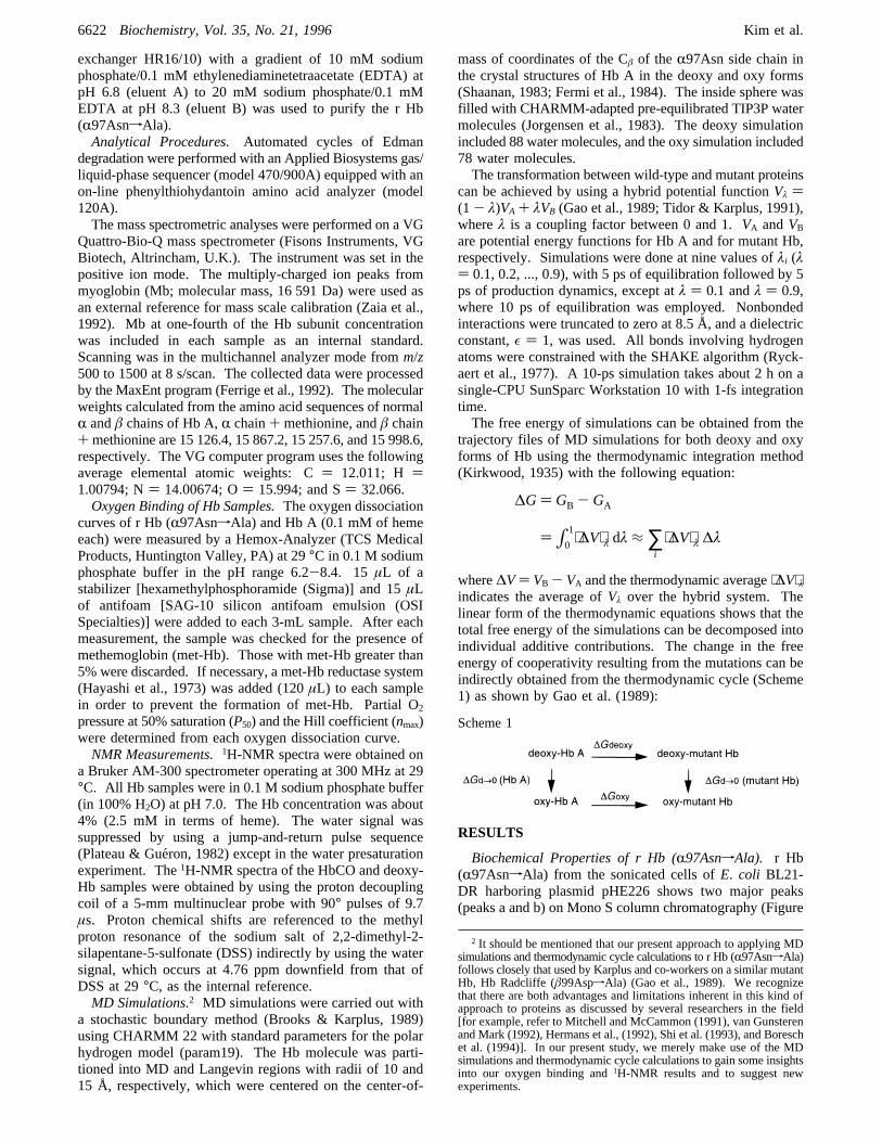
Biochemical Properties of r Hb (R97AsnfAla). r Hb(R97AsnfAla) from the sonicated cells ofE. coli BL21-DR harboring plasmid pHE226 shows two major peaks(peaks a and b) on Mono S column chromatography (Figure
2 It should be mentioned that our present approach to applying MDsimulations and thermodynamic cycle calculations to r Hb (R97AsnfAla)follows closely that used by Karplus and co-workers on a similar mutantHb, Hb Radcliffe (â99AspfAla) (Gao et al., 1989). We recognizethat there are both advantages and limitations inherent in this kind ofapproach to proteins as discussed by several researchers in the field[for example, refer to Mitchell and McCammon (1991), van Gunsterenand Mark (1992), Hermans et al., (1992), Shi et al. (1993), and Boreschet al. (1994)]. In our present study, we merely make use of the MDsimulations and thermodynamic cycle calculations to gain some insightsinto our oxygen binding and1H-NMR results and to suggest newexperiments.
∆G) GB - GA
)∫01⟨∆V⟩λ dλ ≈∑i
⟨∆V⟩λ ∆λ
Scheme 1
6622 Biochemistry, Vol. 35, No. 21, 1996 Kim et al.
+ +
+ +
1). Both peaks in the CO form show a visible opticalspectrum (over the range 350-700 nm) identical to that ofHb A (results not shown).Electrospray mass spectrometry shows that the N-terminal
methionine residues of peak b have been effectively cleavedby the coexpressed MAP (results not shown). For peak b,bothR andâ chains have the correct masses with undetect-able (<2%) N-terminally added methionine. For peak a,about 30% of theR chain and about 70% of theâ chainhave the mass corresponding to that of normalR andâ chainsplus a methionine residue (Table 1). These mass spectro-metric results have been confirmed by Edman degradation.Amino acid sequencing shows that 48% of the combinedRand â chains for peak a contains N-terminally addedmethionine, whereas only 4% of the totalR andâ chainsfor peak b contains N-terminal methionine. Table 1 sum-marizes the mass spectrometric results obtained for r Hb(R97AsnfAla). From the mass spectrometric data and theN-terminal sequence analysis, it is clear that the proteinisolated from peak b represents the correct r Hb(R97AsnfAla). r Hb (R97AsnfAla) from peak b has beenused for all further experiments, unless otherwise specified.
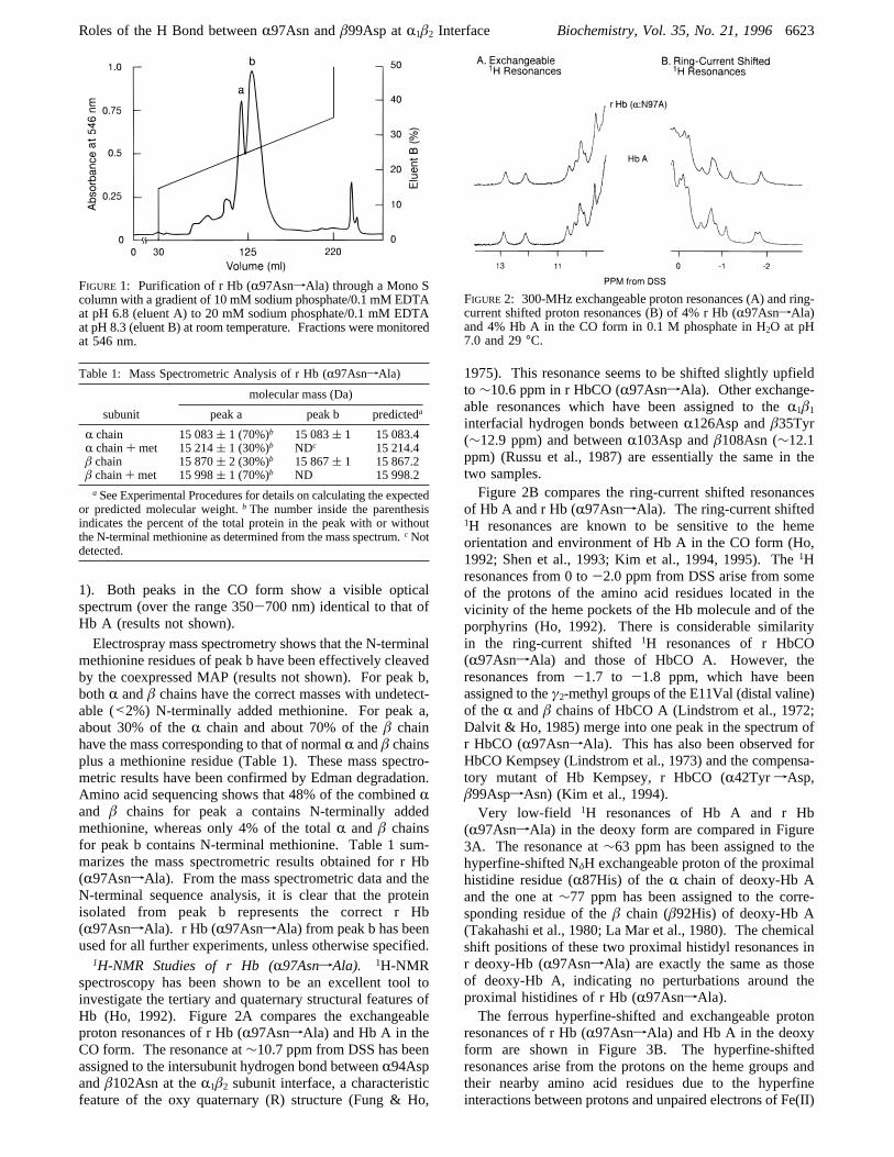
1H-NMR Studies of r Hb (R97AsnfAla). 1H-NMRspectroscopy has been shown to be an excellent tool toinvestigate the tertiary and quaternary structural features ofHb (Ho, 1992). Figure 2A compares the exchangeableproton resonances of r Hb (R97AsnfAla) and Hb A in theCO form. The resonance at∼10.7 ppm from DSS has beenassigned to the intersubunit hydrogen bond betweenR94Aspandâ102Asn at theR1â2 subunit interface, a characteristicfeature of the oxy quaternary (R) structure (Fung & Ho,
1975). This resonance seems to be shifted slightly upfieldto∼10.6 ppm in r HbCO (R97AsnfAla). Other exchange-able resonances which have been assigned to theR1â1
interfacial hydrogen bonds betweenR126Asp andâ35Tyr(∼12.9 ppm) and betweenR103Asp andâ108Asn (∼12.1ppm) (Russu et al., 1987) are essentially the same in thetwo samples.Figure 2B compares the ring-current shifted resonances
of Hb A and r Hb (R97AsnfAla). The ring-current shifted1H resonances are known to be sensitive to the hemeorientation and environment of Hb A in the CO form (Ho,1992; Shen et al., 1993; Kim et al., 1994, 1995). The1Hresonances from 0 to-2.0 ppm from DSS arise from someof the protons of the amino acid residues located in thevicinity of the heme pockets of the Hb molecule and of theporphyrins (Ho, 1992). There is considerable similarityin the ring-current shifted1H resonances of r HbCO(R97AsnfAla) and those of HbCO A. However, theresonances from-1.7 to -1.8 ppm, which have beenassigned to theγ2-methyl groups of the E11Val (distal valine)of theR andâ chains of HbCO A (Lindstrom et al., 1972;Dalvit & Ho, 1985) merge into one peak in the spectrum ofr HbCO (R97AsnfAla). This has also been observed forHbCO Kempsey (Lindstrom et al., 1973) and the compensa-tory mutant of Hb Kempsey, r HbCO (R42TyrfAsp,â99AspfAsn) (Kim et al., 1994).Very low-field 1H resonances of Hb A and r Hb
(R97AsnfAla) in the deoxy form are compared in Figure3A. The resonance at∼63 ppm has been assigned to thehyperfine-shifted NδH exchangeable proton of the proximalhistidine residue (R87His) of theR chain of deoxy-Hb Aand the one at∼77 ppm has been assigned to the corre-sponding residue of theâ chain (â92His) of deoxy-Hb A(Takahashi et al., 1980; La Mar et al., 1980). The chemicalshift positions of these two proximal histidyl resonances inr deoxy-Hb (R97AsnfAla) are exactly the same as thoseof deoxy-Hb A, indicating no perturbations around theproximal histidines of r Hb (R97AsnfAla).The ferrous hyperfine-shifted and exchangeable proton
resonances of r Hb (R97AsnfAla) and Hb A in the deoxyform are shown in Figure 3B. The hyperfine-shiftedresonances arise from the protons on the heme groups andtheir nearby amino acid residues due to the hyperfineinteractions between protons and unpaired electrons of Fe(II)
FIGURE 1: Purification of r Hb (R97AsnfAla) through a Mono Scolumn with a gradient of 10 mM sodium phosphate/0.1 mM EDTAat pH 6.8 (eluent A) to 20 mM sodium phosphate/0.1 mM EDTAat pH 8.3 (eluent B) at room temperature. Fractions were monitoredat 546 nm.
Table 1: Mass Spectrometric Analysis of r Hb (R97AsnfAla)
molecular mass (Da)
subunit peak a peak b predicteda
R chain 15 083( 1 (70%)b 15 083( 1 15 083.4R chain+ met 15 214( 1 (30%)b NDc 15 214.4â chain 15 870( 2 (30%)b 15 867( 1 15 867.2â chain+ met 15 998( 1 (70%)b ND 15 998.2aSee Experimental Procedures for details on calculating the expected
or predicted molecular weight.b The number inside the parenthesisindicates the percent of the total protein in the peak with or withoutthe N-terminal methionine as determined from the mass spectrum.cNotdetected.
FIGURE2: 300-MHz exchangeable proton resonances (A) and ring-current shifted proton resonances (B) of 4% r Hb (R97AsnfAla)and 4% Hb A in the CO form in 0.1 M phosphate in H2O at pH7.0 and 29°C.
Roles of the H Bond betweenR97Asn andâ99Asp atR1â2 Interface Biochemistry, Vol. 35, No. 21, 19966623
+ +
+ +
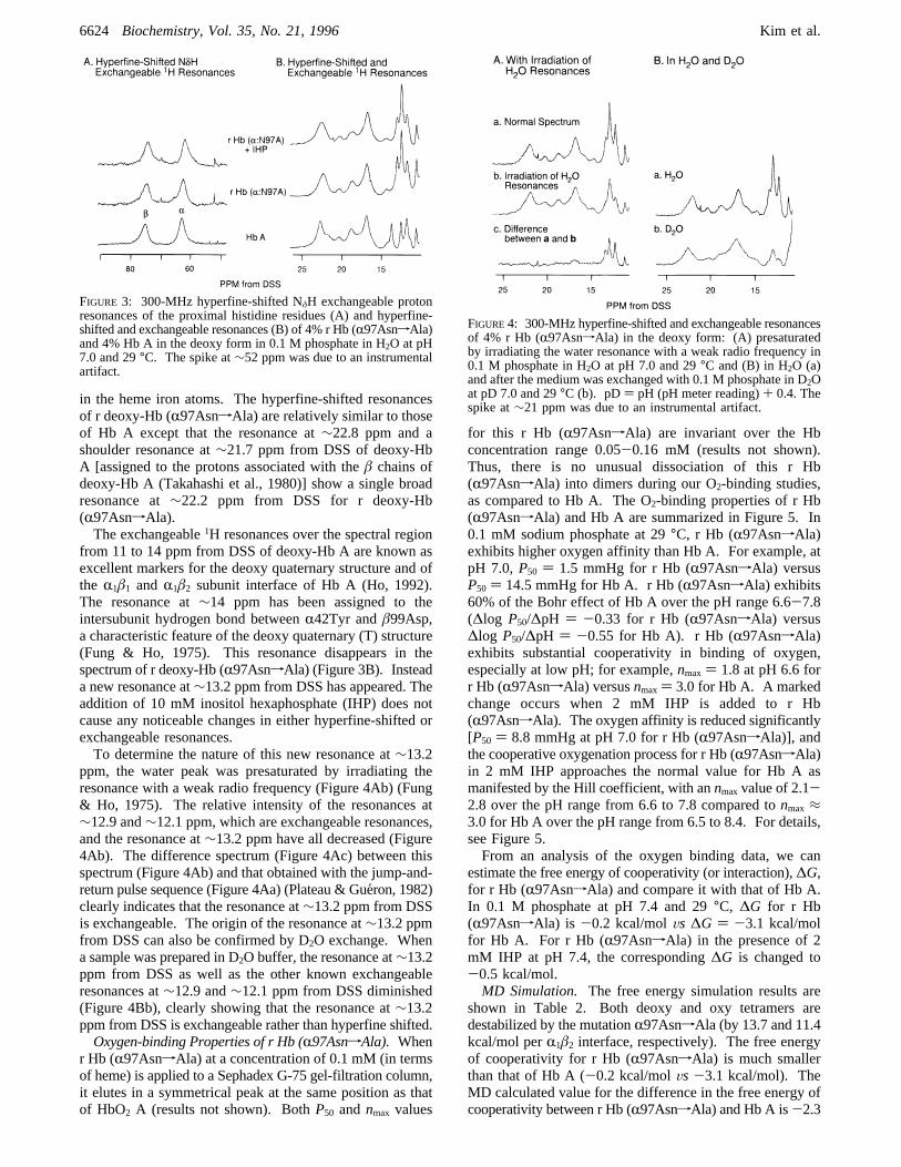
in the heme iron atoms. The hyperfine-shifted resonancesof r deoxy-Hb (R97AsnfAla) are relatively similar to thoseof Hb A except that the resonance at∼22.8 ppm and ashoulder resonance at∼21.7 ppm from DSS of deoxy-HbA [assigned to the protons associated with theâ chains ofdeoxy-Hb A (Takahashi et al., 1980)] show a single broadresonance at∼22.2 ppm from DSS for r deoxy-Hb(R97AsnfAla).The exchangeable1H resonances over the spectral region
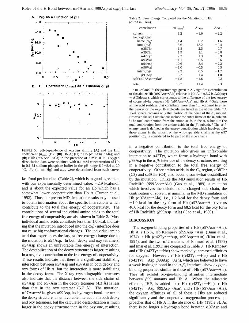
from 11 to 14 ppm from DSS of deoxy-Hb A are known asexcellent markers for the deoxy quaternary structure and ofthe R1â1 andR1â2 subunit interface of Hb A (Ho, 1992).The resonance at∼14 ppm has been assigned to theintersubunit hydrogen bond betweenR42Tyr andâ99Asp,a characteristic feature of the deoxy quaternary (T) structure(Fung & Ho, 1975). This resonance disappears in thespectrum of r deoxy-Hb (R97AsnfAla) (Figure 3B). Insteada new resonance at∼13.2 ppm from DSS has appeared. Theaddition of 10 mM inositol hexaphosphate (IHP) does notcause any noticeable changes in either hyperfine-shifted orexchangeable resonances.To determine the nature of this new resonance at∼13.2
ppm, the water peak was presaturated by irradiating theresonance with a weak radio frequency (Figure 4Ab) (Fung& Ho, 1975). The relative intensity of the resonances at∼12.9 and∼12.1 ppm, which are exchangeable resonances,and the resonance at∼13.2 ppm have all decreased (Figure4Ab). The difference spectrum (Figure 4Ac) between thisspectrum (Figure 4Ab) and that obtained with the jump-and-return pulse sequence (Figure 4Aa) (Plateau & Gue´ron, 1982)clearly indicates that the resonance at∼13.2 ppm from DSSis exchangeable. The origin of the resonance at∼13.2 ppmfrom DSS can also be confirmed by D2O exchange. Whena sample was prepared in D2O buffer, the resonance at∼13.2ppm from DSS as well as the other known exchangeableresonances at∼12.9 and∼12.1 ppm from DSS diminished(Figure 4Bb), clearly showing that the resonance at∼13.2ppm from DSS is exchangeable rather than hyperfine shifted.Oxygen-binding Properties of r Hb (R97AsnfAla). When
r Hb (R97AsnfAla) at a concentration of 0.1 mM (in termsof heme) is applied to a Sephadex G-75 gel-filtration column,it elutes in a symmetrical peak at the same position as thatof HbO2 A (results not shown). BothP50 andnmax values
for this r Hb (R97AsnfAla) are invariant over the Hbconcentration range 0.05-0.16 mM (results not shown).Thus, there is no unusual dissociation of this r Hb(R97AsnfAla) into dimers during our O2-binding studies,as compared to Hb A. The O2-binding properties of r Hb(R97AsnfAla) and Hb A are summarized in Figure 5. In0.1 mM sodium phosphate at 29°C, r Hb (R97AsnfAla)exhibits higher oxygen affinity than Hb A. For example, atpH 7.0,P50 ) 1.5 mmHg for r Hb (R97AsnfAla) versusP50 ) 14.5 mmHg for Hb A. r Hb (R97AsnfAla) exhibits60% of the Bohr effect of Hb A over the pH range 6.6-7.8(∆log P50/∆pH ) -0.33 for r Hb (R97AsnfAla) versus∆log P50/∆pH ) -0.55 for Hb A). r Hb (R97AsnfAla)exhibits substantial cooperativity in binding of oxygen,especially at low pH; for example,nmax ) 1.8 at pH 6.6 forr Hb (R97AsnfAla) versusnmax) 3.0 for Hb A. A markedchange occurs when 2 mM IHP is added to r Hb(R97AsnfAla). The oxygen affinity is reduced significantly[P50 ) 8.8 mmHg at pH 7.0 for r Hb (R97AsnfAla)], andthe cooperative oxygenation process for r Hb (R97AsnfAla)in 2 mM IHP approaches the normal value for Hb A asmanifested by the Hill coefficient, with annmaxvalue of 2.1-2.8 over the pH range from 6.6 to 7.8 compared tonmax≈3.0 for Hb A over the pH range from 6.5 to 8.4. For details,see Figure 5.From an analysis of the oxygen binding data, we can
estimate the free energy of cooperativity (or interaction),∆G,for r Hb (R97AsnfAla) and compare it with that of Hb A.In 0.1 M phosphate at pH 7.4 and 29°C, ∆G for r Hb(R97AsnfAla) is -0.2 kcal/molVs ∆G ) -3.1 kcal/molfor Hb A. For r Hb (R97AsnfAla) in the presence of 2mM IHP at pH 7.4, the corresponding∆G is changed to-0.5 kcal/mol.MD Simulation. The free energy simulation results are
shown in Table 2. Both deoxy and oxy tetramers aredestabilized by the mutationR97AsnfAla (by 13.7 and 11.4kcal/mol perR1â2 interface, respectively). The free energyof cooperativity for r Hb (R97AsnfAla) is much smallerthan that of Hb A (-0.2 kcal/molVs -3.1 kcal/mol). TheMD calculated value for the difference in the free energy ofcooperativity between r Hb (R97AsnfAla) and Hb A is-2.3
FIGURE 3: 300-MHz hyperfine-shifted NδH exchangeable protonresonances of the proximal histidine residues (A) and hyperfine-shifted and exchangeable resonances (B) of 4% r Hb (R97AsnfAla)and 4% Hb A in the deoxy form in 0.1 M phosphate in H2O at pH7.0 and 29°C. The spike at∼52 ppm was due to an instrumentalartifact.
FIGURE4: 300-MHz hyperfine-shifted and exchangeable resonancesof 4% r Hb (R97AsnfAla) in the deoxy form: (A) presaturatedby irradiating the water resonance with a weak radio frequency in0.1 M phosphate in H2O at pH 7.0 and 29°C and (B) in H2O (a)and after the medium was exchanged with 0.1 M phosphate in D2Oat pD 7.0 and 29°C (b). pD) pH (pH meter reading)+ 0.4. Thespike at∼21 ppm was due to an instrumental artifact.
6624 Biochemistry, Vol. 35, No. 21, 1996 Kim et al.
+ +
+ +
kcal/mol per interface (Table 2), which is in good agreementwith our experimentally determined value,-2.9 kcal/mol,and is about the expected value for an Hb which has asomewhat lower cooperativity than Hb A (Turner et al.,1992). Thus, our present MD simulation results may be usedto obtain information about the specific interactions whichcontribute to the total free energy of cooperativity. Thecontributions of several individual amino acids to the totalfree energy of cooperativity are also shown in Table 2. Mostindividual amino acids contribute less than 1.0 kcal, indicat-ing that the mutation introduced into theR1â2 interface doesnot cause big conformational changes. The individual aminoacid that experiences the largest free energy change due tothe mutation isR94Asp. In both deoxy and oxy tetramers,R94Asp shows an unfavorable free energy of interaction.The destabilization of the deoxy structure is larger, resultingin a negative contribution to the free energy of cooperativity.These results indicate that there is a significant stabilizinginteraction betweenR94Asp andR97Asn in both deoxy andoxy forms of Hb A, but the interaction is more stabilizingin the deoxy form. The X-ray crystallographic structuresalso indicate that the distance between the side chains ofR94Asp andR97Asn in the deoxy tetramer (4.3 Å) is lessthan that in the oxy tetramer (5.7 Å). The mutation,R97AsnfAla, givesâ99Asp, its hydrogen bond partner inthe deoxy structure, an unfavorable interaction in both deoxyand oxy tetramers, but the calculated destabilization is muchlarger in the deoxy structure than in the oxy one, resulting
in a negative contribution to the total free energy ofcooperativity. The mutation also gives an unfavorableinteraction toR42Tyr, which forms a hydrogen bond withâ99Asp in theR1â2 interface of the deoxy structure, resultingin a negative contribution to the total free energy ofcooperativity. Other amino acids in the CR region,R38Thr(C3) andR39Thr (C4) also become somewhat destabilizedby the mutation. Unlike the MD simulation results of HbRadcliffe (â99AspfAla) (Gao et al., 1989), a mutationwhich involves the deletion of a charged side chain, thecontribution of solvent is minimal in the MD simulation ofHb (R97AsnfAla), i.e., 1.2 kcal for the deoxy form and-1.0 kcal for the oxy form of Hb (R97AsnfAla) versus46.0 kcal for the deoxy form and 68.5 kcal for the oxy formof Hb Radcliffe (â99AspfAla) (Gao et al., 1989).
DISCUSSION
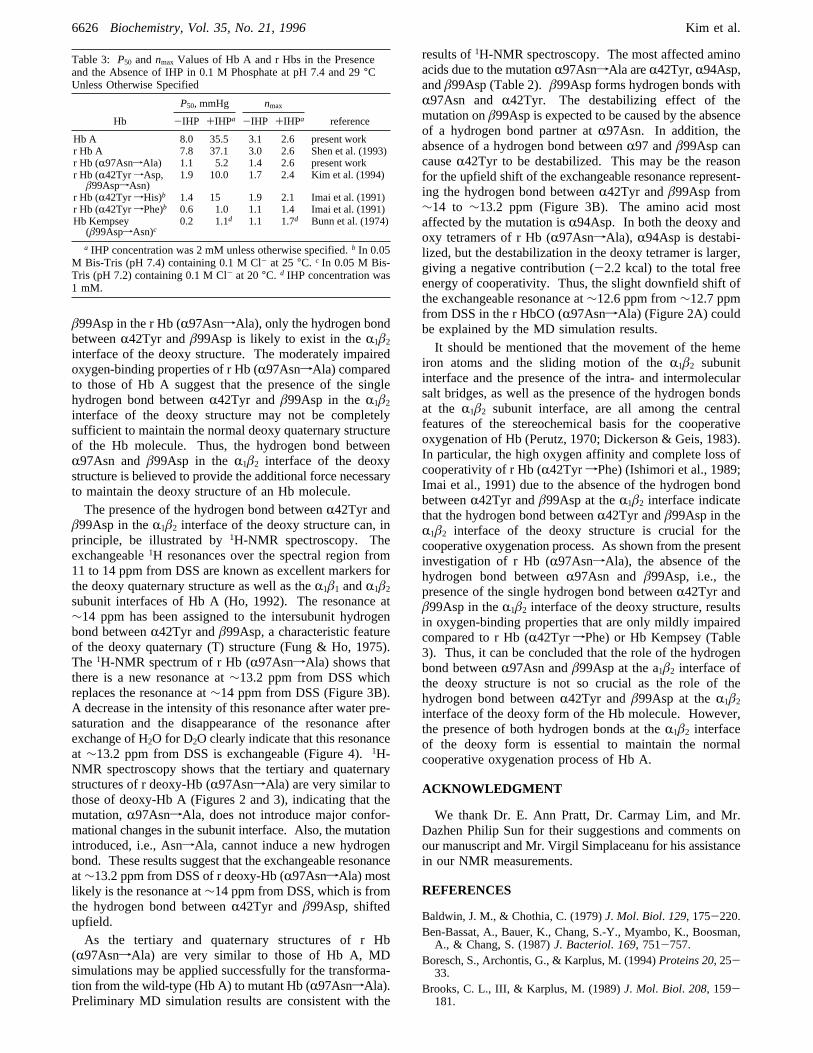
The oxygen-binding properties of r Hb (R97AsnfAla),Hb A, r Hb A, Hb Kempsey (â99AspfAsn) (Bunn et al.,1974), r Hb (R42TyrfAsp, â99AspfAsn) (Kim et al.,1994), and the twoR42 mutants of Ishimori et al. (1989)and Imai et al. (1991) are compared in Table 3. Hb Kempseyand r Hb (R42TyrfPhe) show markedly increased affinitiesfor oxygen. However, r Hb (R42TyrfHis) and r Hb(R42TyrfAsp,â99AspfAsn), which are believed to havea weak hydrogen bond in theR1â2 interface, show oxygen-binding properties similar to those of r Hb (R97AsnfAla).They all exhibit oxygen-binding affinities intermediatebetweenâ99 mutants and Hb A. When the allostericeffector, IHP, is added to r Hb (R42TyrfHis), r Hb(R42TyrfAsp, â99AspfAsn), and r Hb (R97AsnfAla),the oxygen affinities of all of these r Hbs are reducedsignificantly and the cooperative oxygenation process ap-proaches that of Hb A in the absence of IHP (Table 3). Asthere is no longer a hydrogen bond betweenR97Asn and
FIGURE 5: pH-dependence of oxygen affinity (A) and the Hillcoefficient (nmax) (B): (9) Hb A; (O) r Hb (R97AsnfAla); and(b) r Hb (R97AsnfAla) in the presence of 2 mM IHP. Oxygendissociation data were obtained with 0.1 mM concentration of Hbin 0.1 M sodium phosphate buffer in the pH range 6.2-8.4 at 29°C. P50 (in mmHg) andnmax were determined from each curve.
Table 2: Free Energy Computed for the Mutation of r Hb(R97AsnfAla)a
contribution ∆Gdeoxyb ∆Goxy ∆∆Gc
solvent 1.2 -1.0 -2.2hemoglobind
heme (R1)e -1.4 0.2 -1.6intra (R1)f 13.6 13.2 -0.4
R38Thr 1.8 2.5 0.7R39Thr 1.9 1.1 -0.8R42Tyr 2.2 1.3 -0.9R93Val -1.1 -0.5 0.6R94Asp 10.6 8.4 -2.2R96Val -1.0 -0.5 0.5
inter (â2)g 2.2 0.5 -1.7â99Asp 3.2 1.4 -1.8
self (R97AsnfAla)h -1.8 -1.6 0.2
total 13.7 11.4 -2.3a In kcal/mol. b The positive sign given in∆G signifies a contribution
to destabilize Hb (R97AsnfAla) relative to Hb A.c ∆∆G is ∆G(oxy)- ∆G(deoxy), which corresponds to the difference of the free energyof cooperativity between Hb (R97AsnfAla) and Hb A.dOnly thoseamino acid residues that contribute more than 1.0 kcal/mol in eitherthe deoxy- or the oxy-Hb molecule are listed in the above table.eA15-Å sphere contains only that portion of the heme of theR1 subunit.However, the MD simulations include the entire heme of theR1 subunit.f The total contribution from the amino acids in theR1 subunit.g Thetotal contribution from the amino acids in theâ2 subunit.h The self-energy term is defined as the energy contribution which involves onlythose atoms in the mutant or the wild-type side chains at theR97position (CR is considered to be part of the side chain).
Roles of the H Bond betweenR97Asn andâ99Asp atR1â2 Interface Biochemistry, Vol. 35, No. 21, 19966625
+ +
+ +
â99Asp in the r Hb (R97AsnfAla), only the hydrogen bondbetweenR42Tyr andâ99Asp is likely to exist in theR1â2
interface of the deoxy structure. The moderately impairedoxygen-binding properties of r Hb (R97AsnfAla) comparedto those of Hb A suggest that the presence of the singlehydrogen bond betweenR42Tyr andâ99Asp in theR1â2
interface of the deoxy structure may not be completelysufficient to maintain the normal deoxy quaternary structureof the Hb molecule. Thus, the hydrogen bond betweenR97Asn andâ99Asp in theR1â2 interface of the deoxystructure is believed to provide the additional force necessaryto maintain the deoxy structure of an Hb molecule.The presence of the hydrogen bond betweenR42Tyr and
â99Asp in theR1â2 interface of the deoxy structure can, inprinciple, be illustrated by1H-NMR spectroscopy. Theexchangeable1H resonances over the spectral region from11 to 14 ppm from DSS are known as excellent markers forthe deoxy quaternary structure as well as theR1â1 andR1â2
subunit interfaces of Hb A (Ho, 1992). The resonance at∼14 ppm has been assigned to the intersubunit hydrogenbond betweenR42Tyr andâ99Asp, a characteristic featureof the deoxy quaternary (T) structure (Fung & Ho, 1975).The 1H-NMR spectrum of r Hb (R97AsnfAla) shows thatthere is a new resonance at∼13.2 ppm from DSS whichreplaces the resonance at∼14 ppm from DSS (Figure 3B).A decrease in the intensity of this resonance after water pre-saturation and the disappearance of the resonance afterexchange of H2O for D2O clearly indicate that this resonanceat ∼13.2 ppm from DSS is exchangeable (Figure 4).1H-NMR spectroscopy shows that the tertiary and quaternarystructures of r deoxy-Hb (R97AsnfAla) are very similar tothose of deoxy-Hb A (Figures 2 and 3), indicating that themutation,R97AsnfAla, does not introduce major confor-mational changes in the subunit interface. Also, the mutationintroduced, i.e., AsnfAla, cannot induce a new hydrogenbond. These results suggest that the exchangeable resonanceat∼13.2 ppm from DSS of r deoxy-Hb (R97AsnfAla) mostlikely is the resonance at∼14 ppm from DSS, which is fromthe hydrogen bond betweenR42Tyr andâ99Asp, shiftedupfield.As the tertiary and quaternary structures of r Hb
(R97AsnfAla) are very similar to those of Hb A, MDsimulations may be applied successfully for the transforma-tion from the wild-type (Hb A) to mutant Hb (R97AsnfAla).Preliminary MD simulation results are consistent with the
results of1H-NMR spectroscopy. The most affected aminoacids due to the mutationR97AsnfAla areR42Tyr,R94Asp,andâ99Asp (Table 2).â99Asp forms hydrogen bonds withR97Asn and R42Tyr. The destabilizing effect of themutation onâ99Asp is expected to be caused by the absenceof a hydrogen bond partner atR97Asn. In addition, theabsence of a hydrogen bond betweenR97 andâ99Asp cancauseR42Tyr to be destabilized. This may be the reasonfor the upfield shift of the exchangeable resonance represent-ing the hydrogen bond betweenR42Tyr andâ99Asp from∼14 to ∼13.2 ppm (Figure 3B). The amino acid mostaffected by the mutation isR94Asp. In both the deoxy andoxy tetramers of r Hb (R97AsnfAla), R94Asp is destabi-lized, but the destabilization in the deoxy tetramer is larger,giving a negative contribution (-2.2 kcal) to the total freeenergy of cooperativity. Thus, the slight downfield shift ofthe exchangeable resonance at∼12.6 ppm from∼12.7 ppmfrom DSS in the r HbCO (R97AsnfAla) (Figure 2A) couldbe explained by the MD simulation results.It should be mentioned that the movement of the heme
iron atoms and the sliding motion of theR1â2 subunitinterface and the presence of the intra- and intermolecularsalt bridges, as well as the presence of the hydrogen bondsat the R1â2 subunit interface, are all among the centralfeatures of the stereochemical basis for the cooperativeoxygenation of Hb (Perutz, 1970; Dickerson & Geis, 1983).In particular, the high oxygen affinity and complete loss ofcooperativity of r Hb (R42TyrfPhe) (Ishimori et al., 1989;Imai et al., 1991) due to the absence of the hydrogen bondbetweenR42Tyr andâ99Asp at theR1â2 interface indicatethat the hydrogen bond betweenR42Tyr andâ99Asp in theR1â2 interface of the deoxy structure is crucial for thecooperative oxygenation process. As shown from the presentinvestigation of r Hb (R97AsnfAla), the absence of thehydrogen bond betweenR97Asn andâ99Asp, i.e., thepresence of the single hydrogen bond betweenR42Tyr andâ99Asp in theR1â2 interface of the deoxy structure, resultsin oxygen-binding properties that are only mildly impairedcompared to r Hb (R42TyrfPhe) or Hb Kempsey (Table3). Thus, it can be concluded that the role of the hydrogenbond betweenR97Asn andâ99Asp at the a1â2 interface ofthe deoxy structure is not so crucial as the role of thehydrogen bond betweenR42Tyr andâ99Asp at theR1â2
interface of the deoxy form of the Hb molecule. However,the presence of both hydrogen bonds at theR1â2 interfaceof the deoxy form is essential to maintain the normalcooperative oxygenation process of Hb A.
ACKNOWLEDGMENT
We thank Dr. E. Ann Pratt, Dr. Carmay Lim, and Mr.Dazhen Philip Sun for their suggestions and comments onour manuscript and Mr. Virgil Simplaceanu for his assistancein our NMR measurements.
REFERENCES
Baldwin, J. M., & Chothia, C. (1979)J.Mol. Biol. 129, 175-220.Ben-Bassat, A., Bauer, K., Chang, S.-Y., Myambo, K., Boosman,A., & Chang, S. (1987)J. Bacteriol. 169, 751-757.
Boresch, S., Archontis, G., & Karplus, M. (1994)Proteins 20, 25-33.
Brooks, C. L., III, & Karplus, M. (1989)J. Mol. Biol. 208, 159-181.
Table 3: P50 andnmax Values of Hb A and r Hbs in the Presenceand the Absence of IHP in 0.1 M Phosphate at pH 7.4 and 29°CUnless Otherwise Specified
P50, mmHg nmax
Hb -IHP +IHPa -IHP +IHPa reference
Hb A 8.0 35.5 3.1 2.6 present workr Hb A 7.8 37.1 3.0 2.6 Shen et al. (1993)r Hb (R97AsnfAla) 1.1 5.2 1.4 2.6 present workr Hb (R42TyrfAsp,
â99AspfAsn)1.9 10.0 1.7 2.4 Kim et al. (1994)
r Hb (R42TyrfHis)b 1.4 15 1.9 2.1 Imai et al. (1991)r Hb (R42TyrfPhe)b 0.6 1.0 1.1 1.4 Imai et al. (1991)Hb Kempsey
(â99AspfAsn)c0.2 1.1d 1.1 1.7d Bunn et al. (1974)
a IHP concentration was 2 mM unless otherwise specified.b In 0.05M Bis-Tris (pH 7.4) containing 0.1 M Cl- at 25°C. c In 0.05 M Bis-Tris (pH 7.2) containing 0.1 M Cl- at 20°C. d IHP concentration was1 mM.
6626 Biochemistry, Vol. 35, No. 21, 1996 Kim et al.
+ +
+ +
Bunn, H. F., Wohl, R. C., Bradley, T. B., Cooley, M., & Gibson,Q. H. (1974)J. Biol. Chem. 249, 7402-7409.
Bunn, H. F., & Forget, B. G. (1986) inHemoglobin: Molecular,Genetic, and Clinical Aspects, W. B. Saunders, Philadelphia, PA.
Dalvit, C., & Ho, C. (1985)Biochemistry 24, 3398-3407.Dickerson, R. E., & Geis, I. (1983)Hemoglobin: Structure,Function, EVolution, and Pathology, The Benjamin CummingsPublishing Co., Menlo Park, CA.
Dysert, P. A., Head, C. G., Shih, T. B., Jones, R. T., & Schneider,R. G. (1982)Blood 60(Suppl. 1), 53A.
Fermi, G., Perutz, M. F., Shaanan, B., & Fourme, R. (1984)J.Mol.Biol. 175, 159-174.
Ferrige, A. G., Seddon, M. J., Green, B. N., Jarvis, S. A., & Skilling,J. (1992)Rapid Commun. Mass Spectrosc. 6, 707-711.
Fung, L. W.-M., & Ho, C. (1975)Biochemistry 14, 2526-2535.Gao, J., Kuczera, K., Tidor, B., & Karplus, M. (1989)Science 244,1069-1072.
Glynn, K. P., Penner, J. A., Smith, J. R., & Rucknagel, D. L. (1968)Ann. Intern. Med. 69, 769-776.
Hayashi, A., Suzuki, T., & Shin, M. (1973)Biochim. Biophys. Acta310, 309-316.
Hermans, J., Yun, R. H., & Anderson, A. G. (1992)J. Comput.Chem. 13, 429-442.
Ho, C. (1992)AdV. Protein Chem. 43, 153-312.Hoffman, S. J., Looker, D. L., Roehrich, J. M., Cozart, P. E., Durfee,S. L., Tedesco, J. L., & Stetler, G. L. (1990)Proc. Natl. Acad.Sci. U.S.A. 87, 8521-8525.
Imai, K., Fushitani, K., Miyazaki, G., Ishimori, K., Kitagawa, T.,Wada, Y., Morimoto, H., Morishima, I., Shih, D. T.-B., & Tame,J. (1991)J. Mol. Biol. 218, 769-778.
Ishimori, K., Morishima, I., Imai, K., Fushitani, K., Miyazaki, G.,Shih, D., Tame, J., Pagnier, J., & Nagai, K. (1989)J. Biol.Chem.264, 14624-14626.
Jones, R. T., Osgood, E. E., Brimhall, B., & Koler, R. D. (1967)J.Clin. InVest. 46, 1840-1847.
Jorgensen, W. L., Chandrasekar, J., Madura, J. D., Impey, R. W.,& Klein, M. L. (1983) J. Chem. Phys. 79, 926-935.
Kim, H.-W., Shen, T.-J., Sun, D. P., Ho, N. T., Madrid, M., Tam,M. F., Zou, M., Cottam, P. F., & Ho, C. (1994)Proc.Natl. Acad.Sci. U.S.A. 91, 11547-11551.
Kim, H.-W., Shen, T.-J., Sun, D. P., Ho, N. T., Madrid, M., & Ho,C. (1995)J. Mol. Biol. 248, 867-882.
Kirkwood, J. G. (1935)J. Chem. Phys. 3, 300-313.Kunkel, T. M. (1985)Proc. Natl. Acad. Sci. U.S.A. 82, 488-492.La Mar, G. N., Nagai, K., Jue, T., Budd, D., Gersonde, K., Sick,
H., Kagimoto, T., Hayashi, A., & Taketa, F. (1980)Biochem.Biophys. Res. Commun. 96, 1172-1177.
Lindstrom, T. R., Nore´n, I. B. E., Charache, S., Lehmann, H., &Ho, C. (1972)Biochemistry 11, 1677-1681.
Lindstrom, T. R., Baldassare, J. J., Bunn, H. F., & Ho, C. (1973)Biochemistry 12, 4212-4217.
Looker, D. L., Mathews, A. J., Neway, J. O., & Stetler, G. L. (1994)Methods Enzymol. 231, 364-374.
Miller, J. H. (1972)Experiments in Molecular Genetics, Cold SpringHarbor Laboratory Press, Plainview, NY.
Mitchell, M. J., & McCammon, J. A. (1991)J. Comput. Chem. 12,271-275.
Perutz, M. F. (1970)Nature (London) 228, 726-739.Plateau, P., & Gue´ron, M. (1982)J. Am. Chem. Soc. 104, 7310-7311.
Reed, C. S., Hampson, R., Gordon, S., Jones, R. T., Novy, M. J.,Brimhall, B., Edwards, M. J., & Koler, R. D. (1968)Blood 31,623-632.
Russu, I. M., Ho, N. T., & Ho, C. (1987)Biochim. Biophys. Acta914, 40-48.
Ryckaert, J.-P., Ciccotti, G., & Berendsen, H. J. C. (1977)J.Comput. Phys. 23, 327-341.
Shaanan, B. (1983)J. Mol. Biol. 171, 31-59.Shen, T.-J., Ho, N. T., Simplaceanu, V., Zou, M., Green, B. N.,Tam, M. F., & Ho, C. (1993)Proc. Natl. Acad. Sci. U.S.A. 90,8108-8112.
Shi, Y.-y., Mark, A. E., Wang, C.-x., Huang, F., Berendsen, H. J.C., & van Gunsteren, W. F. (1993)Protein 6, 289-295.
Takahashi, S., Lin, A. K.-L. C., & Ho, C. (1980)Biochemistry 19,5196-5202.
Thillet, J., Arous, N., & Rosa, J. B. (1981)Biochim. Biophys. Acta670, 260-264.
Tidor, B., & Karplus, M. (1991)Biochemistry 30, 3217-3228.Turner, G. J., Galacteros, F., Doyle, M. L., Hedlund, B., Pettigrew,D. W., Turner, B. W., Smith, F. R., Moo-Penn, W., Rucknagel,D. L., & Ackers, G. K. (1992)Proteins 14, 333-350.
van Gunsteren, W. F., & Mark, A. E. (1992)Eur. J. Biochem. 204,947-961.
Weatherall, D. J., Clegg, J. B., Callender, S. T., Wells, R. M. G.,Gale, R. E., Huehns, E. R., Perutz, M. F., Viggiano, G., & Ho,C. (1977)Br. J. Haematol. 35, 177-191.
Zaia, J., Annan, R. S., & Biemann, K. (1992)Rapid Commun.MassSpectrosc. 6, 32-36.
BI952518Z
Roles of the H Bond betweenR97Asn andâ99Asp atR1â2 Interface Biochemistry, Vol. 35, No. 21, 19966627
+ +
+ +