Embed Size (px)
Citation preview

Chapter 18
Converting Genetic information in molecules of lifen Replication – producing new DNA molecule
ü Necessary when cell divides during mitosisn Transcription – production of RNAs
ü rRNA-protein synthesis; mRNA-recipe; tRNA-gets amino acidn Translation – conversion of genetic information into
proteinsn Central Dogma
ü DNAàRNAàproteins
Genetic Information
Overview

§Successful information-based system involves conservation and transfer
§DNA - stable structure that maximizes storage and duplication§RNA - more reactive with numerous roles in protein synthesis and
gene expression regulation§Decoding DNA requires DNA-protein interactions
§Major and minor grooves facilitate sequence-specific binding§Contact between amino acid residues and edges of bases
§DNA-binding proteins - most possess twofold axis of symmetry
1. Helix-turn-helix 3. Leucine zipper2. Helix-loop-helix 4. Zinc finger
§Accurate DNA synthesis; effective DNA repair mechanisms§Variation may also be important for adaptability to environments
§Caused by genetic recombination and mutation
From McKee and McKee, Biochemistry, 5th Edition, © 2011 Oxford University Press
Chapter 18: Overview

Semiconservative replication §Separation of 2 original strands §Each serves as a template for synthesis of
a complementary strand (daughter)§1 original strand/1 new strand
§Meselson and Stahl established using radio-labeled 15NH4Cl
§Replication factories – specific nuclear or nucleoid compartments where replication takes place
§DNA Synthesis in Prokaryotes§Helicases - unwind DNA§Primer synthesis – required for initiation§DNA polynucleotide synthesis – synthesis
of complementary strandFigure 18.2 Semiconservative DNA Replication
Section 18.1: Genetic Information: Replication
From McKee and McKee, Biochemistry, 5th Edition, © 2011 Oxford University Press

n Replication¨ Separation of the two original strands
¨ Synthesis of two new daughter strands using the original strands as templates
n Semiconservative replication:each daughter strand contains one template strand and one newly synthesized strand¨ Incorporation of isotopic label as sole
nitrogen source (15NH4Cl)
¨ Observed that 15N-DNA has a higher density than 14N-DNA, and the two can be separated by density-gradient ultracentrifugation

Section 18.1: Genetic Information: Replication
Origin of replication – point where replication will begin
Replication forks– Y shaped region of an unwinding DNA double helix undergoing replication
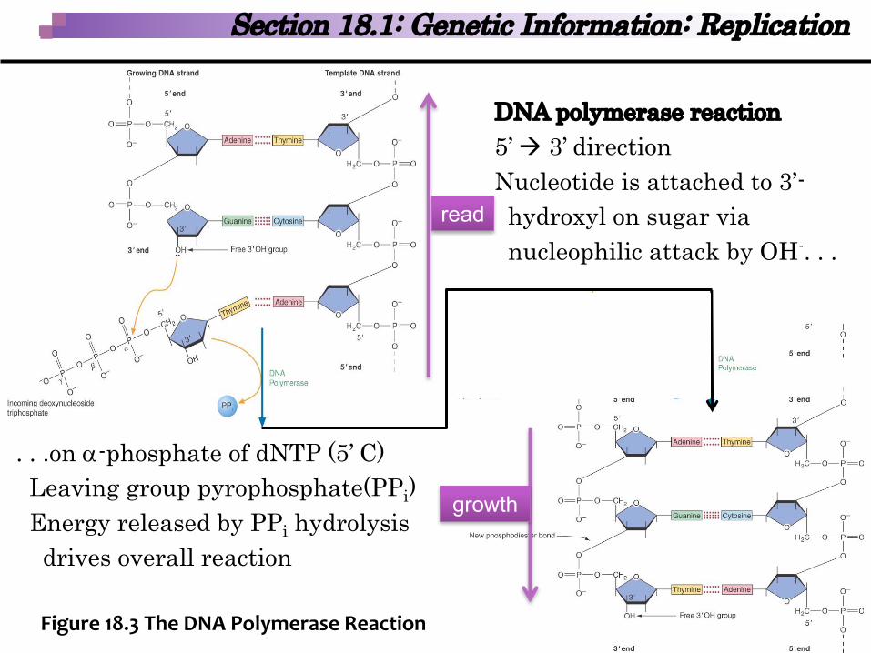
DNA polymerase reaction 5’ à 3’ directionNucleotide is attached to 3’-hydroxyl on sugar vianucleophilic attack by OH-. . .
Figure 18.3 The DNA Polymerase Reaction
Section 18.1: Genetic Information: Replication
read
growth
. . .on a-phosphate of dNTP (5’ C)Leaving group pyrophosphate(PPi)Energy released by PPi hydrolysisdrives overall reaction

§DNA polymerase III (pol III)§Catalyzes the nucleophilic attack of
the 3′-hydroxyl group onto the a-phosphate
§Pol III holoenzyme - at least 10 subunits
§Core polymerase is formed of three subunits: a, e, and t
§b-protein (sliding clamp) is two subunits and forms a donut-shaped ring around the template DNA
§g-complex (clamp loader) transfers sliding clamp
From McKee and McKee, Biochemistry, 5th Edition, © 2011 Oxford University Press
Section 18.1: Genetic Information: Replication
Figure 18.5 Cross Section of the b2-Clamp of DNA Polymerase III

§Replisome - two pol III holoenzymes, primosome andDNA unwinding proteins
§DNA polymerase I - 3 catalytic activities§5’ à 3’ exonuclease activity§5’ à 3’ template directed polymerase activity§3’ à 5’ exonulease activity
§DNA polymerase II, IV, V - DNA repair enzymes known as translesion repair enzymes§Part of the SOS response - prevent cell death
§DNA ligase catalyzes the formation of the phosphodiesterbond between adjoining nucleotides
§DNA topoisomerases – control supercoiling, relieve torque
From McKee and McKee, Biochemistry, 5th Edition, © 2011 Oxford University Press
Section 18.1: Genetic Information: Replication

§Type I topoisomerases produce transient single-strand breaks
§Type II topoisomerases produce transient double-strand breaks
§DNA gyrase—in prokaryotes helps separate replication products & create negative (-) supercoils
§E. coli – high ATP/ADP ratio; DnaA, begins at origin of replication, oriC
§Bidirectional from oriC: 5’ -> 3’ & 3’ -> 5’§Each replication fork:
§helicases – unwind double helix§Replisome – molecular machine that
carries out DNA replication§Replicon - One origin of replication unit
and regulatory sequencesFigure 18.6 Replication of Prokaryotic DNA
From McKee and McKee, Biochemistry, 5th Edition, © 2011 Oxford University Press
Section 18.1: Genetic Information: Replication

§DNA synthesis - 5′à3′ direction§DNA polmerase reaction – nucleotide attaches to 5’ end
§Incoming nucleotide (5’-triP nucleophile) adds to 3’-hydroxyl on sugar on end of chain
§3’-hydroxyl grp attacks P adjacent to sugar being added§Elimination of pyrophosphate, formation of phosphodiester bond
§leading strand – continuously synthesized§lagging strand –semi-continuous
§Okazaki fragments – short 5’->3’ segments; joined by DNA ligase
Figure 18.8 DNA Replication at a Replication Fork
From McKee and McKee, Biochemistry, 5th Edition, © 2011 Oxford University Press
Section 18.1: Genetic Information: Replication

§DNA-binding domain§DnaA proteins bind within oriC (DnaA boxes) – yellow beads§DnaA-DNA complex forms; requires ATP (pink beads) & histone-like
protein (HU); complex opens§DnaB helicase (orange beads) binds to DNA; helix is unwound &
replication fork moves forward§Topoisomerases relieve torque ahead of the replisome§Single Stranded Binding proteins (SSB)– binds to single strand DNA
to keep strands apart (purple)
From McKee and McKee, Biochemistry, 5th Edition, © 2011 Oxford University Press
Section 18.1: Genetic Information: Replication

DNA synthesis requires:§ All 4 deoxyribonuleotide triphosphates & Mg2+ (ATP, CTP, TTP, GTP)§ RNA primer required for pol III to initiate DNA synthesis
§ All 4 ribonucleoside triphosphates (ATP, CTP, UTP<, GTP)§ Leading strand, only a single primer is required§ Lagging strand, a primer is required for each Okazaki fragment
§ Pol III synthesizes at the 3′ end of the primer§ RNA primers are removed by pol I§ DNA ligase then joins Okazaki fragments
§ Tandem operation of pol III complexes requires lagging strand to be looped around replisome
Figure 18.10 E. coli DNA Replication Model
From McKee and McKee, Biochemistry, 5th Edition, © 2011 Oxford University Press
Section 18.1: Genetic Information: Replication

From McKee and McKee, Biochemistry, 5th Edition, © 2011 Oxford University Press
§DNA replication takes place only once each generation in each cell
§Errors in replication (mutations) occur spontaneously only once in every 109 to 1010 base pairs
§Can be lethal to organisms§Proofreading - DNA pol I and III, and postreplication
repair mechanisms§15 eukaryotic DNA polymerases; 3 (a, b, e) in nuclear
replication §e corrects a errors §b is a nuclear repair polymerase
Section 18.1: Genetic Information: Replication

§Replication ends - replication forks meet at the other side of the circular chromosome at the termination site (ter region)ü 6 termination sequences (orange arrows)ü Replication forks become ‘trapped’ in ter region
§DNA-binding protein tus - binds to the ter causing replication arrest
Figure 18.11 Role of Tusin DNA Replication Termination in E. coli
From McKee and McKee, Biochemistry, 5th Edition, © 2011 Oxford University Press
Section 18.1: Genetic Information: Replication
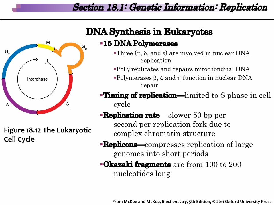
DNA Synthesis in Eukaryotes§15 DNA Polymerases
§Three (a, d, and e) are involved in nuclear DNA replication
§Pol g replicates and repairs mitochondrial DNA§Polymerases b, z and h function in nuclear DNA
repair§Timing of replication—limited to S phase in cell
cycle§Replication rate – slower 50 bp per
second per replication fork due to complex chromatin structure
§Replicons—compresses replication of large genomes into short periods
§Okazaki fragments are from 100 to 200 nucleotides long
Figure 18.12 The Eukaryotic Cell Cycle
From McKee and McKee, Biochemistry, 5th Edition, © 2011 Oxford University Press
Section 18.1: Genetic Information: Replication

Eukaryotic Replication Process – begins with assembly of pre-initiation replication complex (preRC)
§ Preinitiation replication complex (preRC) –initiates replication§ origin replication complex (ORC) binds to DNA
initiation region (origin)§ Recruits Cdc6 & CdrI
§ MCM complex (helicase) is recruited§ Allows replication proteins to load onto
replication fork§ Licensed preRC -active initiation complex
§ Requires pol a/primase, pol e, and accessory proteins
§ Cell cycle regulating kinases then phosphorylate and activate
§ Replication licensing factors (RLFs) -proteins bind to ORC, complete preRCstructure
Figure 18.14 Formation of a PreinitiationReplication Complex
From McKee and McKee, Biochemistry, 5th Edition, © 2011 Oxford University Press
Section 18.1: Genetic Information: Replication
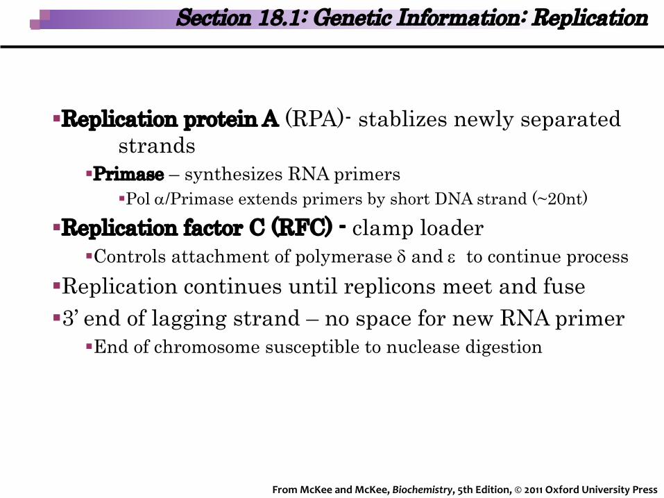
§Replication protein A (RPA)- stablizes newly separated strands
§Primase – synthesizes RNA primers§Pol a/Primase extends primers by short DNA strand (~20nt)
§Replication factor C (RFC) - clamp loader§Controls attachment of polymerase d and e to continue process
§Replication continues until replicons meet and fuse§3’ end of lagging strand – no space for new RNA primer
§End of chromosome susceptible to nuclease digestion
From McKee and McKee, Biochemistry, 5th Edition, © 2011 Oxford University Press
Section 18.1: Genetic Information: Replication
§Telemeres– mini-satellite sequences at end of linear chromosome
§Telomerase – overcomes susceptibility to nuclease digestion
§Ribonucleoprotein with reverse transcriptase activity§RNA base sequence complementary to the TG-rich sequence of
telomeres§Uses sequence to synthesize a single-stranded DNA to extend
the 3′ strand§Telomere end-binding proteins (TEBPs) –binds to GT rich
telomere sequences§Telomere repeat-binding factors (TRFs)– secure 3’ end
§During normal human aging - telomeres shorten§Once reduced to a critical length - chromosome replication
cannot occur;causes cell death§90% of all cancers have hyperactive telomerase
From McKee and McKee, Biochemistry, 5th Edition, © 2011 Oxford University Press
Section 18.1: Genetic Information: Replication

Direct Repairs – photoreactivationrepair of thymine dimers
§ Occurs in bacteria, archaea, protozoa fungi, plants, & animals (not humans
§ DNA damage repaired without removal of nucleotides
§ DNA ligase – repairs breaks inphosphodiester linkages
§ Pyrimidine dimers - restored tooriginal monomeric structure
§ Hydrogen bond - UUsingphotoreactivating enzyme and visible light
Figure 18.18 PhotoreactivationRepair of Thymine Dimers
From McKee and McKee, Biochemistry, 5th Edition, © 2011 Oxford University Press
Section 18.1: Genetic Information: Replication
Average natural mutation – 1/100,000 genes/generation
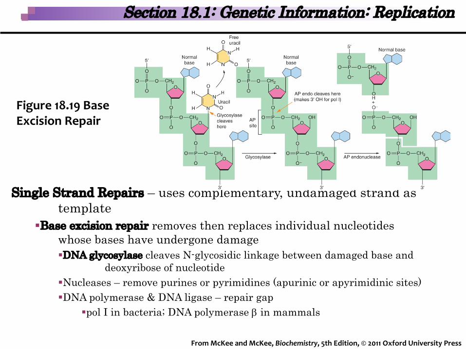
Single Strand Repairs – uses complementary, undamaged strand as template
§Base excision repair removes then replaces individual nucleotides whose bases have undergone damage§DNA glycosylase cleaves N-glycosidic linkage between damaged base and
deoxyribose of nucleotide§Nucleases – remove purines or pyrimidines (apurinic or apyrimidinic sites)§DNA polymerase & DNA ligase – repair gap
§pol I in bacteria; DNA polymerase b in mammals
Figure 18.19 Base Excision Repair
From McKee and McKee, Biochemistry, 5th Edition, © 2011 Oxford University Press
Section 18.1: Genetic Information: Replication

Nucleotide excision repair - removal ofbulky (2-30 nt) lesions; gap is filled
§Global genomic repair§excision enzymes recognize
distortion rather thanbase sequence
§E. coli - excision nuclease cuts DNA
§removes 12 to 13 ntssDNA sequence containing the lesion
§pol I and DNA ligase – repair gap
Figure 18.20 Excision Repair of a Thymine Dimer in E. coliFrom McKee and McKee, Biochemistry, 5th Edition, © 2011 Oxford University Press
Section 18.1: Genetic Information: Replication

§Transcription coupled repair - strand being actively transcribed
§Damage is recognized when RNA polymerase is stalled§Mfd (transcription-repair coupling factor) displaces polymerase
and recruits UvrA2B to initiate damage removal§Mismatch repair (MMR) - corrects helix distorting base
mispairings resulting from proofreading errors or replication slippage
§Key feature is the capacity to distinguish between old and newly synthesized strands
§Methylation of parent strand results in hemimethylated daughter strands
From McKee and McKee, Biochemistry, 5th Edition, © 2011 Oxford University Press
Section 18.1: Genetic Information: Replication

Double-strand breaks (DSBs) - can result in a lethal breakdown of chromosomes
§Caused by radiation, ROS, DNA damaging agents, or as result of replication errors
§Repaired by two mechanisms: non-homologous end joining (NHEJ) and homologous recombination§NHEJ is error prone because there is no requirement
for sequence homology§Recombination will be explained next
From McKee and McKee, Biochemistry, 5th Edition, © 2011 Oxford University Press
Section 18.1: Genetic Information: Replication

DNA Recombination – principle source of genetic variations that make evolution possible
§ Rearrangement of DNA sequences by exchanging segments from different molecules§Two types of recombination:
§General recombination occurs between homologousDNA molecules (most common during meiosis)
§Site-specific recombination—the exchange ofsequences only requires short regions of DNAhomology (e.g., transposition)
From McKee and McKee, Biochemistry, 5th Edition, © 2011 Oxford University Press
Section 18.1: Genetic Information: Replication
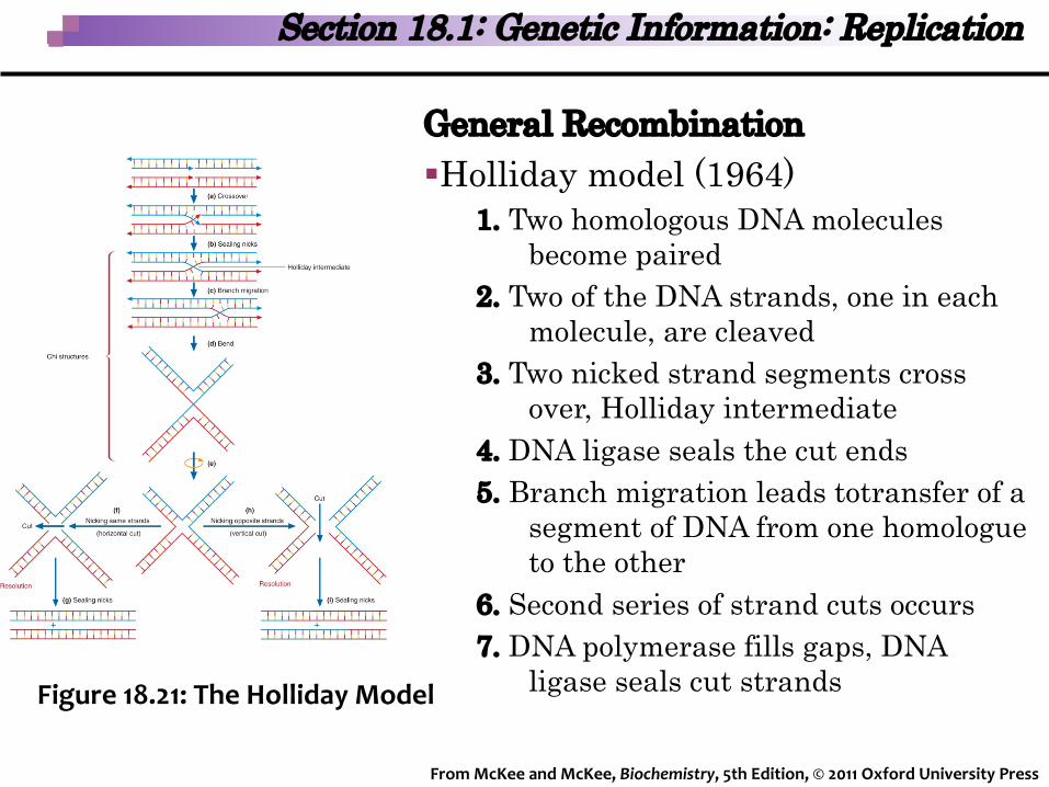
General Recombination§Holliday model (1964)
1. Two homologous DNA moleculesbecome paired
2. Two of the DNA strands, one in eachmolecule, are cleaved
3. Two nicked strand segments cross over, Holliday intermediate
4. DNA ligase seals the cut ends5. Branch migration leads totransfer of a
segment of DNA from one homologue to the other
6. Second series of strand cuts occurs7. DNA polymerase fills gaps, DNA
ligase seals cut strandsFigure 18.21: The Holliday Model
From McKee and McKee, Biochemistry, 5th Edition, © 2011 Oxford University Press
Section 18.1: Genetic Information: Replication

§Messelson-Radding model1. One strand of two homologous DNA molecules
is nicked2. Extension causes displacement of strand on
other side of nick3. D-loop is cleaved, invading strand is ligated to
newly created 3′-end of the homologous strand4. 3′-end of newly synthesized strand & the 5′-end
of a homologous strand are ligated forming a Holliday junction
5. Branch migration may occur6. Strand nicks and Holliday junction resolution
result in a crossover or non-crossover product
From McKee and McKee, Biochemistry, 5th Edition, © 2011 Oxford University Press
Section 18.1: Genetic Information: Replication
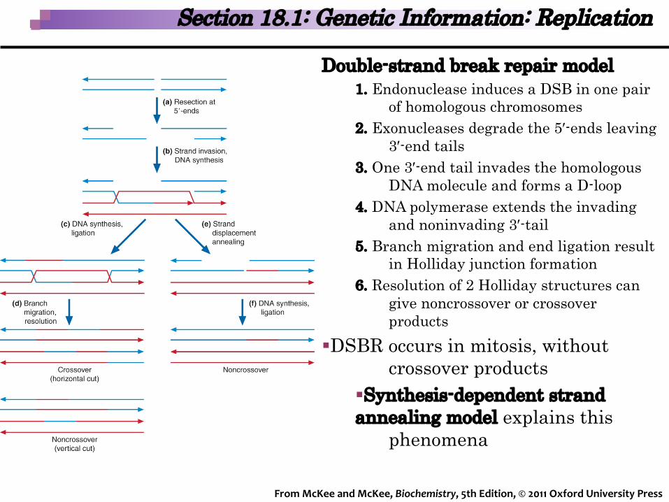
Double-strand break repair model1. Endonuclease induces a DSB in one pair
of homologous chromosomes2. Exonucleases degrade the 5′-ends leaving
3′-end tails3. One 3′-end tail invades the homologous
DNA molecule and forms a D-loop4. DNA polymerase extends the invading
and noninvading 3′-tail 5. Branch migration and end ligation result
in Holliday junction formation6. Resolution of 2 Holliday structures can
give noncrossover or crossover products
§DSBR occurs in mitosis, without crossover products
§Synthesis-dependent strand annealing model explains this
phenomena
From McKee and McKee, Biochemistry, 5th Edition, © 2011 Oxford University Press
Section 18.1: Genetic Information: Replication
§Bacterial Recombination is involved in several formsof intermicrobial DNA transfer:1. Transformation - naked DNA molecules enters the
cell through small holes in the cell wall2. Transduction - bacteriophage inadvertently carries
bacterial DNA to a recipient cell3. Conjugation - unconventional sexual mating
involving passing DNA from a donor cell througha sex pilus to a recipient cell
From McKee and McKee, Biochemistry, 5th Edition, © 2011 Oxford University Press
Section 18.1: Genetic Information: Replication
§Eukaryotic Recombination occurs during first phase of meiosis to ensure accurate homologous chromosome pairing and crossing over§Similar to prokaryotic recombination but has a larger
number of proteins because of the more complex genomes
§Rad52 is believed to be the initial sensor of DSBs§MRN complex – creates scaffold stabilizing DNA ends at
DSBs§Recruits & activates ATM – regulates damage response
§Rad51, BRCA1, and BRCA2 are involved in DSB repair
From McKee and McKee, Biochemistry, 5th Edition, © 2011 Oxford University Press
Section 18.1: Genetic Information: Replication
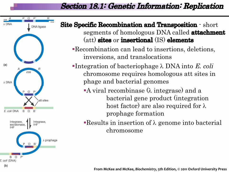
Site Specific Recombination and Transposition - short segments of homologous DNA called attachment(att) sites or insertional (IS) elements
§Recombination can lead to insertions, deletions,inversions, and translocations
§Integration of bacteriophage l DNA into E. colichromosome requires homologous att sites inphage and bacterial genomes§A viral recombinase (l integrase) and a
bacterial gene product (integration host factor) are also required for lprophage formation
§Results in insertion of l genome into bacterial chromosome
From McKee and McKee, Biochemistry, 5th Edition, © 2011 Oxford University Press
Section 18.1: Genetic Information: Replication

Transposition – move DNA sequence from one site to another in genome
§Nonreplicative transposition -double-stranded cut in the donor DNA
§Spliced into the staggered ssDNA cut ends of the target site
§DNA repair system fills the gaps in target DNA
§Transposons – “jumping genes”
Figure 18.27 NonreplicativeTransposition
From McKee and McKee, Biochemistry, 5th Edition, © 2011 Oxford University Press
Section 18.1: Genetic Information: Replication

Replicative transposition -transfer of one strand of donor DNA to target position
§Duplication of the transposonrather than insertion atthe new site
§Involves an intermediate, co-integrate, resolvase (catalyzes the site-specificrecombination)
Figure 18.28 Replicative Transposition
From McKee and McKee, Biochemistry, 5th Edition, © 2011 Oxford University Press
Section 18.1: Genetic Information: Replication

Transcription – creation of RNA from DNA sequences§RNA polymerase – enzyme catalyzing the addition of
ribonucleotides in a 5′à3′ direction§All 4 ribonucleoside triphosphates required, Mg2+
§Primer is not needed, DNA template is required§Only 1 strand of helix is used
§Nontemplate strand (+) - same base sequence as the RNA, except transcript has uracil for thymine
§Template strand (-) - antiparallel to the new RNA strand§Contains initiation & termination signals§Enzyme moves in 3’-to-5’ direction§RNA created in 5’ à 3’
Figure 18.31 DNA Coding Strand
Section 18.2: Transcription
From McKee and McKee, Biochemistry, 5th Edition, © 2011 Oxford University Press

Transcription in Prokaryotes §RNA polymerase in E. coli catalyzes the synthesis of all RNA classes§Core enzyme (a2,b and b′) catalyzes RNA synthesis§Holoenzyme – complete enzyme (a2wbb’s)
§s subunit promotes core assembly and s-factor functions in transcription initiation
§Promoters – RNA polymerase binds to DNA at start of transcription
Figure 18.30 E. coli RNA Polymerase
Section 18.2: Transcription
From McKee and McKee, Biochemistry, 5th Edition, © 2011 Oxford University Press
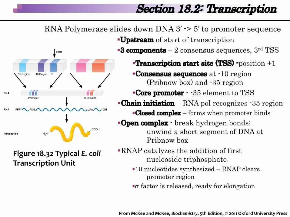
§Transcription start site (TSS) -position +1§Consensus sequences at -10 region
(Pribnow box) and -35 region§Core promoter - -35 element to TSS
§Chain initiation – RNA pol recognizes -35 region§Closed complex – forms when promoter binds
§Open complex - break hydrogen bonds; unwind a short segment of DNA at Pribnow box
§RNAP catalyzes the addition of first nucleoside triphosphate
§10 nucleotides synthesized – RNAP clears promoter region
§s factor is released, ready for elongation
Figure 18.32 Typical E. coli Transcription Unit
Section 18.2: Transcription
From McKee and McKee, Biochemistry, 5th Edition, © 2011 Oxford University Press
RNA Polymerase slides down DNA 3’ -> 5’ to promoter sequence§Upstream of start of transcription§3 components – 2 consensus sequences, 3rd TSS
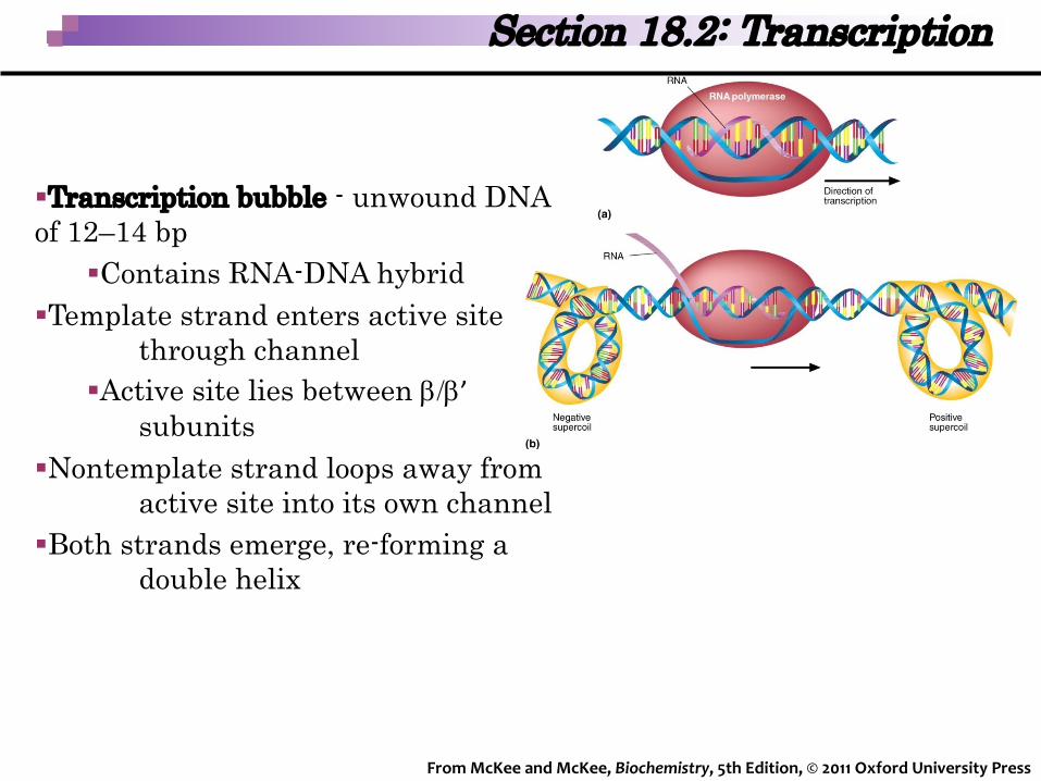
§Transcription bubble - unwound DNA of 12–14 bp
§Contains RNA-DNA hybrid§Template strand enters active site
through channel§Active site lies between b/b’
subunits§Nontemplate strand loops away from
active site into its own channel§Both strands emerge, re-forming a
double helix
Section 18.2: Transcription
From McKee and McKee, Biochemistry, 5th Edition, © 2011 Oxford University Press
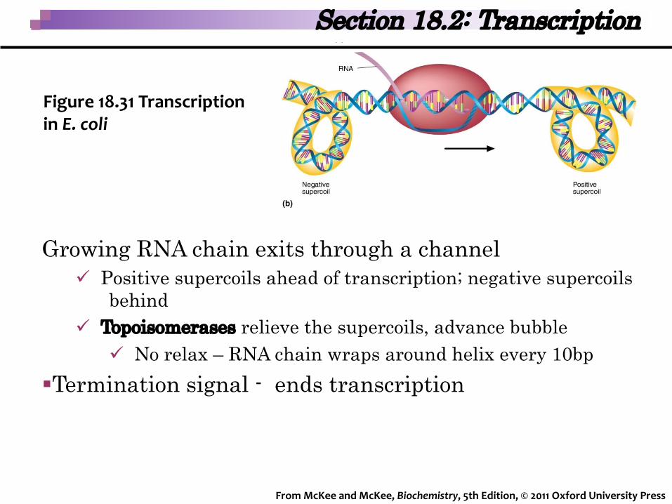
Growing RNA chain exits through a channelü Positive supercoils ahead of transcription; negative supercoils
behindü Topoisomerases relieve the supercoils, advance bubble
ü No relax – RNA chain wraps around helix every 10bp§Termination signal - ends transcription
Section 18.2: Transcription
Figure 18.31 Transcription in E. coli
From McKee and McKee, Biochemistry, 5th Edition, © 2011 Oxford University Press

§Intrinsic termination – controlled by termination sites§2 inverted repeats spaced by few other bases§Sequences of complementary bases, loop back on themselves
§DNA encodes a series of uracils§Forms a stable hairpin causing RNA polymerase to slow or stop
§Series of A-U base pairs between template strand & RNA§RNA transcript is released due to weak base-pair interactions
Figure 18.34 Intrinsic Termination
Section 18.2: Transcription
From McKee and McKee, Biochemistry, 5th Edition, © 2011 Oxford University Press

rho-dependent termination – aid of ATP-dependent helicase rho
factor§Rho binds to a specific recognition
sequence on the nascent RNA chain, upstream fromtermination site§Chases polymerase; when hairpin
loop forms, stalls allowing rho to catch up
§Unwinds the RNA-DNA helix torelease the transcriptFigure 18.35 Rho-Dependent
Termination
Section 18.2: Transcription
From McKee and McKee, Biochemistry, 5th Edition, © 2011 Oxford University Press

Transcription in Eukaryotes§Polymerases are similar in structure and function – 1 vs 3§Initiation factors are distantly related, but perform similar
functions§Regulatory mechanisms differ significantly in both
organisms
Section 18.2: Transcription
From McKee and McKee, Biochemistry, 5th Edition, © 2011 Oxford University Press
§One major difference is the limited access to DNA of the transcription machinery
§Chromatin least partially condensed §Transcription premissive - histone tails
modified by histone acetyl transferases (HATs)
§Chromatin remodeling complexes –weakens histone-DNA contacts
§SWI,SNF, and NURF
§RNA Polymerase Activity§RNA polymerase I (RNAPI) transcribes larger rRNA
(28S, 18S, and 5.8S) in the nucleolus§RNA polymerase II (RNAPII) produces the precursors of
mRNA, miRNAs and most snRNA§RNA polymerase III (RNAPIII) is responsible for
transcribing the precursors for tRNA, 55 rRNA, U6snRNA, and the snoRNAs
§Requires various transcription factors bound to thepromoter to initiate transcription
Section 18.2: Transcription
From McKee and McKee, Biochemistry, 5th Edition, © 2011 Oxford University Press

Eukaryotic promoters- Pol II core promoter can be focused or dispersed
§Focused - transcription start site (TSS) and corepromoter elements (CPE)§TATA box - consensus sequence, TATAA§TFIID - TATA-binding protein (TBP) binds to TATA box.
§Other core elements include the Inr (initiator), BRE (B recognition element), and DPE (downstream promoter element)
§Dispersed genes -multiple TSSs, distributed over a broad region of 50-100 basepairs§Typically occur within CG islands
§Facilitate nucleosome destabilization
Section 18.2: Transcription
From McKee and McKee, Biochemistry, 5th Edition, © 2011 Oxford University Press
RNA polymerase II (RNAP II) §12 subunits in humans
§RBP1, largest subunit, forms part of the enzyme’s active site; binds DNA
§Contains C-terminal domain (CTD)§Unphosphorylated allows RNAPII to bind promoters
§RNAP II machinery - set of five transcription factors and a 20-protein mediator complex§General transcription factors (GTFs): TFIIB, TFIID, TFIIE,
TFIIF, and TFIIH are the minimum required for accurate transcription
§Facilitate promoter recognition, preinitiation complex formation, and ATP-dependent DNA unwinding
Section 18.2: Transcription
From McKee and McKee, Biochemistry, 5th Edition, © 2011 Oxford University Press

§PIC assembly - binding of TBP subunit ofTFIID to the TATA box.
§Other GTFs: TFIIA, TFIIB, TFIIF, mediator
§TFIIH acts as an ATP-dependent helicase§Promoter clearance occurs after 23 nt
§Elongation - promoter clearance; mediator dissociation
§Termination Sequence - poly(A) sequence, (5′-AAUAAA-3′)
§Poly(A) tail- (100–200 adenylate residues) to end of the transcript
Figure 18.44 PreinitiationComplex Formation at a TATA Box
Section 18.2: Transcription
From McKee and McKee, Biochemistry, 5th Edition, © 2011 Oxford University Press

§RNA Processing- enzymatic modification§Trimming -removal of leader (5’) and trailer (3’)
sequences§capping – protects 5’ end from nucleases
§Synthesized when the transcript is about 30 nt long
Figure 18.45 The Methylated Cap of Eukaryotic mRNA
Section 18.2: Transcription
From McKee and McKee, Biochemistry, 5th Edition, © 2011 Oxford University Press
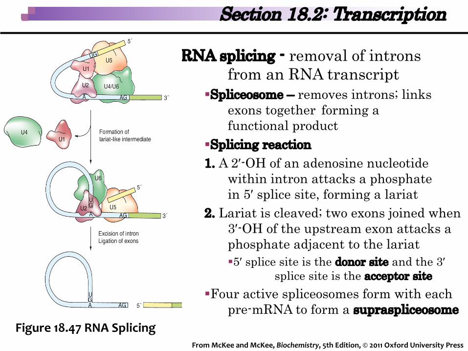
RNA splicing - removal of intronsfrom an RNA transcript
§Spliceosome – removes introns; links exons together forming afunctional product
§Splicing reaction1. A 2′-OH of an adenosine nucleotide
within intron attacks a phosphate in 5′ splice site, forming a lariat
2. Lariat is cleaved; two exons joined when 3′-OH of the upstream exon attacks a phosphate adjacent to the lariat§5′ splice site is the donor site and the 3′
splice site is the acceptor site§Four active spliceosomes form with each
pre-mRNA to form a supraspliceosome
Section 18.2: Transcription
From McKee and McKee, Biochemistry, 5th Edition, © 2011 Oxford University Press
Figure 18.47 RNA Splicing

§lac Operon -control element / structural genes that code for enzymes responsible for lactose metabolism
§Structural genes Z, Y, and A encode for b-galactosidase, lactose permease, and thiogalactoside transacetylase, respectively
§lacI gene encodes for a repressor that binds to the operator ofoperon as a tetramer§lac operon is repressed when its inducer allolactose is not present
due to lac repressor binding
Figure 18.50 Function of the lac Operon
Section 18.3: Gene Expression
From McKee and McKee, Biochemistry, 5th Edition, © 2011 Oxford University Press
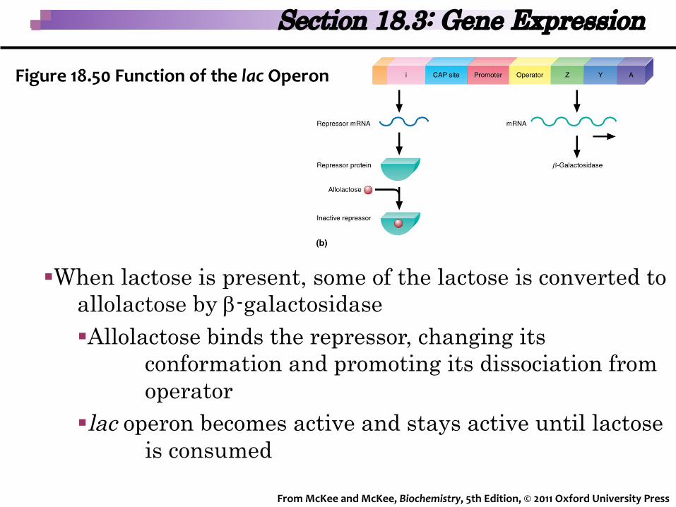
§When lactose is present, some of the lactose is converted toallolactose by b-galactosidase§Allolactose binds the repressor, changing its
conformation and promoting its dissociation fromoperator
§lac operon becomes active and stays active until lactoseis consumed
Figure 18.50 Function of the lac Operon
Section 18.3: Gene Expression
From McKee and McKee, Biochemistry, 5th Edition, © 2011 Oxford University Press

Genomic Control §Chromatin structure
§ Structural organization of genome – chromatin remodeling§Transcription factor-regulated RNA polymerase complex
formation§Particular set of proteins that assembles on a regulatory DNA
sequence§Less common examples include gene rearrangements
and gene amplification
Section 18.3: Gene Expression
From McKee and McKee, Biochemistry, 5th Edition, © 2011 Oxford University Press

RNA processing §Alternative splicing - Joining of
different combinations of exonsto form cell-specific proteins
§Vertebrate tropomyosin gene - 13 to 15 exons; five common in all isoforms§Remaining exons are alternatively used
in different mRNAs
§RNA editing - After transcription, base changes
§RNA stability, translation initiation, alteration of splice sites, and amino acid sequence changes
Figure 18.52 RNA Processing
Section 18.3: Gene Expression
From McKee and McKee, Biochemistry, 5th Edition, © 2011 Oxford University Press

Posttranscriptional Gene Silencing§miRNAs inhibit translation by binding to complementary
sequences in the 3′-UTR of target mRNAs§Humans are estimated to have as many as a thousand
miRNAs believed to regulate about one-third of human genes
§miRNA-mediated gene silencing utilizes components of RNA interference §Limited to protection against viruses & transposons
§Cells use small-interfering RNAs (si-RNAs) to recognize and degrade target mRNAs
Section 18.3: Gene Expression
From McKee and McKee, Biochemistry, 5th Edition, © 2011 Oxford University Press

§mRNA Transport - out of the nucleus requires three phases:processing, docking and passage through nuclear porecomplexes (NPC), and release into the cytoplasm
§Translational Control—Covalent modification of several translation factors has been shown to alter translation rate in response to various stimuli
Section 18.3: Gene Expression
From McKee and McKee, Biochemistry, 5th Edition, © 2011 Oxford University Press

Signal Transduction§Cells alter gene expression patterns in response to
environmental signals§Initiated by binding of a ligand to a receptor; then initiates a signal
transduction cascade§Induce two classes of genes at the end of their signal transduction
cascades§Early response genes are rapidly activated (within 15 minutes);
often transcription factors§Delayed response genes - induced by activities of transcription
factors and proteins produced during the early response phase
Section 18.3: Gene Expression
From McKee and McKee, Biochemistry, 5th Edition, © 2011 Oxford University Press