Embed Size (px)
Citation preview
GI Lymphomas
성균관대학교 의과대학 내과 이준행
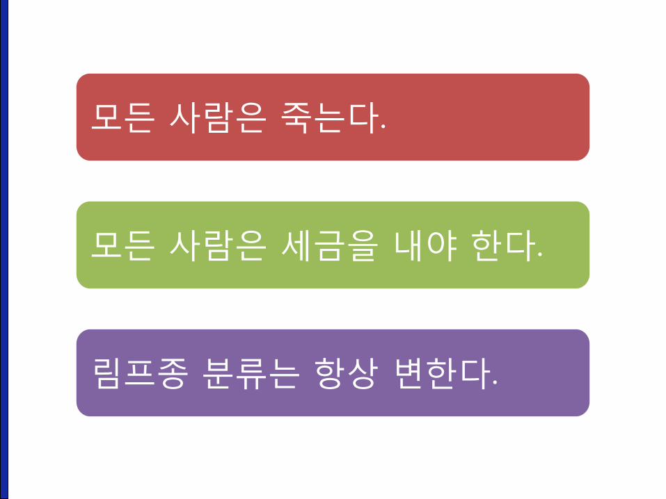
모든 사람은 죽는다.
모든 사람은 세금을 내야 한다.
림프종 분류는 항상 변한다.
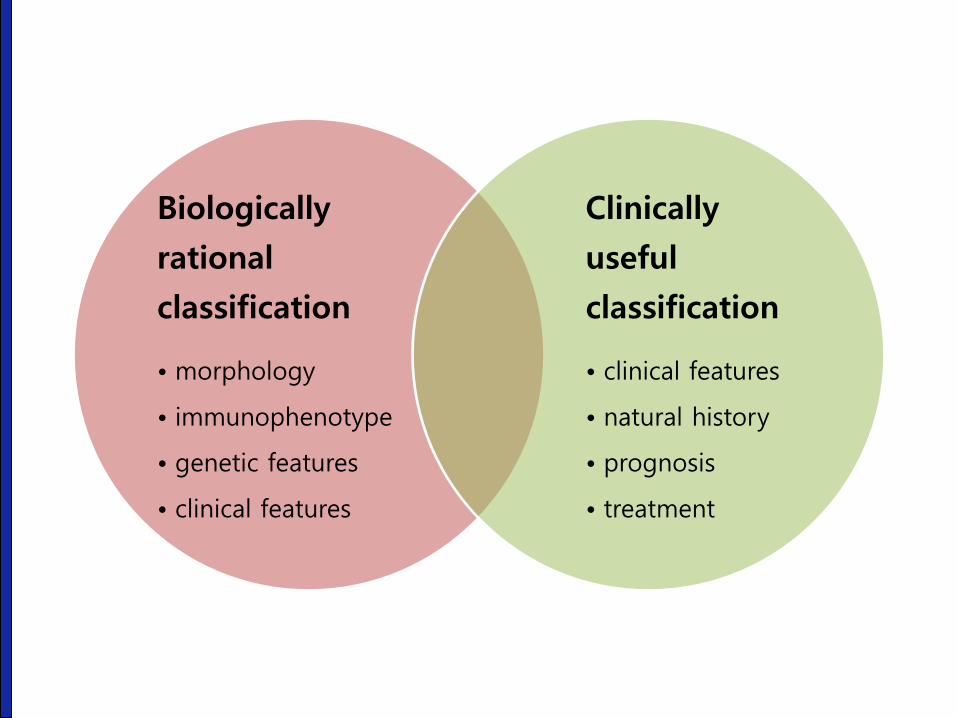
Biologically
rational
classification
• morphology
• immunophenotype
• genetic features
• clinical features
Clinically
useful
classification
• clinical features
• natural history
• prognosis
• treatment

Immunohistochemical markers
• B-cell: CD20, CD79a
• T-cell: CD3, CD45RO
• NK-cell: CD56
• Ki-67: Burkitt lymphoma > DLBL > others
• Mantle cell lympoma: cyclin D1
• HTLV-1 serology
• TdT, CD4, CD5, CD8, CD10, CD30, Bcl-2, cytokeratin AE1/AE3,etc

Rappar-
port
1966
Kiel
1978
Working
Formu-
lation
1982
REAL
1994
WHO
2001
Revised
WHO
2008
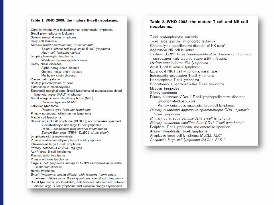

B-cell neoplasms of GI tract
• MALT lymphoma
• Diffuse large B-cell lymphoma (DLBCL)
• Follicular lymphoma
• Mantle cell lymphoma
• Burkitt lymphoma
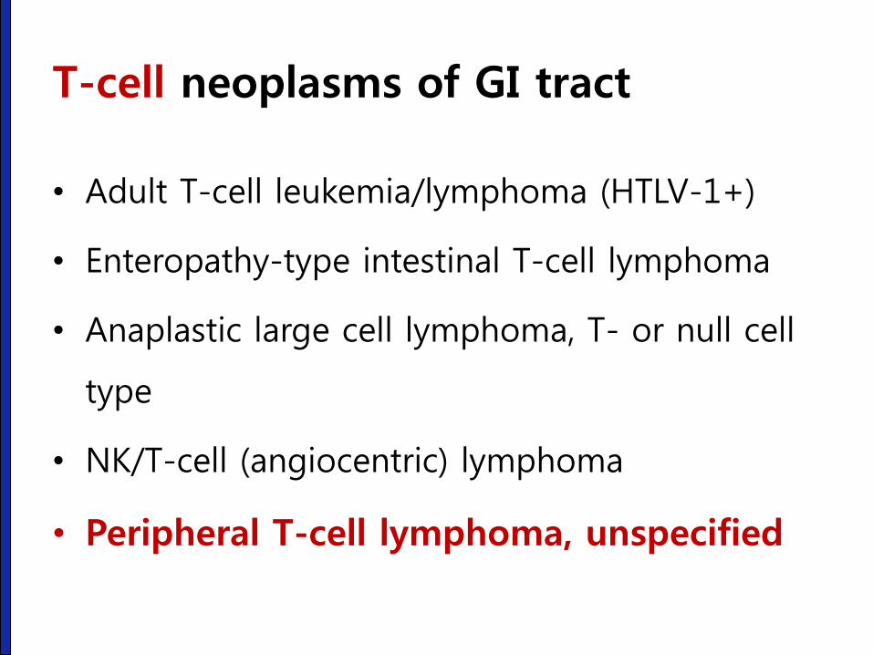
T-cell neoplasms of GI tract
• Adult T-cell leukemia/lymphoma (HTLV-1+)
• Enteropathy-type intestinal T-cell lymphoma
• Anaplastic large cell lymphoma, T- or null cell
type
• NK/T-cell (angiocentric) lymphoma
• Peripheral T-cell lymphoma, unspecified
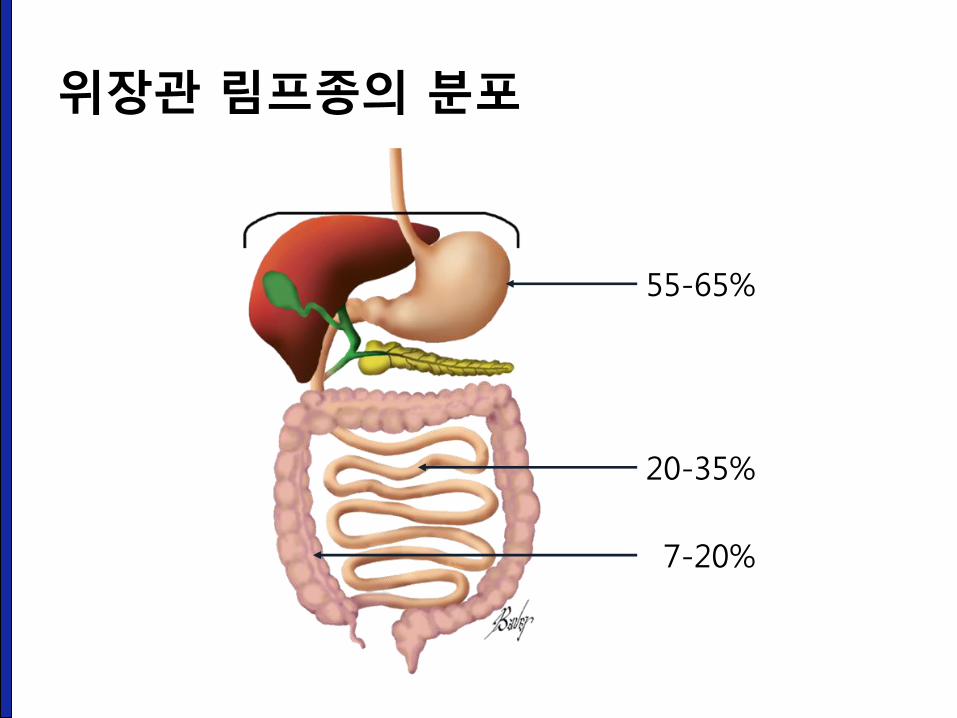
55-65%
20-35%
7-20%
위장관 림프종의 분포
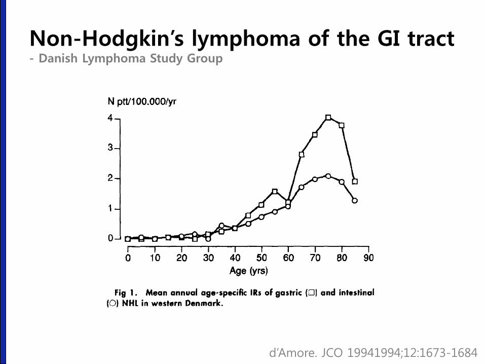
Non-Hodgkin’s lymphoma of the GI tract - Danish Lymphoma Study Group
d’Amore. JCO 19941994;12:1673-1684
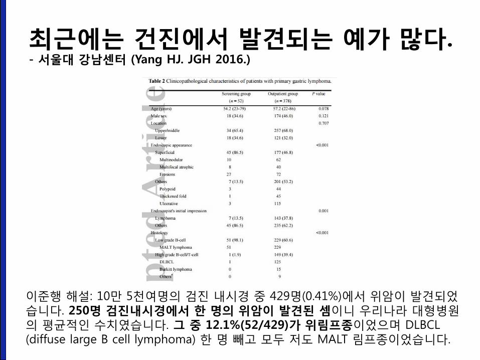
최근에는 건진에서 발견되는 예가 많다. - 서울대 강남센터 (Yang HJ. JGH 2016.)
이준행 해설: 10만 5천여명의 검진 내시경 중 429명(0.41%)에서 위암이 발견되었습니다. 250명 검진내시경에서 한 명의 위암이 발견된 셈이니 우리나라 대형병원의 평균적인 수치였습니다. 그 중 12.1%(52/429)가 위림프종이었으며 DLBCL (diffuse large B cell lymphoma) 한 명 빼고 모두 저도 MALT 림프종이었습니다.
내시경 진단이 항상 쉬운 것은 아니다.
성균관대학교 의과대학 내과 이준행
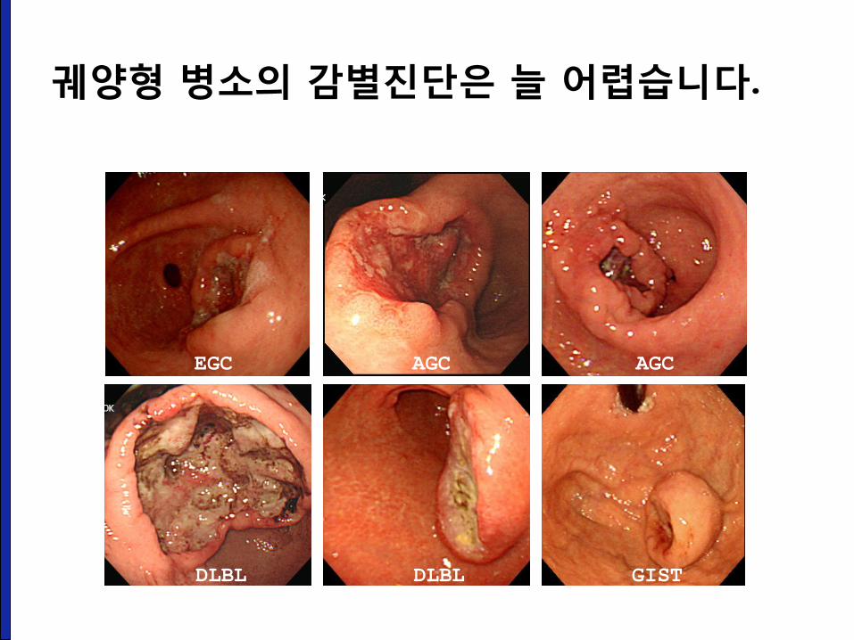
EGC AGC AGC
DLBL DLBL GIST
궤양형 병소의 감별진단은 늘 어렵습니다.
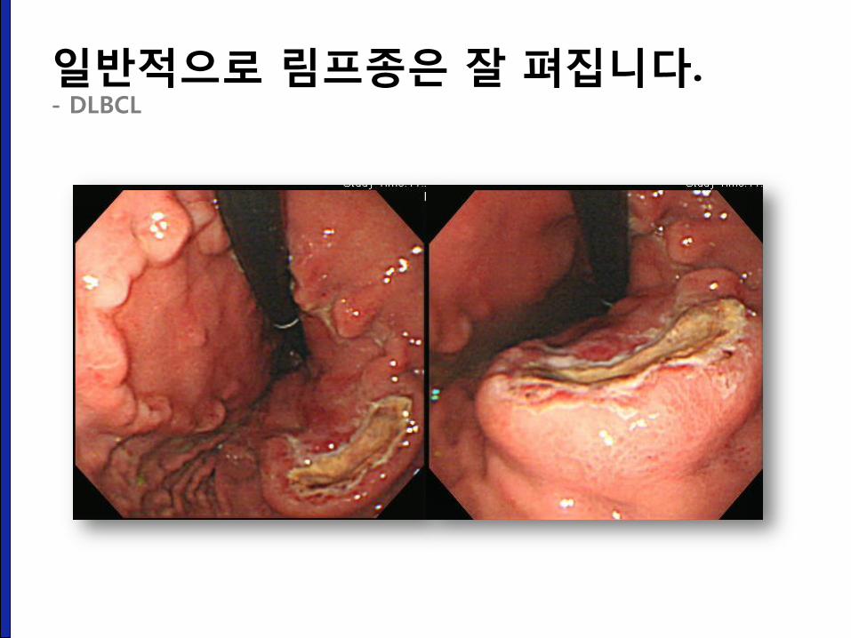
일반적으로 림프종은 잘 펴집니다. - DLBCL
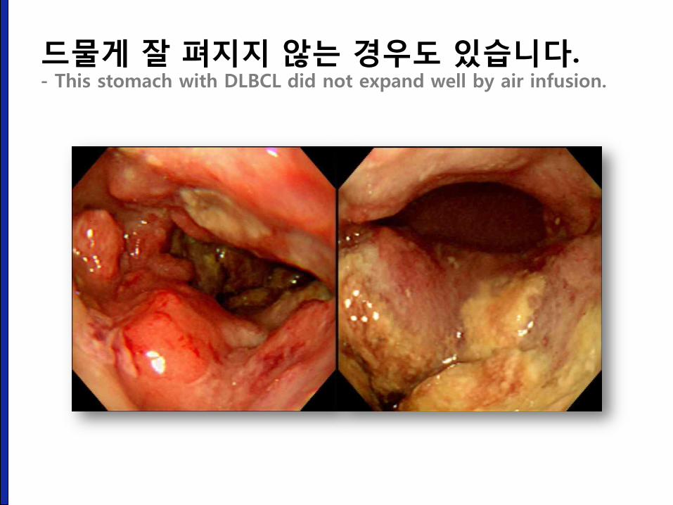
드물게 잘 펴지지 않는 경우도 있습니다. - This stomach with DLBCL did not expand well by air infusion.
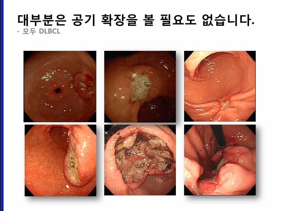
대부분은 공기 확장을 볼 필요도 없습니다. - 모두 DLBCL
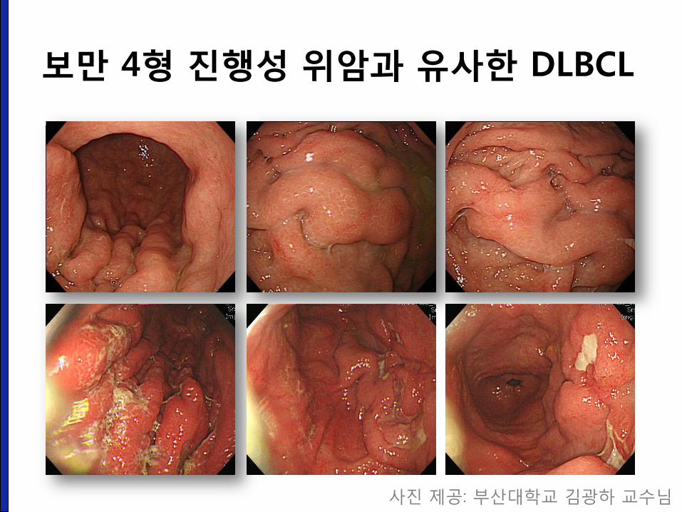
보만 4형 진행성 위암과 유사한 DLBCL
사진 제공: 부산대학교 김광하 교수님
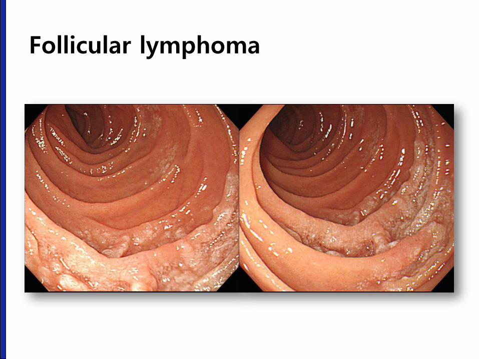
Follicular lymphoma
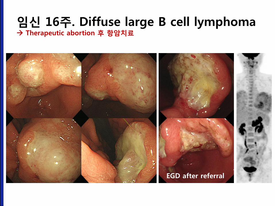
임신 16주. Diffuse large B cell lymphoma Therapeutic abortion 후 항암치료
EGD after referral
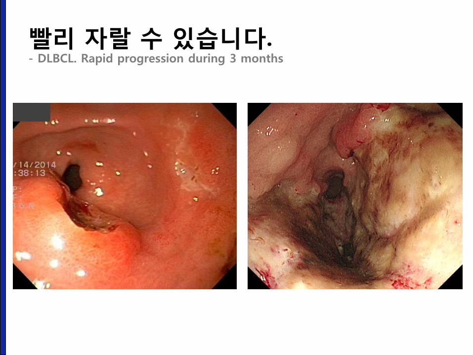
빨리 자랄 수 있습니다. - DLBCL. Rapid progression during 3 months
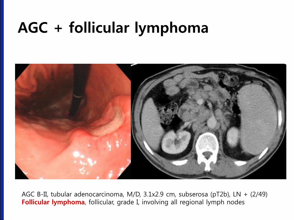
AGC + follicular lymphoma
AGC B-II, tubular adenocarcinoma, M/D, 3.1x2.9 cm, subserosa (pT2b), LN + (2/49) Follicular lymphoma, follicular, grade I, involving all regional lymph nodes
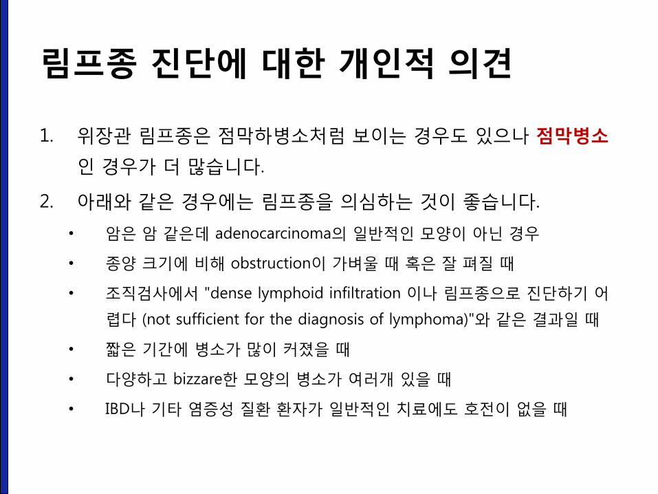
림프종 진단에 대한 개인적 의견
1. 위장관 림프종은 점막하병소처럼 보이는 경우도 있으나 점막병소
인 경우가 더 많습니다.
2. 아래와 같은 경우에는 림프종을 의심하는 것이 좋습니다.
• 암은 암 같은데 adenocarcinoma의 일반적인 모양이 아닌 경우
• 종양 크기에 비해 obstruction이 가벼울 때 혹은 잘 펴질 때
• 조직검사에서 "dense lymphoid infiltration 이나 림프종으로 진단하기 어
렵다 (not sufficient for the diagnosis of lymphoma)"와 같은 결과일 때
• 짧은 기간에 병소가 많이 커졌을 때
• 다양하고 bizzare한 모양의 병소가 여러개 있을 때
• IBD나 기타 염증성 질환 환자가 일반적인 치료에도 호전이 없을 때
병리 검사 해석에 주의하자.
성균관대학교 의과대학 내과 이준행
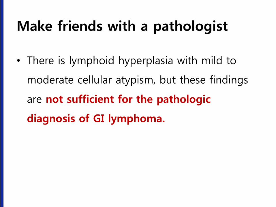
Make friends with a pathologist
• There is lymphoid hyperplasia with mild to
moderate cellular atypism, but these findings
are not sufficient for the pathologic
diagnosis of GI lymphoma.
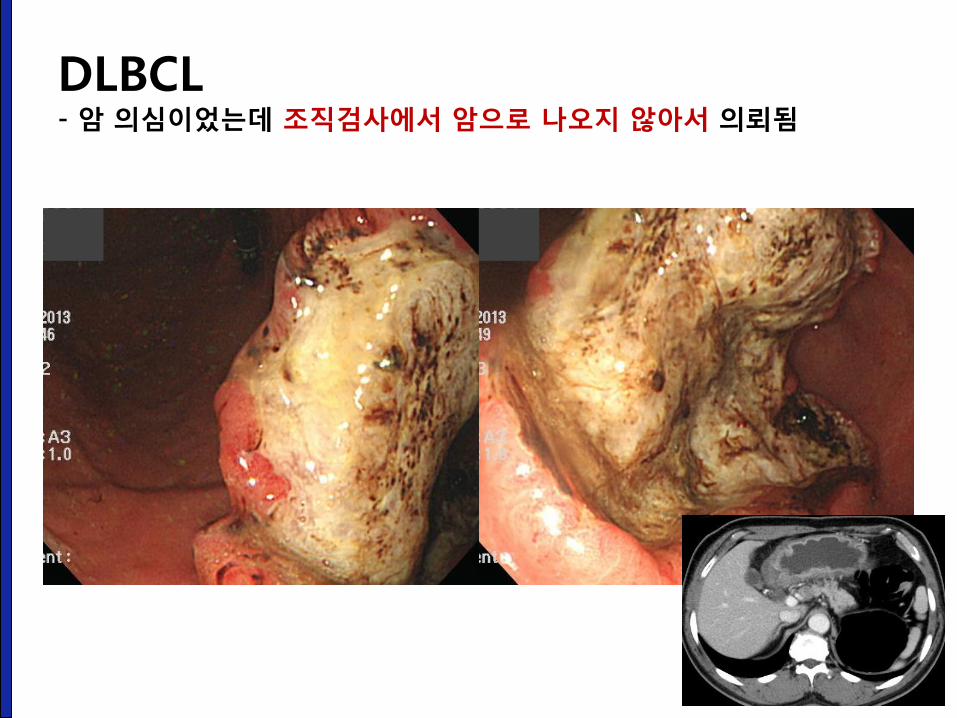
DLBCL - 암 의심이었는데 조직검사에서 암으로 나오지 않아서 의뢰됨
진단이 지연될 수 있다.
성균관대학교 의과대학 내과 이준행
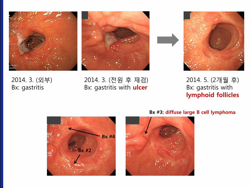
2014. 3. (외부) Bx: gastritis
2014. 3. (전원 후 재검) Bx: gastritis with ulcer
2014. 5. (2개월 후) Bx: gastritis with lymphoid follicles
Bx #2
Bx #3: diffuse large B cell lymphoma
Bx #4
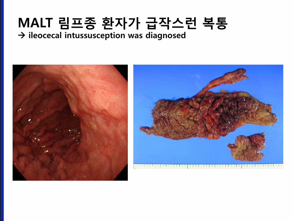
MALT 림프종 환자가 급작스런 복통 ileocecal intussusception was diagnosed

Kim YH, Lee JH, et al. Digest Dis Sci 2005;50:2243-2247
특히 T 세포 림프종 진단이 어렵습니다
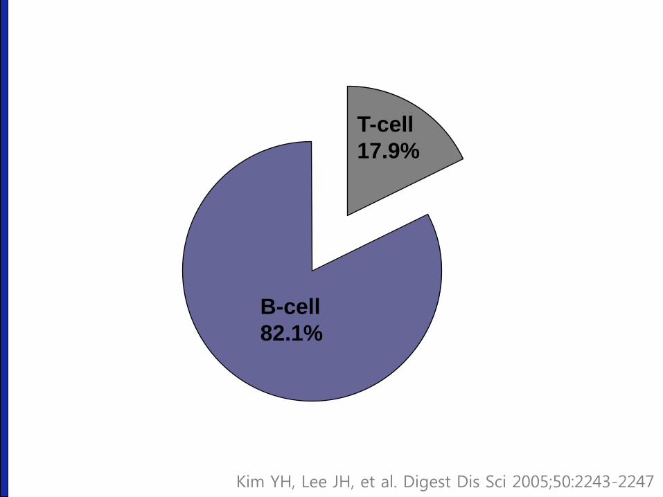
B-cell
82.1%
T-cell
17.9%
Kim YH, Lee JH, et al. Digest Dis Sci 2005;50:2243-2247
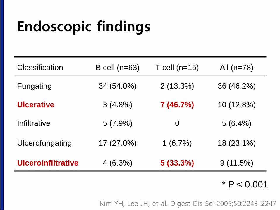
Classification B cell (n=63) T cell (n=15) All (n=78)
Fungating 34 (54.0%) 2 (13.3%) 36 (46.2%)
Ulcerative 3 (4.8%) 7 (46.7%) 10 (12.8%)
Infiltrative 5 (7.9%) 0 5 (6.4%)
Ulcerofungating 17 (27.0%) 1 (6.7%) 18 (23.1%)
Ulceroinfiltrative 4 (6.3%) 5 (33.3%) 9 (11.5%)
* P < 0.001
Kim YH, Lee JH, et al. Digest Dis Sci 2005;50:2243-2247
Endoscopic findings
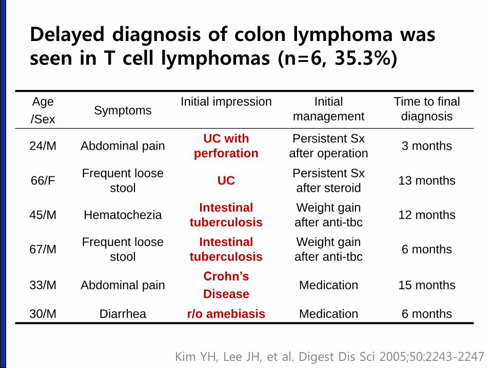
Age
/Sex Symptoms
Initial impression Initial
management
Time to final
diagnosis
24/M Abdominal pain UC with
perforation
Persistent Sx
after operation 3 months
66/F Frequent loose
stool UC
Persistent Sx
after steroid 13 months
45/M Hematochezia Intestinal
tuberculosis
Weight gain
after anti-tbc 12 months
67/M Frequent loose
stool
Intestinal
tuberculosis
Weight gain
after anti-tbc 6 months
33/M Abdominal pain Crohn’s
Disease Medication 15 months
30/M Diarrhea r/o amebiasis Medication 6 months
Delayed diagnosis of colon lymphoma was seen in T cell lymphomas (n=6, 35.3%)
Kim YH, Lee JH, et al. Digest Dis Sci 2005;50:2243-2247
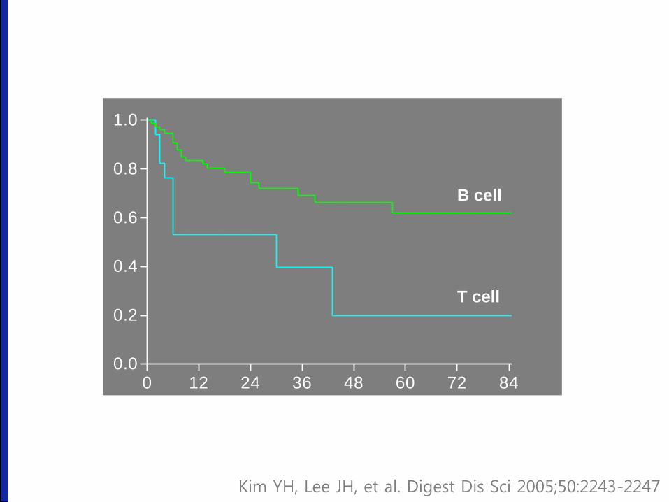
Kim YH, Lee JH, et al. Digest Dis Sci 2005;50:2243-2247
0 12 24 36 48 60 72 840.0
0.2
0.4
0.6
0.8
1.0
T cell
B cell
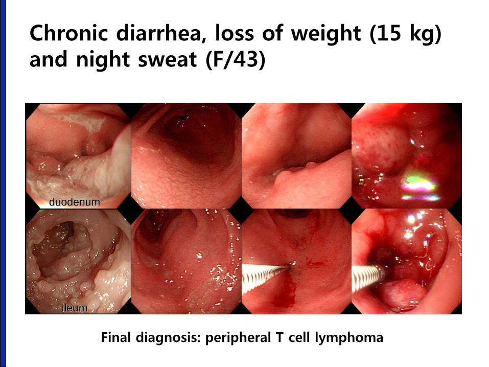
Chronic diarrhea, loss of weight (15 kg) and night sweat (F/43)
ileum
duodenum
Final diagnosis: peripheral T cell lymphoma
다양한 위장관 림프종
진단별 접근법
성균관대학교 의과대학 내과 이준행
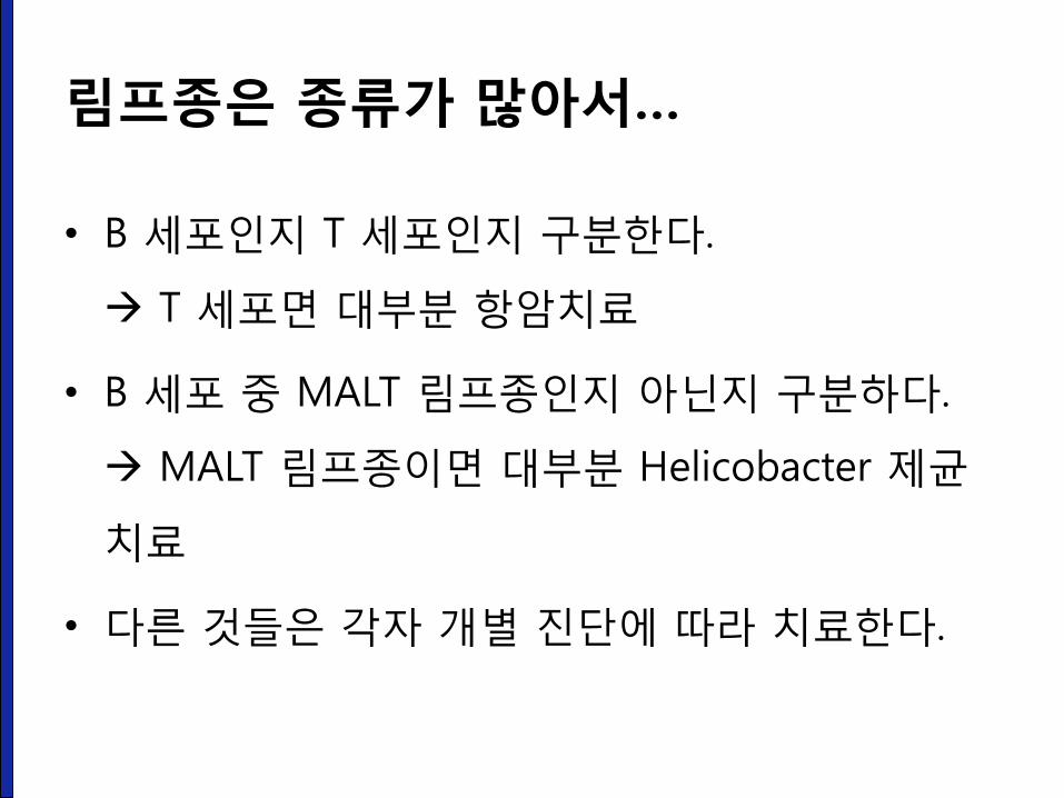
림프종은 종류가 많아서…
• B 세포인지 T 세포인지 구분한다.
T 세포면 대부분 항암치료
• B 세포 중 MALT 림프종인지 아닌지 구분하다.
MALT 림프종이면 대부분 Helicobacter 제균
치료
• 다른 것들은 각자 개별 진단에 따라 치료한다.
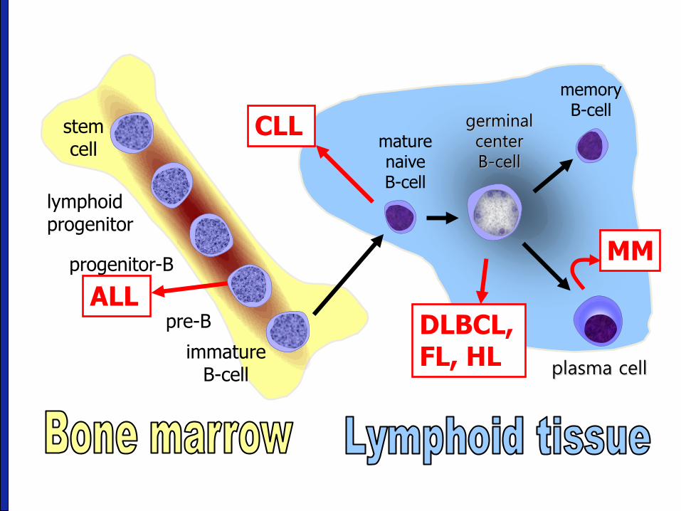
stem cell
lymphoid progenitor
progenitor-B
pre-B
immature B-cell
memory B-cell
plasma cell
DLBCL, FL, HL
ALL
CLL
MM
germinal center B-cell
mature naive B-cell
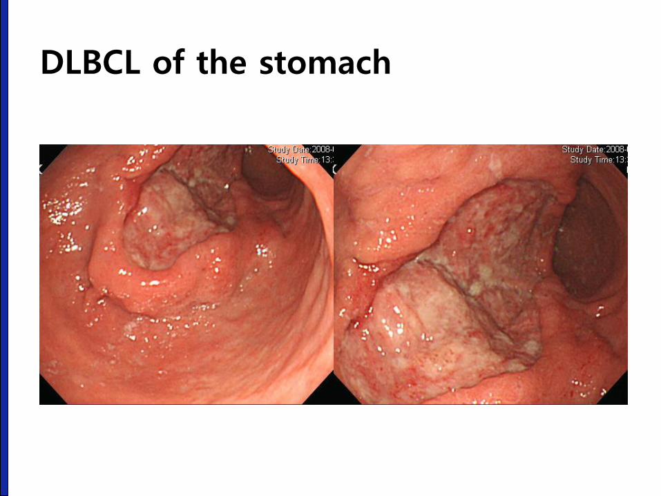
DLBCL of the stomach
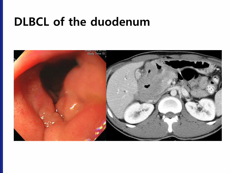
DLBCL of the duodenum
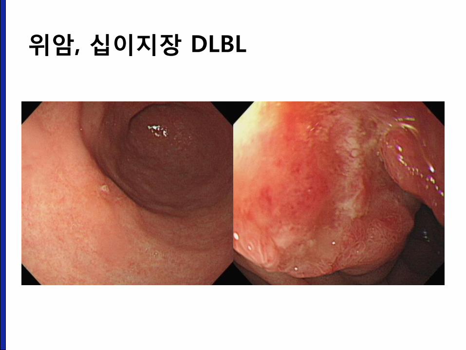
위암, 십이지장 DLBL
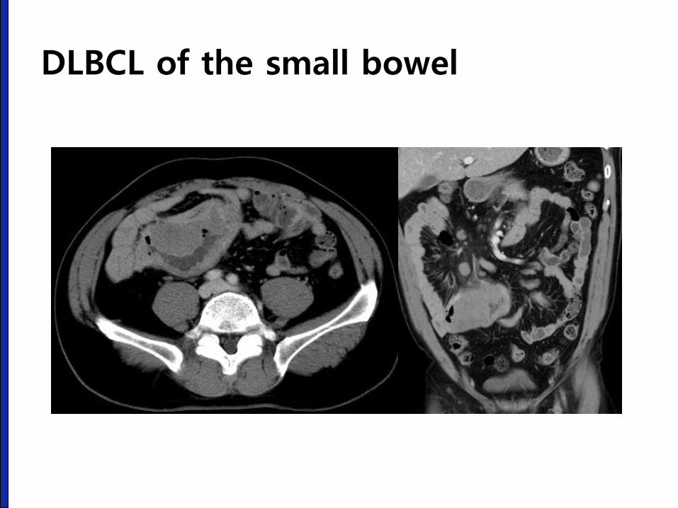
DLBCL of the small bowel
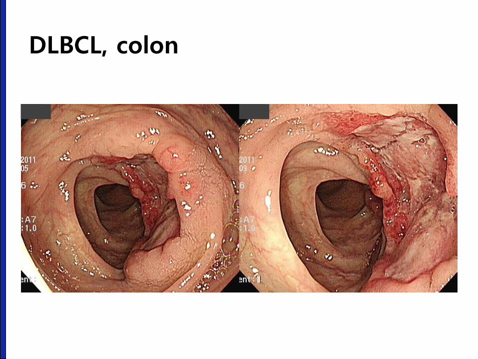
DLBCL, colon
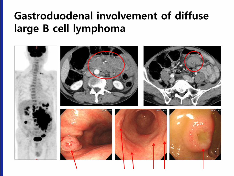
Gastroduodenal involvement of diffuse large B cell lymphoma
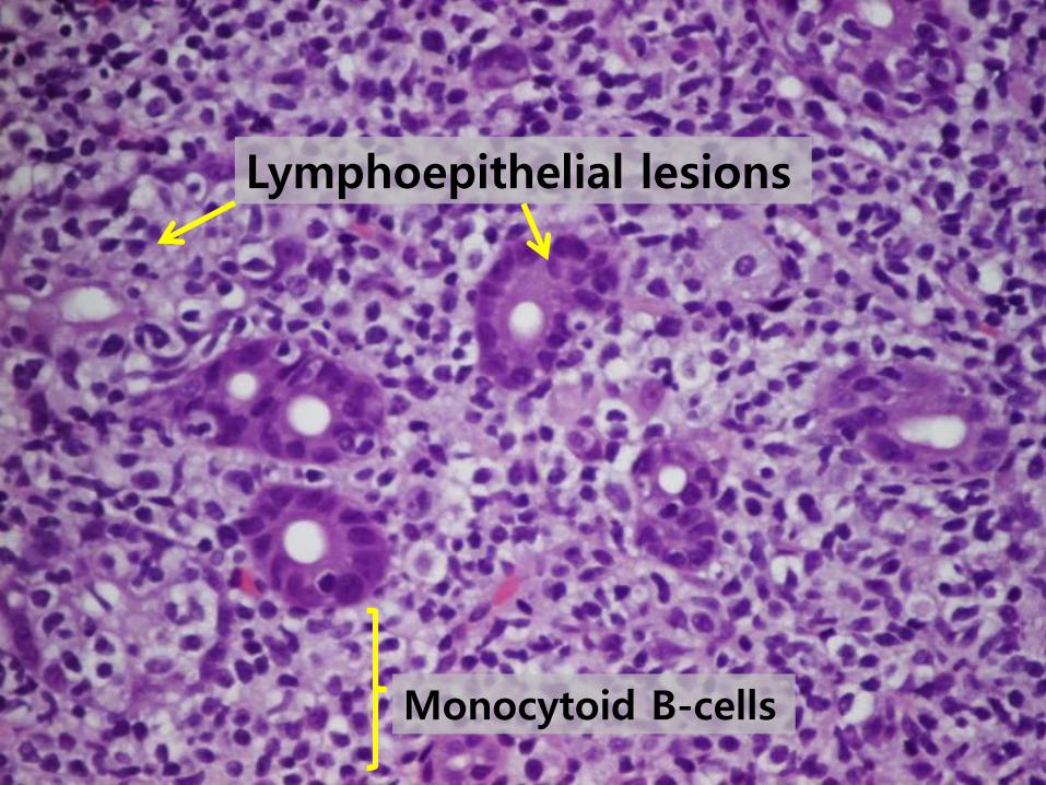
Lymphoepithelial lesions
Monocytoid B-cells
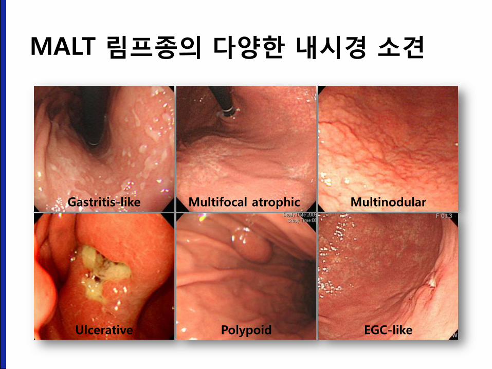
MALT 림프종의 다양한 내시경 소견
Gastritis-like Multifocal atrophic Multinodular
Ulcerative Polypoid EGC-like
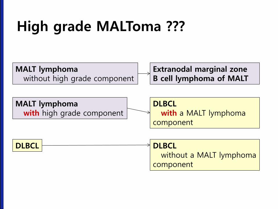
MALT lymphoma without high grade component
MALT lymphoma with high grade component
DLBCL
Extranodal marginal zone B cell lymphoma of MALT
DLBCL with a MALT lymphoma component
DLBCL without a MALT lymphoma component
High grade MALToma ???
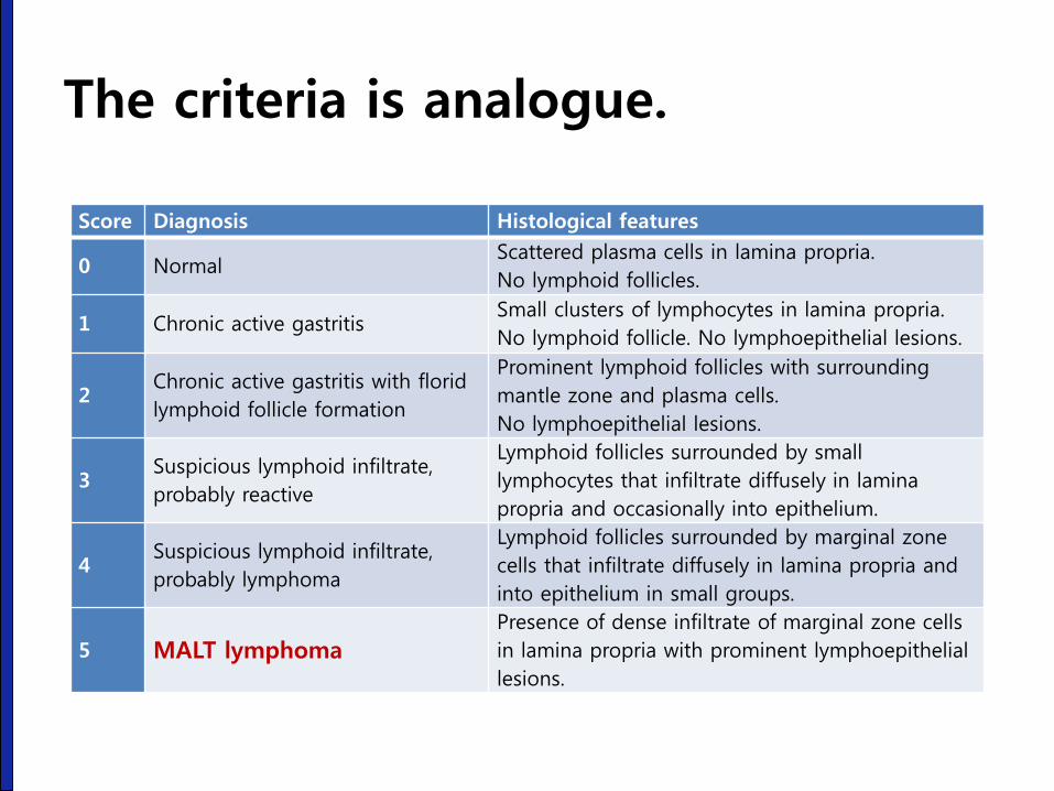
Score Diagnosis Histological features
0 Normal Scattered plasma cells in lamina propria.
No lymphoid follicles.
1 Chronic active gastritis Small clusters of lymphocytes in lamina propria.
No lymphoid follicle. No lymphoepithelial lesions.
2 Chronic active gastritis with florid
lymphoid follicle formation
Prominent lymphoid follicles with surrounding
mantle zone and plasma cells.
No lymphoepithelial lesions.
3 Suspicious lymphoid infiltrate,
probably reactive
Lymphoid follicles surrounded by small
lymphocytes that infiltrate diffusely in lamina
propria and occasionally into epithelium.
4 Suspicious lymphoid infiltrate,
probably lymphoma
Lymphoid follicles surrounded by marginal zone
cells that infiltrate diffusely in lamina propria and
into epithelium in small groups.
5 MALT lymphoma
Presence of dense infiltrate of marginal zone cells
in lamina propria with prominent lymphoepithelial
lesions.
The criteria is analogue.
Choi MK. Korean J Gastroenterol 2011;57:272-280
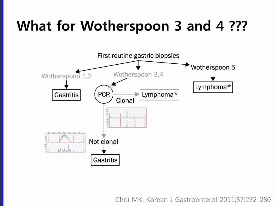
What for Wotherspoon 3 and 4 ???
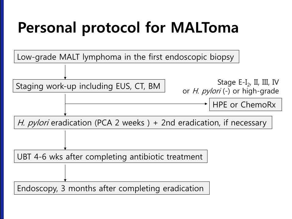
Personal protocol for MALToma
Low-grade MALT lymphoma in the first endoscopic biopsy
Staging work-up including EUS, CT, BM
H. pylori eradication (PCA 2 weeks ) + 2nd eradication, if necessary
UBT 4-6 wks after completing antibiotic treatment
Endoscopy, 3 months after completing eradication
Stage E-I2, II, III, IV or H. pylori (-) or high-grade
HPE or ChemoRx
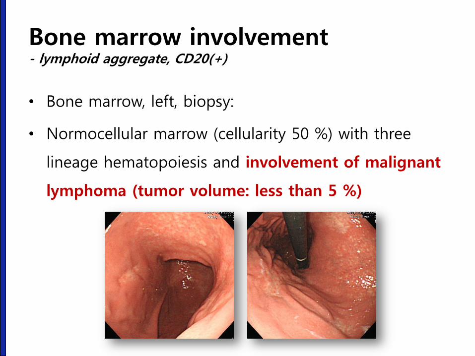
Bone marrow involvement - lymphoid aggregate, CD20(+)
• Bone marrow, left, biopsy:
• Normocellular marrow (cellularity 50 %) with three
lineage hematopoiesis and involvement of malignant
lymphoma (tumor volume: less than 5 %)

Bone marrow involvement - lymphoid aggregate
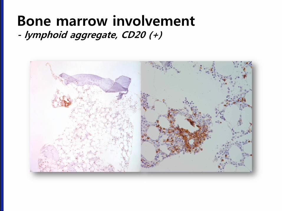
Bone marrow involvement - lymphoid aggregate, CD20 (+)
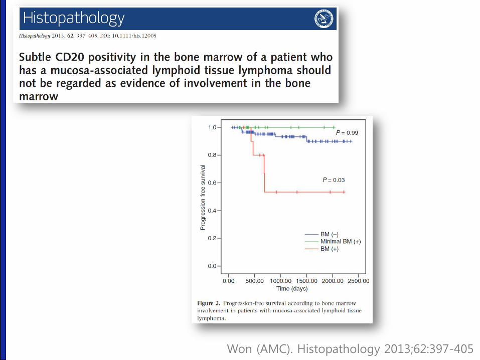
Won (AMC). Histopathology 2013;62:397-405
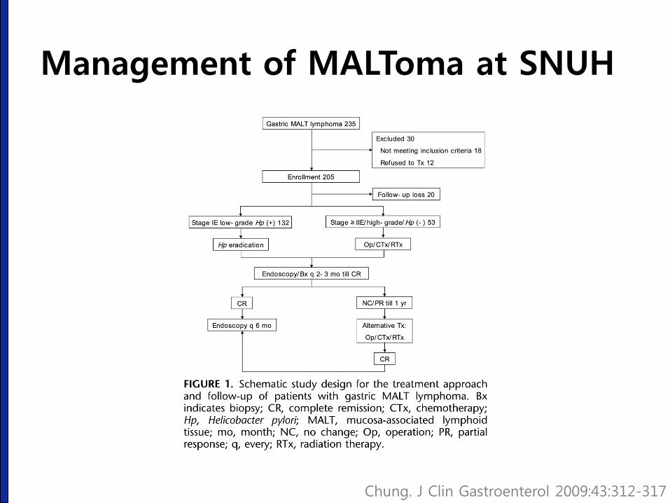
Management of MALToma at SNUH
Chung. J Clin Gastroenterol 2009:43:312-317

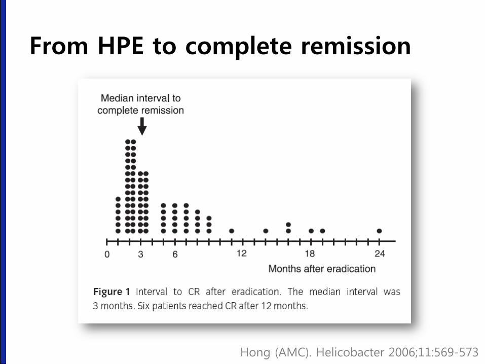
Hong (AMC). Helicobacter 2006;11:569-573
From HPE to complete remission
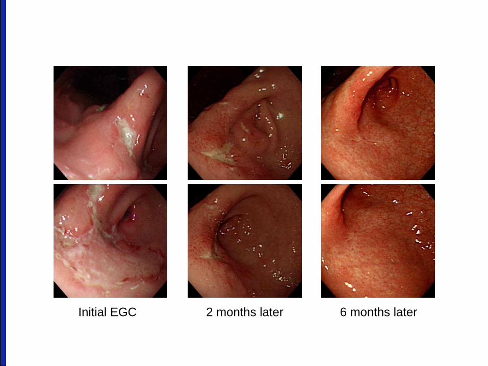
Initial EGC 2 months later 6 months later

2003. 8. 22
(HPE 5개월)
2003. 6.13
(HPE 3개월)
2003. 4. 18
(HPE 1개월)
2003. 2. 7.
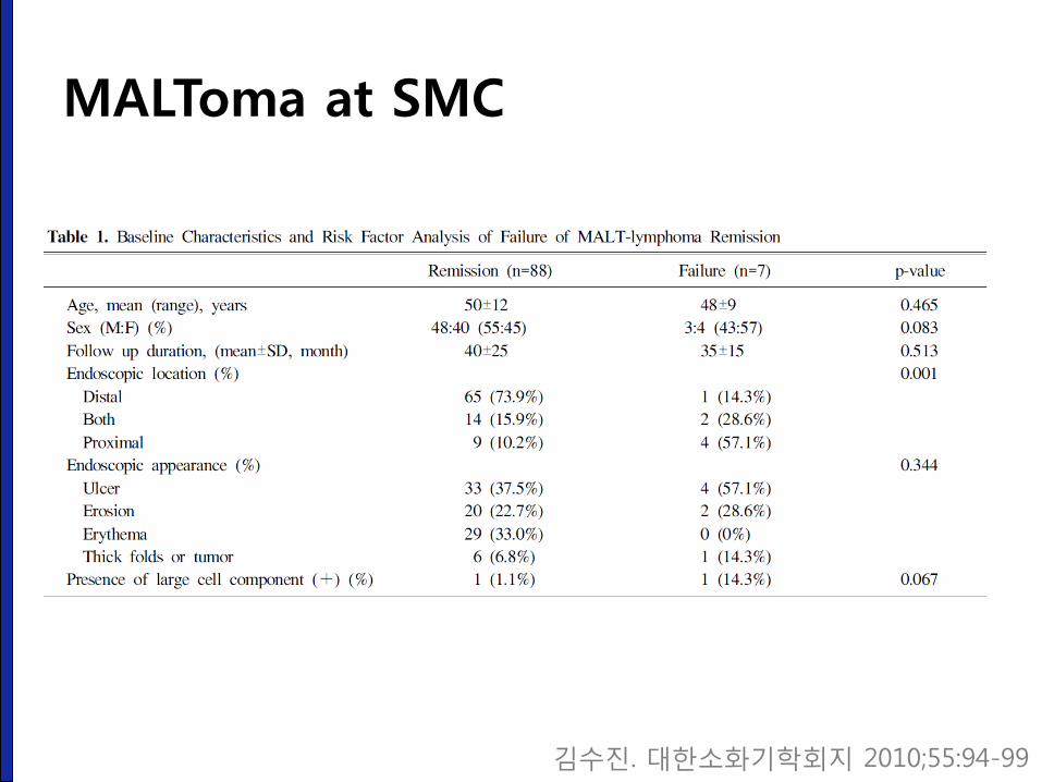
MALToma at SMC
김수진. 대한소화기학회지 2010;55:94-99
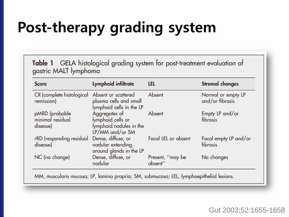
Post-therapy grading system
Gut 2003;52:1655-1658
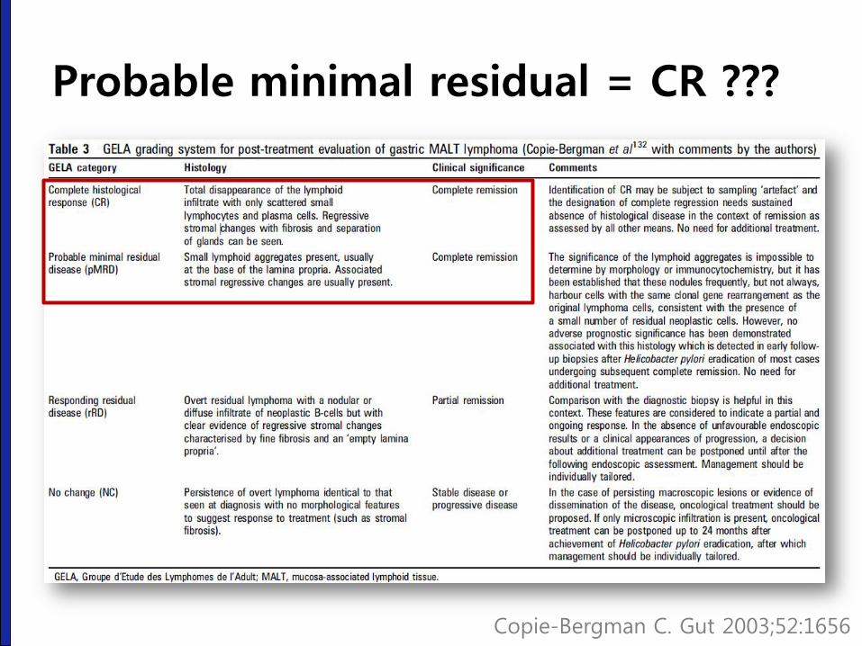
Copie-Bergman C. Gut 2003;52:1656
Probable minimal residual = CR ???
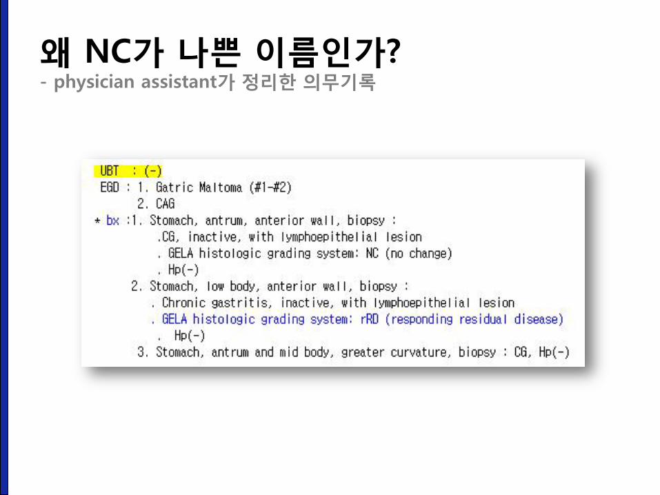
왜 NC가 나쁜 이름인가? - physician assistant가 정리한 의무기록
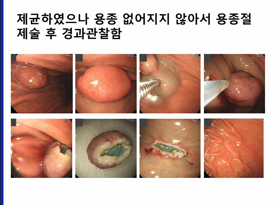
제균하였으나 용종 없어지지 않아서 용종절제술 후 경과관찰함
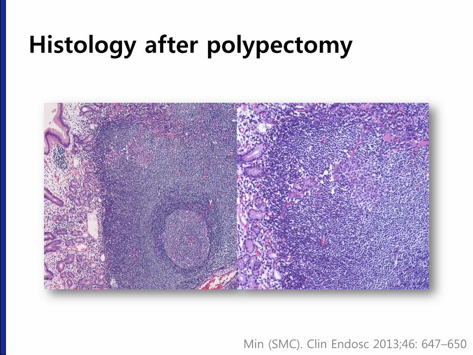
Histology after polypectomy
Min (SMC). Clin Endosc 2013;46: 647–650
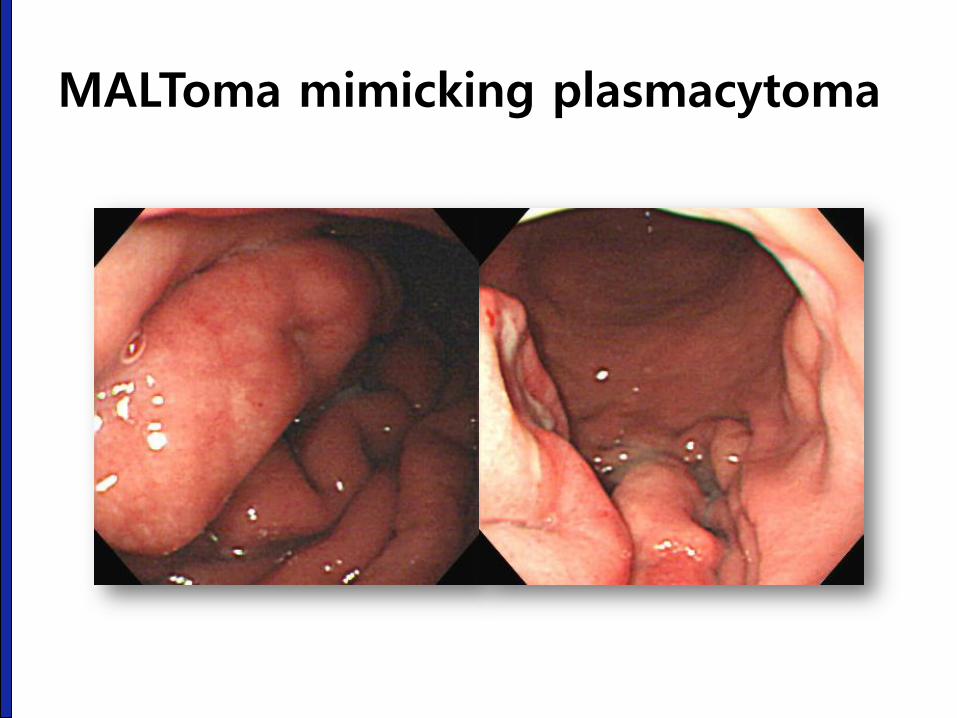
MALToma mimicking plasmacytoma
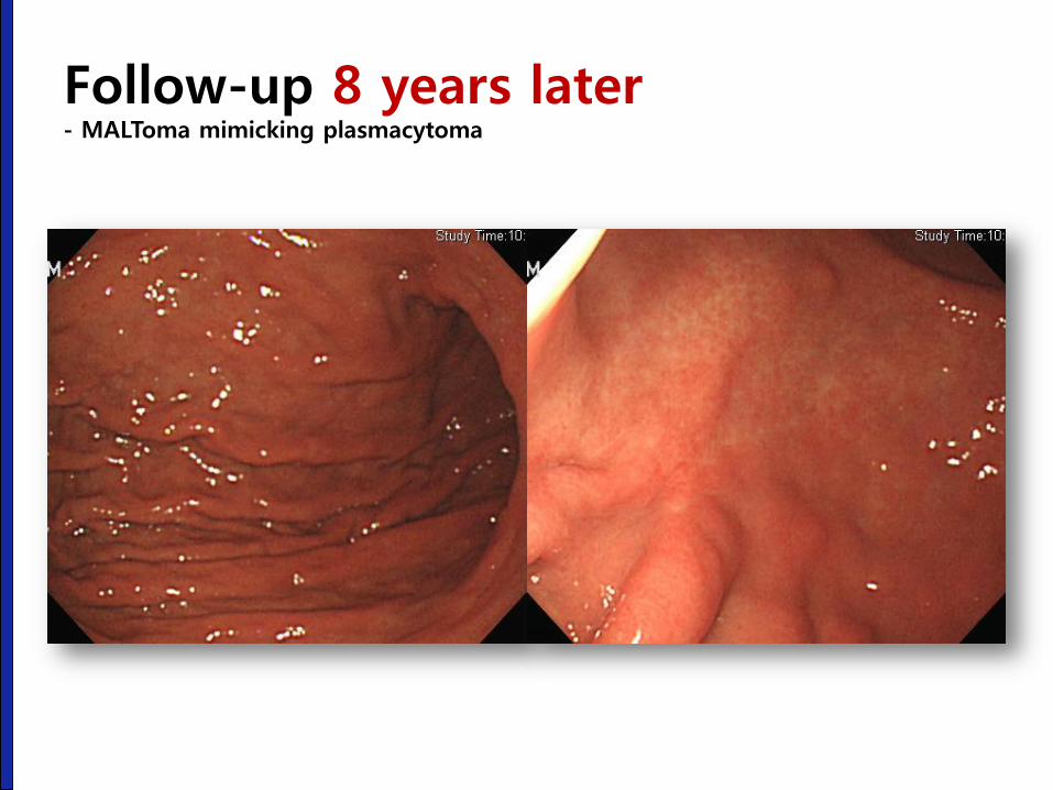
Follow-up 8 years later - MALToma mimicking plasmacytoma
MALT lymphoma simulating an extra-medullary plasmacytoma of the stomach
• Approximately one third of all cases of gastric MALToma
show variable degrees of plasma cell differentiation,
occasionally plasma cells constitute the major population in
the tumor.
• As surgical resection has been the standard treatment for
gastric plasmacyotoma, this case highlights the need for
caution. If H. pylori infection is found in a patient with a
gastric plasmacytoma, it should be eradicated as a first line
of therapy before surgery is considered.
Kodama. Am J Med 1999;107:530-532
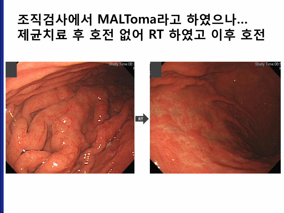
조직검사에서 MALToma라고 하였으나… 제균치료 후 호전 없어 RT 하였고 이후 호전
RT
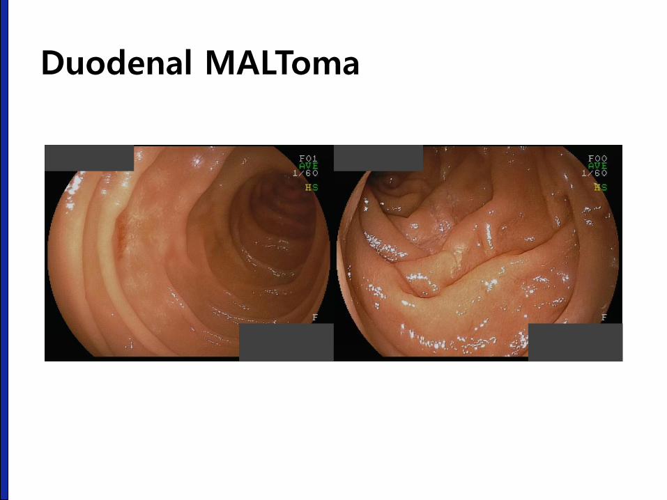
Duodenal MALToma
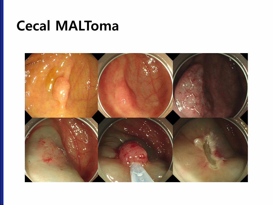
Cecal MALToma
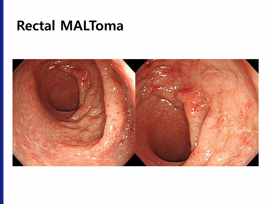
Rectal MALToma
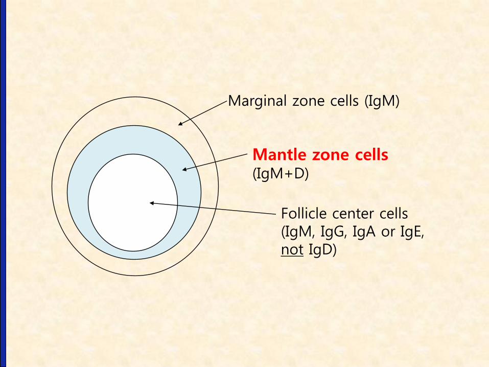
Marginal zone cells (IgM)
Mantle zone cells (IgM+D)
Follicle center cells (IgM, IgG, IgA or IgE, not IgD)

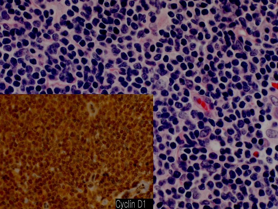
Lymphomatous polyposis (M/66)
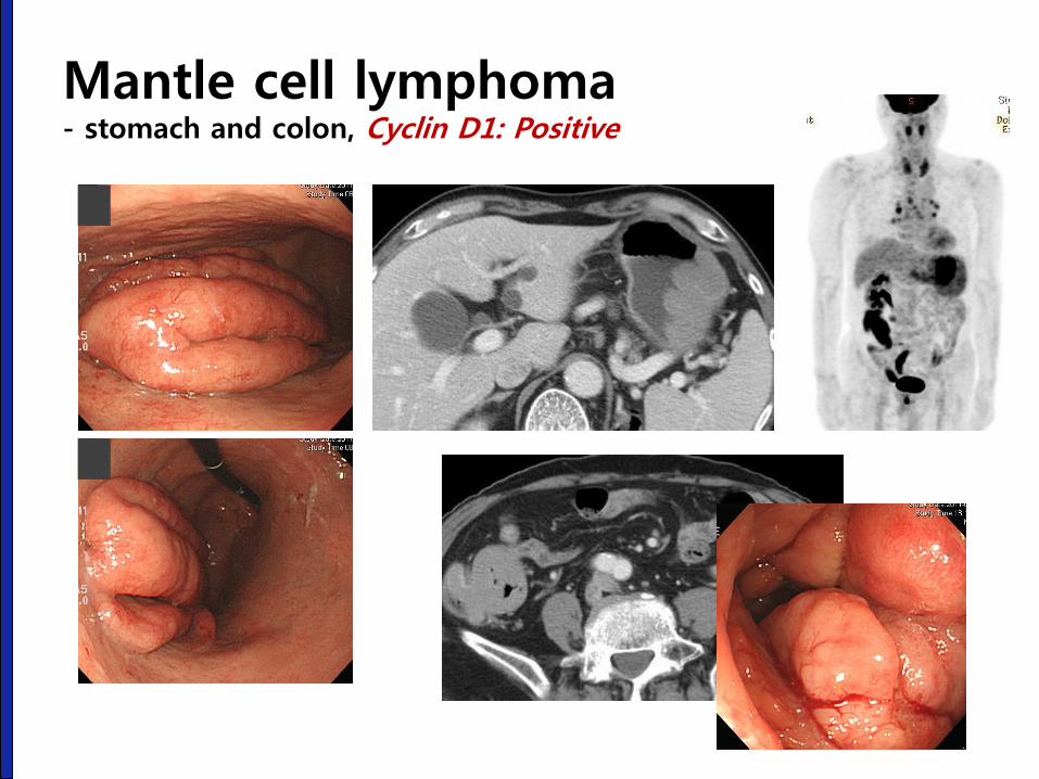
Mantle cell lymphoma - stomach and colon, Cyclin D1: Positive
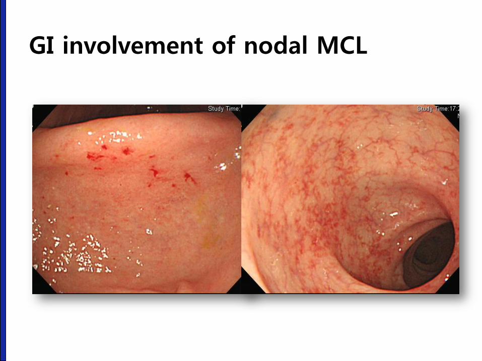
GI involvement of nodal MCL
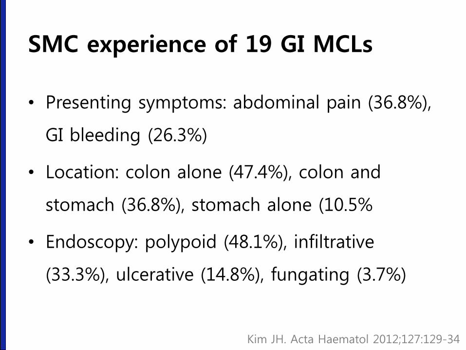
SMC experience of 19 GI MCLs
• Presenting symptoms: abdominal pain (36.8%),
GI bleeding (26.3%)
• Location: colon alone (47.4%), colon and
stomach (36.8%), stomach alone (10.5%
• Endoscopy: polypoid (48.1%), infiltrative
(33.3%), ulcerative (14.8%), fungating (3.7%)
Kim JH. Acta Haematol 2012;127:129-34
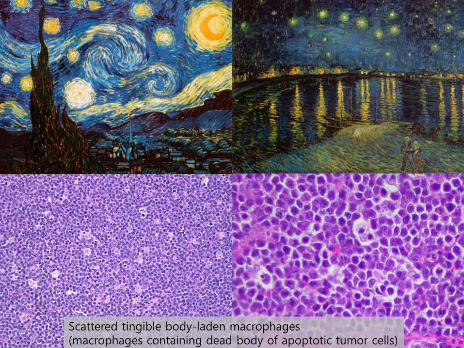
Scattered tingible body-laden macrophages (macrophages containing dead body of apoptotic tumor cells)
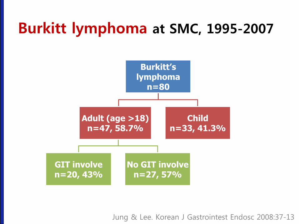
Burkitt lymphoma at SMC, 1995-2007
Burkitt’s lymphoma
n=80
Adult (age >18) n=47, 58.7%
GIT involve n=20, 43%
No GIT involve n=27, 57%
Child n=33, 41.3%
Jung & Lee. Korean J Gastrointest Endosc 2008:37-13
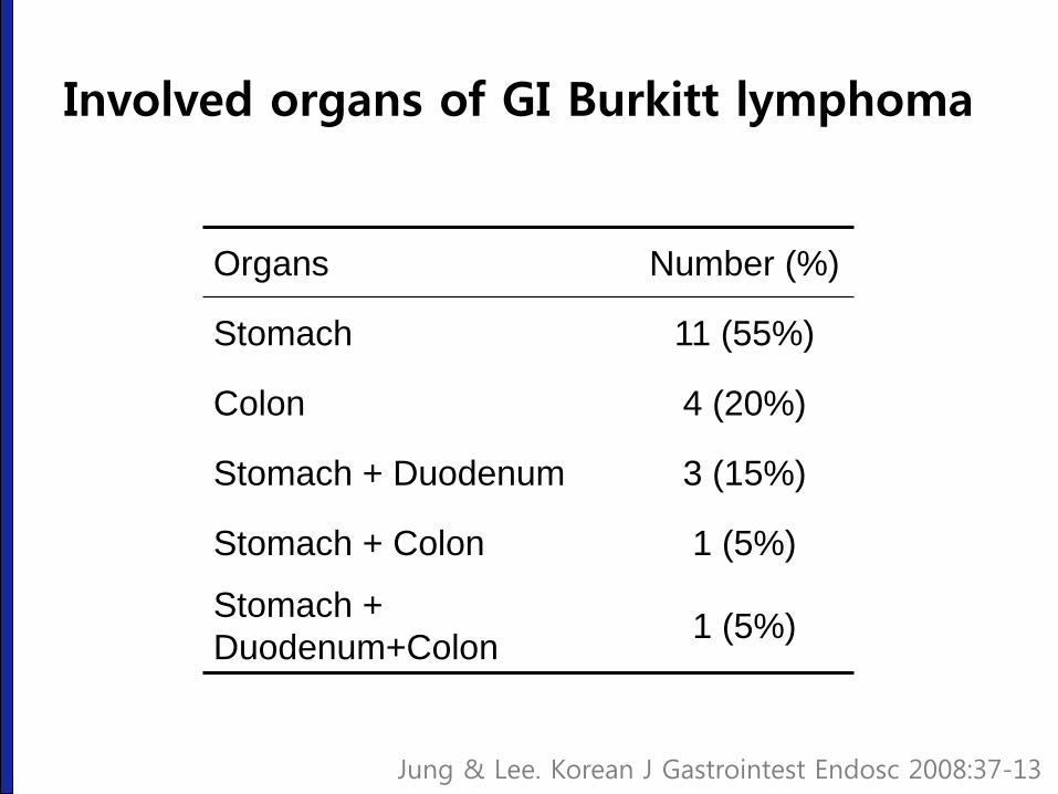
Involved organs of GI Burkitt lymphoma
Organs Number (%)
Stomach 11 (55%)
Colon 4 (20%)
Stomach + Duodenum 3 (15%)
Stomach + Colon 1 (5%)
Stomach +
Duodenum+Colon 1 (5%)
Jung & Lee. Korean J Gastrointest Endosc 2008:37-13
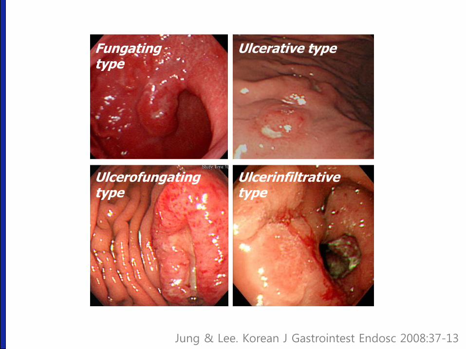
Fungating type
Ulcerative type
Ulcerofungating type
Ulcerinfiltrative type
Jung & Lee. Korean J Gastrointest Endosc 2008:37-13

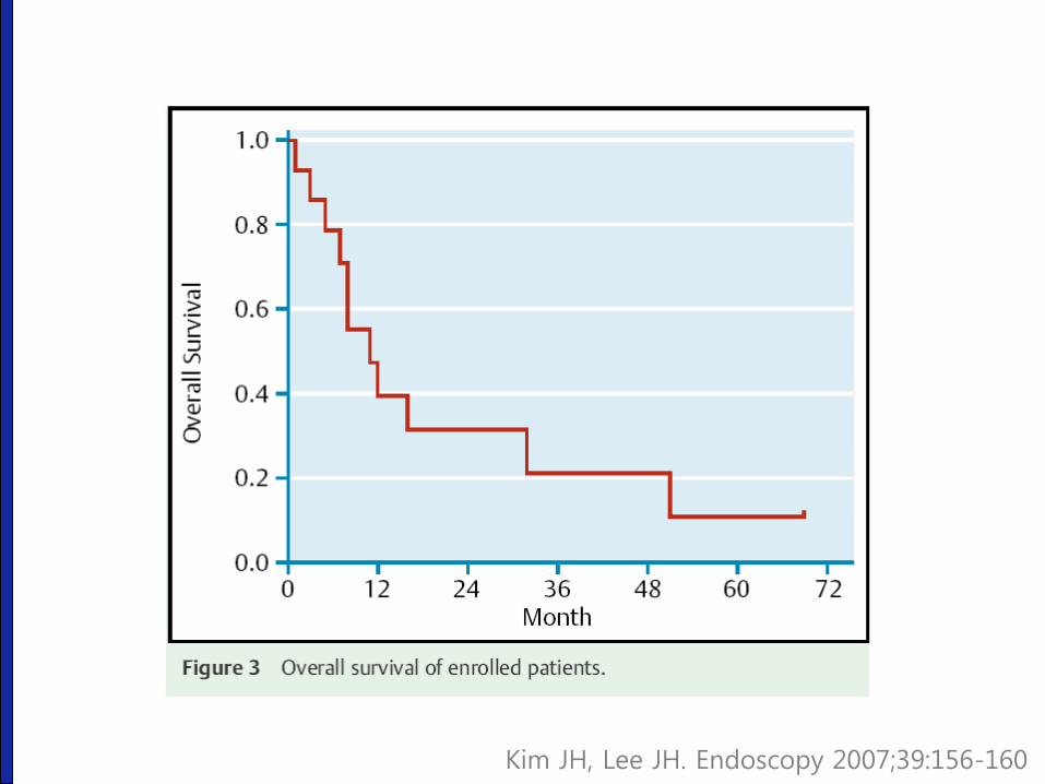
Survival of GI Burkitt lymphoma
0 10 20 30 40 50 600
25
50
75
100
Follow-up duration (months)
Cu
mu
lati
ve s
urv
ival
rate
(%
)
Jung & Lee. Korean J Gastrointest Endosc 2008:37-13
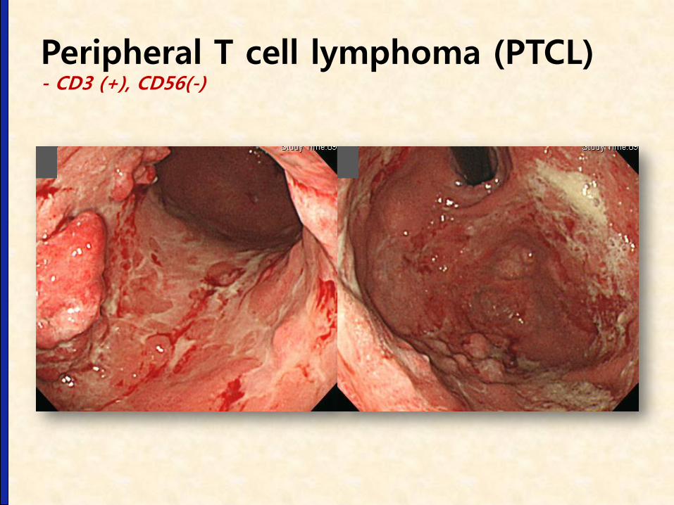
Peripheral T cell lymphoma (PTCL) - CD3 (+), CD56(-)
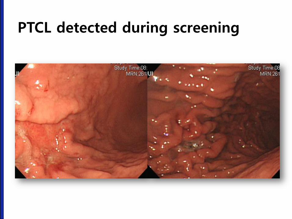
PTCL detected during screening
Kim JH, Lee JH. Endoscopy 2007;39:156-160
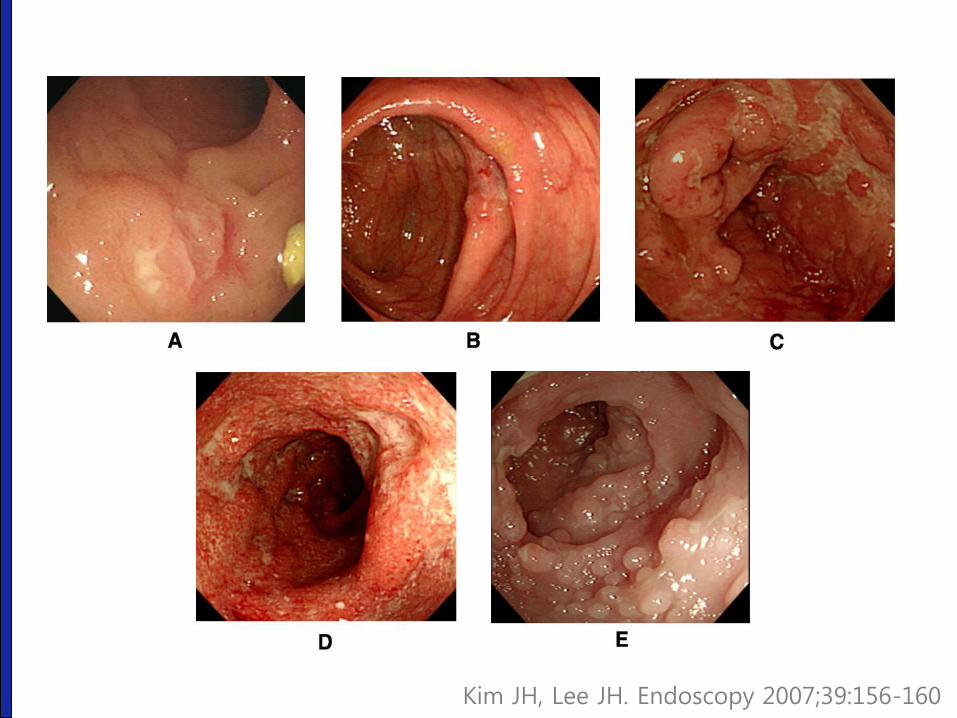
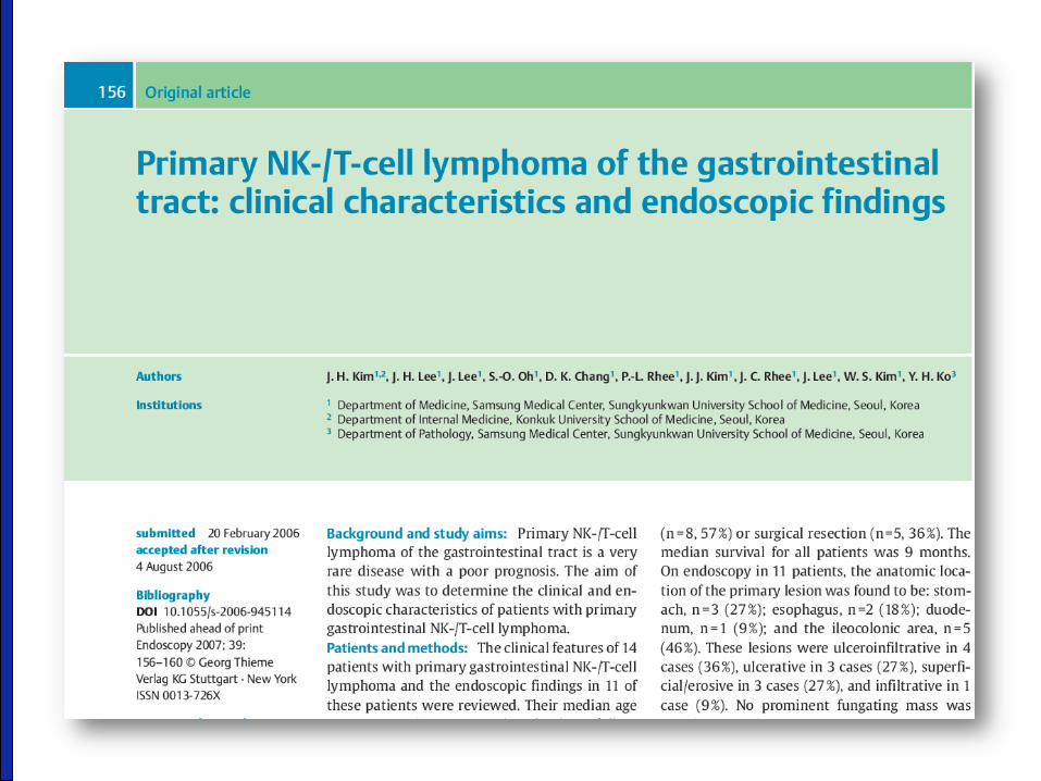
Kim JH, Lee JH. Endoscopy 2007;39:156-160
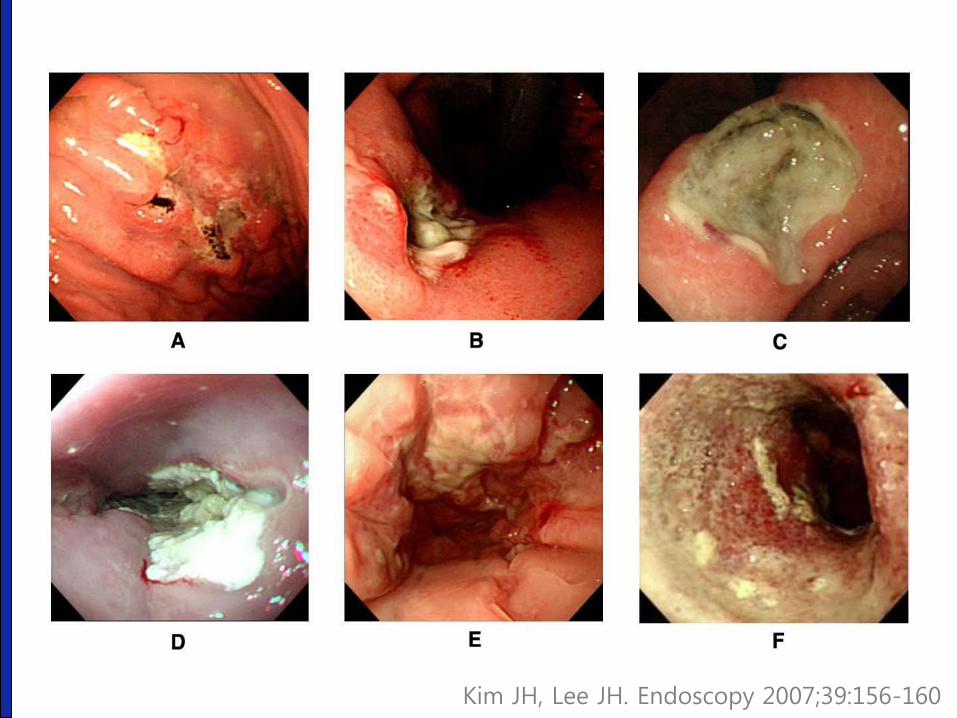
Kim JH, Lee JH. Endoscopy 2007;39:156-160
결론: 위장관 림프종
• 내시경 진단이 항상 쉬운 것은 아니다.
• 병리 검사 결과 해석에 주의하자.
• 우리나라에는 장 T-세포 림프종이 많다.
• 진단이 지연될 수 있다.
• 위장관 림프종은 매우 다양하다.
아래 종설을 참고하시기 바랍니다.
경청해 주셔서 감사합니다.