Embed Size (px)
Citation preview
8/10/2019 lucho 1jjj
http://slidepdf.com/reader/full/lucho-1jjj 1/8
7
Acute Changes in FetalHeart Rate Tracing: When ItBecomes an EmergencyBaha M. Sibai M.D.
John R. Barton M.D.
2
Video Clips on DVD
Continuous electronic fetal heart rate (FHR) monitoring is widely used tomonitor all pregnant women with high-risk medical or obstetric conditions, aswell as most pregnant women undergoing labor and delivery. The objectivesof FHR monitoring during labor are early detection of changes in FHR baselineand patterns in order to identify certain categories that are predictive of fetalhypoxia and acidosis (Table 2-1). Once these changes are identied, the nextstep is for the medical provider to decide on which ones require careful obser-vation and which FHR require immediate delivery.
Guidelines have been published by the National Institute of Child Health andHuman Development (NICHD) working group for denitions, interpretation,
and management recommendations for various categories of FHR tracings. Theresearch group dened three categories as either normal (Category I), indeter-minate (Category II), and abnormal (Category III). These denitions are listedin Table 2-2 (Category I) and Table 2-3 (Category III). Category II is dened asany pattern not included in Category I or III. An example of Category I FHRpatterns is seen in Figure 2-1 , of Category II in Figure 2-2 , and of Category III inFigure 2-3 . The same group also recommended abolishing the term hyperstimula-tion for uterine activity and suggested using the term uterine tachysystole (Table2-4). The denition of baseline variability is described in Table 2-5. Minimal orabsent variability can be due to medications that depress the fetal central nervoussystem, hypoxia, or acidosis (maternal or fetal). The presence or absence of FHRaccelerations was not considered important to dene the three categories.
The NICHD criteria dene normal FHR baseline as a rate of 100 to 160 bpm.Fetal bradycardia is dened as a baseline of < 100 bpm for at least 10 minutes,whereas fetal tachycardia is dened as a baseline of > 160 bpm for at least 10minutes. Fetal tachycardia can be related to various etiologies (Table 2-6). Therefore, management should be individualized based on the etiology, thepersistence of the pattern, and response to corrective factors.
It is important to emphasize that a category II FHR pattern will be seen inalmost all labors, and most of these are reassuring, whereas in others they canbe ominous requiring immediate delivery. Therefore, there are potential pitfallswith the new NICHD classication (Table 2-7).
2-1 PowerPoint Discussion of a Variety ofFetal Heart Rate Tracings
8/10/2019 lucho 1jjj
http://slidepdf.com/reader/full/lucho-1jjj 2/8
8 Management of Acute Obstetric Emergencies
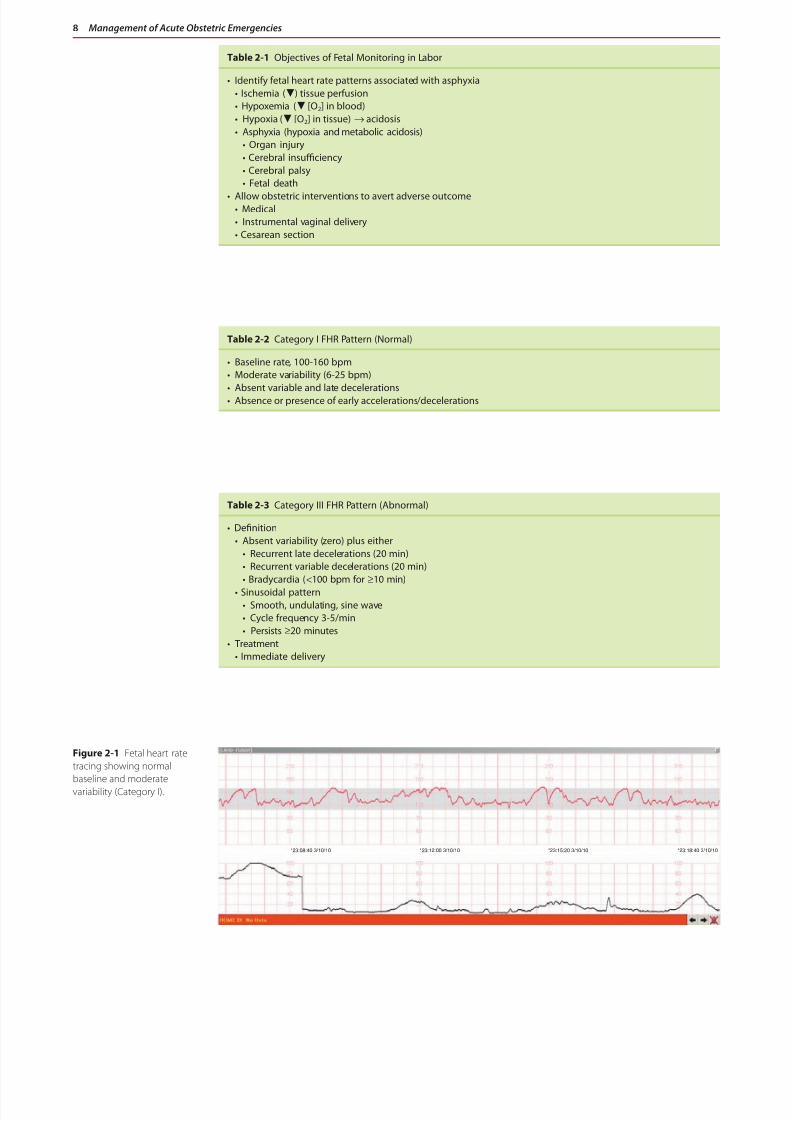
Table 2-1 Objectives of Fetal Monitoring in Labor
• Identify fetal heart rate patterns associated with asphyxia• Ischemia ( ) tissue perfusion• Hypoxemia ( [O2] in blood)• Hypoxia ( [O2] in tissue) → acidosis• Asphyxia (hypoxia and metabolic acidosis)
• Organ injury• Cerebral insufficiency• Cerebral palsy• Fetal death
• Allow obstetric interventions to avert adverse outcome• Medical• Instrumental vaginal delivery• Cesarean section
Table 2-2 Category I FHR Pattern (Normal)
• Baseline rate, 100-160 bpm• Moderate variability (6-25 bpm)
• Absent variable and late decelerations• Absence or presence of early accelerations/decelerations
Table 2-3 Category III FHR Pattern (Abnormal)
• Denition• Absent variability (zero) plus either
• Recurrent late decelerations (20 min)• Recurrent variable decelerations (20 min)• Bradycardia ( < 100 bpm for ≥ 10 min)
• Sinusoidal pattern• Smooth, undulating, sine wave• Cycle frequency 3-5/min• Persists ≥ 20 minutes
• Treatment• Immediate delivery
Figure 2-1 Fetal heart ratetracing showing normalbaseline and moderatevariability (Category I).
*23:08:40 3/10/10 *23:12:00 3/10/10 *23:15:20 3/10/10 *23:18:40 3/10/10

8/10/2019 lucho 1jjj
http://slidepdf.com/reader/full/lucho-1jjj 3/8
Acute Changes in Fetal Heart Rate Tracing: When It Becomes an Emergency 9 2
Figure 2-2 Fetal heart ratetracing revealing moderatevariability and late deceleration(Category II).
*4:33:40 3/5/10 *4:37:00 3/5/10 *4:40:20 3/5/10 *4:43:40 3/5/10
Figure 2-3 Absent variabilityand recurrent late decelerations(Category III).
*17:32:00 2/2/10 *17:35:20 2/2/10 *17:38:40 2/2/10 *17:42:00 2/2/10
Table 2-4 Uterine Tachysystole
• Denition
• More than 5 contractions in 10 minutes• Averaged over 30-minute window• With/without FHR decelerations
• Treatment• Discontinue oxytocin or prostaglandins• Give oxygen by mask • Give terbutaline 0.25 mg IV• Consider delivery if associated with abnormal FHR pattern and no response to therapy
Table 2-5 Baseline FHR Variability
Dened as uctuations that are irregular in amplitude and frequency (visually quantitated as the amplitude ofthe peak-to-trough in bpm).
Amplitude Range Classication
• Undetectable • Absent
• Undetectable to < 5 bpm • Minimal
• 6 to 25 bpm • Moderate
• More than 25 bpm • Marked
8/10/2019 lucho 1jjj
http://slidepdf.com/reader/full/lucho-1jjj 4/8
10 Management of Acute Obstetric Emergencies
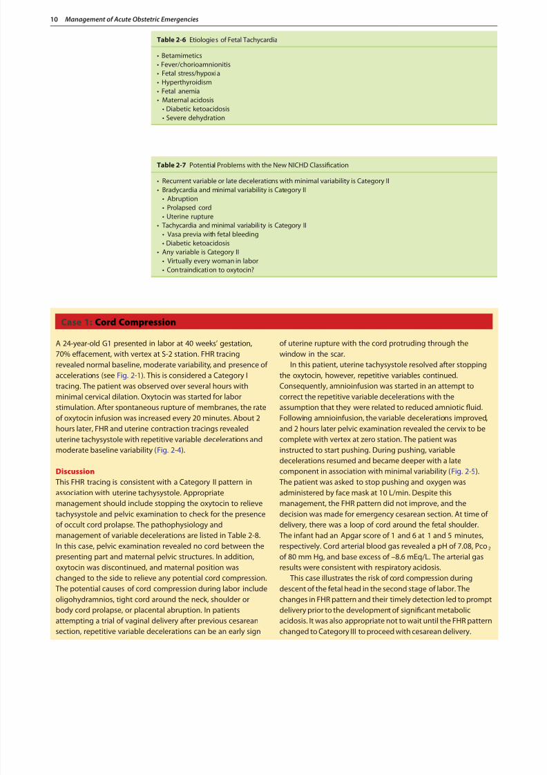
Table 2-6 Etiologies of Fetal Tachycardia
• Betamimetics• Fever/chorioamnionitis• Fetal stress/hypoxia• Hyperthyroidism• Fetal anemia• Maternal acidosis
• Diabetic ketoacidosis• Severe dehydration
Table 2-7 Potential Problems with the New NICHD Classication
• Recurrent variable or late decelerations with minimal variability is Category II• Bradycardia and minimal variability is Category II
• Abruption• Prolapsed cord• Uterine rupture
• Tachycardia and minimal variabili ty is Category II• Vasa previa with fetal bleeding• Diabetic ketoacidosis
• Any variable is Category II• Virtually every woman in labor
• Contraindication to oxytocin?
Case 1: Cord Compression
A 24-year-old G1 presented in labor at 40 weeks’ gestation,70% effacement, with vertex at S-2 station. FHR tracingrevealed normal baseline, moderate variability, and presence ofaccelerations (see Fig. 2-1). This is considered a Category Itracing. The patient was observed over several hours withminimal cervical dilation. Oxytocin was started for labor
stimulation. After spontaneous rupture of membranes, the rateof oxytocin infusion was increased every 20 minutes. About 2hours later, FHR and uterine contraction tracings revealeduterine tachysystole with repetitive variable decelerations andmoderate baseline variability ( Fig. 2-4).
Discussion This FHR tracing is consistent with a Category II pattern inassociation with uterine tachysystole. Appropriatemanagement should include stopping the oxytocin to relievetachysystole and pelvic examination to check for the presenceof occult cord prolapse. The pathophysiology andmanagement of variable decelerations are listed in Table 2-8.In this case, pelvic examination revealed no cord between thepresenting part and maternal pelvic structures. In addition,oxytocin was discontinued, and maternal position waschanged to the side to relieve any potential cord compression. The potential causes of cord compression during labor includeoligohydramnios, tight cord around the neck, shoulder orbody cord prolapse, or placental abruption. In patientsattempting a trial of vaginal delivery after previous cesareansection, repetitive variable decelerations can be an early sign
of uterine rupture with the cord protruding through thewindow in the scar.
In this patient, uterine tachysystole resolved after stoppingthe oxytocin, however, repetitive variables continued.Consequently, amnioinfusion was started in an attempt tocorrect the repetitive variable decelerations with the
assumption that they were related to reduced amniotic uid.Following amnioinfusion, the variable decelerations improved,and 2 hours later pelvic examination revealed the cervix to becomplete with vertex at zero station. The patient wasinstructed to start pushing. During pushing, variabledecelerations resumed and became deeper with a latecomponent in association with minimal variability ( Fig. 2-5). The patient was asked to stop pushing and oxygen wasadministered by face mask at 10 L/min. Despite thismanagement, the FHR pattern did not improve, and thedecision was made for emergency cesarean section. At time ofdelivery, there was a loop of cord around the fetal shoulder. The infant had an Apgar score of 1 and 6 at 1 and 5 minutes,respectively. Cord arterial blood gas revealed a pH of 7.08, Pco 2 of 80 mm Hg, and base excess of –8.6 mEq/L. The arterial gasresults were consistent with respiratory acidosis.
This case illustrates the risk of cord compression duringdescent of the fetal head in the second stage of labor. Thechanges in FHR pattern and their timely detection led to promptdelivery prior to the development of signicant metabolicacidosis. It was also appropriate not to wait until the FHR patternchanged to Category III to proceed with cesarean delivery.
8/10/2019 lucho 1jjj
http://slidepdf.com/reader/full/lucho-1jjj 5/8
Acute Changes in Fetal Heart Rate Tracing: When It Becomes an Emergency 11 2
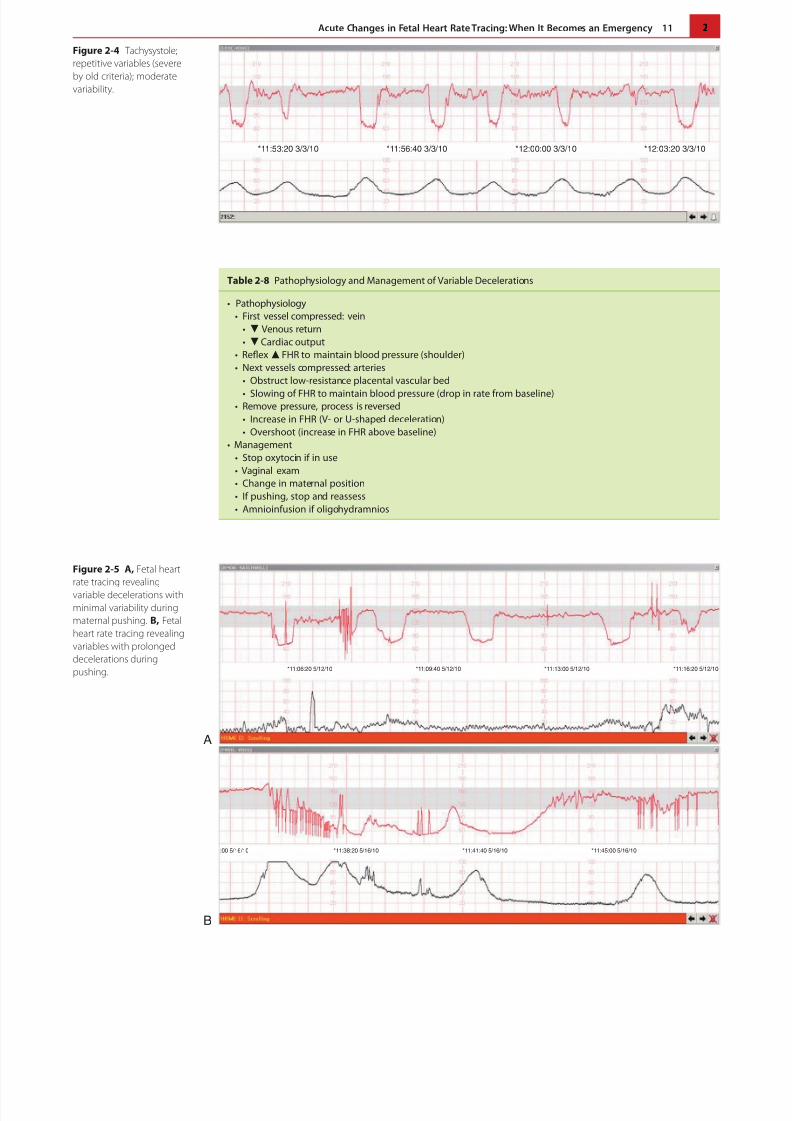
Figure 2-4 Tachysystole;repetitive variables (severeby old criteria); moderatevariability.
*11:53:20 3/3/10 *11:56:40 3/3/10 *12:00:00 3/3/10 *12:03:20 3/3/10
Table 2-8 Pathophysiology and Management of Variable Decelerations
• Pathophysiology• First vessel compressed: vein
• Venous return• Cardiac output
• Reex FHR to maintain blood pressure (shoulder)• Next vessels compressed: arteries
• Obstruct low-resistance placental vascular bed
• Slowing of FHR to maintain blood pressure (drop in rate from baseline)• Remove pressure, process is reversed
• Increase in FHR (V- or U-shaped deceleration)• Overshoot (increase in FHR above baseline)
• Management• Stop oxytocin if in use• Vaginal exam• Change in maternal position• If pushing, stop and reassess• Amnioinfusion if oligohydramnios
Figure 2-5 A, Fetal heartrate tracing revealingvariable decelerations withminimal variability duringmaternal pushing. B, Fetalheart rate tracing revealingvariables with prolongeddecelerations duringpushing.
A
B
*11:06:20 5/12/10 *11:09:40 5/12/10 *11:13:00 5/12/10 *11:16:20 5/12/10
:00 5/16/10 *11:38:20 5/16/10 *11:41:40 5/16/10 *11:45:00 5/16/10
8/10/2019 lucho 1jjj
http://slidepdf.com/reader/full/lucho-1jjj 6/8
12 Management of Acute Obstetric Emergencies
Case 2: Chorioamnionitis
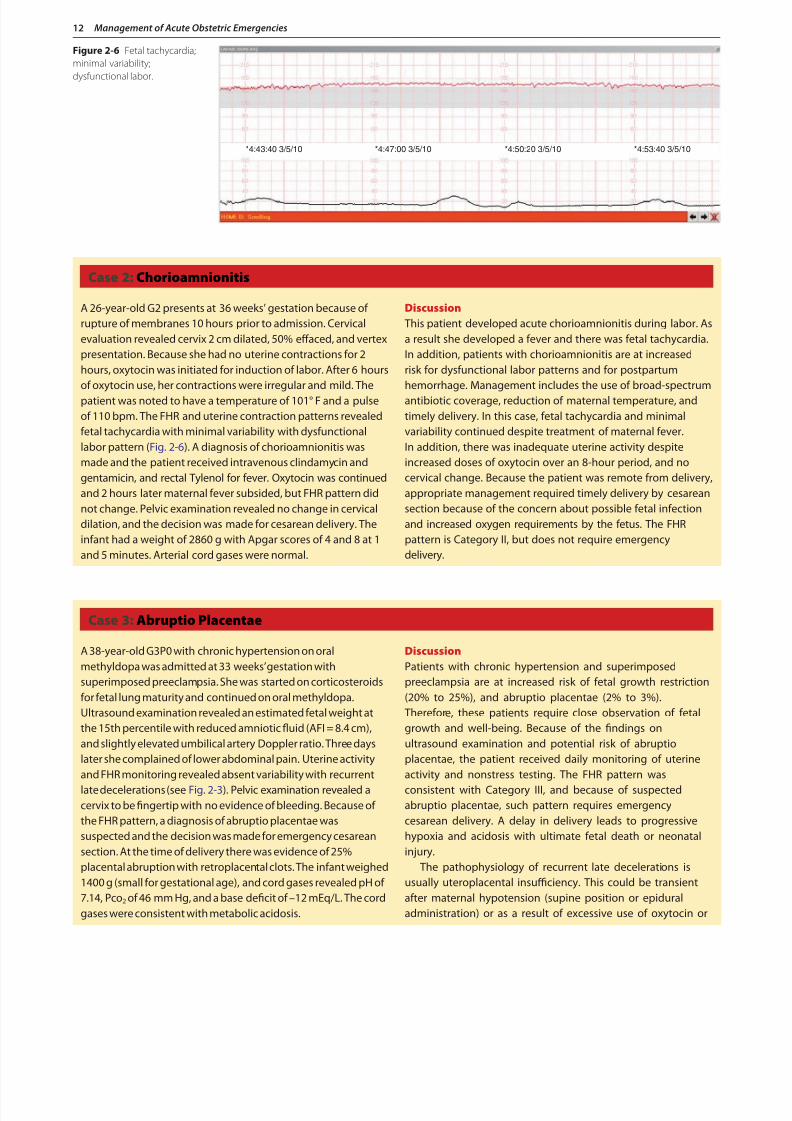
A 26-year-old G2 presents at 36 weeks’ gestation because ofrupture of membranes 10 hours prior to admission. Cervicalevaluation revealed cervix 2 cm dilated, 50% effaced, and vertexpresentation. Because she had no uterine contractions for 2hours, oxytocin was initiated for induction of labor. After 6 hoursof oxytocin use, her contractions were irregular and mild. Thepatient was noted to have a temperature of 101° F and a pulseof 110 bpm. The FHR and uterine contraction patterns revealedfetal tachycardia with minimal variability with dysfunctionallabor pattern ( Fig. 2-6). A diagnosis of chorioamnionitis wasmade and the patient received intravenous clindamycin andgentamicin, and rectal Tylenol for fever. Oxytocin was continuedand 2 hours later maternal fever subsided, but FHR pattern didnot change. Pelvic examination revealed no change in cervicaldilation, and the decision was made for cesarean delivery. Theinfant had a weight of 2860 g with Apgar scores of 4 and 8 at 1and 5 minutes. Arterial cord gases were normal.
Discussion This patient developed acute chorioamnionitis during labor. Asa result she developed a fever and there was fetal tachycardia.In addition, patients with chorioamnionitis are at increasedrisk for dysfunctional labor patterns and for postpartumhemorrhage. Management includes the use of broad-spectrumantibiotic coverage, reduction of maternal temperature, andtimely delivery. In this case, fetal tachycardia and minimalvariability continued despite treatment of maternal fever.In addition, there was inadequate uterine activity despiteincreased doses of oxytocin over an 8-hour period, and nocervical change. Because the patient was remote from delivery,appropriate management required timely delivery by cesareansection because of the concern about possible fetal infectionand increased oxygen requirements by the fetus. The FHRpattern is Category II, but does not require emergencydelivery.
Figure 2-6 Fetal tachycardia;minimal variability;dysfunctional labor.
*4:43:40 3/5/10 *4:47:00 3/5/10 *4:50:20 3/5/10 *4:53:40 3/5/10
Case 3: Abruptio Placentae
A 38-year-old G3P0 with chronic hypertension on oralmethyldopa was admitted at 33 weeks’ gestation withsuperimposed preeclampsia. She was started on corticosteroidsfor fetal lung maturity and continued on oral methyldopa.Ultrasound examination revealed an estimated fetal weight atthe 15th percentile with reduced amniotic uid (AFI = 8.4 cm),and slightly elevated umbilical artery Doppler ratio. Three dayslater she complained of lower abdominal pain. Uterine activityand FHR monitoring revealed absent variability with recurrentlate decelerations (see Fig. 2-3). Pelvic examination revealed acervix to be ngertip with no evidence of bleeding. Because ofthe FHR pattern, a diagnosis of abruptio placentae wassuspected and the decision was made for emergency cesareansection. At the time of delivery there was evidence of 25%placental abruption with retroplacental clots. The infant weighed1400 g (small for gestational age), and cord gases revealed pH of7.14, Pco 2 of 46 mm Hg, and a base decit of –12 mEq/L. The cordgases were consistent with metabolic acidosis.
DiscussionPatients with chronic hypertension and superimposedpreeclampsia are at increased risk of fetal growth restriction(20% to 25%), and abruptio placentae (2% to 3%). Therefore, these patients require close observation of fetalgrowth and well-being. Because of the ndings onultrasound examination and potential risk of abruptioplacentae, the patient received daily monitoring of uterineactivity and nonstress testing. The FHR pattern wasconsistent with Category III, and because of suspectedabruptio placentae, such pattern requires emergencycesarean delivery. A delay in delivery leads to progressivehypoxia and acidosis with ultimate fetal death or neonatalinjury.
The pathophysiology of recurrent late decelerations isusually uteroplacental insufficiency. This could be transientafter maternal hypotension (supine position or epiduraladministration) or as a result of excessive use of oxytocin or
8/10/2019 lucho 1jjj
http://slidepdf.com/reader/full/lucho-1jjj 7/8
Acute Changes in Fetal Heart Rate Tracing: When It Becomes an Emergency 13 2
prostaglandins with associated prolonged tachysystole.In such cases, recurrent decelerations will resolve aftercorrection of the etiology, and labor can then be continuedfor possible vaginal delivery. In contrast, in patients withrecurrent decelerations secondary to chronic uteroplacental
insufficiency (fetal growth restriction or post-termpregnancy) or acute insufficiency (abruptio placentae orruptured uterine scar), management requires promptdelivery by cesarean section unless vaginal delivery isimminent.
Case 3: Abruptio Placentae—cont’d
Case 4: Tachysystole
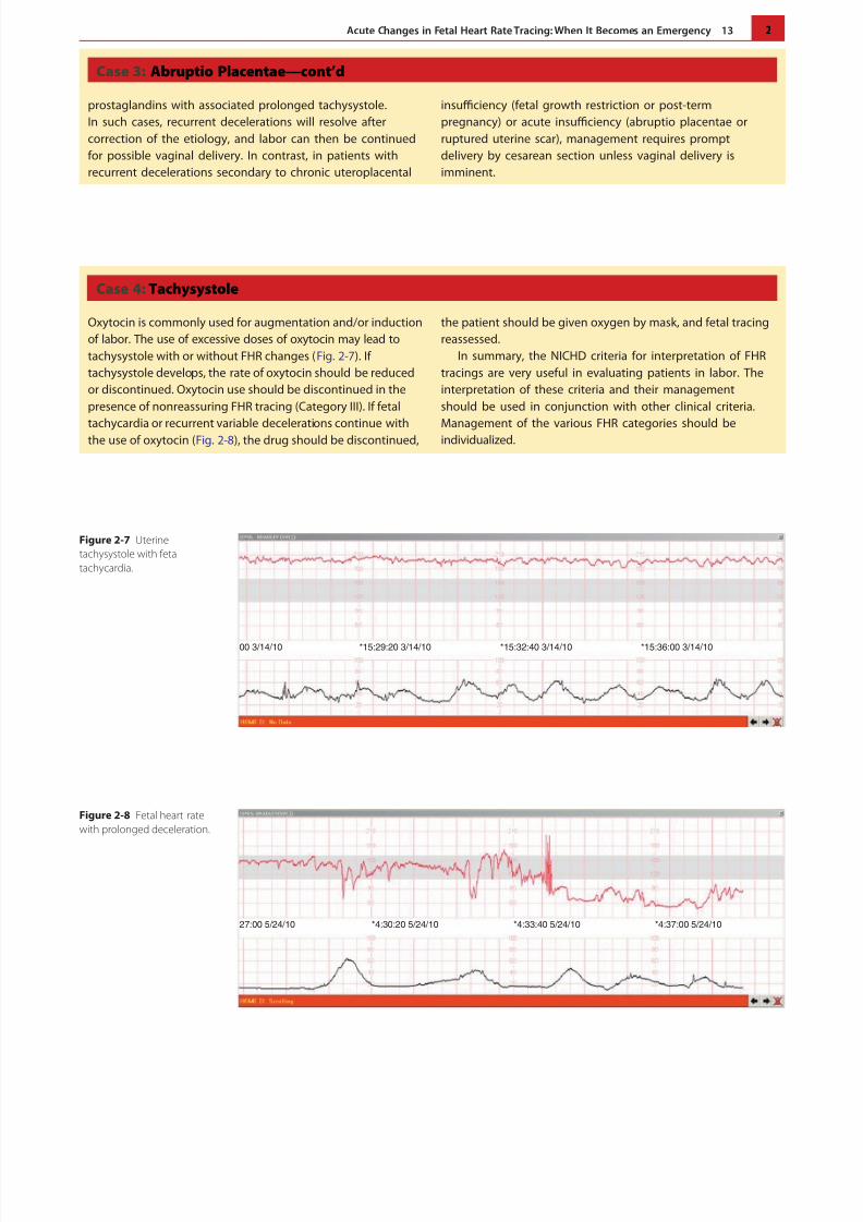
Oxytocin is commonly used for augmentation and/or inductionof labor. The use of excessive doses of oxytocin may lead totachysystole with or without FHR changes ( Fig. 2-7). Iftachysystole develops, the rate of oxytocin should be reducedor discontinued. Oxytocin use should be discontinued in thepresence of nonreassuring FHR tracing (Category III). If fetal
tachycardia or recurrent variable decelerations continue withthe use of oxytocin ( Fig. 2-8), the drug should be discontinued,
the patient should be given oxygen by mask, and fetal tracingreassessed.
In summary, the NICHD criteria for interpretation of FHRtracings are very useful in evaluating patients in labor. Theinterpretation of these criteria and their managementshould be used in conjunction with other clinical criteria.
Management of the various FHR categories should beindividualized.
Figure 2-7 Uterinetachysystole with fetaltachycardia.
00 3/14/10 *15:29:20 3/14/10 *15:32:40 3/14/10 *15:36:00 3/14/10
Figure 2-8 Fetal heart ratewith prolonged deceleration.
27:00 5/24/10 *4:30:20 5/24/10 *4:33:40 5/24/10 *4:37:00 5/24/10

8/10/2019 lucho 1jjj
http://slidepdf.com/reader/full/lucho-1jjj 8/8
14 Management of Acute Obstetric Emergencies
Suggested ReadingsACOG Practice Bulletin: Intrapartum fetal heart rate monitoring: nomenclature interpretation, and
general management principles. Number 106, July 2009.Blix E, Sviggum O, Koss KS: Inter-observer variation in assessment of 845 labour admission tests:
comparisons between midwives and obstetricians in the clinical setting and two experts. BJOG2003;110:1–5.
Freeman RK, Nageotte MP: Comments on American College of Obstetricians and GynecologistsPractice Bulletin No. 106. Am J Obstet Gynecol 2010;202:411–412.
Macones GA, Hawkins GDV, Spong CY, et al: The 2008 National Institute of Child Health and HumanDevelopment workshop report on electronic fetal monitoring. Update on denitions, interpretationand research guidelines. Obstet Gynecol 2008;112:661–666.
Parer JT, Ikeda T, King TL: The 2008 National Institute of Child Health and Human developmentreport on fetal heart rate monitoring. Obstet Gynecol 2009;114:136–138.
Parer JT, King T, Flanders S, et al: Fetal academia and electronic fetal heart rate patterns: is thereevidence of an association? J Matern Fetal Neonatal Med 2006;19:289–294.