Embed Size (px)
Citation preview
Management ofof
Localized Prostate Cancer
Andrew K. Lee, MD, MPHAssociate Professor
M.D. Anderson Cancer Center
Disclosure InformationA d K LAndrew K. Lee
Dr. Lee has indicated no financial relationships, arrangements or affiliationsarrangements or affiliations.
This presentation will not include discussion of investigational or off-label use of a product.
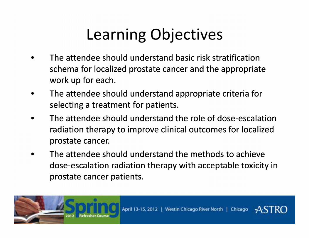
Learning Objectives•• The attendee should understand basic risk stratification The attendee should understand basic risk stratification
schema for localized prostate cancer and the appropriate schema for localized prostate cancer and the appropriate work up for each.work up for each.
•• The attendee should understand appropriate criteria for The attendee should understand appropriate criteria for selecting a treatment for patientsselecting a treatment for patientsselecting a treatment for patients.selecting a treatment for patients.
•• The attendee should understand the role of doseThe attendee should understand the role of dose--escalation escalation radiation therapy to improve clinical outcomes for localized radiation therapy to improve clinical outcomes for localized prostate cancer.prostate cancer.
•• The attendee should understand the methods to achieve The attendee should understand the methods to achieve dosedose escalation radiation therapy with acceptable toxicity inescalation radiation therapy with acceptable toxicity indosedose--escalation radiation therapy with acceptable toxicity in escalation radiation therapy with acceptable toxicity in prostate cancer patients.prostate cancer patients.
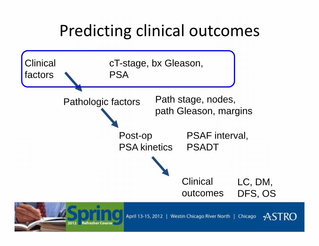
Predicting clinical outcomes
Clinical factors
cT-stage, bx Gleason, PSAfactors
Pathologic factors
PSA
Path stage, nodes, th Gl i
Post-op
path Gleason, margins
PSAF interval,Post op PSA kinetics
PSAF interval, PSADT
Clinical outcomes
LC, DM, DFS, OS
Localized prostate cancer
Localized PCaT1-2
Low riskT1-2
Gleason 6
PSA 10
IntermediateT2b
Gleason 7
PSA 10 20
HighT2c
Gleason 8-10
PSA 20PSA <10
Surgery
PSA 10-20
Surgery
PSA >20
EBRT + HTBrachyRx
EBRT
Active surveillance
EBRT +/- HT
EBRT + Brachy
Brachy alone (select)
EBRT + HT
EBRT + Brachy +HT
Surgery (select)
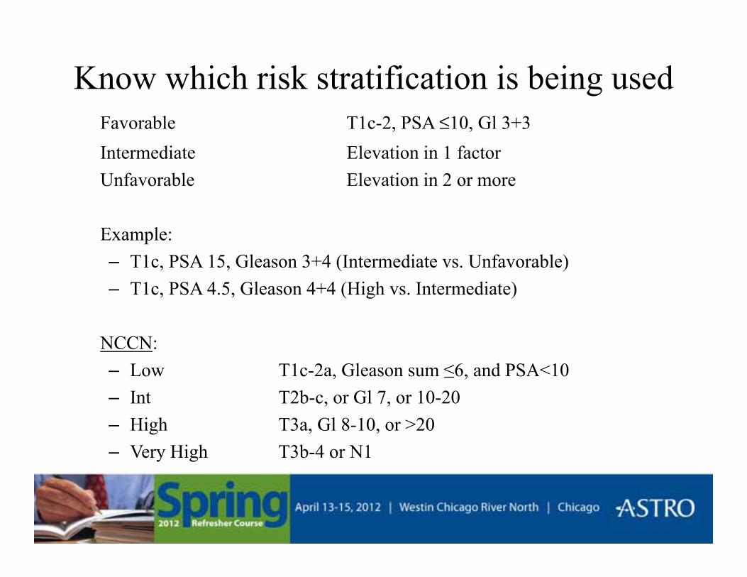
Know which risk stratification is being usedFavorable T1c-2, PSA ≤10, Gl 3+3Intermediate Elevation in 1 factorUnfavorable Elevation in 2 or moreUnfavorable Elevation in 2 or more
Example:– T1c, PSA 15, Gleason 3+4 (Intermediate vs. Unfavorable)– T1c, PSA 4.5, Gleason 4+4 (High vs. Intermediate)
NCCN: – Low T1c-2a, Gleason sum ≤6, and PSA<10– Int T2b c or Gl 7 or 10 20– Int T2b-c, or Gl 7, or 10-20– High T3a, Gl 8-10, or >20– Very High T3b-4 or N1
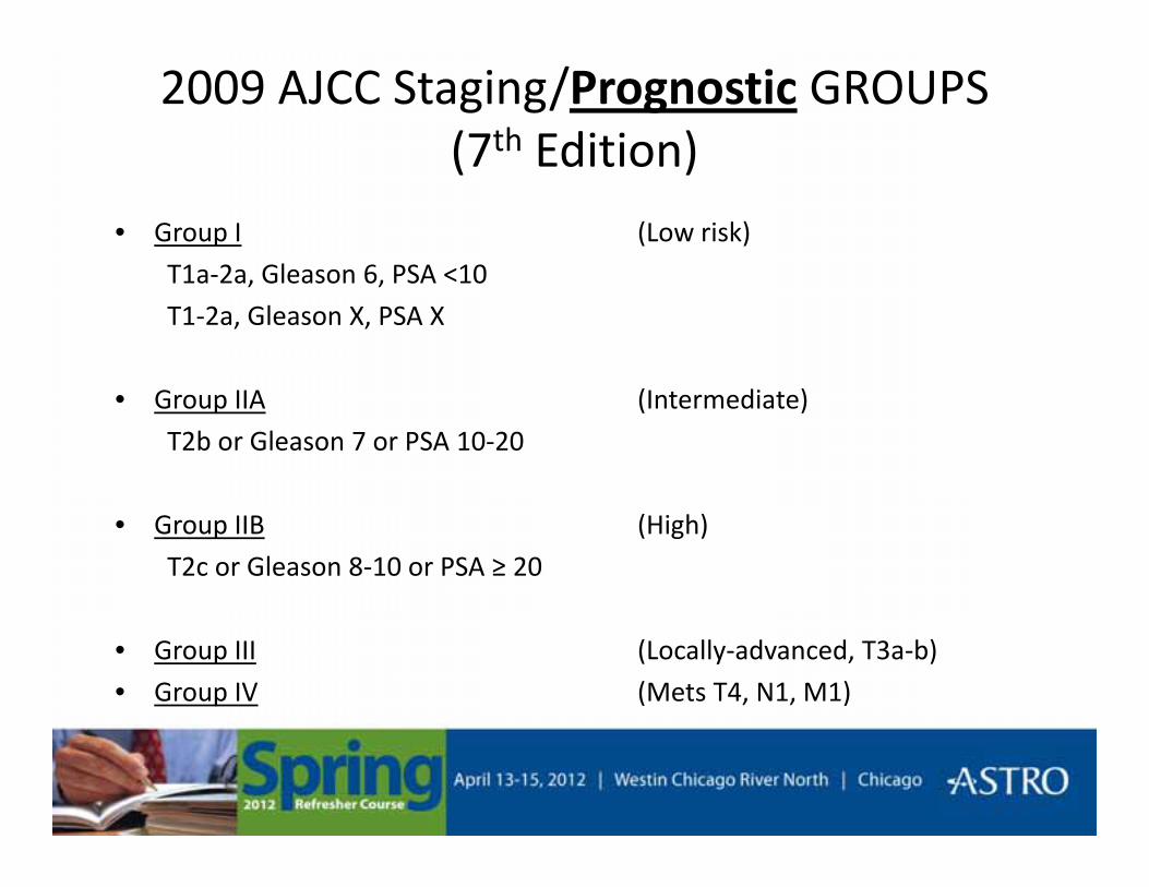
2009 AJCC Staging/Prognostic GROUPS(7th Edition)(7 Edition)
• Group I (Low risk)
T1a-2a Gleason 6 PSA <10T1a-2a, Gleason 6, PSA <10
T1-2a, Gleason X, PSA X
• Group IIA (Intermediate)
T2b or Gleason 7 or PSA 10-20
• Group IIB (High)
T2c or Gleason 8-10 or PSA ≥ 20
• Group III (Locally-advanced, T3a-b)
• Group IV (Mets T4, N1, M1)
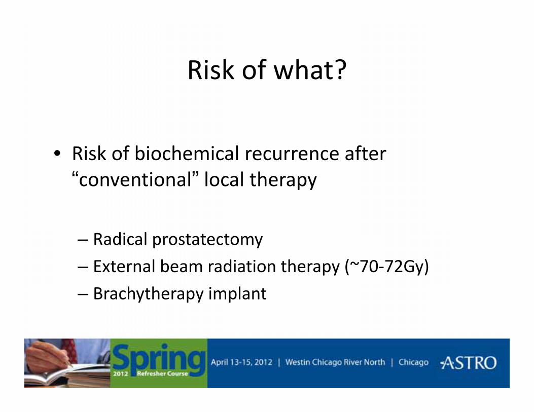
Risk of what?Risk of what?
• Risk of biochemical recurrence after “conventional” local therapyconventional local therapy
l– Radical prostatectomy
– External beam radiation therapy (~70-72Gy)
– Brachytherapy implant
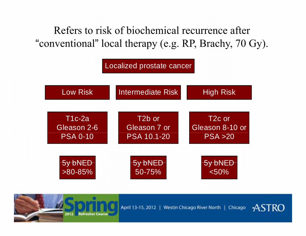
Refers to risk of biochemical recurrence after “conventional” local therapy (e g RP Brachy 70 Gy)conventional local therapy (e.g. RP, Brachy, 70 Gy).
Localized prostate cancer
Low Risk Intermediate Risk High Risk
T1c-2aGleason 2-6
T2b orGleason 7 or
T2c orGleason 8-10 or
5y bNED
PSA 0-10
5y bNED
PSA 10.1-20
5y bNED
PSA >20
5y bNED>80-85%
5y bNED50-75%
5y bNED<50%
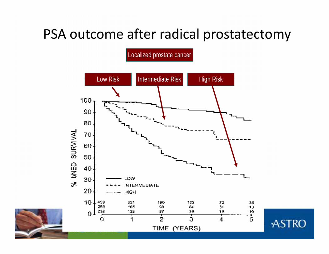
PSA outcome after radical prostatectomy
Low Risk Intermediate Risk High Risk
Localized prostate cancer
Low Risk Intermediate Risk High Risk
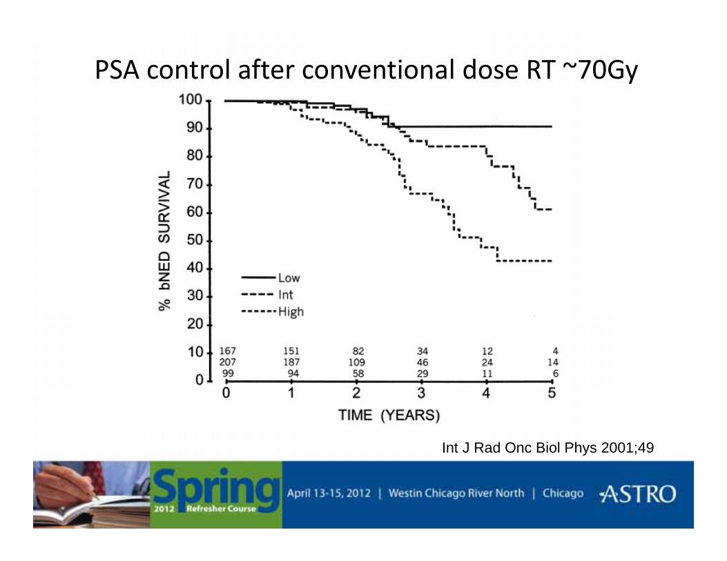
PSA control after conventional dose RT ~70Gy
Int J Rad Onc Biol Phys 2001;49
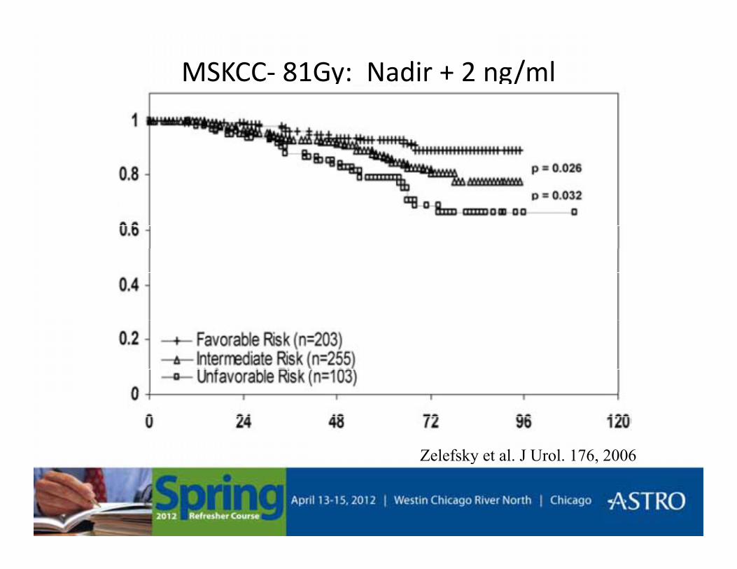
MSKCC- 81Gy: Nadir + 2 ng/ml
Zelefsky et al. J Urol. 176, 2006y
Why risk stratify?Why risk stratify?
• Match a given state of disease with the appropriate level of therapypp p py
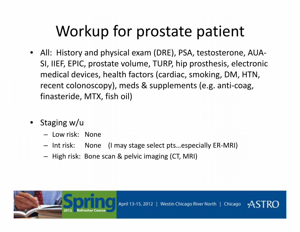
Workup for prostate patient• All: History and physical exam (DRE), PSA, testosterone, AUA-
SI, IIEF, EPIC, prostate volume, TURP, hip prosthesis, electronic di l d i h lth f t ( di ki DM HTNmedical devices, health factors (cardiac, smoking, DM, HTN,
recent colonoscopy), meds & supplements (e.g. anti-coag, finasteride, MTX, fish oil)
• Staging w/u– Low risk: None
– Int risk: None (I may stage select pts…especially ER-MRI)
– High risk: Bone scan & pelvic imaging (CT, MRI)
Treatment options for low-risk patientsTreatment options for low risk patients
• Radical prostatectomy
• External beam radiation therapy (>74Gy @ 1.8-2Gy)
• Brachytherapy implant
• Active surveillance
• Similar PSA control regardless of therapy (~85-90% at 5 years)
Active surveillance (AS)• In general, for low risk patients with life expectancy <10 years or pts
who want to defer Rx for a few years
Ob i AS “ i h i f d l d i i ”• Observation vs. AS “with opportunity for delayed intervention”– Approximately 40% will come off AS w/in 5 years (Klotz. JCO 2010)– Consider repeat TRUS-Bx (10-12 core including anterior horns and
central one) prior to AScentral zone) prior to AS– Repeat PSA @ 3-6 month intervals– Repeat TRUS Bx @ 1y (compare w/ entrance bx)
If i ifi t diff ti t it PSA d t TRUS– If no significant difference continue to monitor PSA and repeat TRUS Bx @ 2-3 y
• Consider Rx for “significant” PSA increase Gleason score change DRE findings• Consider Rx for significant PSA increase, Gleason score change, DRE findings
• Benefits vs. Risks (e.g. repeat bx, missed opportunity for cure, more aggressive Rx, limit Rx options, development of comorbidities may impact definitive Rx)
Patient selection for brachytherapy• cT1c-2a
• Gleason < 7These criteria are beginning to expand to include some int risk features• Gleason < 7
• PSA ≤ 10
int risk features
• Prostate volume < 50-60 cc (pubic arch interference)
• AUA score < 15 (post-implant obstruction)
• No prior TURP (Urethral necrosis, post-implant incontinence, dosimetry)co t e ce, dos et y)
• Hypertrophic median lobe (post-implant obstruction, dosimetry)
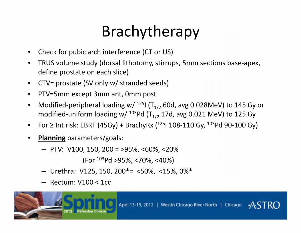
Brachytherapy• Check for pubic arch interference (CT or US)
• TRUS volume study (dorsal lithotomy, stirrups, 5mm sections base-apex, define prostate on each slice)define prostate on each slice)
• CTV= prostate (SV only w/ stranded seeds)
• PTV=5mm except 3mm ant, 0mm post
/ 125• Modified-peripheral loading w/ 125I (T1/2 60d, avg 0.028MeV) to 145 Gy or modified-uniform loading w/ 103Pd (T1/2 17d, avg 0.021 MeV) to 125 Gy
• For ≥ Int risk: EBRT (45Gy) + BrachyRx (125I 108-110 Gy, 103Pd 90-100 Gy)
• Planning parameters/goals:
– PTV: V100, 150, 200 = >95%, <60%, <20%
(For 103Pd >95% <70% <40%)(For Pd >95%, <70%, <40%)
– Urethra: V125, 150, 200*= <50%, <15%, 0%*
– Rectum: V100 < 1cc
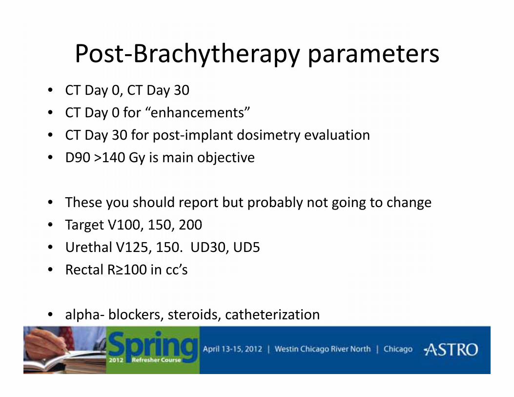
Post-Brachytherapy parameters• CT Day 0, CT Day 30
• CT Day 0 for “enhancements”
• CT Day 30 for post-implant dosimetry evaluation
• D90 >140 Gy is main objective
• These you should report but probably not going to change
• Target V100 150 200• Target V100, 150, 200
• Urethal V125, 150. UD30, UD5
• Rectal R≥100 in cc’sRectal R≥100 in cc s
• alpha- blockers, steroids, catheterization
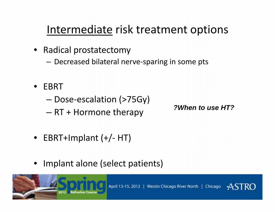
Intermediate risk treatment options
• Radical prostatectomy– Decreased bilateral nerve-sparing in some pts
• EBRT– Dose-escalation (>75Gy)– RT + Hormone therapy ?When to use HT?
• EBRT+Implant (+/- HT)
• Implant alone (select patients)
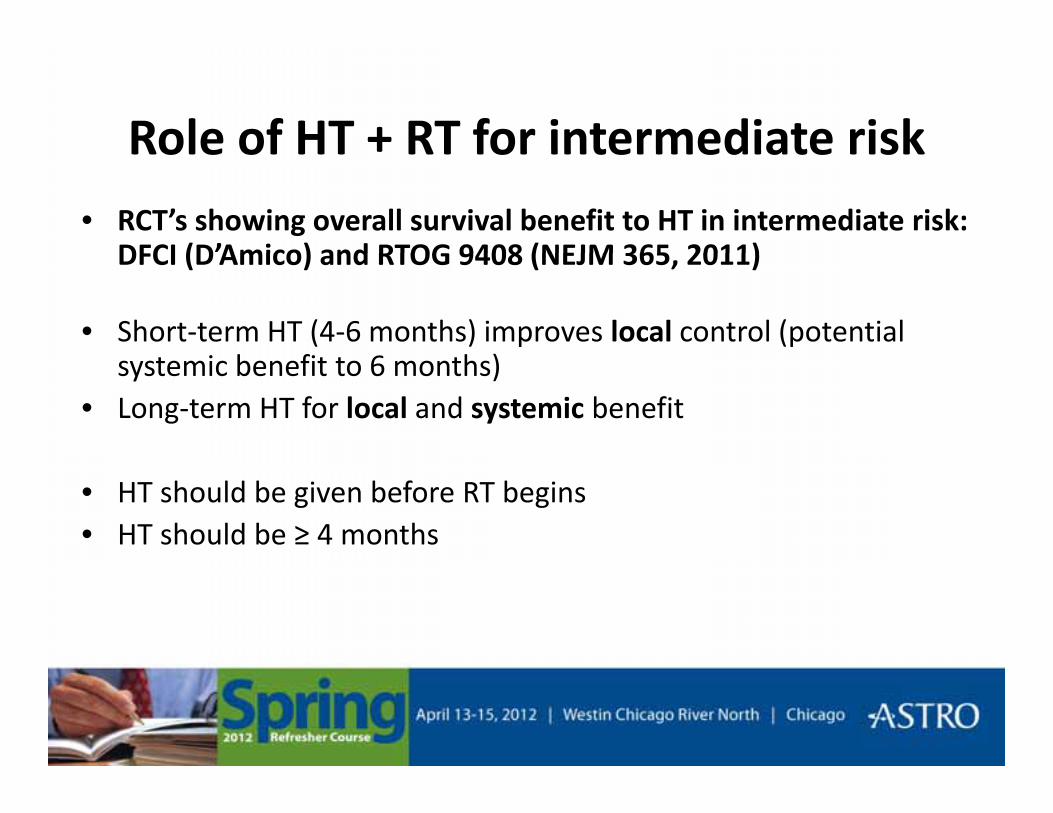
Role of HT + RT for intermediate riskRole of HT + RT for intermediate risk• RCT’s showing overall survival benefit to HT in intermediate risk:
DFCI (D’Amico) and RTOG 9408 (NEJM 365 2011)DFCI (D Amico) and RTOG 9408 (NEJM 365, 2011)
• Short-term HT (4-6 months) improves local control (potential i b fi 6 h )systemic benefit to 6 months)
• Long-term HT for local and systemic benefit
• HT should be given before RT begins • HT should be ≥ 4 months
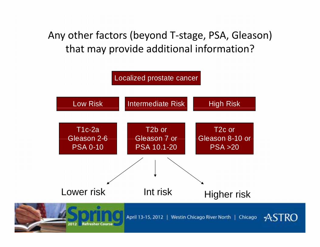
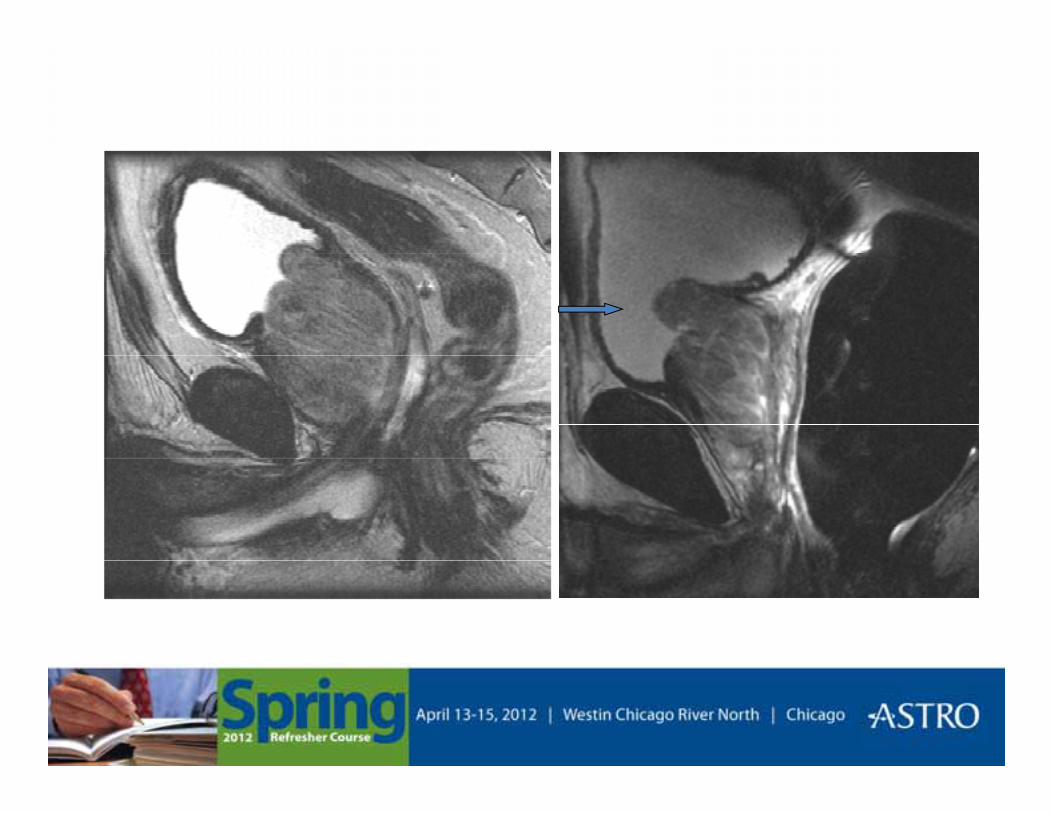
Any other factors (beyond T-stage, PSA, Gleason) th t id dditi l i f ti ?that may provide additional information?
Localized prostate cancer
Low Risk Intermediate Risk High Risk
Localized prostate cancer
T1c-2aGleason 2-6
T2b orGleason 7 or
T2c orGleason 8-10 orGleason 2-6
PSA 0-10Gleason 7 orPSA 10.1-20
Gleason 8-10 orPSA >20
Int riskLower risk Higher risk
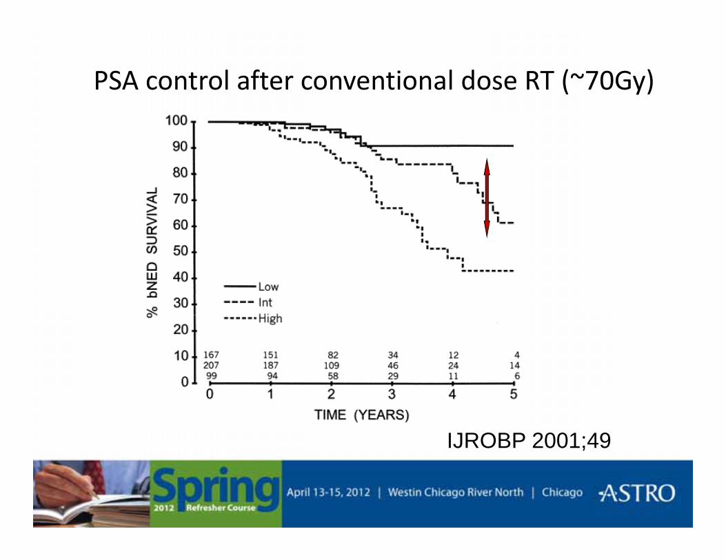
PSA control after conventional dose RT (~70Gy)
IJROBP 2001;49
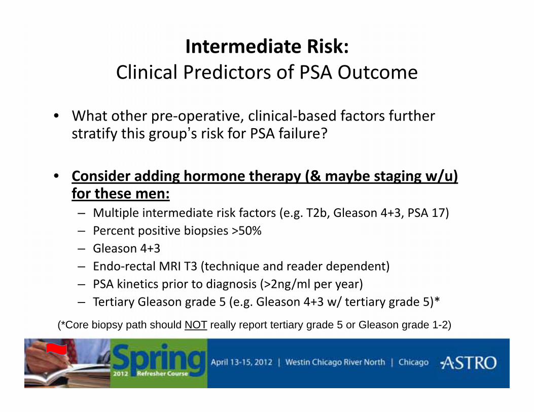
Intermediate Risk:Clinical Predictors of PSA OutcomeClinical Predictors of PSA Outcome
• What other pre-operative, clinical-based factors further if hi ’ i k f PSA f il ?stratify this group’s risk for PSA failure?
• Consider adding hormone therapy (& maybe staging w/u)Consider adding hormone therapy (& maybe staging w/u) for these men:– Multiple intermediate risk factors (e.g. T2b, Gleason 4+3, PSA 17)– Percent positive biopsies >50%Percent positive biopsies >50%– Gleason 4+3– Endo-rectal MRI T3 (technique and reader dependent)
PSA kinetics prior to diagnosis (>2ng/ml per year)– PSA kinetics prior to diagnosis (>2ng/ml per year)– Tertiary Gleason grade 5 (e.g. Gleason 4+3 w/ tertiary grade 5)*
(*Core biopsy path should NOT really report tertiary grade 5 or Gleason grade 1-2)
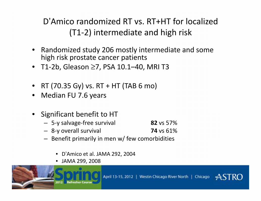
D’Amico randomized RT vs. RT+HT for localized (T1-2) intermediate and high risk
• Randomized study 206 mostly intermediate and some high risk prostate cancer patients
• T1-2b, Gleason ≥7, PSA 10.1–40, MRI T3
• RT (70.35 Gy) vs. RT + HT (TAB 6 mo)RT (70.35 Gy) vs. RT HT (TAB 6 mo)• Median FU 7.6 years
• Significant benefit to HT• Significant benefit to HT – 5-y salvage-free survival 82 vs 57%– 8-y overall survival 74 vs 61%
Benefit primarily in men w/ few comorbidities– Benefit primarily in men w/ few comorbidities
• D’Amico et al. JAMA 292, 2004• JAMA 299, 2008
D’Amico RCT commentsD Amico RCT comments
• Mixture of intermediate-high risk patientsMixture of intermediate high risk patients– Included PSA <40, Gleason 8-10
• Relatively low dose (70Gy)• Relatively low dose (70Gy)
• Preferential benefit in healthier patients
• HT may be detrimental in less healthyy y
• Systemic benefit to 6 months?
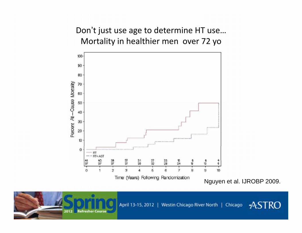
Don’t just use age to determine HT use…Mortality in healthier men over 72 yoMortality in healthier men over 72 yo
Nguyen et al. IJROBP 2009.
What is the role of HT w/ dose-l d kescalation RT in Intermediate risk?
• Single institution reports suggest good outcomes w/ high dose RT alone/ g
• Failure rate may still be >15%
• RTOG 0815 will hopefully answer this• RTOG 0815 will hopefully answer this• RCT: Dose-escalated RT (EBRT +/- LDR,HDR) +/- TAB
• Primary endpoint is overall survival• Primary endpoint is overall survival
MDACC retrospective analysis for int riskMDACC retrospective analysis for int risk
• 636 men w/ int risk who had >75Gy (1995-636 men w/ int risk who had >75Gy (19952009)
• RT alone 45% vs RT + HT 55% (median 6mo)• RT alone 45% vs. RT + HT 55% (median 6mo)
• Recursive partitioning analysis defined “f bl ” “ f bl ” i i k“favorable” vs. “unfavorable” int risk
• Gleason 4+3 and/or ≥ 50% positive cores
Bian et al. Annals of Oncology 2012, epub

Preferential benefit in 5y-FFS for “unfavorable” i di i k h i d HTintermediate risk who received HT
Unfavorable int risk Favorable int risk
Practical considerations of HT + RT• Begin HT at least 2 mos prior to RT
– Leuprolide (LHRH agonist) or goserelin (GnRH agonist)– Bicalutamide (androgen receptor blocker)
If high AUA SI then start bicalutamide >2 weeks prior & consider adding– If high AUA-SI, then start bicalutamide >2 weeks prior & consider adding alpha blocker (e.g. Flomax, Uroxatral, Rapaflo)
• Consider total androgen blockade prior to RTg p– Prostate volume may reduce >30% in first 2-3 months – Total androgen blockade results in faster volume reduction than LHRH
agonist monotherapyW bl l h h di i d d– Want stable target volume through radiation course, decrease dose to rectum
• Consider pre-HT planning target volume for patients with locally-advancedConsider pre HT planning target volume for patients with locally advanced (T3) disease– Prostate volume reduction may be concentric but tumor regression may
not be (neoadjuvant HT studies prior to RP)
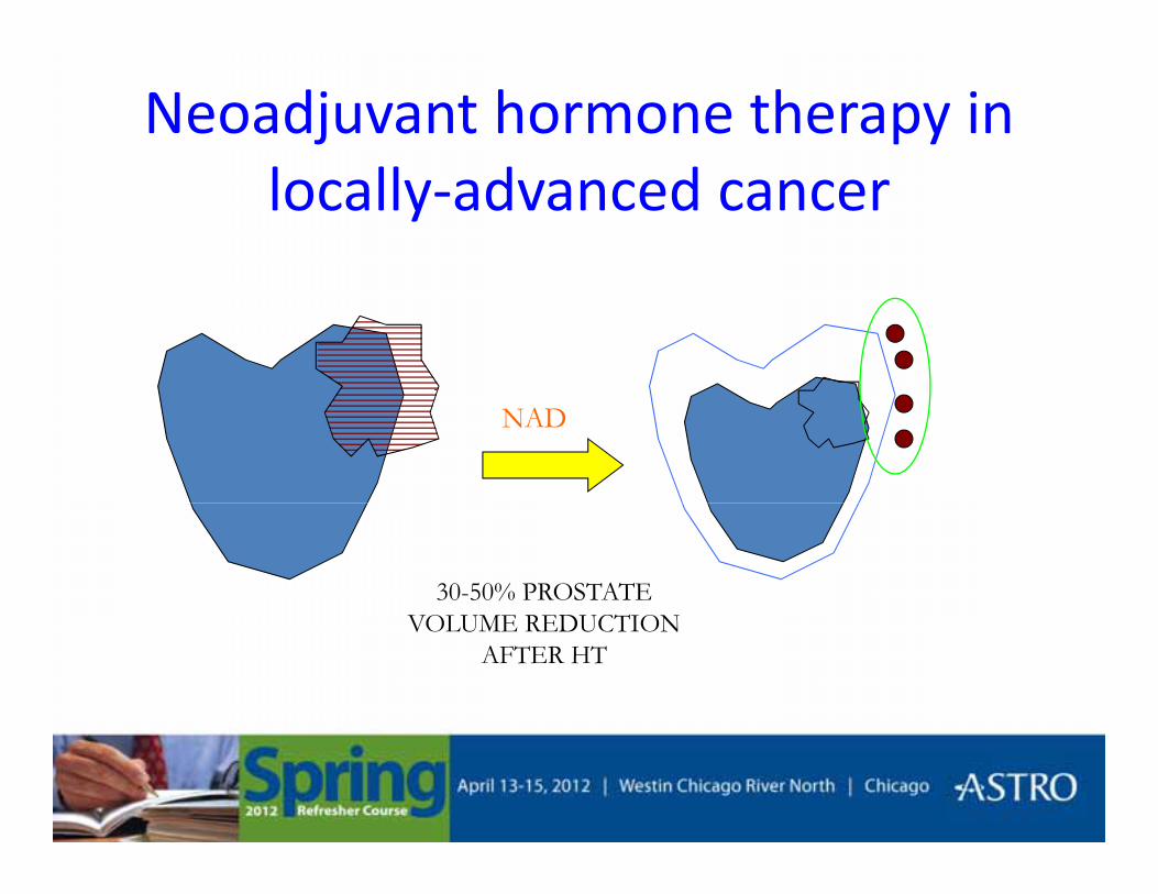
Neoadjuvant hormone therapy in locally-advanced cancer
NAD
30-50% PROSTATE VOLUME REDUCTIONVOLUME REDUCTION
AFTER HT
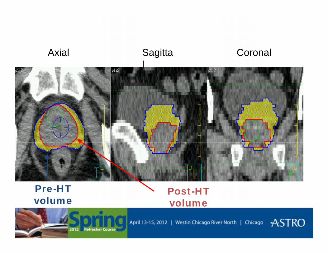
Axial Sagittal
Coronal
Pre-HT volume
Post-HT volumevolume

Evolution of RT + HT
• RTOG 85-31 • Role of adjuvant HT
• RTOG 86-10 • Neo-adjuvant HT
• EORTC 22863 • Concurrent long-term
• RTOG 92-02 • Long-term > short-term
• RTOG 9408/ DFCI • HT for intermediate risk
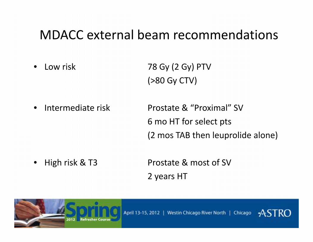
MDACC external beam recommendations
• Low risk 78 Gy (2 Gy) PTV
(>80 Gy CTV)
I t di t i k P t t & “P i l” SV• Intermediate risk Prostate & “Proximal” SV
6 mo HT for select pts
(2 mos TAB then leuprolide alone)(2 mos TAB then leuprolide alone)
• High risk & T3 Prostate & most of SVg
2 years HT
Randomized studies showing benefit to higher dose
• MDACC randomized study of 70 vs. 78 Gy (prescribed to isocenter)– Benefit for 78 Gy including low risk– No difference in distant mets or overall survival
[JCO 18, 2000…Updated IJROBP 2008][ , p ]
• Proton randomized study LLUMC & MGH70 2 Gy vs 79 2 Gy (1 8Gy fxn)– 70.2 Gy vs. 79.2 Gy (1.8Gy fxn)
– First proton boost 19.8 vs. 28.8 CGE followed by photon 50.4 GyPSA control benefit in all patients including low risk– PSA control benefit in all patients including low risk
[JAMA 294:1233-39, 2005…updated JCO 2010]

Randomized studies showing benefit continued…
• Dutch multicenter trial 68 vs 78 Gy (isocenter)– 664 men (Low 18%, Int 27%, High 55%)( , , g )– 3D-CRT various techniques– 22% HT (11% short-, 11% long-term)
• 5-y FFF 64 vs 54% (Median FU 51 months)• Benefit primarily in intermediate and high riskBenefit primarily in intermediate and high risk• No difference in clinical failure, survival• No significant difference in toxicity
– (3D-CRT, rectal DVH constraints)
[Peeters et al J Clin Oncol 2006;24][Peeters et al. J Clin Oncol 2006;24]
MDACC Dose-escalation Study Update
• Median FU 8.7 y (9.5y for alive)
• For entire group 8y FFF 59 vs. 78%
• For PSA >10 8y FFF 39 vs. 78%
• Improved “clinical failure rates” for higher dose• No difference is overall survivalNo difference is overall survival
• Side effect essentially stable from prior reports[IJROBP 2008][IJROBP 2008]
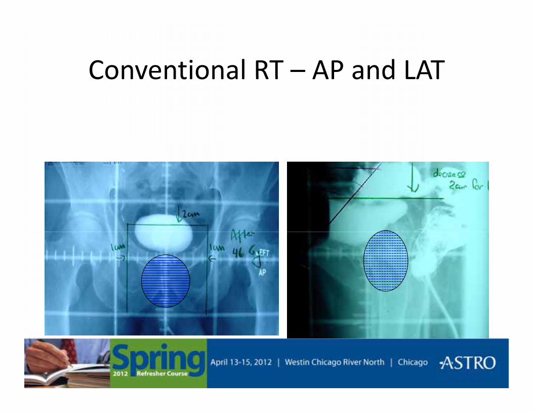
Conventional RT – AP and LATConventional RT AP and LAT
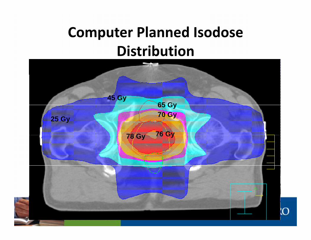
Computer Planned Isodose Distribution
45 Gy65 Gy
25 Gy 70 Gy
76 Gy78 Gy
65 Gy
76 Gy78 Gy
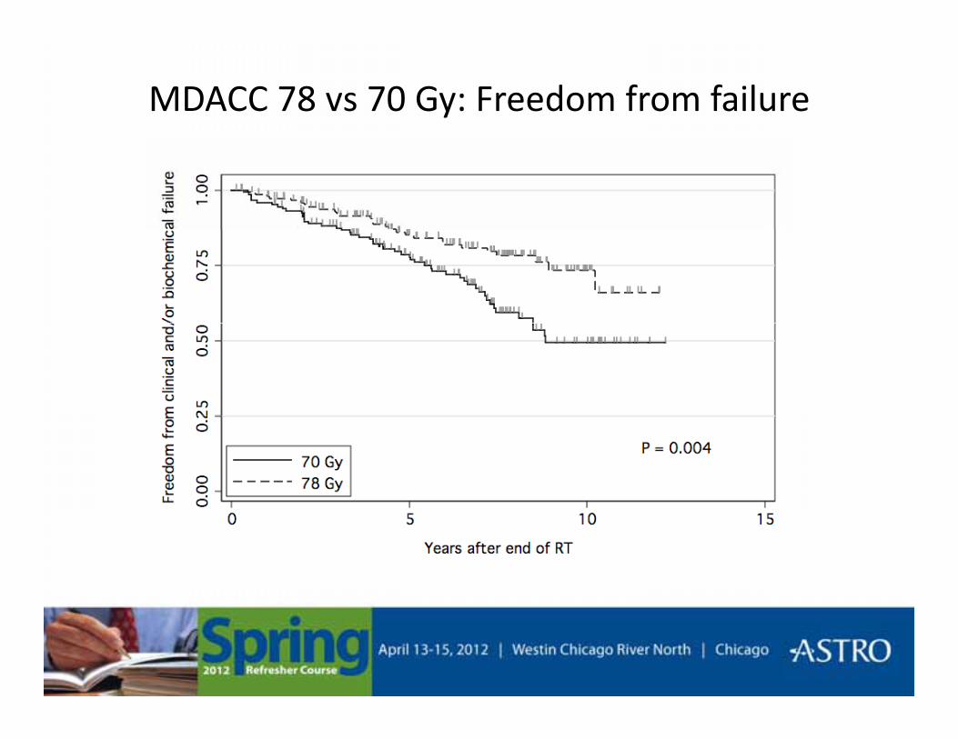
MDACC 78 vs 70 Gy: Freedom from failure
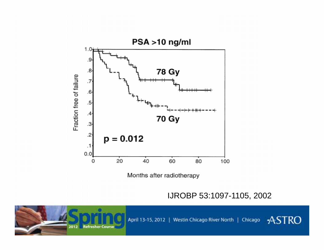
IJROBP 53:1097-1105, 2002
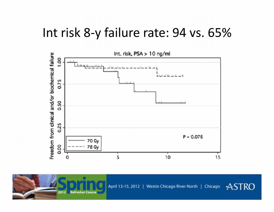
Int risk 8-y failure rate: 94 vs. 65%y
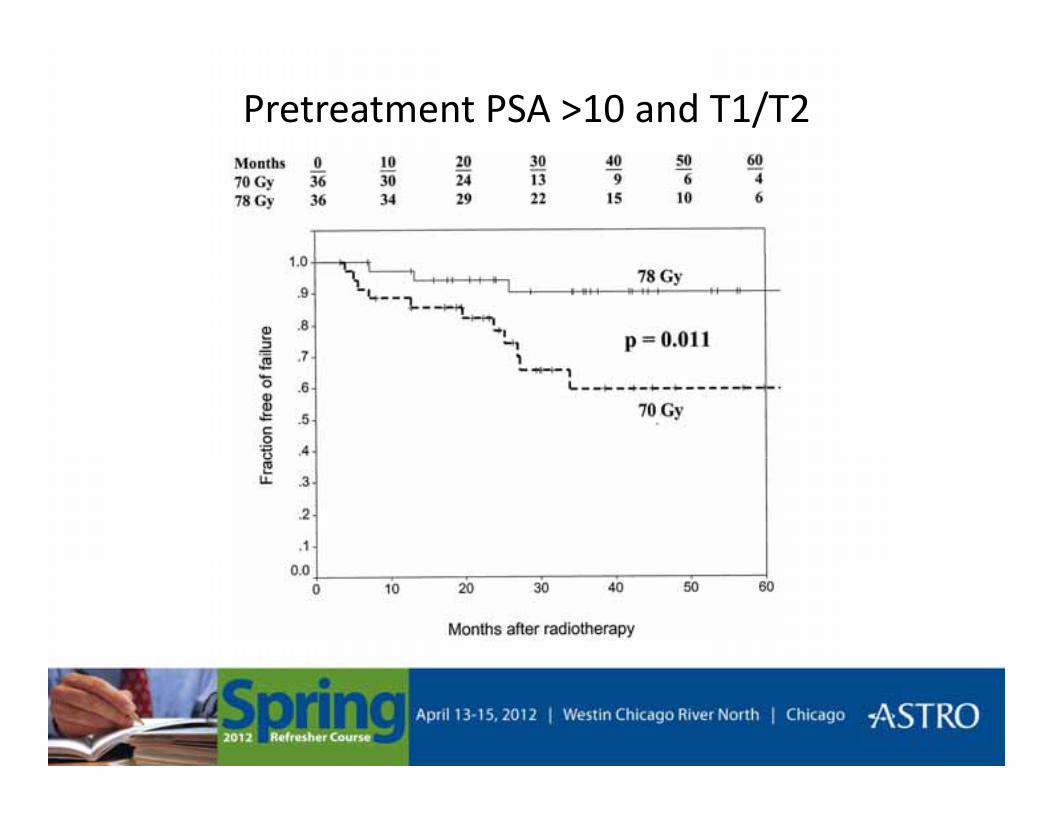
Pretreatment PSA >10 and T1/T2
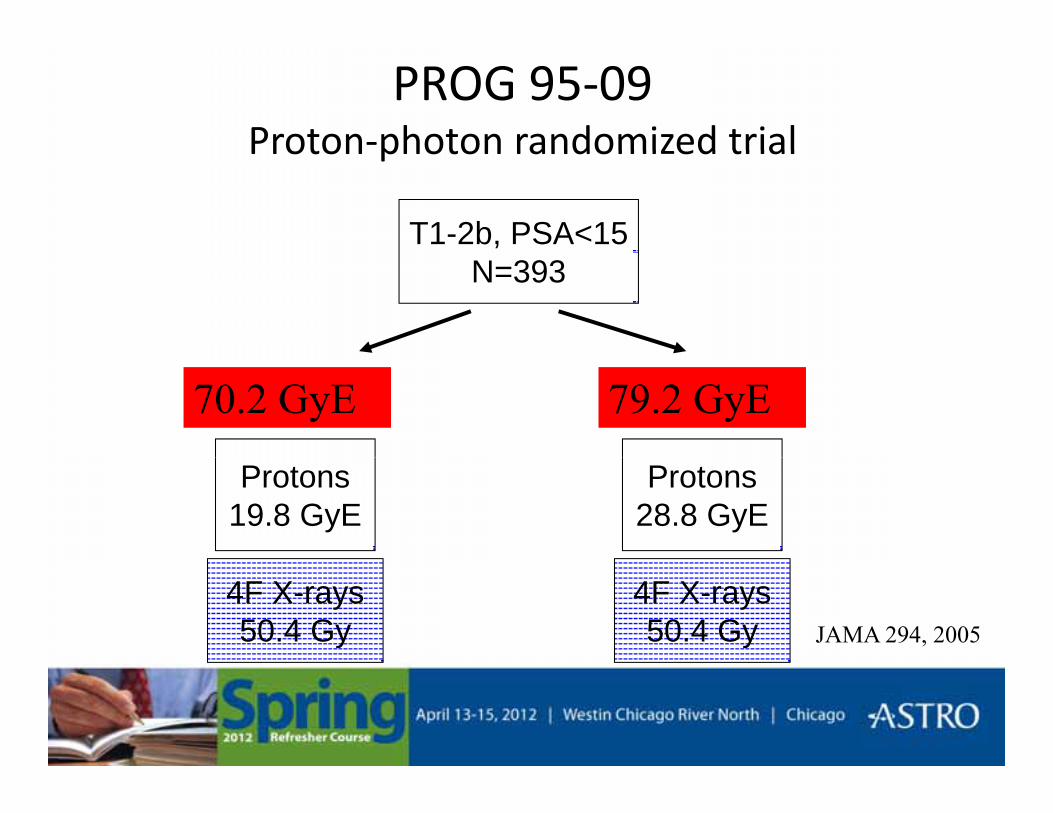
PROG 95-09Proton-photon randomized trialProton photon randomized trial
T1-2b, PSA<15N=393
70.2 GyE 79.2 GyE
Protons19.8 GyE
Protons28.8 GyE
4F X-rays50.4 Gy
4F X-rays50.4 Gy JAMA 294, 2005
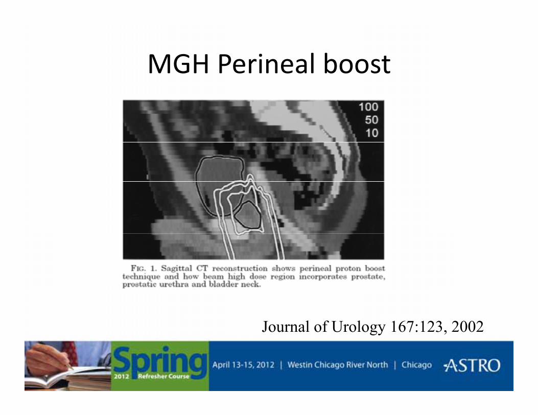
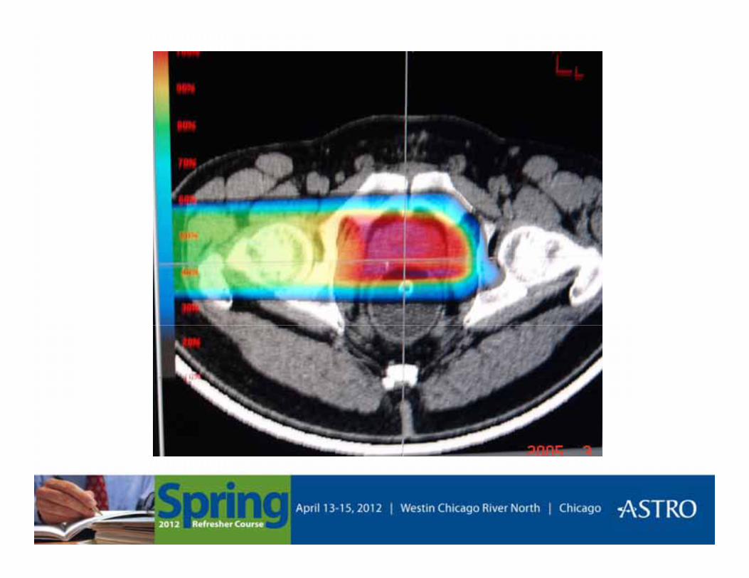
MGH Perineal boost
Journal of Urology 167:123, 2002

PROG 9509PROG 9509--UPDATEUPDATEJ Clin Oncol 2010J Clin Oncol 2010J Clin Oncol 2010J Clin Oncol 2010
• Difference in bNED survival between arms persists i h di f ll f 9with median follow-up of 9 years
• No difference in Gr>3 GI/GU morbidity between arms using data from validated patient questionnairequestionnaire
F ti t i hi h d i d l• Fewer patients in high dose arm required salvage hormones
C. RossiC. Rossi--LLUMC. LLUMC.
20122012--0404--1818
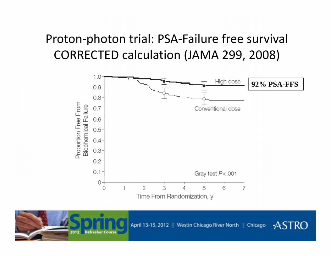
Proton-photon trial: PSA-Failure free survivalCORRECTED calculation (JAMA 299, 2008)
92% PSA-FFS
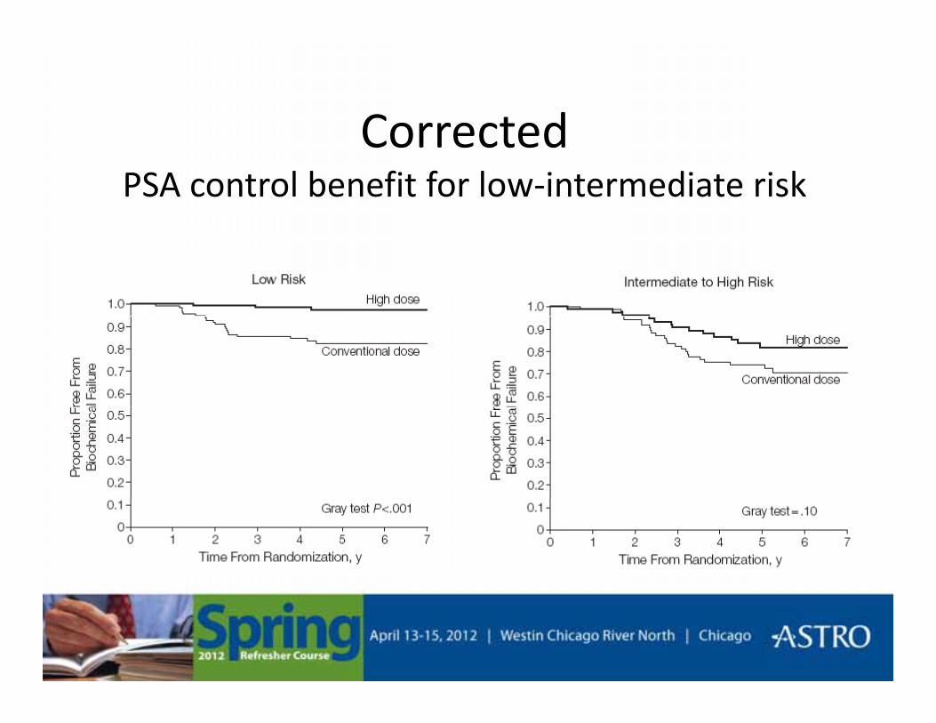
CorrectedCorrected PSA control benefit for low-intermediate risk
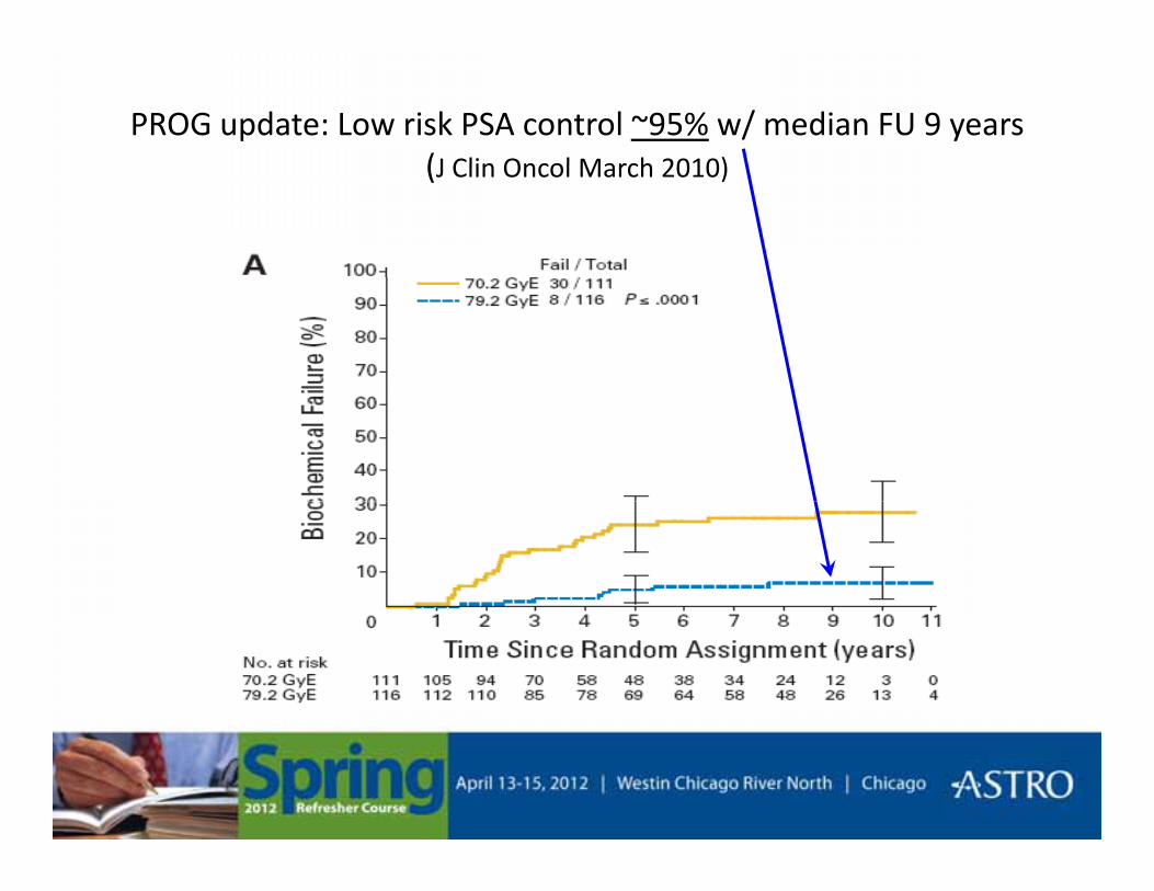
PROG update: Low risk PSA control ~95% w/ median FU 9 years(J Clin Oncol March 2010)(J Clin Oncol March 2010)
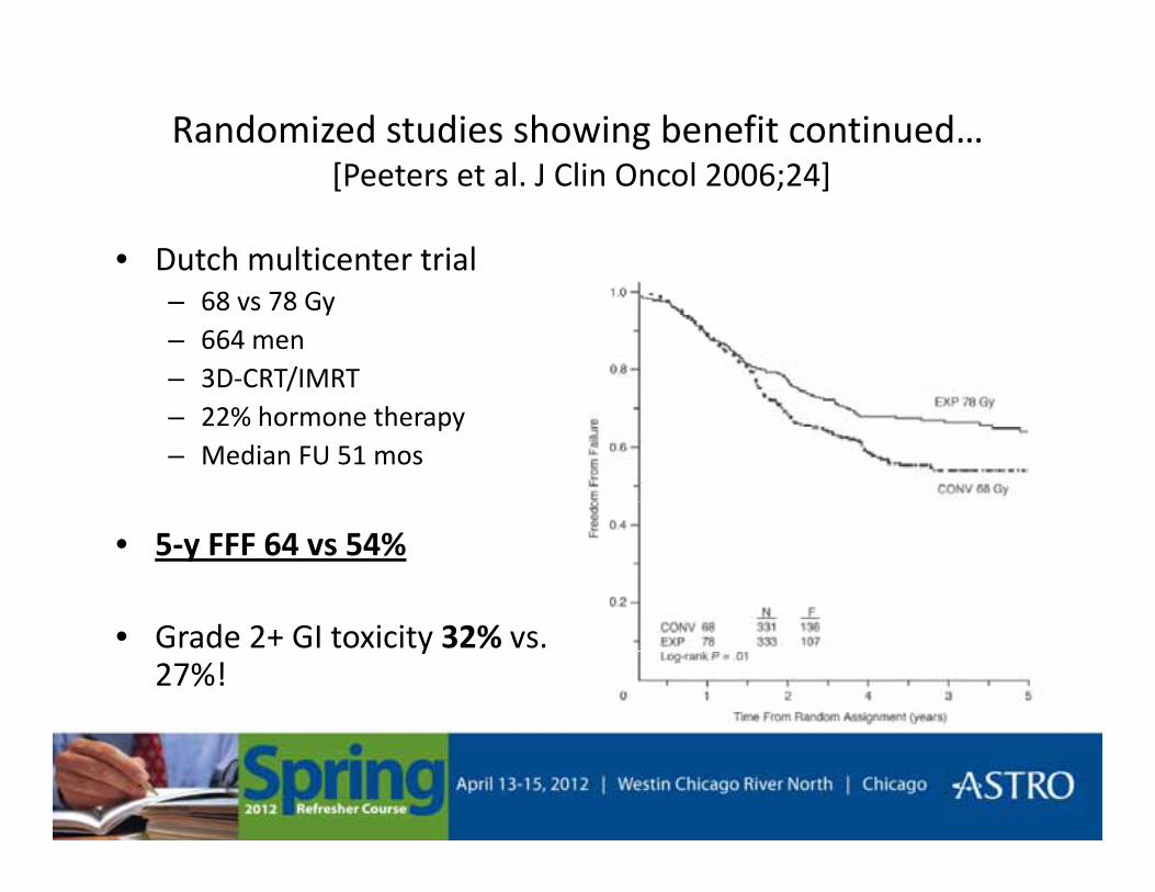
Randomized studies showing benefit continued…[P l J Cli O l 2006 24][Peeters et al. J Clin Oncol 2006;24]
• Dutch multicenter trial – 68 vs 78 Gy – 664 men – 3D-CRT/IMRT/– 22% hormone therapy– Median FU 51 mos
• 5-y FFF 64 vs 54%
• Grade 2+ GI toxicity 32% vs. 27%!
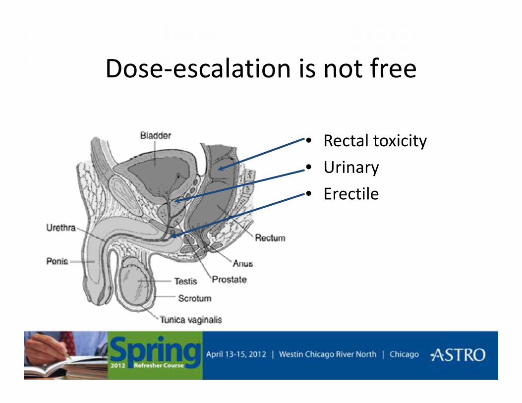
Dose-escalation is not freeDose escalation is not free
• Rectal toxicity
• Urinary
• Erectile

More Grade ≥2 rectal complications in 78 Gy arm[ ][IJROBP 53, 2002]
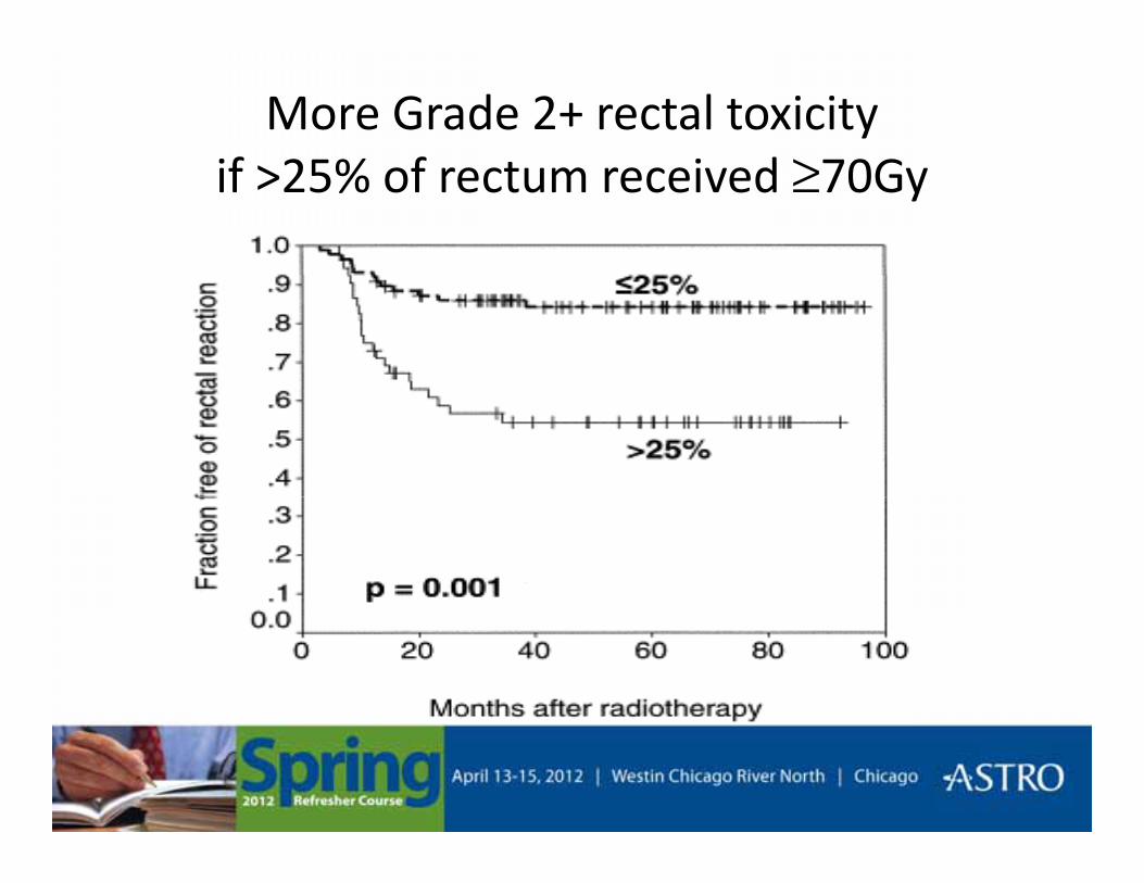
More Grade 2+ rectal toxicity if 25% f t i d ≥70Gif >25% of rectum received ≥70Gy
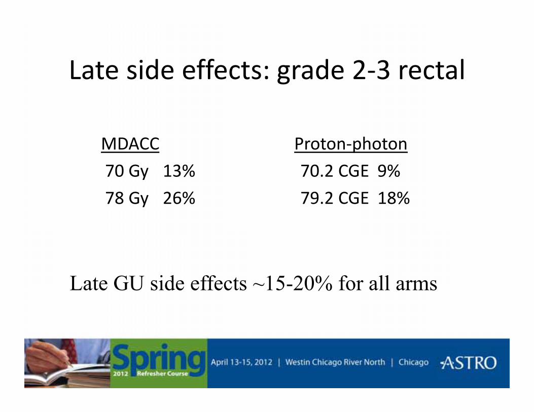
Late side effects: grade 2-3 rectalLate side effects: grade 2 3 rectal
MDACC
70 Gy 13%
Proton-photon
70.2 CGE 9%
78 Gy 26% 79.2 CGE 18%
Late GU side effects ~15 20% for all armsLate GU side effects ~15-20% for all arms
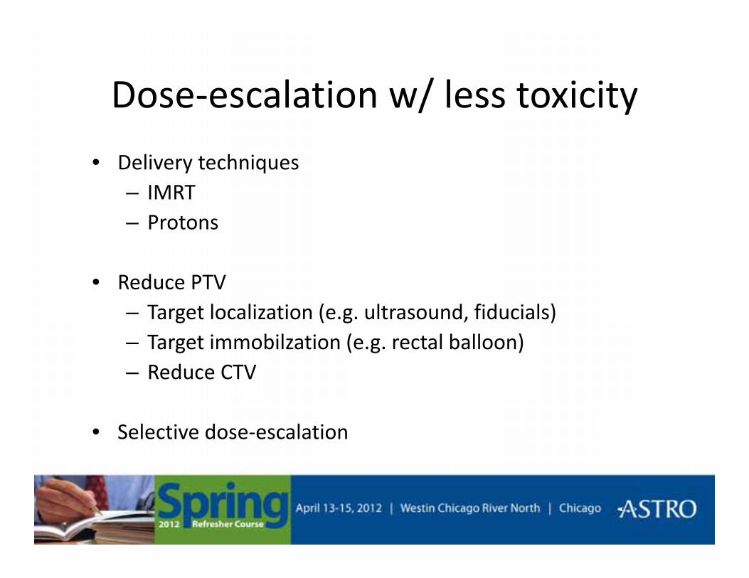
Dose-escalation w/ less toxicityDose escalation w/ less toxicity
• Delivery techniques– IMRT– Protons
• Reduce PTV– Target localization (e.g. ultrasound, fiducials)– Target immobilzation (e.g. rectal balloon)– Reduce CTV
• Selective dose-escalation
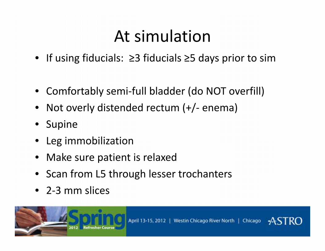
At simulation• If using fiducials: ≥3 fiducials ≥5 days prior to sim
• Comfortably semi-full bladder (do NOT overfill)
• Not overly distended rectum (+/- enema)Not overly distended rectum (+/ enema)
• Supine
• Leg immobilization• Leg immobilization
• Make sure patient is relaxed
• Scan from L5 through lesser trochanters• Scan from L5 through lesser trochanters
• 2-3 mm slices

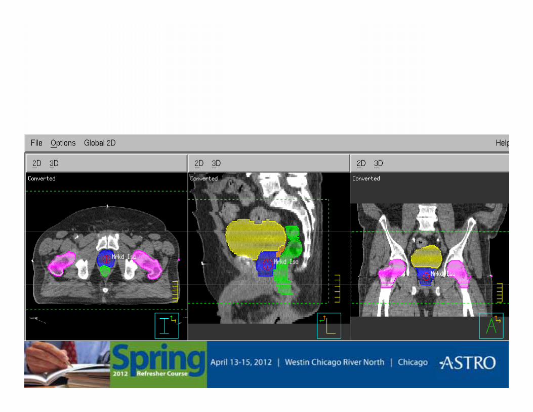
Defining Structures• Rectum (anal verge to anterior flexion of sigmoid)
• Bladder (whole bladder)( )
• Femoral heads (down to lesser trochanter)
• CTV
Low risk prostate onlyLow risk = prostate only
Int risk = prostate + prox SV
High risk = prostate + SV (+/- nodes)
• Use zoom
• Use window and level (e.g. “neck”)( g )
• Use other planes of view (especially sagittal for apex)
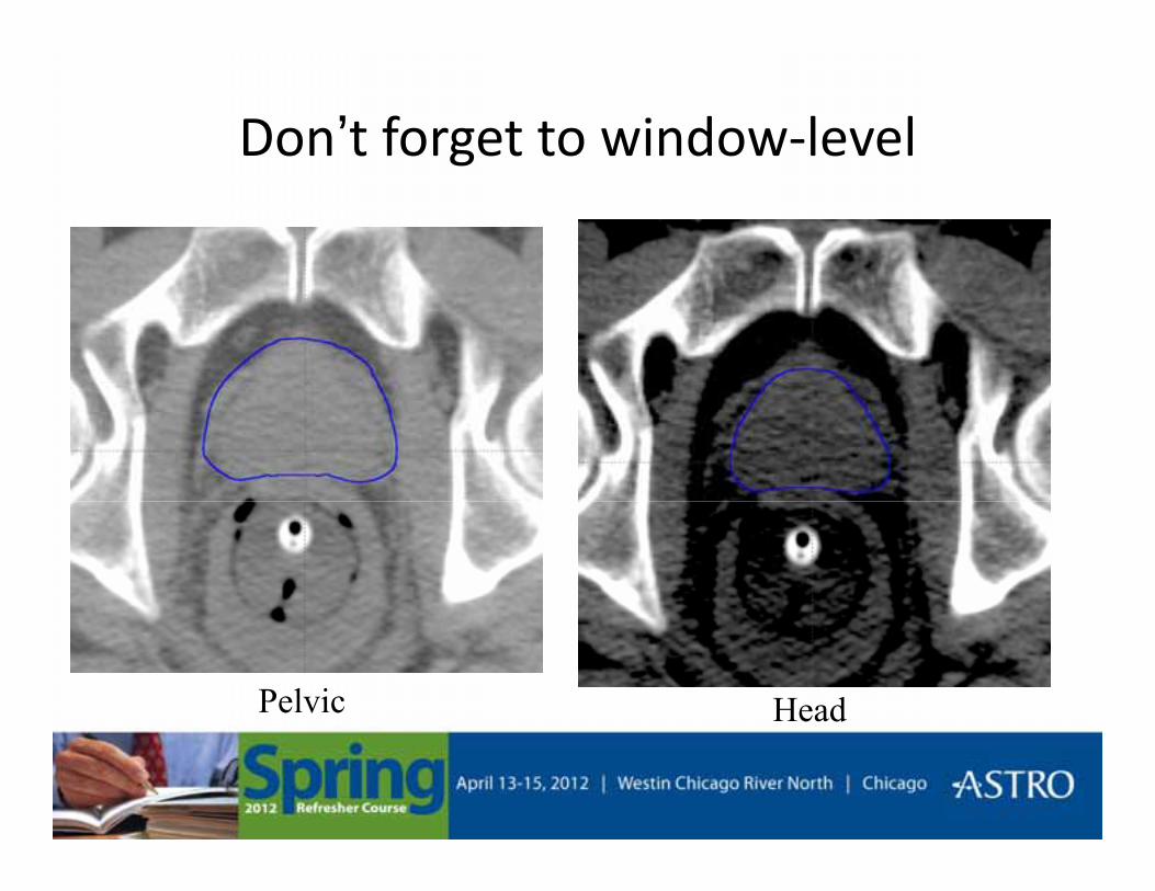
Don’t forget to window-levelg
Pelvic Head
Rectum and BladderRectum and Bladder
• Want to contour the rectum- not the peri-Want to contour the rectum not the perirectal muscles.
• If you using ultrasound-based guidance, pay i l i bl ddspecial attention to bladder-prostate
interface.– Also need to contour non-CTV portion of SV as
reference structure

BAT Alignment (sagittal)BAT Alignment (sagittal)
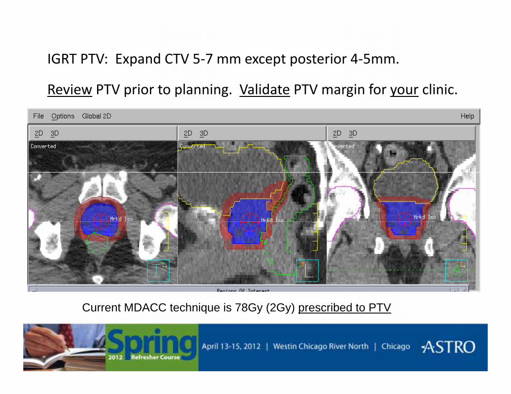
IGRT PTV: Expand CTV 5-7 mm except posterior 4-5mm.
Review PTV prior to planning. Validate PTV margin for your clinic.
Current MDACC technique is 78Gy (2Gy) prescribed to PTV
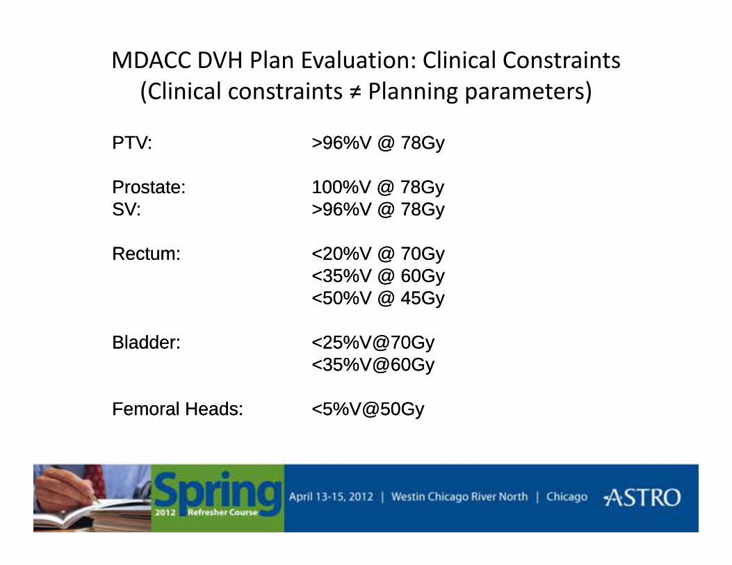
MDACC DVH Plan Evaluation: Clinical Constraints(Clinical constraints ≠ Planning parameters)
PTV: >96%V @ 78GyPTV: >96%V @ 78Gy
Prostate: 100%V @ 78GySV: >96%V @ 78Gy
R t 20%V @ 70G
Prostate: 100%V @ 78GySV: >96%V @ 78Gy
R t 20%V @ 70GRectum: <20%V @ 70Gy<35%V @ 60Gy<50%V @ 45Gy
Rectum: <20%V @ 70Gy<35%V @ 60Gy<50%V @ 45Gy
Bladder: <25%V@70Gy<35%V@60Gy
Bladder: <25%V@70Gy<35%V@60Gy
Femoral Heads: <5%V@50GyFemoral Heads: <5%V@50Gy
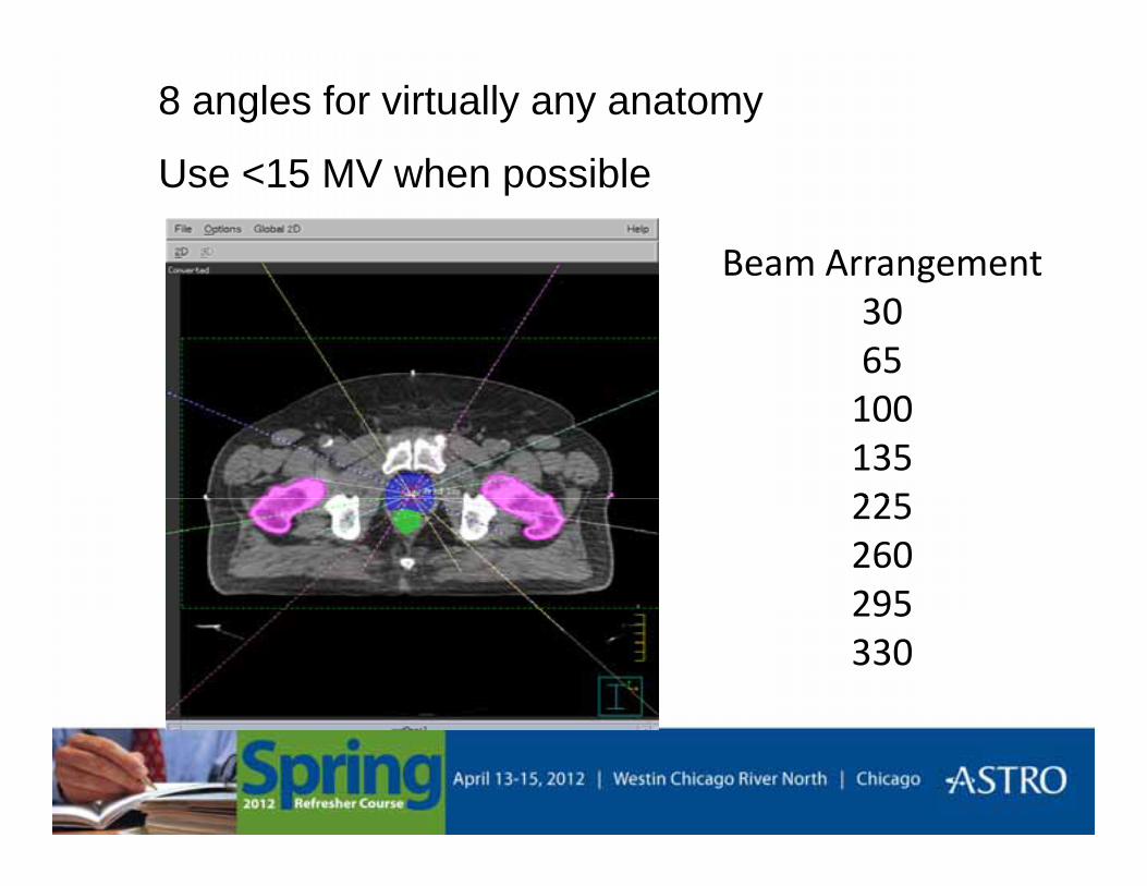
8 angles for virtually any anatomy
U <15 MV h ibl
Beam Arrangement
Use <15 MV when possible
ea a ge e t3065
100135225225260295295330
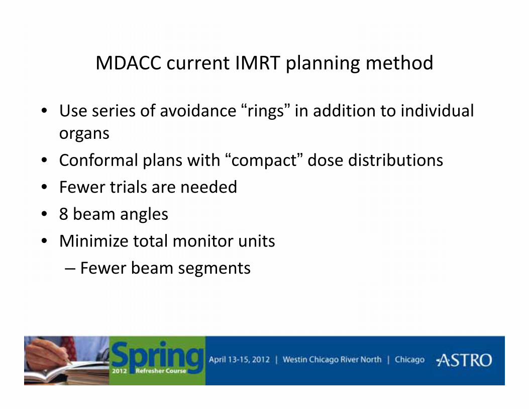
MDACC current IMRT planning method
• Use series of avoidance “rings” in addition to individual organs
• Conformal plans with “compact” dose distributions
• Fewer trials are needed
• 8 beam angles
• Minimize total monitor units
– Fewer beam segments
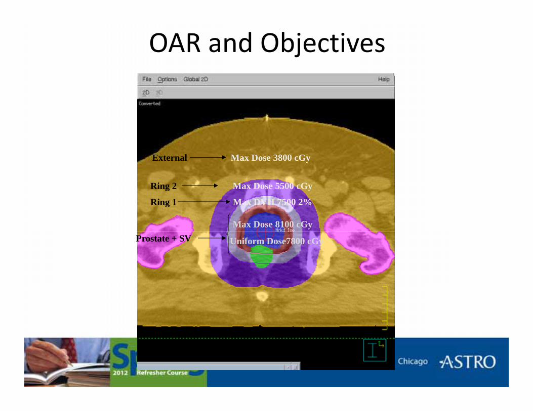
OAR and Objectives
Max Dose 3800 cGyExternal
Max Dose 5500 cGy
Max Dose 8100 cGy
Max DVH 7500 2%
Ring 2
Ring 1
Uniform Dose7800 cGyProstate + SV
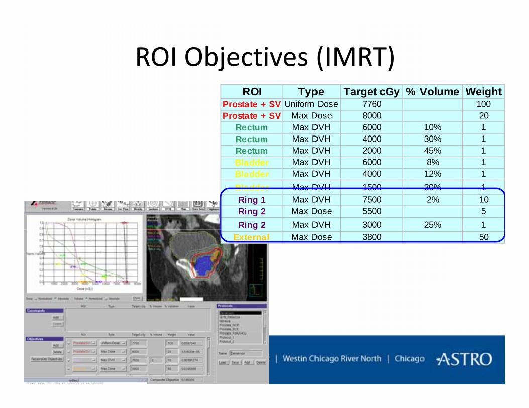
ROI Objectives (IMRT)ROI Type Target cGy % Volume Weight
Prostate + SV Uniform Dose 7760 100Prostate + SV Max Dose 8000 20
Rectum Max DVH 6000 10% 1Rectum Max DVH 6000 10% 1Rectum Max DVH 4000 30% 1Rectum Max DVH 2000 45% 1Bladder Max DVH 6000 8% 1Bladder Max DVH 4000 12% 1Bladder Max DVH 1500 30% 1Ring 1 Max DVH 7500 2% 10Ring 2 Max Dose 5500 5Ring 2 Max DVH 3000 25% 1g
External Max Dose 3800 50
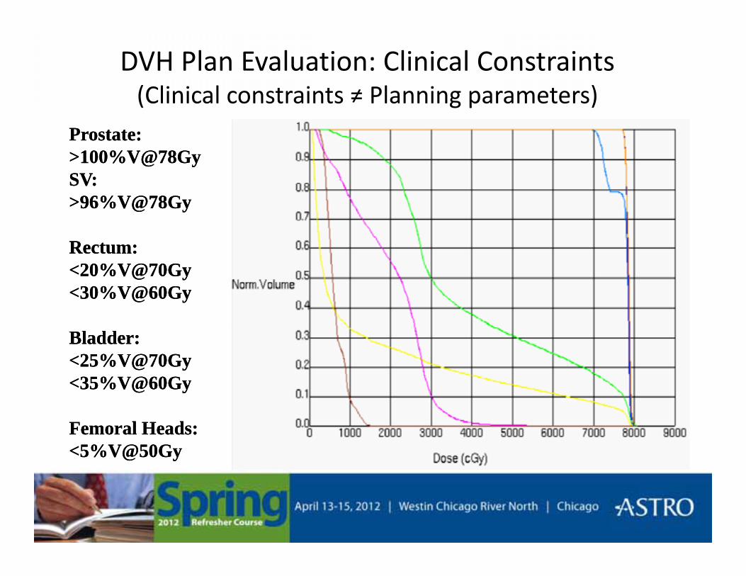
DVH Plan Evaluation: Clinical Constraints(Clinical constraints ≠ Planning parameters)( g p )
Prostate:>100%V@78GySV:
Prostate:>100%V@78GySV:SV:>96%V@78Gy
Rectum:
SV:>96%V@78Gy
Rectum:Rectum:<20%V@70Gy<30%V@60Gy
Rectum:<20%V@70Gy<30%V@60Gy
Bladder:<25%V@70Gy<35%V@60Gy
Bladder:<25%V@70Gy<35%V@60Gy
Femoral Heads:<5%V@50GyFemoral Heads:<5%V@50Gy
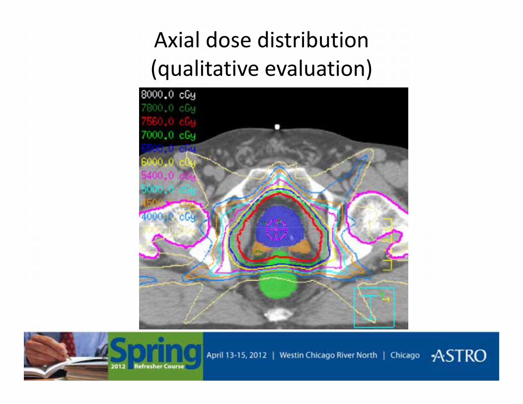
Axial dose distribution (qualitative evaluation)(qualitative evaluation)
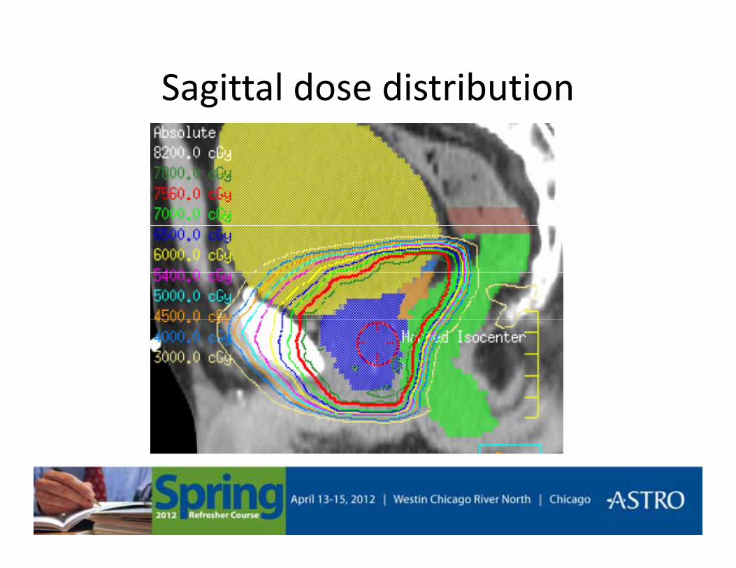
Sagittal dose distributiong
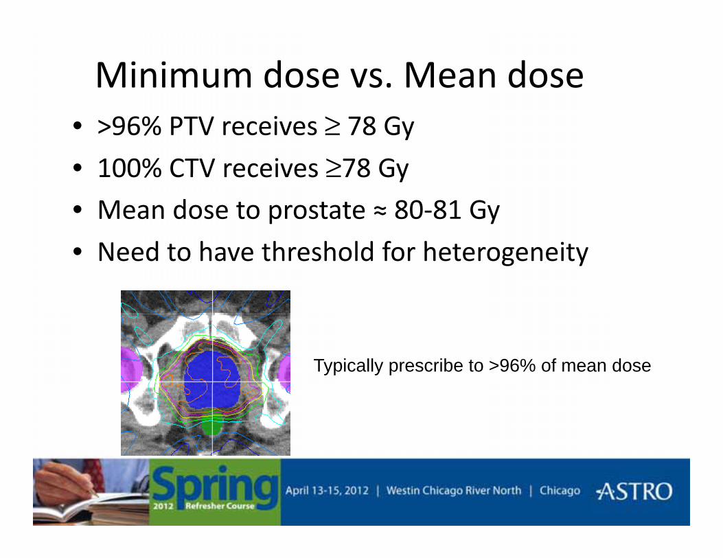
Minimum dose vs. Mean dose• >96% PTV receives ≥ 78 Gy
• 100% CTV receives ≥78 Gy100% CTV receives ≥78 Gy• Mean dose to prostate ≈ 80-81 Gy
N d t h th h ld f h t it• Need to have threshold for heterogeneity
Typically prescribe to >96% of mean dose
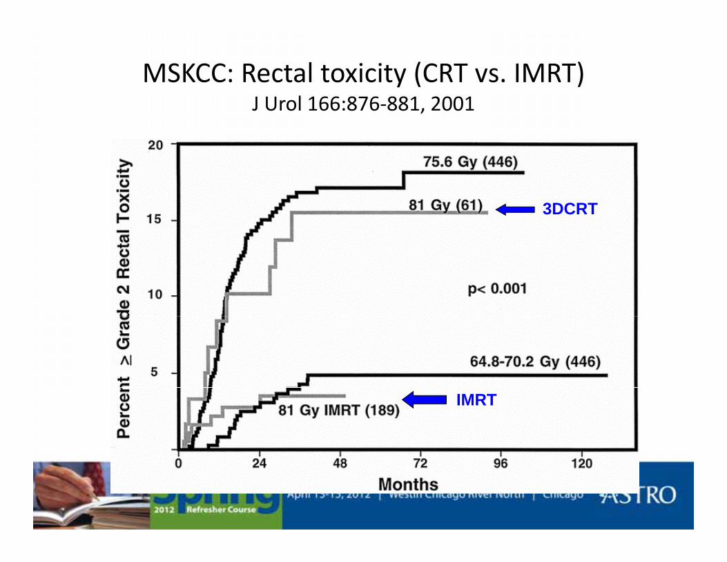
MSKCC: Rectal toxicity (CRT vs. IMRT)J Urol 166:876 881 2001J Urol 166:876-881, 2001
3DCRT
IMRT
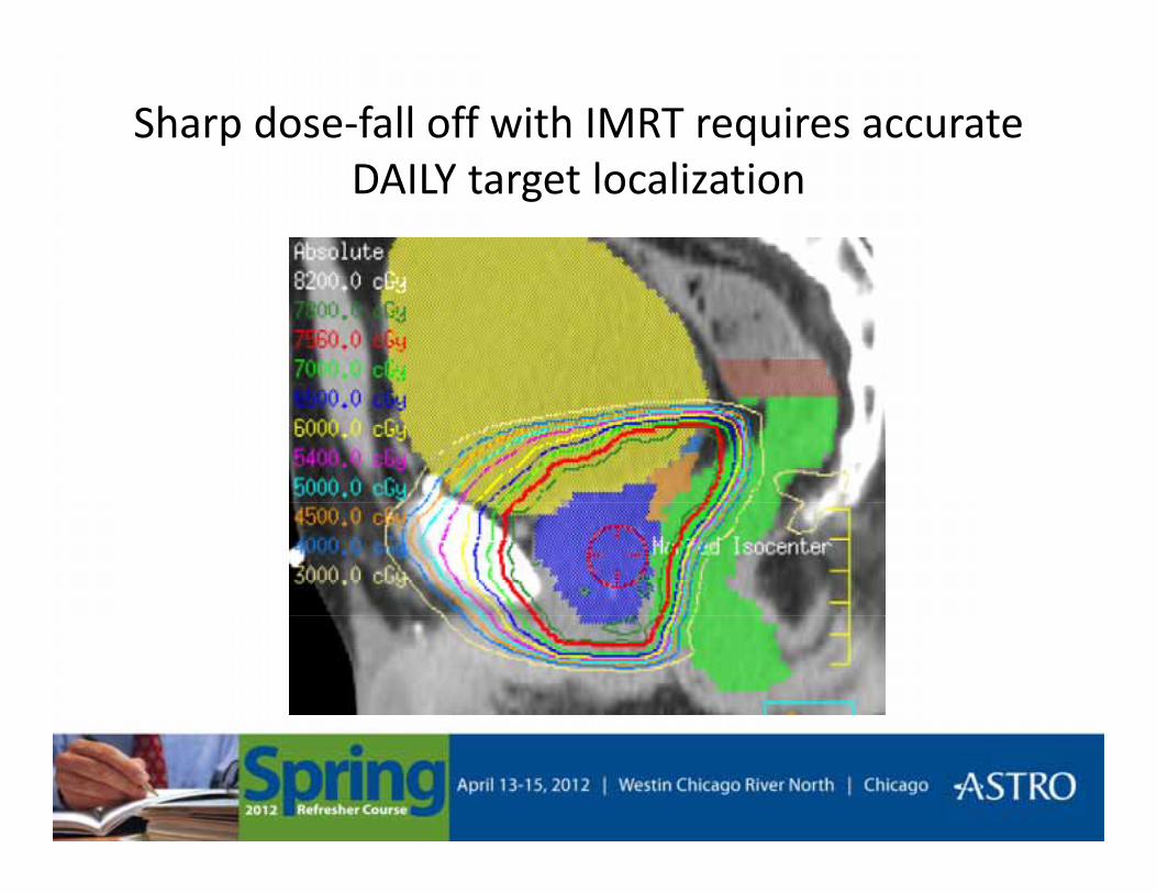
Sharp dose-fall off with IMRT requires accurate DAILY l li iDAILY target localization
Reducing PTV through IGRT (Image Guided Radiation Therapy)(Image Guided Radiation Therapy)
• Requires daily imaging of the target
• Decrease “systematic” setup errorl– From simulation to treatment
• Correct for INTER fractional movement• Correct for INTER-fractional movement– Pelvis– Rectal and bladder filling g
• May not account for INTRA-fractional movement

IGRT
• Portal imaging
• Ultrasound
• Fiducial markers (intraprostatic)
• Volumetric on-board imaging
– In-room CT
– Cone-beam CT
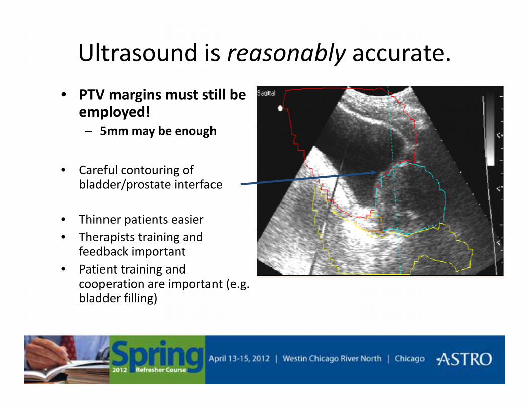
Ultrasound is reasonably accurate.
• PTV margins must still be employed!
5 b h– 5mm may be enough
• Careful contouring of bladder/prostate interface
• Thinner patients easier• Therapists training and
feedback important• Patient training and
ti i t t (cooperation are important (e.g. bladder filling)
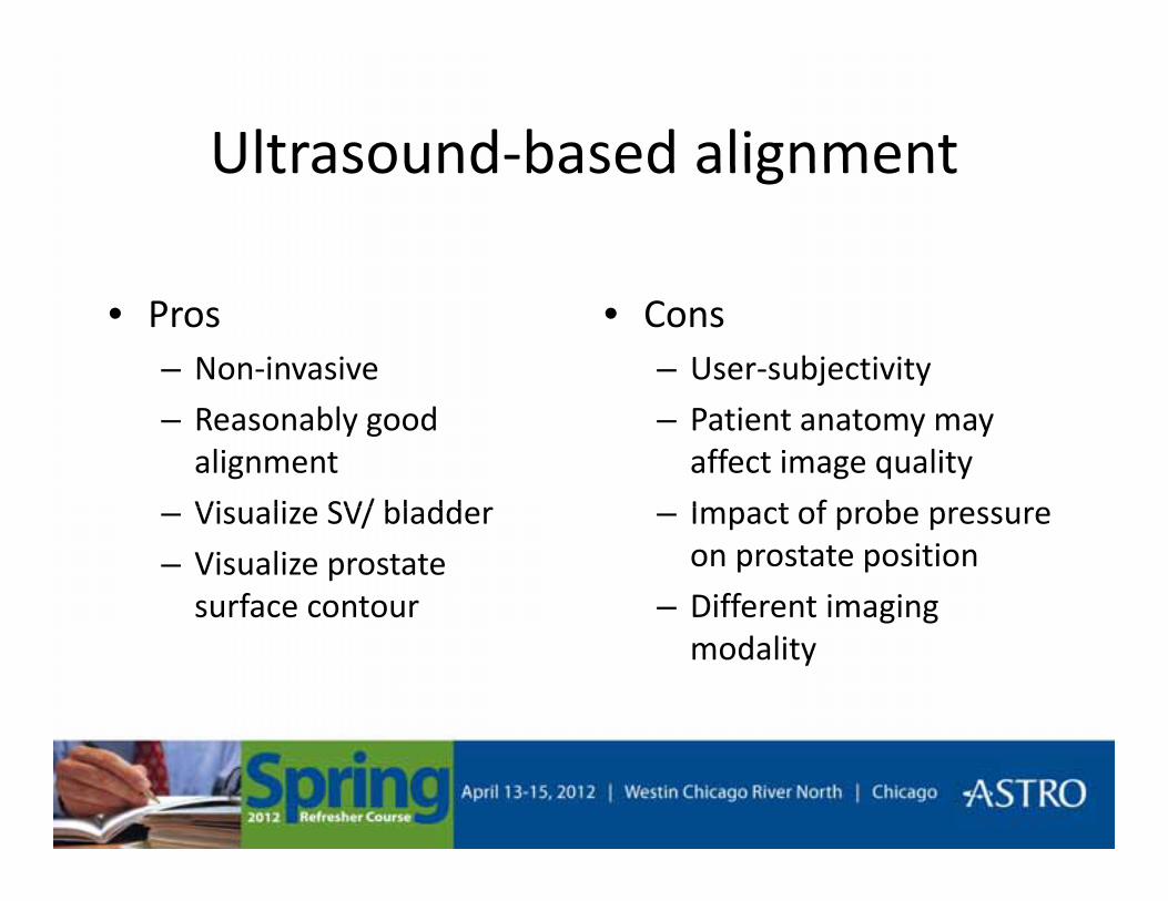
Ultrasound-based alignmentUltrasound based alignment
• Pros– Non-invasive
• Cons– User-subjectivity
– Reasonably good alignment
Visualize SV/ bladder
– Patient anatomy may affect image quality
Impact of probe pressure– Visualize SV/ bladder
– Visualize prostate surface contour
– Impact of probe pressure on prostate position
– Different imaging g gmodality
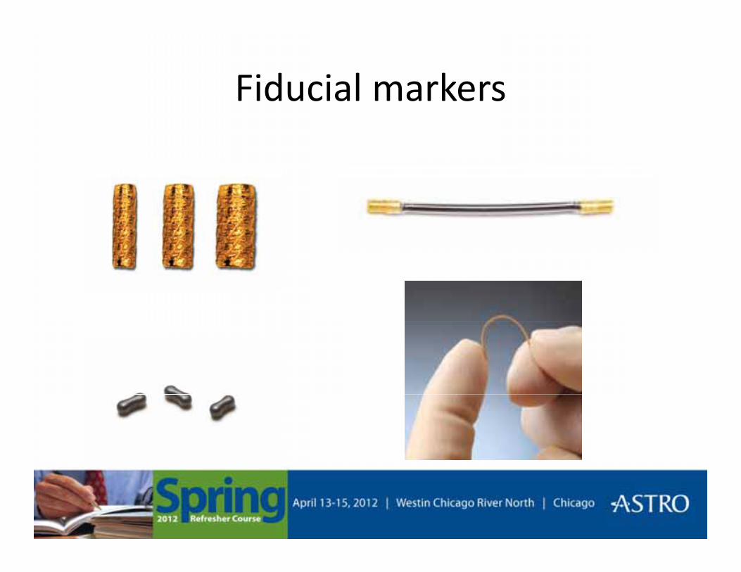
Fiducial markers
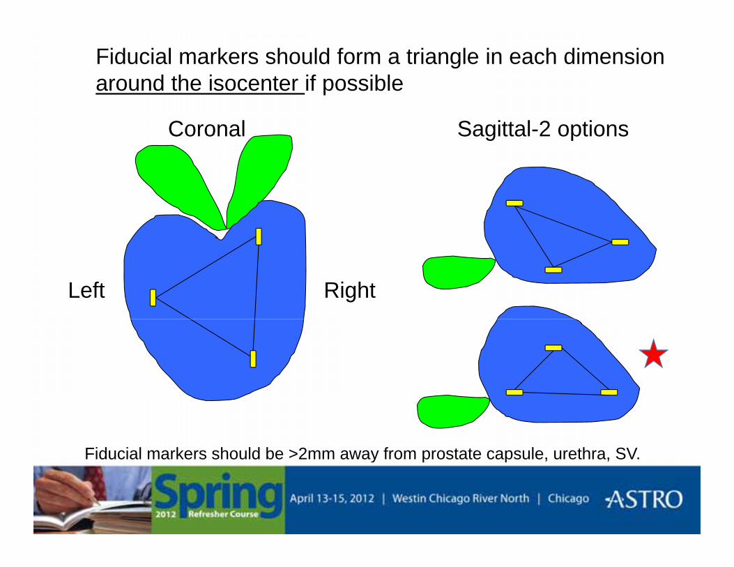
Fiducial markers should form a triangle in each dimension around the isocenter if possible
Coronal Sagittal-2 options
Left Right
Fiducial markers should be >2mm away from prostate capsule, urethra, SV.y p p
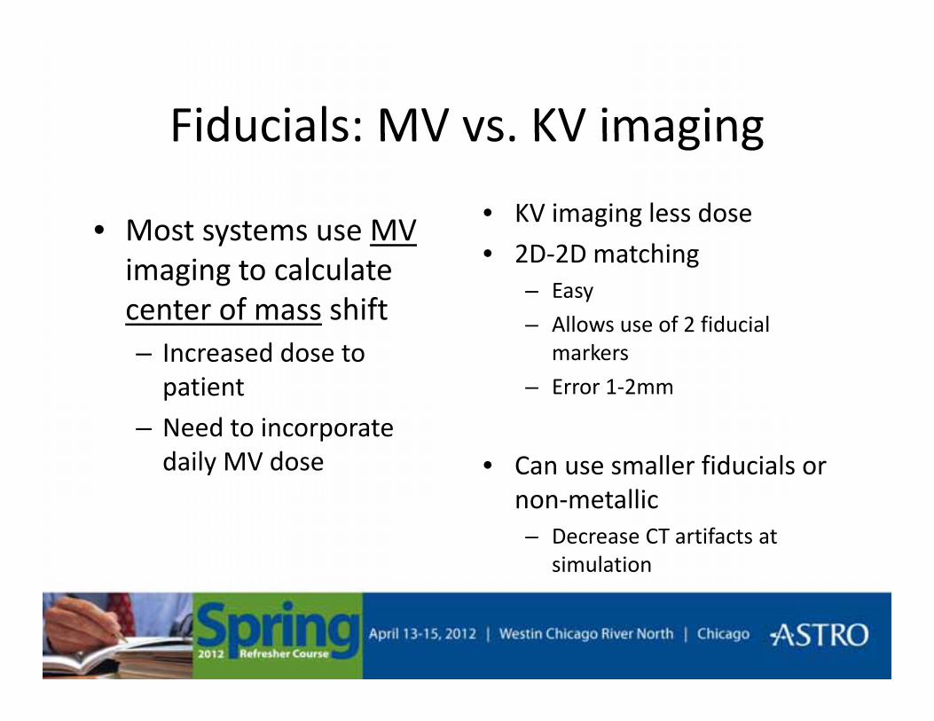
Fiducials: MV vs KV imagingFiducials: MV vs. KV imaging
M t t MV • KV imaging less dose• Most systems use MVimaging to calculate center of mass shift
g g
• 2D-2D matching– Easy
center of mass shift– Increased dose to
patient
– Allows use of 2 fiducial markers
– Error 1-2mm
– Need to incorporate daily MV dose • Can use smaller fiducials or
non metallicnon-metallic– Decrease CT artifacts at
simulation
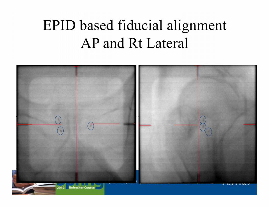
EPID based fiducial alignmentAP d R L lAP and Rt Lateral
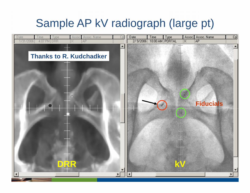
Sample AP kV radiograph (large pt)
Thanks to R. Kudchadker
Fiducials
DRR kV
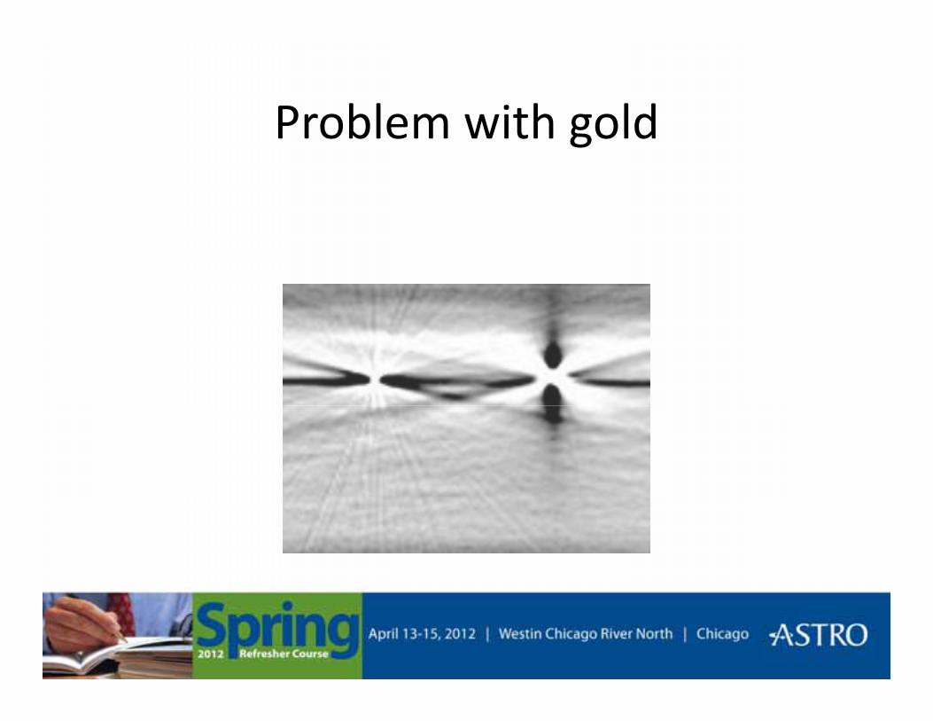
Problem with goldProblem with gold
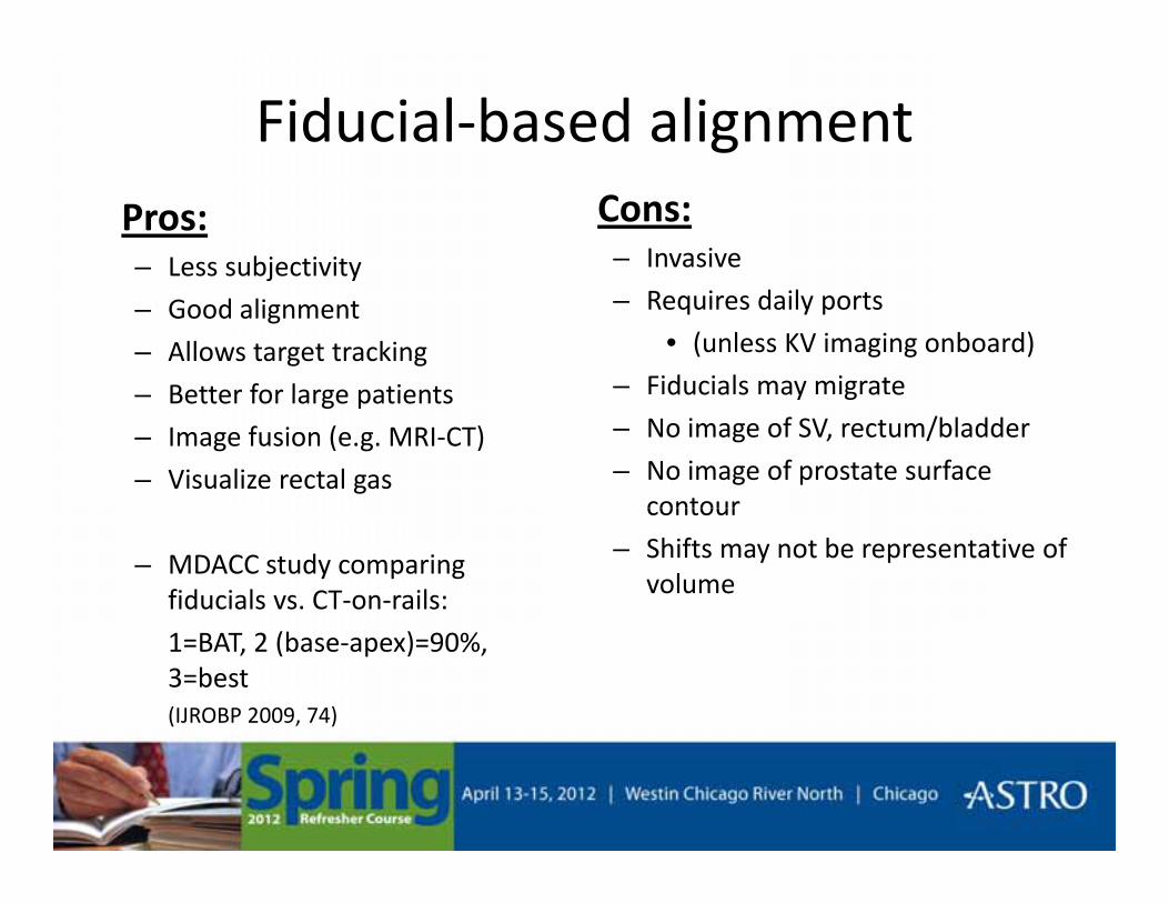
Fiducial-based alignmentPros:– Less subjectivity
Cons:– Invasivej y
– Good alignment
– Allows target tracking
– Better for large patients
– Requires daily ports
• (unless KV imaging onboard)
– Fiducials may migrate– Better for large patients
– Image fusion (e.g. MRI-CT)
– Visualize rectal gas
duc a s ay g a e
– No image of SV, rectum/bladder
– No image of prostate surface contour
– MDACC study comparing fiducials vs. CT-on-rails:
contour
– Shifts may not be representative of volume
1=BAT, 2 (base-apex)=90%, 3=best(IJROBP 2009, 74)
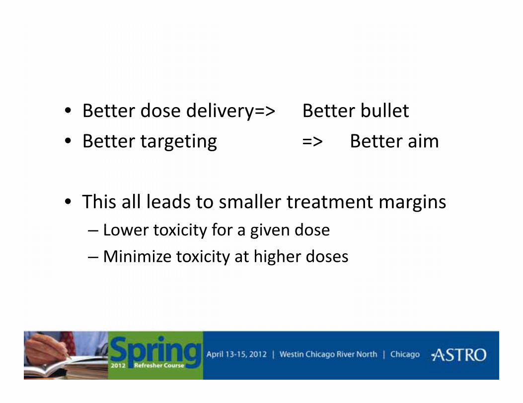
• Better dose delivery=> Better bullet
• Better targeting => Better aim
• This all leads to smaller treatment marginsLower toxicity for a given dose– Lower toxicity for a given dose
– Minimize toxicity at higher doses
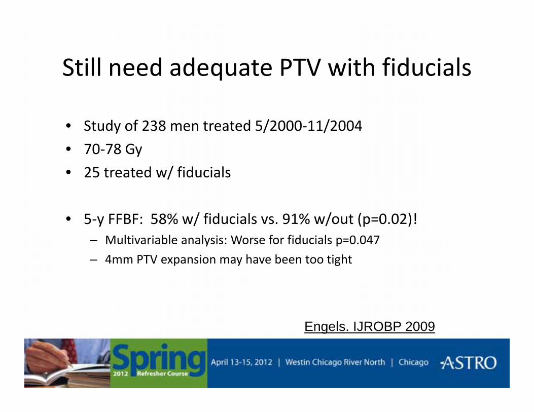
Still need adequate PTV with fiducialsq
• Study of 238 men treated 5/2000-11/2004Study of 238 men treated 5/2000 11/2004
• 70-78 Gy
• 25 treated w/ fiducials
• 5-y FFBF: 58% w/ fiducials vs. 91% w/out (p=0.02)!– Multivariable analysis: Worse for fiducials p=0.047
– 4mm PTV expansion may have been too tight
Engels. IJROBP 2009
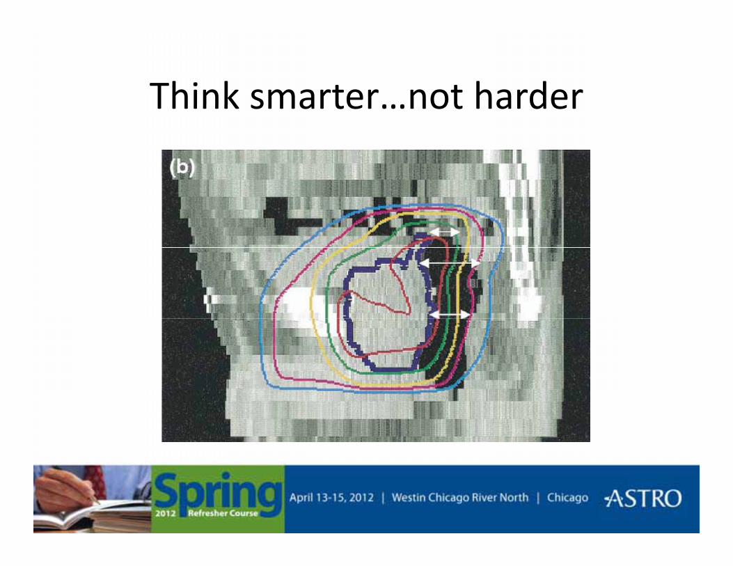
Think smarter…not harderThink smarter…not harder
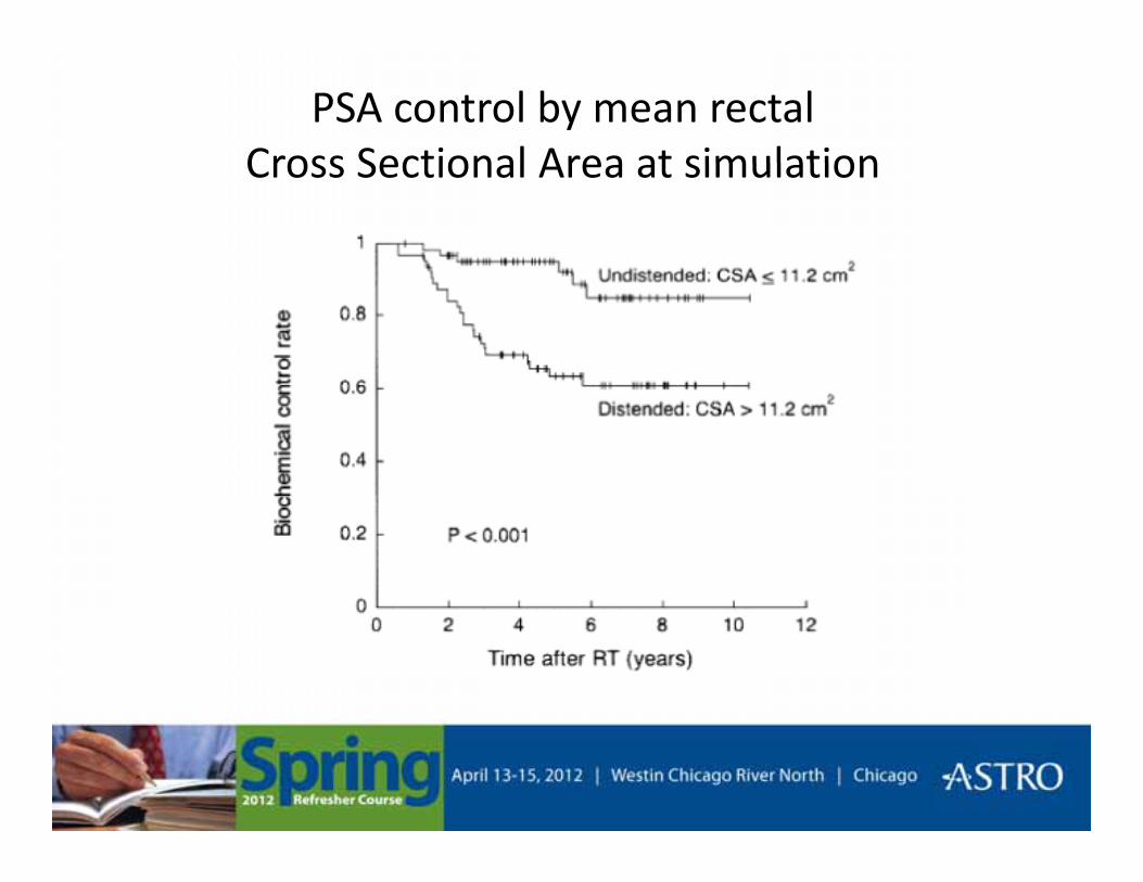
PSA control by mean rectal Cross Sectional Area at simulationCross Sectional Area at simulation
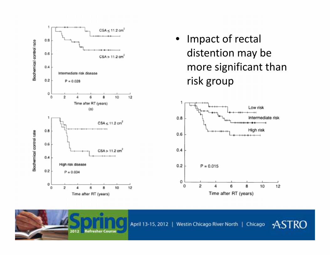
• Impact of rectal distention may be more significant than risk grouprisk group
Take home message
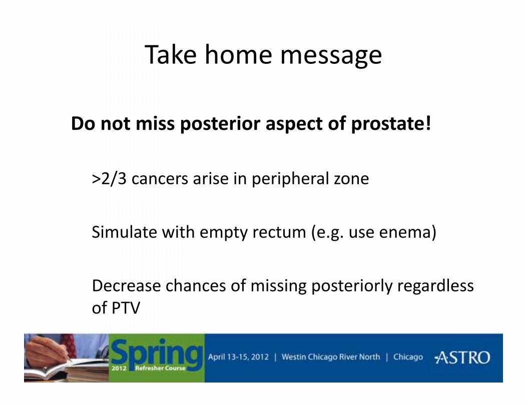
Do not miss posterior aspect of prostate!Do not miss posterior aspect of prostate!
>2/3 cancers arise in peripheral zone>2/3 cancers arise in peripheral zone
Si l t ith t t ( )Simulate with empty rectum (e.g. use enema)
D h f i i i l dlDecrease chances of missing posteriorly regardless of PTV
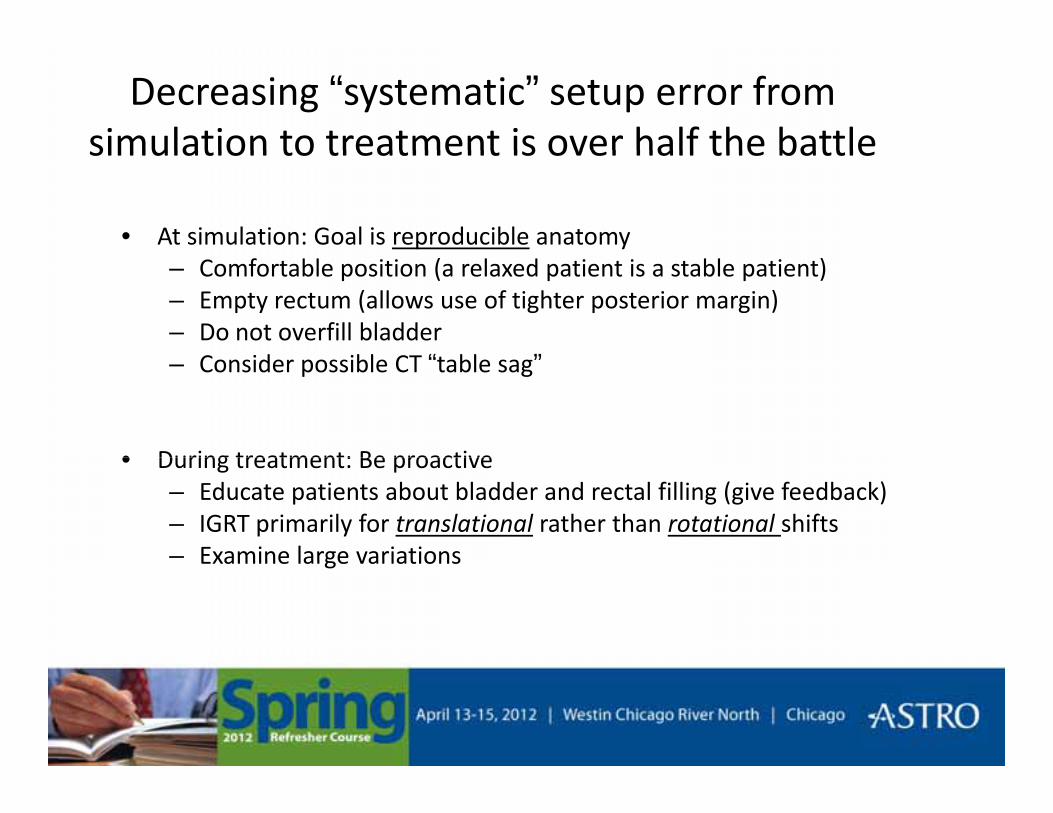
Decreasing “systematic” setup error from simulation to treatment is over half the battlesimulation to treatment is over half the battle
• At simulation: Goal is reproducible anatomy– Comfortable position (a relaxed patient is a stable patient)– Empty rectum (allows use of tighter posterior margin)– Do not overfill bladder
C id ibl CT “ bl ”– Consider possible CT “table sag”
• During treatment: Be proactive• During treatment: Be proactive – Educate patients about bladder and rectal filling (give feedback)– IGRT primarily for translational rather than rotational shifts – Examine large variationsExamine large variations
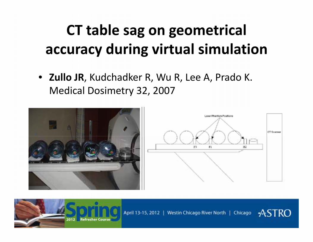
CT table sag on geometrical d i i t l i l tiaccuracy during virtual simulation
Z ll JR K d h dk R W R L A P d K• Zullo JR, Kudchadker R, Wu R, Lee A, Prado K. Medical Dosimetry 32, 2007
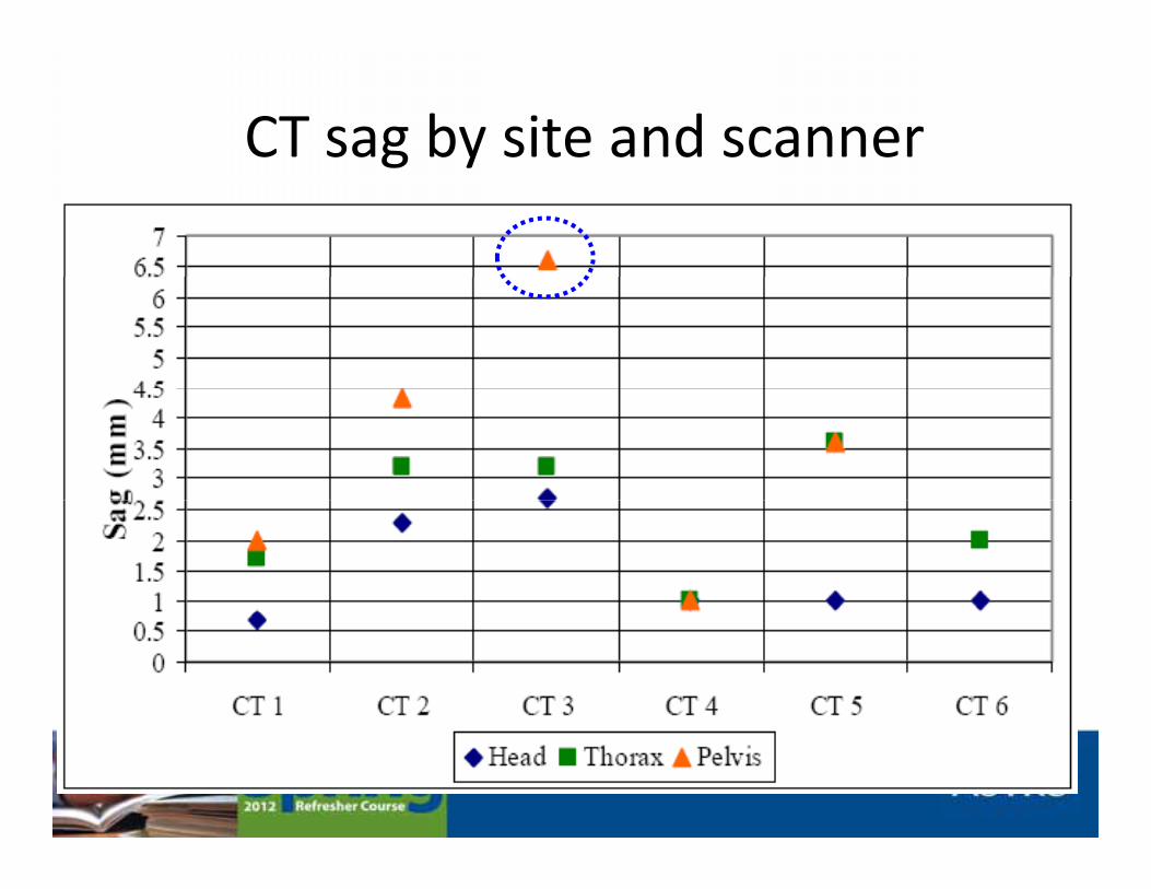
CT sag by site and scannerg y
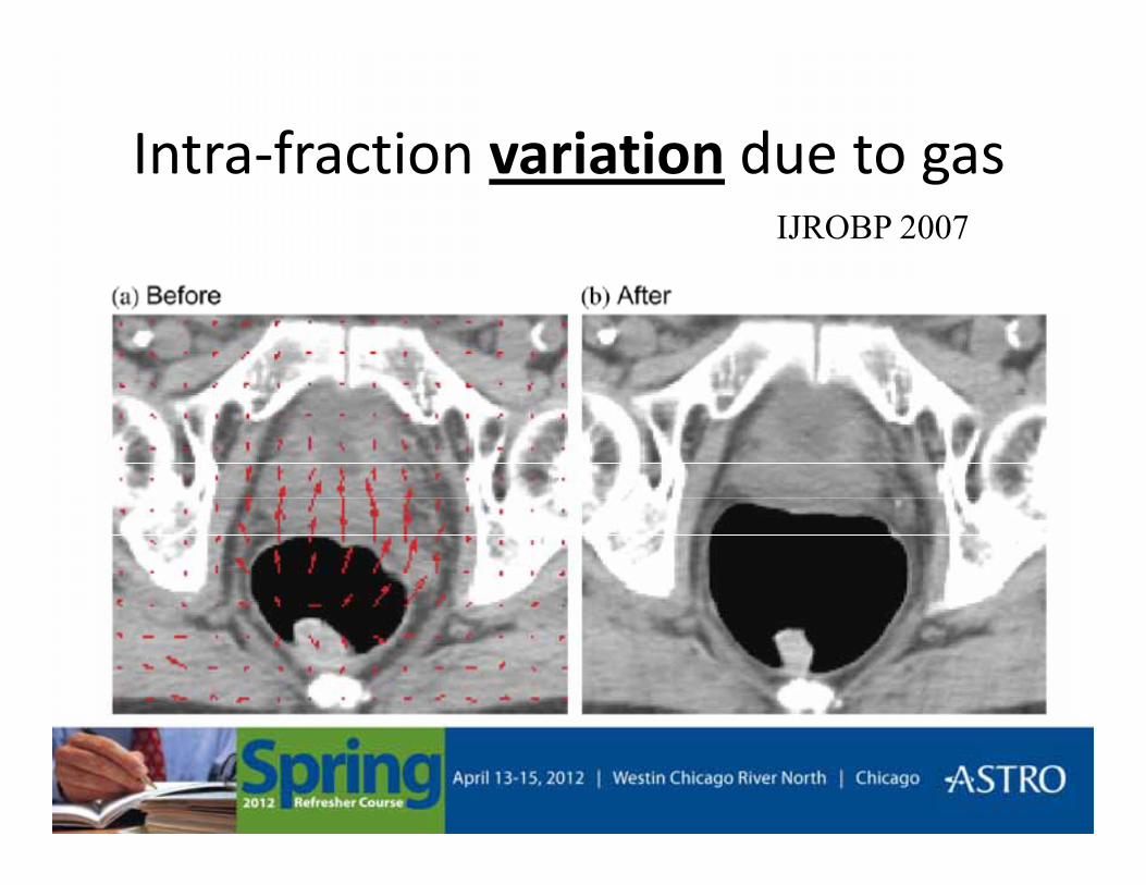
Intra-fraction variation due to gasIntra fraction variation due to gasIJROBP 2007
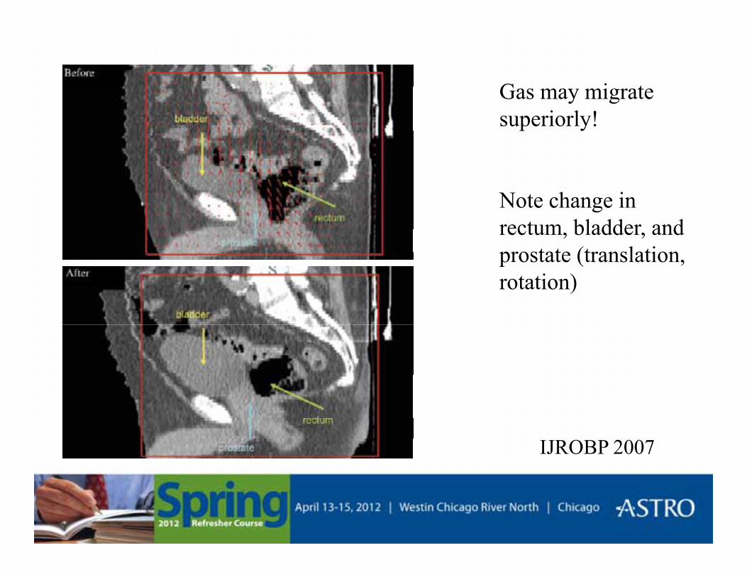
Gas may migrate superiorly!
Note change in rectum, bladder, and prostate (translationprostate (translation, rotation)
IJROBP 2007
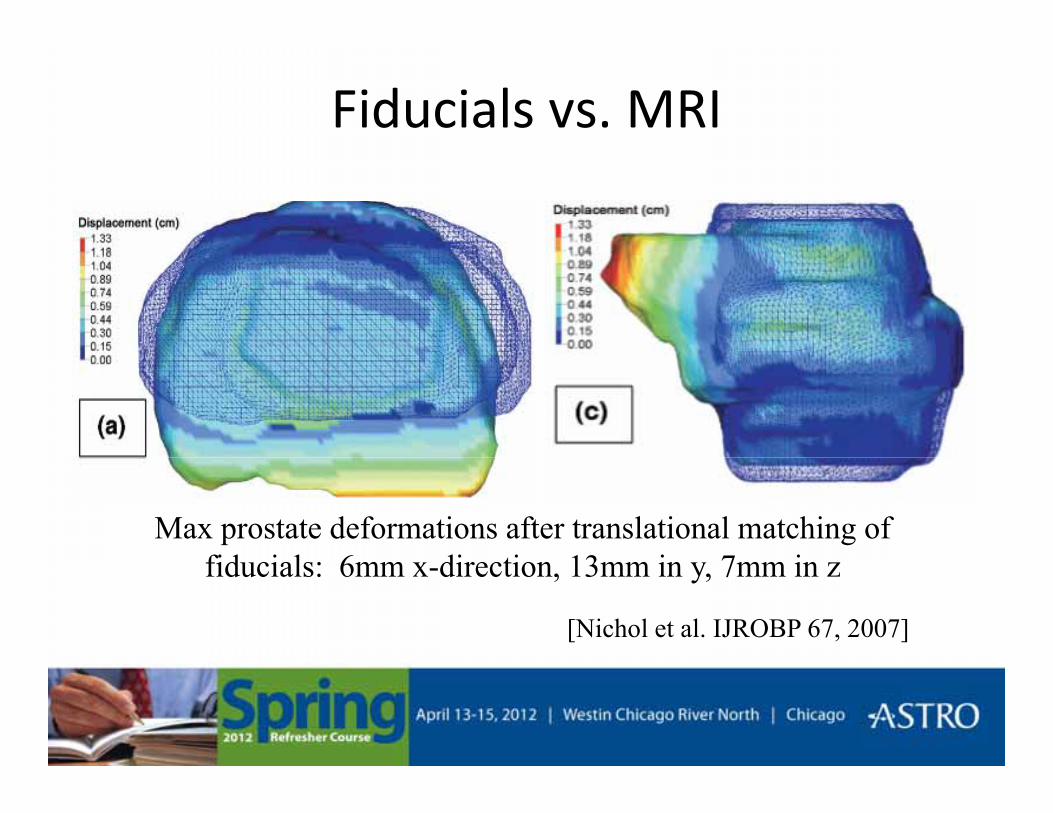
Fiducials vs. MRI
Max prostate deformations after translational matching of fid i l 6 di ti 13 i 7 ifiducials: 6mm x-direction, 13mm in y, 7mm in z
[Nichol et al. IJROBP 67, 2007]
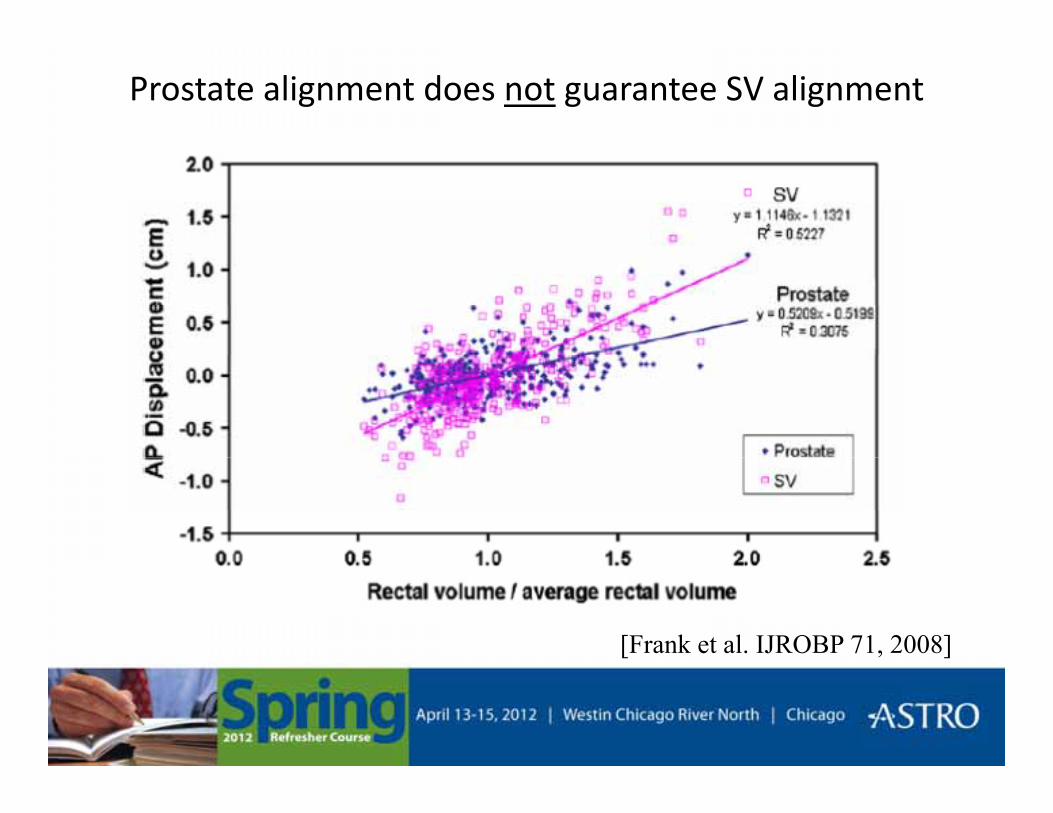
Prostate alignment does not guarantee SV alignment
[Frank et al. IJROBP 71, 2008]
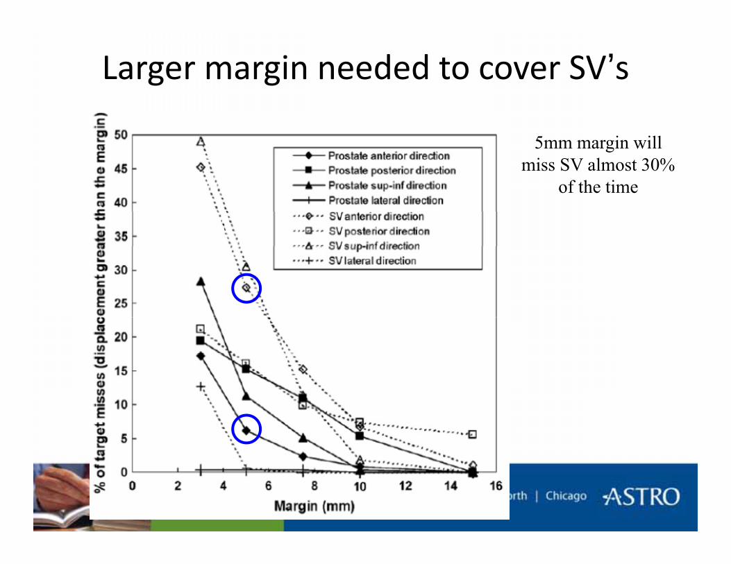
Larger margin needed to cover SV’s
5mm margin will miss SV almost 30%
of the time
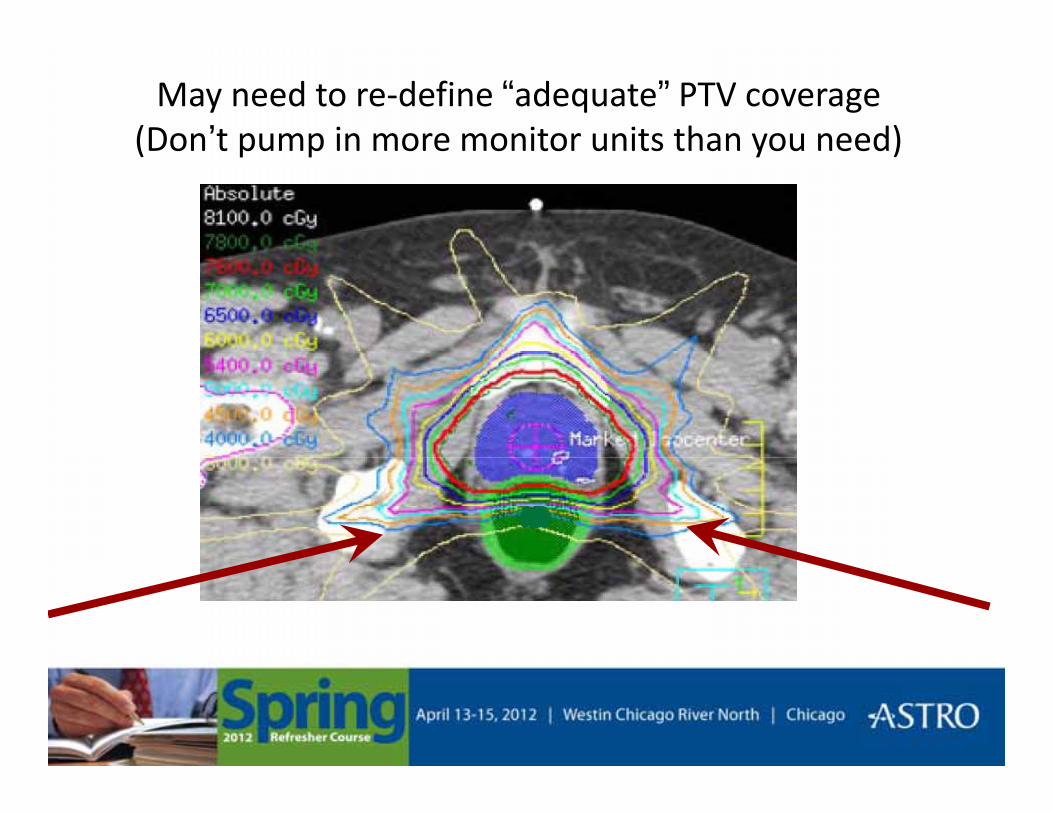
May need to re-define “adequate” PTV coverage(Don’t pump in more monitor units than you need)(Don t pump in more monitor units than you need)

Localized prostate cancer
Localized PCaT1-2
Low riskT1-2
Gleason 6
PSA 10
IntermediateT2b
Gleason 7
PSA 10 20
HighT2c
Gleason 8-10
PSA 20PSA <10
Surgery
PSA 10-20
Surgery
PSA >20
EBRT + HTBrachyRx
EBRT
Active surveillance
EBRT +/- HT
EBRT + Brachy
Brachy alone (select)
EBRT + HT
EBRT + Brachy +HT
Surgery (select)
MDACC external beam recommendations• Low risk 78 Gy (2 Gy) PTV
(>80 Gy CTV)
• Intermediate risk Prostate & “Proximal” SV6 mo HT for select pts(≥ 2 mos TAB then leuprolide alone)( p )
• High risk & T3 Prostate & most of SV2 years HT
A relaxed patient is a stable patient (reproducibility)Even with IGRT use appropriate PTV marginsI ti t ( CT) l hift d i t t tInvestigate (e.g. re-CT) large shifts during treatmentStart HT ≥ 2 months prior to RT
Thank youThank you